Interaction Between Cytokines and Oxidative Stress in Acute Pancreatitis
Upload
independentCategory
view
0download
0
REVIEW
tRNA structural and functional changes induced by oxidativestress
Barbara Nawrot • Elzbieta Sochacka •
Markus Duchler
Received: 10 June 2011 / Revised: 4 July 2011 / Accepted: 7 July 2011 / Published online: 11 August 2011
� The Author(s) 2011. This article is published with open access at Springerlink.com
Abstract Oxidatively damaged biomolecules impair
cellular functions and contribute to the pathology of a
variety of diseases. RNA is also attacked by reactive
oxygen species, and oxidized RNA is increasingly recog-
nized as an important contributor to neurodegenerative
complications in humans. Recently, evidence has accu-
mulated supporting the notion that tRNA is involved in
cellular responses to various stress conditions. This review
focuses on the intriguing consequences of oxidative mod-
ification of tRNA at the structural and functional level.
Keywords tRNA � Oxidative damage �Modified nucleosides � Thiouridine � Oxidative stress �Stress signaling
Introduction
The evolution of higher life forms is firmly associated with
the utilization of oxygen, which is essential for energy
production in the mitochondrial respiratory chain. Oxida-
tive modifications of biomolecules were adopted to
function as signals in intracellular communication. How-
ever, the high reactivity of oxygen must be tightly
controlled to avoid unwanted oxidative damage. During
mitochondrial energy production, oxygen is partially
converted into reactive oxygen species (ROS). About
1–2% of oxygen leaks out of the respiratory chain in the
form of superoxide (O2•-), which is transformed into
hydrogen peroxide (H2O2). In the presence of Fenton-
reactive metals, such as the ferrous cation (Fe2?), H2O2 is
split into a highly aggressive hydroxyl radical (OH•) and a
hydroxyl anion (OH-). The majority of ROS arise as
byproducts of cellular energy production, whereas a
smaller portion is directly created by oxygenases. The
intracellular level of ROS is tightly regulated by anti-oxi-
dative enzymes and nonenzymatic antioxidants. ROS act as
second messengers and are essential components of signal
transduction pathways [1]. However, when the generation
of ROS exceeds their degradation by antioxidant enzymes,
a condition of oxidative stress arises. Overly elevated
levels of ROS cause oxidative damage to proteins, nucleic
acids, polysaccharides, and lipids, leading to disturbances
of cellular functions and eventually cell death [2].
Oxidative damage of biomolecules is increasingly
understood to serve a critical role in the pathogenesis of
human diseases, especially those involving neurodegener-
ation [3]. Neurons are rich in polyunsaturated lipids, which
are highly susceptible to oxidative stress damage. Also,
oxidative damage of nucleic acids was identified as a key
contributor to Alzheimer’s disease; the amount of
8-hydroxy-20-deoxyguanosine (8-oxo-dG), one of the
major products of nucleic acid oxidation, is increased in the
brains and cerebrospinal fluid of Alzheimer’s disease
patients. Not only DNA but also up to 50% of messenger
RNAs (mRNAs) are oxidatively damaged in the affected
brain areas of Alzheimer’s patients [4]. DNA damage can
result in a loss of genetic information, constituting a more
persistent insult than any injury of proteins and RNA
molecules that are quickly resynthesized. Most mRNA
molecules are relatively short-lived, so RNA oxidation
B. Nawrot � M. Duchler (&)
Department of Bioorganic Chemistry, Centre of Molecular
and Macromolecular Studies, Polish Academy of Sciences,
112, Sienkiewicza Street, 90-363 Lodz, Poland
e-mail: [email protected]
E. Sochacka
Institute of Organic Chemistry, Technical University of Lodz,
Zeromskiego 116, 90-924 Lodz, Poland
Cell. Mol. Life Sci. (2011) 68:4023–4032
DOI 10.1007/s00018-011-0773-8 Cellular and Molecular Life Sciences
123

reflects a kind of steady-state balance of oxidative damage
[5]. Oxidized mRNAs are not translated properly. Conse-
quently, protein levels are reduced and normal protein
function is lost [6]. Protein aggregation, as it is observed in
neurodegenerative diseases, may be a consequence. Oxi-
dative damage of mRNA is thought to contribute not only
to neuronal cell death in Alzheimer’s disease but also to the
pathology of Parkinson’s disease, epilepsy, atherosclerosis,
and amyotrophic lateral sclerosis (characterized by the
progressive degeneration of motor neurons) [6].
In addition to mRNA, ribosomal RNAs (rRNAs) and
transfer RNAs (tRNAs) are also targeted by ROS. The
binding capacity to Fenton-reactive ions is higher for rRNA
than for tRNA, thus rRNA was found to be more heavily
oxidized than tRNA [7]. Although repair mechanisms have
been described for RNA modified by alkylation [8], no
salvage activity was reported for oxidatively damaged RNA
which therefore undergoes enzymatic nucleolysis [9]. How
the damaged RNA is distinguished and marked for degra-
dation is largely unknown. Several RNA-binding proteins
bind 8-oxo-G with higher affinity than to non-oxidized
nucleotides, which could mark oxidized RNA for degra-
dation [10], including the human polynucleotide
phosphorylase (Pnp) and the Y box-binding protein-1
(YB-1) [11]. In comparison to mRNA, tRNAs are less
susceptible to degradation as a result of stabilization by
their tertiary structure and a high content of base modifi-
cations. In bacteria, a kind of quality-control process detects
mutant tRNAs at the level of tRNA precursors and promotes
their degradation [12]. Whether and how an oxidative
damage of mature tRNA is recognized and translated into a
degradation signal is currently unknown. However, during
recent years, evidence has accumulated showing that oxi-
dized tRNA plays a role in stress response regulation.
Stress induces tRNA cleavage
A detailed analysis of the cellular small RNA content by
deep sequencing revealed an unexpectedly high abundance
of tRNA fragments [13]. The length of tRNA fragments was
either 22 nucleotides, which is the typical size of microR-
NAs (miRNAs), or in the range of 30–40 nucleotides, which
corresponds to tRNA halves. miRNAs compose a class of
highly expressed, noncoding small RNAs involved in post-
transcriptional regulation of mRNA abundance. miRNAs
bind to the 3’-untranslated regions of target mRNAs via
imperfect base-pairing to inhibit their translation. The sec-
ond abundant class of RNA fragments was found to be
generated under various stress conditions by single cleavage
of tRNA molecules in the anticodon loop [14]. Several
findings support the assumption that such instances of tRNA
cleavage occur as specific events, rather than by degradation
in the course of metabolic turnover: (1) tRNA cleavage is
triggered by certain stress conditions, including nutritional
deficiency, heat shock, hypothermia, hypoxia, and oxidative
stress (Fig. 1), but not by others, such as irradiation. Simi-
larly, some apoptotic inducers (which actually also cause
stress), such as Fas ligand, promote tRNA cleavage, while
others, such as staurosporine, are inactive [15]. (2) Some
tRNA species are more vulnerable than others, e.g., nutri-
tional stress-induced cleavage of tRNA is specific for
methionine (tRNAMet) and tRNAVal, but not for tRNATyr
[14]. Similarly, oxidative stress mediated by H2O2 results in
strong cleavage of tRNAArg and some cleavage of tRNATrp,
but not of tRNATyr [15]. The data published so far are
incomplete; a comprehensive investigation of all tRNA
species exposed to various types of stress is still lacking. (3)
The specificity of this process is further emphasized by the
fact that tRNA cleavage is a conserved response to oxidative
stress among many eukaryotic species, from Saccharomy-
ces cerevisiae and Arabidopsis to human cells [15].
tRNAs are cleaved by specific enzymes
Stress-induced tRNA cleavage is carried out by specific
enzymes (Fig. 2). In yeast, tRNAs are cleaved by the Rny1
RNase [16]. In mammalian cells, angiogenin, a secreted
ribonuclease, is required for stress-induced endonucleolytic
cleavage of tRNAs [14]. Both RNases are normally spatially
segregated from cytoplasmic tRNAs, but they are released
into the cytoplasm under conditions of oxidative stress [16].
Angiogenin was originally recognized as an angiogenic
factor secreted by tumor cells into the surrounding medium
exhibiting enhanced secretion under hypoxic conditions.
In vivo, receptors on the surface of endothelial cells bind
and internalize angiogenin. Importantly, the induction of
new blood vessel outgrowth is dependent on its ribonuclease
activity. In endothelial cells, angiogenin is concentrated in
the nucleolus and also partially localized to the cytoplasm,
where it is bound by the ribonuclease inhibitor RNH1 [17].
Depletion of RNH1 increases tRNA cleavage consistent
with a role for cytoplasmic angiogenin [18].
Fig. 1 Triggers of tRNA cleavage. Various stress conditions induce
cleavage of tRNAs in the anticodon loop
4024 B. Nawrot et al.
123

Yet another finding supports the view that tRNA cleav-
age is not just a simple degradation process: the cleavage is
controlled by RNA methylation. The DNA methyltrans-
ferase Dnmt2 is capable of methylating tRNA molecules as
well, in particular tRNAAsp, tRNAVal and tRNAGly. Dnmt2-
mediated methylation was shown to protect tRNAs against
ribonuclease cleavage by angiogenin [19].
The levels of full-length tRNAs do not decline signifi-
cantly during stress-induced tRNA cleavage; only a small
proportion of tRNA is targeted. Is there any function, then,
of the tRNA halves generated in a controlled way as a
response to many, but not all, stress conditions?
tRNA halves promote stress granule assembly
When exposed to environmental stress, eukaryotic cells
activate stress response programs. Energy-expensive
processes, such as transcription and translation, are reduced
to conserve energy for survival and the repair of stress-
induced damage. The expression of common housekeeping
genes is blocked, whereas the expression of genes that
repair stress-induced damage and promote cell survival is
increased. Cleaved tRNA may contribute to the transla-
tional arrest by inhibitory interactions with the translation
machinery. After cleavage, tRNA might persist in a fully
folded conformation, but the nicked anticodon loop would
prevent correct interaction with its respective codon
(Fig. 3). Elongation, therefore, would be stalled [20]. In
association with specialized proteins, tRNA halves might
directly regulate gene expression (see below). In addition
to interfering with protein translation, tRNA halves were
demonstrated capable of inducing the formation of stress
granules (SG) [21]. SG appear as part of the stress response
program as cytoplasmic aggregates in which nontranslated
transcripts accumulate. It is thought that SG compose the
Fig. 2 tRNAs are cleaved by
specific enzymes. Bacterial
endonucleases, PrrC, colicin D,
and colicin E5, cleave specific
subsets of bacterial tRNAs of
invading species in the anticodon
region. A similar tRNA
endonuclease activity is mediated
by c-toxin from the dairy yeast
Kluyveromyces lactis, to arrest the
growth of Saccharomycescerevisiae. Under oxidative
stress, tRNAs are cleaved by
Rny1 in yeast and by angiogenin
in mammalian cells. The
nucleolytic activity of angiogenin
is further regulated by RNH1, an
inhibitory protein, and by tRNA
methylation
Fig. 3 Functions of damaged
tRNAs. The nicked anticodon
loop in cleaved tRNA prevents
correct interaction with its
respective codon and elongation
is stalled. The 50-half tRNA
halves induce the assembly of
stress granules, cytoplasmic
aggregates which associate with
P-bodies to function in selective
degradation of mRNA. At least
one tRNA-derived fragment
was required for the
proliferation of prostate cancer
cells. In association with
specialized proteins, tRNA
halves contribute to the
regulation of gene expression by
guiding endonucleolytic
cleavage of target mRNAs
tRNA under oxidative stress 4025
123

sites where mRNAs are sorted into stored, degraded, or
translated ones. SG are physically associated with P-bod-
ies, another kind of cytoplasmic foci present in resting cells
that are enriched in RNA-degrading enzymes (Fig. 3).
P-bodies serve as a site of mRNA degradation.
SG formation was demonstrated to be enhanced by
angiogenin [21], which provided a traceable link to tRNA
cleavage. When cells were transfected with tRNA frag-
ments derived from stress-induced cleavage, transfection of
50-tRNA halves induced SG formation, while no such
effect was observed with the 30-tRNA fragments. These
results convincingly exhibit the signaling component of
tRNA-derived fragments in the cellular response to stress.
tRNA fragments in RNA-mediated translational
silencing
Post-transcriptional regulation of gene expression includes
the miRNA-mediated gene-silencing pathway, in which
selected mRNAs are prevented from translation. One of the
major silencing routes employs the RNA-induced silencing
complex (RISC). Non-coding RNA transcripts (pre-miR-
NAs) are processed by Drosha and Dicer nucleases and
loaded onto Argonaute (AGO) proteins, which are compo-
nents of the RISC. RISC-associated miRNAs recognize and
interact with complementary sequences of target mRNAs,
which are then silenced by degradation or stalled translation.
Also, tRNA fragments were found to be associated with AGO
proteins (Fig. 3) [22]. In HeLa cells, tRNA-derived *19
nucleotide-long fragments, mainly processed from the 50-end,
are highly abundant [23]. In murine embryonic stem cells, a
small RNA fragment was identified that mapped to a tRNAIle
gene. For this tRNAIle, an alternative fold was predicted
beside the tRNA cloverleaf structure, which could serve as a
substrate for the Dicer RNA processing enzyme [24]. tRNA-
derived small fragments with low inherent silencing activity
might compete with other small RNA species for association
with AGO, and due to their high abundance, significantly
diminish the silencing activity of ‘regular’ miRNAs [25].
Some small RNA fragments cleaved from tRNA in prostate
cancer cell lines were further characterized [26]. They were
18–25-nucleotides long, representing 50-ends or 30-ends of
mature tRNAs or tRNA precursors. At least one of them,
derived from the 30-end of a tRNASer precursor transcript,
was shown to be necessary for proliferation of these cancer
cells (Fig. 3). This fragment was cleaved from a pre-tRNA in
the cytoplasm by RNase Z (ELAC2), an enzyme involved in
tRNA maturation [27]. During this maturation process,
RNase Z removes a 30-trailer from pre-tRNA.
RNase Z not only trims tRNA precursors but is also
capable of cleaving target RNA at any desired site by a
silencing mechanism similar to that exerted by the RISC
[28]. For that silencing activity, RNase Z has to be loaded
with a guide RNA that brings the enzyme into contact with
the target RNA. By co-immunoprecipitation, small RNA
species associated with RNase Z were identified in human
cell lines. Among those small RNAs was the 50-half of
tRNAGlu, which indeed functioned as guide RNA in RNase
Z-mediated cleavage (Fig. 3). A mRNA targeted by the
tRNAGlu-half/RNase Z complex was identified as coding
for PPM1F, a serine/threonine protein phosphatase which
specifically dephosphorylates Thr-286 of the calcium/cal-
modulin-dependent protein kinase II (CaMKII) [29].
CaMKII is a multifunctional enzyme that is involved in
inducing apoptosis upon endoplasmatic reticulum stress
[30]. Downregulation of PPM1F, as a direct consequence
of tRNA cleavage, could therefore promote apoptosis in
cells under stress. How gene silencing with small tRNA-
derived fragments might be associated with stress is a
fascinating question that warrants further research.
Cleavage of tRNA in the anticodon loop by bacterial
and fungal endonucleases
Halves of tRNA molecules were also found in bacterial
species, in which tRNA anticodon damage is an activity of
a kind of innate immune system against invading species.
The bacterial tRNA endonucleases, PrrC, colicin D, and
colicin E5, cleave specific subsets of bacterial tRNAs in the
anticodon region (Fig. 2) [31–33]. Notably, the presence of
modified nucleosides within the anticodon loop seems to
determine which tRNAs are targeted. PrrC cleaves tRNA-Lys at the 50 phosphate of the 5-methylaminomethyl-2-
thiouridine (mnm5S2U) wobble base in position 34 [31, 34,
35]. Colicin D shows specificity for all four tRNAArg iso-
accepting molecules, and cleaving them at nucleotide 38
impairs protein synthesis and promotes cell death [33].
tRNAs for Tyr, His, Asn, and Asp, which contain the
modified base queuine at the wobble position, are cut by
colicin E5 at the 30 phosphate of the wobble nucleotide. A
similar tRNA endonuclease activity mediated by c-toxin
was found in the dairy yeast Kluyveromyces lactis, which
arrests the growth of Saccharomyces cerevisiae. The c-
toxin is secreted as a component of a larger protein com-
plex and transported into the cytoplasm of target cells,
where it cleaves three tRNA species specific for Glu, Gln
and Lys at the 30 site of the modified wobble uridine
(mcm5S2U, Fig. 2) [36]. Modifications of the ribose in the
wobble base suppressed the cleavage [37]. In contrast to
the stress signaling function of tRNA halves described
above, no such function was described for the cleavage
products of bacterial and fungal tRNA endonucleases.
Instead, tRNA cleavage in bacteria and yeast results in
inhibition of protein translation and induction of cell death.
4026 B. Nawrot et al.
123

Oxidative dethiolation of tRNAs
In addition to tRNA cleavage under stress conditions, single
nucleotides in tRNAs are chemically altered by high levels
of ROS. tRNA contains a variety of modified nucleotides,
some of which serve as targets for oxidative modifications.
The oxidative dethiolation of sulfur-containing nucleotides
is one example that has been investigated in detail.
Sulfur-containing tRNAs
There are more than 100 post-transcriptionally modified
nucleosides present in all types of RNA, and most of them
are located in tRNA. They function in modifying the trans-
lation process by precise decoding of the genetic
information [38–42]. The tRNA Modification Databases
[43, 44] contain a comprehensive list of modified nucleo-
sides identified to date in tRNA. Among them, 18 are
modified pyrimidine nucleosides containing either sulfur or
selenium at the nucleobase moiety. The structures of known
thio- and seleno-pyrimidine nucleosides are shown in Fig. 4.
Most of them are 2-thiouridines, which are substituted with a
side chain at the carbon C5, in addition to a sulfur atom at
C2. The 2-thiouridines most typically appear in the first
position of the tRNA anticodon (position 34, wobble posi-
tion). Three tRNAs, specific for lysine (tRNALys3), glutamic
acid (tRNAGlu) and glutamine (tRNAGln), contain hyper-
modified 2-thiouridines in the wobble position. The
2-thiouridine (S2U) unit in position 34 facilitates predomi-
nantly Watson–Crick base pairing with adenosine and
restricts the wobble pairing with G in the third position of the
codon [39, 45]. The S2U modification in the anticodon loop
of tRNA is also a recognition element for the cognate
aminoacyl tRNA synthetases (aa-RS) [46–48]. Notably, the
human tRNALys3 serves as a primer for the reverse
transcription of the human immunodeficiency virus type 1
(HIV-1) RNA; it contains 5-methoxycarbonylmethyl-2-
thiouridine (mcm5S2U) in position 34, which is required for
the formation of the initial complex with the viral RNA [49,
50]. In addition to S2U, selenium-containing uridine (Se2U)
has been identified in tRNAs from bacterial, mammalian and
plant species, and this modified unit is located at the wobble
position of three tRNAs specific for Glu, Gln and Lys (the
same as for S2U) [51–56]. All these modified units contain
thiocarbonyl/selenocarbonyl function, which is prone to
attack of oxidizing agents [57].
Loss of function of sulfur-containing tRNAs
in oxidative conditions in vitro
The earliest report on oxidative damage of tRNA describes
the inactivation of specific E. coli or rabbit liver tRNA
species for amino acid acceptor activity via treatment with
dilute iodine–potassium iodide and recovery of functional
tRNAs via the action of appropriate reducing agents [58].
Chemical modification of sulfur-containing pyrimidine
nucleosides in E. coli tRNA with various oxidizing
reagents showed that, in the presence of hydrogen peroxide
or cyanogen bromide (CNBr), the entire 5-methylamino-
methyl-2-thiouridine nucleoside was dethiolated, although
a detailed structure of products was not reported [59]. The
CNBr-oxidized tRNA species specific for Glu, Gln and Lys
lost their potential to be aminoacylated by their cognate
aa-RSs. It has been shown (either at the nucleoside or entire
tRNA level) that the 2-thiouridines are oxidized by H2O2 to
uridine and to another ‘‘H2O2-altered’’, unidentified prod-
uct, but not to a ring-opened or disulfide structure [60].
Dethiolation of E. coli tRNAGlu in oxidative conditions led
Fig. 4 The structures of sulfur-
and selenium-containing
pyrimidine nucleosides. S2U
and Se2U nucleosides present in
transfer RNAs, according to
Agris et al., http://rna-mdb.cas.
albany.edu/RNAmods/ [43] and
Dunin-Horkawicz et al., http://
modomics.genesilico.pl/ [44]
tRNA under oxidative stress 4027
123

to more than 95% conversion of the 2-thiouridine 34 to
uridine, as assigned by base composition analysis [61].
Notably, the H2O2-oxidized tRNAGlu lost much of its
binding affinity to tRNAPhe, which has the complementary
anticodon sequence. The stability of the tRNAGlu/tRNAPhe
complex after mnm5S2U oxidation was significantly
reduced, as determined by the temperature jump relaxation
method. The melting temperature of this complex dropped
by 20�C in comparison to the Tm of the parental tRNAGlu/
tRNAPhe complex. The rationale for the observed phe-
nomena was the decrease of the stacking interaction and
change in uridine conformational flexibility, in comparison
to the S2U nucleoside. Other experiments have shown that
probing the tRNALys3 (mcm5S2U at the wobble position)
with potassium peroxynitrite (ONOOK), which generates
hydroxyl radicals in the absence of heavy metal ions,
results in dethiolation and subsequent strand scission at the
wobble position, but the non-thiolated substrate is not
cleaved [62]. Hydroxyl radicals are thought to promote
cleavage of the polynucleotide backbone. Despite these
intense studies, only very limited information has been
made available on the structure of the dethiolated nucleo-
sides and the origin of the observed loss of tRNA function.
Dethiolation of S2U on nucleoside level
The removal of the sulfur atom from 2-thiopyrimidine
nucleosides proceeds either under reductive conditions or
under various oxidative conditions in a process called oxi-
dative desulfuration (desulfurization or dethiolation).
Reduction of S2U gives the corresponding 4-pyrimidinone
nucleoside (H2U). The action of oxidants may remove
sulfur in favor of hydrogen (desulfuration to H2U) or in
favor of oxygen (oxidation to U) (Fig. 5), and these reac-
tions usually occur simultaneously with a ratio dependent on
the kind of oxygen agent and reaction conditions [57, 63].
Dethiolation of S2U under reductive conditions
It has been reported that dethiolation of 2-thiopyrimidine
nucleosides proceeds in moderate yield under reductive
conditions in the reaction with dibenzoyldiazene [64] or
under treatment with dipotassium diazenedicarboxylate
[65]. However, desulfuration of 2-thiothymidine by Raney-
nickel reduction worked more efficiently to produce cor-
responding 4-pyrimidinone derivatives at an isolated yield
of 61% [66].
Dethiolation of S2U under oxidative conditions
Oxidative dethiolation of the 2-thiopyrimidine moiety has
been observed upon treatment with various oxidizing
agents: aqueous iodine [67], m-chloroperbenzoic acid [67],
dimethyldioxirane [68] and trans-2-phenylsulfonyl-3-phe-
nyloxaziridine (PSO) [69]. The reactivity of the
2-thiocarbonyl function towards aqueous iodine was found
to be the main cause of the low efficiency of 2-thionucle-
oside incorporation into the oligonucleotide chain during
the standard phosphoramidite oligonucleotide synthesis, as
desulfuration occurred during the oxidation step routinely
performed with this oxidizing agent [67, 70–73]. During the
evaluation of alternative oxidizing agents, it was discovered
that treatment of 2-thiothymidine with m-chloroperbenzoic
acid/pyridine solution rapidly produced the corresponding
4-pyrimidinone 20-deoxynucleoside in high yield [67].
Recently, the same oxidizing reagent was successfully
applied in a RNA nucleotide synthesis for the efficient,
selective transformation of 2-thiouridine into 4-pyrimidi-
none ribonucleoside [74]. Treatment of the sugar-protected
2-thiouridine derivative with dimethyldioxirane revealed
incomplete removal of the sulfur atom. Under these con-
ditions, the thiocarbonyl function at the C2 position of the
pyrimidine ring was partially dethiolated, leading to the
4-pyrimidinone nucleoside (yield 43%), and the disulfide of
2-thiouridine was formed with 20% yield [68]. We have
reported that the S2U nucleoside in the presence of PSO,
undergoes selective transformation to the H2U nucleoside.
The loss of the sulfur atom from 2-thiouridine was also
observed under H2O2 treatment in aqueous solution. Under
these conditions, the 2-thionucleoside is converted to the
4-pyrimidinone nucleoside and uridine [64, 75], and the
process is pH-dependent (Sochacka, unpublished results).
The development of the synthesis of the corresponding
S2U- and H2U-phosphoramidites has provided model oli-
gonucleotides for further studies on tRNA dethiolation.
Dethiolation of S2U on the RNA oligonucleotide level
For the first time, the effective desulfuration of the
2-thiouridine built into the 50-TdA(S2U)dGdC-30 pentamer
could be achieved by the treatment of oligomer in ‘organic
conditions’ with PSO in a water/acetonitrile solution [69].
The obtained product, as identified by MALDI-TOF mass
spectrometry, did not contain sulfur and, in accordance
Fig. 5 Transformation of 2-thiouridine and 20-deoxy-2-thiouridine to
products of desulfuration. The oxidative dethiolation of S2U (dS2U)
results in the production of 4-pyrimidinone nucleoside H2U (dH2U)
and uridine U (20-deoxyuridine, dU)
4028 B. Nawrot et al.
123

with the results obtained at the nucleoside level, was
identified as the 4-pyrimidinone nucleoside. Further stud-
ies, carried out under in vitro oxidative stress conditions in
the presence of aqueous H2O2, demonstrated that the S2U-
containing RNA, which was homo-sequential to the anti-
codon loop of tRNALys3, was transformed predominantly
to H2U-RNA [76]. The affinity of H2U-RNA to its Wat-
son-Crick complement (A opposite of H2U), as determined
by UV/VIS melting experiments, was much smaller than
the affinity of S2U-RNA or U-RNA to the complementary
RNA. These novel results indicated that the sulfur-con-
taining nucleosides present in tRNA chains are transformed
preferentially to the H2U analog than to uridine when
dethiolated under oxidative stress conditions, which is
commonly accepted as the sole dethiolation product.
Biological consequences of desulfuration
of 2-thiouridine in tRNA
It has been recognized that the 2-thiocarbonyl group of
2-thiouracil nucleosides strongly influences their confor-
mation and plays a key role in the modulation of base pair
recognition [77, 78]. These properties are crucial for the
decoding ability of natural 2-thiouracil ribonucleosides
located in the anticodon wobble position of many tRNAs
[38–40]. 2-Thiouridines preferentially adopt a rigid C30-endo sugar ring conformation [79, 80], for which the long-
range electrostatic effect between the 20-OH group and the
sulfur atom plays a dominant role [81]. The S2U-A base
pair in RNA duplexes is more stable than that of a parent
U-A one [82, 83]. Furthermore, due to steric hindrance and
the weaker H-bonding ability of sulfur relative to oxygen,
2-thiouracil ribonucleosides make the S2U-G wobble base
pairs less stable than base pairs containing uridine [80, 82–
84]. The specific hybridization properties of 2-thiouracil
ribonucleosides led to their practical use in the antisense
strategy and single-nucleotide polymorphism (SNP) anal-
ysis [85, 86, 87, 88].
Our recent studies on 4-pyrimidinone ribonucleoside
conformation in a solid state and in solution (NMR) have
demonstrated that the conformational characteristics of the
ribofuranose ring is dramatically affected by desulfuration
[74]. The 4-pyrimidinone nucleoside predominantly adopts
the C20-endo form (S conformer) in aqueous solution
(Fig. 6), and this ribose pucker is also fixed for H2U
molecules in the crystal state. The loss of 2-thiocarbonyl/2-
carbonyl function significantly influences the conformation
of nucleosides. The observed differences between the S2U,
U and H2U ribose folding (Table 1) may have important
biological consequences on tRNA fate and cellular func-
tion, because dethiolation of natural 2-thiouridines is
plausible under conditions of oxidative stress in the cell.
Notably, 4-pyrimidinone nucleosides are nucleoside
analogs lacking both the N3-amide hydrogen and the
thiocarbonyl/carbonyl function at the C2 position in the
heterobase moiety, characteristic for 2-thiouridines and
natural uridines, and thus offer entirely different possibil-
ities for base pairing within RNA and DNA duplexes
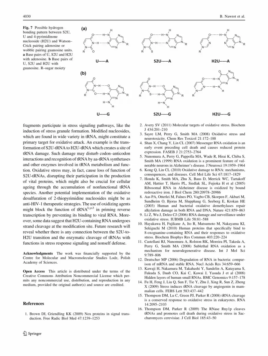
(Fig. 7). Interestingly, the UV-melting profiles of S2U-,
U- and H2U-RNAs hybridized to wobble complements (G
opposite of modification site) are almost identical, what
indicates that the mode of wobble base pairing is less
affected by dethiolation [76]. Plausible modes of hydrogen
bond patterns of H2U-A and H2U-G are shown in Fig. 7.
It is clear that shifting guanine residues along the
4-pyrimidinone moiety offers two hydrogen bonds that
might compensate for the original wobble hydrogen bonds.
This hypothesis should be addressed in future studies.
Moreover, some data reveal that dH2U-containing DNA
oligonucleotides undergo strand cleavage in certain con-
ditions [89]. We have also reported that H2U-containing
RNA is unstable in basic conditions and decomposes to its
abasic form, followed by strand scission [76]. These pre-
liminary results need to be further confirmed to consider
the H2U-containing anticodon loop as a strand scission
site, whereby such a possibility would offer new insight
into tRNA oxidative damage.
Conclusions
Oxidative damage of biomolecules, including RNA, con-
tributes to neurodegenerative and other diseases. tRNA is
cleaved under oxidative stress, and the resulting tRNA
Fig. 6 Crystal structure of H2U. The crystal structure shows
conformational details with respect to the plane passing through
atoms C10, C40 and O40 (C20-endo)
Table 1 Population of sugar S and N conformers (%) in aqueous
solution calculated on the basis of 3JH–H coupling constants
Nucleoside S (%) N (%) Reference
H2U 62 38 [74]
S2U 29 71 [90]
U 47 53 [91]
tRNA under oxidative stress 4029
123
fragments participate in stress signaling pathways, like the
induction of stress granule formation. Modified nucleosides,
which are found in wide variety in tRNA, might constitute a
primary target for oxidative attack. An example is the trans-
formation of S2U-tRNA to H2U-tRNA which creates a site of
tRNA damage. Such damage may disturb codon–anticodon
interactions and recognition of tRNA by aa-tRNA synthetases
and other enzymes involved in tRNA metabolism and func-
tion. Oxidative stress may, in fact, cause loss of function of
S2U-tRNAs, disrupting their participation in the production
of vital proteins, which might also be crucial for cellular
ageing through the accumulation of nonfunctional tRNA
species. Another potential implementation of the oxidative
desulfuration of 2-thiopyrimidine nucleosides might be as
anti-HIV-1 therapeutic strategies. The use of oxidizing agents
might block the function of tRNALys3 in priming reverse
transcription by preventing its binding to viral RNA. More-
over, some data suggest that H2U-containing RNA undergoes
strand cleavage at the modification site. Future research will
reveal whether there is any connection between the S2U-to-
H2U transition and the enzymatic cleavage of tRNAs with
functions in stress response signaling and nonself defense.
Acknowledgments The work was financially supported by the
Centre for Molecular and Macromolecular Studies Lodz, Polish
Academy of Sciences.
Open Access This article is distributed under the terms of the
Creative Commons Attribution Noncommercial License which per-
mits any noncommercial use, distribution, and reproduction in any
medium, provided the original author(s) and source are credited.
References
1. Brown DI, Griendling KK (2009) Nox proteins in signal trans-
duction. Free Radic Biol Med 47:1239–1253
2. Avery SV (2011) Molecular targets of oxidative stress. Biochem
J 434:201–210
3. Sayre LM, Perry G, Smith MA (2008) Oxidative stress and
neurotoxicity. Chem Res Toxicol 21:172–188
4. Shan X, Chang Y, Lin CL (2007) Messenger RNA oxidation is an
early event preceding cell death and causes reduced protein
expression. FASEB J 21:2753–2764
5. Nunomura A, Perry G, Pappolla MA, Wade R, Hirai K, Chiba S,
Smith MA (1999) RNA oxidation is a prominent feature of vul-
nerable neurons in Alzheimer’s disease. J Neurosci 19:1959–1964
6. Kong Q, Lin CL (2010) Oxidative damage to RNA: mechanisms,
consequences, and diseases. Cell Mol Life Sci 67:1817–1829
7. Honda K, Smith MA, Zhu X, Baus D, Merrick WC, Tartakoff
AM, Hattier T, Harris PL, Siedlak SL, Fujioka H et al (2005)
Ribosomal RNA in Alzheimer disease is oxidized by bound
redoxactive iron. J Biol Chem 280:20978–20986
8. Aas PA, Otterlei M, Falnes PO, Vagbo CB, Skorpen F, Akbari M,
Sundheim O, Bjoras M, Slupphaug G, Seeberg E, Krokan HE
(2003) Human and bacterial oxidative demethylases repair
alkylation damage in both RNA and DNA. Nature 421:859–863
9. Li Z, Wu J, Deleo CJ (2006) RNA damage and surveillance under
oxidative stress. IUBMB Life 58:81–588
10. Hayakawa H, Fujikane A, Ito R, Matsumoto M, Nakayama KI,
Sekiguchi M (2010) Human proteins that specifically bind to
8-oxoguanine-containing RNA and their responses to oxidative
stress. Biochem Biophys Res Commun 403:220–224
11. Castellani RJ, Nunomura A, Rolston RK, Moreira PI, Takeda A,
Perry G, Smith MA (2008) Sublethal RNA oxidation as a
mechanism for neurodegenerative disease. Int J Mol Sci
9:789–806
12. Deutscher MP (2006) Degradation of RNA in bacteria: compar-
ison of mRNA and stable RNA. Nucl Acids Res 34:659–666
13. Kawaji H, Nakamura M, Takahashi Y, Sandelin A, Katayama S,
Fukuda S, Daub CO, Kai C, Kawai J, Yasuda J et al (2008)
Hidden layers of human small RNAs. BMC Genomics 9:157–178
14. Fu H, Feng J, Liu Q, Sun F, Tie Y, Zhu J, Xing R, Sun Z, Zheng
X (2009) Stress induces tRNA cleavage by angiogenin in mam-
malian cells. FEBS Lett 583:437–442
15. Thompson DM, Lu C, Green PJ, Parker R (2008) tRNA cleavage
is a conserved response to oxidative stress in eukaryotes. RNA
14:2095–2103
16. Thompson DM, Parker R (2009) The RNase Rny1p cleaves
tRNAs and promotes cell death during oxidative stress in Sac-
charomyces cerevisiae. J Cell Biol 185:43–50
(a)
(b)
Fig. 7 Possible hydrogen
bonding pattern between S2U,
U and 4-pyrimidinone
nucleoside (H2U) and Watson–
Crick pairing adenosine or
wobble pairing guanosine units.
a Base pairs of U, S2U and H2U
with adenosine. b Base pairs of
U, S2U and H2U with
guanosine. R–sugar moiety
4030 B. Nawrot et al.
123

17. Tsuji T, Sun Y, Kishimoto K, Olson KA, Liu S, Hirukawa S, Hu
GF (2005) Angiogenin is translocated to the nucleus of HeLa
cells and is involved in ribosomal RNA transcription and cell
proliferation. Cancer Res 65:1352–1360
18. Yamasaki S, Ivanov P, Hu GF, Anderson P (2009) Angiogenin
cleaves tRNA and promotes stress-induced translational repres-
sion. J Cell Biol 185:35–42
19. Schaefer M, Pollex T, Hanna K, Tuorto F, Meusburger M, Helm
M, Lyko F (2010) RNA methylation by Dnmt2 protects transfer
RNAs against stress-induced cleavage. Genes Dev 24:1590–1595
20. Thompson DM, Parker R (2009) Stressing out over tRNA
cleavage. Cell 138:215–219
21. Emara MM, Ivanov P, Hickman T, Dawra N, Tisdale S, Kedersha
N, Hu GF, Anderson P (2010) Angiogenin-induced tRNA-
derived stress-induced RNAs promote stress-induced stress
granule assembly. J Biol Chem 285:10959–10968
22. Burroughs AM, Ando Y, Hoon ML, Tomaru Y, Suzuki H,
Hayashizaki Y, Daub CO (2011) Deep-sequencing of human
Argonaute-associated small RNAs provides insight into miRNA
sorting and reveals Argonaute association with RNA fragments of
diverse origin. RNA Biol 8:158–177
23. Cole C, Sobala A, Lu C, Thatcher SR, Bowman A, Brown JW,
Green PJ, Barton GJ, Hutvagner G (2009) Filtering of deep
sequencing data reveals the existence of abundant dicer-depen-
dent small RNAs derived from tRNAs. RNA 15:2147–2160
24. Babiarz JE, Ruby JG, Wang Y, Bartel DP, Blelloch R (2008)
Mouse ES cells express endogenous shRNAs, siRNAs, and other
Microprocessor-independent, Dicer-dependent small RNAs.
Genes Dev 22:2773–2785
25. Haussecker D, Huang Y, Lau A, Parameswaran P, Fire AZ, Kay
MA (2010) Human tRNA-derived small RNAs in the global
regulation of RNA silencing. RNA 16:673–695
26. Lee YS, Shibata Y, Malhotra A, Dutta A (2009) A novel class of
small RNAs: tRNA-derived RNA fragments (tRFs). Genes Dev
23:2639–2649
27. Phizicky EM, Hopper AK (2010) tRNA biology charges to the
front. Genes Dev 24:1832–1860
28. Elbarbary RA, Takaku H, Uchiumi N, Tamiya H, Abe M,
Takahashi M, Nishida H, Nashimoto M (2009) Modulation of
gene expression by human cytosolic tRNase Z(L) through 50-half-
tRNA. PLoS One 4:e5908
29. Harvey BP, Banga SS, Ozer HL (2004) Regulation of the
multifunctional Ca2?/calmodulin-dependent protein kinase II
by the PP2C phosphatase PPM1F in fibroblasts. J Biol Chem
279:24889–24898
30. Timmins JM, Ozcan L, Seimon TA, Li G, Malagelada C, Backs J,
Backs T, Bassel-Duby R, Olson EN, Anderson ME, Tabas I
(2009) Calcium/calmodulin-dependent protein kinase II links ER
stress with Fas and mitochondrial apoptosis pathways. J Clin
Invest 119:2925–2941
31. Amitsur M, Levitz R, Kaufmann G (1987) Bacteriophage T4
anticodon nuclease, polynucleotide kinase and RNA ligase
reprocess the host lysine tRNA. EMBO J 6:2499–2503
32. Ogawa T, Tomita K, Ueda T, Watanabe K, Uozumi T, Masaki H
(1999) A cytotoxic ribonuclease targeting specific transfer RNA
anticodons. Science 283:2097–2100
33. Tomita K, Ogawa T, Uozumi T, Watanabe K, Masaki H (2000) A
cytotoxic ribonuclease which specifically cleaves four isoac-
cepting arginine tRNAs at their anticodon loops. Proc Natl Acad
Sci USA 97:8278–8283
34. Jiang Y, Meidler R, Amitsur M, Kaufmann G (2001) Specific
interaction between anticodon nuclease and the tRNA(Lys)
wobble base. J Mol Biol 305:377–388
35. Jiang Y, Blanga S, Amitsur M, Meidler R, Krivosheyev E,
Sundaram M, Bajji AC, Davis DR, Kaufmann G (2002) Struc-
tural features of tRNALys favored by anticodon nuclease as
inferred from reactivities of anticodon stem and loop substrate
analogs. J Biol Chem 277:3836–3841
36. Lu J, Huang B, Esberg A, Johansson MJ, Bystrom AS (2005) The
kluyveromyces lactis gamma-toxin targets tRNA anticodons.
RNA 11:1648–1654
37. Keppetipola N, Jain R, Meineke B, Diver M, Shuman S (2009)
Structure-activity relationships in Kluyveromyces lactis gamma-
toxin, a eukaryal tRNA anticodon nuclease. RNA 15:1036–1044
38. Agris PF (2004) Decoding the genome: a modified view. Nucl
Acids Res 32:223–238
39. Agris PF, Vendeix FAP, Graham WD (2007) tRNA’s wobble
decoding of the genome: 40 years of modification. J Mol Biol
366:1–13
40. Yokoyama S, Nishimura S (1995) Modified nucleosides and
codon recognition. In: Soll D, RajBhandary U (eds) tRNA:
structure, biosynthesis and function. ASM Press, Washington DC,
pp 207–223
41. Curran JF (1998) Modified nucleosides in translation. In: Gros-
jean H, Benne B (eds) Modification and editing of RNA. ASM
Press, Washington DC, pp 493–516
42. Watanabe K (2007) Role of modified nucleosides in the trans-
lation function of tRNAs from extreme thermophilic bacteria and
animal mitochondria. Bull Chem Soc Jpn 80:1253–1267
43. Agris PF, Crain PF, Rozenski J, Fabris D, Vendeix FAP. The
RNA modification database. http://biochem.ncsu.edu/RNAmods/.
44. Dunin-Horkawicz S, Czerwoniec A, Gajda MJ, Feder M,
Grosjean H, Bujnicki JM (2006) MODOMICS: a database of
RNA modification pathways. Nucl Acids Res 34:D145–D149.
http://modomics.genesilico.pl/
45. Yarian C, Townsend H, Czestkowski W, Sochacka E, Malkiewicz
AJ, Guenther R, Miskiewicz A, Agris PF (2002) Accurate trans-
lation of the genetic code depends on tRNA modified nucleosides.
J Biol Chem 277:16391–16395
46. Beuning PJ, Musier-Forsyth K (1999) Transfer RNA recognition
by aminoacyl-tRNA synthetases. Biopolymers 52:1–28
47. Sylvers LA, Rogers KC, Shimizu M, Ohtsuka E, Soll D (1993) A
2-thiouridine derivative in tRNAGlu is a positive determinant for
aminoacylation by Escherichia coli glutamyl-tRNA synthetase.
Biochemistry 32:3836–3841
48. Gustilo EM, Dubois DY, Lapointe J, Agris PF (2007) E. coliglutamyl-tRNA synthetase is inhibited by anticodon stem-loop
domains and a minihelix. RNA Biol 4:85–92
49. Isel C, Marquet R, Keith G, Ehresmann C, Ehresmann B (1993)
Modified nucleotides of tRNA(3Lys) modulate primer/template
loop-loop interaction in the initiation complex of HIV-1 reverse
transcription. J Biol Chem 268:25269–25272
50. Bilbille Y, Vendeix FA, Guenther R, Malkiewicz A, Ariza X,
Vilarrasa J, Agris PF (2009) The structure of the human tRNA-
Lys3 anticodon bound to the HIV genome is stabilized by
modified nucleosides and adjacent mismatch base pairs. Nucl
Acids Res 37:3342–3353
51. Ching WM, Wittwer AJ, Tsai L, Stadtman TC (1984) Distribu-
tion of two selenonucleosides among the selenium-containing
tRNAs from Methanococcus vannielii. Proc Natl Acad Sci USA
81:57–60
52. Ching WM (1984) Occurrence of selenium-containing tRNAs in
mouse leukemia cells. Proc Natl Acad Sci USA 81:3010–3013
53. Chen CS, Stadtman TC (1980) Selenium-containing tRNAs from
Clostridium sticklandii: cochromatography of one species with
L-prolyl-tRNA. Proc Natl Acad Sci USA 77:1403–1407
54. Ching WM, Stadtman TC (1982) Selenium-containing tRNAGlu
from Clostridium sticklandii: correlation of aminoacylation with
selenium content. Proc Natl Acad Sci USA 79:374–377
55. Wittwer AJ (1983) Specific incorporation of selenium into lysine-
and glutamate-accepting tRNAs from Escherichia coli. J Biol
Chem 258:8637–8641
tRNA under oxidative stress 4031
123

56. Wen TN, Li C, Chen CS (1988) Ubiquity of selenium-containing
tRNA in plants. Plant Sci 57:185–193
57. Corsaro A, Pistara V (1998) Conversion of the thiocarbonyl
group into the carbonyl group. Tetrahedron 54:15027–15062
58. Carbon J, Hung L, Jones DS (1965) A reversible oxidative
inactivation of specific transfer RNA species. Proc Natl Acad Sci
USA 53:979–986
59. Rao PYS, Cherayil JD (1974) Studies on chemical modification
of thionucleosides in transfer ribonucleic acid of Escherichia coli.Biochem J 143:285–294
60. Watanabe K (1980) Reaction of 2-thioribothymidine and
4-thiouridine with hydrogen peroxide in transfer ribonucleic acids
from Thermus thermophilus and Escherichia coli studied by
circular dichroism. Biochemistry 19:5542–5549
61. Houssier C, Degee P, Nicoghosian K, Grosjean H (1988) Effect
of uridine dethiolation in anticodon triplet of tRNA (Glu) on its
association with tRNA(Phe). J Biomol Struct Dynam 5:1259–
1265
62. Gotte M, Marquet R, Isel C, Anderson VE, Keith G, Gross HJ,
Ehresmann C, Ehresmann B, Heumann H (1996) Probing the
higher order structure of RNA with peroxonitrous acid. FEBS
Lett 390:226–228
63. Brown DJ, Evans RF, Cowden WB, Fenn MD (2008) Thiopyr-
imidines. In: Taylor EC (ed) Chemistry of heterocyclic
compounds: the pyrimidines, vol chap 52. Wiley, London,
pp 553–609
64. Mitsunobu O, Ito N, Saita S, Ogihara T, Tamaoki H, Nagasawa
H, Suzuki H, Kimira J (1982) Studies on nucleosides and
nucleotides. X. Desulfurization of 20,30-O-isopropylidene-2-
thiouridine by dibenzoyldiazene. Tetrahedron Lett 23:517–520
65. Ogihara T, Mitsunobu O (1982) Desulfurization of 2-thiouridines
by dipotassium diazenedicarboxylate. Chem Lett 11:1621–1624
66. Rajur SB, McLaughlin LW (1992) The synthesis of oligode-
oxynucleotides containing 2-thiothymidine and 5-methyl-4-
pyrimidinone base analoques. Tetrahedron Lett 41:6081–6084
67. Kuimelis RG, Nambiar KP (1993) Efficient desulfurization of
2-thiopyrimidine nucleosides to the corresponding 4-pyrimidi-
nones. Tetrahedron Lett 34:3813–3816
68. Saladino R, Mincione E, Cearsini C, Mezzetti M (1996) Trans-
formations of thiopyrimidine and thiopurine nucleosides following
oxidation with dimethyldioxirane. Tetrahedron 52:6759–6780
69. Sochacka E, Fratczak I (2004) Efficient desulfurization of
2-thiopyrimidine nucleosides to corresponding 4-pyrimidinone
analogues using trans-2-phenylsulfonyl-3-phenyloxaziridine.
Tetrahedron Lett 45:6729–6731
70. Kuimelis RG, Nambiar KP (1994) Synthesis of oligodeoxynu-
cleotides containing 2-thiopyrimidine residues–new protection
scheme. Nucl Acids Res 22:1429–1436
71. Kumar RK, Davis DR (1995) Synthesis of oligoribonucleotides
containing 2-thiouridine: Incorporation of 2-thiouridine phos-
phoramidite without base protection. J Org Chem 60:7726–7727
72. Sochacka E (2001) Efficient assessment of modified nucleoside
stability under conditions of automated oligonucleotide synthesis:
characterization of the oxidation and oxidative desulfurization of
2-thiouridine. Nucleosides Nucleotides Nucleic Acids 20:1871–
1879
73. Okamoto I, Seio K, Sekine M (2006) Improved synthesis of
oligonucleotides containing 2-thiouridine derivatives by use of
diluted iodine solution. Tetrahedron Lett 47:583–585
74. Kraszewska K, Kaczynska I, Jankowski S, Karolak-Wojcie-
chowska J, Sochacka E (2011) Desulfurization of 2-thiouracil
nucleosides: conformational studies of 4-pyrimidinone nucleo-
sides. Bioorg Med Chem 19:2443–2449
75. Sochacka E, Malkiewicz A (1991) XIV-th international tRNA
workshop, Rydzyna, Poland
76. Sochacka E, Kraszewska K, Sochacki M, Sobczak M, Janicka M,
Nawrot B (2011) The 2-thiouridine unit in the RNA strand is
desulfured predominantly to 4-pyrimidinone nucleoside under in
vitro oxidative stress conditions. Chem Commun 47:4914–4916
77. Davis RD (1998) Biophysical and conformational properties of
modified nucleosides in RNA (nuclear magnetic resonance
studies). In: Grosjean H, Benne R (eds) Modification and editing
of RNA. ASM Press, Washington DC, pp 85–102
78. Guerra CF, Baerends EJ, Bickelhaupt FM (2008) Watson–Crick
base pairs with thiocarbonyl groups: how sulfur changes the
hydrogen bonds in DNA. Cent Eur J Chem 6:15–21
79. Yamamoto Y, Yokoyama S, Miyazawa T, Watanabe K, Higuchi
S (1983) NMR analyses on the molecular mechanism of the
conformational rigidity of 2-thioribothymidine, a modified
nucleoside in extreme thermophile tRNAs. FEBS Lett 157:95–99
80. Smith WS, Sierzputowska-Gracz H, Sochacka E, Malkiewicz A,
Agris PF (1992) Chemistry and structure of modified uridine
dinucleosides are determined by thiolation. J Am Chem Soc
114:7990–7997
81. Zhang R, Erikson LA (2010) Theoretical study on conformational
preferences of ribose in 2-thiouridine—the role of the 20OH
group. Phys Chem Chem Phys 12:3690–3697
82. Kumar RK, Davis DR (1997) Synthesis and studies on the effect
of 2-thiouridine and 4-thiouridine on sugar conformation and
RNA duplex stability. Nucl Acids Res 25:1272–1280
83. Testa SM, Disney MD, Turner DH, Kierzek R (1997) Thermo-
dynamics of RNA-RNA duplexes with 2- or 4-thiouridines:
implications for antisense design and targeting a group I intron.
Biochemistry 38:16655–16662
84. Agris PF, Sierzputowska-Gracz H, Smith W, Malkiewicz A,
Sochacka E, Nawrot B (1992) Thiolation of uridine carbon-2
restricts the motional dynamics of the transfer RNA wobble
position nucleoside. J Am Chem Soc 114:2652–2656
85. Shohda K, Okamoto I, Wada S, Seio K, Sekine M (2000) Synthesis
and properties of 20-O-methyl-2-thiouridine and oligoribonucleo-
tides containing 20-O-methyl-2-thiouridine. Bioorg Med Chem Lett
10:1795–1798
86. Okamoto I, Seio K, Sekine M (2008) Study of the base dis-
crimination ability of DNA and 20-O-metylated RNA oligomers
containing 2-thiouracil bases towards complementary RNA or
DNA strands and their application to single base mismatch
detection. Bioorg Med Chem 16:6034–6041
87. Sipa K, Sochacka E, Kazmierczak-Baranska J, Maszewska M,
Janicka M, Nowak G, Nawrot B (2007) Effect of base modifi-
cations on structure, thermodynamic stability, and gene silencing
activity of short interfering RNA. RNA 13:1301–1316
88. Sierant M, Paduszynska A, Kazmierczak-Baranska J, Nacmias B,
Sorbi S, Bagnoli S, Sochacka E, Nawrot B (2011) Specific
silencing of L392V PSEN1 mutant allele by RNA interference.
Int J Alzheimers Dis 2011:809218
89. Iocono JA, Gildea B, McLaughlin LM (1990) Mild acid hydro-
lysis of 2-pyrimidinone—containing DNA fragment generates
apurinic/apyrimidinic sites. Tetrahedron Lett 31:175–178
90. Sierzputowska-Gracz H, Sochacka E, Małkiewicz A, Kuo K,
Gehrke CW, Agris PF (1987) Chemistry and structure of modi-
fied uridines in the anticodon, wobble position of transfer RNA
are determined by thiolation. J Am Chem Soc 109:7171–7177
91. Schleich T, Blackburn BJ, Lapper RD, Smith ICP (1972) A
nuclear magnetic resonance study of the influence of aqueous
sodium perchlorate and temperature on the solution conformation
of uracil nucleosides and nucleotides. Biochemistry 11:137–145
4032 B. Nawrot et al.
123
Copyright © 2022 FDOKUMEN