Crystal Structure of Archaeosine tRNA-guanine Transglycosylase
13
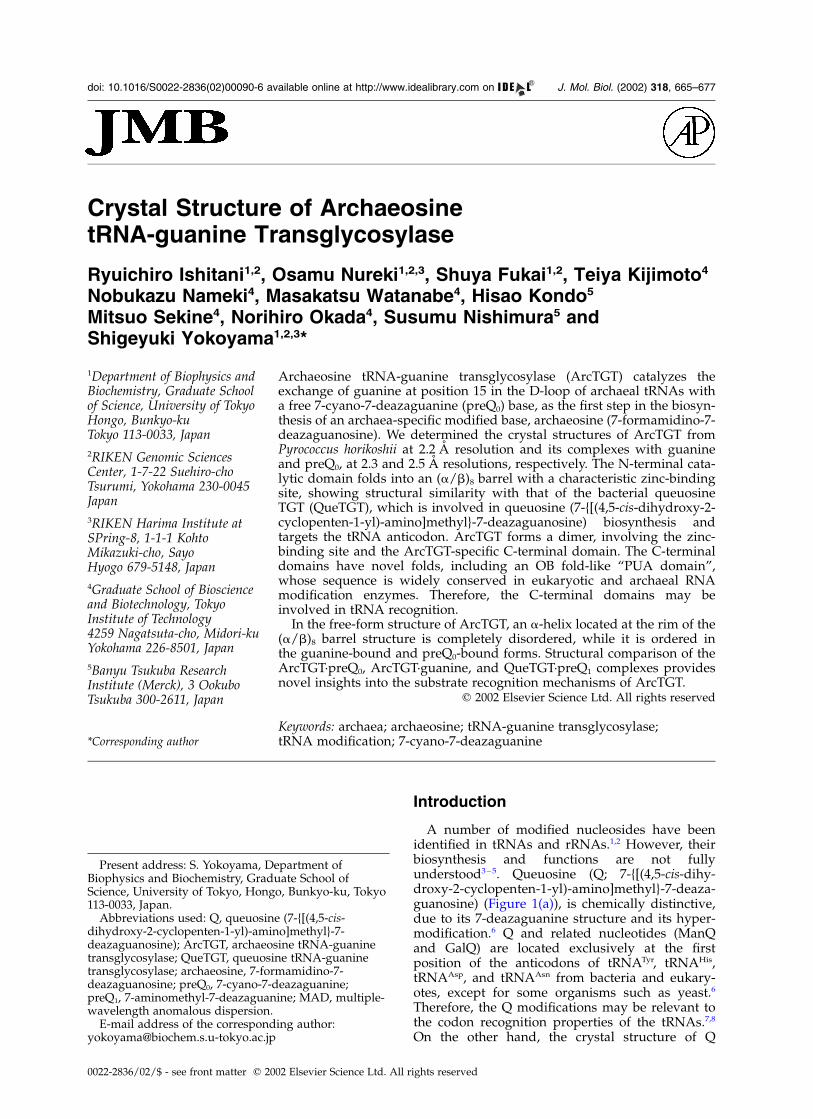
Crystal Structure of Archaeosine tRNA-guanine Transglycosylase Ryuichiro Ishitani 1,2 , Osamu Nureki 1,2,3 , Shuya Fukai 1,2 , Teiya Kijimoto 4 Nobukazu Nameki 4 , Masakatsu Watanabe 4 , Hisao Kondo 5 Mitsuo Sekine 4 , Norihiro Okada 4 , Susumu Nishimura 5 and Shigeyuki Yokoyama 1,2,3 * 1 Department of Biophysics and Biochemistry, Graduate School of Science, University of Tokyo Hongo, Bunkyo-ku Tokyo 113-0033, Japan 2 RIKEN Genomic Sciences Center, 1-7-22 Suehiro-cho Tsurumi, Yokohama 230-0045 Japan 3 RIKEN Harima Institute at SPring-8, 1-1-1 Kohto Mikazuki-cho, Sayo Hyogo 679-5148, Japan 4 Graduate School of Bioscience and Biotechnology, Tokyo Institute of Technology 4259 Nagatsuta-cho, Midori-ku Yokohama 226-8501, Japan 5 Banyu Tsukuba Research Institute (Merck), 3 Ookubo Tsukuba 300-2611, Japan Archaeosine tRNA-guanine transglycosylase (ArcTGT) catalyzes the exchange of guanine at position 15 in the D-loop of archaeal tRNAs with a free 7-cyano-7-deazaguanine (preQ 0 ) base, as the first step in the biosyn- thesis of an archaea-specific modified base, archaeosine (7-formamidino-7- deazaguanosine). We determined the crystal structures of ArcTGT from Pyrococcus horikoshii at 2.2 A ˚ resolution and its complexes with guanine and preQ 0 , at 2.3 and 2.5 A ˚ resolutions, respectively. The N-terminal cata- lytic domain folds into an (a/b) 8 barrel with a characteristic zinc-binding site, showing structural similarity with that of the bacterial queuosine TGT (QueTGT), which is involved in queuosine (7-{[(4,5-cis-dihydroxy-2- cyclopenten-1-yl)-amino]methyl}-7-deazaguanosine) biosynthesis and targets the tRNA anticodon. ArcTGT forms a dimer, involving the zinc- binding site and the ArcTGT-specific C-terminal domain. The C-terminal domains have novel folds, including an OB fold-like “PUA domain”, whose sequence is widely conserved in eukaryotic and archaeal RNA modification enzymes. Therefore, the C-terminal domains may be involved in tRNA recognition. In the free-form structure of ArcTGT, an a-helix located at the rim of the (a/b) 8 barrel structure is completely disordered, while it is ordered in the guanine-bound and preQ 0 -bound forms. Structural comparison of the ArcTGT·preQ 0 , ArcTGT·guanine, and QueTGT·preQ 1 complexes provides novel insights into the substrate recognition mechanisms of ArcTGT. q 2002 Elsevier Science Ltd. All rights reserved Keywords: archaea; archaeosine; tRNA-guanine transglycosylase; tRNA modification; 7-cyano-7-deazaguanine *Corresponding author Introduction A number of modified nucleosides have been identified in tRNAs and rRNAs. 1,2 However, their biosynthesis and functions are not fully understood 3–5 . Queuosine (Q; 7-{[(4,5-cis-dihy- droxy-2-cyclopenten-1-yl)-amino]methyl}-7-deaza- guanosine) (Figure 1(a)), is chemically distinctive, due to its 7-deazaguanine structure and its hyper- modification. 6 Q and related nucleotides (ManQ and GalQ) are located exclusively at the first position of the anticodons of tRNA Tyr , tRNA His , tRNA Asp , and tRNA Asn from bacteria and eukary- otes, except for some organisms such as yeast. 6 Therefore, the Q modifications may be relevant to the codon recognition properties of the tRNAs. 7,8 On the other hand, the crystal structure of Q 0022-2836/02/$ - see front matter q 2002 Elsevier Science Ltd. All rights reserved Present address: S. Yokoyama, Department of Biophysics and Biochemistry, Graduate School of Science, University of Tokyo, Hongo, Bunkyo-ku, Tokyo 113-0033, Japan. E-mail address of the corresponding author: [email protected] Abbreviations used: Q, queuosine (7-{[(4,5-cis- dihydroxy-2-cyclopenten-1-yl)-amino]methyl}-7- deazaguanosine); ArcTGT, archaeosine tRNA-guanine transglycosylase; QueTGT, queuosine tRNA-guanine transglycosylase; archaeosine, 7-formamidino-7- deazaguanosine; preQ 0 , 7-cyano-7-deazaguanine; preQ 1 , 7-aminomethyl-7-deazaguanine; MAD, multiple- wavelength anomalous dispersion. doi: 10.1016/S0022-2836(02)00090-6 available online at http://www.idealibrary.com on B w J. Mol. Biol. (2002) 318, 665–677
Transcript of Crystal Structure of Archaeosine tRNA-guanine Transglycosylase
Crystal Structure of ArchaeosinetRNA-guanine Transglycosylase
Ryuichiro Ishitani1,2, Osamu Nureki1,2,3, Shuya Fukai1,2, Teiya Kijimoto4
Nobukazu Nameki4, Masakatsu Watanabe4, Hisao Kondo5
Mitsuo Sekine4, Norihiro Okada4, Susumu Nishimura5 andShigeyuki Yokoyama1,2,3*
1Department of Biophysics andBiochemistry, Graduate Schoolof Science, University of TokyoHongo, Bunkyo-kuTokyo 113-0033, Japan
2RIKEN Genomic SciencesCenter, 1-7-22 Suehiro-choTsurumi, Yokohama 230-0045Japan
3RIKEN Harima Institute atSPring-8, 1-1-1 KohtoMikazuki-cho, SayoHyogo 679-5148, Japan
4Graduate School of Bioscienceand Biotechnology, TokyoInstitute of Technology4259 Nagatsuta-cho, Midori-kuYokohama 226-8501, Japan
5Banyu Tsukuba ResearchInstitute (Merck), 3 OokuboTsukuba 300-2611, Japan
Archaeosine tRNA-guanine transglycosylase (ArcTGT) catalyzes theexchange of guanine at position 15 in the D-loop of archaeal tRNAs witha free 7-cyano-7-deazaguanine (preQ0) base, as the first step in the biosyn-thesis of an archaea-specific modified base, archaeosine (7-formamidino-7-deazaguanosine). We determined the crystal structures of ArcTGT fromPyrococcus horikoshii at 2.2 A resolution and its complexes with guanineand preQ0, at 2.3 and 2.5 A resolutions, respectively. The N-terminal cata-lytic domain folds into an (a/b)8 barrel with a characteristic zinc-bindingsite, showing structural similarity with that of the bacterial queuosineTGT (QueTGT), which is involved in queuosine (7-{[(4,5-cis-dihydroxy-2-cyclopenten-1-yl)-amino]methyl}-7-deazaguanosine) biosynthesis andtargets the tRNA anticodon. ArcTGT forms a dimer, involving the zinc-binding site and the ArcTGT-specific C-terminal domain. The C-terminaldomains have novel folds, including an OB fold-like “PUA domain”,whose sequence is widely conserved in eukaryotic and archaeal RNAmodification enzymes. Therefore, the C-terminal domains may beinvolved in tRNA recognition.
In the free-form structure of ArcTGT, an a-helix located at the rim of the(a/b)8 barrel structure is completely disordered, while it is ordered inthe guanine-bound and preQ0-bound forms. Structural comparison of theArcTGT·preQ0, ArcTGT·guanine, and QueTGT·preQ1 complexes providesnovel insights into the substrate recognition mechanisms of ArcTGT.
q 2002 Elsevier Science Ltd. All rights reserved
Keywords: archaea; archaeosine; tRNA-guanine transglycosylase;tRNA modification; 7-cyano-7-deazaguanine*Corresponding author
Introduction
A number of modified nucleosides have beenidentified in tRNAs and rRNAs.1,2 However, theirbiosynthesis and functions are not fullyunderstood3 – 5. Queuosine (Q; 7-{[(4,5-cis-dihy-droxy-2-cyclopenten-1-yl)-amino]methyl}-7-deaza-guanosine) (Figure 1(a)), is chemically distinctive,due to its 7-deazaguanine structure and its hyper-modification.6 Q and related nucleotides (ManQand GalQ) are located exclusively at the firstposition of the anticodons of tRNATyr, tRNAHis,tRNAAsp, and tRNAAsn from bacteria and eukary-otes, except for some organisms such as yeast.6
Therefore, the Q modifications may be relevant tothe codon recognition properties of the tRNAs.7,8
On the other hand, the crystal structure of Q
0022-2836/02/$ - see front matter q 2002 Elsevier Science Ltd. All rights reserved
Present address: S. Yokoyama, Department ofBiophysics and Biochemistry, Graduate School ofScience, University of Tokyo, Hongo, Bunkyo-ku, Tokyo113-0033, Japan.
E-mail address of the corresponding author:[email protected]
Abbreviations used: Q, queuosine (7-{[(4,5-cis-dihydroxy-2-cyclopenten-1-yl)-amino]methyl}-7-deazaguanosine); ArcTGT, archaeosine tRNA-guaninetransglycosylase; QueTGT, queuosine tRNA-guaninetransglycosylase; archaeosine, 7-formamidino-7-deazaguanosine; preQ0, 7-cyano-7-deazaguanine;preQ1, 7-aminomethyl-7-deazaguanine; MAD, multiple-wavelength anomalous dispersion.
doi: 10.1016/S0022-2836(02)00090-6 available online at http://www.idealibrary.com onBw
J. Mol. Biol. (2002) 318, 665–677

suggested that the 7-substituent group protrudesfrom the anticodon in the direction opposite thatof the codon recognition.9 Q may therefore beinvolved in some biological processes other thanprotein biosynthesis, such as development, differen-tiation, aging, and cancer.3,10 – 12
The biosynthesis of Q is carried out on tRNAmolecules by a base-exchange reaction, which iscatalyzed by queuosine tRNA-guanine transglyco-sylase (QueTGT). In bacteria, the QueTGT cata-lyzes the replacement of the guanine base atthe first position of the anticodon (position 34) oftRNA with a free precursor, 7-aminomethyl-7-deazaguanine (preQ1).
13,14 The preQ1-tRNA is
further modified to Q by several steps15 – 17 (Figure1(a)). In contrast, the eukaryotic QueTGT incorpo-rates a fully-modified Q base into position 34 ofthe tRNA by a single base-exchange reaction.18
QueTGT cleaves the N-glycosidic bond of theribose to the guanine base and regenerates that tothe modified base, without consuming any energysource such as ATP.
Archaeosine (7-formamidino-7-deazaguanosine;Figure 1(a)) is the only other hypermodifiednucleoside with a 7-deazaguanine framework.Most archaeal tRNAs have archaeosine at position15 in the D loop,19 a position that is not modifiedin the tRNAs from the other two primary phylo-genetic domains.20 Archaeosine was discoveredfirst in the tRNAs from Thermoplasma acidophilum,and has now been found in many archaealspecies.21 The chemical structure of archaeosinewas determined to be 7-formamidino-7-deaza-guanosine by Gregson et al.19 (Figure 1(a)), whoproposed that archaeosine functions in the stabili-zation of the tRNA structure. The biosyntheticpathway of archaeosine is closely related to thatof queuosine. The archaeosine TGT (ArcTGT)replaces the guanine base at position 15 of thearchaeal tRNAs with the free precursor, 7-cyano-7-deazaguanine (preQ0), and then the incorporatedpreQ0 is further modified into archaeosine on thetRNA22 (Figure 1(a)).
The crystal structure of a bacterial QueTGT fromZymomonas mobilis has been solved.23 It suggestedthat the base-exchange reaction proceeds via twoconsecutive nucleophilic substitutions involvingan aspartate residue and that, in the process, aQueTGT·tRNA covalent intermediate is formed.23,24
The N-terminal region of ArcTGT shows sequencehomology to the full-length QueTGT, and the puta-tive catalytic aspartate residue is strictly conserved.Therefore, both TGTs are considered to catalyze thebase-exchange reaction by quite similar mecha-nisms (Figure 1(b)). On the other hand, the sub-strate specificity of ArcTGT is different from thatof QueTGT. As substrates, the ArcTGT utilizesonly the preQ0 and guanine bases,22 whereas thebacterial QueTGT utilizes the preQ1, preQ0, andguanine bases.13,14
Compared to QueTGT, ArcTGT has an extraC-terminal region composed of about 300 aminoacid residues, which shows no sequence similaritywith any other protein with known structure. Thisstructural difference is likely to be related to thedifference in tRNA recognition between ArcTGTand QueTGT: ArcTGT modifies position 15 in theD-loop of tRNA, while QueTGT recognizes thetRNA anticodon stem-loop and modifies position34 of the tRNA anticodon loop.25,26 The C-terminalregion of ArcTGT has partial sequence homologyto a putative RNA-binding domain, the “PUAdomain”.27 By a sequence profile analysis, thePUA domain was identified in a variety of RNA-modifying enzymes, such as the CBF5-like pseudo-uridine synthases, and therefore was proposed tobind RNA. The tertiary structure of the PUA
Figure 1. (a) Biosynthetic pathways of 7-deazaguaninederivatives in archaea (upper) and bacteria (lower). TGTand QueA represent tRNA-guanine transglycosylaseand tRNA ribosyltransferase-isomerase, respectively.The pathways marked with question marks have notbeen determined. (b) Hypothetical reaction mechanismof ArcTGT.
666 Crystal Structure of Archaeosine

domain has not been reported. Here, we presentthe crystal structures of the Pyrococcus horikoshiiArcTGT alone and in its complexes with guanineand with preQ0.
Results and Discussion
Structure determination
The crystal structure of P. horikoshii ArcTGT (Mr
of 66,000) was determined by the multiple-wave-length anomalous dispersion (MAD) method,using a crystal of the selenomethionine derivative(Table 1). The final model has an R-factor of 22.7%(free R-factor of 26.1%) against the diffraction dataof the native crystal up to 2.2 A resolution (Table2). The crystal structure of ArcTGT contains two
molecules per asymmetric unit, and each moleculeis bound with one zinc ion and a putative mag-nesium ion (Figure 2). The ArcTGT structure con-sists of the N-terminal catalytic domain (residues6–360) and the C-terminal region with unidentifiedfunctions (residues 361–582) (Figure 3). TheC-terminal region is divided into three domains,C1, C2, and C3 (Figure 3). Six residues at the Nterminus and ten residues (97–106) near the cata-lytic site are disordered (see Figure 6(a)).
Dimer structure
In the homodimeric structure, the two subunitsinteract closely through the zinc-binding site, witha CXCX2CX22H motif, and a part of the C-terminaldomains (Figure 2). The intersubunit contact area
Table 1. Data collection and phasing statistics
Data collectionHigh-energy remote Low-energy remote Peak Edge
Wavelength (A) 0.97392 0.98203 0.97916 0.97931Resolution (A) 50–3.2 3.26–3.2 50–3.2 3.26–3.2 50–3.2 3.26–3.2 50–3.2 3.26–3.2Total reflections 383,965 18,196 381,037 17,292 383,410 18,283 385,326 17,907Unique reflections 31,670 1548 31,757 1557 31,708 1544 31,624 1551Completeness (%) 99.8 99.9 99.8 99.8 99.8 99.9 99.8 99.9I . 3s(I ) (%) 91.3 74.5 88.2 67.0 90.7 73.2 92.1 76.1Rsym (%) 8.7 19.5 8.9 23.5 8.4 20.2 9.2 18.7
Phasing statistics 50–3.2 A 50–3.2 A 50–3.2 A 50–3.2 APhasing power Iso Ano Iso Ano Iso Ano Iso AnoCentric 1.653 – 1.520 – 1.604 – 0 –Acentric 2.500 2.445 2.235 0.8165 2.400 3.064 0 2.482
Figure of meritCentric 0.51Acentric 0.66
Data collectionNative Guanine preQ0
Wavelength (A) 0.90000 1.02000 1.02000Resolution (A) 50–2.2 2.24–2.2 50–2.3 2.34–2.3 50–2.5 2.54–2.5Total reflections 530,165 17,411 567,949 18,480 282,730 8,560Unique reflections 88,967 4241 82,107 3973 63,242 2975Completeness (%) 95.3 93.0 98.4 96.7 96.0 92.3I /s(I ) 34.0 7.10 19.6 3.78 8.69 2.14Rsym 9.2 25.6 10.7 38.2 8.4 20.2
Table 2. Refinement statistics
Native Guanine preQ0
Resolution (A) 50–2.2 50–2.3 50–2.5
Number ofReflections 91,138 82,093 63,193Protein atoms 9,304 9,304 9,304Water molecules 295 184 142
r.m.s.d.Bond lengths (A) 0.00634 0.00669 0.00730Bond angles (8) 1.28 1.33 1.37Impropers (8) 0.857 0.872 0.916
Ramachandran plot90.7 90.7 87.1
Additionally allowed (%) 9.0 8.5 12.3Generously allowed (%) 0.3 0.7 0.6
Rwork (%) 22.7 22.91 23.15Rfree (%) 26.1 27.12 27.85sA coordinate error (A) 0.27 0.47 0.67
Crystal Structure of Archaeosine 667

is about 2200 A2. Correspondingly, the molecularmass of ArcTGT in solution was estimated bydynamic light-scattering experiments to be about124 kDa (Rh ¼ 4.67 nm) at 20 8C and 131 kDa(Rh ¼ 4.77 nm) at 60 8C, which supports the dimer
formation. Therefore, the a2 dimer is likely to bethe functional form of ArcTGT.
On the other hand, the Z. mobilis QueTGT doesnot form a dimer,28 while the Escherichia coliQueTGT seems to oligomerize in solution.29 Theputative dimerization manner of the E. coliQueTGT was proposed from the symmetry-relatedinteraction of the Z. mobilis QueTGT in the crystal.23
The dimerization manner of ArcTGT, because itinvolves the ArcTGT specific C-terminal domains(Figure 2), is fundamentally different from that ofthe E. coli QueTGT.
Catalytic domain
The major part of the N-terminal domain ofArcTGT is an (a/b)8 barrel with the top end openand with the bottom end closed by the N-terminalthree-stranded antiparallel b-sheet (Figure 3(a)).The conserved Asp95, a putative catalytic residue,is located on the inner side of the open end ofthe barrel (Figure 5(a)). The zinc-binding site isinserted near the C terminus of the barrel (Figures2 and 3). These structural features are quite similarto those of the full-length Z. mobilis QueTGT,23
as expected from the considerable sequencehomology of the P. horikoshii ArcTGT domain tothe Z. mobilis QueTGT (26% sequence identityover 356 residues) (Figure 4).
The electrostatic surface potential and thesolvent-accessible surface of the entire dimericArcTGT were calculated, as shown in Figure 5(b).The charge distribution of the catalytic domain ofArcTGT is considerably different from that ofQueTGT (Figure 5(b) and (c)). In particular, thepositively charged patch near the zinc-binding siteof ArcTGT is narrower than that of QueTGT. Thebasic patch of Z. mobilis QueTGT contains Arg286and Arg289, which are conserved only amongQueTGTs and not conserved in ArcTGTs.30 It hasbeen proposed that the basic patch and thesearginine residues of QueTGT recognize the anti-codon stem of the tRNA.23,30 Therefore, theseelectrostatic surface properties may reflect thedifference in the base-exchange site in the tRNAbetween ArcTGT and QueTGT (positions 15 and34, respectively).
Moreover, there are several structural differencesbetween the catalytic domains of ArcTGT and thefull-length QueTGT. First, QueTGT has an anti-parallel b-sheet, which is inserted between thehelices that correspond to helices a5 and a6 inArcTGT (Figures 4(a) and 7). Second, ArcTGT hashelix a9, which is not present in QueTGT, near thecatalytic pocket (Figures 4(a) and 7). Third,ArcTGT has additional helices, a16 and a17, at theC-terminal end of the catalytic domain (Figures 3and 4(b)). These additional helices may enable thecatalytic domain to connect properly with theArcTGT-specific C-terminal domains.
Figure 2. Overall structure of the dimeric archaeosinetRNA-guanine transglycosylase from Pyrococcus horikoshii.In (a) and (b), the ArcTGT structure is viewed fromopposite directions. The a-helices and the b-strands ofone subunit are colored brown and yellowish green,and those of the other subunit are colored purple andpink, respectively. Zinc and magnesium ions are shownas metallic balls. All of the graphics were preparedusing the programs DINO (http://www.bioz.unibas.ch/~xray/dino) and POVRAY(http://www.povray.org/).
668 Crystal Structure of Archaeosine

C-terminal domains
The C-terminal domains, C1, C2, and C3, ofArcTGT have no sequence similarity to any otherprotein with known structure. Domain C1 (resi-dues 361–431) is composed of loops and fourb-strands (Figure 3). Some ArcTGTs have longinsertions at the end of this domain (Figure 4).Domain C1 is involved in the dimerization, asdescribed above (Figure 2(b)). Domain C2 (resi-dues 432–503) is formed by a four-stranded anti-parallel b-sheet lined with two a-helices (Figure3). Domain C3 encompasses residues 504–582 andis highly conserved among all ArcTGTs. Thisdomain has sequence homology to the “PUAdomain”, which was detected by a sequence profileanalysis and was predicted to be rich inb-strands.27 The PUA domain sequence is presentin the eukaryotic CBF5 C synthases and someputative RNA-modifying enzymes from eukary-otes and archaea.27 In the present crystal structure,domain C3 consists of two a-helices and sixb-strands, and folds into a b-sandwich structure(Figure 3). The spatial arrangement of these secon-dary structure elements within domain C3 issimilar to the oligonucleotide-binding (OB)-fold,31
whereas their arrangement in the sequence is com-pletely different. Furthermore, domain C3 is bound
with a putative magnesium ion, which is coordi-nated octahedrally by main-chain carbonyl oxygenatoms (Ala528, Val531, Met566, Ile567, andPhe569) and one water molecule (Figures 2(a) and4(b)).
In ArcTGT, domains C2 and C3 have conservedbasic residues (Arg470, Arg483, Arg522, Lys576,Arg578, and Lys579) on the surfaces of theirb-sheets (Figure 4(b)). Intriguingly, the catalyticdomain has conserved basic residues (Lys13,Arg15, and Lys20) on the outer surface of theN-terminal three-stranded b-sheet, which closesthe barrel (Figure 3(b)). These basic residues of thethree domains form a continuous, positivelycharged patch (Figure 5(b)), to which tRNA maybind. Three of the basic residues are conserved inthe PUA domains of eukaryotic CBF5 C synthases(Figure 4(b)), which are considered to modifyrRNA and snRNAs.32 Within the protein globuleof one subunit, the positively charged patch islocated on the side opposite to the catalytic site atthe open end of the barrel (Figure 3(a)). On theother hand, in the homodimeric structure, theputative tRNA-binding surface of one subunit ison the same side as the catalytic site of the othersubunit (Figure 5(a) and (b)). Therefore, it ispossible that the C-terminal domains of one sub-unit recognize the tRNA, and the catalytic site of
Figure 3. Domain architecture of the ArcTGT subunit. (a) Ribbon diagram of the ArcTGT subunit. The catalyticdomain, domains C1, C2, and C3 are colored yellow, emerald green, sky blue, and dark blue, respectively. Zinc andmagnesium ions are shown as metallic balls. (b) Topology diagram of the ArcTGT structure. a-Helices are representedwith circles or tubes, and b-strands are shown with rectangles or arrows.
Crystal Structure of Archaeosine 669

Figure 4 (legend opposite)
670 Crystal Structure of Archaeosine

the other subunit catalyzes the base-exchangereaction.
Base-bound structures
In the free form of ArcTGT, residues 97–106 nearthe catalytic site are completely disordered (Figure6(a)), and the catalytic site is exposed to the sol-vent. Before and after the base-exchange reaction,the conformational flexibility of the disorderedregion may facilitate the diffusion of the nucleo-tide at position 15 of tRNA into/out of thecatalytic pocket. Therefore, we soaked the native
crystals in mother liquor containing guanine orpreQ0, and determined the complex structures at2.3 A and 2.5 A resolution, respectively (Tables 1and 2).
In the guanine-bound form, the disordered resi-dues are partially ordered and fold into an a-helix(helix a5), and the electron density correspondingto the guanine base is evident (Figure 6(b)). Theside-chain of Ser98 and the backbones of Ser98and Phe99 anchor the guanine base tightly. Theelectron density of the Phe99 side-chain is partiallyobserved, as the Cd1 and C11 atoms of Phe99 areclose to the C8 atom of the guanine base (,3.8 A).
Figure 4. Sequence alignments of ArcTGTs and related proteins. (a) Structure-based sequence alignment of the full-length Z. mobilis QueTGT and the catalytic domain of ArcTGTs (PyrHo for Pyrococcus horikoshii, PyrAb for Pyrococcusabyssi, MetJa for Methanococcus jannaschii, MetTh for Methanothermobacter thermautotrophicus, and ZmQue for Z. mobilisQueTGT, respectively). The numbering refers to the P. horikoshii ArcTGT sequence. Conserved amino acids are high-lighted in blue for basic, red for acidic, green for hydrophilic, and yellow for hydrophobic residues. The zinc-bindingresidues are indicated with arrows. The secondary structural elements are shown below the sequences, withtheir numbers. (b) Sequence alignment of the C-terminal regions of the ArcTGTs from four archaea (PyrHo forPyrococcus horikoshii, PyrAb for Pyrococcus abyssi, MetJa for Methanococcus jannaschii, and MetTh for Methanothermobacterthermautotrophicus, respectively). The PUA domains of two eukaryotic CBF5 C synthases (SchPo-CBF5 for the CBF5 Csynthases from Schizosaccharomyces pombe and Hs-dyskerin for the human orthologue of CBF5) are aligned withdomain C3 of ArcTGT. The thick vertical line indicates the boundary between the catalytic domain and the C-terminaldomains. The numbering refers to the P. horikoshii ArcTGT sequence. Conserved amino acid residues are highlighted inblue for basic, red for acidic, green for hydrophilic, and yellow for hydrophobic residues. Magnesium-binding residuesare indicated with arrows. The secondary structural elements are shown below the sequences, with the same coloringscheme as in Figure 3. The sequence alignment was carried out with the program CLUSTAL-X.47
Figure 5. Comparison of the over-all structures of ArcTGT andQueTGT. (a) Ribbon representationof the overall structure of the homo-dimeric ArcTGT. In subunit A,a-helices and b-strands are coloredpurple and pink, respectively,while in subunit B, the catalyticdomain, domains C1, C2, and C3of subunit B are colored in thesame manner as in Figure 3. Zincand magnesium ions are repre-sented as metallic balls. The cata-lytic Asp95 residue is indicatedwith a stick model. (b) and (c)Distribution of the electrostaticpotential on the solvent-accessiblesurface of ArcTGT and QueTGT.Positively charged regions arecolored blue and negativelycharged regions are colored red,with the intensity of the colorbeing proportional to the localpotential (range 210 to þ10). Thecatalytic domain of subunit A ofArcTGT is enclosed with a line.The electrostatic potential was cal-culated with the program GRASP,48
and the molecular surface wasgenerated with the programMSMS.49 The coordinates of theZ. mobilis QueTGT (1PUD) weretaken from the Protein Data Bank.
Crystal Structure of Archaeosine 671

The Ser98 residue is ordered and makes hydrogenbonds with Ser96. Nevertheless, the other side-chains of helix a5 are disordered completely(Figure 6(b)). The guanine-bound form may be rel-evant to the first nucleophilic substitution step ofthe base-exchange reaction: G15 is bound to thecatalytic pocket, the C10 atom of G15 is attackedby Asp95, and a covalent bond is formed betweenthe tRNA and the enzyme (Figure 1(b)). Theordered part of helix a5 may block the solventfrom entering the catalytic pocket and facilitatethe nucleophilic substitution.
In the preQ0-bound form, the electron density ofpreQ0 is observed at the same position as theguanine base, in the guanine-bound form; the adja-cent structure, however, is more ordered andresidues 97–106, including helix a5, are observedclearly (Figures 6(c) and 7(a)). The side-chain ofMet102 is ordered because of the contact at 3.8 Abetween the d sulfur atom of Met102 and thecyano-group nitrogen atom of preQ0 (Figure 6(c)).The preQ0-bound form may be relevant to thesecond nucleophilic substitution step of the base-exchange reaction: the free preQ0 base is caught
Figure 6. The catalytic sites of the(a), free (b) guanine-bound, and (c)preQ0-bound forms of ArcTGT(stereo views). Residues 94–110and substrates are shown by stickmodels. Color codes for atomicB-factors: blue, below 20 A2, andyellow, above 100 A2. Other colorcodes correspond to values between20 and 100 A2. The disordered resi-dues are transparent. The 2Fo 2 Fc
omit electron density maps con-toured at 1.1s is displayed in darkgreen for the residues 94–110 andthe substrates.
672 Crystal Structure of Archaeosine

within the catalytic pocket, N9 of preQ0 attacks theribose C10 at position 15 of the tRNA, and theN-glycosidic bond between the base and ribosemoieties of the tRNA is regenerated (Figure 1(b)).The induced-fit of helix a5 may also facilitate thenucleophilic substitution, as in the guanine-boundform.
Even in the base-free structure of QueTGT,23 thea-helix corresponding to helix a5 was observedwith high B-factors (Figure 7). In the crystals ofQueTGT,23 the a helix interacts with the othersymmetric molecule through the QueTGT-specificantiparallel b-sheet (Figures 4(a) and 7).
Base recognition mechanism
In the complexes with guanine and preQ0, asshown in Figure 8(a) and (b), the Od1 and Od2
atoms of Asp130, at the edge of strand b7, makehydrogen bonds with the N1 and N2 atoms of thebase, and the N12 atom of Gln169, at the edge ofstrand b8, makes a hydrogen bond with the O6atom. The a-NH group of Gly196 makes a hydro-gen bond with the O6 atom of the base, which isinvolved in the long hydrogen bond network com-posed of the O6 atom of the base, the a-NH groupof Gly196, the a-CO group of Gly195, and the Nh1
and Nh2 atoms of Arg177. The Asp130, Gln169,and Arg177 residues are strictly conserved in theprimary structure (Figure 4(a)), and in the tertiarystructures of ArcTGT and QueTGT (Figure 8).Furthermore, the substrate base stacks with thearomatic ring of Phe229 (Figure 8(a)), and the five-membered ring of the base makes van der Waalsinteractions with Phe99 (Figure 6(b) and (c)).Therefore, in both TGTs, the conserved hydrogenbonds described above are buried in this hydro-phobic environment, which facilitate the specificrecognition of the common structure of theguanine base and the 7-deazaguanine derivativeto distinguish them from other bases.
In the guanine-bound form of ArcTGT, the N7atom of guanine makes hydrogen bonds with awell-ordered water molecule, which in turn hydro-gen bonds with the a-NH group of Val198 (Figure8(a)). On the other hand, in the preQ0-bound form,the exocyclic cyano moiety of preQ0 replaces thewell-ordered water molecule observed in theguanine-bound form, and hydrogen bonds withthe a-NH group of Val198 (Figure 8(b)). Moreover,the p-electron-bearing cyano moiety makes vander Waals interactions with the side-chains ofPhe99 and Met102, as described above (Figure 6).
In the preQ1-bound form of QueTGT,23 the side-chain of Ala232, which corresponds to Val198 inArcTGT (Figure 4(a)), faces away from the catalyticpocket. Moreover, the a-CO group of the adjoiningLeu231 residue faces the 7-substituent group side,and hydrogen bonds with the exocyclic amino-methyl moiety of preQ1 (Figure 8(c) and (d)). Instriking contrast, in the preQ0-bound form ofArcTGT, the side-chain of Val198 faces the catalyticpocket, and the a-NH group faces the 7-substituentgroup side (Figure 8(a), (b), and (d)). These struc-tural differences can be ascribed to the insertedhelix a9, which does not exist in QueTGT (Figures4(a) and 7). Therefore, the substrate pocket ofArcTGT cannot bind a bulky and positivelycharged 7-substituent group, such as the amino-methyl moiety of preQ1 and the formamidinomoiety of the archaeosine base (Figure 1(a)). Theseobservations coincide with the fact that ArcTGTutilizes the preQ0 and guanine bases as substrates,but not the preQ1 and archaeosine bases.22
In QueTGT, the Ser residue corresponding toSer96 was proposed to play an important role inthe substrate orientation.33 In contrast, in ArcTGT,
Figure 7. The catalytic domain of the preQ0-boundform of (a) ArcTGT and (b) the base-free form ofQueTGT. The viewpoints of (a) and (b) are adjusted tothat of Figure 6. The color of helix a5 corresponds to theatomic B-factors, as indicated below. The arrows indicatethe major structural differences between the ArcTGT andQueTGT, which are discussed in the text.
Crystal Structure of Archaeosine 673

Ser96 is located 4.0 A away from the N2 atom ofpreQ0 (Figure 6(b) and (c)). Therefore, Ser96 cannotmake a direct hydrogen bond with the N2 atom ofpreQ0. To investigate the role of the Ser96 residue
in ArcTGT, we mutated Ser96 to Ala (S96A), andmeasured the enzymatic activity. The Vmax/Km
value of the S96A mutant is reduced by about20-fold as compared with the wild-type enzyme,
Figure 8. (a) Guanine and (b)preQ0 recognition by ArcTGT, and(c) preQ1 recognition by QueTGT(stereo views). Amino acid residuesinvolved in the base recognitionare indicated as stick models. Thebase substrates are indicated asball-and-stick models. Hydrogenbonds are shown by colored brokenlines. (d) Superposition of the activesites of the ArcTGT and QueTGT(stereo view). Residues of ArcTGTand QueTGT involved in the baserecognition are shown by stickmodels, in ivory and cyan,respectively. The arrows indicatethe major structural differencesbetween the ArcTGT and QueTGTactive site, which are discussed inthe text.
674 Crystal Structure of Archaeosine

whereas in the corresponding mutant of the E. coliQueTGT, the activity is reduced by about 11,000-fold.34 This result supports the idea that the Ser96residue of ArcTGT is not involved in the baserecognition directly. On the other hand, Ser96hydrogen bonds with the Og atom of Ser98 onhelix a5, which is structurally ordered upon bind-ing to the base substrate, as described above, andprovides Phe99 and Met102 for the base recog-nition (Figure 6). Therefore, these observationsmay correlate with the 20-fold decrease in theactivity of the S96A mutant.
Finally, the Od1 atom of Asp95 makes a directhydrogen bond with the N2 atom of the base sub-strate (Figure 8(a) and (b)). The Od2 atom of Asp95is located within 3 A of the supposed position ofthe anomeric C10 atom of G15 (data not shown),which is in good agreement with the hypothesisthat the nucleophilic aspartate residue attacks C10
of the ribose moiety from the a-face.24 To verifythe role of Asp95 in ArcTGT, we constructed theAsp95 to Ala mutant (D95A). In the case of theZ. mobilis QueTGT, the corresponding D102Amutation eliminated the enzymatic activity.24
Similarly, the D95A mutation in ArcTGT abolishedthe enzymatic activity. A detailed analysis of therole of Asp95 must await the determination of theArcTGT·tRNA complex structure.
Materials and Methods
Crystallization and data collection
Recombinant P. horikoshii ArcTGT was over-expressedin E. coli, purified, and crystallized. The selenomethio-nine-labeled ArcTGT was prepared and crystallized.The experimental details are described elsewhere.35 Thedatasets of the native crystal and the selenomethioninederivative were collected at stations BL44XU andBL41XU at SPring-8 (Harima, Japan), respectively. Thenative crystal belongs to the tetragonal space groupP43212, with unit-cell parameters a ¼ b ¼ 99.28 A,c ¼ 363.74 A, and the selenomethionine-labeled crystalbelongs to the same space group, with unit-cell para-meters a ¼ b ¼ 100.15 A, c ¼ 363.64 A. The data wereprocessed and scaled with the program HKL2000.36
Structure determination and refinement
The scaled dataset at the peak wavelength of the MADexperiment up to 3.2 A was used to calculate the normal-ized structure factor with the program DREAR,37 and theresulting data were input into the program SnB37 tolocate the selenium atoms. After several trials of SnB, 22consistent peaks were picked out of the 32 atomsexpected in the asymmetric unit, and were input intothe program SHARP38 to calculate the initial phases andthe heavy-atom refinement. The resulting initial phaseswere refined with density modification using the pro-gram SOLOMON.39 The non-crystallographic averagingwith phase extension up to 2.8 A was performed withDM.40
The model was built using the program O,41 and wasrefined by energy minimization and simulated annealingprocedures with the program CNS42,43 using the reflec-
tions of the native dataset from 50.0 to 2.2 A. The coordi-nates of the disordered residues 97–106 are taken fromthe preQ0-bound form structure (described below),and are included in the final refinement procedure.The Ramachandran plot analysis with the programPROCHECK44 showed that 90.8% of the residues in thefinal structure are in the most favored and additionallyallowed regions.
Determination of the complex structures
Native ArcTGT crystals were soaked for three days ina preQ0 or guanine-containing cryo-protectant buffercomposed of 140 mM Hepes buffer (pH 7.5) containing670 mM NaH2PO4, 670 mM KH2PO4, 1180 mM CH3-
COONa, and 15% (v/v) glycerol. A dataset was collectedunder cryogenic conditions at station BL45PX at SPring-8(Harima, Japan) using an RAXIS-V imaging platedetector (RIGAKU). Using the native crystal structure asa search model, molecular replacement was performedby AMoRe.45 The solution was further refined with theprogram CNS, by energy minimization and simulatedannealing procedures.
Dynamic light-scattering
Dynamic light-scattering was performed on a Dyna-Pro MS dynamic light-scattering instrument (ProteinSolutions) using the program DYNAMICS from ProteinSolutions. Samples were prepared using the same proto-col as for the crystallized samples, and were dissolvedin 10 mM Tris–HCl buffer (pH 7.5) containing 5 mMMgCl2, 5 mM Zn(CH3COO)2, 5 mM 2-mercaptoethanol,and 410 mM NaCl, to a concentration of 2.0 mg/ml.Prior to the measurement, the samples were filteredthrough a 0.1 mm Anodisc13 glass filter (Whatman).
Mutational analyses
Each of the mutations (D95A and S96A) was intro-duced into the Phtgt/pET3a vector,46 using the Quick-Change Site-Directed Mutagenesis Kit (Stratagene). Theresultant expression vectors were transformed into theE. coli BL21(DE3)-tRNARI strain, which carries the tRNAgenes for Arg and Ile minor codons (AGR and AUA,respectively). These transformants were cultivated at37 8C in LB medium (1% (w/v) Tryptone, 0.5% (w/v)NaCl, and 0.5% (v/v) yeast extract) containing 25 mg/mlof ampicillin and 33 mg/ml of chloramphenicol. Afterinduction with isopropyl-1-thio-b-D-galactopyranoside(final concentration, 0.1 mM) at A600 , 0.5 for threehours, the cells were harvested by centrifugation.Approximately 7 g of cells were obtained from 2.5 litersof culture. A cell-free extract was prepared by sonicationof the cell pellet in 50 volumes of sonication buffer(50 mM Tris–HCl (pH 7.5), 5 mM MgCl2, 0.5 mM EDTA,400 mM NaCl, 1 mM DTT, 0.5 mM PMSF, 50 mg/ml ofDNase I). The crude extract was heated at 85 8C for 30minutes and was examined by SDS-PAGE. The in vitrotranscript of tRNAVal from P. horikoshii was prepared asdescribed.46 The enzymatic activities of the mutant andwild-type enzymes were assayed as described.46
Protein Data Bank accession numbers
The coordinates of free, guanine-bound, and preQ0-bound form of ArcTGT have been deposited in the
Crystal Structure of Archaeosine 675

RCSB Protein Data Bank, with the accession codes 1IQ8,1IT7, and 1IT8, respectively.
Acknowledgments
We acknowledge the contributions of DrsM. Kawamoto, N. Kamiya, A. Nakagawa, S. Adachi,and Y. Kawano for the synchrotron data collection atSPring-8. We are grateful to Dr I. Matsui, of the NationalInstitute of Bioscience and Human Technology, Japan, forproviding the P. horikoshii genomic DNA. We thankDr D. Suck for providing the coordinates of the Z. mobilisQueTGT·preQ1 complex, and for helpful comments onthe manuscript. This work was supported, in part, byGrants-in-Aid for Scientific Research to S.Y., O.N., andN.O. from the Ministry of Education, Science, Sportsand Culture of Japan.
References
1. Limbach, P. A., Crain, P. F. & McCloskey, J. A. (1994).Summary: the modified nucleosides of RNA. Nucl.Acids Res. 22, 2183–2196.
2. McCloskey, J. A. & Crain, P. F. (1998). The RNAmodification database—1998. Nucl. Acids Res. 26,196–197.
3. Bjork, G. R. (1995). Biosynthesis and function ofmodified nucleosides. In tRNA: Structure, Bio-synthesis, and Function (Soll, D. & RajBhandary, U.,eds), pp. 165–205.
4. Bjork, G. R., Ericson, J. U., Gustafsson, C. E.,Hagervall, T. G., Jonsson, Y. H. & Wikstrom, P. M.(1987). Transfer RNA modification. Annu. Rev.Biochem. 56, 263–287.
5. Persson, B. C. (1993). Modification of tRNA as aregulatory device. Mol. Microbiol. 8, 1011–1016.
6. Nishimura, S. (1983). Structure, biosynthesis, andfunction of queuosine in transfer RNA. Prog. Nucl.Acid Res. Mol. Biol. 28, 49–73.
7. Grosjean, H. J., de Henau, S. & Crothers, D. M.(1978). On the physical basis for ambiguity in geneticcoding interactions. Proc. Natl Acad. Sci. USA, 75,610–614.
8. Meier, F., Suter, B., Grosjean, H., Keith, G. & Kubli, E.(1985). Queuosine modification of the wobble basein tRNAHis influences in vivo decoding properties.EMBO J. 4, 823–827.
9. Yokoyama, S., Miyazawa, T., Iitaka, Y., Yamaizumi,Z., Kasai, H. & Nishimura, S. (1979). Three-dimen-sional structure of hyper-modified nucleoside Qlocated in the wobbling position of tRNA. Nature,282, 107–109.
10. Dirheimer, G., Baranowski, W. & Keith, G. (1995).Variations in tRNA modifications, particularly oftheir queuine content in higher eukaryotes. Itsrelation to malignancy grading. Biochimie, 77,99–103.
11. Kersten, H. (1984). On the biological significance ofmodified nucleosides in tRNA. Prog. Nucl. Acid Res.Mol. Biol. 31, 59–114.
12. Okada, N., Shindo-Okada, N., Sato, S., Itoh, Y. H.,Oda, K. & Nishimura, S. (1978). Detection of uniquetRNA species in tumor tissues by Escherichia coliguanine insertion enzyme. Proc. Natl Acad. Sci. USA,75, 4247–4251.
13. Okada, N. & Nishimura, S. (1979). Isolation andcharacterization of a guanine insertion enzyme, aspecific tRNA transglycosylase, from Escherichia coli.J. Biol. Chem. 254, 3061–3066.
14. Okada, N., Noguchi, S., Kasai, H., Shindo-Okada, N.,Ohgi, T., Goto, T. & Nishimura, S. (1979). Novelmechanism of post-transcriptional modification oftRNA. Insertion of bases of Q precursors into tRNAby a specific tRNA transglycosylase reaction. J. Biol.Chem. 254, 3067–3073.
15. Frey, B., McCloskey, J. A., Kersten, W. & Kersten, H.(1988). New function of vitamin B12: cobamide-dependent reduction of epoxyqueuosine to queuo-sine in tRNAs of Escherichia coli and Salmonellatyphimurium. J. Bacteriol. 170, 2078–2082.
16. Kinzie, S. D., Thern, B. & Iwata-Reuyl, D. (2000).Mechanistic studies of the tRNA-modifying enzymeQueA: a chemical imperative for the use of AdoMetas a “ribosyl” donor. Org. Letters, 2, 1307–1310.
17. Slany, R. K. & Kersten, H. (1994). Genes, enzymesand coenzymes of queuosine biosynthesis in pro-caryotes. Biochimie, 76, 1178–1182.
18. Shindo-Okada, N., Okada, N., Ohgi, T., Goto, T. &Nishimura, S. (1980). Transfer ribonucleic acidguanine transglycosylase isolated from rat liver.Biochemistry, 19, 395–400.
19. Gregson, J. M., Crain, P. F., Edmonds, C. G., Gupta,R., Hashizume, T., Phillipson, D. W. & McCloskey,J. A. (1993). Structure of the archaeal transfer RNAnucleoside G*-15 (2-amino-4,7-dihydro-4-oxo-7-beta-D-ribofuranosyl-1H-pyrrolo[2,3-d ]pyrimidine-5-carb-oximidamide (archaeosine)). J. Biol. Chem. 268,10076–10086.
20. Sprinzl, M., Horn, C., Brown, M., Ioudovitch, A. &Steinberg, S. (1998). Compilation of tRNA sequencesand sequences of tRNA genes. Nucl. Acids Res. 26,148–153.
21. Edmonds, C. G., Crain, P. F., Gupta, R., Hashizume,T., Hocart, C. H., Kowalak, J. A. et al. (1991). Post-transcriptional modification of tRNA in thermophilicarchaea (Archaebacteria). J. Bacteriol. 173, 3138–3148.
22. Watanabe, M., Matsuo, M., Tanaka, S., Akimoto, H.,Asahi, S., Nishimura, S. et al. (1997). Biosynthesis ofarchaeosine, a novel derivative of 7-deazaguanosinespecific to archaeal tRNA, proceeds via a pathwayinvolving base replacement on the tRNA polynucleo-tide chain. J. Biol. Chem. 272, 20146–20151.
23. Romier, C., Reuter, K., Suck, D. & Ficner, R. (1996).Crystal structure of tRNA-guanine transglycosylase:RNA modification by base exchange. EMBO J. 15,2850–2857.
24. Romier, C., Reuter, K., Suck, D. & Ficner, R. (1996).Mutagenesis and crystallographic studies ofZymomonas mobilis tRNA-guanine transglycosylasereveal aspartate 102 as the active site nucleophile.Biochemistry, 35, 15734–15739.
25. Curnow, A. W. & Garcia, G. A. (1995). tRNA-guaninetransglycosylase from Escherichia coli. Minimal tRNAstructure and sequence requirements for recognition.J. Biol. Chem. 270, 17264–17267.
26. Nakanishi, S., Ueda, T., Hori, H., Yamazaki, N.,Okada, N. & Watanabe, K. (1994). A UGU sequencein the anticodon loop is a minimum requirementfor recognition by Escherichia coli tRNA-guaninetransglycosylase. J. Biol. Chem. 269, 32221–32225.
27. Aravind, L. & Koonin, E. V. (1999). Novel predictedRNA-binding domains associated with the trans-lation machinery. J. Mol. Evol. 48, 291–302.
676 Crystal Structure of Archaeosine

28. Reuter, K. & Ficner, R. (1995). Sequence analysis andoverexpression of the Zymomonas mobilis tgt geneencoding tRNA-guanine transglycosylase: purifi-cation and biochemical characterization of theenzyme. J. Bacteriol. 177, 5284–5288.
29. Garcia, G. A., Koch, K. A. & Chong, S. (1993). tRNA-guanine transglycosylase from Escherichia coli. Overexpression, purification and quaternary structure.J. Mol. Biol. 231, 489–497.
30. Romier, C., Meyer, J. E. & Suck, D. (1997). Slightsequence variations of a common fold explain thesubstrate specificities of tRNA-guanine transglycosyl-ases from the three kingdoms. FEBS Letters, 416,93–98.
31. Murzin, A. G. (1993). OB(oligonucleotide/oligo-saccharide binding)-fold: common structural andfunctional solution for non-homologous sequences.EMBO J. 12, 861–867.
32. Lafontaine, D. L., Bousquet-Antonelli, C., Henry, Y.,Caizergues-Ferrer, M. & Tollervey, D. (1998). Thebox H þ ACA snoRNAs carry Cbf5p, the putativerRNA pseudouridine synthase. Genes Dev. 12,527–537.
33. Gradler, U., Ficner, R., Garcia, G. A., Stubbs, M. T.,Klebe, G. & Reuter, K. (1999). Mutagenesis andcrystallographic studies of Zymomonas mobilis tRNA-guanine transglycosylase to elucidate the role ofserine 103 for enzymatic activity. FEBS Letters, 454,142–146.
34. Reuter, K., Chong, S., Ullrich, F., Kersten, H. &Garcia, G. A. (1994). Serine 90 is required forenzymic activity by tRNA-guanine transglycosylasefrom Escherichia coli. Biochemistry, 33, 7041–7046.
35. Ishitani, R., Nureki, O., Kijimoto, T., Watanabe, M.,Kondo, H., Nameki, N. et al. (2001). Crystallizationand preliminary X-ray analysis of the archaeosinetRNA-guanine transglycosylase from Pyrococcushorikoshii. Acta Crystallog. sect. D 57, 1659–1662.
36. Otwinowski, Z. & Minor, W. (1997). Processing ofX-ray diffraction data collected in oscillation mode.Methods Enzymol. 276, 307–326.
37. Weeks, C. M. & Miller, R. (1999). The design andimplementation of SnB v2.0. J. Appl. Crystallog. 32,120–124.
38. de La Fortelle, E. & Bricogne, G. (1997). Maximum-likelihood heavy-atom parameter refinement formultiple isomorphous replacement and multiwave-length anomalous diffraction methods. MethodsEnzymol. 276, 472–494.
39. Abrahams, J. P. & Leslie, A. G. W. (1996). Methodsused in the structure determination of bovinemitochondrial F1 ATPase. Acta Crystallog. sect. D 52,30–42.
40. Collaborative Computational Project, Number 4(1994). The CCP4 suite: programs for protein crystal-lography. Acta Crystallog. sect. D 50, 760–763
41. Jones, T. A., Zou, J. Y., Cowan, S. W. & Kjeldgaard(1991). Improved methods for binding proteinmodels in electron density maps and the location oferrors in these models. Acta Crystallog. sect. A 47,110–119.
42. Adams, P. D., Pannu, N. S., Read, R. J. & Brunger,A. T. (1997). Cross-validated maximum likelihoodenhances crystallographic simulated annealingrefinement. Proc. Natl Acad. Sci. USA, 94, 5018–5023.
43. Brunger, A. T., Adams, P. D., Clore, G. M., DeLano,W. L., Gros, P., Grosse-Kunstleve, R. W. et al. (1998).Crystallography and NMR system: a new softwaresuite for macromolecular structure determination.Acta Crystallog. sect. D 54, 905–921.
44. Laskowski, R. A., MacArthur, M. W., Morris, A. L. &Thornton, J. M. (1993). PROCHECK: a program tocheck the stereochemical quality of protein struc-tures. J. Appl. Crystallog. 26, 283–291.
45. Navaza, J. (1994). AMoRe: an automated packagefor molecular replacement. Acta Crystallog. sect. A50, 157–163.
46. Watanabe, M., Nameki, N., Matsuo-Takasaki, M.,Nishimura, S. & Okada, N. (2000). tRNA recognitionof tRNA-guanine transglycosylase from a hyper-thermophilic archaeon, Pyrococcus horikoshii. J. Biol.Chem. 276, 2387–2394.
47. Thompson, J. D., Gibson, T. J., Plewniak, F.,Jeanmougin, F. & Higgins, D. G. (1997). The CLU-STAL_X windows interface: flexible strategies formultiple sequence alignment aided by quality analy-sis tools. Nucl. Acids Res. 25, 4876–4882.
48. Nicholls, A., Sharp, K. A. & Honig, B. (1991). Proteinfolding and association: insights from the interfacialand thermodynamic properties of hydrocarbons. Pro-teins: Struct. Funct. Genet. 11, 281–296.
49. Sanner, M. F., Olson, A. J. & Spehner, J.-C. (1995). Fastand Robust Computation of Molecular Surfaces. Pro-ceedings of the 11th ACM Symposium on Compu-tational Geometry, ACM Press, Vancouver C6-C7,pp. 104–106.
Edited by K. Nagai
(Received 7 November 2001; received in revised form 4 February 2002; accepted 8 February 2002)
Crystal Structure of Archaeosine 677