Branding Strategy Berbasis Ekonomi Kreatif, Triple Helix vs. Quadruple Helix
Upload
moscowstateCategory
view
1download
0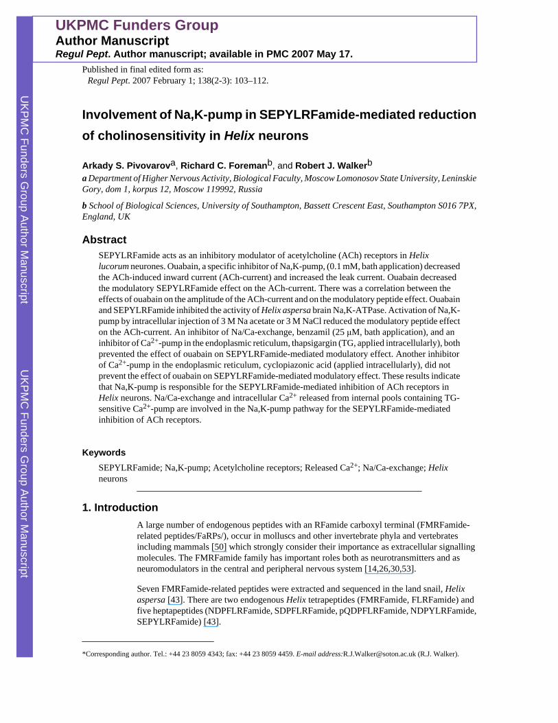
Involvement of Na,K-pump in SEPYLRFamide-mediated reductionof cholinosensitivity in Helix neurons
Arkady S. Pivovarova, Richard C. Foremanb, and Robert J. Walkerba Department of Higher Nervous Activity, Biological Faculty, Moscow Lomonosov State University, LeninskieGory, dom 1, korpus 12, Moscow 119992, Russia
b School of Biological Sciences, University of Southampton, Bassett Crescent East, Southampton S016 7PX,England, UK
AbstractSEPYLRFamide acts as an inhibitory modulator of acetylcholine (ACh) receptors in Helixlucorum neurones. Ouabain, a specific inhibitor of Na,K-pump, (0.1 mM, bath application) decreasedthe ACh-induced inward current (ACh-current) and increased the leak current. Ouabain decreasedthe modulatory SEPYLRFamide effect on the ACh-current. There was a correlation between theeffects of ouabain on the amplitude of the ACh-current and on the modulatory peptide effect. Ouabainand SEPYLRFamide inhibited the activity of Helix aspersa brain Na,K-ATPase. Activation of Na,K-pump by intracellular injection of 3 M Na acetate or 3 M NaCl reduced the modulatory peptide effecton the ACh-current. An inhibitor of Na/Ca-exchange, benzamil (25 μM, bath application), and aninhibitor of Ca2+-pump in the endoplasmic reticulum, thapsigargin (TG, applied intracellularly), bothprevented the effect of ouabain on SEPYLRFamide-mediated modulatory effect. Another inhibitorof Ca2+-pump in the endoplasmic reticulum, cyclopiazonic acid (applied intracellularly), did notprevent the effect of ouabain on SEPYLRFamide-mediated modulatory effect. These results indicatethat Na,K-pump is responsible for the SEPYLRFamide-mediated inhibition of ACh receptors inHelix neurons. Na/Ca-exchange and intracellular Ca2+ released from internal pools containing TG-sensitive Ca2+-pump are involved in the Na,K-pump pathway for the SEPYLRFamide-mediatedinhibition of ACh receptors.
KeywordsSEPYLRFamide; Na,K-pump; Acetylcholine receptors; Released Ca2+; Na/Ca-exchange; Helixneurons
1. IntroductionA large number of endogenous peptides with an RFamide carboxyl terminal (FMRFamide-related peptides/FaRPs/), occur in molluscs and other invertebrate phyla and vertebratesincluding mammals [50] which strongly consider their importance as extracellular signallingmolecules. The FMRFamide family has important roles both as neurotransmitters and asneuromodulators in the central and peripheral nervous system [14,26,30,53].
Seven FMRFamide-related peptides were extracted and sequenced in the land snail, Helixaspersa [43]. There are two endogenous Helix tetrapeptides (FMRFamide, FLRFamide) andfive heptapeptides (NDPFLRFamide, SDPFLRFamide, pQDPFLRFamide, NDPYLRFamide,SEPYLRFamide) [43].
*Corresponding author. Tel.: +44 23 8059 4343; fax: +44 23 8059 4459. E-mail address:[email protected] (R.J. Walker).
UKPMC Funders GroupAuthor ManuscriptRegul Pept. Author manuscript; available in PMC 2007 May 17.
Published in final edited form as:Regul Pept. 2007 February 1; 138(2-3): 103–112.
UKPM
C Funders G
roup Author Manuscript
UKPM
C Funders G
roup Author Manuscript
Endogenous molluscan FMRFamide-related peptides (FaRPs): FMRFamide,SKPYMRFamide, SEPYLRFamide and its N-terminally modified analogues, EPYLRFamide,SEGYLRFamide, SRPYLRFamide, SKPYLRFamide and acetyl-SKPYMRFamide act asinhibitory neuromodulators of cholinergic transmission in H. aspersa [1,38,41] and Helixlucorum [36,39,40,42] parietal ganglia.
The modulation of synaptic transmission by FaRPs can occur through a change in the sensitivityof the postsynaptic membrane to transmitters since it was found that all the FaRPs reducedreversibly the inward current in H. aspersa and H. lucorum identified neurons to localacetylcholine (ACh) application onto the soma. Reduction of ACh-induced current(metabotropic effect) by SEPYLRFamide and its N-terminally modified analogues atconcentrations 0.01–10.0 μM was not associated with any changes in the leak current. Atconcentration of 50 μM the peptides evoked an electrogenic effect, i.e., an increase (by 15–25%) of inward leak current with full recovery after wash. [39].
Elevation of free intracellular Ca2+ reduces ACh-induced inward Cl− currents in Lymnaeastagnalis [12,52] and Helix pomatia [3] dialysed neurons. This means that an artificial increaseof intracellular free Ca2+ in the cytoplasm has a similar effect to that of FaRPs on somaticacetylcholine receptors. Hence FaRPs may inhibit cholinosensitivity in Helix neurons via anelevation of intracellular Ca2+. This assumption is supported by the observation thatFMRFamide increases basal intracellular Ca2+ in Helix neurons since FMRFamide amplifiesa fluorescent signal from H. aspersa identified neurons loaded with Ca2+-sensitive dye fluo-3[36].
Our electrophysiological data show that intracellular free Ca2+ is involved in the intracellularmechanism for the reduction of cholinosensitivity in Helix neurons by FaRPs [36,38,41].Ryanodine receptors and IP3 receptors may be involved in the inhibitory metabotropic effectsof FaRPs on somatic cholinergic receptors of identified Helix neurons [36,38,41,42].
We searched the literature for a possible cellular regulator to explain the inhibitorymetabotropic effect evoked by the FaRPs on ACh receptors. One possible regulator is the Na,K-pump. It is known from the literature that inhibition of the Na,K-pump by ouabain evokes adecrease of ACh-induced currents of H. pomatia dialysed neurons [3]. Ouabain can elevateintracellular calcium via the activation of the reverse mode of Na/Ca-exchange (Na+-efflux,Ca2+-influx) [1,11,13,15,20,31,45,46] and stimulate Ca2+ release from internal stores [4,32,46,47].
Previous work has shown that FMRFamide increases intracellular Ca2+ via inhibition inNaout-dependent Ca2+-efflux (Na/Ca-exchange in the normal mode) [16,24,51] and activationof Ca2+-mobilization via ryanodine receptors and IP3 receptors [18,19,36,38,41,54].
Ouabain reduces reversibly ACh-induced inward currents in identified H. lucorum neurons(LPa2, LPa3, RPa3, RPa2) [33,35,37] that were used in our investigations with FaRPs [36,38,39,40,42]. This ouabain-mediated metabotropic effect is Ca2+-dependent.
There are identical effects of FaRPs and ouabain on the amplitude of ACh-induced currents inHelix neurons and on intracellular Ca2+ level. This provides the basis to postulate a workinghypothesis: FaRPs may inhibit postsynaptic cholinosensitivity in Helix neurons partly viainitial Na,K-pump inhibition by means of G-proteins followed by elevation of intracellular freeCa2+.
In this study we investigated the participation of Na,K-pump in the modulatory effect ofendogenous molluscan heptapeptide, SEPYLRFamide, on cholinosensitivity in Helixneurones. The role of the following intracellular regulators were investigated, G-proteins, Na/
Pivovarov et al. Page 2
Regul Pept. Author manuscript; available in PMC 2007 May 17.
UKPM
C Funders G
roup Author Manuscript
UKPM
C Funders G
roup Author Manuscript
Ca-exchange and intracellular calcium released from internal pools in the modulatory peptide-mediated cascade involving inhibition of Na,K-pump.
2. Materials and methods2.1. Recording of transmembrane currents
2.1.1. Animals—Snails, H. lucorum, were collected locally in the Sevastopol region, Crimea,Ukraine. Experiments were performed using an isolated ganglia preparation at roomtemperature (18–22 °C) during 2002–2004.
Animals were anaesthetized in cold saline and the circumoesophageal nerve ring was removedfor study. Circumoesophageal ganglia were pinned down, dorsal side up, on a silicon rubber-coated flow chamber with a bath volume of 1.0 ml. The connective tissue sheath coveringneurons was removed prior to the experiments after preliminary enzymatic preparation(digestase/Seatec/, 0.5%, 30–40 min at room temperature (18–22 °C). Ganglia were constantlysuperfused (0.5–0.8 ml/min) with normal Helix saline containing (in mM): NaCl, 100; KCl, 4;CaCl2,10;MgCl2, 4; Tris buffer, 10; adjusted to pH 7.5 with HCl.
2.1.2. Electrophysiological recording—Experiments were carried out on identifiedLPa2, LPa3, RPa3 and RPa2 neurons from left and right parietal ganglia of H. lucorum. Thesecells are command elements of withdrawal responses to noxious stimuli [22].
Single-barrel glass microelectrodes were pulled using a PUL-1 puller (World PrecisionInstruments) from Pyrex glass (1.5 mm outer diameter) and filled with 2 M potassium acetate;resistance 8–120 MΩ. The electrodes were connected by a Ag–AgCl microelectrode holder(World Precision Instruments design) to a Micro-Electrode Amplifier MEZ-8101 (NihonKohden) and Voltage Clamp Amplifier CEZ-1100 (Nihon Kohden) that were used in VirtualGround Mode for two-electrode voltage clamp experiments. A briquette of Ag–AgCl(Medicor) was used as reference electrode. The neurons were clamped at −75 mV. Currentswere entered on a PC through the analogous-digital interface L-154 (L-CARD, Moscow) andrecorded using CONAN 3.5 software (InCo, Moscow, Russia).
ACh was applied ionophoretically (interstimulus interval 5 min) onto the neuronal soma usingthe current source isolated from the ground. The ionophoretic solution was as follows: 1 MACh chloride (Sigma) in distilled water (pH 7.0). The resistance of the ionophoretic pipettewas 14–46 MΩ. The reference pipette was filled with normal Helix saline with a resistance of1–3 MΩ. Cationic currents (685–877 nA; 0.1–3.0 s) were used for ionophoresis. A LaboratoryElectrostimulator ESL-2 (Kaunas Research Institute of Radiometrical Engineering) was usedto control the duration of the ionophoretic current. Ejection of positive currents through anionophoretic pipette filled only with distilled water had no effect on the cells. A negativeretention current (10 nA) was passed continuously through the ionophoretic pipette in order toprevent the spontaneous diffusion of ACh.
For an estimation of changes of stationary membrane conductance the holding potential involtage clamp mode was shifted periodically in a negative direction (rectangular pulses, 10mV, 5 s) for 9 s up to the start of ionophoretic ACh application. Change of amplitude of theinward leak current evoked by the negative shift of a holding potential was directly proportionalto the change in resting membrane conductance.
2.1.3. Drugs—SEPYLRFamide (Department of Biochemistry, University of Southampton;Alta Bioscience, University of Birmingham) was locally applied under pressure at aconcentration of 5 mM using piston ejector KM-2 (Chernogolovka). Peptide was applied froma glass micropipette with a broken tip, inner diameter of micropipette tip was 4.3–10.6 μM.
Pivovarov et al. Page 3
Regul Pept. Author manuscript; available in PMC 2007 May 17.
UKPM
C Funders G
roup Author Manuscript
UKPM
C Funders G
roup Author Manuscript
Volume of applied solution was 4–14 nL; duration of application was 5 or 10 s; speed ofapplication was 0.4–2.8 nL/s; distance of micropipette tip from the recording cell was 2–3 celldiameter (0.2–0.3 mm) to prevent mechanical action of ejected solution: interval betweenrepeated peptide applications was 60 min. Red dye ruthenium red (10 mM, Sigma) was locallyapplied under pressure to evaluate this type of application.
An inhibitor of Na,K-pump, ouabain (0.1 mM), and an inhibitor of Na/Ca-exchange, benzamil(25 μM), were bath applied. Other compounds entered the cells by passive loading (diffusion)from intracellular voltage recording microelectrodes during a sixty minute period: sodiumacetate, sodium chloride, inhibitors of Ca-ATPase in endoplasmic and sarcoplasmic reticulum(thapsigargin, cyclopiazonic acid). All drugs were obtained from Sigma.
SEPYLRFamide (5 mM) was dissolved in Helix saline. Cyclopiazonic acid (0.1 M) andthapsigargin (0.1 mM) were dissolved in dimethyl sulfoxide (DMSO, 5%; Sigma) and 2 Mpotassium acetate. These solutions were stored at −18 °C.
Stock solutions of ouabain (10 mM) and benzamil (5 mM) were prepared using Helix salineand stored at +4 °C. Sodium acetate (3 M) and sodium chloride (3 M) were dissolved in distilledwater and stored at room temperature (18–22 °C).
The results were obtained from 167 neurons in 167 preparations. The resting potential of thecells was −61.10 ± 0.62 mV. The input resistance of neurons was 4.46 ± 0.18 MΩ.
2.1.4. Data analysis—The data are expressed as means ± standard error of the mean(S.E.M.). Each amplitude of ACh-induced current was normalised to the last three responsesbefore each peptide application. In the text and figures, n represents the number of neurons.Significant differences for comparisons between two groups (control and experimental) wereevaluated using nonparametric Wilcoxon's test. Student's t-test was used for comparisonsbetween two linear regression slopes. A computer statistical software STADIA 6.2 (InCo,Moscow, Russia) was used for statistical analysis. A P value of less than 0.05 was consideredto be statistically significant.
2.2. Measurement of Na,K-ATPase activity2.2.1. Animals—Snails (H. aspersa.L) were collected locally in Southampton.Circumoesophageal nerve rings were extracted. Experiments were performed during October–December.
2.2.2. Homogenisation—H. aspersa were dissected, and the brain removed and weighed.The brain tissue was mixed with 5 ml per gram of medium containing: 0.32 M sucrose, 1mMEDTA, 0.1% deoxycholate, 50 mM Tris/MES, 250 μM ammonium molybdate at pH 7.4 [26with some modification]. The homogenate was processed by differential centrifugation toseparate the nuclear and mitochondrial fractions. This mixture was homogenised at 4 °C usinga Teflon glass homogeniser until all tissue was broken down. A crude pellet containingmitochondria, nerve ending membranes, and myelin was separated by centrifugation at 10,000×g for 10 min. Clear supernatant was removed and centrifuged in a Beckman ultracentrifugeat 100,000 ×g for 60 min. This double-stage homogenization was undertaken to obtain lightmicrosomal fraction enriched by small vesicles originating from the plasma membrane andmembranes of the endoplasmic reticulum.
The final sediment was resuspended in a medium containing 0.32 M sucrose, 1 mM EDTA,50 mM Tris/MES, 250 μM ammonium molybdate at pH 7.4. The aliquots were stored at −75
Pivovarov et al. Page 4
Regul Pept. Author manuscript; available in PMC 2007 May 17.
UKPM
C Funders G
roup Author Manuscript
UKPM
C Funders G
roup Author Manuscript
°C. Brain samples were diluted in Tris/MES buffer with 250 μM ammonium molybdate at pH7.4 for ATPase assay.
2.2.3. ATPase assay—Measurement of ATPase activity within membrane vesicles wasmonitored by colorimetric determination of free inorganic phosphate released (Pi) from ATPusing the catalyst polyvinylpyrrolidone [34].
Diluting 3 mM KH2PO4 in Tris/MES buffer with 250 μM ammonium molybdate at pH 7.4produced twelve standard phosphate solutions between 0 and 96 nM PO4 per 50 μl. Each ofthese solutions was applied in triplicate to each assay to produce a standard phosphate curve.
A 96 well Tissue Culture Testplate (TPP, Switzerland) was used to perform this assay. Thereaction was started by addition of 30–40 μl of reaction mixture to each experimental well.The reaction mixture (10 ml) consisted of: 4 ml Tris/Mes buffer with 250 μM ammoniummolybdate at pH 7.4, 1 ml 20 mM ATP, 1 ml 20 mM MgSO4, 1 ml 500 mM KCl, 3 ml H20.The tissue was then incubated on ice under appropriate conditions for 30 min.
After the first incubation the tissue samples were added to the experimental wells in triplicate.The total volume in each experimental well was 50 μl with reaction mixture and experimentalsample.
Reaction mixture alone was added to three wells to serve as a blank. The 96 well microtitreplate was then incubated at 37 °C for 60 min, gently mixing every 20 min.
Once incubation was complete the reaction was stopped by adding 8 μM 10% sodium dodecylsulphate to each well. Then 150 μl Ohnishi A/B reagent and 15 μl Ohnishi Colour Developerwere added to each well and mixed thoroughly using a multipipette. A colour developed overa 20 min period. The plate was then read at 620 nm of wavelength to obtain values for opticaldensity using a Spectrophotometer Dynatech MR5000 (Anthos Labtec Instruments).
In order to determinate the level of protein in the extracts a protein assay was carried out usingHelix brain tissue. Samples of the extract were added in triplicate at 1:10, 1:100 and 1:1000concentrations. A standard curve of known human serum album (HSA) concentration was alsoadded to the plate. The standard curve consisted of values between 0 and 2 mg/ml (HSA). BACreagent was then added to all experimental plates and colour was allowed to develop for 30min. The plate was then read at 620 nm wavelength and protein content calculated.
2.2.4. Drugs—SEPYLRFamide was obtained from Alta Bioscience (University ofBirmingham, UK) and Department of Biochemistry (University of Southampton, UK).Ouabain, an inhibitor of Na, K-ATPase; adenosine 5′-trisphosphate; non-hydrolizable GTPanalogue, guanosine-5′-O-(3-thiotriphosphate) tetralithium salt (GTP-γ-S); Na azide, aninhibitor of mitochondrial ATPase, were all obtained from Sigma.
All drugs were dissolved in water medium contained 50 mM Tris/MES, 0.32 M sucrose and1 mM EDTA, pH 7.4. Stock solutions of SEPYLRFamide (2 mM) and GTP-γ-S (2 mM) werestored at −18 °C. Stock solution of ouabain (20 mM) was stored at +4 °C. Stock solution ofNa azide (8 mM) was stored at room temperature (18–22 °C).
2.2.5. Data analysis—The data are expressed as mean ± standard error of the mean (S.E.M.).Significant differences for comparisons between two groups (control and experimental) wereevaluated using nonparametric Mann–Whitney rank sum test. The computer statistical softwareSigmaStat 3.0 was used for statistical analysis. A P value of less than 0.05 was considered tobe statistically significant.
Pivovarov et al. Page 5
Regul Pept. Author manuscript; available in PMC 2007 May 17.
UKPM
C Funders G
roup Author Manuscript
UKPM
C Funders G
roup Author Manuscript

3. Results3.1. Control experiments with pressure application of ruthenium red onto neuron soma
In control experiments (n = 4) a dye, ruthenium red (10 mM), was applied under pressure ontothe H. lucorum identified neurons RPa3 or LPa3 using the same parameters of application aswith SEPYLRFamide. Red solution reached the soma of the target neuron 1–2 s after the endof pressure ejection of ruthenium red. The volume of ejected coloured cone exceeded the sizeof the target cell. This means that there was dilution of the applied solution.
Several conclusions could be made from this. It was calculated that the degree of dilution ofthe applied solution adjacent to the soma was around 100 times. We can therefore assume thatthe concentration of the applied peptide around the neuronal soma was approximately 50 μM.Following single peptide application, the peptide concentration in the flow chamber was 0.02–0.07 μM. This concentration is smaller compared with the threshold concentrations ofFMRFamide-related peptides for an inhibitory action on ACh-induced inward current (0.1–1.0μM) [39,40]. So we can assume that SEPYLRFamide application under pressure mediates thelocal effect on the target neuron.
3.2. SEPYLRFamide-evoked reduction of ACh-induced inward currentsThe first experiment was a control to investigate the effect of pressure on the amplitude of theACh-induced current using Helix saline ejection. In these experiments (n = 11) pressureapplication of Helix saline did not change the ACh-current amplitude. 20 s after the end ofapplication the amplitude of ACh-current was 101.00 ± 5.83% (Table 1).
This control was followed by three SEPYLRFamide applications with a 60 min interval beforeeach application of peptide.
The first local application of SEPYLRFamide (5 mM) under pressure evoked time-dependentreversible reduction of ACh-current in all LPa2, LPa3, RPa3 and RPa2 neurons that wereinvestigated (Fig. 1). The minimal amplitude of the ACh-current occurred 20 s following theend of the local application of SEPYLRFamide, 57.73 ± 4.04%, n = 18 cells, P < 0.001) (Table1). Reduction of the ACh-current amplitude to the 2nd and 3rd SEPYLRFamide applicationdid not differ significantly from the peptide effect to the 1st application (Table 1). This meansthat repeated peptide applications did not evoke significant changes in membrane sensitivityto SEPYLRFamide.
3.3. Effect of ouabain on ACh-induced inward current and leak currentBefore we investigated the effect of ouabain on SEPYLRFamide-evoked reduction of ACh-induced inward currents we checked for any effects of ouabain on neurons. Ouabain (0.1 mM,bath application, n = 18) evoked three clear effects in Helix neurons that developed graduallyand reached stable levels after 30 min exposure: 1) the slow inward current (5–15 nA, notshown), 2) reduction of the ACh-current amplitude to 81.99 ± 5.78%, P < 0.05 (Tables 2 and3) increase of the leak current amplitude to 150.90 ± 5.78%, P < 0.01 (Table 3).
3.4. Effect of ouabain on SEPYLRFamide-evoked reduction of ACh-induced inward currentsThe experiment included three consecutive SEPYLRFamide applications with a 60 mininterval: before ouabain, after 30 min of the application of ouabain and after washout.
Ouabain decreased the ability of SEPYLRFamide to reduce ACh-current amplitude (Table 1).Amplitude of the ACh-current 20 s after peptide application was 51.90 ± 3.94% before ouabain(n = 18), 62.90 ± 3.94% after the application of ouabain (n = 18) and 48.09 ± 5.30% (n = 15)
Pivovarov et al. Page 6
Regul Pept. Author manuscript; available in PMC 2007 May 17.
UKPM
C Funders G
roup Author Manuscript
UKPM
C Funders G
roup Author Manuscript
after wash. There was a statistically significant reduction by ouabain of the peptide effect (P< 0.05) and recovery of peptide effect after wash (P < 0.05).
There was a significant correlation between the effects of ouabain on the amplitude of the ACh-current and on the modulatory peptide effect (r = 0.58, Pearson coefficient; P < 0.001; linearregression slope = 0.63). There was no correlation between the effects of ouabain on theamplitude of the leak current and on the modulatory peptide effect (r = 0.15, Pearsoncoefficient; P > 0.05; linear regression slope = 0.08).
3.5. Influence of intracellular action of sodium ions on SEPYLRFamide-evoked reduction ofACh-induced inward currents
Passive loading of the cell with sodium ions from an intracellular electrode during a 60 minperiod will raise the level of intracellular sodium ions and activate the Na,K-pump. Activationof Na,K-ATPase by intracellular injection of 3 M Na acetate or 3 M NaCl reduced themodulatory peptide effect on the ACh-current (Table 1). The minimal amplitude of the ACh-current 20 s after the first SEPYLRFamide pressure application following 60 min sodiuminjection was 82.38 ± 3.92% (n = 18 cells, P < 0.001). It is significantly smaller (P < 0.001)as compared with the reduction of the ACh-current to the first SEPYLRFamide applicationwithout injection of sodium ions (57.73 ± 4.04%, n = 30).
3.6. Effect of benzamil on the effect of ouabain on SEPYLRFamide-evoked reduction of ACh-induced inward currents
The specific inhibitor of Na/Ca-exchange in plasmalemma, benzamil (BZ, bath application,25 μM), decreased the peptide-mediated reduction of ACh-current (Table 1). The minimalamplitude of the ACh-current 20 s after the first SEPYLRFamide pressure applicationfollowing 30 min of BZ application was 72.48 ± 7.29% (n = 12 cells, P < 0.001). It issignificantly smaller (P < 0.05) as compared with the reduction of the ACh-current to the firstSEPYLRFamide application without the action of BZ (55.54 ± 2.93%, n = 48).
Reductions of the ACh-current amplitude to the 2nd and 3rd SEPYLRFamide application inthe presence of BZ did not differ significantly from the peptide effect to the 1st application(Table 1). This means that repeated peptide application after BZ application did not changethe membrane sensitivity to SEPYLRFamide.
BZ blocked the effect of ouabain on the SEPYLRFamide-mediated modulatory effect. Afterapplication of BZ, ouabain did not change the ability of SEPYLRFamide to reduce the ACh-current amplitude (Table 1). The amplitude of the ACh-current 20 s after the end of peptideapplication was 76.13 ± 6.70% before ouabain (n = 15), 78.15 ± 7.47% in the presence ofouabain (n = 15) and 81.96 ± 8.41% (n = 8) after wash. The differences between these peptideeffects were not significant.
BZ blocked ouabain-induced reduction of the ACh-current but did not change the increase inthe leak current by ouabain. In the presence of BZ the amplitude of ACh-current was 100.90± 4.87%, (n=15, P > 0.05, Table 2) after 30 min bath application of ouabain. Ouabain increasedthe leak current amplitude to 136.90 ± 8.38%, P< 0.01 (Table 3).
3.7. Effect of thapsigargin on the effect of ouabain on SEPYLRFamide-evoked reduction ofACh-induced inward currents
The specific inhibitor of endoplasmic reticulum Ca2+-ATPase, thapsigargin (TG, passiveloading the cell from an intracellular electrode during 60 min period, 0.1 mM), decreased thepeptide-mediated reduction of ACh-current (Table 1). The minimal amplitude of the ACh-
Pivovarov et al. Page 7
Regul Pept. Author manuscript; available in PMC 2007 May 17.
UKPM
C Funders G
roup Author Manuscript
UKPM
C Funders G
roup Author Manuscript
current 20 s after the first SEPYLRFamide pressure application following 60 min of TG loadingwas 77.25 ± 7.27% (n =13 cells, P < 0.001). It is significantly smaller (P < 0.01) as comparedwith the reduction of the ACh-current to the first SEPYLRFamide application without injectionof TG (55.54 ± 2.93%, n =48).
Reductions of the ACh-current amplitude to the 2nd and 3rd SEPYLRFamide application inthe presence of TG did not differ from the peptide effect to the 1st application (Table 1). Thismeans that repeated peptide application after TG injection did not change the membranesensitivity to SEPYLRFamide.
TG blocked the effect of ouabain on the SEPYLRFamide-mediated modulatory effect. Afterintracellular injection of TG, ouabain did not change the ability of SEPYLRFamide to reducethe ACh-current amplitude (Table 1). The amplitude of the ACh-current 20s after the end ofpeptide application was 67.46 ± 6.08% before ouabain (n = 16), 68.12 ± 8.50% in the presenceof ouabain (n = 11) and 77.89 ± 5.36% (n = 9) after wash. The differences between these peptideeffects were not significant.
TG blocked ouabain-induced reduction of the ACh-current but did not change the increase inthe leak current by ouabain. In the presence of intracellular TG the amplitude of ACh-currentwas 99.78 ± 5.06%, (n =11, P > 0.05, Table 2) after 30 min bath application of ouabain. Ouabainincreased the leak current amplitude to 176.90 ± 15.65%, P < 0.001 (Table 3).
3.8. Influence of cyclopiazonic acid on the effect of ouabain on SEPYLRFamide-evokedreduction of ACh-induced inward currents
Another specific inhibitor of endoplasmic reticulum Ca2+-ATPase, cyclopiazonic acid (CPA,passive loading the cell from an intracellular electrode during 60 min period, 0.1 mM)decreased the peptide-mediated reduction of ACh-current (Table 1). The minimal amplitudeof the ACh-current 20 s after the first SEPYLRFamide pressure application following 60 minof CPA injection was 67.26 ± 4.92% (n=16 cells, P < 0.001). It is significantly smaller (P<0.05) as compared with the reduction of ACh-current to the first SEPYLRFamide applicationwithout injection of CPA (55.54 ±2.93%, n=48).
Reductions of the ACh-current amplitude to the 2nd and 3rd SEPYLRFamide application onthe background of CPA was greater as compared with the peptide effect to the 1st application(combined group, lines 9.1 + 10.1, Table 1). This means that CPA decreased the peptide-mediated reduction of ACh-current only to the 1st peptide application.
CPA did not prevent the effect of ouabain on the SEPYLRFamide-mediated modulatory effect.After intracellular injection of CPA ouabain decreased the ability of SEPYLRFamide to reducethe ACh-current amplitude (Table 1). Amplitude of ACh-current 20 s after the end of peptideapplication was 70.25 ± 4.52% before ouabain (n =17), 85.07 ± 5.92% in the presence ofouabain (n =13) and 70.51 ± 4.54% (n =12) after wash. There was a statistically significantreduction by ouabain of the peptide effect (P < 0.05) and recovery of peptide effect after wash(P < 0.05).
CPA did not change the ouabain-induced reduction of the ACh-current and increase of the leakcurrent. On the background of intracellular injection of CPA, ouabain (0.1 mM, bathapplication, n = 14) evoked a reduction of the ACh-current amplitude to 81.78 ± 5.06%, P <0.05 (Table 2) and an increase in the leak current amplitude to 159.20 ± 5.06%, P < 0.001(Table 3).
Pivovarov et al. Page 8
Regul Pept. Author manuscript; available in PMC 2007 May 17.
UKPM
C Funders G
roup Author Manuscript
UKPM
C Funders G
roup Author Manuscript
3.9. ATPase activity in Helix brain extractsThe presence of ATPase activity in the extracts was first established. It was shown that brainextracts produced 81 nM phosphate per 5 μl extract per hour. From the optical density andprotein assay data, average phosphate produced was calculated. The average protein contentin brain extracts was 8.23 mg/ml. The average phosphate produced by the brain was 0.561 ±0.032 μM/ml/h or 921.76 μM of phosphate per gram protein per hour (n=9).
3.10. Effect of ouabain and SEPYLRFamide on activity of purified Na,K-ATPaseThe next biochemical experiment was to demonstrate the effects of ouabain andSEPYLRFamide on purified Na,K-ATPase (2 mU/μl) activity. Ouabain (5 mM) decreasedactivity of pure Na,K-ATPase to 30.34 ± 5.87% (n =9, P<0.001). SEPYLRFamide (0.5 mM)did not change activity of pure Na,K-ATPase (data not shown).
These results demonstrate that ouabain reduced activity of purified Na,K-ATPase on anaverage by 70%. But SEPYLRFamide did not change the activity of the purified enzyme.
3.11. Effect of ouabain, SEPYLRFamide and GTP-γ-S on Na, K-ATPase activity in extractsfrom H. aspersa brain (circumoesophageal ganglia)
These experiments were performed using the light microsomal fraction that is enriched bysmall vesicles originating from plasma membrane and membranes of endoplasmic reticulum,as described in method section. Brain extracts were dissolved in the medium containing 1 mMEDTA which suppresses Ca2+-ATPase activity in the endoplasmic reticulum and thereforeATP hydrolysis will be due to only two ATPases, viz, Na,K-ATPase and ecto-ATPase. Someof these ecto-ATPases are inhibited by 1 mM azide. Therefore all the following experimentswere made in the presence of 2 mM Na azide. It is reasonable to conclude that after this ATPhydrolysis was due to only Na,K-ATPase.
Ouabain, 5 mM, inhibited brain Na,K-ATPase activity by 10%. Enzyme activity in the presenceof ouabain was 90.65 ± 4.23% (n=11, P=0.032) relatively to control activity without ouabain(Table 4).
SEPYLRFamide, 0.5 mM, also inhibited brain Na,K-ATPase activity by about 10%. Enzymeactivity in the presence of SEPYLRFamide was 87.38 ± 9.66% (n=10, P=0.003) relative tocontrol enzyme activity without SEPYLRFamide (Table 4).
GTP-γ-S, 0.5 mM, inhibited on average by 15% brain Na,K-ATPase activity. Enzyme activityin the presence of GTP-γ-S was 85.14 ± 1.88% (n=15, P<0.001) relative to control activitywithout the non-hydrolysable GTP analogue (Table 4).
4. DiscussionThe endogenous Helix heptapeptide SEPYLRFamide evoked a reversible reduction in thesomatic membrane cholinosensitivity in identified H. lucorum neurons as it decreasedacetylcholine-induced inward current.
Our data show that Na,K-ATPase may be involved in the intracellular mechanism that isresponsible for the SEPYLRFamide-mediated inhibition of neuronal cholinosensitivity.Ouabain, a specific inhibitor of Na,K-ATPase, decreased the ACh-current and increased theleak current. Ouabain decreased the modulatory peptide effect on the ACh-current. There wasa significant correlation between the effects of ouabain on the amplitude of the ACh-currentand on the modulatory peptide effect. There was no correlation between the effects of ouabainon the amplitude of the leak current and on the modulatory peptide effect. Activation of Na,K-
Pivovarov et al. Page 9
Regul Pept. Author manuscript; available in PMC 2007 May 17.
UKPM
C Funders G
roup Author Manuscript
UKPM
C Funders G
roup Author Manuscript

ATPase by intracellular injection of 3 M Na acetate or 3 M NaCl reduced the modulatorypeptide effect on the ACh-current. These results indicate that SEPYLRFamide inhibits Na,K-pump. Our biochemical results support this theoretical proposition.
Why did activation of the Na,K-ATPase through a rise in intracellular Na+ reduce themodulatory peptide effect on ACh-current? We assume that extracellular SEPYLRFamide andintracellular free Na+ bind different sites of the membrane Na, K-ATPase. It is likely that inour experiments intracellular injection of Na+ evoked greater activation of Na,K-ATPasecompared with the inhibition of this enzyme by SEPYLRFamide. As a result, elevation ofintracellular Na+ reduced the inhibitory effect of peptide on the Na,K-ATPase activity.
Inhibition of ouabain-sensitive neuronal Na,K-ATPase by FMRFamide-related peptide,SEPYLRFamide, has been shown for the first time. Other biologically active substances areknown to inhibit the activity of Na,K-ATPase. For example, this enzyme is inhibited by adrenalcortical hormone (“endogenous ouabain”) [9], mastoporan [28], 5-hydroxytrytamine [49],atrial natriuretic peptide 99–126 [5], neurotensin [29], leptin [6] and parathyroid hormone[25]. It has been shown recently that parathyroid hormone inhibits Na+, K+-ATPase activityby serine phosphorylation of the α1 subunit through protein kinase C-(PKC) and extracellularsignal regulated kinase-(ERK) dependent pathways [25].
In our experiments ouabain inhibited purified Na,K-ATPase activity by 70%. SEPYLRFamidedid not change the activity of the purified enzyme. This means that there are no binding sitesfor SEPYLRFamide on the pure Na,K-ATPase. Ouabain, SEPYLRFamide and GTP-γ-S(activator of G-proteins) inhibited the activity of H. aspersa brain Na,K-ATPase. We canpropose that SEPYLRFamide inhibits plasma membrane Na,K-ATPase indirectly, probably,via activation of G-proteins.
An inhibitor of plasmalemma Na/Ca-exchange, benzamil, and a specific inhibitor ofendoplasmic reticulum Ca2+-ATPase, thapsigargin, prevented the effect of ouabain on theSEPYLRFamide-mediated modulatory effect. Benzamil and TG prevented the ouabain-mediated reduction of acetylcholine-induced inward current but did not change the increase ofleak current. Another specific inhibitor of the endoplasmic reticulum Ca2+-ATPase,cyclopiazonic acid, did not prevent the effect of ouabain on the SEPYLRFamide-mediatedmodulatory effect. CPA did not change the ouabain-mediated reduction of acetylcholine-induced inward current and increase of leak current. These results allow us to propose that Na/Ca-exchange and intracellular Ca2+ released from internal Ca2+-depots containing TG-sensitive Ca2+-ATPase may be involved in the Na,K-ATPase pathway for theSEPYLRFamide-mediated inhibition of acetylcholine receptors.
Intracellular Ca2+ released from internal pools containing CPA-sensitive Ca2+-ATPase is notinvolved in the Na,K-ATPase pathway for the SEPYLRFamide-mediated inhibition ofacetylcholine receptors. How can these results be explained? Two different types of internalCa2+-depots have been found in H. pomatia neurons. These two calcium pools do not overlapand at least part of the caffeine-sensitive store (with ryanodine receptors) is located close tothe cellular membrane while the inositol 1,4,5-trisphosphate-sensitive pool is located in thecentre of the cell [27]. We propose that caffeine-sensitive stores (with ryanodine receptors) inthe Helix neurons used in this study may contain TG-sensitive Ca2+-ATPase.
Taking into account our results and those from the literature we propose the following threeintracellular mechanisms for the SEPYLRFamide-mediated inhibitory modulatory effect onHelix neurone cholinosensitivity without and with Na,K-pump (Fig. 2). These mechanismshave the same final target, the elevation of free intracellular Ca2+, and are closely related. (1)SEPYLRFamide → activation of membrane receptor → activation of G-protein → elevationof inositol-1,4,5-trisphosphate (IP3, second messenger) level → activation of IP3 receptors →
Pivovarov et al. Page 10
Regul Pept. Author manuscript; available in PMC 2007 May 17.
UKPM
C Funders G
roup Author Manuscript
UKPM
C Funders G
roup Author Manuscript

Ca2+ release from internal store → elevation of intracellular free Ca2+ [42]. (2)SEPYLRFamide → activation of membrane receptor → activation of G-protein → elevationof cyclic adenosine diphosphate-ribose (cADPR, second messenger) level → activation ofryanodine receptors → Ca2+ release from internal store → elevation of intracellular freeCa2+ [36]. (3) SEPYLRFamide → activation of membrane receptor → activation of G-protein→ activation of neuronal signal molecule → inhibition of membrane Na,K-pump → elevationof intracellular content of Na+ → inhibition of Na/Ca-exchange in normal mode (Na+-influx,Ca2+-efflux) and activation of Na/Ca-exchange in reverse mode (Na+-efflux, Ca2+-influx) →elevation of intracellular free Ca2+ → activation of Ca2+-sensitive receptors (channels) inendoplasmic reticulum (IP3 receptors and ryanodine receptors) → Ca2+ release from internalstores → elevation of intracellular free Ca2+. Inhibition of the Na,K-ATPase must stop thehydrolysis of ATP and evoke a corresponding elevation in the intracellular ATP level that canactivate ryanodine-sensitive Ca2+ release channel [48].So it is not excluded that peptide-mediated inhibition of Na,K-ATPase evokes Ca2+ release via elevated ATP and followingactivation of ryanodine receptors. The last pathway, including peptide-mediated inhibition ofNa,K-ATPase, evokes Ca2+ release that amplifies Ca2+ signals evoked by the other twopeptide-mediated pathways. This process is limited by negative feedback: high Ca2+
inconcentration inhibits Ca2+ release via the receptors in Ca2+-depots [7,8,10,44].
It should be noted that the scheme in Fig. 2 contains three G-protein boxes. However, this doesnot mean that the peptide receptor is connected with three different G-proteins. We do not haveexperimental evidence which would enable us to conclude that the three intracellular pathwaysinclude activation of the same G-protein. This remains an open question.
How does elevation of intracellular free Ca2+ mediate the reduction of membranecholinosensitivity? It has been shown that regulation of AMPA receptor trafficking inhippocampal neurons results in changes in receptor number at a postsynaptic membrane, andhence modifications in synaptic strength, which are proposed to underlie learning and memory[17,21]. AMPA receptor internalization triggered by NMDA receptor activation is Ca2+-dependent, requires protein phosphatases, and is followed by rapid membrane reinsertion[17]. A novel postsynaptic Ca2+-binding protein provides a direct mechanistic link betweenNMDA receptor-mediated Ca2+ influx and AMPA-receptor endocytosis [21]. In considerationof these results we assume that the cytoplasmic Ca2+ which is elevated by SEPYLRF-amideactivates an internalization of acetylcholine receptors via endocytosis that leads to a reversiblereduction of cholinosensitivity in the non-synaptic membrane.
The postsynaptic membrane in H. lucorum neurons contains acetylcholine receptors [50]. Inconclusion the above mechanism may be responsible for the reduction in cholinergic synaptictransmission to molluscan neurons by FMRFamide-related peptides [2,23,30].
Acknowledgement
This work was supported by the Wellcome Trust (grant #066177/Z/01/Z).
References1. Alvarez Leefmans FJ, Merediz Alonso G, Fernandez Calderon JR. The consequences of sodium pump
inhibition in the neurons and its relevance to the physiopathology of the cell damage produced byanoxic ischemia. Gac Med Mex 1994;130:347–54. [PubMed: 7607364]
2. Arvanov VL, Chen ML, Sharma R, Walker RJ, Ayrapetyan SN. Low concentrations of neuroactivepeptides modulate cholinergic transmission and cyclic AMP levels in Helix aspersa. Acta Biol Hung1992;43:89–97. [PubMed: 1338559]
Pivovarov et al. Page 11
Regul Pept. Author manuscript; available in PMC 2007 May 17.
UKPM
C Funders G
roup Author Manuscript
UKPM
C Funders G
roup Author Manuscript

3. Arvanov VL, Stepanyan AS, Ayrapetyan SN. The effects of cAMP, Ca2+, and phorbol esters onouabain-induced depression of acetylcholine responses in Helix neurons. Cell Mol Neurobiol1992;12:153–61. [PubMed: 1318166]
4. Balduini W, Costa LG. Characterization of ouabain-induced phosphoinositide hydrolysis in brain slicesof the neonatal rat. Neurochem Res 1990;15:1023–9. [PubMed: 1963925]
5. Beltowski J, Gorny D, Marciniak A. The mechanism of Na+,K+-ATPase inhibition by atrial natriureticfactor in rat renal medulla. J Physiol Pharmacol 1998;49:271–83. [PubMed: 9670110]
6. Beltowski J, Marciniak A, Wojcicka G. Leptin decreases renal medullary Na+,K+-ATPase activitythrough phosphatidylinositol 3-kinase dependent mechanism. J Physiol Pharmacol 2004;55:391–407.[PubMed: 15213361]
7. Berridge MJ. Inositol trisphosphate and calcium signalling. Nature 1993;361:315–25. [PubMed:8381210]
8. Berridge MJ. The endoplasmic reticulum: a multifunctional signaling organelle. Cell Calcium 2002;32(5–6):235–49. [PubMed: 12543086]
9. Blaustein MP. Endogenous ouabain: role in the pathogenesis of hypertension. Kidney Int1996;49:1748–53. [PubMed: 8743490]
10. Bootman MD, Berridge MJ, Roderick HL. Calcium signalling: more messengers, more channels,more complexity. Curr Biol 2002;12:R563–5. [PubMed: 12194839]
11. Charlemagne D. Molecular and cellular level of action of digitalis. Herz 1993;18:79–85. [PubMed:7684015]
12. Chemeris NK, Kazachenko VN, Kislov AN, Kurchikov AA. Inhibition of acetylcholine responsesby intracellular calcium in Lymnaea stagnalis neurons. J Physiol 1982;323:1–19. [PubMed: 6284913]
13. Condrescu M, Gardner JP, Chernaya G, Aceto JF, Kroupis C, Reeves JP. ATP-dependent regulationof sodium–calcium exchange in Chinese hamster ovary cells transfected with the bovine cardiacsodium–calcium exchanger. J Biol Chem 1995;270:9137–46. [PubMed: 7721828]
14. Cottrell GA, Lin JW, Llinas R, Price DA, Sugimori M, Stanley EF. FMRFamide-related peptidespotentiate transmission at the squid giant synapse. Exp Physiol 1992;77:881–9. [PubMed: 1362646]
15. Deitmer JW, Eckert R, Schlue WR. Changes in the intracellular free calcium concentration of Aplysiaand leech neurones measured with calcium-sensitive microelectrodes. Can J Physiol Pharm1987;65:934–9.
16. DiPolo R, Beauge L. Cardiac sarcolemmal Na/Ca-inhibiting peptides XIP and FMRF-amide alsoinhibit Na/Ca exchange in squid axons. Am J Physiol 1994;267:C307–11. [PubMed: 8048489]
17. Ehlers MD. Reinsertion or degradation of AMPA receptors determined by activity-dependentendocyting sorting. Neuron 2000;28:511–25. [PubMed: 11144360]
18. Ellis AM, Huddart H. Excitation evoked by FMRFamide and FLRFamide in the heart of Buccinumundatum and evidence for inositol 1,4,5-trisphosphate as an RF-tetrapeptide second messenger. JComp Physiol B 2000;170:351–6. [PubMed: 11083516]
19. Falconer SW, Carter AN, Downes CP, Cottrell GA. The neuropeptide Phe-Met-Arg-Phe-NH2(FMRFamide) increases levels of inositol 1,4, 5-trisphosphate in the tentacle retractor muscle ofHelix aspersa. Exp Physiol 1993;78:757–66. [PubMed: 8311943]
20. Goldman WF, Yarowsky PJ, Juhaszova M, Krueger BK, Blaustein MP. Sodium/calcium exchangein rat cortical astrocytes. J Neurosci 1994;14:5834–43. [PubMed: 7523629]
21. Hanley JG, Henley JM. PICK1 is a calcium-sensor for NMDA-induced AMPA receptor trafficking.EMBO J 2005;24:3266–78. [PubMed: 16138078]
22. Ierusalimskii VN, Zakharov IS, Palikhova TA, Balaban PM. The nervous system and the mappingof the neurons in the gastropod Helix lucorum L. Z Vyss Nervn Deat Im IP Pavlova 1992;42:1075–89.
23. Keating C, Lloyd PE. Differential modulation of motor neurons that innervate the same muscle butuse different excitatory transmitters in Aplysia. J Neurophysiol 1999;82:1759–67. [PubMed:10515965]
Pivovarov et al. Page 12
Regul Pept. Author manuscript; available in PMC 2007 May 17.
UKPM
C Funders G
roup Author Manuscript
UKPM
C Funders G
roup Author Manuscript

24. Khananshvili D, Price DC, Greenberg MJ, Sarne Y. Phe-Met-Arg-Phe-NH2 (FMRFa)-relatedpeptides inhibit Na+–Ca2+ exchange in cardiac sarcolemma vesicles. J Biol Chem 1993;268:200–5.[PubMed: 8416928]
25. Khundmiri SJ, Dean WL, McLeish KR, Lederer ED. Parathyroid hormone-mediated regulation ofNa+–K+-ATPase requires ERK-dependent translocation of protein kinase Calpha. J Biol Chem2005;280:8705–13. [PubMed: 15637080]
26. Koyabashi M, Muneoka Y. Structure and action of molluscan neuropeptides. Zool Sci 1990;7:801–14.
27. Kostyuk PG, Kirischuk SI. Spatial heterogeneity of caffeine- and inositol 1,4,5-trisphosphate-inducedCa2+ transients in isolated snail neurons. Neuroscience 1993;53:943–7. [PubMed: 8506027]
28. Langel U, Pooga M, Kairane C, Zilmer M, Bartfai T. A galanin–mastoparan chimeric peptide activatesthe Na+,K+-ATPase and reverses its inhibition by ouabain. Regul Pept 1996;62:47–52. [PubMed:8738882]
29. Lopez Ordieres MG, Rodriquez de Lores Arnaiz G. Neurotensin inhibits neuronal Na+–K+-ATPaseactivity through high affinity peptide receptor. Peptides 2000;21:571–6. [PubMed: 10822114]
30. Man-Son-Hing H, Zoran MJ, Lukowiak K, Haydon PG. A neuromodulation of synaptic transmissionacts on the secretory apparatus as well as on ion channels. Nature 1989;341:237–9. [PubMed:2476676]
31. Meyer-Lehnert H, Backer A, Kramer HJ. Inhibitors of Na–K-ATPase in human urine: effects ofouabain-like factors and of vanadium–diascorbate on calcium mobilization in rat vascular smoothmuscle cells: comparison with the effects of ouabain, angiotensin II, and arginine–vasopressin. AmJ Hypertens 2000;13:364–9. [PubMed: 10821337]
32. Myles ME, Fain JN. Carbachol, but not norepinephrine, NMDA, ionomycin, ouabain, or phorbolmyristate acetate, increases inositol 1,3,4,5-tetrakisphosphate accumulation in rat brain corticalslices. J Neurochem 1994;62:2333–9. [PubMed: 8189237]
33. Nistratova VL, Pivovarov AS. Inositoltrisphospate receptors and ryanodine receptors in regulationof cholinosensitivity of Helix lucorum neurones by Na,K-pump during habituation. Z Vyss NervnDeat Im IP Pavlova 2004;54:554–64.
34. Ohnishi T, Gall RS, Mayer ML. An improved assay of inorganic phosphate in the presence ofextralabile phosphate compounds: application to the ATPase assay in the presence ofphosphocreatine. Anal Biochem 1975;69:261–7. [PubMed: 129016]
35. Pivovarov AS, Boguslavsky DV. Na,K-pump regulates reduction of cholinosensitivity of Helixlucorum neurones in cellular analog of habituation: role of intracellular calcium. Z Vyss Nervn DeatIm IP Pavlova 2000;50:855–66.
36. Pivovarov AS, Chad JE, Walker RJ. Involvement of ryanodine receptors in EPYLRFamide-mediatedreduction of acetylcholine-induced inward currents in Helix lucorum identified neurones. Regul Pept2000;88:83–93. [PubMed: 10706956]
37. Pivovarov AS, Nistratova VL, Boguslavskii DV. Participation of Na/Ca-exchange and intracellularmobilized Ca2+ in regulating depression of cholino-sensitive Helix lucorum neurons to a cellularanalog of habituation. Z Vyss Nervn Deat Im IP Pavlova 2001;51:723–32.
38. Pivovarov AS, Sharma R, Walker RJ. Inhibitory action of SKPYMRFamide on acetylcholinereceptors of Helix aspersa neurons: role of second messengers. Gen Pharmacol 1995;26:495–505.[PubMed: 7789722]
39. Pivovarov AS, Sharma R, Walker RJ. Structure-activity relationships of the Helix neuropeptide,SEPYLRFamide, and its N-terminally modified analogues on identified Helix lucorum neurones.Brain Res 1999;821:294–308. [PubMed: 10064816]
40. Pivovarov AS, Walker RJ. Direct and modulatory effects of FMRFamide, SKPYMRFamide andacety1-SKPYMRFamide on LPa2, LPa3, and RPa3 identified neurons of Helix lucorum. Regul Pept1996;67:169–78. [PubMed: 8988517]
41. Pivovarov AS, Walker RJ. Effects of SEPYLRFamide on acetylcholine-induced currents of Helixaspersa neurones: role of ryanodine receptors. Invertebr neurosci 1999;4:17–24.
42. Pivovarov AS, Walker RJ. EPYLRFamide-mediated reduction of acetylcholine-induced inwardcurrents in Helix lucorum identified neurones: Role of NAADP-dependent and IP3-dependent Ca2
Pivovarov et al. Page 13
Regul Pept. Author manuscript; available in PMC 2007 May 17.
UKPM
C Funders G
roup Author Manuscript
UKPM
C Funders G
roup Author Manuscript

+ release from internal stores, calmodulin and Ca2+/calmodulin-dependent protein kinase II. RegulPept 2003;111:31–9. [PubMed: 12609746]
43. Price DA, Lesser W, Lee TD, Doble KE, Greenberg MJ. Seven FMRFamide-related and two SCP-related cardioactive peptides from Helix. J Exp Biol 1990;154:421–37. [PubMed: 1980513]
44. Roderick HL, Berridge MJ, Bootman MD. Calcium-induced calcium release. Curr Biol2003;13:R425. [PubMed: 12781146]
45. Saghian AA, Ayrapetyan SN, Carpenter DO. Low concentrations of ouabain stimulate Na/Caexchange in neurons. Cell Mol Neurobiol 1996;16:489–98. [PubMed: 8879751]
46. Sarkozi S, Szentesi P, Jona I, Csernoch L. Effects of cardiac glycosides on excitation-contractioncoupling in frog skeletal muscle fibres. J Physiol (Lond) 1996;495:611–26. [PubMed: 8887770]
47. Schiebinger RJ, Cragoe EJ Jr. Ouabain. A stimulator of atrial natriuretic peptide secretion and itsmechanism of action. Circ Res 1993;72:1035–43. [PubMed: 8386595]
48. Sitsapesan R, McGarry SJ, Williams AJ. Cyclic ADP-ribose competes with ATP for the adeninenucleotide binding site on the cardiac ryanodine receptor Ca(2+)-release channel. Circ Res1994;75:596–600. [PubMed: 8062431]
49. Stepp LR, Novakoski MA. Effect of 5-Hydroxytryptamine on sodium- and potassium-dependentadenosine triphosphatase and its reactivity toward ouabain. Arch Biochem Biophys 1997;337:43–53. [PubMed: 8990266]
50. Ter-Markarian AG, Palikhova TA, Sokolov EN. The action of atropine and d-tubocurarine on themonosynaptic connections between identified neurons in the central nervous system of the ediblesnail. Z Vyss Nervn Deat Im IP Pavlova 1990;40:183–4.
51. Van Eylen F, Gourlet P, Vandermeers A, Lebrun P, Herchuelz A. Inhibition of Na/Ca exchange byPhe-Met-Arg-Phe-NH2 (FMRFa)-related peptides in intact rat pancreatic B-cells. Mol CellEndocrinol 1994;106:R1–5. [PubMed: 7534729]
52. Vulfius CA, Ryazansky VD, Krasts IV, Ilyasov FE. Intracellular Ca2+ modulates Cl− current evokedby acetylcholine in Lymnaea neurons. Neurosci Lett 1998;249:5–8. [PubMed: 9672375]
53. Walker RJ. Neuroactive peptides with an RFamide or Famide carboxyl terminals. Comp BiochemPhysiol 1992;102C:213–22.
54. Willoughby D, Yeoman MS, Benjamin PR. Inositol-1,4,5-trisphosphate and inositol-1,3,4,5-tetrakisphosphate are second messenger targets for cardioactive neuropeptides encoded on theFMRFamide gene. J Exp Biol 1999;202:2581–93. [PubMed: 10482718]
Pivovarov et al. Page 14
Regul Pept. Author manuscript; available in PMC 2007 May 17.
UKPM
C Funders G
roup Author Manuscript
UKPM
C Funders G
roup Author Manuscript
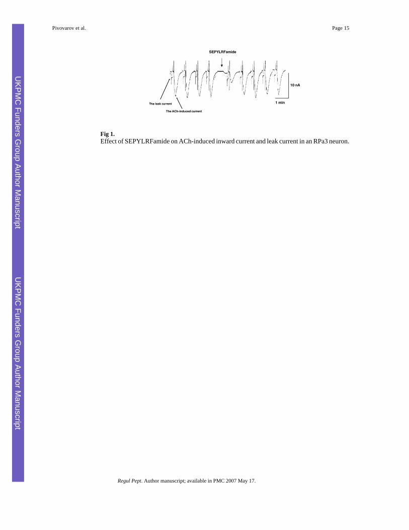
Fig 1.Effect of SEPYLRFamide on ACh-induced inward current and leak current in an RPa3 neuron.
Pivovarov et al. Page 15
Regul Pept. Author manuscript; available in PMC 2007 May 17.
UKPM
C Funders G
roup Author Manuscript
UKPM
C Funders G
roup Author Manuscript
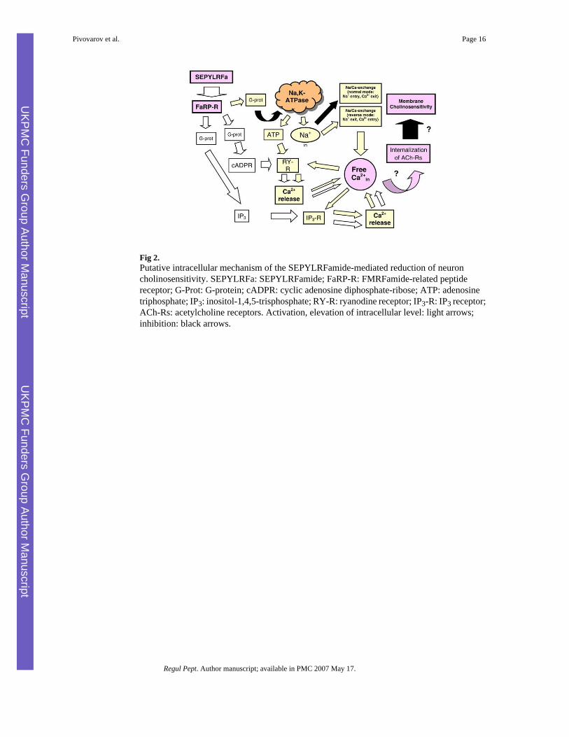
Fig 2.Putative intracellular mechanism of the SEPYLRFamide-mediated reduction of neuroncholinosensitivity. SEPYLRFa: SEPYLRFamide; FaRP-R: FMRFamide-related peptidereceptor; G-Prot: G-protein; cADPR: cyclic adenosine diphosphate-ribose; ATP: adenosinetriphosphate; IP3: inositol-1,4,5-trisphosphate; RY-R: ryanodine receptor; IP3-R: IP3 receptor;ACh-Rs: acetylcholine receptors. Activation, elevation of intracellular level: light arrows;inhibition: black arrows.
Pivovarov et al. Page 16
Regul Pept. Author manuscript; available in PMC 2007 May 17.
UKPM
C Funders G
roup Author Manuscript
UKPM
C Funders G
roup Author Manuscript
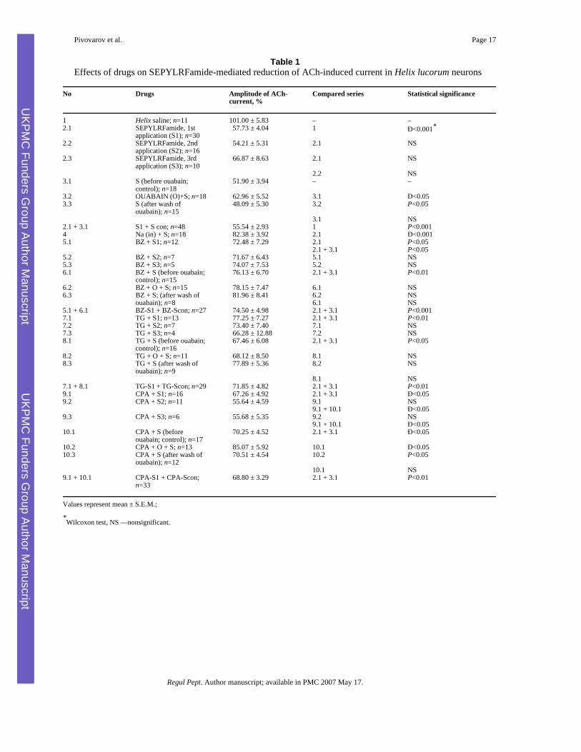
Table 1Effects of drugs on SEPYLRFamide-mediated reduction of ACh-induced current in Helix lucorum neurons
No Drugs Amplitude of ACh-current, %
Compared series Statistical significance
1 Helix saline; n=11 101.00 ± 5.83 – –2.1 SEPYLRFamide, 1st
application (S1); n=30 57.73 ± 4.04 1 Đ<0.001*
2.2 SEPYLRFamide, 2ndapplication (S2); n=16
54.21 ± 5.31 2.1 NS
2.3 SEPYLRFamide, 3rdapplication (S3); n=10
66.87 ± 8.63 2.1 NS
2.2 NS3.1 S (before ouabain;
control); n=18 51.90 ± 3.94 – –
3.2 OUABAIN (O)+S; n=18 62.96 ± 5.52 3.1 Đ<0.053.3 S (after wash of
ouabain); n=15 48.09 ± 5.30 3.2 P<0.05
3.1 NS2.1 + 3.1 S1 + S con; n=48 55.54 ± 2.93 1 P<0.0014 Na (in) + S; n=18 82.38 ± 3.92 2.1 Đ<0.0015.1 BZ + S1; n=12 72.48 ± 7.29 2.1 P<0.05
2.1 + 3.1 P<0.055.2 BZ + S2; n=7 71.67 ± 6.43 5.1 NS5.3 BZ + S3; n=5 74.07 ± 7.53 5.2 NS6.1 BZ + S (before ouabain;
control); n=15 76.13 ± 6.70 2.1 + 3.1 P<0.01
6.2 BZ + O + S; n=15 78.15 ± 7.47 6.1 NS6.3 BZ + S; (after wash of
ouabain); n=8 81.96 ± 8.41 6.2 NS
6.1 NS5.1 + 6.1 BZ-S1 + BZ-Scon; n=27 74.50 ± 4.98 2.1 + 3.1 P<0.0017.1 TG + S1; n=13 77.25 ± 7.27 2.1 + 3.1 P<0.017.2 TG + S2; n=7 73.40 ± 7.40 7.1 NS7.3 TG + S3; n=4 66.28 ± 12.88 7.2 NS8.1 TG + S (before ouabain;
control); n=16 67.46 ± 6.08 2.1 + 3.1 P<0.05
8.2 TG + O + S; n=11 68.12 ± 8.50 8.1 NS8.3 TG + S (after wash of
ouabain); n=9 77.89 ± 5.36 8.2 NS
8.1 NS7.1 + 8.1 TG-S1 + TG-Scon; n=29 71.85 ± 4.82 2.1 + 3.1 P<0.019.1 CPA + S1; n=16 67.26 ± 4.92 2.1 + 3.1 Đ<0.059.2 CPA + S2; n=11 55.64 ± 4.59 9.1 NS
9.1 + 10.1 Đ<0.059.3 CPA + S3; n=6 55.68 ± 5.35 9.2 NS
9.1 + 10.1 Đ<0.0510.1 CPA + S (before
ouabain; control); n=17 70.25 ± 4.52 2.1 + 3.1 Đ<0.05
10.2 CPA + O + S; n=13 85.07 ± 5.92 10.1 Đ<0.0510.3 CPA + S (after wash of
ouabain); n=12 70.51 ± 4.54 10.2 P<0.05
10.1 NS9.1 + 10.1 CPA-S1 + CPA-Scon;
n=33 68.80 ± 3.29 2.1 + 3.1 P<0.01
Values represent mean ± S.E.M.;
*Wilcoxon test, NS —nonsignificant.
Pivovarov et al. Page 17
Regul Pept. Author manuscript; available in PMC 2007 May 17.
UKPM
C Funders G
roup Author Manuscript
UKPM
C Funders G
roup Author Manuscript

Table 2Effects of OUABAIN (0.1 mM) on ACh-induced current in Helix lucorum neurons
No Drugs ACh-current, (%control)
Comparedseries
Statisticalsignificance
1 Helix saline; n=10 99.94 ± 7.61 – –2 Ouabain (only);
n=18 81.99 ± 5.78 1 Đ<0.05*
3 Benzamil+ouabain;n=15
100.90 ± 4.87 1 NS2 P<0.05
4 Thapsigargin+ouabain, n=11
99.39 ± 5.64 1 NS2 P<0.05
5 Cyclopiazonic acid+ouabain, n=14
81.78 ± 5.06 1 Đ<0.052 NS
Values represent mean ± S.E.M.;
*Wilcoxon test, NS —nonsignificant.
Pivovarov et al. Page 18
Regul Pept. Author manuscript; available in PMC 2007 May 17.
UKPM
C Funders G
roup Author Manuscript
UKPM
C Funders G
roup Author Manuscript
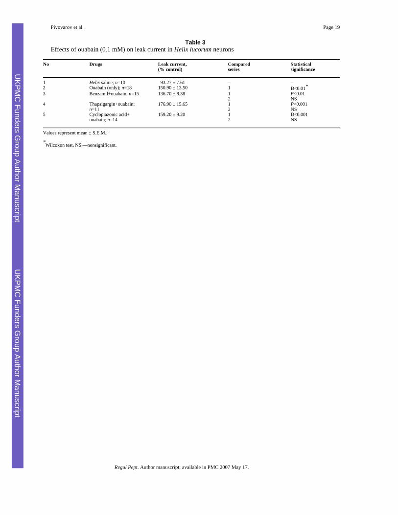
Table 3Effects of ouabain (0.1 mM) on leak current in Helix lucorum neurons
No Drugs Leak current,(% control)
Comparedseries
Statisticalsignificance
1 Helix saline; n=10 93.27 ± 7.61 – –2 Ouabain (only); n=18 150.90 ± 13.50 1 Đ<0.01*3 Benzamil+ouabain; n=15 136.70 ± 8.38 1 P<0.01
2 NS4 Thapsigargin+ouabain;
n=11176.90 ± 15.65 1 P<0.001
2 NS5 Cyclopiazonic acid+
ouabain; n=14159.20 ± 9.20 1 Đ<0.001
2 NS
Values represent mean ± S.E.M.;
*Wilcoxon test, NS —nonsignificant.
Pivovarov et al. Page 19
Regul Pept. Author manuscript; available in PMC 2007 May 17.
UKPM
C Funders G
roup Author Manuscript
UKPM
C Funders G
roup Author Manuscript
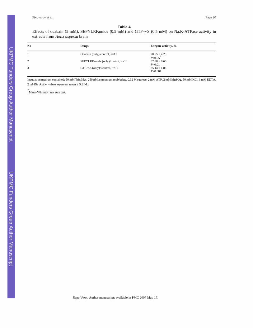
Table 4Effects of ouabain (5 mM), SEPYLRFamide (0.5 mM) and GTP-γ-S (0.5 mM) on Na,K-ATPase activity inextracts from Helix aspersa brain
No Drugs Enzyme activity, %
1 Ouabain (only)/control, n=11 90.65 ± 4.23P<0.05*
2 SEPYLRFamide (only)/control, n=10 87.38 ± 9.66P<0.01
3 GTP-γ-S (only)/Control, n=15 85.14 ± 1.88P<0.001
Incubation medium contained: 50 mM Tris/Mes, 250 μM ammonium molybdate, 0.32 M sucrose, 2 mM ATP, 2 mM MgSO4, 50 mM KCl, 1 mM EDTA,2 mMNa Azide; values represent mean ± S.E.M.;
*Mann-Whitney rank sum test.
Pivovarov et al. Page 20
Regul Pept. Author manuscript; available in PMC 2007 May 17.
UKPM
C Funders G
roup Author Manuscript
UKPM
C Funders G
roup Author Manuscript
Copyright © 2022 FDOKUMEN