Neuronal Voltage-Gated Potassium Channel Complex Autoimmunity in Children
7
Neuronal Voltage-Gated Potassium Channel Complex Autoimmunity in Children Radhika Dhamija, MD † , Deborah L. Renaud, MD † , Sean J. Pittock, MD †‡ , Andrew McKeon, MD †‡ , Daniel H. Lachance, MD †‡ , Katherine C. Nickels, MD † , Elaine C. Wirrell, MD † , Nancy L. Kuntz, MD † , Mary D. King, MD § , and Vanda A. Lennon, MD, PhD* †‡ Autoimmunity targeting voltage-gated potassium channel complexes have not been systematically docu- mented in children. Identified in the Neuroimmunol- ogy Laboratory records of Mayo Clinic were 12 seropositive children, 7 among 252 Mayo Clinic pedi- atric patients tested on a service basis for serologic ev- idence of neurologic autoimmunity (June 2008-April 2010), 4 during the assay’s preimplementation valida- tion (before June 2008) and 1 non-Mayo patient with available clinical information. Neurologic manifesta- tions were subacute and multifocal. Three had global developmental regression, 6 movement disorders, 4 dysarthria, 3 seizures, 1 Satoyoshi syndrome, 1 painful red feet, 2 insomnia, 2 gastrointestinal dysmotility, and 2 small fiber neuropathy. Neoplasia was found in 1 child. Treating physicians recorded improvement in all 7 children who received immunotherapy. Neuro- logic symptom relapse occurred in 3 of 6 children af- ter ceasing immunotherapy. These findings highlight a diverse clinical spectrum of neuronal potassium channel complex autoimmunity in children, and they illustrate benefit from early initiated immunotherapy, with a tendency to relapse when therapy ceases. Diag- nosis is generally delayed in the process of eliminating neurodegenerative causes. Currently 2.7% of pediat- ric sera evaluated for neurologic autoimmunity are positive for neuronal potassium channel complex- reactive immunoglobulin G. The frequency and full spectrum of neurologic accompaniments remains to be determined. Ó 2011 Elsevier Inc. All rights reserved. Dhamija R, Renaud DL, Pittock SJ, McKeon A, Lachance DH, Nickels KC, Wirrell EC, Kuntz NL, King MD, Lennon VA. Neuronal voltage-gated potassium channel complex autoimmunity in children. Pediatr Neurol 2011; 44:275-281. Introduction Voltage-gated potassium channels (VGKC) are tetra- meric signaling proteins containing 6–transmembrane do- main alpha subunits (S1–6) encoded by approximately 50 genes in humans. Each includes a voltage-sensitive fourth domain [1]. VGKCs in neurons of both the central and pe- ripheral nervous systems play an important role in mem- brane repolarization, axonal conduction, and synaptic transmission [1,2]. Autoantibodies targeting VGKCs were initially reported in patients with acquired neuromyotonia, a disorder of peripheral nerve hyperexcitability (also called Isaac syndrome) [3,4], and subsequently in patients with a continuum of autoimmune neuromuscular hyperexcitability disorders with and without central nervous system accompaniments (Morvan syndrome, cramp-fasciculation syndrome, rippling muscle disease) [5-7], rapid eye movement sleep disorders, intractable epilepsy [8,9], and potentially reversible encephalopathies [10-12]. Recent reports suggest that the autoantibodies identified by immunoprecipitation of VGKCs complexed with 125 I-a- dendrotoxin (a ligand that binds with high affinity to Kv1.1, 1.2, and 1.6 channels) target neuronal proteins that coprecipitate with solubilized potassium channels. Two From the Departments of *Immunology, † Neurology, and ‡ Laboratory Medicine and Pathology, Mayo Clinic, Rochester, Minnesota, and § Children’s University Hospital, Temple Street, Dublin, Ireland. Communication should be addressed to: Dr. Lennon; Neuroimmunology Laboratory; Mayo Clinic; 200 First St SW; Rochester, MN 55905. E-mail: [email protected] Received August 31, 2010; accepted October 20, 2010. Ó 2011 Elsevier Inc. All rights reserved. doi:10.1016/j.pediatrneurol.2010.10.015 0887-8994/$ - see front matter Dhamija et al: VGKC Neurologic Autoimmune Spectrum 275
Transcript of Neuronal Voltage-Gated Potassium Channel Complex Autoimmunity in Children
Neuronal Voltage-Gated Potassium Channel
Complex Autoimmunity in ChildrenRadhika Dhamija, MD†, Deborah L. Renaud, MD†, Sean J. Pittock, MD†‡,
Andrew McKeon, MD†‡, Daniel H. Lachance, MD†‡, Katherine C. Nickels, MD†,Elaine C. Wirrell, MD†, Nancy L. Kuntz, MD†, Mary D. King, MD§,
and Vanda A. Lennon, MD, PhD*†‡
Autoimmunity targeting voltage-gated potassiumchannel complexes have not been systematically docu-mented in children. Identified in the Neuroimmunol-ogy Laboratory records of Mayo Clinic were 12seropositive children, 7 among 252 Mayo Clinic pedi-atric patients tested on a service basis for serologic ev-idence of neurologic autoimmunity (June 2008-April2010), 4 during the assay’s preimplementation valida-tion (before June 2008) and 1 non-Mayo patient withavailable clinical information. Neurologic manifesta-tions were subacute and multifocal. Three had globaldevelopmental regression, 6 movement disorders, 4dysarthria, 3 seizures, 1 Satoyoshi syndrome, 1 painfulred feet, 2 insomnia, 2 gastrointestinal dysmotility, and2 small fiber neuropathy. Neoplasia was found in 1child. Treating physicians recorded improvement inall 7 children who received immunotherapy. Neuro-logic symptom relapse occurred in 3 of 6 children af-ter ceasing immunotherapy. These findings highlighta diverse clinical spectrum of neuronal potassiumchannel complex autoimmunity in children, and theyillustrate benefit from early initiated immunotherapy,with a tendency to relapse when therapy ceases. Diag-nosis is generally delayed in the process of eliminatingneurodegenerative causes. Currently 2.7% of pediat-ric sera evaluated for neurologic autoimmunity arepositive for neuronal potassium channel complex-reactive immunoglobulin G. The frequency and fullspectrum of neurologic accompaniments remainsto be determined. � 2011 Elsevier Inc. All rightsreserved.
From the Departments of *Immunology, †Neurology, and ‡LaboratoryMedicine and Pathology, Mayo Clinic, Rochester, Minnesota, and§Children’s University Hospital, Temple Street, Dublin, Ireland.
� 2011 Elsevier Inc. All rights reserved.doi:10.1016/j.pediatrneurol.2010.10.015 � 0887-8994/$ - see front matter
Dhamija R, Renaud DL, Pittock SJ, McKeon A, LachanceDH, Nickels KC, Wirrell EC, Kuntz NL, King MD,Lennon VA. Neuronal voltage-gated potassium channelcomplex autoimmunity in children. Pediatr Neurol 2011;44:275-281.
Introduction
Voltage-gated potassium channels (VGKC) are tetra-meric signaling proteins containing 6–transmembrane do-main alpha subunits (S1–6) encoded by approximately 50genes in humans. Each includes a voltage-sensitive fourthdomain [1]. VGKCs in neurons of both the central and pe-ripheral nervous systems play an important role in mem-brane repolarization, axonal conduction, and synaptictransmission [1,2]. Autoantibodies targeting VGKCs wereinitially reported in patients with acquired neuromyotonia,a disorder of peripheral nerve hyperexcitability (also calledIsaac syndrome) [3,4], and subsequently in patients witha continuum of autoimmune neuromuscular hyperexcitabilitydisorders with and without central nervous systemaccompaniments (Morvan syndrome, cramp-fasciculationsyndrome, rippling muscle disease) [5-7], rapid eyemovement sleep disorders, intractable epilepsy [8,9], andpotentially reversible encephalopathies [10-12]. Recentreports suggest that the autoantibodies identified byimmunoprecipitation of VGKCs complexed with 125I-a-dendrotoxin (a ligand that binds with high affinity toKv1.1, 1.2, and 1.6 channels) target neuronal proteins thatcoprecipitate with solubilized potassium channels. Two
Communication should be addressed to:Dr. Lennon; Neuroimmunology Laboratory; Mayo Clinic; 200 First StSW; Rochester, MN 55905.E-mail: [email protected] August 31, 2010; accepted October 20, 2010.
Dhamija et al: VGKC Neurologic Autoimmune Spectrum 275

Table 1. Clinical characteristics of 12 children
Patient
No.
Age at
Onset/Sex
Age at
Diagnosis
Duration of
Follow-up from
Diagnosis
VGKC
(nmol/L)
Neurologic
Manifestations
Antecedent
Event (1-4 wk) MRI Brain
1 5 yr/M 5 yr 3 mo 0.05 Motor tics — Increased T2 signal,
left thalamus
2 3 yr/M 6.5 yr 1.5 yr 0.06 Global regression,
seizures and chorea
Salmonella
gastroenteritis
Normal
3 10 mo/F 3 yr 6 mo 0.11 Symptomatic
generalized
epilepsy,
developmental
delay
— Bilateral increased
T2 signal,
mesial temporal
lobes
4 7 yr/M 9 yr 3 mo 0.13 Painful red feet, cramps,
hypohidrosis
— —
5 8 mo/M 2 yr 1 yr 0.15 Episodic flushing and
overheating, hypohidrosis
— —
6 6 yr/F 6.5 yr 2 mo 0.19 Acute disseminated
encephalomyelitis,
migraine headaches,
tremor
Influenza Symmetric areas of
signal abnormality,
white matter of both
cerebral hemispheres
7 14 yr/M 15 yr 8 yr 0.20 Continuous muscle
fiber activity
(Satoyoshi
syndrome)
— —
8 9 yr/F 15 yr 3 yr 0.25 Dysarthria
(worse when
dysmotility is
worse)
Viral illness Normal
9 8 yr/F 9 yr 2 yr 0.43 Developmental regression,
myoclonus, dystonia,
hyperkinetic dysarthria
— Normal
10 8 yr/F 8 yr 7 yr 0.95 Limbic encephalitis
(insomnia, seizures),
chorea, hyperkinetic
dysarthria
Viral illness Increased T2 signal,
amygdala and mesial
temporal lobes
11 29 mo/F 5 yr 1 yr 1.42 Global developmental
regression, autism,
insomnia
Influenza
vaccine
Normal
12* 9 yr/F 9 yr 6 mo 0.54 Cerebellar ataxia,
intermittent
perioral
myoclonus,
dysarthria
— Normal
* Patient not seen at Mayo Clinic
Abbreviations:
CSF = Cerebrospinal fluid
hpf = High-power field
MRI = Magnetic resonance imaging
VGKC = Voltage-gated potassium channels
276 PEDIATRIC NEUROLOGY Vol. 44 No. 4

Gastrointestinal
Symptoms CSF Findings Immunotherapy
Immunotherapy
Response
Other Clinical
Manifestation or
Autoantibodies
Family History of
Autoimmunity
— — None — — —
— Cell 1/hpf, glucose
73 mg/dL,
protein
25 mg/dL
Yes, IV corticosteroids,
IV immune globulin,
Plasma exchange,
Cyclophosphamide
Mild improvement in
seizure frequency
Eosinophilia Diabetes mellitus,
lupus
— Cell 1/hpf, glucose
59 mg/dL,
protein
32 mg/dL
Yes, Prednisone, IV
immune globulin
Moderate improvement
in seizure frequency,
relapsed after IV
immune globulin,
stopped
Striational
antibody
(1:240)
Rheumatoid
arthritis
— — None — — Hypothyroidism,
psoriasis
Diarrhea — None — No Hashimoto thyroiditis,
Graves disease
— Cell 0/hpf,
glucose
55 mg/dL,
protein
16 mg/dL
None — Antinuclear
antibody
Type 1 diabetes
mellitus, Connective
tissue
disease
— — Yes, Prednisone, IV
immune globulin
Mild improvement in
muscle spasms
Alopecia,
joint pain,
antinuclear
antibody,
striational
antibody
(1:120)
—
Dysmotility — Yes, IV
immune globulin
Moderate recovery in
dysmotility and
dysarthria, relapsed
after, IV
immune globulin,
stopped
Hypothyroidism,
lupus
— Cell 1/hpf, glucose
54 mg/dL,
protein 45 mg/dL
None — No Hypothyroidism,
type 1 diabetes
mellitus
— Cell 1/hpf, glucose
73 mg/dL,
protein 25 mg/dL
Yes, IV corticosteroids,
plasma exchange,
6-mercaptopurine
Marked, complete
recovery
— —
— — Yes, Prednisone, IV
immune globulin,
mycophenolate
mofetil
Moderate recovery
with better eye
contact and
communication,
relapsed after IV
immune globulin
stopped
— —
— Cells 4/hpf, glucose
61 mg/dL,
protein, 232 mg/dL
Oral corticosteroids,
tumor removal
Marked complete
recovery
Neuroblastoma,
antineuronal
nuclear
autoantibody
type 1
(1:3,840)
—
Dhamija et al: VGKC Neurologic Autoimmune Spectrum 277

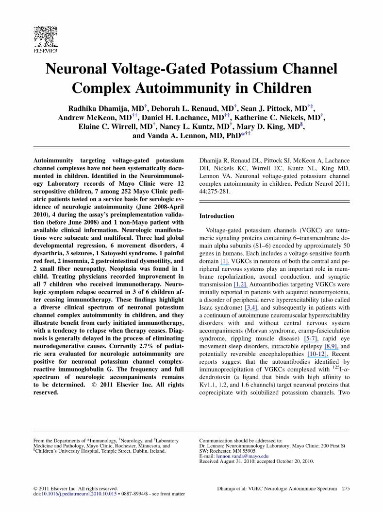
Figure 1. Magnetic resonance image of the brain of patient 10. Arrowsindicate amygdala and mesial temporal lobes. Note increased T2 signal(A) and resolution after treatment (B).
major antigens identified to date are the leucine-rich, gli-oma inactivated 1 protein (Lgi1), bound by immune globu-lin (Ig) G in serum of patients with limbic encephalitis, anda neurexin, contactin-associated protein 2 (Caspr2), boundby IgG in serum of some patients with neuromuscularhyperexcitability [13,14].
The clinical spectrum of VGKC complex autoimmunitycurrently recognized in adult patients includes idiopathicand paraneoplastic disorders of the central and peripheralsomatic, autonomic, and enteric nervous systems [15-18].In a paraneoplastic context, these autoantibodies are mostcommonly associated in adults with adenocarcinomas,small-cell lung carcinoma, thymoma, and hematologicmalignancies [15]. Little information has been reportedconcerning clinical correlations of VGKC complex auto-immunity in children. Here we report manifestations docu-mented in seropositive patients younger than 18 years.
Methods
We identified 12 patients younger than age 18 years whose serum IgG
immunoprecipitated VGKC complexes solubilized from brain tissue.
Seven were among 252 Mayo Clinic patients tested on a service basis
for serologic evidence of neurologic autoimmunity (June 2008 to April
2010), and another 4 were identified during the assay’s preimplementation
validation (before June 2008); we additionally included 1 non–Mayo pa-
tient for whom clinical information was available. Clinical information for
Mayo Clinic patients was obtained by retrospective chart review. The
study was approved by the Mayo institutional review board.
Contemporary evaluation of pediatric patients suspected to have neuro-
logic autoimmunity includes, in the Mayo Neuroimmunology Laboratory,
radioimmunoprecipitation assays to detect cation channel autoantibodies
(voltage-gated calcium channels [P/Q-type and N-type] and a-dendro-
toxin-sensitive potassium channel complexes; nicotinic acetylcholine re-
ceptors [muscle-type and neuronal ganglionic-type]) and glutamic acid
decarboxylase-65; enzyme-linked immunosorbent assay to detect skeletal
muscle striational autoantibodies, and indirect immunofluorescence (com-
posite substrate of mouse tissues) optimized to detect antineuronal nuclear
autoantibody type 1, collapsin response-mediator protein 5 IgG [19,20],
Purkinje cell cytoplasmic autoantibody type 2 [21], N-methyl-D-aspartic
acid receptor, and other autoantibodies reactive with hippocampal or basal
ganglionic synaptic proteins [22].
Autoantibodies reactive with neuronal VGKC complexes are identified
by radioimmunoprecipitation with digitonin-solubilized porcine brain
synaptic membrane proteins ligated with 125I-a-dendrotoxin, which binds
with high affinity to Kv1.1, 1.2, and 1.6 VGKCs [7,15]. The normal value
range is 0.00 to 0.02 nmol/L. False positive binding of 125I-a-dendrotoxin
is excluded [23].
Results
We identified 12 patients younger than age 18 yearswhose autoantibodies bound to VGKC complexes. Eightwere identified through service evaluation for serologicmarkers of neurologic autoimmunity (June 2008-April2010). In none was testing for VGKC autoantibody specif-ically requested by the physician. Four more were identi-fied through additional testing based on clinical suspicionduring the assay’s preimplementation validation. Elevenwere assessed at Mayo Clinic, and clinical informationfor the 12th patient was obtained by contacting the pa-tient’s physician. Before serologic testing was considered,
278 PEDIATRIC NEUROLOGY Vol. 44 No. 4
all children had undergone extensive diagnostic studies, asindicated by the clinical history, to exclude metabolic andgenetic disorders. Demographic and clinical characteristicsof the children are listed in Table 1.
Demographics
Eight of the 12 patients were girls; 11 were Caucasian; 1was Asian. Median age at symptom onset was 7.5 years (8months to 14 years).
Clinical Presentation
Before the onset of neurologic symptoms, 5 patients hada prodrome of fatigue, upper respiratory symptoms, and fe-ver. Neurologic manifestations in all cases were multifocaland subacute in onset (manifesting over weeks). The dura-tion between onset of neurologic symptoms and detectionof VGKC antibodies ranged from <1 month to 6 years. Di-agnosis was delayed because an autoimmune etiology wasnot suspected. Three patients manifested global develop-mental regression, and 1 had autistic features. Six patientshad movement disorders, including chorea (2), dystonia(1), myoclonus (2), tremor (1), motor tics (1), and cerebel-lar ataxia (1). Four patients had dysarthria. Two patientshad prominent insomnia. Three patients had seizures, 1with limbic encephalitis and 2 with new-onset symptom-atic generalized epilepsy. Several patients had evidenceof peripheral or autonomic nervous system involvement:1 peripheral nerve hyperexcitability with painful musclespasms (diagnosis, Satoyoshi syndrome), 1 painful redfeet, 2 gastrointestinal dysmotility, and 2 small fiber neu-ropathy. The only child who had typical symptoms of lim-bic encephalitis was an 8-year-old Caucasian girl (patient10) who experienced subacute onset of seizures, chorea,hyperkinetic dysarthria, and insomnia after a prodromeof sore throat and fever. Serum sodium was low (126mEq/L), suggesting hypothalamic derangement. Magneticresonance imaging of the brain demonstrated increased T2
signal in the amygdala and mesial temporal lobes (Fig 1).

Autoimmune Serology and Family History
The median value for VGKC immunoprecipitation was0.19 nmol/L (range 0.05-1.42 nmol/L; normal range0.00-0.02 nmol/L). None of the patients had a personal his-tory of autoimmune disorders, but 4 had serologic evidenceof autoimmunity: 2 had antinuclear antibody, 1 had anti-neuronal nuclear autoantibody type 1 (positive at end pointdilution 1: 3,840; normal value is negative at 1:240), and 2had striational antibody (positive at 1:120 and 1:240; nor-mal value is negative at 1:60). A family history of autoim-munity was documented in 7 patients (Table 1).
Cerebrospinal Fluid Findings
Cerebrospinal fluid was obtained from 6 patients. Theinterval between onset of neurologic symptoms and perfor-mance of the cerebrospinal fluid tap ranged from 1 monthto 3.5 years (Table 1). Nucleated cell numbers were nor-mal, but protein was elevated in one.
Neoplasia
We found a single neoplasm (neuroblastoma at the liverhilum) in 1 of 5 patients (patient 12) who underwent exten-sive evaluation for malignancy (computed tomographicscan of chest, abdomen, and pelvis; metaiodobenzylguani-dine scan; whole-body positron emission tomography).The patient had cerebellar ataxia, dysarthria, and periodicmyoclonus, and was seropositive for antineuronal nuclearautoantibody type 1.
Treatment
Seven of 12 patients received one or more immunother-apies, including intravenous and oral corticosteroids,intravenous immune globulin, plasma exchange, 6-mercap-topurine, cyclophosphamide, andmycophenolate mofetil. Inall cases, the therapy was acute (6 corticosteroids, 5 intrave-nous immune globulin, and 2 plasma exchange); 5 of 7patients received combination treatment. Corticosteroid-sparing agents were introduced in 3 patients (6-mercaptopu-rine, cyclophosphamide, and mycophenolate mofetil). Thetreating physicians reported mild symptom improvementsin 2 (decreased frequency of seizures in 1; muscle spasmsin 1), moderate in 3 (decreased frequency of seizures in 1;improvement in gastrointestinal motility and dysarthria in1; improved communication skills in 1) and marked in 2(resolution of limbic symptoms in 1; resolution of cerebellarataxia and dysarthria in 1). Neurologic symptoms disap-peared after neuroblastoma resection in the patient whowas positive for antineuronal nuclear autoantibody type 1.Themedian interval between symptom onset and start of im-munotherapy was 2.4 years (range 1 month to 6 years). Im-munotherapy was commenced within 4 weeks of symptomonset in both patients whose symptoms improved markedly.Relapses occurred after cessation of immunotherapy in 3 of6 patients. One patient (patient 10; Table 1) had not re-
sponded to initial plasma exchange (5 treatments), but im-proved remarkably after intermittent high-dose oralcorticosteroid therapy was commenced. Recovery was com-plete 3 months later. In the next 2 years, while receivingmonthly IV corticosteroid maintenance therapy, she experi-enced a single relapse when corticosteroid cessation was at-tempted. Seven years after symptom onset (and aftercorticosteroids had been withdrawn from the patient for 5years), she had no recurrent neurologic problems, and noVGKC complex-reactive autoantibody was detectable. Inpatient 11, the serum antibody level fell progressively duringintravenous immune globulin treatment (from 1.42 to 0.78nmol/L) but, after the family’s decision to stop treatment,it rose (to 2.17 nmol/L), and developmental and social skillsregressed.
Discussion
VGKC complex-reactive autoantibodies are an underap-preciated biomarker of autoimmune neurologic disease.They are at this time the most common of neural autoanti-bodies identified in adult patients evaluated for paraneoplas-tic autoimmunity in the Mayo Clinic NeuroimmunologyLaboratory, being found in 3%of sera submitted for compre-hensive testing [24]. By comparison, the detection frequen-cies of antineuronal nuclear autoantibody type 1 (‘‘anti-Hu’’), collapsin response-mediator protein 5 IgG, andvoltage-gated calcium channel autoantibodies (P/Q-typeandN-type) are 0.3%, 0.3%, and 1%.This study has revealeda similar high frequency ofVGKCcomplex-reactive autoan-tibodies (2.7%) in pediatric patients. Thus the newly recog-nized neurologic entity of VGKC complex autoimmunityseems not to be rare in children. Its clinical spectrum in pe-diatric patients is likely broader than presently recognized.
It was recently established that VGKC proteins per se aregenerally not the antigenic target of these autoantibodies.Nevertheless, radioimmunoprecipitation remains the opti-mal screening assay to detect VGKC complex antibodies.Twomajor proteins that have been identified as primary an-tigens coprecipitate as complexes with 125I-a-dendrotoxin-ligated VGKCs in radioimmunoprecipitation assays. Theseare the secreted leucine-rich, glioma inactivated 1 (Lgi1)protein (associated with limbic encephalitis) andcontactin-associated protein 2 (Caspr2) (associated withneuromuscular hyperexcitability) [13,14]. Lgi1 binds toand bridges presynaptic disintegrin and metalloproteinasedomain-containing protein 23 (ADAM23) and postsynapticADAM22 protein forming a trans-synaptic complex withpresynaptic Kv1.1 channels. Caspr2 colocalizes withKv1.1 and Kv1.2 in paranodal axons and hippocampus.The clinical heterogeneity of VGKC complex autoimmu-nity may reflect different VGKC-associated antigenic tar-gets (such as Lgi1, Caspr2), different epitopes on theseproteins, or as yet unidentified VGKC-complexed proteins.
The spectrum and severity of neurologic manifestationsof VGKC complex autoimmunity remain to be determined.It is conceivable that milder, spontaneously remitting
Dhamija et al: VGKC Neurologic Autoimmune Spectrum 279

phenotypes may exist. Our experience to date documentsa diversity of clinical presentations in children who haveVGKC complex autoimmunity, and it justifies early test-ing for serologic markers of autoimmunity in childrenmanifesting any subacute neurologic disorder of unknowncause. In all but 2 of the presented cases, the diagnosiswas delayed because an autoimmune pathogenesis wasnot considered earlier. Investigation in larger case seriesis needed to ascertain the frequency of the unprecedentedassociations that we noted with developmental delay orregression, autism, and Satoyoshi syndrome. A personalor family history of autoimmunity, seropositivity fororgan-specific or non-organ-specific autoantibodies, ora prodromal ‘‘viral’’ illness should raise a suspicion forneurologic autoimmunity. The young age of the patientsin our study likely explains their lack of a personal historyof autoimmunity. Follow-up through adulthood is neededto establish this association. Neoplasia was detected inonly 1 patient in this initial series of pediatric cases.That patient was additionally seropositive for antineuronalnuclear autoantibody type 1, a recognized marker of neu-roblastoma [25]. The frequency of cancer in children sero-positive for VGKC complex autoantibodies has not yetbeen determined, but in adult patients it is 33% [15].Thus an occult neoplasm should be considered in pediatricpatients.
Conclusion
Treatment was started within 4 weeks of symptom onsetin both of the patients who had greatest recoveries. Promptand aggressive immunomodulatory therapy is therefore ad-vocated, but there is no definitive evidence-based approachto treating VGKC complex autoimmunity. Therapeuticapproaches reported to be beneficial in adult patients in-clude plasmapheresis, corticosteroids (oral and intrave-nous), and intravenous immune globulin in combinationwith corticosteroid-sparing immunosuppressant medica-tions [26]. For initial therapy of adults and children, werecommend pulse corticosteroids in high dose (IV methyl-prednisolone, 1 g or 30-50 mg/kg, for 5 days), and forpatients who cannot tolerate the adverse effects of cortico-steroids, who have diabetes, or who are at risk for diabetes,we recommend intravenous immune globulin (0.4 g/kgdaily for 3-5 days). Plasmapheresis (total of 5-7 exchanges,provided on alternate days) is usually reserved for patientswho cannot tolerate either corticosteroids or intravenousimmune globulin. On the basis of our experience to date,we recommend that the child be re-evaluated for objectiveevidence of improvement after the first 6 weeks of therapy.A favorable response justifies use of a corticosteroid-sparing agent (e.g., oral azathioprine or mycophenolatemofetil). Rituximab or intravenous cyclophosphamidemay be options if these initial approaches are not beneficialor if relapses occur after treatment [26]. Larger studies areneeded to better define the optimal type, dose, and durationof immunotherapy.
280 PEDIATRIC NEUROLOGY Vol. 44 No. 4
References
[1] Gutman GA, Chandy KG, Grissmer S, et al. International Union
of Pharmacology. LIII. Nomenclature and molecular relationships of
voltage-gated potassium channels. Pharmacol Rev 2005;57:473-508.
[2] Yellen G. The voltage-gated potassium channels and their rela-
tives. Nature 2002;419:35-42.
[3] Hart IK, Waters C, Vincent A, et al. Autoantibodies detected to
expressed K+ channels are implicated in neuromyotonia. Ann Neurol
1997;41:238-46.
[4] Sinha S, Newsom-Davis J, Mills K, Byrne N, Lang B, Vincent A.
Autoimmune aetiology for acquired neuromyotonia (Isaacs’ syndrome).
Lancet 1991;338:75-7.
[5] Vernino S, Lennon VA. Ion channel and striational antibodies de-
fine a continuum of autoimmune neuromuscular hyperexcitability. Muscle
Nerve 2002;26:702-7.
[6] LiguoriR,VincentA, Clover L, et al.Morvan’s syndrome: Periph-
eral and central nervous system and cardiac involvementwith antibodies to
voltage-gated potassium channels. Brain 2001;124:2417-26.
[7] Vernino S, Kryzer TJ, Lennon VA. Autoimmune autonomic neu-
ropathy and neuromuscular hyperexcitability disorders. In: Rose NR,
Hamilton RG, Detrick B, editors. Manual of Clinical and Laboratory Im-
munology. 6th ed. Washington, DC: ASM Press, 2002:1013-7.
[8] MajoieHJ, de Baets M, Renier W, Lang B, Vincent A. Antibodies
to voltage-gated potassium and calcium channels in epilepsy. Epilepsy
Res 2006;71:135-41.
[9] McKnight K, Jiang Y, Hart Y, et al. Serum antibodies in epilepsy
and seizure-associated disorders. Neurology 2005;65:1730-6.
[10] Thieben MJ, Lennon VA, Boeve BF, Aksamit AJ, Keegan M,
Vernino S. Potentially reversible autoimmune limbic encephalitis
with neuronal potassium channel antibody. Neurology 2004;62:1177-82.
[11] Vincent A, Buckley C, Schott JM, et al. Potassium channel
antibody-associated encephalopathy: A potentially immunotherapy-
responsive form of limbic encephalitis. Brain 2004;127:701-12.
[12] Pozo-Rosich P, Clover L, Saiz A, Vincent A, Graus F. Voltage-
gated potassium channel antibodies in limbic encephalitis. Ann Neurol
2003;54:530-3.
[13] LaiM, Huijbers MG, Lancaster E, et al. Investigation of LGI1 as
the antigen in limbic encephalitis previously attributed to potassium chan-
nels: A case series. Lancet Neurol 2010;9:776-85.
[14] Irani SR, Alexander S, Waters P, et al. Antibodies to Kv1
potassium channel-complex proteins leucine-rich, glioma inactivated
1 protein and contactin-associated protein-2 in limbic encephalitis,
Morvan’s syndrome and acquired neuromyotonia. Brain 2010;133:
2734-48.
[15] Tan KM, Lennon VA, Klein CJ, Boeve BF, Pittock SJ. Clinical
spectrum of voltage-gated potassium channel autoimmunity. Neurology
2008;70:1883-90.
[16] Knowles CH, Lang B, Clover L, et al. A role for autoantibodies
in some cases of acquired non-paraneoplastic gut dysmotility. Scand J
Gastroenterol 2002;37:166-70.
[17] Dhamija R, Tan KM, Pittock SJ, Foxx-Orenstein A,
Benarroch E, Lennon VA. Serologic profiles aiding the diagnosis of au-
toimmune gastrointestinal dysmotility. Clin Gastroenterol Hepatol 2008;
6:988-92.
[18] Viallard JF, Vincent A, Moreau JF, Parrens M, Pellegrin JL,
Ellie E. Thymoma-associated neuromyotonia with antibodies against
voltage-gated potassium channels presenting as chronic intestinal
pseudo-obstruction. Eur Neurol 2005;53:60-3.
[19] Cikes N, Momoi MY, Williams CL, et al. Striational autoanti-
bodies: Quantitative detection by enzyme immunoassay in myasthenia
gravis, thymoma, and recipients of d-penicillamine or allogeneic bone
marrow. Mayo Clin Proc 1988;63:474-81.
[20] Lennon VA. The case for a descriptive generic nomenclature:
Clarification of immunostaining criteria for PCA-1, ANNA-1, and
ANNA-2 autoantibodies. Neurology 1994;44:2412-5.
[21] Joshi DD, Anderson PM, Matsumoto J, et al. Chondroblastoma
with elevated creatine kinase and paraneoplastic neurologic autoimmu-
nity. Pediatr Hematol Oncol 2003;25:900-4.

[22] Dalmau J, Gleichman AJ, Hughes EG, et al. Anti-NMDA-recep-
tor encephalitis: Case series and analysis of the effects of antibodies. Lan-
cet Neurol 2008;7:1091-8.
[23] Apiwattanakul M, McKeon A, Pittock SJ, Kryzer TJ,
Lennon VA. Eliminating false-positive results in serum tests for neuro-
muscular autoimmunity. Muscle Nerve 2010;41:702-4.
[24] Aston PA, Lennon VA, McKeon A, Lachance D, Klein C,
Pittock SJ. Neuronal voltage-gated potassium channel (VGKC) auto-
antibodies in routine paraneoplastic evaluation: Frequency and clini-
cal correlates [abstract]. Ann Neurol 2010;68(Suppl. 14):S32.
[25] Antunes NL, Khakoo Y, Matthay KK, et al. Antineuronal anti-
bodies in patients with neuroblastoma and paraneoplastic opsoclonus-my-
oclonus. J Pediatr Hematol Oncol 2000;22:315-20.
[26] McKeon A, Lennon VA, Pittock SJ. Immunotherapy-responsive
dementias and encephalopathies. Continuum Lifelong Learning Neurol
2010;16:80-101.
Dhamija et al: VGKC Neurologic Autoimmune Spectrum 281