Are spontaneous otoacoustic emissions generated by self-sustained cochlear oscillators
Upload
independentCategory
view
0download
0
ARTICLE
Voltage-gated Potassium Channel (Kv) Subunits Expressed in theRat Cochlear Nucleus
Zoltan Rusznak, Gabor Bakondi, Krisztina Pocsai, Agnes Por, Lıvia Kosztka, Balazs Pal,Denes Nagy, and Geza Szucs
Department of Physiology, Research Centre of Molecular Medicine, Medical and Health Science Centre, University ofDebrecen, Debrecen, Hungary (ZR,GB,KP,LK,BP,DN,GS), and Department of Pathology, Hajdu-Bihar Megyei Kenezy GyulaCounty Infirmary, Debrecen, Hungary (AP)
SUMMARY Because the neuronal membrane properties and firing characteristics arecrucially affected by the depolarization-activated K1 channel (Kv) subunits, data about the Kvdistribution may provide useful information regarding the functionality of the neuronssituated in the cochlear nucleus (CN). Using immunohistochemistry in free-floating slices, thedistribution of seven Kv subunits was described in the rat CN. Positive labeling was observedfor Kv1.1, 1.2, 1.6, 3.1, 3.4, 4.2, and 4.3 subunits. Giant and octopus neurons showed par-ticularly strong immunopositivity for Kv3.1; octopus neurons showed intense Kv1.1- and1.2-specific reactions also. In the latter case, an age-dependent change of the expressionpattern was also documented; although both young and older animals produced definitelabeling for Kv1.2, the intensity of the reaction increased in older animals and was accom-panied with the translocation of the Kv1.2 subunits to the cell surface membrane. The gran-ule cell layer exhibited strong Kv4.2-specific immunopositivity, and markedly Kv4.2-positiveglomerular synapses were also seen. It was found that neither giant nor pyramidal cells wereuniform in terms of their Kv expression patterns. Our data provide new information aboutthe Kv expression of the CN and also suggest potential functional heterogeneity of the giantand pyramidal cells. (J Histochem Cytochem 56:443–465, 2008)
KEY WORDS
hearing
rhodamine
immunohistochemistry
confocal microscopy
age dependence
glomerular synapse
THE MAMMALIAN cochlear nucleus (CN) is an assemblypoint of numerous types of neurons forming local net-works that receive and process the primary acousticinformation arriving from the cochlea (Brawer et al.1974; Disterhoft et al. 1980; Webster and Trune 1982;Hackney et al. 1990). The nucleus is divided intothree morphologically distinct parts (anteroventral,posteroventral, and dorsal divisions; aVCN, pVCN,and DCN, respectively), each contacted by the incom-ing and branching axons of the auditory nerve (Moore1986). Because of the fact that these regions of the CNplay different roles in the processing of the auditoryinformation, the parallel secondary pathways emergingfrom the CN and targeting higher centers in the braincarry activity patterns, providing information about
different features of the sound (Cant and Benson 2003;Malmierca 2003).
To understand the function of the CN, it is essentialto know how the various cell types contribute to itsoverall task. Thus far, numerous authors have providedvaluable information about the morphological andfunctional properties of the individual CN neurons,revealing that the behavior and firing characteristics ofthe projection cells of the CN are markedly different.The bushy neurons (Brawer et al. 1974) of the VCNproduce rapidly adapting (type II) response pattern[i.e., they fire a single action potential at the start ofsuprathreshold electrical stimulation (Manis and Marx1991; Schwarz and Puil 1997; Doughty et al. 1998)].They can, therefore, precisely recognize the onset of theacoustic stimulation, which is a function that is indis-pensable for the sound source localization. Moreover,the specific membrane properties of the bushy cellsenable them to “recognize” the fine structure of sounds,allowing them to encode the phase of low-frequencysounds and performing amplitude modulation of high-
Correspondence to: Geza Szucs, MD, PhD, Department of Physi-ology, Medical and Health Science Centre, University of Debrecen,PO Box 22, H-4012 Debrecen, Hungary. E-mail: [email protected]
Received for publication November 29, 2007; accepted Janu-ary 14, 2008 [DOI: 10.1369/jhc.2008.950303].
TheJournal
ofHistoch
emistry&
Cytoch
emistry
C The Histochemical Society, Inc. 0022-1554/08/$3.30 443
Volume 56(5): 443–465, 2008
Journal of Histochemistry & Cytochemistry
http://www.jhc.org
by guest on January 26, 2016jhc.sagepub.comDownloaded from

frequency sounds. Octopus cells are found in the pVCNonly (Harrison and Irving 1966; Osen 1969a), each ofthem receiving synaptic inputs from numerous (at least60) auditory axons (Osen 1969b; Oertel et al. 2000).Octopus cells fire action potentials only if the activity ofthese synapses develops within a very narrow time in-terval (Golding et al. 1995); thus, they are regarded ascoincidence detectors of the CN (Oertel et al. 2000).
In the DCN, the pyramidal (fusiform) neurons(Lorente de No 1981; Webster and Trune 1982) canproduce variable activity patterns (termed onset, chop-per, and build-up) depending on the strength of theincoming stimuli and on the membrane potential justbefore the stimulation (Kane 1974; Kanold and Manis1999). These neurons receive inputs through differentpathways; thus, they may play an integrative role. It hasalso been shown that pyramidal cells detect spectralnotches that are the cues for monoaural (vertical) soundsource localization (Reiss and Young 2005). As for theother projection cells of the DCN [giant and Purkinje-like cells (Kane et al. 1981; Rossi and Borsello 1993;Zhang and Oertel 1993; Hurd and Feldman 1994)],very little is known about their firing properties; thusfar, no obvious function has been attributed to them.
Although the CN projection cells are able to producemarkedly different firing patterns in response to thesame acoustic stimuli, the exact explanation of theirfiring behavior is still poorly understood. Generally, theneuronal response pattern is most effectively deter-mined by the organization and activity of the synapticinputs and by their intrinsic membrane properties. Themembrane characteristics are, on the other hand, cru-cially affected by the presence, distribution, and subunitcomposition of the various voltage-gated K1 channels,which have pivotal roles in determining the excitability,membrane time constant, and the major features ofthe action potential firing pattern. The depolarization-activated K1 channels fall into three major categories(for reviews, see Coetzee et al. 1999; Rudy and Mcbain2001; Dodson and Forsythe 2004), each having differ-ent molecular composition, electrophysiological proper-ties, and functional relevance. The molecular assemblyof the low voltage–activated (LVA), dendrotoxin-sensitive channels requires the presence of K1 channel(Kv)1.1, 1.2, or 1.6 subunits; whereas the assembly ofrapidly inactivating, transient K1 channels requiresKv1.4, 4.2, 4.3, or 3.4 subunits. Finally, the molecularcomposition of the members of the high voltage–activated (HVA), non- or very slowly inactivating,delayed-rectifier channels is rather variable, containinga number of possible subunits (e.g., Kv3.1 or 3.2).
Individual representatives of each major K1 currenttype have been described in the various portions of theauditory pathway, including the CN (Rusznak et al.1997; Rothman and Manis 2003; Pal et al. 2004; Caoet al. 2007). Because the various K1 currents have dif-
ferent roles in determining the electrophysiologicalcharacteristics of the individual neuron types, it is im-portant to identify the subunits contributing to the as-sembly of the functional K1 channels and to establishtheir distribution. Although numerous studies are avail-able regarding the presence of the various Kv channelsubunits in the CN at either the mRNA or protein level,several problems hinder the interpretation of the ex-perimental data. Among others, expression of the Kvsubunits showed remarkable age and species depen-dence, making the direct comparison of the availabledata complicated. An additional problem is that theidentification of the cell types in functional and histo-chemical experiments is often difficult and may notbe unambiguously achieved. Although these problemscould be solved by a comprehensive study investigatingthe expression patterns of a large number of Kv sub-units using the same species and age, preferably withthe combined application of other methods helping cellidentification, such a study has not been completedthus far.
The aim of this study was, therefore, to describe theexpression patterns of seven Kv subunits in the adultrat CN. To ensure precise cell identification, severalmethods, including retrograde labeling, confocal mi-croscopy, and double immunolabeling, were used. Thedata presented here indicate that the investigated cellsof the CN possess several types of Kv subunits. TheKv4.2 and Kv3.1 expressions of the granule and mostof the giant cells, respectively, were so prominent thatthey might be considered as good markers for assistingcell identification in future studies. Moreover, stronglyKv4.2-positive glomerular synapses were shown in thecochlear granule cell region. Finally, octopus and bushycells were strongly Kv1.2 positive, and definite age-dependent changes could be noted in the distributionpattern of this subunit.
Materials and Methods
Animal Care
The experiments were conducted on young (11–17 day)and older (at least 25 days, but usually 1–3 months old)Wistar rats (both sexes) using a protocol that wasauthorized by the Committee of Animal Research ofthe University of Debrecen and was in accordance withthe appropriate international and Hungarian laws. Theanimals were bred in the departmental animal house,and they lived in an environment with natural day-night cycles. Food and water were available ad libitum.During the experimental procedures, the animals weresubjected to the smallest possible pain and discomfort.
Immunolabeling
The preparation technique used was essentially thesame as described earlier (Pal et al. 2005; Por et al.
TheJournal
ofHistoch
emistry&
Cytoch
emistry
444 Rusznak, Bakondi, Pocsai, Por, Kosztka, Pal, Nagy, Szucs
by guest on January 26, 2016jhc.sagepub.comDownloaded from

2005). All dissection steps were carried out in ice-coldlow-Na1 artificial cerebrospinal fluid (ACSF) containing(in mM) 220 sucrose, 2.5 KCl, 10 glucose, 26 NaHCO3,1.25 NaH2PO4, 2 CaCl2, 1 MgCl2, 3 myo-inositol,0.5 ascorbic acid, and 2 Na-pyruvate (the pH was set to7.2; the osmolarity was 320 mOsm; all chemicals weresupplied by Sigma-Aldrich, St. Louis, MO, unless indi-cated otherwise).
After the decapitation of the animal, the brain wasquickly removed; the brain stem (containing the CN)was prepared and placed into a 4% paraformaldehydesolution for 4 hr (4C). After this, the fixed tissue waswashed (three times for 10 min) in 0.1 M phosphatebuffer (PB; 0.1 M Na2HPO4 3 2 H2O and 0.1 MNaH2PO4 3 H2O; pH 5 7.4). A vibrating microtome(Campden Instruments; Loughborough, UK orMicromInternational; Walldorf, Germany) was used to cut50- to 60-mm-thick, parasagittal slices that were rinsedfirst in PB for 10 min and then three times in Tris-buffered saline solution (TBS; 8 mM Tris-base; 42 mMTrizma HCl; 150 mM NaCl; pH 5 7.4) for 10 min atroom temperature.
The immunolabelings were carried out on free-floating slices using a protocol described earlier in detail(Pocsai et al. 2007). Both single and double labelingtechniques were used; in the latter cases, the Kv-specificreactions were combined with glial fibrillar acidicprotein (GFAP; an astrocyte-specific marker) or synap-tophysin labeling. Because synaptophysin is an estab-lished molecular marker for the presynaptic vesiclemembrane, synaptic terminals making contact with thevarious cells could be visualized, producing a firm out-line of some of the neurons (e.g., bushy cells), helping todetermine their precise identification. Moreover, com-bined application of the Kv- and synaptophysin-specificimmunolabelings could also be used to raise the pos-sibility of potential presynaptic localization of theKv subunits.
In all cases, blocking and permeabilization wereachieved using TBS supplemented with 0.1% Triton X-100 and 10% normal horse or goat serum (dependingon the type of secondary antibody applied) for 1 hr at4C. Samples were incubated with the primary anti-bodies diluted in TBS containing 1% normal horse orgoat serum and 0.1% Triton X-100 (either overnightat room temperature or for 48 hr at 4C). Types, dilu-tions, and sources of the primary antibodies are listed inTable 1.
When the incubation with the primary antibodieswas terminated, the slices were rinsed in TBS (threetimes for 15 min) and incubated with the appropriatefluorochrome-conjugated secondary antibody (3 hr atroom temperature or 24 hr at 4C; Table 1) diluted inTBS containing 1% horse or goat serum and 0.1%Triton X-100 and washed in TBS again (three timesfor 10 min). At the end of the procedure, the slices
were mounted using a 4¶,6-diamidino-2-phenylindole(DAPI)-containing mounting medium (Vector Labora-tories; Burlingame, CA). DAPI was used for the specificlabeling of the cell nuclei that allowed more precisemorphological identification of the various structures(cell bodies or selected parts of the CN, such as thegranule cell layer).
In cases of double labelings, the appropriate Kv-specific antibody and the other antibody (anti-GFAP oranti-synaptophysin) were applied simultaneously. Be-fore the application of the primary antibodies, blockingand permeabilization were achieved using TBS sup-plemented with 10% normal bovine serum and 0.1%Triton X-100 (1 hr, 4C). The primary and secondaryantibodies were diluted in TBS containing 1% bovineserum and 0.1% Triton X-100; otherwise, the majorsteps of the immunoreactions were the same as de-scribed above.
The data presented in this work were obtained from36 animals (5 young and 31 older rats from both sexes);young animals were exclusively used for the study ofthe age dependence of the Kv1.2 subunit expression.When the same Kv-specific antibody was used, no ap-preciable difference was observed in the distributionpattern of the immunopositivity in the “old” group,regardless of the actual age of the animal. In all cases,both cochlear nuclei were prepared and sliced, provid-ing 8–10 (young animals) or 10–20 (older animals)cochlear slices. In the majority of the cases, all sliceswere used for testing the distribution of the same sub-unit, but in eight instances, the slices were distributed
Table 1 Characteristics of the primary and secondary antibodies
Antibody Dilution Raised in Company
Anti-Kv1.1 1:100 Rabbit Alomonea
Anti-Kv1.2 1:100 Rabbit AlomoneAnti-Kv1.6 1:100 Rabbit AlomoneAntiKv3.1b 1:100 Rabbit AlomoneAnti-Kv3.1b 1:200 Rabbit Sigmab
Anti-Kv3.4 1:100 Rabbit AlomoneAnti-Kv3.4 1:200 Rabbit SigmaAnti-Kv4.2 1:100 Rabbit AlomoneAnti-Kv4.2 1:200 Rabbit SigmaAnti-Kv4.3 1:100 Rabbit AlomoneAnti-Kv4.3 1:200 Rabbit SigmaAnti-GFAP 1:1,000 Goat Santa-Cruzc
Anti-synaptophysin 1:2,000 Mouse BioGenexd
Anti-rabbit FITC 1:1,000 Goat Vectore
Anti-rabbit Texas Red 1:1,000 Goat VectorAnti-goat FITC 1:1,000 Rabbit VectorAnti-mouse Texas Red 1:1,000 Mouse Vector
aAlomone Labs; Jerusalem, Israel.bSigma-Aldrich Corp.; St. Louis, MO.cSanta Cruz Biotechnology; Santa Cruz, CA.dBioGenex; San Ramon, CA.eVector Laboratories; Burlingame, CA.The anti-synaptophysin antibody was monoclonal, whereas the remaining pri-mary antibodies were all polyclonal ones prepared against synthetic peptides.
TheJournal
ofHistoch
emistry&
Cytoch
emistry
Kv Subunits in the Rat Cochlear Nucleus 445
by guest on January 26, 2016jhc.sagepub.comDownloaded from

among several (two to seven) tubes containing differentKv-specific primary antibodies. The number of animalsused for the study of the individual antibodies wasas follows: 7 (Kv1.1); 11 (Kv1.2; 5 young and 6 olderanimals); 6 (Kv1.6); 9 (Kv3.1b); 11 (Kv3.4); 9 (Kv4.2);and 11 (Kv4.3).
Retrograde Labeling
Retrograde labeling of certain projection neurons ofthe CN was achieved in six animals, according to aprotocol described earlier (Pocsai et al. 2007). In theseexperiments, the aim was to label all projection neuronsof the DCN that targeted the inferior colliculus. For thispurpose, tetramethylrhodamine-dextran crystals (lysinefixable, molecular mass 5 3000; Molecular Probes,Eugene, OR; referred to as rhodamine hereafter) wereapplied into incisions severing the dorsal acoustic striaor into sagittal incisions made on the ventral surfaceof the brain stem at the level of the trapezoid body.To avoid photobleaching of the retrograde tracer, allsteps of the labeling procedure were performed in adark room.
The application of the rhodamine was immediatelyfollowed by the incubation of the brain stem in normalACSF (its composition was similar to that of the low-Na1 ACSF except that it contained 125 mM NaClinstead of sucrose), which was continuously oxygen-ated; the length of the incubation period was 8–12 hr.At the end of the procedure, the preparation was trans-ferred into 4% paraformaldehyde solution and kept at4C for 12 hr. After this, the brain stem was rinsed inPB (three times), and 50- to 60-mm-thick sagittal sliceswere cut, which subsequently underwent the immuno-labeling procedure as described above. An example of afree-floating slice containing rhodamine-filled projec-tion cells is shown in Figure 1. This and similar sectionsallowed more precise assessment of the distribution andmorphology of the pyramidal and giant cells and en-sured more reliable cell identification. Moreover, whenrhodamine filling was used in combination with immu-nohistochemistry, the distributions of the Kv-specificimmunopositivities presented by the rhodamine-labeledcells could be more easily determined.
Microscopy
The basic assessment and visualization of the immu-noreactions and the retrograde labeling were achievedusing a conventional fluorescence microscope (Eclipse600W; Nikon, Tokyo, Japan), where the images wereacquired using Spot RT v3.5 software. However, inthe majority of the study, a laser scanning confocalmicroscope (LSM 510 microscope; Zeiss, Oberkochen,Germany) was used. In these instances, the FITC-conjugated secondary antibodies were detected using a488-nm argon laser for the excitation; the emission was
recorded using a BP 505-530 filter. For the excitation ofTexas red–conjugated secondary antibodies, a 543-nmhelium/neon laser was used, and the emitted light wasrecorded by passing the light beam through an LP 560filter. For the visualization of the DAPI, a diode laserwas used (excitation wavelength: 405 nm), whereasthe emission was recorded between 420 and 490 nmusing an LP420 filter. The size of the individual imagesvaried according to the area of interest, but it wasusually either 20483 2048 or 10243 1024 pixels. Low-magnification images were acquired using 310 or 320objectives, whereas high-magnification images were ob-tained by a 340 oil- or a 363 water-immersion objec-tive. Besides acquiring single optical sections, Z-stackimages were also produced in a number of cases. Thethickness of the individual optical sections varied be-tween 0.8 and 2 mm. Both the image acquisition and thereconstruction of the Z-stack images were performedusing the Zeiss LSM Image Browser program. In somecases, the digital images were corrected for brightnessand contrast, but no other forms of image processing
Figure 1 Rhodamine-labeled neurons in the cochlear nucleus (CN).To backfill the projection neurons of the dorsal cochlear nucleus(DCN), rhodamine was applied to a sagittal incision made on theventral surface of the brain stem. The cells were visualized usingconventional fluorescence microscopy. VCN, ventral cochlear nucleus.Bar 5 100 mm.
TheJournal
ofHistoch
emistry&
Cytoch
emistry
446 Rusznak, Bakondi, Pocsai, Por, Kosztka, Pal, Nagy, Szucs
by guest on January 26, 2016jhc.sagepub.comDownloaded from

were carried out. The final illustrations were createdusing Photoshop 7.0 (Adobe Systems Incorporated; SanJose, CA).
Cell Identification
Because the expression of the various Kv subunits wasstudied in a morphologically and functionally hetero-geneous group of cells, an identification protocol wasused that ensured the correct and reliable determinationof the neuron types. Granule cells could be easilyidentified, because they were the most numerous andsmallest neurons of the CN. The small (5–8 mm),usually spherical cell body, the thin, often bifurcating orbranching processes, and the typical localization (eitherthe most superficial layer of the CN or a belt-like regionseparating the ventral and dorsal CN) made their iden-tification straightforward. Bushy cells were sought inthe VCN, near the entry point of the acoustic nerve,where they often formed columns or smaller groups.Although multipolar cells are also present in the VCN,they are known to be situated farther away from thisregion; thus, it was unlikely that bushy cells were mis-taken for them. Additionally, synaptophysin labelingalso helped the identification of the bushy cells, becausetheir spherical or slightly elongated somata were clearlyand distinctly marked by the synaptophysin-positivenerve terminals. All cells identified as bushy ones werecarefully studied during the confocal analysis, and itwas ensured that no multiple processes emerged fromthe cell body over the entire depth of the given neuron.Octopus cells were looked for in the pVCN, close tothe DCN. These neurons usually formed groups, wherethe individual cells possessed somewhat elongated cellbodies from which numerous processes emerged, usu-ally proceeding in the same direction. Identification ofthe octopus cells was also aided by the synaptophysin-specific immunolabeling, because the outlines of boththe cell bodies and the processes could be well visualized.
The correct and reliable identification of the giantand pyramidal cells proved to be the most challenging.In these cases, even the synaptophysin labeling had onlylimited value, because the density of the synaptophysin-positive terminals was so high in the DCN that it didnot circumscribe the individual cells but rather filled upthe entire space between the neuronal cell bodies. Insome cases, the cell bodies of the giant and pyramidalcells could be noted as synaptophysin-negative, emptyareas within the dense synaptophysin-positive DCN.In fact, if the sectioning plane was suitable, the shapeand size of these neuronal cell bodies could be well as-sessed, and occasionally, this information was enoughfor positive identification. However, rhodamine label-ing was also used in a number of cases to enhancecorrect identification of the pyramidal and giant cells.
Regardless of the method applied, the distinctionbetween giant and pyramidal cells was made after care-
fully considering their size, shape, localization withinthe nucleus, and arrangement of the visible processes.Giant neurons were usually situated in the deeper layersof the DCN or in the VCN, close to the VCN/DCNborder; they had polygonal somata with multiple pro-cesses. The size of their cell bodies (.25 mm) alsohelped with their correct identification. Cells showingthe same morphological clues were also observed afterusing rhodamine labeling. Pyramidal neurons, on theother hand, were less numerous and more difficult toobserve; most of them had a triangular cell body, withthe largest diameter being ,25 mm.
Although no specific measures were applied to sep-arate putative giant cells from commissural cells, thesize of the cells identified as giant cells in this studymade it unlikely that they were confused with com-missural cells. It has been shown that the most commontypes of commissural inhibitory cells (termed COM1 andCOM3) were 18–28 (average: 23 mm) and 15–20 mm(average: 18 mm), respectively. Occasionally a “giant”type of commissural cell (COM2) could also be ob-served, showing a diameter of 28–33 mm, but theoccurrence of this cell type was very low (Alibardi2000,2006). It must be noted, however, that, althoughthe techniques and precautions applied in this studymade it unlikely that commissural cells were mistakenlyidentified as giant cells, they could not eliminate suchmisidentifications completely.
In all cases, it was carefully considered whether themorphological features of the cells provided enoughinformation for their unequivocal identification. Cellswhose type could not be unambiguously determinedwere omitted from the analysis, and no further effortswere made to correctly identify them. One possiblereason for such exclusion was when the sectioningplane ran through the cell body, so that precise iden-tification of these severed neurons could not be per-formed. In the case of rhodamine filling, cells wereexcluded from the analysis if the processes were notvisible or the orientation of the cell body was such thatit hindered the proper assessment of the number andorigin of the appendages.
Validation of the Immunohistochemistry Data
All antibodies used in this study were commerciallyavailable products (details and specifics of the primaryand secondary antibodies are summarized in Table 1).In the case of the polyclonal antibodies, all staining wasabolished when the primary antibodies were preincu-bated with their immunizing peptides. Positive controlexperiments were also employed, using either cerebellaror CN sections. Because the presence and expressionof some Kv subunits were already reported on certaincell types of the CN (at mRNA, protein, or functionallevels), these cell types could be used as positive con-
TheJournal
ofHistoch
emistry&
Cytoch
emistry
Kv Subunits in the Rat Cochlear Nucleus 447
by guest on January 26, 2016jhc.sagepub.comDownloaded from

trols. The presented data were collected over a period of3 years, and the immunolabeling patterns were con-sistent and reproducible, even if different lots of theantibodies were used. As part of the validation proce-dure, each antibody was tested in Western blot ex-periments, using whole brain preparations. In theseinstances, the immunopositive bands appeared at theexpected molecular mass, and no signs of nonspecificbinding or cross-reactivity could be observed.
Although the previously described control experi-ments all indicated that the antibodies recognized thecorrect Kv subunits, it was also carefully consideredwhether cross-reaction occurred between the individualantibodies and other Kv subunits. Because the labelingpatterns provided by the various antibodies produceddistinctly different results, it was unlikely that they rec-ognized the same proteins. This finding was in perfectagreement with the results of Adamson et al. (2002),who showed the lack of cross-reactivity even betweenthe antibodies specific for the most closely related Kv1subunits (Kv1.1, 1.2, and 1.3).
As an additional way to verify the presented data,antibodies specific for some of the tested Kv subunits(Kv3.1b, 3.4, 4.2, and 4.3) were purchased from twodifferent sources (see Table 1 for details), and thedistribution of the immunopositivities was compared.These experiments always resulted in the same labelingpatterns. Although the individual antibodies recogniz-ing a given Kv subunit were targeted against the sameepitopes, these experiments could not be regarded
as ideal controls; together with the results of the othercontrol experiments, they also confirmed the validityof the presented data.
Results
Distribution of the Kv4.2-specific Immunopositivityin the CN
The presence and distribution of the Kv4.2-specificimmunopositivity was studied using two antibodies,purchased from two different sources. As Figures 2Aand 2B show, strong Kv4.2-specific immunopositivitywas observed in the CN, whose distribution was thesame irrespective of the primary antibody applied. Fig-ures 2C–2F provide a more detailed view of the dorsal(Figures 2C and 2D) and ventral (Figures 2E and 2F)parts of the CN. Recognition of the various parts ofthe nucleus was aided by simultaneously performedsynaptophysin-specific labelings (Figures 2D and 2F).Although both the DCN and VCN showed Kv4.2-specific positivity, the DCN showed stronger immuno-labeling. In the DCN, the strongest reaction was presentin the granule cell region (Figure 2C, filled arrows) andin some areas of the deep part of the DCN (examplesare indicated by empty arrows in Figure 2C).
Prominent labeling of the aVCN could also be ob-served, and outlines of individual cells could be seen(such a region is marked with a star in Figures 2E and2F). Higher-magnification images of the aVCN (Fig-ure 2G) showed a characteristic necklace-like organiza-
Figure 2 Distribution of the Kv4.2-specific immunopositivity in theCN. (A,B) Low-magnification imagesshowing the distribution of the Kv4.2-specific immunopositivity after usingprimary antibodies purchased fromAlomone Labs, Jerusalem, Israel (A)and Sigma (B). (C) Kv4.2-specific im-munolabeling of the DCN. Filled ar-rows indicate examples of the granulecell regions; empty arrows show Kv4.2positivity in the deep parts of theDCN. (D) Synaptophysin-specific label-ing of the same area shown in C. (E)Kv4.2-specific immunolabeling of theVCN. Star marks the anterior part ofthe VCN, where the strongly Kv4.2-positive outlines of the globular bushycell bodies are visible. ANR, acousticnerve root. (F) Synaptophysin-specificlabeling of the same area shown in E.(G) Columns of strongly Kv4.2-positivebushy cells situated among the Kv4.2-negative acoustic nerve fibers. (H) High-magnification view of a markedlyKv4.2-positive bushy cell. Arrow indi-cates the initial part of one of the pro-cesses. (I) A Kv4.2-positive granule cell.(J–L) Individual 1-mm-thick sections
presenting the same Kv4.2-positive giant cell taken at various optical depths. The arrow in L indicates one of the emerging processes of thecell. Bars: A–F 5 200 mm; G 5 100 mm; H,I 5 20 mm; J–L 5 10 mm.
TheJournal
ofHistoch
emistry&
Cytoch
emistry
448 Rusznak, Bakondi, Pocsai, Por, Kosztka, Pal, Nagy, Szucs
by guest on January 26, 2016jhc.sagepub.comDownloaded from
tion of spherical cells that corresponded to the globularbushy cells. The Kv4.2 labeling was not restricted tothe cell bodies of the bushy neurons, but it was clearlypresent on the surface of the processes as well (Fig-ure 2H). Because the Kv4.2 expression of this cell typehas been documented earlier (Pal et al. 2005), their la-beling could be regarded as a positive control for theseexperiments. Kv4.2 expression of the cochlear axonswas insignificant [compare the neurons and either theareas between the cell rows or the entry point of theacoustic nerve root (marked area in Figures 2E and 2F)].
Higher-magnification images showed that small neu-rons situated in the deep parts of the DCN also ex-hibited strong Kv4.2-specific labeling (an example isshown in Figure 2I). In the presented case, the size of thecell body was 6 3 10 mm, whereas its thin, bifurcatingprocess thickened as it ran away from the cell body andshowed a clearly beaded appearance. On the basis oftheir morphology, this and similar cells were recognizedas cochlear granule neurons (Oertel and Wu 1989).Kv4.2-positive giant cells were difficult to find, but theycould be noted in some cases. Figures 2J–2L show sec-tions (each with a thickness of 1 mm) taken from thesame cell (dimensions of the cell body: 28 3 50 mm),whose contour showed strong Kv4.2-specific immuno-labeling. Moreover, the section presented in Figure 2Lshows a Kv4.2-positive process emerging from the cellbody (arrow).
Figure 3A1 shows a high-magnification imageshowing the distribution of the Kv4.2 positivity in thegranule cell region of the CN. The Kv4.2-specific im-munoreaction was not only present in the cell surfacemembrane of the granule cells, but it could also be seenbetween the individual neurons, forming nearly spher-ical, strongly Kv4.2-positive structures with an averagediameter of 76 2 (SD) mm (on the basis of 22 randomlyselected structures). To establish the morphological basisof these structures, double labeling experiments wereconducted, where the Kv4.2-specific reaction was com-plemented with the application of synaptophysin- orGFAP-specific markers. The result of the synaptophysin-specific labeling of the area presented in Figure 3A1 isshown in Figure 3A2, along with the overlay image(Figure 3A3). It is obvious that the intensely Kv4.2-positive structures in the granule cell region were alsostrongly positive for synaptophysin (some examples aremarked with arrows). The colocalization of the Kv4.2-and synaptophysin-specific immunolabelings couldeven more easily assessed using higher magnifications(Figures 3B1–B3, arrows indicate regions with particu-larly prominent colocalization). The distribution of theKv4.2- and synaptophysin-positive structures is fur-ther shown in Figure 2C, where the nuclei of the gran-ule cells are also visualized using DAPI staining. It wasconcluded that the Kv4.2-positive structures observedin the granule cell region resembled the morphology
of glomerular synapses that have been described in thecerebellum (Shibata et al. 2000; Shibasaki et al. 2004;Strassle et al. 2005) and in the CN (Alibardi 2004). Atthe same time, the glial expression of the Kv4.2 sub-units was excluded because no colocalization of theGFAP- and Kv4.2-specific immunolabelings was ob-served (Figures 3D1–D2).
Figures 3E1–3F3 are high-magnification images takenfrom the octopus cell region. As seen, the Kv4.2-specificimmunopositivity showed somewhat patchy distribution,suggesting clustering of the channel protein. Moreover,some of the Kv4.2-positive dots showed colocaliza-tion with the synaptophysin positive synaptic terminals(some of them are indicated by arrows), raising thepossibility of presynaptically localized Kv4.2 channels.
As Figures 3G1–3G3 show, some of the fusiform(pyramidal) cells also showed Kv4.2-specific immuno-positivity. When the Kv4.2 expression of rhodamine-labeled pyramidal cells was studied, three of the fourclearly labeled and positively identified pyramidal cellspresented Kv4.2-specific immunolabeling, whereasKv4.2-specific labeling could be noted in one of fiveretrogradely labeled giant neurons (data not shown).
Distribution of the Kv4.3-specific Immunopositivityin the CN
Both DCN and VCN showed Kv4.3-specific immuno-positivity. Figure 4A shows the distribution of theKv4.3-specific immunolabeling in the deep part of theDCN, showing strongly positive neuronal cell bodies(arrows) within the positively labeled neuropil. Thesuperficial regions of the DCN were also clearly posi-tive, although the distribution of the Kv4.3-specific im-munopositivity was different (Figure 4B): in this area,several strongly positive, small cells could be observed(a few examples are marked with arrows), situated in ameshwork formed by a large number of Kv4.3-positiveprocesses. Figure 4C shows strongly Kv4.3-positive cells(arrows) and neuronal processes at higher magnifica-tion. Although the precise classification of these cellscould not be performed, they might have correspondedto granule cells or other interneurons of the CN. Theependymal layer covering the surface of the DCN showedno appreciable Kv4.3 immunopositivity (this region ismarked with a star in Figures 4C and 4D).
When the projection neurons of the CN were stud-ied, the strong immunopositivity of the bushy cells wasevident, which affected both the surface of the cell body(Figure 4E) and the processes (marked with arrow inFigure 4F). Figure 4G was taken from the aVCN, nearthe entry point of the acoustic nerve, and it shows acluster of three Kv4.3-positive bushy cells (marked withstars). The strong immunolabeling of the cell surfacemembrane (including that of the processes) was evidentin all three neurons. Kv4.3-positive cross-sections of
TheJournal
ofHistoch
emistry&
Cytoch
emistry
Kv Subunits in the Rat Cochlear Nucleus 449
by guest on January 26, 2016jhc.sagepub.comDownloaded from

Figure 3 Distribution of the Kv4.2-specificimmunopositivity in the CN. (A1–A3) High-magnification view of the granule cellregion, showing the distribution of the Kv4.2-(A1) and synaptophysin-specific (A2) immuno-positivities, as well as the overlay image (A3).Arrows indicate examples of notable colocali-zation. (B1–B3) Higher-magnification view ofthe granule cell region showing the Kv4.2positivity of the glomerular synapses (B1:Kv4.2 immunopositivity; B2: synaptophysinlabeling of the same area; B3: overlayimage). Arrows indicate examples of notablecolocalization. (C) Overlay image of thegranule cell region showing the distributionof the Kv4.2- (green) and synaptophysin-(red) specific immunopositivity and the cellnuclei (blue). (D1) Kv4.2 immunopositivity ofthe granule cell layer and glomerular synap-ses of the CN. (D2) Same asD1, but the resultsof glial fibrillar acidic protein (GFAP)-specific(white) and 4’,6-diamidino-2-phenylindole(DAPI) labelings (blue) are also presented.(E1–E3) High-magnification view of an octo-pus cell (E1: Kv4.2 labeling; E2: synaptophysinlabeling; E3: overlay image). (F1–F3) Anotheroctopus cell, allowing the visualization oftwo of the emerging processes. The panelsare arranged in the same way as before(E1–E3). Arrows indicate regions showingpronounced colocalizations of the Kv4.2-and synaptophysin-specific labeling. (G1–G3)A pyramidal cell showing strongly Kv4.2-positive contours. The panels are arrangedin the same way as before (E1–E3). Bars: A1–A3,C 5 20 mm, B1–B3,E1–E3,F1–F3 5 10 mm;D1,D2 5 50 mm; G1–G3 5 15 mm.
TheJournal
ofHistoch
emistry&
Cytoch
emistry
450 Rusznak, Bakondi, Pocsai, Por, Kosztka, Pal, Nagy, Szucs
by guest on January 26, 2016jhc.sagepub.comDownloaded from

Figure 4 Distribution of the Kv4.3-specificimmunopositivity in the CN. (A) Distributionof the Kv4.3-specific immunopositivity in thedeep part of the DCN. Arrows indicate twocells with strong Kv4.3 positivity. (B) Kv4.3positivity of the superficial part of the DCN.Two examples of strongly Kv4.3-positive cellssituated among the Kv4.3-positive nervefibers are marked with arrows. (C) Higher-magnification view of the most superficialpart of the DCN. Arrows indicate two stronglypositive neuronal cell bodies; star marks theependymal layer covering the surface ofthe DCN and showing insignificant Kv4.3labeling. (D) The same area depicted in C,visualizing the distribution of the cell nuclei.(E) High-magnification image of the cell bodyof a bushy cell. (F) Another bushy cell showingKv4.3 positivity; arrow indicates an emergingprocess. (G) Optical cross-sections of the cellbodies of a cluster of three strongly Kv4.3-positive bushy neurons (single stars). Arrowsindicate examples of cross-sections of variousneuronal processes. (H) Composite image of agiant cell; arrows indicate two visible processes.The image presented is the vertical projectionof 18 individual images taken at 1-mm intervals.Bars: A,B 5 50 mm; C–H 5 20 mm.
TheJournal
ofHistoch
emistry&
Cytoch
emistry
Kv Subunits in the Rat Cochlear Nucleus 451
by guest on January 26, 2016jhc.sagepub.comDownloaded from

several other processes could also be shown (arrows).Interestingly, intense Kv4.3-like immunopositivity wasseen intracellularly as well, showing rough, granulardistribution and usually concentrating near the cellnucleus. Although the intracellular Kv4.3 immuno-positivity might have simply represented K1 channelsubunits currently synthesized or stored, the presenteddistribution pattern raised the possibility of a non-specific reaction between the primary antibody and cer-tain intracellular proteins. However, this unexpecteddistribution of the Kv4.3 immunopositivity was a stableand consistently reproducible phenomenon, and all pos-sible verification techniques suggested that it reflectedthe true distribution of the Kv4.3 subunits. Never-theless, this phenomenon was not studied further.
Strong Kv4.3-specific immunopositivity could alsobe noted in the case of the octopus (data not shown)and giant neurons (Figure 4H, arrows indicate two pro-cesses with strong immunopositivity on the surface).When the Kv4.3-specific immunoreaction was combinedwith rhodamine labeling, all unambiguously identifiedgiant cells (n58) showed positivity. Convincing iden-tification of one pyramidal cell could also be achieved,which was Kv4.3 positive as well.
Although the Kv4.2- and Kv4.3-specific immuno-labelings indicated the expression and presence of thesesubunits, it could not differentiate between homotet-rameric or Kv4.2/Kv4.3-heterotetrameric channel for-mations. Because Kv subunits belonging to the same
major family can combine with each other (i.e., sub-units belonging to the Kv3 or Kv1 family), this remarkstands for the rest of the results also.
Distribution of the Kv3.4-specific Immunopositivityin the CN
Besides the Kv4.2 and Kv4.3 subunits, Kv3.4 subunitsmay also contribute to the assembly of K1 channelsproducing transient current. As shown in Figure 5,Kv3.4 subunits were expressed in the CN. The low-magnification image presented in Figure 5A showsthe distribution of the Kv3.4-specific immunopositivityin the entire CN. As seen, both the VCN and DCNshowed labelings, but the entry point of the acousticnerve, the intranuclear fascicles of the cochlear nerve,and the most superficial layer of the DCN were spared.In the deep part of the DCN, positively labeled neuronalcell bodies could be observed. Figures 5B and 5C arefurther sagittal sections of the CN shown in Figure 5A,but they were cut in more medial parts of the nucleus;thus, only the DCN situated on top of the rest of thebrain stem is visible. In these and similar sections,the strongly Kv3.4-positive DCN was in sharp contrastwith the faintly labeled brain stem, suggesting thatKv3.4 subunits are more strongly expressed in the CNthan in the nearby regions.
Figure 6 provides an overview of the distributionof the Kv3.4-specific immunopositivity of the CN at
Figure 5 Low-magnification view ofthe distribution of the Kv3.4-specificimmunopositivity in the CN. (A) A sec-tion showing the Kv3.4 positivity of anentire CN (sagittal section). The imagepresented was produced after mergingtwo individual images taken with thesame settings from the dorsal (DCN)and ventral (VCN) parts of the nucleus.ANR, acoustic nerve root. (B,C) Moremedial sagittal sections of the same CNshown in A. In these sections, only thestrongly Kv3.4-positive DCN could beobserved. Bar 5 200 mm.
TheJournal
ofHistoch
emistry&
Cytoch
emistry
452 Rusznak, Bakondi, Pocsai, Por, Kosztka, Pal, Nagy, Szucs
by guest on January 26, 2016jhc.sagepub.comDownloaded from

higher magnification. Figures 6A1–6A3 show theKv3.4- and the synaptophysin-specific immunopositivi-ties of the granule cell region of the DCN. Both thesurface membrane of the granule cells and the glomer-ular synapses showed Kv3.4 positivity, although theintensity of the labeling was less pronounced than thatof the Kv4.2 immunoreaction. Nevertheless, colocali-zation of the Kv3.4- and synaptophysin-specific label-ings could also be observed (Figures 6B1–6B3; arrow),suggesting that this channel subunit is also involved inthe shaping of the electrical properties of the glomeru-lar synapses.
Strong Kv3.4 immunopositivity could be observedin the more superficial layer of the DCN, with partic-
ularly intense labeling of the neuropil (Figures 6C1–6C3), whereas the cell bodies of the individual cellsappeared as Kv3.4-negative regions (an example ismarked with an arrow). The labeling intensity of theneuropil was less pronounced in the deeper regions ofthe DCN; thus, the Kv3.4-specific immunopositivityof the individual cells could be more easily judged.Figures 6D1–6D3 show the Kv3.4-positive cell body(marked with a star) of a pyramidal neuron (the di-mensions of the cell body were 14 3 27 mm), whereasFigures 6E1–6E3 show the large (14 3 50 mm), elon-gated, Kv3.4-positive, polygonal cell body of a giantcell. It is worth noting that, regardless of the cell typestudied, the Kv3.4-specific labeling of the cell membrane
Figure 6 High-magnification view of the distribution of the Kv3.4-specific immunopositivity in the CN. (A1–A3) Kv3.4- (A1) and synaptophysin-(A2) specific immunopositivity of the granule cell region of the CN. The overlay image (A3) shows the distribution of the cell nuclei as well.(B1–B3) High-magnification view of the granule cell layer, depicting the Kv3.4 positivity of the glomerular synapses. The arrangement ofthe panels is the same as in A1–A3; arrow indicates an example for notable colocalization of the synaptophysin- and Kv4.2-specificimmunolabeling. (C1–C3) Kv3.4 positivity of themost superficial layer of the DCN (C1), the distribution of the cell nuclei in the presented region(C2), and the overlay image (C3). Arrow indicates the cell body of a cell appearing as an empty region within a strongly Kv3.4-positivemeshwork. (D1–D3) A Kv3.4-positive pyramidal cell; star marks the precise location of the cell body. The arrangement of the individual panelsis the same as in C1–C3. (E1–E3) A Kv3.4-positive giant cell. (F1–F3) A Kv3.4-positive octopus cell. (G) A cluster of three Kv3.4-positive octopuscell bodies (arrows). (H) Image taken from the aVCN, showing the spherical cell bodies of three bushy neurons (arrows). Star indicates fasciclesof the acoustic nerve fibers showing no prominent immunopositivity. (I) Outlines of a strongly Kv3.4-positive bushy cell. (J1) Kv3.4-positivebushy cells; note the presence of immunopositive dots on the cell surface (filled arrows). (J2) Synaptophysin-specific immunolabeling of thesame area. (J3) Overlay image of J1 and J2. Empty arrows in J1–J3 indicate regions with notable colocalization of the Kv3.4- and synaptophysin-specific labelings. Bars: A1–A3,D1–J3 5 20 mm; B1–B3 5 10 mm; C1–C3 5 50 mm.
TheJournal
ofHistoch
emistry&
Cytoch
emistry
Kv Subunits in the Rat Cochlear Nucleus 453
by guest on January 26, 2016jhc.sagepub.comDownloaded from

had punctate distribution. When the Kv3.4-specificimmunoreaction was combined with retrograde labeling,one unambiguously identified, Kv3.4-positive pyramidalcell could be noted, whereas the outlines of rhodamine-filled giant cells did not show appreciably strongerimmunopositivity than that of the neuropil (n53).
In the VCN, octopus cells showed intense Kv3.4immunopositivity. Figures 6F1–6F3 show an octopuscell (note the characteristic, highly eccentric nucleus ofthe cell), whereas Figure 6G shows a cluster of octopuscells, where the contours of the cell bodies are stronglypositive (arrows). The cells here appear to be embeddedin a strongly Kv3.4-positive meshwork that mighthave corresponded to the processes of the presentedand other octopus neurons. Figure 6H shows severalstrongly Kv3.4-positive bushy cell bodies (filled arrows),along with the lack of the immunopositivity in thenearby acoustic nerve fibers (star). The high-magnifi-cation image shown in Figure 6I illustrates the punctatedistribution of the Kv3.4-specific immunopositivityon the surface of the soma of a bushy neuron. The punc-tate nature of the Kv3.4 labeling is present in Fig-ures 6J1–6J3 also, showing two bushy cells. Because anoptical plane near the surface of the cells was selected,some of the immunopositive dots are present on thesurface of the cells (examples are marked with filledarrows). Some of the Kv3.4-positive areas on the cellsurface showed colocalization with the synaptophysin-specific labeling (empty arrows), suggesting that Kv3.4subunits may be present presynaptically as well.
Distribution of the Kv1.2-specific Immunopositivity inthe CN
It has been described earlier that some cells of the CNpossess an LVA K1 current corresponding to a dendro-toxin-sensitive component (Golding et al. 1995,1999;Rothman and Manis 2003; Cao et al. 2007), whosegenesis is known to be associated with ionic channelscontaining Kv1.1, 1.2, and 1.6 subunits. The study ofthe distribution of the Kv1.2 subunits was particularlyinteresting, because a controversy was present in theliterature concerning the ontogenetic alterations of theexpression pattern of this subunit in the CN (Caminoset al. 2005; Bortone et al. 2006). In these experiments,specific emphasis was laid on contrasting the Kv1.2-specific labeling patterns of young and older animals.
Figure 7 gives an overview of the data describing thepresence and distribution of the Kv1.2-specific immu-nopositivity in both old and young rats. Generally,animals belonging to both age groups presented clearKv1.2-specific immunopositivity, but marked differ-ences could be noted in the expression patterns. As seenin Figures 7A and 7B, bushy cells of the old animalspresented strong immunopositivity, which was partic-ularly intense on the cell surface, providing a sharpoutline of the cell body. The synaptophysin- and Kv1.2-
specific double labelings (Figure 7A) indicated that,although presynaptic localization of the Kv1.2 labelingmight be possible (arrows indicate colocalization ofthe Kv1.2- and synaptophysin-specific immunopositiv-ities); the majority of the Kv1.2 immunopositive patchesshowed no simultaneous synaptophysin positivity.
The bushy cells of young rats proved to be Kv1.2positive as well (Figures 7C and 7D), but the intensityof the immunoreaction was less prominent. Moreover,in these cases, the immunolabeling appeared to be pri-marily intracellular, and the previously demonstratedmarked cell surface labeling was entirely missing. Thisobservation suggested that, although the Kv1.2 proteinswere expressed in the bushy cells of young animals also,they were not yet available in the cell surface membrane.
Exactly the same conclusions could be reached whenthe octopus cells were studied (Figures 7E–7H). Theseneurons presented such a strong Kv1.2-specific immu-nopositivity that their groups could be easily identifiedin the pVCN. In old animals, the cell surface showedparticularly intense, patchy immunolabeling, and someof these immunopositive patches showed clear colocali-zation with the synaptophysin-positive areas (Figure 7E,arrows). In younger rats, the Kv1.2-specific labelingwas also present, but it did not produce such a sharpoutline of the cell as in the older animals (Figures 7Gand 7H). Figures 7I–7K show the distinctly patchy dis-tribution of the Kv1.2-specific immunopositivity pre-sented by the octopus neurons showing a different cellat higher magnification. Figure 7I shows the morpho-logical features of a typical octopus cell, whose outlineis clearly marked. The presence and distribution of thesynaptic terminals making contact with the octopuscell are also seen, some of them showing colocalizationwith the Kv1.2-positive patches. Similar observationscould be made using the image shown in Figure 7J thatwas taken 3 mm above the one shown in Figure 7I. Inthis view, a more superficial plane of the cell is pre-sented that allows the visualization of several synapticboutons covering the soma of the octopus cell and itsprocesses. When studying the relation between theKv1.2-specific immunopositivity and the synaptophysin-positive boutons, higher-magnification views were alsoapplied, and an example is shown in Figure 7K. It isclear that the Kv1.2-positive patches were often situ-ated in close proximity of or around the synaptic ter-minals (a few examples are indicated by empty arrowsin Figures 7J and 7K).
It was also established that the granule cell layer andthe cochlear glomeruli did not present noteworthyKv1.2 positivity. The majority of the reliably identifiedpyramidal neurons of the DCN were found to be Kv1.2positive (six of seven rhodamine-filled pyramidal cells),and Kv1.2 positivity could also be established in severalgiant cells (24 of 34 cells). Figure 7L shows a typicalgiant cell seen in the DCN of a young animal. It is worth
TheJournal
ofHistoch
emistry&
Cytoch
emistry
454 Rusznak, Bakondi, Pocsai, Por, Kosztka, Pal, Nagy, Szucs
by guest on January 26, 2016jhc.sagepub.comDownloaded from

noting that, in young animals, the Kv1.2 immunopo-sitivity affected mostly the cell bodies and the proximalprocesses, and the immunopositivity of these structureswas more pronounced than that of the surroundingregion of the DCN. Consequently, the cell bodies ofthe individual cells could be easily noted. In the adultanimals, however, the Kv1.2-specific labeling of theneuropil of the DCN was so pronounced that theindividual cells could not be distinguished, unless retro-
grade rhodamine filling was used before the Kv1.2-specific immunolabeling.
Distributions of the Kv1.6- and Kv1.1-specificImmunopositivities in the CN
Figure 8 presents the distribution of the Kv1.6-specificimmunoreaction at low magnification. Figure 8A showsa sagittal section of an entire CN, where both the DCN
Figure 7 Kv1.2 expression pattern of the CN. (A) Overlay image showing the distribution of the Kv1.2- (green) and synaptophysin-specific(red), as well as the DAPI (blue) labelings of the VCN of an old (2 months) animal. The strongly Kv1.2-positive outlines of three bushy cells arevisible; arrows indicate examples of areas where colocalization of the Kv1.2- and synaptophysin-specific signals is present. (B) The same areashown in A, but the red channel is omitted to enhance visibility of the Kv1.2-specific immunolabeling. (C) Kv1.2-specific immunolabeling of asection prepared from the VCN of a young (12 days) animal. The result of the DAPI labeling is also presented. (D) Same as C, but the distributionof the synaptophysin-specific immunopositivity is also shown. (E,F) Kv1.2-specific labeling of an octopus cell of an old animal; the arrangementof the panels and the meaning of the arrows is the same as in A and B. (G,H) Kv1.2-specific immunoreaction of an octopus cell in a younganimal; the arrangement of the panels is the same as in C and D. (I) Overlay image showing a single optical section of an octopus cell of an oldanimal. Green signal shows the distribution of the Kv1.2-specific immunopositivity; red signal represents the result of the synaptophysin-specific labeling. (J) The same cell shown in I, but the optical section was obtained 3 mm above the one presented in I, allowing the study of thelocalization of the presynaptic terminals (red) and the Kv1.2-specific immunopositivity (green) on the cell surface. Arrows indicate exampleswhere the synaptic boutons are surrounded by intensely Kv1.2-positive patches, suggesting close proximity of the presynaptic terminals andK1 channels containing Kv1.2 subunits. (K) High-magnification image showing the relation between the localization of the presynapticterminals (red) and the Kv1.2-specific immunopositivity (green) on the surface of an octopus cell. The meaning of the arrow is the same as in J.(L) Distribution of the Kv1.2-specific immunopositivity in the DCN of a young animal; the cell nuclei are also shown. Arrow indicates a Kv1.2-positive giant cell. Bars: A–H 5 20 mm; I–K 5 10 mm; L 5 50 mm.
TheJournal
ofHistoch
emistry&
Cytoch
emistry
Kv Subunits in the Rat Cochlear Nucleus 455
by guest on January 26, 2016jhc.sagepub.comDownloaded from

and the VCN are visible. As seen, the Kv1.6-specificimmunopositivity was more pronounced in the ventralpart of the CN than in the DCN. It must also be notedthat, although the cell bodies situated in the VCN wereclearly positive, the acoustic nerve fibers (a few exam-ples are indicated by filled arrows) showed no or neg-ligible immunopositivity. In the DCN, the central partof the nucleus showed prominent labeling, whereasneither the most superficial parts nor the layer sepa-rating the VCN and the DCN was strongly positive forKv1.6 (empty arrows). Figure 8B shows a different,more medially located section from the same CN, whereindividual Kv1.6-positive cells could be observed in thecore region of the DCN (star).
Figure 9 provides an overview of the Kv1.6 expres-sion pattern at higher magnification. Bushy cells wereKv1.6 positive (Figure 9A) that affected both theircell bodies and the initial segments of their processes(arrow). Octopus cells were also strongly positive. Fig-ure 9B shows a cluster of three octopus neurons in thepVCN, whereas Figure 9C shows a single cell at highermagnification. The section was selected in such a waythat the surface of the cell is depicted. The characteris-tic, elongated cell body (largest diameter: 25 mm) andthe processes that initiate from the same side of the
soma are all characteristic features of the octopus cells.As seen, the Kv1.6-specific immunopositivity showedpunctate distribution on the cell body, and these sub-units appeared to be expressed in the initial segments ofthe processes as well. Figure 9D shows a Kv1.6-positivepyramidal cell (note the triangular cell body whose di-mensions were 153 18mm,markedwith a star), whereasFigures 9E and 9F show two giant cells. Although the cellshown in Figure 9E had a polygonal, whereas the oneshown in Figure 9F had an elongated, more fusiform cellbody, their dimensions (16 3 39 and 16 3 34 mm, re-spectively) and localization (both situated in the deepestparts of the DCN) ensured their proper identification.
Another typical example of the giant cells is shownin Figures 9G and 9H, where a rhodamine-filled giantcell showed weak but definitely present Kv1.6 immuno-positivity (arrow). Considering all experiments, Kv1.6expression was noted in eight positively identified giantcells, whereas four rhodamine-filled giant neurons didnot show appreciable Kv1.6 positivity. As for the pyra-midal neurons, six cells appeared to be Kv1.6 positiveand two cells showed no Kv1.6 expression.
Figure 10 gives excerpts of the results of the experi-ments aimed at the detection of Kv1.1 distribution inthe CN. As seen in Figure 10A, strong Kv1.1 positivity
Figure 8 Low-magnification view ofthe distribution of the Kv1.6-specificimmunopositivity in the CN. (A) Kv1.6positivity of a sagittal section of an en-tire CN. The image presentedwas recon-structed after merging three individualimages taken with the same settingsfrom the dorsal (DCN), anteroventral(aVCN) and posteroventral (pVCN)parts of the nucleus. Filled arrows indi-cate examples of the Kv1.6-negativeintranuclear acoustic nerve fascicles.Empty arrows show the border be-tween the DCN and VCN, showing noprominent Kv1.6 positivity. ANR, acous-tic nerve root. (B) Another sagittal sec-tion of the same CN shown in A; in thiscase, a larger (and more profound) por-tion of the DCN is in view. Star indicatesthe deep part of the DCN, where theKv1.6 positivity is more pronounced thanin the superficial region. Bar 5 200 mm.
TheJournal
ofHistoch
emistry&
Cytoch
emistry
456 Rusznak, Bakondi, Pocsai, Por, Kosztka, Pal, Nagy, Szucs
by guest on January 26, 2016jhc.sagepub.comDownloaded from

Figure 9 High-magnification view of thedistribution of the Kv1.6-specific immuno-positivity in the CN. (A) A Kv1.6-positivebushy cell; arrow marks the initial partof a Kv1.6-positive process emerging fromthe spherical cell body. (B) A cluster ofthree Kv1.6-positive octopus cells. (C) High-magnification view of an octopus cell; onlythe surface of the cell body is visualized. (D)The triangular cell body of a Kv1.6-positivepyramidal neuron; star marks its nucleus.(E,F) Two Kv1.6-positive giant cells of thedeep region of the DCN showing differentmorphologies. (G) A rhodamine-labeledgiant cell (arrow). (H) The same areashown in G, demonstrating the result ofthe Kv1.6-specific immunolabeling. Ar-row indicates the cell body of the cellshown in the preceding panel. Bars:A–F520 mm; G,H 5 25 mm.
TheJournal
ofHistoch
emistry&
Cytoch
emistry
Kv Subunits in the Rat Cochlear Nucleus 457
by guest on January 26, 2016jhc.sagepub.comDownloaded from
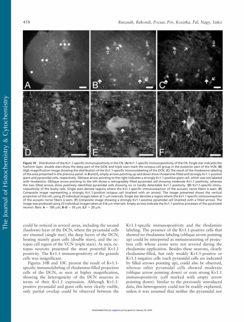
could be noticed in several areas, including the second(fusiform) layer of the DCN, where the pyramidal cellsare situated (single star), the deep layers of the DCN,hosting mainly giant cells (double stars), and the oc-topus cell region of the VCN (triple stars). As seen, oc-topus neurons presented the most powerful Kv1.1positivity. The Kv1.1 immunopositivity of the granulecells was insignificant.
Figures 10B and 10C present the result of Kv1.1-specific immunolabeling of rhodamine-filled projectioncells of the DCN, as seen at higher magnification,showing the heterogeneity of the DCN neurons interms of their Kv1.1 expression. Although Kv1.1-positive pyramidal and giant cells were clearly visible,only partial overlap could be observed between the
Kv1.1-specific immunopositivity and the rhodaminelabeling. The presence of the Kv1.1-positive cells thatshowed no rhodamine labeling (oblique arrow pointingup) could be interpreted as immunostaining of projec-tion cells whose axons were not severed during therhodamine application. Besides these neurons, clearlyrhodamine-filled, but only weakly Kv1.1-positive orKv1.1-negative cells (such pyramidal cells are indicatedby filled arrows pointing up), could also be observed,whereas other pyramidal cells showed moderate(oblique arrow pointing down) or even strong Kv1.1immunopositivity (cell marked with empty arrowpointing down). Similar to the previously introduceddata, this heterogeneity could not be readily explained,unless it was assumed that neither the pyramidal nor
Figure 10 Distribution of the Kv1.1-specific immunopositivity in the CN. (A) Kv1.1-specific immunopositivity of the CN. Single star indicates thefusiform layer, double stars show the deep part of the DCN, and triple stars mark the octopus cell group in the posterior part of the VCN. (B)High-magnification image showing the distribution of the Kv1.1-specific immunolabeling of the DCN. (C) The result of the rhodamine labelingof the area presented in the previous panel. In B and C, empty arrows pointing up and down show rhodamine-filled and strongly Kv1.1-positivegiant and pyramidal cells, respectively. Oblique arrow pointing to the right indicates a strongly Kv1.1-positive giant cell, which was not labeledwith rhodamine. Oblique arrow pointing to the left shows a retrogradely filled pyramidal cell showing moderate Kv1.1 positivity, whereasthe two filled arrows show positively identified pyramidal cells showing no or hardly detectable Kv1.1 positivity. (D) Kv1.1-specific immu-nopositivity of the bushy cells. Single stars denote regions where the Kv1.1-specific immunoreaction of the acoustic nerve fibers is seen. (E)Composite image representing a strongly Kv1.1-positive octopus cell (marked with an arrow). The image presented shows the verticalprojection of the cell, using 25 individual images taken at 1-mm intervals. Single star denotes a regionwhere the Kv1.1-specific immunoreactionof the acoustic nerve fibers is seen. (F) Composite image showing a strongly Kv1.1-positive pyramidal cell (marked with a filled arrow). Theimage was produced using 23 individual images taken at 0.8-mm intervals. Empty arrows indicate the Kv1.1-positive processes of the pyramidalneuron. Bars: A 5 100 mm; B–D 5 50 mm; E,F 5 20 mm.
TheJournal
ofHistoch
emistry&
Cytoch
emistry
458 Rusznak, Bakondi, Pocsai, Por, Kosztka, Pal, Nagy, Szucs
by guest on January 26, 2016jhc.sagepub.comDownloaded from

the giant cells formed homogeneous populations. Thissuggestion is further substantiated by the results of allsimilar experiments, where 29 giant and 9 pyramidalcells could be labeled by applying rhodamine. Of thesecells, only 18 giant (an example is shown in Figures 10Band 10C, marked with empty arrow pointing up) and 6pyramidal neurons showed Kv1.1 positivity.
Figures 10D–10F show confocal images taken fromthe CN and show the Kv1.1-specific distribution pat-tern of bushy (Figure 10D), octopus (Figure 10E,arrow), and pyramidal cells (Figure 10F, filled arrow).Besides the positivity of the presented cells, intense,
patchy labeling of the acoustic nerve fibers (see theareas marked with stars) and neuronal processes of theindividual cells were also prominent findings (e.g., emptyarrows in Figure 10F).
Distribution of the Kv3.1-specific Immunopositivityin the CN
The low-magnification image shown in Figure 11Ashows the distribution of the Kv3.1b-specific immuno-positivity of the CN. Although both the DCN and theVCN showed definite labeling, the intensity of the im-
Figure 11 Distribution of the Kv3.1-specific immunopositivity in the CN. (A) Kv3.1b-specific immunopositivity of a sagittal section of the CN.(B) The same area shown in A, but the result of the retrograde labeling of the projection neurons is visualized. (C) Overlay image of A and Balong with the result of the DAPI labeling. Single and double empty arrows mark the superficial and the deep granule cell layers, respectively.Filled arrows indicate two rhodamine-filled neurons, showing prominent Kv3.1-specific immunoreaction. (D–F) The organization of the imagesis the same as in the cases of A–C, but higher-magnification views are provided from a different preparation. Filled arrow pointing to the rightindicates a strongly Kv3.1-positive and rhodamine-filled giant cell; empty arrow shows a Kv3.1-negative, rhodamine-filled pyramidal neuron,and the oblique arrow points at a weakly Kv3.1-positive, rhodamine-labeled pyramidal neuron. (G) Overlay, high-magnification image of aKv3.1-positive, rhodamine-filled giant cell, indicated by arrow (low-magnification view of the same cell is shown in A–C). (H) Same area shownin G, but only the red (rhodamine) channel is visualized. (I) Composite image representing strongly Kv3.1-positive octopus cells (arrows). Theimage was produced using the vertical projection of 22 individual images taken at 1-mm intervals. (J) One single section from the stack usedfor constructing the composite image shown in the preceding panel. (K) A composite image depicting the strong Kv3.1 positivity of an octopuscell process. The image is the vertical projection of 25 individual images taken at 1-mm intervals. Bars: A–C 5 100 mm; D–F 5 50 mm; G,H 525 mm; I–K 5 20 mm.
TheJournal
ofHistoch
emistry&
Cytoch
emistry
Kv Subunits in the Rat Cochlear Nucleus 459
by guest on January 26, 2016jhc.sagepub.comDownloaded from

munoreaction was appreciably stronger in the VCNthan in the DCN. The result of the rhodamine labelingof the same preparation is shown in Figure 11B, wherethe backfilled projection cells are clearly visible. Thebackfilled neurons situated in the DCN were recognizedas pyramidal and giant neurons; distinctions betweenthe individual cells could be made on the basis of theirlocalization, size, and shape at higher magnification (seeFigure 11G). Applying the same criteria, the rhodamine-labeled cells in the VCN were also identified as giantcells. Figure 11C is the overlay image of Figures 11Aand 11B; in this panel, the result of DAPI labeling is alsopresented. The densely packed nuclei of the granulecells made the identification of the granule cell regionseasy; single empty arrows indicate the granule cell layercovering the surface of the CN; double empty arrows,on the other hand, indicate the granule cell layer sepa-rating the ventral and dorsal parts of the CN. As pre-sented in Figure 11C, the Kv3.1b positivity of the cellsurface of the individual neurons could be easily ob-served in the VCN, some of them showing simulta-neous rhodamine labeling as well (filled arrows). Theprojection neurons of the DCN were also positivefor Kv3.1b, although their labeling intensity was lesspronounced than that of the cells found in the VCN.It could also be established that the Kv3.1 positivity ofthe granule cells was low compared with the projectioncells of either the DCN or the VCN.
Figures 11D–11F show higher-magnification imagesof the DCN obtained from a different preparation,where several rhodamine-filled giant and pyramidalcells were present. Considering all similar experiments,most rhodamine-filled giant cells (27 of 33) appeared tobe Kv3.1 positive (an example is indicated by a filledarrow pointing to the right in Figures 11D–11F). Pyra-midal cells behaved in a similar fashion; of the 15 posi-tively identified pyramidal cells, 3 appeared to possessnegligible Kv3.1-specific immunopositivity (such a cellis marked with an empty arrow in Figures 11D–11F),whereas 12 pyramidal cells were Kv3.1 positive (oneexample is marked with oblique arrow). The Kv3.1immunopositivity of the giant cells was generallystronger than that of the pyramidal neurons. Anothertypical rhodamine-filled and strongly Kv3.1b-positivegiant cell is shown in Figures 11G and 11H (arrow; thepresented panels are high-magnification images of oneof the cells presented in Figure 11C). It is worth notingthat, in the proximity of the giant cell, several smaller(14–17 mm in diameter), spherical cells were also pres-ent, showing strong Kv3.1-specific reaction. Becausethese cells were not labeled with rhodamine, they mostlikely corresponded to cochlear interneurons, whosefurther classification was not attempted.
In the VCN, both bushy and octopus neuronsshowed intense Kv3.1b-specific reaction. Figure 11I isa vertical composite image showing a group of octopus
cells (one of the sections used for the production of thecomposite image is presented in Figure 11J). The sur-face membrane of the octopus cells showed prominentimmunolabeling, which affected both the cell bodiesand the processes. It was also a prominent feature thatthe Kv3.1-specific immunopositivity was not homoge-neous but it showed clear clustering (see the areasmarked with arrows for good examples). The intenseKv3.1-specific immunolabeling and the clustering ofthe immunopositivity were clearly present in the neu-ronal processes of the octopus cells also (Figure 11K).
Discussion
The most important aim of this study was to providea comprehensive description of the Kv subunit expres-sion of positively identified neurons of the CN. Al-though the presence and distribution pattern of certainKv channel proteins have been established before, thisis the first study using experimental animals of the samespecies and age to describe the precise, cell-specific dis-tribution of seven different Kv subunits. Besides detect-ing the presence or absence of certain subunits, thusallowing the prediction of the membrane properties ofthe individual cell types, it was shown that some Kvsubunits are preferentially expressed by certain CNneurons, and they may serve as cell-specific markers inthe identification of these cells. Octopus cells, for ex-ample, exhibited particularly strong Kv1.1 and Kv1.2expressions, most of the giant cells were strongly Kv3.1positive, whereas Kv4.2 expression was the most sig-nificant in the granule cells. Moreover, this study showsKv4.2 positivity in the glomerular synapses of the CN,resembling the same structures in the cerebellum andfurther supporting the view about the common originand close morphological similarity between the CN andthe cerebellum. Last, but not least, on the basis of theheterogeneity concerning their Kv expression patterns,it is suggested that neither the pyramidal nor the giantneurons form homogenous cell populations; thus, itcannot be ruled out that functional/morphological sub-groups exist within these cell classes.
Kv Subunit Expression Pattern of the CN Neurons
The firing properties of the major types of CN neuronsshow remarkable differences, possibly as the conse-quence of the diversity of their voltage-gated K1 cur-rents. Interestingly, however, as this study shows, mostof the CN neurons seem to express all major types ofthe voltage-gated K1 currents, including the transient,the delayed rectifier, and the LVA components.
The expression of certain Kv subunits of the CNneurons has been studied before; Tables 2–4 are in-tended to present a synopsis of the most importantresults of some earlier studies. These tables also accom-modate the data of this study, providing a convenient
TheJournal
ofHistoch
emistry&
Cytoch
emistry
460 Rusznak, Bakondi, Pocsai, Por, Kosztka, Pal, Nagy, Szucs
by guest on January 26, 2016jhc.sagepub.comDownloaded from

way to compare them with the already published resultsaccomplished on positively identified CN neurons. Insome of the earlier studies, however, the precise classi-fication of the individual cell types was not attempted,making the interpretation of the data less straight-forward; in these instances, the results were indicatedas being relevant to the VCN or DCN in general. As forthe intensity of the labelings, the “this study” term re-flects subjective decisions, when the intensity of theimmunopositivity shown by the individual cell typeswas compared with the background labeling of thesections and/or to the immunopositivity of other cells
within the nucleus. Although this method does notallow the assessment of the absolute intensity of theimmunoreactions, it was appropriate for the recogni-tion of strong immunopositivity presented by the in-dividual types of neurons and for the comparison ofthe immunopositivities of the different classes of cells.It must also be noted that different laboratories haveoften studied the presence of the various Kv subunits atdifferent levels [mRNA (Sequier et al. 1990; Fitzakerleyet al. 2000; Grigg et al. 2000); protein (Wang et al.1994; Rosenberger et al. 2003; Caminos et al. 2005); orfunctional measurements using subunit-specific chan-
Table 3 Expression of Kv subunits in the cochlear nucleus: subunits contributing to the genesis of transiently activating currents
Kv4.2 (LVA) Kv4.3 (LVA) Kv3.4 (HVA)
Localization This study References This study References This study References
Acoustic fibers Neg Neg NegVCN 1 RNA rata 1/2 RNA rate
Bushy cells Strong 1 Protein ratb Strong 1 Protein ratb Strong 1 protein ratb
1 Current ratb 1 Current ratb 1 current ratb
Octopus cells Strong 1 RNA mousec Strong 2 RNA mousec StrongDCN 1 RNA rata,d 1 RNA ratd Strong (neuropil) 2 RNA rate
Pyramidal cells 3/1 1 RNA mousec 1/0 1/2 RNA mousec 1/0Giant cells 1/4 1 RNA mousec 8/0 1/? RNA mousec 0/3Granule cells Strong, glomerular synapses 1/2 RNA mousec Present 2 RNA mousec Present, glomerular synapses
“This study” indicates data of the present work; “references” means results published earlier in aBortone et al. (2006); bPal et al. (2005); cFitzakerley et al. (2000);dSerodio and Rudy (1998); and eWeiser et al. (1994). Intensities of these data were assessed by comparing the labeling intensity of any given cell to other cells ofthe cochlear nucleus and/or to that of the background. “Strong” and “present” indicate easily recognizable and notable but not prominent immunoreactions,respectively; “neg” refers to no or hardly detectable immunopositivity. In the cases of the giant and pyramidal cells, the numbers refer to the retrogradely labeledand positively identified cells; the first number indicates the number of positive cells, and the second indicates the number of negative cells. In the case of the earlierdata, the presence (1) or absence (2) of the given subunit is indicated at RNA, protein, or functional levels (current) in the relevant species, whereas 1/2 or 1/?denote that no unequivocal conclusion was drawn from the published data. Kv, K1 channel; LVA, low voltage–activated; HVA, high voltage–activated; VCN, ventralcochlear nucleus; DCN, dorsal cochlear nucleus.
Table 2 Expression of Kv subunits in the cochlear nucleus: DTX-sensitive, LVA subunits
Kv1.1 Kv1.2 Kv1.6
Localization This study References This study References This study References
Acoustic fibers Strong 1 Protein rata,b Strong 1 Protein rata,b NegVCN 1 Protein mousec 1 Protein mousec
1 RNA ratd
Bushy cells Strong 1 Protein rata,b,e,f Strong 1 Protein rata,b,e,f Present 1 Protein rate,f
1 Current rate 1 Current rate 1 Current rate
Octopus cells Strong 1 Current mouseg Strong 1 Current mouseg Present1 Protein bath 1 RNA mousei
1 RNA mousei
1 Protein ratj
DCN 1 Protein mousec 1 RNA rata
1 RNA ratd
Pyramidal cells 6/3 1/2 Protein bath 6/1 6/2Giant cells 18/11 1/2 Protein bath 24/10 8/4Granule cells Neg 2 Protein bath Neg 2 RNA mousei Neg
1/2 RNA mousei
“This study” indicates data of the present work; “references” means results published earlier in aCaminos et al. (2005); bBortone et al. (2006); cWang et al. (1994);dSequier et al. (1990); eDodson et al. (2003); fPal et al. (2005); gBal and Oertel (2001); hRosenberger et al. (2003); iGrigg et al. (2000); and jJung et al. (2005). Intensitiesof these data were assessed by comparing the labeling intensity of any given cell to other cells of the cochlear nucleus and/or to that of the background. “Strong” and“present” indicate easily recognizable and notable but not prominent immunoreactions, respectively; “neg” refers to no or hardly detectable immunopositivity.In the cases of the giant and pyramidal cells, the numbers refer to the retrogradely labeled and positively identified cells; the first number indicates the numberof positive cells and the second indicates the number of negative cells. In the case of the earlier data, the presence (1) or absence (2) of the given subunit is indicatedat RNA, protein, or functional levels (current) in the relevant species, whereas 1/2 denotes that no unequivocal conclusion was drawn from the published data. Kv,K1 channel; DTX, dendrotoxin; LVA, low voltage–activated; VCN, ventral cochlear nucleus; DCN, dorsal cochlear nucleus.
TheJournal
ofHistoch
emistry&
Cytoch
emistry
Kv Subunits in the Rat Cochlear Nucleus 461
by guest on January 26, 2016jhc.sagepub.comDownloaded from

nel blockers (Bal and Oertel 2001; Dodson et al. 2003;Pal et al. 2004)], which is also indicated in Tables 2–4.
Table 2 summarizes the results of experiments aimedat the detection of the dendrotoxin-sensitive LVAchannel subunits. As seen, the expression of the Kv1.1subunit has been well documented in several speciesand in various cell types, especially in the VCN (Sequieret al. 1990; Wang et al. 1994; Grigg et al. 2000; Bal andOertel 2001; Dodson et al. 2003; Rosenberger et al.2003; Caminos et al. 2005; Jung et al. 2005; Pal et al.2005; Bortone et al. 2006). In the DCN, however, thesituation is somewhat less clear; although the generalpresence of Kv1.1 protein has been noted (Sequier et al.1990; Wang et al. 1994), cell-specific labeling was re-ported in the bat only (Rosenberger et al. 2003). Con-sequently, the results shown in this study are the firstshowing the heterogeneity of the giant and pyramidalcells in terms of their Kv1.1 expression patterns. Thusfar, only limited data are available about the presenceof Kv1.2 and Kv1.6 subunits in the CN. Nevertheless,Kv1.2 expression was noted in the VCN (Wang et al.1994), and it was shown that both bushy (Pal et al.2004; Caminos et al. 2005; Bortone et al. 2006) and oc-topus neurons (Grigg et al. 2000; Bal and Oertel 2001)
possessed this subunit. In contrast to the VCN, the po-tential Kv1.2 expression of the DCN was reported atthe mRNA level only (Caminos et al. 2005). Results ofthis study indicate that Kv1.2 subunits are expressed byboth pyramidal and giant neurons of the CN.
In a recent study, when investigating developmentalchanges occurring in the Kv1.1 and Kv1.2 expressionof the rat CN, an age-dependent increase was noted inthe expression intensity at mRNA and protein levels.The increase was present from birth up to the 25thpostnatal day, and it was followed by a moderate de-cline to the adult values (Bortone et al. 2006). In adifferent work (Caminos et al. 2005), however, focus-ing on Kv1.1 and Kv1.2 expression of the VCN (includ-ing bushy cells), a continuous and gradual decline of theexpression levels was reported. This reduction startedat about the 14th postnatal day and resulted in very lowexpression levels in adult animals. Moreover, it wasnoted that the Kv1.2 subunits disappeared from the cellbodies, and at the same time, a continuous increaseof the Kv1.2 expression was observed in the acousticnerve fibers, reaching rather intense levels by the21st postnatal day. These findings implied that, notonly the intensity, but the localization of the Kv1.2 ex-pression might also change during postnatal development.
In this study, the expression of the Kv1.2 subunitwas noted in the CN isolated from either young or oldanimals, (resembling the data presented by Bortoneet al. 2006) the intensity of the Kv1.2-specific immuno-positivity increased, and it involved the neuropil of theDCN as well. It is very important to note that a charac-teristic translocation of the Kv1.2 subunits was shownin this study, especially in the case of the octopus andbushy cells. Compared with the young animals, wherethe Kv1.2 immunopositivity was present intracellularlyonly, in older animals, the immunopositivity was notonly stronger intracellularly, but a pronounced cell sur-face labeling could also be observed. This observationsuggests that, not only the amount of the channel pro-tein might change accompanying the maturation of theauditory pathway, but the density of the functional K1
channels containing Kv1.2 subunits might also increasein the cell surface membrane. Although further work isneeded to verify the functional significance of this find-ing, it is a tempting possibility that the increased densityof the Kv1.2 channels of the cell membrane might haveroles in the maturation process of the auditory system.
Table 3 shows the results of numerous previous stud-ies concerning the presence and distribution of Kvsubunits contributing to the assembly of K1 channelsproducing transient (inactivating) current. Besides thedata presented in Table 3, it should also be mentionedthat mRNA encoding a subunit belonging to the Kv4family has been described in the rat (Drewe et al. 1992).Although the exact type of subunit was not specified, itis unlikely that the mRNA found encoded Kv4.1 pro-
Table 4 Expression of Kv3.1 subunit in the cochlear nucleus
Kv3.1b (HVA)
Localization This study References
Acoustic fibers NegVCN 1 RNA rata,b,c,d
1 Protein rata
Bushy cells Strong 1 Protein rate
Octopus cells Strong 2 RNA mousef
1 RNA ratg,h
2 Protein rath
1 Protein rati
DCN 1 RNA ratc,d
1/2 RNA ratb
Pyramidal cells 12/3 1 RNA ratg,h
2 Protein rath
Giant cells 27/6 1 RNA ratg,h
1/? Protein rath
Granule cells Neg 1 RNA mousef
1/? RNA ratg,h
1/? RNA rath
“This study” indicates data of the present work; “references” means resultspublished earlier in aWeiser et al. (1995); bWeiser et al. (1994); cPerney et al.(1992); dBortone et al. (2006); ePal et al. (2005); fGrigg et al. (2000); gLi et al.(2001); hPerney and Kaczmarek (1997); and iJung et al. (2005). Intensities ofthese data were assessed by comparing the labeling intensity of any givencell to other cells of the cochlear nucleus and/or to that of the background.“Strong” indicates easily recognizable immunoreaction; “neg” refers to no orhardly detectable immunopositivity. In the case of the giant and pyramidalcells, the numbers refer to the retrogradely labeled and positively identifiedcells; the first number indicates the number of positive cells and the secondindicates the number of negative cells. In the case of the earlier data, thepresence (1) or absence (2) of the given subunit is indicated at RNA, protein, orfunctional levels (current) in the relevant species, whereas 1/2 or 1/? denotethat no unequivocal conclusion was drawn from the published data. Kv, K1
channel; HVA, high voltage–activated; VCN, ventral cochlear nucleus; DCN,dorsal cochlear nucleus.
TheJournal
ofHistoch
emistry&
Cytoch
emistry
462 Rusznak, Bakondi, Pocsai, Por, Kosztka, Pal, Nagy, Szucs
by guest on January 26, 2016jhc.sagepub.comDownloaded from

teins, because specific studies indicated the lack of thismRNA in both the rat (Serodio and Rudy 1998) andmouse (Fitzakerley et al. 2000).
Because most of the previous attempts tried to iden-tify the K1 channel subunits producing transient cur-rents at the mRNA level, the data presented here areparticularly important because they prove that Kv4.2,Kv4.3, and Kv3.4 proteins are present in the CN. It isworth mentioning that, although glomerular synapseshave been shown in the DCN (Kane 1974; Alibardi2004), this study is the first to show strong Kv4.2 andmoderate Kv3.4 expression in these structures.
The most significant Kv subunit contributing to thegenesis of delayed rectifier K1 current in the CN is theKv3.1 (Table 4). In fact, the majority of the authorsagreed that Kv3.1-specific mRNA is present in the CN,although the overall picture is more complex whentrying to find the Kv3.1 subunits at the protein level.The often confusing data may be the consequence ofdevelopment-related issues (Bortone et al. 2006): thechanges in the amount of the mRNA necessarily pre-cede the modifications occurring at the protein level,causing some apparent discrepancies in the experimen-tal data yielded at the mRNA and protein levels, re-spectively. Nevertheless, these findings of strong Kv3.1expression of the bushy cells are in agreement with ourprevious data (Pal et al. 2005). The intense Kv3.1 ex-pression of the octopus cells is also a noteworthy resultof this study. As for the DCN, both the pyramidal andthe giant neurons showed some variability of Kv3.1expression, and this non-uniform occurrence of immu-nopositivity may explain why earlier studies did notalways note definite Kv3.1 positivity of these cells.
Functional Implications
Despite the lack of direct studies of the activity of theCN neurons, some functional assumptions could bemade on the basis of their Kv expression. In the case ofpyramidal cells, the activity of various LVA K1 channelsubunits (Kv1.1, 1.2, 1.6, 4.2, and 4.3) may contributeto the setting of their resting membrane potential that,in turn, plays a decisive role in determining their firingpatterns (Kane 1974; Manis et al. 2003). The limitednumber of earlier papers suggested similar membraneproperties and firing characteristics of the giant andpyramidal neurons (Zhang and Oertel 1993; Ding et al.1999; Babalian et al. 2003), and this view was sup-ported by these experiments indicating similar Kv ex-pression patterns of these two cell types. However, inaccordance with some earlier reports (Perney andKaczmarek 1997; Fitzakerley et al. 2000; Rosenbergeret al. 2003), both pyramidal and the giant neuronsshowed some variability, suggesting that there might bedifferent subtypes within both groups presenting non-uniform functional properties.
Earlier electrophysiological experiments on granulecells showed the presence of a rapidly inactivating, LVAtransient (A-type) and a slowly inactivating (delayedrectifier) current (Rusznak et al. 1997). Our presentfindings indicate that the transient current is mainly theconsequence of the activation of K1 channels contain-ing Kv4.2 subunits. Based on the similar distributionpattern of the Kv4.2 subunits in the CN and in thecerebellum (unpublished data), it is proposed that thesesubunits may have the same roles in the CN as de-scribed originally in the cerebellar glomerular synapses(Shibata et al. 2000; Shibasaki et al. 2004; Strassle et al.2005). In the case of the bushy cells, this study showedthe expression of Kv subunits whose presence can ex-plain the K1 current components described earlier(Manis and Marx 1991; Dodson et al. 2003; Rothmanand Manis 2003; Cao et al. 2007).
Functional studies have indicated that the coinci-dence detector role (Golding et al. 1995; Oertel et al.2000) of the octopus cells is ensured by voltage-gatedK1 channels representing all three major functionalgroups. These findings are clearly in accordance withthis conclusion, because Kv subunits from all threefunctional classes could be detected. Definite, age-dependent translocation of the Kv1.2 subunits and sig-nificant expression of the Kv4.2 protein were also notedin the octopus neurons, and both phenomena may berelated to developmental changes affecting the func-tions of these cells.
Acknowledgments
This work was supported by grants from the HungarianScience Foundation (Hungarian Research Fund, T-046067,NK-61412) and from The Wellcome Trust (CollaborativeResearch Initiative Grant 075243/Z/04/Z).
We thank Dr. Beatrix Dienes for kind and professionalhelp on some of the confocal microscopy sessions and IbolyaVarga for skilled technical assistance.
Literature Cited
Adamson CL, Reid MA, Mo ZL, Bowne-English J, Davis RL (2002)Firing features and potassium channel content of murine spiralganglion neurons vary with cochlear location. J Comp Neurol447:331–350
Alibardi L (2000) Cytology, synaptology and immunocytochemistryof commissural neurons and their putative axonal terminals in thedorsal cochlear nucleus of the rat. Ann Anat 182:207–220
Alibardi L (2004) Mossy fibers in granule cell areas of the ratdorsal cochlear nucleus from intrinsic and extrinsic origin inner-vate unipolar brush cell glomeruli. J Submicrosc Cytol Pathol 36:193–210
Alibardi L (2006) Review: cytological characteristics of commissuraland tuberculo-ventral neurons in the rat dorsal cochlear nucleus.Hear Res 216–217:73–80
Babalian AL, Ryugo DK, Rouiller EM (2003) Discharge propertiesof identified cochlear nucleus neurons and auditory nerve fibers inresponse to repetitive electrical stimulation of the auditory nerve.Exp Brain Res 153:452–460
Bal R, Oertel D (2001) Potassium currents in octopus cells of themammalian cochlear nucleus. J Neurophysiol 86:2299–2311
TheJournal
ofHistoch
emistry&
Cytoch
emistry
Kv Subunits in the Rat Cochlear Nucleus 463
by guest on January 26, 2016jhc.sagepub.comDownloaded from

Bortone DS, Mitchell K, Manis PB (2006) Developmental time courseof potassium channel expression in the rat cochlear nucleus. HearRes 211:114–125
Brawer JR, Morest DK, Kane EC (1974) The neuronal architecture ofthe cochlear nucleus of the cat. J Comp Neurol 155:251–300
Caminos E, Vale C, Lujan R, Martinez-Galan JR, Juiz JM (2005)Developmental regulation and adult maintenance of potassiumchannel proteins (Kv 1.1 and Kv 1.2) in the cochlear nucleus of therat. Brain Res 1056:118–131
Cant NB, Benson CG (2003) Parallel auditory pathways: projectionpatterns of the different neuronal populations in the dorsal andventral cochlear nuclei. Brain Res Bull 60:457–474
Cao XJ, Shatadal S, Oertel D (2007) Voltage-sensitive conductancesof bushy cells of the mammalian ventral cochlear nucleus. JNeurophysiol 97:3961–3975
Coetzee WA, Amarillo Y, Chiu J, Chow A, Lau D, McCormack T,Moreno H, et al. (1999) Molecular diversity of K1 channels. AnnNY Acad Sci 868:233–285
Ding J, Benson TE, Voigt HF (1999) Acoustic and current-pulse re-sponses of identified neurons in the dorsal cochlear nucleus ofunanesthetized, decerebrate gerbils. J Neurophysiol 82:3434–3457
Disterhoft JF, Perkins RE, Evans S (1980) Neuronal morphology ofthe rabbit cochlear nucleus. J Comp Neurol 192:687–702
Dodson PD, Billups B, Rusznak Z, Szucs G, Barker MC, Forsythe ID(2003) Presynaptic Kv1.2 channels suppress synaptic terminalhyperexcitability following action potential invasion. J Physiol550:27–33
Dodson PD, Forsythe ID (2004) Presynaptic K1 channels: electrifyingregulators of synaptic terminal excitability. Trends Neurosci 27:210–217
Doughty JM, Barnes-Davies M, Rusznak Z, Harasztosi C, ForsytheID (1998) Contrasting Ca21 channel subtypes at cell bodies andsynaptic terminals of rat anteroventral cochlear bushy neurones.J Physiol 512:365–376
Drewe JA, Verma S, Frech G, Joho RH (1992) Distinct spatial andtemporal expression patterns of K1 channel mRNAs from differ-ent subfamilies. J Neurosci 12:538–548
Fitzakerley JL, Star KV, Rinn JL, Elmquist BJ (2000) Expressionof Shal potassium channel subunits in the adult and developingcochlear nucleus of the mouse. Hear Res 147:31–45
Golding NL, Ferragamo MJ, Oertel D (1999) Role of intrinsic con-ductances underlying responses to transients in octopus cells of thecochlear nucleus. J Neurosci 19:2897–2905
Golding NL, Robertson D, Oertel D (1995) Recordings from slicesindicate that octopus cells of the cochlear nucleus detect coincidentfiring of auditory nerve fibers with temporal precision. J Neurosci15:3138–3153
Grigg JJ, Brew HM, Tempel BL (2000) Differential expression ofvoltage-gated potassium channel genes in auditory nuclei of themouse brainstem. Hear Res 140:77–90
Hackney CM, Osen KK, Kolston J (1990) Anatomy of the cochlearnuclear complex of guinea pig. Anat Embryol (Berl) 182:123–149
Harrison JM, Irving R (1966) The organization of the posterior ven-tral cochlear nucleus in the rat. J Comp Neurol 126:391–401
Hurd LB, Feldman ML (1994) Purkinje-like cells in rat cochlearnucleus. Hear Res 72:143–158
Jung DK, Lee SY, Kim D, Joo KM, Cha CI, Yang HS, Lee WB, et al.(2005) Age-related changes in the distribution of Kv1.1 and Kv3.1in rat cochlear nuclei. Neurol Res 27:436–440
Kane EC (1974) Synaptic organisation in the dorsal cochlear nucleusof the cat: a light and electron microscopic study. J Comp Neurol155:301–330
Kane ES, Puglisi SG, Gordon BS (1981) Neuronal types in the deepdorsal cochlear nucleus of the cat: I. giant neurons. J Comp Neurol198:483–513
Kanold PO, Manis PB (1999) Transient potassium currents regu-late the discharge patterns of dorsal cochlear pyramidal cells. JNeurosci 19:2195–2208
Li W, Kaczmarek LK, Perney TM (2001) Localization of two high-threshold potassium channel subunits in the rat central auditorysystem. J Comp Neurol 437:196–218
Lorente de No R (1981) The Primary Acoustic Nuclei, New York,Raven Press
Malmierca MS (2003) The structure and physiology of the rat audi-tory system: an overview. Int Rev Neurobiol 56:147–211
Manis PB, Marx SO (1991) Outward currents in isolated ventralcochlear nucleus neurons. J Neurosci 11:2865–2880
Manis PB, Molitor SC, Wu H (2003) Subthreshold oscillations gen-erated by TTX-sensitive sodium currents in dorsal cochlear nu-cleus pyramidal cells. Exp Brain Res 153:443–451
Moore JK (1986) Cochlear nuclei: relationship to the auditory nerve.In Altschuler RA, Hoffman DW, Bobbin RP, eds. Neurobiologyof Hearing: The Cochlea. New York, Raven Press, 283–301
Oertel D, Bal R, Gardner SM, Smith PH, Joris PX (2000) Detectionof synchrony in the activity of auditory nerve fibers by octopuscells of the mammalian cochlear nucleus. Proc Natl Acad Sci USA97:11773–11779
Oertel D, Wu SH (1989) Morphology and physiology of cells inslice preparation of the dorsal cochlear nucleus of mice. J CompNeurol 283:228–247
Osen KK (1969a) Cytoarchitecture of the cochlear nuclei in the cat.J Comp Neurol 136:453–484
Osen KK (1969b) The intrinsic organization of the cochlear nuclei.Acta Otolaryngol 67:352–359
Pal B, Por A, Pocsai K, Szucs G, Rusznak Z (2005) Voltage-gated andbackground K1 channel subunits expressed by the bushy cells ofthe rat cochlear nucleus. Hear Res 199:57–70
Pal B, Rusznak Z, Harasztosi C, Szucs G (2004) Depolarization-activated K1 currents of the bushy neurons of the rat cochlear nu-cleus in a thin brain slice preparation. Acta Physiol Hung 91:83–98
Perney TM, Kaczmarek LK (1997) Localization of a high thresholdpotassium channel in the rat cochlear nucleus. J CompNeurol 386:178–202
Perney TM, Marshall J, Martin KA, Hockfield S, Kaczmarek LK(1992) Expression of the mRNAs for the Kv3.1 potassium channelgene in the adult and developing rat brain. J Neurophysiol 68:756–766
Pocsai K, Pal B, Pap P, Bakondi G, Kosztka L, Rusznak Z, Szucs G(2007) Rhodamine backfilling and confocal microscopy as a toolfor the unambiguous identification of neuronal cell types: a studyof the neurons of the rat cochlear nucleus. Brain Res Bull 71:529–538
Por A, Pocsai K, Rusznak Z, Szucs G (2005) Presence and distribu-tion of three calcium binding proteins in projection neurons ofthe adult rat cochlear nucleus. Brain Res 1039:63–74
Reiss LA, Young ED (2005) Spectral edge sensitivity in neural circuitsof the dorsal cochlear nucleus. J Neurosci 25:3680–3691
Rosenberger MH, Fremouw T, Casseday JH, Covey E (2003)Expression of the Kv1.1 ion channel subunit in the auditorybrainstem of the big brown bat, Eptesicus fuscus. J Comp Neurol462:101–120
Rossi F, Borsello T (1993) Ectopic Purkinje cells in the adult rat:olivary innervation and different capabilities of migration and de-velopment after grafting. J Comp Neurol 337:70–82
Rothman JS, Manis PB (2003) Differential expression of three distinctpotassium currents in the ventral cochlear nucleus. J Neurophysiol89:3070–3082
Rudy B, McBain CJ (2001) Kv3 channels: voltage-gated K1 channelsdesigned for high-frequency repetitive firing. Trends Neurosci 24:517–526
Rusznak Z, Forsythe ID, Brew HM, Stanfield PR (1997) Membranecurrents influencing action potential latency in granule neurons ofthe rat cochlear nucleus. Eur J Neurosci 9:2348–2358
Schwarz DW, Puil E (1997) Firing properties of spherical bushy cellsin the anteroventral cochlear nucleus of the gerbil. Hear Res 114:127–138
Sequier JM, Brennand J, Barhanin J, Lazdunski M (1990) Regionalexpression of a MCD-peptide and dendrotoxin I-sensitive voltage-dependent potassium channel in rat brain. FEBS Lett 263:163–165
Serodio P, Rudy B (1998) Differential expression of Kv4 K1 channelsubunits mediating subthreshold transient K1 (A-type) currents inrat brain. J Neurophysiol 79:1081–1091
TheJournal
ofHistoch
emistry&
Cytoch
emistry
464 Rusznak, Bakondi, Pocsai, Por, Kosztka, Pal, Nagy, Szucs
by guest on January 26, 2016jhc.sagepub.comDownloaded from

Shibasaki K, Nakahira K, Trimmer JS, Shibata R, Akita M,Watanabe S, Ikenaka K (2004) Mossy fibre contact triggersthe targeting of Kv4.2 potassium channels to dendrites andsynapses in developing cerebellar granule neurons. J Neuro-chem 89:897–907
Shibata R, Nakahira K, Shibasaki K, Wakazono Y, Imoto K, IkenakaK (2000) A-type K1 current mediated by the Kv4 channel regulatesthe generation of action potential in developing cerebellar granulecells. J Neurosci 20:4145–4155
Strassle BW, Menegola M, Rhodes KJ, Trimmer JS (2005) Light andelectron microscopic analysis of KChIP and Kv4 localization in ratcerebellar granule cells. J Comp Neurol 484:144–155
Wang H, Kunkel DD, Schwartzkroin PA, Tempel BL (1994) Locali-zation of Kv1.1 and Kv1.2, two K channel proteins, to synaptic
terminals, somata, and dendrites in the mouse brain. J Neurosci14:4588–4599
Webster DB, Trune DR (1982) Cochlear nuclear complex of mice.Am J Anat 163:103–130
Weiser M, Bueno E, Sekirnjak C, Martone ME, Baker H, Hillman D,Chen S, et al. (1995) The potassium channel subunit KV3.1b islocalized to somatic and axonal membranes of specific populationsof CNS neurons. J Neurosci 15:4298–4314
Weiser M, Vega-Saenz de Miera E, Kentros C, Moreno H, Franzen L,Hillman D, Baker H, et al. (1994) Differential expression of Shaw-related K1 channels in the rat central nervous system. J Neurosci14:949–972
Zhang S, Oertel D (1993) Giant cells of the dorsal cochlear nucleus ofmice: intracellular recordings in slices. J Neurophysiol 69:1398–1408
TheJournal
ofHistoch
emistry&
Cytoch
emistry
Kv Subunits in the Rat Cochlear Nucleus 465
by guest on January 26, 2016jhc.sagepub.comDownloaded from
Copyright © 2022 FDOKUMEN