Dried leaf extract of Olea europaea ameliorates islet-directed autoimmunity in mice
12
Dried leaf extract of Olea europaea ameliorates islet-directed autoimmunity in mice Tamara Cvjetic ´anin 1 , Djordje Miljkovic ´ 1 , Ivana Stojanovic ´ 1 , Dragana Dekanski 2 and Stanislava Stos ˇic ´-Grujic ˇic ´ 1 * 1 Department of Immunology, Institute for Biological Research ‘Sinis ˇa Stankovic ´’, University of Belgrade, Bulevar Despota Stefana 142, 11060 Belgrade, Serbia 2 Biomedical Research, R&D Institute, Galenika a.d., Belgrade, Serbia (Received 11 August 2009 – Revised 29 October 2009 – Accepted 11 November 2009 – First published online 22 December 2009) The health-promoting effects of various constituents of the olive tree (Olea europaea) are mainly associated with hypoglycaemic and insulin- sensitising activities and have been widely demonstrated in the metabolic syndrome and type 2 diabetes. However, their biological activity in autoimmune type 1 diabetes (T1D) is poorly characterised. Therefore, the influence of O. europaea-derived components present in dry olive leaf extract (DOLE) was examined in two established preclinical models of human T1D, which differ in some aspects of diabetogenesis: multiple low-dose streptozotocin-induced diabetes in susceptible C57BL/6 and CBA/H mouse strains; cyclophosphamide-accelerated diabetes in non-obese diabetic mice. In both T1D models, in vivo administration of DOLE significantly reduced clinical signs of diabetes (hyperglycaemia and body weight loss) and led to complete suppression of histopathological changes in pancreatic islets. In line with these, insulin expression and release were restored in DOLE-treated mice. Interestingly, inducible NO synthase expression and NO production were significantly elevated in peripheral tissues but were down-regulated within the local environment of the endocrine pancreas. This interference was reflected in NO-mediated suppression of T lymphocyte proliferation and lower production of the proinflammatory cytokines interferon-g, IL-17 and TNF-a in the spleen, with subsequent blockade of b-cell destruction. The results suggest that DOLE interferes with development of autoimmune diabetes by down- regulating production of proinflammatory and cytotoxic mediators. Therefore, the potential use of a DOLE-enriched diet for prophylaxis/treatment of human T1D, and possibly other autoimmune diseases, is worthy of further investigation. Type 1 diabetes: Olea europaea leaves: Cytokines: Nitric oxide The term diabetes mellitus is used to describe a variety of chronic metabolic disorders characterised by elevated blood glucose levels now afflicting 3 % of the world population (1,2) . Diabetes mellitus can broadly be classified into two main types based on individual aetiologies. Type 1 diabetes (T1D) that accounts for approximately 5–10% of all cases of diabetes, is an inflammatory autoimmune disease in which pancreatic insulin-producing b-cells are selectively destroyed by cells of the immune system (3) culminating in a state of hypoinsulinaemia and hyperglycaemia (4) . Type 2 diabetes (T2D) that is estimated to represent 90–95 % of all cases is due to b-cell failure or various degrees of insulin resistance (5) . The only possible cure for T1D is control of the T cell autoimmunity against b-cells together with recovery and/or replacement of the destroyed b-cell mass. Over many years, various immunomodulatory regimens were tested with the aim of blocking autoimmunity against b-cell mass and promoting b-cell preservation. Despite considerable progress in the management of T1D with conventional drugs, a single effective immune-based therapeutic approach has not been identified so far (2,6 – 9) . This provides an impetus for the development of new therapeutic strategies using various animal models (10,11) . However, any single animal model is unlikely to address the issue of population hetero- geneity, so the application of different models has been instructive about many diverse scenarios that might occur in human T1D (12) . Benefits of the Mediterranean-style diet have been known for centuries, and it has been traditionally used to prevent and treat different metabolic disorders. Phenolic components of commonly consumed foods, such as red wine and virgin olive oil, or components of other olive (Olea europaea) organs, such as leaves, rich in these constituents, have been shown to possess strong antioxidant and anti-inflammatory properties (13) . The chemical composition of dry olive leaf extract (DOLE) is very complex, comprising oleuropein, caffeic acid, luteolin, luteolin-7-O-glucoside, apigenin- 7-O-glucoside, quercetin, chrysoeriol and others (14) . Due to * Corresponding author: Stanislava Stos ˇic ´-Grujic ˇic ´, fax þ 381 11 2761 433, email [email protected] Abbreviations: b.wt., body weight; CY, cyclophosphamide; DOLE, dry olive leaf extract; IFN, interferon; iNOS, inducible NO synthase; i.p., intraperitoneal; MLDS, multiple low-dose streptozotocin; NOD, non-obese diabetic; p.i., post-induction; p.o., per oral; Panc, pancreatic islets; PC, peritoneal cells; PLN, pancreatic lymph nodes; IFN, interferon; Spl, splenocytes; T1D, type 1 diabetes; T2D, type 2 diabetes. British Journal of Nutrition (2010), 103, 1413–1424 doi:10.1017/S0007114509993394 q The Authors 2009 British Journal of Nutrition
-
Upload
independent -
Category
Documents
-
view
1 -
download
0
Transcript of Dried leaf extract of Olea europaea ameliorates islet-directed autoimmunity in mice

Dried leaf extract of Olea europaea ameliorates islet-directed autoimmunity
in mice
Tamara Cvjeticanin1, Djordje Miljkovic1, Ivana Stojanovic1, Dragana Dekanski2
and Stanislava Stosic-Grujicic1*1Department of Immunology, Institute for Biological Research ‘Sinisa Stankovic’, University of Belgrade, Bulevar Despota Stefana
142, 11060 Belgrade, Serbia2Biomedical Research, R&D Institute, Galenika a.d., Belgrade, Serbia
(Received 11 August 2009 – Revised 29 October 2009 – Accepted 11 November 2009 – First published online 22 December 2009)
The health-promoting effects of various constituents of the olive tree (Olea europaea) are mainly associated with hypoglycaemic and insulin-
sensitising activities and have been widely demonstrated in the metabolic syndrome and type 2 diabetes. However, their biological activity in
autoimmune type 1 diabetes (T1D) is poorly characterised. Therefore, the influence of O. europaea-derived components present in dry olive
leaf extract (DOLE) was examined in two established preclinical models of human T1D, which differ in some aspects of diabetogenesis: multiple
low-dose streptozotocin-induced diabetes in susceptible C57BL/6 and CBA/H mouse strains; cyclophosphamide-accelerated diabetes in non-obese
diabetic mice. In both T1D models, in vivo administration of DOLE significantly reduced clinical signs of diabetes (hyperglycaemia and body
weight loss) and led to complete suppression of histopathological changes in pancreatic islets. In line with these, insulin expression and release
were restored in DOLE-treated mice. Interestingly, inducible NO synthase expression and NO production were significantly elevated in
peripheral tissues but were down-regulated within the local environment of the endocrine pancreas. This interference was reflected in NO-mediated
suppression of T lymphocyte proliferation and lower production of the proinflammatory cytokines interferon-g, IL-17 and TNF-a in the spleen,
with subsequent blockade of b-cell destruction. The results suggest that DOLE interferes with development of autoimmune diabetes by down-
regulating production of proinflammatory and cytotoxic mediators. Therefore, the potential use of a DOLE-enriched diet for prophylaxis/treatment
of human T1D, and possibly other autoimmune diseases, is worthy of further investigation.
Type 1 diabetes: Olea europaea leaves: Cytokines: Nitric oxide
The term diabetes mellitus is used to describe a variety ofchronic metabolic disorders characterised by elevated bloodglucose levels now afflicting 3 % of the world population(1,2).Diabetes mellitus can broadly be classified into two maintypes based on individual aetiologies. Type 1 diabetes (T1D)that accounts for approximately 5–10 % of all cases ofdiabetes, is an inflammatory autoimmune disease in whichpancreatic insulin-producing b-cells are selectively destroyedby cells of the immune system(3) culminating in a state ofhypoinsulinaemia and hyperglycaemia(4). Type 2 diabetes(T2D) that is estimated to represent 90–95 % of all cases isdue to b-cell failure or various degrees of insulin resistance(5).The only possible cure for T1D is control of the T cellautoimmunity against b-cells together with recovery and/orreplacement of the destroyed b-cell mass. Over manyyears, various immunomodulatory regimens were testedwith the aim of blocking autoimmunity against b-cell massand promoting b-cell preservation. Despite considerableprogress in the management of T1D with conventional
drugs, a single effective immune-based therapeutic approachhas not been identified so far(2,6 – 9). This provides an impetusfor the development of new therapeutic strategies usingvarious animal models(10,11). However, any single animalmodel is unlikely to address the issue of population hetero-geneity, so the application of different models has beeninstructive about many diverse scenarios that might occur inhuman T1D(12).
Benefits of the Mediterranean-style diet have been knownfor centuries, and it has been traditionally used to preventand treat different metabolic disorders. Phenolic componentsof commonly consumed foods, such as red wine and virginolive oil, or components of other olive (Olea europaea)organs, such as leaves, rich in these constituents, have beenshown to possess strong antioxidant and anti-inflammatoryproperties(13). The chemical composition of dry oliveleaf extract (DOLE) is very complex, comprising oleuropein,caffeic acid, luteolin, luteolin-7-O-glucoside, apigenin-7-O-glucoside, quercetin, chrysoeriol and others(14). Due to
* Corresponding author: Stanislava Stosic-Grujicic, fax þ381 11 2761 433, email [email protected]
Abbreviations: b.wt., body weight; CY, cyclophosphamide; DOLE, dry olive leaf extract; IFN, interferon; iNOS, inducible NO synthase; i.p., intraperitoneal; MLDS,
multiple low-dose streptozotocin; NOD, non-obese diabetic; p.i., post-induction; p.o., per oral; Panc, pancreatic islets; PC, peritoneal cells; PLN, pancreatic lymph
nodes; IFN, interferon; Spl, splenocytes; T1D, type 1 diabetes; T2D, type 2 diabetes.
British Journal of Nutrition (2010), 103, 1413–1424 doi:10.1017/S0007114509993394q The Authors 2009
British
Journal
ofNutrition

all these useful active ingredients, olive oil and oliveleaf constituents have been used increasingly as comple-mentary and alternative medicine to improve metabolicdisorders(15 – 19). Some of these beneficial effects include reac-tive oxygen species scavenge, inhibition of LDL oxidation,production of NO, down-regulation of adhesion moleculesand inhibition of inflammatory cytokines(20 – 22). It has alsobeen reported that constituents of olive leaves have anti-hyperglycaemic and insulin-sensitising activities, suggestinga beneficial metabolic effect in T2D(23 – 26). However, thedevelopment of therapeutic approaches for T1D has beenneglected in favour of efforts to advance therapies for thelarger T2D population.
In view of the increasing prevalence, there is a growingneed to develop integrated approaches towards preventionand treatment of T1D by exploring potentials offered bytraditional phytotherapies as alternative and/or supplementarytherapy. Recently, we have shown that DOLE-enriched dietameliorated central nervous system autoimmunity in ratsthrough reduction of proinflammatory cytokine production,which implied that this extract had a potent immunomodula-tory effect(27). Therefore, in the present study we investigatedthe effects of commercially available standardised DOLE onpancreatic islet (Panc)-directed autoimmunity. For this pur-pose, we used two models of T1D: cyclophosphamide (CY)-accelerated diabetes in non-obese diabetic (NOD) mice(28);multiple low-dose streptozotocin (MLDS)-induced diabetesin susceptible mouse strains(29), because hyperglycaemia andinsulitis can be easily induced in a relatively short period oftime in a high percentage of animals. In both the diseasemodels, inflammatory autoreactive T cells and macrophagesmediate the autoimmune status, causing islet b-cell destruc-tion either directly or through secretion of proinflammatorycytokines and free radicals(30). However, in contrast toMLDS-induced diabetes, CY-induced diabetes in NOD miceis based on selective elimination of inhibitory cells thatwould otherwise prevent development of the disease(28).Therefore, the ultimate translational objective is to use bothof these animal models to test the hypothesis that DOLE pro-tects against T1D development and to determine potentialmechanisms involved in this protective effect.
Materials and methods
Reagents
Standardised to 18–26 % of oleuropein, DOLE EFLAw 943was purchased from Frutarom Switzerland Ltd (Wadenswil,Switzerland). The extract was manufactured from the driedleaves of O. europaea L., applying an ethanol (80 % m/m)extraction procedure. After a patented filtration process(EFLAw Hyperpure), the crude extract was dried. Further-more, stability and microbiological purity were confirmed bythe manufacturer. The extract was standardised to 19·8 % ofoleuropein content by the manufacturer. It was further phyto-chemically analysed for the presence of various components inthe previous report of Dekanski and collaborators. There itwas detected that the extract contained oleuropein (19·8 %),total flavonoids (0·29 %), including luteoline-7-O-glucoside(0·04 %), apigenine-7-O-glucoside (0·07 %) and quercetin(0·04 %), as well as tannins (0·52 %) and caffeic acid
(0·02 %)(14). All other reagents were purchased from Sigma(St Louis, MO, USA) unless otherwise noted.
Type 1 diabetes induction and in vivo treatment protocols
All experimental procedures were approved by the Insti-tutional Animal Care and Use Committee at the Institutefor Biological Research ‘Sinisa Stankovic’, University ofBelgrade and run in accordance to the requirements of theEuropean Union regarding handling of experimental animals.Specific pathogen-free inbred C57BL/6, CBA/H and NOD/ShiLtJ mice were provided by the Central Animal HouseFacility of our institute. Mice were maintained under conven-tional conditions with free access to a standard laboratorychow and potable water. Animals and feed were regularlychecked by appropriate microbiological examinations, whichshowed that the animals were not infected with commonmouse pathogens and that the feed was free of microbiologicalcontamination.
Immunoinflammatory T1D was induced in male mice ofthe C57BL/6 and CBA/H strains with multiple low dosesof streptozotocin (MLDS, 40 mg/kg body weight (b.wt.),intraperitoneal (i.p.) for five consecutive days) as describedpreviously(31). The mice were age-matched (8–12 weeks ofage) with b.wt. ranging from 25 to 30 g. Treatment withDOLE was per oral (p.o.) by gavage in 0·2 ml of PBS(100 mg/kg b.wt. per d, divided into two daily doses), ori.p. (40 mg/kg b.wt per d), given as a prevention regimen(days 25–21 of diabetes post induction (p.i.)) or as a reversalregimen (days 5–21 p.i.). Control MLDS-induced mice weretreated with an equivalent amount of vehicle. A group ofnon-diabetic control mice were treated i.p. with DOLE only.The mice subjected to morphological examination of pancrea-tic glands and ex vivo analyses were sacrificed on day 15 p.i.,unless otherwise stated. Each experimental group used forin vivo analyses consisted of six to seven mice per group,whereas for ex vivo analyses each experimental group hadfour to five mice per group.
For induction of T1D with CY, nulliparous non-pregnantnon-diabetic female NOD mice, 8–10 weeks old (b.wt.range 20–25 g) were injected twice with the drug at a doseof 300 mg/kg b.wt., i.p., with a 2-week interval betweenthe injections. DOLE was administered i.p. (40 mg/kg b.wt.per d), or p.o. (100 mg/kg b.wt. per d, divided into two dailydoses) as a continuous treatment until the end of the exper-iments, starting 1 d after the first diabetogenic challengewith CY. Control mice were i.p. administered with an equiv-alent amount of vehicle or left untreated. The number of miceused for every experiment was seven to sixteen per group.
Both in MLDS- and CY-induced diabetes, bloodsamples were taken from the tail tip of non-fasting mice forblood glucose determination, with a handheld glucometer(GlucoSure Plus; ApexBio, Hsinchu, Taiwan, ROC). Micewere considered hyperglycaemic when blood glucoseconcentration exceeded 10 mmol/l (for MLDS induction) and8·8 mmol/l (for CY induction).
The dose of DOLE used in the present study was calculatedaccording to a clinical study in which DOLE at 1000 mg daily(divided in two doses) effectively reduced blood pressure(32).We used the metabolic body size or food intake rather thanb.wt. as the criterion for extrapolation of the dosage from
T. Cvjeticanin et al.1414
British
Journal
ofNutrition

human subjects to mice(33). The estimated quantity ofDOLE expressed per unit of human diet was 20 mg/g ofdry food. For the mouse, this consumption corresponded toa DOLE dose of 100 mg/kg. Moreover, our previous resultsindicated that a similar amount (80 mg/kg) was very effectivein experimentally induced gastric lesions in rats(14), as wellas for preventing experimental autoimmune encephalomyelitisin rats(27).
Histology and immunohistochemistry
For histological analysis, pancreases were placed in formalinand embedded in paraffin. Five-micrometer sections wereprepared from different levels (200mm apart), stained withhaematoxylin and eosin and examined for the presence ofmononuclear cell infiltration by light microscopy. Multipleindependent sections were analysed at varying depths foreach pancreas in a blind fashion. At least twenty islets perpancreas were graded for insulitis according to an arbitraryscale as follows: 0, intact islets; 1, area of mononuclear cellinfiltration within an islet ,25 %; 2, 25–50 %; 3, .50 %; 4,small retracted islets with some residual infiltrates. Themean score for each group was calculated by dividingthe total score by the number of islets examined. Repre-sentative islets were photographed using a Zeiss microscopeat 200 £ magnification.
Immunohistochemical analyses were performed on 5mmparaffin thick transverse sections of pancreas. After deparaffi-nisation, tissue sections were treated using a microwave anti-gen retrieval procedure in 0·01 M sodium citrate buffer. Afterblocking endogenous peroxidase, slides were incubated for1 h with the appropriate dilution of primary antibodies,followed by the Rabbit ExtrAvidin peroxidase staining kitaccording to the manufacturer’s instructions. Primary anti-bodies were rabbit anti-nitrotyrosine (1:500), and rabbit anti-inducible NO synthase (iNOS; 1:400). Staining was developedwith diaminobenzidine (DakoCytomation, Carpinteria, CA,USA), and sections were counterstained with haematoxylin.A minimum of twenty islets per animal were analysed foreach marker.
Cell preparations and culture
Pancreatic lymph nodes (PLN), spleens, resident peritonealcells (PC) and pancreata were collected from individualMLDS-induced mice or from CY-induced NOD mice treatedwith DOLE or its vehicle, on day 15 of diabetes p.i. ResidentPC were collected by peritoneal lavage with cold PBS. Toisolate PLN cells and splenocytes (Spl), organs were mechani-cally disrupted, passed through 40mm nylon mesh filter andcollected by centrifugation. Erythrocytes from single cellsuspensions were lysed using erthrocytes lysis buffer(eBioscience, San Diego, CA, USA). Pancreata were usedfor isolation of the Panc by collagenase digestion, as pre-viously described(12), followed by handpicking. Cell culturesupernatants, used for detection of cytokine and NO pro-duction were obtained by culturing the cells (1 £ 106 PC,5 £ 106 Spl or 3 £ 106 PLN cells) in twenty-four-well cultureplates in 1 ml of Roswell Park Memorial Institute-1640containing 5 % fetal calf serum, 2 mM-glutamine, 0·01 %sodium pyruvate, 5 £ 1025
M-2-mercaptoethanol and antibiotics
(culture medium). After 48 h incubation at 378C in ahumidified atmosphere with 5 % CO2, media were harvestedfor cytokine and NO assay.
In vitro suppression assay
The in vitro interaction between PC and Spl was assessedby co-culture. To measure proliferation, Spl (5 £ 105 perwell) were co-cultured with different numbers of PC(1 £ 103–1 £ 104). Cells were plated in ninety-six-well flat-bottom plates in culture medium and stimulated with1mg/ml concanavalin A. After 48 h in co-culture, 37·5 kBq(1mCi) of [3H]thymidine (ICN, Costa Mesa, CA, USA)was added to each well, and cells were harvested 18 h later.Incorporated radioactivity was measured in a liquid scintil-lation counter (Beckman Coulter, Fullerton, CA, USA).
Assessment of insulin and cytokines
For estimation of insulin concentration, non-fasted micewere bled from the orbital plexus. Serum was obtainedand examined for mouse insulin using an ELISA kit(Mercodia, Uppsala, Sweden) according to the manufacturer’sinstructions. Cytokines in both serum and cell culturesupernatants were determined by sandwich ELISA usingMaxiSorp plates (Nunck, Rochild, Denmark) and anti-mouse paired antibodies according to the manufacturer’sinstructions. Samples were analysed in duplicate for murineIL-17, IL-1b, TNF-a (BD Pharmingen, San Diego, CA,USA), IL-6, IL-10 (eBioscience), interferon (IFN)-g andIL-4 (R&D, Minneapolis, MN, USA). The results werecalculated using standard curves made on the basis of knownconcentrations of the appropriate recombinant cytokines.
Assay of nitric oxide release
Nitrite accumulation, as an indirect measure of NO release,was determined in cell culture supernatants using the Griessreaction. In brief, triplicate aliquots of cell-free supernatantswere mixed with an equal volume of Griess reagent (a 1:1mixture of 0·1 % naphthylethylenediamine dihydrochlorideand 1 % sulphanilamide in 5 % H3PO4). The absorbanceat 570 nm was determined in a microplate reader (LKB5060-006; LKB, Vienna, Austria) and compared to a standardcurve for NaNO2.
RNA isolation and real-time PCR
Total RNA was extracted from a pool of freshly isolatedSpl, PLN cells and PC (5 £ 106) or Panc from each exper-imental group using a mi-Total RNA Isolation Kit (Metabion,Martinsried, Germany) according to the manufacturer’sinstructions. RNA (1mg) was reverse transcribed usingMoloney murine leukaemia virus RT and random hexamerprimers (both from Fermentas, Vilnius, Lithuania). PCRamplification of cDNA (1ml per 20ml of PCR) was carriedout in an ABI PRISM 7000 Sequence Detection System(Applied Biosystems, Warrington, UK) using SYBRGreenPCR master mix (Applied Biosystems) as follows: 10 min at508C for deoxy-uridine triphosphate activation; 10 min at958C for initial denaturation of cDNA followed by forty
Olive leaf protects against type 1 diabetes 1415
British
Journal
ofNutrition
cycles (15 s of denaturation at 958C and 60 s for primerannealing and chain extension step). Primer pairs (Metabion,Munich, Germany) were: IL-17, 50-GGG AGA GCT TCATCT GT-30 and 50-GAC CCT GAA AGT GAA GGG-30 (Gen-Bank accession no. NM 010552.3); IFN-g, 50-CAT CAG CAACAA CAT AAG CGT CA-30 and 50-CTC CTT TTC CGCTTC CTG A-30 (GenBank accession no. NM 008337.2);TNF-a, 50-CCA CGT AGC AAA CCA C-30 and 50-TGGGTG AGG AGC ACG TAG T-30 (GenBank accession no.NM 013693.2); IL-4, 50-ATC CTG CTC TTC TTT CTCG-30 and 50-GAT GCT CTT TAG GCT TTC C-30 (GenBankaccession no. NM 021283.1); IL-10, 50-TGT GAA AATAAG AGC AAG GCA GTG-30 and 50-CAT TCA TGGCCT TGT AGA CAC C-30 (GenBank accession no. NM010548·1); IL-1b, 50-TGT CGT TGC TTG GTT CTCCTT-30 and 50-GCT GAA AGC TCT CCA CCT CAA TG-30
(GenBank accession no. NM 008361.3); iNOS, 50-CCATAA TAC TGG TTG ATG AAC T-30 and 50-AAG CTAAAT CCT ACC AAA GTG A-30 (GenBank accession no.NM 010927.2); insulin, 50-CCA TCA GCA AGC AGG TTAT-30 and 50-GGG TGT GTA GAA GAA GCC A-30 (GenBankaccession no. NM 008386.3); b-actin, 50-GGA CCT GACAGA CTA CC-30 and 50-GGC ATA GAG GTC TTT ACGG-30 (NM 007393.2). Data were quantitatively analysedusing SDS 2.1 software (Applied Biosystems). The expressionof these genes was calculated according to the formula:22(Cti2Cta), where Cti is the cycle threshold of the geneof interest and Cta is the cycle threshold value of b-actin.The obtained values of the samples from MLDS group werearbitrarily attributed a value of one (an arbitrary unit).The efficiency of real time PCR was in the optimal range of90–110 % (slope of standard curves 3·1–3·6) for all of theprimer pairs used.
Flow cytometry
Cells (3 £ 105 per sample) were stained for surface markerswith 0·5mg of fluorescein isothiocyanate conjugate-labelledanti-CD4, anti-CD8 or F4/80 antibody (eBioscience). Tregcells were detected by a Mouse Regulatory T cell StainingKit (PE-Cy5 Foxp3 FJK-16s, fluorescein isothiocyanateconjugate CD4, PE CD25) according to the manufacturer’sinstructions (eBioscience). Appropriate isotype controls withthe same fluorophore were used (eBioscience). Stained cellswere detected on FACSCalibur and analysed by CellQuestPro software (BD Biosciences, San Diego, CA, USA).
Statistical analysis
The results are provided as means and standard deviations.Statistical analysis of differences was made using one-wayANOVA, followed by Student–Newman–Keuls test formultiple comparisons, or Student’s t-test, as appropriate.The Spearman r value was calculated to determine the typeof correlation (21–0 ¼ inverse correlation). Differencesin diabetes incidence were analysed by Fisher’s exact prob-ability test. The statistical package used was Primer, version1.0 (McGraw-Hill, Columbus, OH, USA). P,0·05 wasconsidered to be significant.
Results
Treatment with dry olive leaf extract reduces clinical signs ofmultiple low-dose streptozotocin-induced diabetes
Diabetes was induced with MLDS in two T1D-susceptibleinbred mouse strains, C57BL/6 and CBA/H. In order toavoid direct interference with the initial streptozotocinaction upon b-cells by DOLE, i.p. treatment with the extractwas started 24 h after completion of MLDS administration(reversal regimen). Control mice that were challenged withMLDS and received vehicle i.p., developed persistent lateonset hyperglycaemia over a 2-week period after MLDS injec-tions (Fig. 1(a) and (b)). Although MLDS induced differentdegrees of hyperglycaemia in the two mouse strains,DOLE treatment (40 mg/kg b.wt.) led to a clear reductionin blood glucose levels in both strains (Fig. 1(a) and (b)).The extract showed a similar protective effect when givenp.o. (100 mg/kg b.wt. per d, divided into two daily doses;Fig. 1(a)). Moreover, in mice treated with DOLE by a preven-tive regimen, diabetes was avoided (not shown). In addition,when a control non-diabetic group of mice was treated withDOLE i.p. for 5 weeks, the animals remained normoglycaemicfor the entire period of observation (not shown), indicatingthat DOLE acts via the immune system, rather than throughmetabolic pathways. Importantly, prolonged treatment (for7 weeks) with the extract appeared to be well toleratedby the mice as judged from their behaviour and generalappearance (data not shown). Moreover, DOLE treatmentdid not affect b.wt. gain of treated mice (Fig. 1(c)), suggestingthat the extract does not induce toxicity in mice.
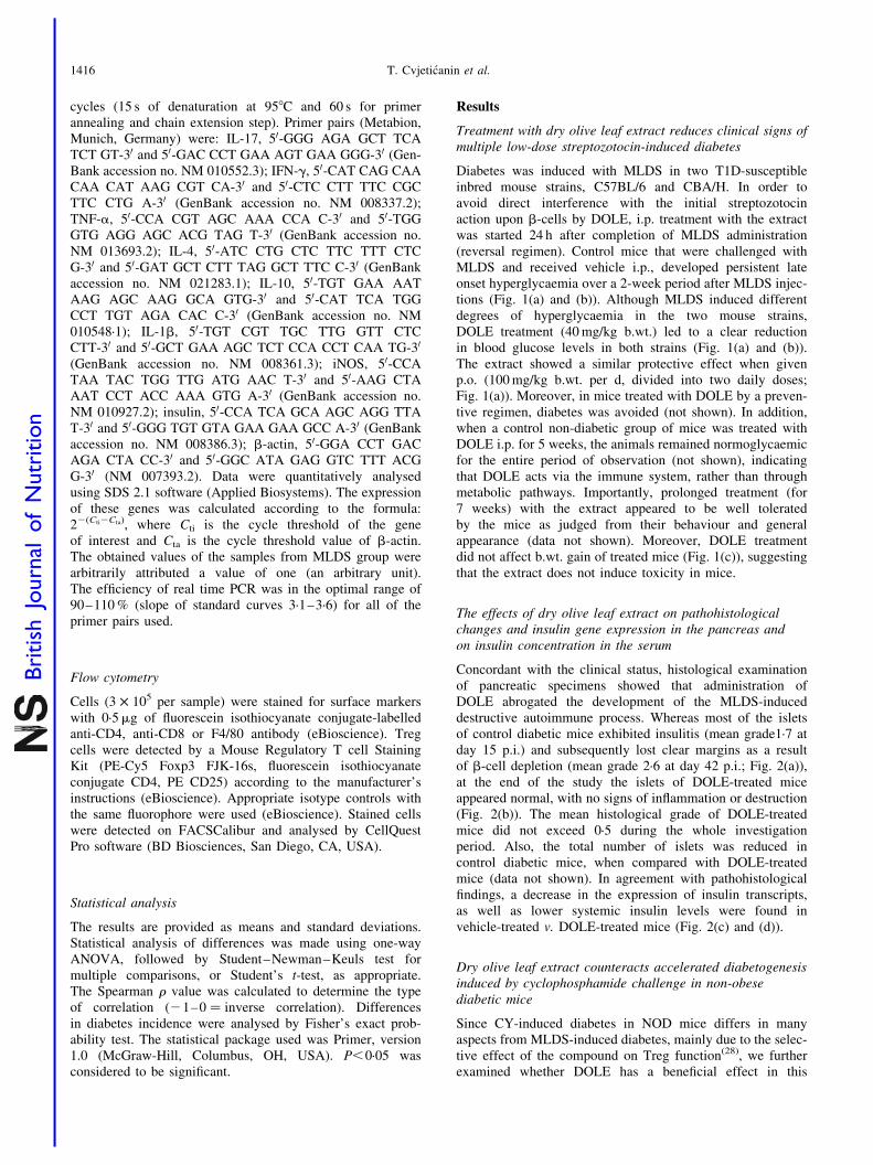
The effects of dry olive leaf extract on pathohistologicalchanges and insulin gene expression in the pancreas andon insulin concentration in the serum
Concordant with the clinical status, histological examinationof pancreatic specimens showed that administration ofDOLE abrogated the development of the MLDS-induceddestructive autoimmune process. Whereas most of the isletsof control diabetic mice exhibited insulitis (mean grade1·7 atday 15 p.i.) and subsequently lost clear margins as a resultof b-cell depletion (mean grade 2·6 at day 42 p.i.; Fig. 2(a)),at the end of the study the islets of DOLE-treated miceappeared normal, with no signs of inflammation or destruction(Fig. 2(b)). The mean histological grade of DOLE-treatedmice did not exceed 0·5 during the whole investigationperiod. Also, the total number of islets was reduced incontrol diabetic mice, when compared with DOLE-treatedmice (data not shown). In agreement with pathohistologicalfindings, a decrease in the expression of insulin transcripts,as well as lower systemic insulin levels were found invehicle-treated v. DOLE-treated mice (Fig. 2(c) and (d)).
Dry olive leaf extract counteracts accelerated diabetogenesisinduced by cyclophosphamide challenge in non-obesediabetic mice
Since CY-induced diabetes in NOD mice differs in manyaspects from MLDS-induced diabetes, mainly due to the selec-tive effect of the compound on Treg function(28), we furtherexamined whether DOLE has a beneficial effect in this
T. Cvjeticanin et al.1416
British
Journal
ofNutrition

model of autoimmune diabetogenesis, as well. As expected,approximately 80 % of the control mice treated with vehicledeveloped an acute form of diabetes within 2 weeks of theCY injections (Table 1). In contrast, DOLE treatment, giveneither i.p. or by oral gavage every day from day 1 of CY treat-ment, completely abolished the diabetogenic process, since
none of the DOLE-treated mice developed hyperglycaemia(Table 1). Concurring with the clinical findings, histologicalstudies of pancreases showed that DOLE treatment reducedthe extensive leucocyte infiltration observed in mice treated
(a) (b)
10*
(c)
(d)
8
Insu
lin m
RN
A e
xpre
ssio
n(a
rbit
rary
un
its)
Ser
um
insu
lin (
µg/l)
6
4
2
0MLDS MLDS+DOLE
MLDS MLDS+DOLE
1·8
1·6
1·4
1·2
1·0
0·8
0·6
0·4
0·2
0·0
*
Fig. 2. Histopathology and insulin expression of pancreatic islets (Panc) and
serum insulin of multiple low-dose streptozotocin (MLDS)-treated mice.
(a and b) Light micrographs showing morphological profiles of Panc by day 42
after disease induction (haematoxylin and eosin staining): (a) control MLDS-
induced and (b) dry olive leaf extract (DOLE)-treated MLDS-induced C57BL/6
mice. (c) Insulin gene expression and (d) serum insulin concentration in mice
described in Fig. 1(b), sacrificed on day 15 post induction. Gene expression
is presented in arbitrary units, and the actual value (22(Cti2Cta)) for the
gene expression was 0·013. Results from a representative experiment are
presented as the means and standard deviations for four to five mice per
group. *P,0·05 refers to corresponding control MLDS mice.
25
20
15
10
50 7
(a)
Glu
cose
(m
mo
l/l)
14 21
**
**
* *
* *
*
28 35 42 49
45
35
25
15
50 7
(b)
Glu
cose
(m
mo
l/l)
14 21
*
*
**
**
28 35 42 49
100
110
120
130
140
150
90
800 7
(c)
Bo
dy
wei
gh
t (%
ch
ang
e)
14 21
**
**
**
**
**
28Time (d)
35 42 49
Fig. 1. Effect of dry olive leaf extract (DOLE) treatment on hyperglycaemia
and body weight (b.wt.) of multiple low-dose streptozotocin (MLDS)-treated
mice. (a) Plasma glucose levels in control CBA/H mice or (b) C57BL/6 mice,
receiving MLDS (40 mg/kg per d for five consecutive days), or mice treated
with MLDS and DOLE, administered intraperitoneally (i.p.) or per orally (p.o.)
from days 5 to 21. (c) Percentage change in b.wt. from the start of the exper-
iment determined in the mice described in (a). Results from a representative
experiment are presented as the means and standard deviations for six to
seven mice per group. *P,0·05 refers to corresponding control MLDS
mice. (a) –S–, MLDS; –B–, MLDS þ i.p. DOLE; –O–, MLDS þ p.o. DOLE.
(b) –S–, MLDS; –B–, MLDS þ i.p. DOLE. (c) –S–, MLDS; –B–, MLDS þ
i.p. DOLE; –O–, MLDS þ p.o. DOLE.
Olive leaf protects against type 1 diabetes 1417
British
Journal
ofNutrition

with vehicle (Fig. 3(a) and (b)). While control mice developedsevere destructive insulitis (Fig. 3(a)), either intact isletsor stationary peri-insulitis were mostly observed in DOLE-treated mice, as exemplified in Fig. 3(b). Consistent with
these findings, DOLE-treated mice retained their capacityto secrete insulin, as revealed by higher serum insulinconcentrations in these animals (Fig. 3(c)).
In vivo treatment with dry olive leaf extract differentiallymodulates systemic and local nitric oxide production
To elucidate the mechanisms by which DOLE treatmentprevents disease onset, functional ex vivo studies were per-formed during early progression of the disease induced byCY or MLDS (days 7 and 15 of diabetes induction, respect-ively) on both peripheral compartments and the pancreas.First, we examined production of NO. In CY-induced NODmice, the production of NO by PC obtained from mice thathad received DOLE treatment p.o. was significantly up-regulated in comparison with control diabetic mice, asrevealed by nitrite formation by these cells (Fig. 4(a)).Similarly, peripheral immune cells, present in the peritonealcavity, spleen and PLN of DOLE-treated MLDS-inducedmice produced much more NO in comparison with cellsfrom vehicle-treated animals (Fig. 4(b)). In line with theseresults, iNOS expression in peripheral compartments wasalso significantly higher in mice that had received DOLE com-pared with vehicle-treated diabetic control mice (Fig. 4(c)). Incontrast, nitrite accumulation in cell culture supernatants ofthe Panc isolated from DOLE-treated mice was significantlyreduced when compared with islets of control diabetic mice(Fig. 4(b)), whereas local iNOS staining of pancreatic sectionswas barely detectable (Fig. 4(d)). Concordantly, immuno-staining for nitrotyrosine, a collective marker for oxidativeand nitrative stress, was less prominent in pancreata ofDOLE-treated v. vehicle-treated diabetic mice (Fig. 4(e)).
In vivo treatment with dry olive leaf extract induces in vitronitric oxide-mediated suppressor function of peritoneal cells
In order to examine whether DOLE-induced up-regulationof peripheral NO production influences T cell activation,we performed a suppression assay of the conventional T cellresponse to mitogen. To this, different concentrations of PCisolated from DOLE-treated or vehicle-treated MLDS-treatedmice were co-cultured with normal untreated lymph node-derived lymphocytes stimulated in vitro with concanavalinA. In contrast to PC derived from vehicle-treated mice,PC obtained from DOLE-treated animals displayed dose-dependent suppressive effects on T cell proliferation(Fig. 5(a)). Moreover, a clear negative correlation (r 20·81,P,0·05; linear regression) was observed between the dose-dependent increase in NO production (Fig. 5(b)) and theconcomitant reduction in [3H]thymidine incorporation(Fig. 5(a)). To determine whether alteration of the PC subsetsratio following DOLE treatment might contribute to theobserved suppression of T cell response, phenotype analysisof cell populations within the peritoneal cavity was performed.Administration of DOLE did not significantly alter the percen-tage of CD4þ cells (11·0 (SD 3·0) v. 9·1 (SD 2·6) for controland DOLE-treated mice, respectively) or CD8þ cells (8·3(SD 1·9) v. 7·7 (SD 2·7) for control and DOLE-treated mice,respectively), but it significantly increased the frequency ofF4/80þ cells, i.e. macrophages (43·9 (SD 1·6) v. 53·6 (SD 7·0)for control and DOLE-treated mice, respectively; P¼0·040)
(a) (b)
1·6(c)*
1·4
1·2
1·0
Ser
um
insu
lin (
µg/l)
0·8
0·6
0·4
0·2
0·0CY CY+DOLE
Fig. 3. Histopathology of pancreatic islets and serum insulin of cyclo-
phosphamide (CY)-treated mice. (a and b) Representative examples of the
light microscopic evaluation of the islets of Langerhans after 2 weeks of
CY-accelerated diabetes: (a) vehicle-treated and (b) dry olive leaf extract
(DOLE)-treated CY-accelerated non-obese diabetic (NOD) mice. (c) Serum
insulin concentration of the mice described in (a) and (b). Results from a
representative experiment are presented as the means and standard
deviations for four to five mice per group. *P,0·05 refers to corresponding
control CY-accelerated NOD mice.
Table 1. Inhibition of diabetes by dry olive leaf extract (DOLE) incyclophosphamide (CY)-treated non-obese diabetic mice (NOD)*
Diabetes incidence
Treatment with DOLE Diabetic/total % P†
DOLE p.o.‡ 0/9 0·0 0·0003DOLE i.p.§ 0/7 0·0 0·0005Non-treatedk 11/14 78·6Vehicle i.p.{ 13/16 81·2
p.o., per orally; i.p., intraperitoneally.* Diabetes was induced in euglycaemic female NOD mice by CY, as described in
‘Materials and methods’, and development of the disease was monitored within2 weeks post induction of diabetes.
† The incidence of diabetes in each group was compared using Fisher’s exactprobability test.
‡ The mice were treated with DOLE p.o. (100 mg/kg per d, divided in two dailydoses) from the first injection of CY till the end of the experiment.
§ The mice were treated with DOLE i.p. (40 mg/kg per d) from the first injection ofCY till the end of the experiment.
kThe mice were left untreated.{The mice were treated with vehicle i.p. from the first injection of CY till the end of
the experiment.
T. Cvjeticanin et al.1418
British
Journal
ofNutrition

among PC. Although there was no significant increase inthe absolute PC number at the two time points examined,a trend towards greater absolute cell number was noted inthe DOLE-treated group (Table 2), suggesting immigrationof cells into the peritoneum as an explanation for the increasein absolute cell number.
In vivo treatment with dry olive leaf extract modulatescytokine production
Because proinflammatory cytokines contribute to T1Dpathogenesis, we examined whether DOLE treatment mayinfluence MLDS-induced cytokine production in the spleen.Spleens were collected 15 d after diabetes induction, andcytokine expression and release were assayed ex vivo withoutfurther stimulation. The results clearly showed that Spl(Fig. 6(a)) isolated from DOLE-treated mice releasedsignificantly reduced amounts of proinflammatory cytokines,including IFN-g, IL-17 and TNF-a compared with control
diabetic animals. Moreover, we also evaluated cytokinegene expression in both experimental groups and found thatIFN-g expression in the spleen of DOLE-treated mice wasdecreased when compared with that in the vehicle-treatedgroup, whereas there was no significant difference betweenthe two groups regarding the other cytokines examined(Fig. 6(b)). Also, it was important to find out if DOLE hadan effect on Treg cell frequency in peripheral lymphoidtissues. To that extent the percentage of CD4þCD25þFoxP3þ
(i.e. Treg) cells among PLN cells was determined in bothMLDS- and CY-induced diabetes. DOLE did not induce asignificant alteration in Treg percentage in either theMLDS model (11·5 (SD 1·2) v. 9·9 (SD 2·2) for control andDOLE-treated mice, respectively) or the CY model (14·6(SD 0·6) v. 14·0 (SD 1·6)). Also, the absolute cellularity ofPLN was not affected by treatment with DOLE in eitherMLDS-induced diabetes (Table 2) or CY-accelerated diabetesin NOD mice (1·88 (SD 0·78) v. 1·92 (SD 0·62), in millions,for control and DOLE-treated mice, respectively).
16
Nit
rite
acc
um
ula
tio
n (
µM)
iNO
S m
RN
A e
xpre
ssio
n(a
rbit
rary
un
its)
Nit
rite
acc
um
ula
tio
n (
µM)
* *
*
*
*
(a) (b)
(c)
CY CY+DOLE
14
12
10
8
6
4
2
0
6
5
4
3
2
1
0
16
14
12
10
8
6
4
2
0
MLDS
MLDS+DOLE MLDS+DOLE
MLDS
PC Spl
(d) (e)
PLNC Panc
PC Spl PLNC
*
*
*
Fig. 4. Effect of dry olive leaf extract (DOLE) treatment on nitric oxide generation in lymphoid tissues and pancreatic islets, inducible nitric oxide synthase
(iNOS) expression in lymphoid tissues, presence of iNOS and nitrotyrosine in pancreatic islets (Panc). (a) Peritoneal cells (PC) were isolated from control and
DOLE-treated cyclophosphamide (CY)-accelerated non-obese diabetic (NOD) mice and nitrite accumulation was measured in the 48 h culture supernatants of
cells. (b) Nitrite accumulation in the 48 h culture supernatants of PC, splenocytes (Spl), pancreatic lymph node cells (PLNC) and Panc of control and DOLE-treated
multiple low-dose streptozotocin (MLDS)-induced C57BL/6 mice (A, MLDS; B, MLDS þ DOLE). (c) Inducible NO synthase (iNOS) mRNA expression in the cells
of the mice (A, MLDS; B, MLDS þ DOLE) described in (b). Gene expression is presented in arbitrary units, and the actual values (22(Cti2Cta)) for iNOS gene
expression were 0·00 087 for PC, 0·000093 for Spl and 0·000052 for PLNC (d). Immunostaining for iNOS and (e) nitrotyrosine of Panc of the mice described in
(b). (a–c) Results from one of three separate experiments with similar results are given as the means and standard deviations for five mice per group. *P,0·05
refers to corresponding control (a) CY-accelerated NOD mice, or (b and c) MLDS mice.
Olive leaf protects against type 1 diabetes 1419
British
Journal
ofNutrition

Discussion
The present study is the first demonstration that O. europaea-derived components, present in DOLE, confer protectionfrom T1D by interfering with an islet-directed autoimmuneresponse. This interference was reflected in NO-mediated sup-pression of T lymphocyte proliferation and lower productionof proinflammatory cytokines (TNF-a, IFN-g and IL-17)in the peripheral lymphoid compartments as well as lessoxidative and nitrative damage within the pancreas.
Olive tree constituents have long been known asanti-diabetic herbal agents in the metabolic syndrome and
T2D, due to their antioxidant(16) and glucose-lowering effectsassociated with insulin-sensitising activity, increased peripheralglucose utilisation and improved insulin release(25,26,34 – 37).The anti-diabetic properties of O. europaea L. leaf constituentswere evaluated in several experimental models of ‘toxic’ forms
60
50
40
30
20
10
01·25
(a)P
rolif
erat
ion
(cp
m ×
100
0)
2·5
*
5 10
25
20
15
10
5
01·25
(b)
Nit
rite
acc
um
ula
tio
n (
µM)
2·5
PC (×103 cells)
*
*
5 10
Fig. 5. In vitro suppressor function and nitric oxide generation of peritoneal
cells (PC). (a and b) PC were collected from dry olive leaf extract (DOLE)-
treated or vehicle-treated multiple low-dose streptozotocin (MLDS)-induced
mice on day 15 of diabetes post induction. These cells were co-cultured at
the indicated concentrations with 3 £ 105 cervical lymph node cells (LNC)
prepared from healthy untreated mice, along with 1mg/ml concanavalin A.
(a) Proliferation of LNC was assessed 66 h later by incorporation of [3H]-
thymidine over the last 18 h of culture. Data points show the mean thymidine
incorporation counts and standard deviations for triplicate wells (–S–, MLDS;
–B–, MLDS þ DOLE). (b) Nitrite accumulation in the culture supernatant
was measured after 48 h of cultivation. Horizontal line: LNC alone. *P,0·05
refers to corresponding control MLDS mice. cpm, Cycles per minute.
Table 2. Effect of dry olive leaf extract (DOLE) on peritoneal cell andpancreatic lymph node cell number
(Mean values and standard deviations)
Treatment group
MLDS* MLDS þ DOLE†
Mean SD Mean SD
PC‡ ( £106)/mouseExpt 1 3·8 1·9 5·5 2·3Expt 2 3·4 1·1 4·3 0·5Expt 3 3·4 2·6 4·4 1·8
PLNC‡ ( £106)/mouseExpt 1 5·3 2·4 5·5 0·8Expt 2 2·2 0·8 3·6 1·4Expt 3 3·0 1·0 3·1 1·9
MLDS, multiple low doses of streptozotocin; PC, peritoneal cells; PLNC, pancreaticlymph node cells; i.p., intraperitoneally; p.i., post induction.
* MLDS was administered i.p. to C57BL/6 mice at a daily dose of 40 mg/kg per dfor 5 d (four mice per group per experiment).
† DOLE was administered i.p. at a daily dose of 40 mg/kg per d from day 5 ofMLDS diabetes p.i. (four mice per group per expt).
‡ Cells were obtained after 14 d (Expts 1 and 2) or 38 d (Expt 3) of diabetes p.i.
2500(a)
(b)
Cyt
oki
ne
secr
etio
n (
pg
/ml)
2000
1500
500
1000
0
2
mR
NA
exp
ress
ion
(arb
itra
ry u
nit
s) 1·5
1
0·5
0IL-17 IL-6 IL-4 IL-10TNF-α IL-1β IFN-γ
*
*
*
*
Fig. 6. Effect of dry olive leaf extract (DOLE) treatment on cytokine profile in
splenocytes (Spl). Cytokine concentration in culture supernatants (A, MLDS;
B, LDS þ DOLE) (a) and cytokine gene expression (b) of Spl from vehicle-
treated and DOLE-treated multiple low-dose streptozotocin (MLDS)-induced
C57BL/6 mice after 48 h of cultivation. Gene expression is presented in arbi-
trary units, and the actual values (22(Cti2Cta)) for the analysed cytokines were
as follows: 0·000017 for IL-17; 0·000090 for IL-6; 0·0052 for TNF-a;
0·000037 for IL-4; 0·00 045 for IL-10; 0·013 for IL-1b; 0·0021 for interferon-g
(IFN-g). Data are means and standard deviations of four experiments, each
with four to five mice per group. *P,0·01 refers to corresponding control
MLDS mice.
T. Cvjeticanin et al.1420
British
Journal
ofNutrition

or dietary-induced T2D(23,24,26,34,38,39). To our knowledge, thisis the first study on the effects of O. europaea-derivedcomponents in T1D in two established mouse models: MLDS-induced immunoinflammatory diabetes; CY-accelerated NODdiabetes. We used the MLDS-induced diabetes model, as theautoimmune process is initiated by minor chemical destructionof b-cells and shedding of target autoantigens, in contrast tothe non-immune ‘toxic’ form of diabetes induced by a singlehigh dose of streptozotocin. As a consequence, immune cellsrespond to selfb-cell antigens so progression of hyperglycaemiaand insulitis evolves during a 2-week period(11,29). Alterna-tively, CY-induced diabetes was used because it is known thatthe autoimmune response directed against pancreatic b-cellsrelies on selective depletion of Treg cells(28). We showed thatin both models, DOLE efficiently counteracted diabetogenesis,indicating that the suppressive effect on the islet-directedT cell immune response is most likely mediated by commonevents and independently of Treg cells. In support of this isour finding that there was no difference in the frequency ofTreg cells within PLN in vehicle- and DOLE-treated micein which diabetes was induced with CY. The common effect ofDOLE in MLDS- and CY-induced diabetes was an increase ofNO generation in peripheral lymphoid tissues. This finding isof importance for understanding the beneficial effect of DOLEin diabetes, as it is well known that NO produced in lymphoidtissues has immunoregulatory functions, including anti-proli-ferative, pro-apoptotic and cytokine-shifting effects on T cellsduring their activation(40 – 42). In addition, PC isolated fromDOLE-treated mice, but not PC isolated from vehicle-treatedmice, were able to inhibit proliferation of mitogen-activatedT cells. Importantly, the observed inhibition was inversely pro-portional to the level of NO produced in these co-cultures. Onepossible explanation for the enhanced capacity of PC to produceNO is the observed increase in frequency of cells responsible forits production among PC, i.e. of F4/80þ macrophages. Together,our findings suggest that DOLE acts through up-regulation ofNO in peripheral lymphoid tissues, which consequently leadsto limitation of T cell activation and finally to restriction of theautoimmune attack upon pancreatic b-cells. The capacity ofDOLE to up-regulate NO generation by lymphoid cells is inaccordance with the previously reported amplifying effects ofthe O. europaea bioactive component, oleuropein, on the releaseof NO and the expression of iNOS by macrophages during theresponse to endotoxin challenge in vitro (20). However, severalstudies showed that O. europaea-derived components, such asflavonoids and phenols, are capable of inhibiting inflammationthrough inhibition of NO production and increasing antioxidantenzyme activity(22,23,35,36). Although, we do not report inhi-bition of NO production in peripheral compartments by DOLEtreatment, we do show that DOLE inhibits NO synthesis in pan-creatic b-cells. This again is very important for the anti-diabeto-genic effect of DOLE, as it is recognised that NO released inPanc is mostly cytotoxic and leads to dysfunction and destruc-tion of insulin-producing cells(43,44). Damaging effects of NOmay be attributable to the reaction with superoxide anions toyield peroxynitrite, a potent nitrating and nitrosylating agent.Since oxidative stress in diabetes coexists with reducedantioxidant status of the pancreas, which can further increasethe deleterious effects of free radicals, it is tempting tospeculate that DOLE has a protective effect in diabetes atleast partly, through decreasing local oxidative and nitrative
damage. Indeed, reduced immunostaining of DOLE-treatedPanc for nitrotyrosine, a collective marker for reactiveoxygen species and reactive nitrogen species(45), furthersupported this assumption.
The other important factor influenced by DOLE is the cyto-kine profile of Spl. We showed that, under the influence ofthe extract, typical pro-inflammatory cytokines IFN-g,TNF-a and IL-17 were down-regulated in MLDS-induceddiabetes. On the contrary, anti-inflammatory cytokine (IL-4and IL-10) generation was unaffected by DOLE. Therefore,it seems that DOLE promotes a profile shift of Spl cytokineproduction from pro-inflammatory to anti-inflammatory, orfrom Th1/Th17 to Th2. The importance of Th1 cells andIFN-g, as well as of TNF-a for autoimmune diabetes pathol-ogy has been described in many papers(44). According to thecurrent understanding of T1D pathogenesis, initial injury tothe islets leads to the presentation of exposed autoantigenson antigen-presenting cells to autoreactive T cells within theregional lymph nodes. The antigen-presenting cells stimulateeffector Th1 cells which produce IFN-g, and this cytokineactivates additional macrophages and polymorphonuclearcells. These cells release IL-1b, TNF-a and NO, which aredirectly toxic to b-cells. Moreover, the antigen-presentingcells promote CD8þT cells which use FasL, perforin/gran-zyme and TNF-a to trigger b-cell apoptosis(44). The role ofTh17 cells and their signature cytokine IL-17 in diabetes isstill not clear. It has been suggested that Th17 cells are import-ant for the development of diabetes in NOD mice and thatIL-17-directed therapy could be useful in the treatment ofthe disease(46). It has also been shown that islet-reactiveTh17 cells primarily function by promoting inflammation,and that their conversion to Th1 cells in lymphopenic hostsresults in diabetes(47). Importantly, IFN-g seems indispensablefor Th17-induced diabetes, whereas Th17 cells play an acces-sory role by promoting inflammation produced by Th1 orCD8þT cells. In the line with these results, in both MLDS-induced and human diabetes, inflammatory infiltrates are com-posed of both IFN-g- and IL-17-producing cells(12,48,49).Moreover, it was suggested that IL-17 cooperates withTNF-a in inflammation associated with autoimmunity(50).Having in mind all of the findings mentioned above, inhibitionof IFN-g, TNF-a and IL-17 by DOLE in regional lymphoidtissue, the place of initiation and perpetuation of the diabeto-genic process, seem to be a valid explanation for the beneficialeffect of the extract in T1D. Importantly, we have recentlydemonstrated the efficiency of DOLE in experimental auto-immune encephalomyelitis, an animal model of multiplesclerosis(27). There, the application of the extract also led todown-regulation of IFN-g and IL-17 production during thedisease course. Thus, it seems that DOLE counteractspathogenic autoimmune responses dependent on IFN-g andIL-17-producing cells.
The increasing interest in olive tree (O. europaea) constitu-ents is mainly attributed to its antioxidant effects, due tothe high concentration of phenolic compounds. In additionto their activities as antioxidants, some phenolics havebeen shown to possess anti-inflammatory activity(22). Thisis supported by experiments in which phenolic compoundssignificantly decreased the production of pro-inflammatorymediators, such as TNF-a, IL-1b and PGE2
(21,51 – 53). FeedingNOD mice diets rich in polyphenols derived from grape was
Olive leaf protects against type 1 diabetes 1421
British
Journal
ofNutrition

recently found to decrease the autoimmune inflammatoryprocess associated with T1D(53). Here we add that DOLE,rich in phenolic compound oleuropein inhibits inflammationin T1D and protects mice from disease progression. Twoobservations of particular relevance from the clinical pointof view are the apparently low toxicity of DOLE, as demon-strated by the lack of characteristic b.wt. loss that occursin MLDS-induced diabetes, and the equal efficacy of theextract given either i.p. or p.o. to prevent the disease. There-fore, our finding that DOLE applied orally to mice suppressedT1D, show that oral administration of phenolic compoundscould be a feasible way of treating subjects suffering fromdiabetes. However, the proper evaluation of DOLE efficiencyin the treatment of patients with newly diagnosed T1Dwill require preclinical studies in animals with establisheddisease carried out under a therapeutic regimen of application.Nonetheless, the capacity of DOLE to counteract the dia-betogenic effects of MLDS even when applied as a reversalregimen, started 1 d after the last toxin dose had beengiven, is encouraging for translation of these findings to aclinical setting.
Regarding DOLE, it has to be emphasised that it is amixture of various chemicals with potential bioreactivity(14).Therefore, the putative immunomodulating effects of otherprimary and secondary compounds of DOLE cannot beexcluded. Indeed, oleanolic, ursolic and maslinic acids, threemain olive leaf triterpenic compounds(54) were recentlyshown to contribute to the anti-diabetic effect of oliveleaves(24,55,56). Further, although as a consequence of gentleprocessing of olive leaf constituents the phenolic componentof DOLE contains almost exclusively oleuropein, it has tobe understood that the extract constituents are extensivelymetabolised in the body. As a consequence, the singlephenol products of oleuropein breakdown tyrosol and hydro-xytyrosol may contribute to its health-promoting properties,as well. In line with such a possibility, numerous studieshave clearly demonstrated that these compounds exhibitbeneficial health effects in various oxidative stress associateddisorders due to their antioxidant and anti-inflammatoryactivities(22,35,36,57,58). Therefore, the effect of DOLE onT1D should not be attributed only to the phenolic fraction –oleuropein, but also to other components, such as triterpeniccompounds (oleanolic acid, maslinic and ursolic acid), tanninsand flavonoids, as well as to oleuropein metabolites. To thatextent, it is of importance to investigate the individual effectof each compound on the T1D course. However, the obviousadvantage of DOLE is that it is an easily attainable productof olive leaves, not requiring purification of any fractions ofthe extract before application in T1D models, and possiblyfor human subjects.
In conclusion, we have provided evidence that oliveleaf constituents are effective inhibitors of T1D. The resultsalso suggest that the inhibitory effect of DOLE may be due,at least in part, to the NO-mediated suppression of T lympho-cyte proliferation and lower production of proinflammatorycytokines (IFN-g, IL-17 and TNF-a) in the spleen, andsubsequent blockade of b-cell destruction. Though the activecomponents and precise mode of action of DOLE are notyet ascertained, its powerful anti-diabetogenic activity, alongwith the safety profile observed in vivo, suggest that itwould be worth undertaking additional studies to explore
the possibility that DOLE possesses the desired therapeuticproperties applicable to human T1D.
Acknowledgements
The study was supported by the Serbian Ministry of Science(grant 143029B).The contributions of the authors were asfollows: T. C. and I. S. carried out in vivo treatments,conducted glucose, insulin and cytokine analyses, performedreal-time PCR and conducted gene expression analyses.D. M. conducted flow cytometry analysis, contributed todata analysis and helped to write the manuscript. D. D.planned and carried out the feeding experiments. S. S.-G.designed the study, performed the cell proliferation assay,conducted histology and immunohistochemistry analyses,coordinated the project and wrote the manuscript. The authorswould like to thank Bosko Milovanovic, GeographicalInstitute – Serbian Academy of Sciences and Arts for thehelp with statistical analysis of our data. None of the authorshas any conflicts of interest.
References
1. Gianni C, Mohn A & Chiarelli F (2009) Technology and the
issue cost/benefit in diabetes. Diabetes Metab Res Rev 25,
Suppl. 1, S34–S44.
2. Craig ME, Hattersley A, Donaghue TC, et al. (2009) ISPAD
clinical practice consensus guidelines 2009 compendium.
Definition, epidemiology and classification of diabetes in
children and adolescents. Pediatr Diabetes 10, Suppl. 12,
3–12.
3. Tisch R & McDevitt H (1996) Insulin-dependent diabetes
mellitus. Cell 85, 291–297.
4. Daaboul J & Schatz D (2003) Overview of prevention and
intervention trials for type 1 diabetes. Rev Endocr Metab Dis
4, 317–323.
5. Watson S & Miller K (2004) Encyclopedia of the Human
Body: The Endocrine System. Westport, CT: Greenwood Pub-
lishing.
6. Mastrandrea L, Yu J, Behrens T, et al. (2009) Etanercept
treatment in children with new-onset type 1 diabetes. Pilot
randomized, placebo-controlled, double-blind study. Diabetes
Care 32, 1244–1249.
7. Staeva-Vieira T, Peakman M & von Herrath M (2007)
Translational mini-review series on type 1 diabetes: immune-
based therapeutic approaches for type 1 diabetes. Clin Exp
Immunol 148, 17–31.
8. Kawano Y, Irie J, Nakatani H, et al. (2009) Pioglitazone might
prevent the progression of slowly progressive type 1 diabetes.
Inter Med 48, 1037–1039.
9. Rother KJ, Brown RJ, Morales MM, et al. (2009) Effect of
ingested interferon-a on b-cell function in children with new
onset type 1 diabetes. Diabetes Care 32, 1250–1255.
10. Shoda LK, Young DL, Ramanujan S, et al. (2005) A compre-
hensive review of interventions in the NOD mouse and
implications for translation. Immunity 23, 115–126.
11. Stosic-Grujicic S, Cvetkovic I, Mangano K, et al. (2007)
A potent immunomodulatory compound (S,R)-3-phenyl-4,
5-dihydro-5-isoxasole acetic acid, prevents spontaneous and
accelerated forms of autoimmune diabetes in NOD mice and
inhibits the immunoinflammatory diabetes induced by multiple
low doses of streptozotocin in CBA/H mice. J Pharmacol Exp
Ther 320, 1038–1049.
T. Cvjeticanin et al.1422
British
Journal
ofNutrition

12. Stosic-Grujicic S, Cvjeticanin T & Stojanovic I (2009)
Retinoids differentially regulate the progression of autoimmune
diabetes in three preclinical models in mice. Mol Immunol 47,
79–86.
13. Perona JS, Cabello-Moruno R & Ruiz-Gutierrez V (2006)
The role of virgin olive oil components in the modulation of
endothelial function. J Nutr Biochem 17, 429–445.
14. Dekanski D, Janicijevic-Hudomal S, Tadic V, et al. (2009)
Phytochemical analysis and gastroprotective activity of an
olive leaf extract. J Serb Chem Soc 74, 367–377.
15. Ramırez-Tortosa MC, Suarez A, Gomez MC, et al. (1999)
Effect of extra-virgin olive oil and fish-oil supplementation on
plasma lipids and susceptibility of low-density lipoprotein to
oxidative alteration in free-living Spanish male patients with
peripheral vascular disease. Clin Nutr 18, 167–174.
16. Vissers MN, Zock PL & Katan MB (2004) Bioavailability and
antioxidant effects of olive oil phenols in humans: a review.
Eur J Clin Nutr 58, 955–965.
17. Esposito K, Ciotola M & Giugliano D (2007) Mediterranean
diet and the metabolic syndrome. Mol Nutr Food Res 51,
1268–1274.
18. Fito M, de la Torre R, Farre-Albaladejo M, et al. (2007)
Bioavailability and antioxidant effects of olive oil phenolic com-
pounds in humans: a review. Ann Ist Super Sanita 43, 375–381.
19. Panagiotakos DB & Polychronopoulos E (2005) The role of
Mediterranean diet in the epidemiology of metabolic syndrome;
converting epidemiology to clinical practice. Lipids Health Dis 4, 7.
20. Visioli F, Bellosta S & Galli C (1998) Oleuropein, the bitter
principle of olives, enhances nitric oxide production by mouse
macrophages. Life Sci 62, 541–546.
21. Carluccio MA, Siculella L, Ancora MA, et al. (2003) Olive oil
and red wine antioxidant polyphenols inhibit endothelial
activation: antiatherogenic properties of Mediterranean diet
phytochemicals. Arterioscler Thromb Vasc Biol 23, 622–629.
22. Bitler CM, Viale TM, Damaj B, et al. (2005) Hydrolyzed olive
vegetation water in mice has anti-inflammatory activity. J Nutr
135, 1475–1479.
23. Al-Azzawie HF & Alhamdani MS (2006) Hypoglycemic and
antioxidant effects of oleuropein in alloxan-diabetic rabbits.
Life Sci 78, 1371–1377.
24. Sato H, Genet C, Strehle A, et al. (2007) Anti-hyperglycemic
activity of TGR5 agonist isolated from Olea europaea. Biochem
Biophys Res Commun 362, 793–798.
25. Said O, Fulder S, Khalil K, et al. (2008) Maintaining a physio-
logical blood glucose level with ‘glucolevel’, a combination of
four anti-diabetes plants used in the traditional Arab herbal
medicine. Evid Based Complement Alternat Med 5, 421–428.
26. Eidi A, Eidi M & Darzi R (2009) Antidiabetic effect of Olea
europea L. in normal and diabetic rats. Phytother Res 23,
347–350.
27. Miljkovic DJ, Dekanski D, Miljkovic Z, et al. (2009) Dry
olive leaf extract ameliorates experimental autoimmune
encephalomyelitis. Clin Nutr 28, 346–350.
28. Brode S, Raine T, Zaccone P, et al. (2006) Cyclophosphamide-
induced type-1 diabetes in the NOD mouse is accelerated with a
reduction of CD4þCD25þFoxp3þ regulatory T cells. J Immunol
177, 6603–6612.
29. Like AA & Rossini AA (1976) Streptozotocin-induced
pancreatic insulitis: new model of diabetes mellitus. Science
193, 415–417.
30. Bach JF (1995) Insulin-dependent diabetes mellitus as a
beta-cell targeted disease of immunoregulation. J Autoimmun
8, 439–463.
31. Cvetkovic I, Al-Abed Y, Miljkovic D, et al. (2005) Critical
role of macrophage migration inhibitory factor activity in
experimental autoimmune diabetes. Endocrinology 146,
2942–2951.
32. Perrinjaquet-Moccetti T, Busjahn A, Schmidlin C, et al. (2008)
Food supplementation with an olive (Olea europaea L.) leaf
extract reduces blood pressure in borderline hypertensive
monozygotic twins. Phytother Res 22, 1239–1242.
33. Rucker RB (2007) Allometric scaling, metabolic body size and
interspecies comparisons of basal nutritional requirements.
J Anim Physiol Anim Nutr (Berl) 91, 148–156.
34. Gonzalez M, Zarzuelo A, Gamez MJ, et al. (1992) Hypo-
glycemic activity of olive leaf. Planta Med 58, 513–515.
35. Briante R, Patumi M, Terenziani S, et al. (2002) Olea europea
L. leaf extract and derivatives: antioxidant properties. J Agric
Food Chem 50, 4934–4940.
36. Visioli F, Poli A & Galli C (2002) Antioxidant and other
biological activities of phenols from olives and olive oil. Med
Res Rev 22, 65–75.
37. Jemai H, Bouaziz M, Fki I, et al. (2008) Hypolipidimic
and antioxidant activities of oleuropein and its hydrolysis
derivative-rich extracts from Chemlali olive leaves. Chem Biol
Interact 176, 88–98.
38. Bennani-Kabchi N, Fdhil H, Cherrah Y, et al. (2000)
Therapeutic effect of Olea europea var. oleaster leaves on
carbohydrate and lipid metabolism in obese and prediabetic
sand rats (Psammomys obesus). Ann Pharm Fr 58, 271–277.
39. Coskun O, Kanter M, Korkmaz A, et al. (2005) Quercetin, a
flavonoid antioxidant, prevents and protects streptozotocin-
induced oxidative stress and b-cell damage in rat pancreas.
Pharmacol Res 51, 117–123.
40. Albina JE, Abate JA & Henry WL Jr (1991) Nitric oxide pro-
duction is required for murine resident peritoneal macrophages
to suppress mitogen-stimulated T cell proliferation. Role of
IFN-gamma in the induction of the nitric oxide-synthesizing
pathway. J Immunol 147, 144–148.
41. Bogdan C (2001) Nitric oxide and the immune response.
Nat Immunol 2, 907–916.
42. Lemaire G, Guittet O, Vesin MF, et al. (2009) Glutathione
depletion reveals impairment of antigen processing and inhi-
bition of cathepsin activity by nitric oxide in antigen-presenting
cells. Mol Immunol 46, 1100–1108.
43. Stassi G, De Maria R, Trucco G, et al. (1997) Nitric oxide
primes pancreatic beta cells for Fas-mediated destruction in
insulin-dependent diabetes mellitus. J Exp Med 186,
1193–1200.
44. Pearl-Yafe M, Kaminitz A, Yolcu ES, et al. (2007) Pancreatic
islets under attack: cellular and molecular effectors. Curr
Pharm Des 13, 749–760.
45. Ischiropoulos H, Zhu L, Chen J, et al. (1992) Peroxynitrite-
mediated tyrosine nitration catalyzed by superoxide dismutase.
Arch Biochem Biophys 298, 431–437.
46. Emamaullee JA, Davis J, Merani S, et al. (2009) Inhibition
of Th17 cells regulates autoimmune diabetes in NOD mice.
Diabetes 58, 1302–1311.
47. Martin-Orozco N, Chung Y, Chang SH, et al. (2009) Th17 cells
promote pancreatic inflammation but only induce diabetes
efficiently in lymphopenic hosts after conversion into Th1
cells. Eur J Immunol 39, 216–224.
48. Miljkovic D, Cvetkovic I, Momcilovic M, et al. (2005) Inter-
leukin-17 stimulates inducible nitric oxide synthase-dependent
toxicity in mouse beta cells. Cell Mol Life Sci 62, 2658–2668.
49. Tesmer LA, Lundy SK, Sarkar S, et al. (2008) Th17 cells in
human disease. Immunol Rev 223, 87–113.
50. Hartupee J, Liu C, Novotny M, et al. (2009) IL-17 signaling for
mRNA stabilization does not require TNF receptor-associated
factor 6. J Immunol 182, 1660–1666.
51. Blonska M, Czuba ZP & Krol W (2003) Effect of flavone
derivatives on interleukin-1 beta (IL-1 beta) mRNA expression
and IL-1 beta protein synthesis in stimulated RAW 264.7
macrophages. Scand J Immunol 57, 162–166.
Olive leaf protects against type 1 diabetes 1423
British
Journal
ofNutrition

52. Miles EA, Zoubouli P & Calder PC (2005) Differential anti-
inflammatory effects of phenolic compounds from extra virgin
olive oil identified in human whole blood cultures. Nutrition
21, 389–394.
53. Zunino SJ, Storms DH & Stephensen CB (2007) Diets rich in
polyphenols and vitamin A inhibit the development of type 1
autoimmune diabetes in nonobese diabetic mice. J Nutr 137,
1216–1221.
54. Sanchez-Avila N, Priego-Capote F, Ruiz-Jimenez J, et al.
(2009) Fast and selective determination of triterpenoic com-
pounds in olive leaves by liquid chromatography–tandem
mass spectrometry with multiple reaction monitoring after
microwave-assisted extraction. Talanta 78, 40–48.
55. Jang S-M, Yee S-T, Choi J, et al. (2009) Ursolic acid enhances
the cellular immune system and pancreatic beta-cell function
in streptozotocin-induced diabetic mice fed a high-fat diet.
Int Immunopharmacol 9, 113–119.
56. Liu J, Sun H, Duan W, et al. (2007) Maslinic acid reduces blood
glucose in KK-Ay mice. Biol Pharm Bull 30, 2075–2078.
57. Lory D, Incani A, Deiana M, et al. (2009) Protective effect of
hydroxytyrosol and tyrosol against oxidative stress in kidney
cells. Toxic Ind Health 25, 301–310.
58. Andrikopoulos NK, Kaliora AC, Assimopoulou AN, et al. (2002)
Inhibitory activity of minor polyphenolic and nonpolyphenolic
constituents of olive oil against in vitro low-density lipoprotein
oxidation. J Med Food 5, 1–7.
T. Cvjeticanin et al.1424
British
Journal
ofNutrition