European Integration through Gaullism and Europeanism, STUDIA EUROPAEA, 2006
Upload
tonekaboniauCategory
view
0download
0
Spectrochimica Acta Part A: Molecular and Biomolecular Spectroscopy 127 (2014) 61–66
Contents lists available at ScienceDirect
Spectrochimica Acta Part A: Molecular andBiomolecular Spectroscopy
journal homepage: www.elsevier .com/locate /saa
Green synthesis of silver nanoparticles using Delphinium denudatum rootextract exhibits antibacterial and mosquito larvicidal activities
http://dx.doi.org/10.1016/j.saa.2014.02.0301386-1425/� 2014 Elsevier B.V. All rights reserved.
⇑ Corresponding author. Tel.: +91 44 27264066x14.E-mail address: [email protected] (G. Suresh).
Gopal Suresh a,⇑, Poosali Hariharan Gunasekar a, Dhanasegaran Kokila a, Durai Prabhu b,Devadoss Dinesh b, Nagaiya Ravichandran c, Balasubramanian Ramesh d, Arunagirinathan Koodalingam d,Ganesan Vijaiyan Siva c
a Department of Microbiology, Sri Sankara Arts & Science College, Enathur, Kanchipuram 631 561, Tamilnadu, Indiab Department of Zoology, University of Madras, Guindy Campus, Chennai 600 025, Tamilnadu, Indiac Department of Biotechnology, University of Madras, Guindy Campus, Chennai 600 025, Tamilnadu, Indiad Department of Biotechnology, Sri Sankara Arts & Science College, Enathur, Kanchipuram 631 561, Tamilnadu, India
h i g h l i g h t s
� Green synthesis of silver nanoparticlewas performed using Delphiniumdenudatum extract.� The synthesized silver nanoparticle
was characterized.� Nanoparticle shows antibacterial
activity against human pathogens.� Potent larvicidal activity was noticed
against mosquito Aedes aegypti.
g r a p h i c a l a b s t r a c t
a r t i c l e i n f o
Article history:Received 30 November 2013Received in revised form 25 January 2014Accepted 9 February 2014Available online 22 February 2014
Keywords:Delphinium denudatumGreen synthesisNanoparticlesAntibacterialMosquito larvicidal
a b s t r a c t
Green synthesis of silver nanoparticles (AgNPs) using aqueous root extract of Delphinium denudatum (Dd)by reduction of Ag+ ions from silver nitrate solution has been investigated. The synthesized DdAgNPswere characterized by using UV–Vis spectroscopy, X-ray diffraction (XRD), Field emission scanning elec-tron microscope (FESEM) and Fourier transform infrared spectroscopy (FTIR). The prepared DdAgNPsshowed maximum absorbance at 416 nm and particles were polydispersed in nature, spherical in shapeand the size of the particle obtained was 685 nm. The DdAgNPs exhibited antibacterial activity againstStaphylococcus aureus ATCC 6538, Bacillus cereus NCIM 2106, Escherichia coli ATCC 8739 and Pseudomonasaeruginosa ATCC 9027. The DdAgNPs showed potent larvicidal activity against second instar larvae ofdengue vector Aedes aegypti with a LC50 value of 9.6 ppm.
� 2014 Elsevier B.V. All rights reserved.
Introduction
Nanotechnology concerns with the development of experimen-tal processes for the synthesis of nanoparticles of different sizes,
shapes and controlled disparity. This provides an efficient controlover many of the physical and chemical properties with variouspotential applications including pharmaceuticals and medicine[1]. To date, a metallic nanoparticle are mostly prepared fromnoble metals viz., platinum (Pt), gold (Au), silver (Ag) and led(Pb), among those Ag is the metal of choice in the field of biologicalsystem [2]. Green synthesis of nanoparticles is an emerging branch

62 G. Suresh et al. / Spectrochimica Acta Part A: Molecular and Biomolecular Spectroscopy 127 (2014) 61–66
of nanotechnology in which environmentally benign materials likeplant leaf extract, bacteria and fungi where used for the synthesis.It offers numerous benefits of eco-friendliness and compatibilityfor pharmaceutical and biomedical applications as they do notuse toxic chemicals in the synthesis protocols [3].
Disease causing microbes that have become resistant to drugtherapy are an increasing public health problem. Therefore thereis an urgent need to develop new bactericides. The antibacterialeffects of Ag salts have been noticed since antiquity and Ag is cur-rently used to control bacterial growth in a variety of applications,including dental work, catheters, and burn wounds. In fact, it is wellknown that Ag ions and Ag-based compounds are highly toxic tomicrobes, showing strong biocidal effects [4]. Mosquitoes areimportant vectors of human diseases, especially in the tropics as itkills millions of people every year [5]. Still mosquitoes are impor-tant vector insect in public health viewpoint owing to the problemsassociated with chemical insecticides, including toxicity to non-target organisms, environmental and human health concerns andunavailability of vaccines for many mosquito borne diseases [6,7].Control of mosquito populations is the only available option toreduce the incidence of vector-borne diseases like malaria, filariasis,dengue and chikungunya in many tropical countries especially India[5,8]. The Ag NPs which are less likely to cause ecological damagehave been identified as a potential replacement of syntheticchemical insecticides, hence the need to use green synthesized AgNPs for the control of disease vectors.
The Delphinium denudatum Wall, popularly known as Jadwar,family Ranunculacease, is one of the most important drugs usedin indigenous medicine in India especially in Unani system. Theroots are bitter and reported to be useful in a variety of ailments;as antidote for aconite poisoning, antiepileptic, antimicrobial,opium deaddiction, diuretic, analgesic, antipyretic, cardio, hepato-cellular protective, stimulant, tonic, treated for hemorrhoid, insan-ity, tooth aches, rheumatism, snake bite, etc. [9]. However, metalreducing property and anti-mosquito property of D. denudatumare not known. Hence, the present study is designed to greensynthesize of silver nanoparticles using aqueous extract ofD. denudatum, characterization of synthesized nanoparticles andto investigate the possible antibacterial and mosquito larvicidalactivities.
Materials and methods
Collection of plant sample
The dried roots of D. denudatum were procured from authorizedsuppliers from Hyderabad. The identity and authenticity of D.denudatum roots was confirmed with the help of taxonomists fromCentre for Advance Studies in Botany, University of Madras,Chennai.
Bacterial strains
The bacterial strains Staphylococcus aureus ATCC 6538, Bacilluscereus NCIM 2106, Escherichia coli ATCC 8739, Pseudomonasaeruginosa ATCC 9027 were gifted from Christian Medical College,Vellore. The strains were maintained on nutrient agar slants at4 �C.
Rearing mosquito larvae
The Aedes ageypti mosquitoes were reared as described inKoodalingam et al. [10], and maintained in Department of Biotech-nology, Sri Sankara Arts and Science College, Kanchipuram. Thelarvae were maintained in plastic bowl containing unchlorinated
tap water and fed with powdered dog biscuit and baker’s yeast(3:1 ratio).
Hot water extraction
The D. denudatum roots were washed with sterile double dis-tilled water to remove the surface contamination and were shadedried for ten days. The dried plant material was powdered by usingmechanical grinder. 4 g of fine powder was taken and mixed with40 ml of sterile double distilled water and kept in water bath at60 �C for 2 h. The slurry is then filtered by Whatman No. 1 filterpaper. The filtered extract was stored in refrigerator at 4 �C forfurther studies.
Biosynthesis of silver nanoparticles
For the biosynthesis of silver nanoparticles, 1.5 ml of plantextract was mixed with 30 ml of 1 mM aqueous silver nitrate solu-tion and incubated at room temperature in dark condition for 2 h.The bio-reduction of the silver ions in the solution was monitoredperiodically by measuring the UV–Vis spectroscopy (200–800 nm)of the solutions. The formation of a yellowish brown-colored solu-tion indicated the formation of the silver nanoparticles. The silvernanoparticles obtained from the solution were purified by re-peated centrifugation at 12,000 rpm for 20 min followed by disper-sion of the pellet in sterile deionized water three times to removethe water-soluble biomolecules such as proteins and secondarymetabolites. The water–suspended nanoparticles were lyophilized.After freeze dried silver nanoparticles were used to characterizethe structure and composition.
Characterization of green synthesized silver nanoparticals
UV–Vis absorption spectra were measured using a Cecil spec-trophotometer (CE7001). Crystalline metallic silver nanoparticleswere examined using an X-ray diffractometer (Phillips 1830) oper-ated at a voltage of 40 kV and a current of 30 mA with Cu Ka1 radi-ation in h–2h configurations. All XRD data were collected under theexperimental conditions in the angular range 3� 6 2h 6 50�. Thecrystalline silver nanoparticle was calculated from the width ofthe XRD peaks, using the Debye–Scherrer formula D = 0.94k/bcosh(where D is the mean diameter of nanoparticles, k is wavelengthof X-ray radiation source and b is the angular FWHM of the XRDpeak at the diffraction angle h). FT-IR spectra for root extract ofD. denudatum powder and silver nanoparticles were obtained inthe range 4000–450 cm�1 with Perkin–Elmer Spectrum One-FT-IR 4200 spectrophotometer, by KBr pellet method. Field emissionscanning electron microscope (FESEM) analysis of synthesized sil-ver nanoparticles was done using a (HITACHI SU6600).
Antibacterial analysis
Agar well diffusion methodThe preliminary antibacterial activities of DdAgNPs were
employing the Mueller Hinton agar (MHA) well diffusion methodat various concentrations such as 100, 250, 500 lg/well. Sterile1 mM AgNO3 solution was used as blank exhibited no activityagainst any of the used organisms. Amoxicillin disc (10 lg) wasused as standard drug. The minimum inhibitory concentrationagainst the same microbes used in the preliminary screening wascarried out using micro dilution susceptibility method [11].
Micro-broth dilution methodEach well of the microtitre plate, containing 100 ll of the
respective DdAgNPs solution (dilute with sterile Muller Hintonbroth) followed was added with test bacterial suspension (5 ll;

Fig. 1. (A) UV–Vis spectra recorded as a function of the reaction time for thereaction of 1 mM AgNO3 solution with Delphinium denudatum root extracts. (B)Inset photographs showing color variations after different reaction time intervals.
G. Suresh et al. / Spectrochimica Acta Part A: Molecular and Biomolecular Spectroscopy 127 (2014) 61–66 63
1 � 108 CFU/ml). Each batch included a growth control well (noantimicrobial agent) and a negative control well (un-inoculated).After inoculation, each tray was covered with a lid to prevent evap-oration during incubation. The micro dilution trays were incubatedat 37 �C for 16–20 h in ambient incubator prior to reading. MIC wasdetermined based on the growth observed in the lowest dilution ofthe well after streaking on MHA plates [11].
Mosquito larvicidal assay
The toxicity of silver nanoparticles synthesized using D. denud-atum root extract was evaluated by small scale mosquito bioassaymethod using 12 well tissue culture plate (Tarsons, India). 10 earlysecond instar larvae were placed in each well of tissue culture platecontaining 3 ml of test medium. The test medium was prepared byadding various concentrations of silver nanoparticles in unchlori-nated tap water. The larvae were tested against initial concentra-tions of DdAgNPs such as 10, 100 and 1000 ppm. Control wellreceived 3 ml of tap water alone without addition of silver nano-particles. Larval food (yeast and dog biscuit) was added to eachwell. The tissue culture plates were incubated at room temperature(26 ± 2 �C), water loss was compensated by addition of distilledwater. Larval mortality was recorded after 24 and 48 h exposureperiods.
Fig. 2. The XRD pattern of DdAgNPs.
Results and discussion
Green synthesis of nano-scale silver particles
The development of easy, reliable and eco-friendly methodshelps to increase interest in the synthesis and application of nano-particles that are beneficial for mankind [12]. The reduction of sil-ver nitrate into AgNPs during exposure to plant extracts is followedby a gradual increase in color development from clear to yellowishbrown, as a result of the surface plasmon resonance (SPR) phenom-enon. The root extracts without AgNO3 did not show any change incolor. UV–Vis spectroscopy is one of the most widely used tech-nique for structural characterization of silver nanoparticles. Thepale brown color developed with increase of incubation time.The absorption spectrum of the pale brown color silver colloidsshowed a notable peak at 416 nm, indicating the presence oflone spherical or roughly spherical AgNPs (Fig. 1). The optical
absorption spectra of metal nanoparticles are dominated by SPR,which shift to longer wavelengths with increasing particle size[13]. The position and shape of plasmon absorption of silvernanoclusters are strongly dependent on the particle size, dielectricmedium, and surface-adsorbed species [14,15]. According to Mie’stheory [16], only a single SPR band is expected in the absorptionspectra of spherical nanoparticles, whereas anisotropic particlescould give rise to two or more SPR bands depending on the shapeof the particles. The number of SPR peaks increases as the symme-try of the nanoparticle decreases [17]. The result obtained in thisinvestigation is very interesting in terms of the identification ofpotential hill station plant for the synthesis of silver nanoparticles.Significant color change before two hours indicated the fast reduc-tion of the AgNO3 by the aqueous root extract. Shankar et al. [18]reported the rapid biosynthesis of stable silver, gold and bi-metal-lic Ag/Au core shell nanoparticles with 20 g of Azadirachta indicaleaf biomass and 1 mM aqueous AgNO3, with a 90% reduction ofthe metal ions within 4 h. The dissimilarity in the rates of biore-duction observed may be due to the differences in the activitiesof the enzymes present in the A. indica and D. denudatum aqueousleaf and root extracts, respectively.
X-ray diffraction (XRD) analysis
The XRD pattern showed thirteen intense peaks (12.25�, 13.12�,16.07�, 16.68�, 18.60�, 20.30�, 27.24�, 27.63�, 28.30�, 30.31�, 30.89�,38.45�, and 44.87�) in the whole spectrum of 2h value ranging from10 to 70. The typical XRD pattern revealed that the samples con-tained mixed phases (cubic, face-centered cubic) of silver nanopar-ticles. The average estimated particle shape of the nanoparticleswas derived from the FWHM of the peak corresponding to the(0.66) and (0.69) planes with cubic (2h = 38.45�), face-centered cu-bic (2h = 44.87�) structures of the silver nanoparticles correspond-ing to (111) and (200) planes of silver, respectively, were observedand compared with the standard powder diffraction card of JCPDSfile number 01-1167. The average size of silver nanoparticles asestimated from the FWHM of the (111) and (200) planes of silverusing the Debye–Scherrer formula is 2.19 nm. This finding clearlyindicates that the silver nanoparticles formed by the reduction ofAg+ ions by the roots extracts are crystalline in nature. The uniden-tified peaks are may be due to the crystalline nature of the cappingagent on the nanoparticles (Fig. 2).
FESEM and FTIR analysis
The high resolution study of the nanoparticles using FESEM re-vealed that the DdAgNPs are polydispersed and spherical in shape.

Fig. 3. FESEM image of silver nanoparticles that were synthesized by Delphiniumdenudatum root extracts.
64 G. Suresh et al. / Spectrochimica Acta Part A: Molecular and Biomolecular Spectroscopy 127 (2014) 61–66
The size of the particles obtained was not more than 85 nm (Fig. 3).FTIR measurements of both the aqueous root extract and the syn-thesized dried silver nanoparticles were carried out to identify thepossible biomolecules responsible for the reduction of the Ag+ ionsand capping of the bioreduced silver nanoparticles synthesized bythe root extract. The major peaks in the FTIR spectrum of AgNPspeaks were observed at 3354, 2952, 2063, 1651, 1419, 1383,1354, 1171, 1093, 780, 672 and 605 cm�1, Similarly, in the FTIRspectrum of the D. denudatum aqueous root extract, the peaks wereobserved at 3430, 2923, 2086, 1630, 1406, 1257, 1152, 1029, 860,762 and 585 cm�1. While comparing the peaks of the spectrum ofthe aqueous extract of D. denudatum and DdAgNPs, a total disap-pearance of the peaks at �1257 and �860 cm�1 and strengtheningin the intensity of peaks at 1651, 1419, 1354, 1171 cm�1 were ob-served after the bioreduction (Fig. 4). These changes may be pres-ence of the polyols and phenols were mainly responsible for thereduction of Ag+ ions and were oxidized to unsaturated carbonylgroups, which led to a broad peak at �1660 cm�1 [19]. Thesegroups indicate that the silver nanoparticles synthesized from
Fig. 4. The Merge FTIR spectra of the Delphinium denu
the extract are surrounded by some proteins and metabolites suchas terpenoids that have amine, alcohol, ketone, aldehyde and car-boxylic acid functional groups [20].
Antibacterial screening
In the current investigation, the antibacterial effect of DdAgNPsat different concentrations (100–500 lg) was quantitativelyassessed on the basis of the zone of inhibition (Table 1, SuppFig. 1). According to the Clinical and Laboratory StandardInstitutes, [11] guide lines, the standard bacterial ATCC and NCIMstrains were used in this study. DdAgNPs exhibited strong antibac-terial activity against all the test organisms even at the lowest con-centrations used. The DdAgNPs exhibited a potent lethal effectagainst Bacillus cereus NCIM 2106 and Escherichia coli ATCC 8739with a zone of inhibition of 10 mm and 8 mm, respectively whencompared to standard antibiotic.
The micro-broth dilution method was done for gram positiveand gram negative bacterial strains, the results are shown inTable 2. Dilution ranges of 100 ll (1000 lg), 50 ll(500 lg),25 ll(250 lg), 10 ll(100 lg), 5 ll(50 lg) and 1 ll(10 lg) ofDdAgNPs were used. The bacterial strains Staphylococcus aureusATCC 6538, Bacillus cereus NCIM 2106, Escherichia coli ATCC8739 and Pseudomonas aeruginosa ATCC 9027 showed MIC ofDdAgNPs were 250 lg, 500 lg, 500 lg and 250 lg, respectively.Agar well diffusion results were compared with MIC results isvaried. This finding once again ascertains the importance of MICdetermination.
Silver is well known as one of the most universal antimicrobialsubstance. Silver ions and silver-based compounds are highly toxicto microorganisms, which showed a strong biocidal effect againstmany microbial species. The mechanism of inhibitory action of sil-ver ions on microorganisms is partially known. It is believed thatDNA loses its replication ability and cellular proteins become inac-tivated on Ag+ treatment [21]. In addition, it was also shown thatAg+ binds to functional groups of proteins, resulting in proteindenaturation [22]. However, the silver ions or salts have only
datum aqueous root extract (A) and DdAgNPs (B).
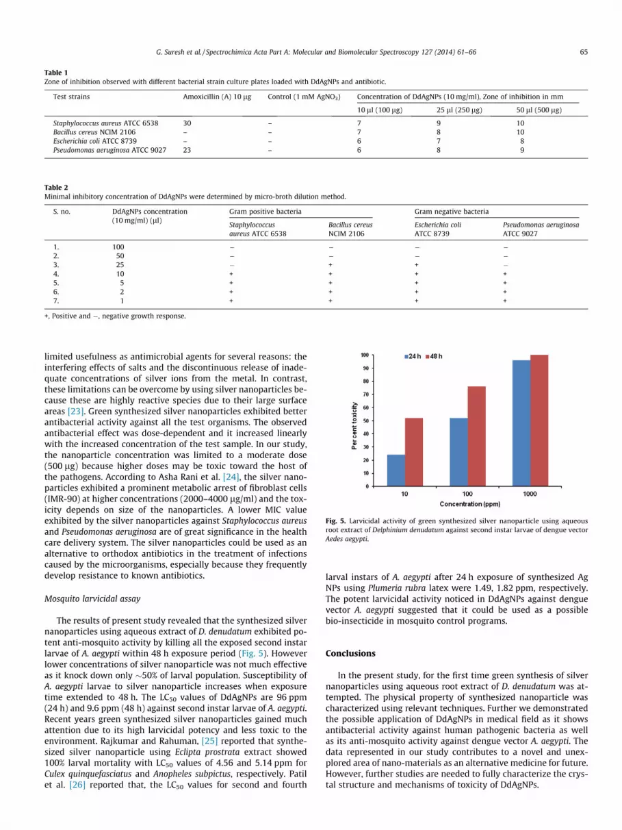
Table 1Zone of inhibition observed with different bacterial strain culture plates loaded with DdAgNPs and antibiotic.
Test strains Amoxicillin (A) 10 lg Control (1 mM AgNO3) Concentration of DdAgNPs (10 mg/ml), Zone of inhibition in mm
10 ll (100 lg) 25 ll (250 lg) 50 ll (500 lg)
Staphylococcus aureus ATCC 6538 30 – 7 9 10Bacillus cereus NCIM 2106 – – 7 8 10Escherichia coli ATCC 8739 – – 6 7 8Pseudomonas aeruginosa ATCC 9027 23 – 6 8 9
Table 2Minimal inhibitory concentration of DdAgNPs were determined by micro-broth dilution method.
S. no. DdAgNPs concentration(10 mg/ml) (ll)
Gram positive bacteria Gram negative bacteria
Staphylococcusaureus ATCC 6538
Bacillus cereusNCIM 2106
Escherichia coliATCC 8739
Pseudomonas aeruginosaATCC 9027
1. 100 � � � �2. 50 � � � �3. 25 � + + �4. 10 + + + +5. 5 + + + +6. 2 + + + +7. 1 + + + +
+, Positive and �, negative growth response.
Fig. 5. Larvicidal activity of green synthesized silver nanoparticle using aqueousroot extract of Delphinium denudatum against second instar larvae of dengue vectorAedes aegypti.
G. Suresh et al. / Spectrochimica Acta Part A: Molecular and Biomolecular Spectroscopy 127 (2014) 61–66 65
limited usefulness as antimicrobial agents for several reasons: theinterfering effects of salts and the discontinuous release of inade-quate concentrations of silver ions from the metal. In contrast,these limitations can be overcome by using silver nanoparticles be-cause these are highly reactive species due to their large surfaceareas [23]. Green synthesized silver nanoparticles exhibited betterantibacterial activity against all the test organisms. The observedantibacterial effect was dose-dependent and it increased linearlywith the increased concentration of the test sample. In our study,the nanoparticle concentration was limited to a moderate dose(500 lg) because higher doses may be toxic toward the host ofthe pathogens. According to Asha Rani et al. [24], the silver nano-particles exhibited a prominent metabolic arrest of fibroblast cells(IMR-90) at higher concentrations (2000–4000 lg/ml) and the tox-icity depends on size of the nanoparticles. A lower MIC valueexhibited by the silver nanoparticles against Staphylococcus aureusand Pseudomonas aeruginosa are of great significance in the healthcare delivery system. The silver nanoparticles could be used as analternative to orthodox antibiotics in the treatment of infectionscaused by the microorganisms, especially because they frequentlydevelop resistance to known antibiotics.
Mosquito larvicidal assay
The results of present study revealed that the synthesized silvernanoparticles using aqueous extract of D. denudatum exhibited po-tent anti-mosquito activity by killing all the exposed second instarlarvae of A. aegypti within 48 h exposure period (Fig. 5). Howeverlower concentrations of silver nanoparticle was not much effectiveas it knock down only �50% of larval population. Susceptibility ofA. aegypti larvae to silver nanoparticle increases when exposuretime extended to 48 h. The LC50 values of DdAgNPs are 96 ppm(24 h) and 9.6 ppm (48 h) against second instar larvae of A. aegypti.Recent years green synthesized silver nanoparticles gained muchattention due to its high larvicidal potency and less toxic to theenvironment. Rajkumar and Rahuman, [25] reported that synthe-sized silver nanoparticle using Eclipta prostrata extract showed100% larval mortality with LC50 values of 4.56 and 5.14 ppm forCulex quinquefasciatus and Anopheles subpictus, respectively. Patilet al. [26] reported that, the LC50 values for second and fourth
larval instars of A. aegypti after 24 h exposure of synthesized AgNPs using Plumeria rubra latex were 1.49, 1.82 ppm, respectively.The potent larvicidal activity noticed in DdAgNPs against denguevector A. aegypti suggested that it could be used as a possiblebio-insecticide in mosquito control programs.
Conclusions
In the present study, for the first time green synthesis of silvernanoparticles using aqueous root extract of D. denudatum was at-tempted. The physical property of synthesized nanoparticle wascharacterized using relevant techniques. Further we demonstratedthe possible application of DdAgNPs in medical field as it showsantibacterial activity against human pathogenic bacteria as wellas its anti-mosquito activity against dengue vector A. aegypti. Thedata represented in our study contributes to a novel and unex-plored area of nano-materials as an alternative medicine for future.However, further studies are needed to fully characterize the crys-tal structure and mechanisms of toxicity of DdAgNPs.
66 G. Suresh et al. / Spectrochimica Acta Part A: Molecular and Biomolecular Spectroscopy 127 (2014) 61–66
Acknowledgements
Authors are thankful to Dr. K.R. Venkatesan, Principal, Sri San-kara Arts and Science College for providing lab facilities and alsothank to IIT Madras for recording FT-IR spectra.
Appendix A. Supplementary material
Supplementary data associated with this article can be found, inthe online version, at http://dx.doi.org/10.1016/j.saa.2014.02.030.
References
[1] M. Dubey, S. Bhadauria, B.S. Kushwah, Dig. J. Nanomater. Bios. 4 (3) (2009)537–543.
[2] V. Parashar, P. Rashmi, S. Bechan, C.P. Avinash, Dig. J. Nanomater. Bios. 4 (1)(2009) 45–50.
[3] N. Roy, A. Barik, Int. J. Nanotech. Appl. 4 (2) (2001) 95–101.[4] M. Khalil, E.H. Ismail, K.Z. El-Baghdady, M. Doaa, Arab. J. Chem. (2013), http://
dx.doi.org/10.1016/j.arabjc.2013.04.007.[5] D.M. Morens, A.S. Fauci, PLoS Pathog. 9 (7) (2013) e1003467.[6] V. Shetty, D. Sanil, N.J. Shetty, Pest Manage. Sci. 69 (2013) 257–267.[7] S.J. Thomas, T.P. Endy, Curr. Opin. Infect. Dis. 26 (2013) 429–434.[8] T.J. John, L. Dandona, V.P. Sharma, M. Kakkar, Lancet 377 (9761) (2011) 252–
269.
[9] Q. Nizami, M.A. Jafri, Indian J. Tradit. Know. 5 (4) (2006) 463–467.[10] A. Koodalingam, P. Mullainadhan, M. Arumugam, Parasitol. Res. 105 (2009)
1425–1434.[11] CLSI, Methods for Dilution Antimicrobial Susceptibility Tests for Bacteria that
Grow Aerobically; Approved Standard-Seventh Edition Document, 2005, M7–A7.
[12] D. Bhattacharya, R.K. Gupta, Crit. Rev. Biotechnol. 25 (4) (2005) 199–204.[13] R. Brause, H. Moeltgen, K. Kleinermanns, Appl. Phys. B 75 (2002) 711–716.[14] U. Kreibig, M. Vollmer, Optical Properties of Metal Clusters, Springer, Berlin,
Germany, 1995.[15] P. Mulvaney, Langmuir 12 (1996) 788–800.[16] G. Mie, Ann. Phys. 25 (1908) 377–445.[17] I.O. Sosa, C. Noguez, R.G. Barrera, J. Phys. Chem. B 107 (2003) 6269–6275.[18] S.S. Shankar, A. Rai, A. Ahmad, M. Sastry, J. Colloid Interface Sci. 275 (2) (2004)
496–502.[19] R. Veerasamy, T.Z. Xin, S. Gunasagaran, T.F.W. Xiang, E.F.C. Yang, N. Jeyakumar,
S.A. Dhanaraj, J. Saudi Chem. Soc. 15 (2011) 113–120.[20] Y.S. Jae, S.K. Beom, Bioprocess. Biosyst. Eng. 32 (2009) 79–84.[21] Q.L. Feng, J. Wu, G.Q. Chen, F.Z. Cui, T.M. Kim, J.O. Kim, J. Biomed. Mater. Res. 52
(2000) 662.[22] J.A. Spadaro, T.J. Berger, S.D. Barranco, S.E. Chapin, R.O. Becker, Microb. Agents
Chemother. 6 (1974) 637.[23] A. Nabikhan, K. Kandasamy, A. Raj, N.M. Alikunhi, Colloids Surf. B Biointerfaces
79 (2) (2010) 488–493.[24] P.V. Asha Rani, G.L. Kah Mun, M.P. Hande, S. Valiyaveetti, ACS Nano 3 (2)
(2009) 279–290.[25] G. Rajakumar, A. Abdul Rahuman, Acta Trop. 118 (2011) 196–203.[26] C.D. Patil, S.V. Patil, H.P. Borase, B.K. Salunke, R.B. Salunkhe, Parasitol. Res. 110
(5) (2012) 1815–1822.
Copyright © 2022 FDOKUMEN