The influence of mechanical properties on wall stress and distensibility of the dilated ascending...
11
2003;126:842-850 J Thorac Cardiovasc Surg M. Sundt, III Ruth J. Okamoto, Haodong Xu, Nicholas T. Kouchoukos, Marc R. Moon and Thoralf dilated ascending aorta The influence of mechanical properties on wall stress and distensibility of the http://jtcs.ctsnetjournals.org/cgi/content/full/126/3/842 located on the World Wide Web at: The online version of this article, along with updated information and services, is 2003 American Association for Thoracic Surgery Association for Thoracic Surgery and the Western Thoracic Surgical Association. Copyright © is the official publication of the American The Journal of Thoracic and Cardiovascular Surgery on June 15, 2013 jtcs.ctsnetjournals.org Downloaded from
-
Upload
independent -
Category
Documents
-
view
0 -
download
0
Transcript of The influence of mechanical properties on wall stress and distensibility of the dilated ascending...

2003;126:842-850 J Thorac Cardiovasc SurgM. Sundt, III
Ruth J. Okamoto, Haodong Xu, Nicholas T. Kouchoukos, Marc R. Moon and Thoralf dilated ascending aorta
The influence of mechanical properties on wall stress and distensibility of the
http://jtcs.ctsnetjournals.org/cgi/content/full/126/3/842located on the World Wide Web at:
The online version of this article, along with updated information and services, is
2003 American Association for Thoracic Surgery Association for Thoracic Surgery and the Western Thoracic Surgical Association. Copyright ©
is the official publication of the AmericanThe Journal of Thoracic and Cardiovascular Surgery
on June 15, 2013 jtcs.ctsnetjournals.orgDownloaded from

The influence of mechanical properties on wall stress anddistensibility of the dilated ascending aortaRuth J. Okamoto, DSca,b
Haodong Xu, MD, PhDc
Nicholas T. Kouchoukos, MDd
Marc R. Moon, MDe
Thoralf M. Sundt III, MDf
Objectives: We sought to determine how intrinsic mechanical properties of dilatedascending aorta influence in vivo distensibility and wall stress, potential contribut-ing factors to the risk of aortic rupture and dissection.
Methods: Fresh tissue from patients undergoing ascending aortic replacement forMarfan syndrome (n � 8, age 38 � 14 years), bicuspid aortic valve with associatedaneurysm (n � 20, age 55 � 12 years), or idiopathic degenerative aneurysm (n �7, age 64 � 9 years) was analyzed for elastic properties and circumferential residualstress. Histologic specimens were graded for elastin fragmentation, cystic changes,fibrosis, and necrosis. Distensibility and wall stresses were predicted with a cylin-drical mathematical model of the aorta.
Results: Elastic properties of aortic tissue were nonlinear and extensibility decreasedwith age. Moderate or severe elastin fragmentation was prevalent in tissue frompatients with the Marfan syndrome and more common in tissue from youngerpatients. Model-predicted mean circumferential stress did not vary significantly withage or patient group, but increased with aortic lumen diameter and systolic bloodpressure. Model-predicted distensibility decreased with age in bicuspid aortic valveand degenerative aneurysm groups. Differences in distensibility were correlatedwith differences in elastic properties.
Conclusions: The age dependence of elastic properties of dilated ascending aorta isconsistent with that of normal aorta observed in previous studies. Increases in meancircumferential stress with blood pressure and diameter support the clinical impor-tance of blood pressure control and serial evaluation of aortic diameter. Decliningwall strength with age may increase the risk of rupture or dissection.
Aortic rupture and dissection are mechanical failures of the aorticwall occurring with increased frequency in the dilated aorta.1
Histologic evidence of degenerative changes of the media inaneurysmal aorta have been observed in the Marfan syndrome andbicuspid aortic valve (BAV) disease.2,3 The mechanical propertiesof the dilated aorta in these conditions, however, have not been
well characterized. We have recently reported ex vivo mechanical properties ofdilated ascending aorta.4 To relate these properties to the risk of mechanical failure,we applied these findings to a cylindrical mathematical model. Using fresh tissuefrom patients undergoing surgical resection of ascending aortic aneurysms associ-ated with Marfan syndrome, BAV, or degenerative disease, we performed mechan-ical testing to determine elastic properties ex vivo. We used these patient-specificdata in a mathematical model to predict distensibility and average wall stressesunder physiologic loading. Mechanical properties, predicted in vivo behavior, andhistologic abnormalities were examined and compared in these 3 conditions.
From the Departments of Mechanical En-gineering,a Biomedical Engineering,b andPathology,c and Division of CardiothoracicSurgery,e Washington University, St Louis,Mo; Missouri Baptist Medical Center,d StLouis, Mo; and Division of CardiovascularSurgery,f Mayo Clinic, Rochester, Minn.
Funding provided by The Whitaker Foun-dation (R.J.O.). Additional funding fromthe National Marfan Foundation (R.J.O.).
Received for publication April 15, 2002;revisions requested June 12, 2002; revi-sions received March 31, 2003; acceptedfor publication May 1, 2003.
Address for reprints: Ruth J. Okamoto,DSc, Washington University, Campus Box1185, One Brookings Dr, St Louis, MO63130-4899 (E-mail: [email protected]).
J Thorac Cardiovasc Surg 2003;126:842-50
Copyright © 2003 by The American Asso-ciation for Thoracic Surgery
0022-5223/2003 $30.00 � 0
doi:10.1016/S0022-5223(03)00728-1
Surgery for Acquired Cardiovascular Disease Okamoto et al
842 The Journal of Thoracic and Cardiovascular Surgery ● September 2003
ACD
on June 15, 2013 jtcs.ctsnetjournals.orgDownloaded from

MethodsPatientsAscending aorta was obtained from 35 patients (18-79 years old)undergoing graft replacement of the ascending aorta for aneurys-mal dilatation at the Washington University Medical Center andthe Missouri Baptist Medical Center. The study protocol wasapproved by the Human Studies Committees of both institutionsand the patients gave informed consent. Clinical groups weredefined by the presence of Marfan syndrome, BAV, or idiopathicdegenerative aneurysm (DA). Patients were also grouped accord-ing to the predominant valvular hemodynamic abnormality (ste-nosis, regurgitation, or mixed), referred to as “valve pathologygroups” hereafter.
HistologyCircumferentially oriented specimens were cut from the aortictissue adjacent to regions used for mechanical testing, fixed in 10%formalin, embedded in paraffin blocks, and sectioned at 4- or 5-�mthickness with at least 4 serial sections obtained from each block.Histologic abnormalities were graded by the scheme of 0 (normal)to 3 (severe) proposed by Schlatmann and Becker.5 The patholo-gist (H.X.) was blinded to clinical data. Necrosis, defined as a focalloss of smooth muscle cell nuclei in the media, was evaluatedusing sections stained with hematoxylin and eosin. Cystic medialnecrosis (CMN), defined as cystic regions in the media containingmucopolysaccharides,5 was evaluated on sections stained withMovat pentachrome. Elastic fiber fragmentation and fibrosis wereevaluated on sections stained with Hart’s elastin and van Giesonstains, respectively.
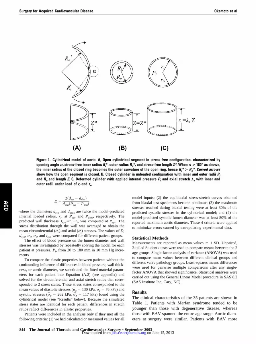
Mechanical TestingAortic tissue was stored in refrigerated saline solution (4°C) untiltesting. Mechanical testing methods have been previously de-scribed.4 Tissue was received as a short tube cut from above thesinuses of Valsalva. For most patients with the Marfan syndrome,the axial dimension of the tube was very short and a separate pieceof tissue from the noncoronary sinus large enough for biaxialtesting was obtained. Approximately square, biaxial test specimens(2.0-2.5 cm per side) with the edges aligned to the circumferentialand axial directions of the aorta were subjected to equibiaxial andstrip biaxial stretching protocols. Force and deformation data werecombined with specimen dimensions to obtain Cauchy stresses(force/unit deformed area), stretch ratios (deformed length/un-loaded length), and strains in each direction. The elastic propertieswere determined by fitting the experimental stress-strain data to amaterial law that allowed for nonlinear behavior and for anisotropy(ie, different properties in the circumferential and axial directions).Details of this law are given in the appendix. A Levenberg-Marquardt optimization algorithm was used to find the best-fitmaterial parameters for each specimen tested. When sufficienttissue was available, circumferential residual stress was character-ized by measuring the opening angle6 of a 3- to 5-mm–wide ring.The closed ring was placed in room temperature saline solutionand a radial cut was made in the ring. The opening angle, � (Figure1, A) was defined as the angle created between the 2 end points andthe midpoint of the circumference. When the intima was on theouter side of the curved ring, the opening angle was greater than180°.
Cylindrical ModelWe used a thick-walled cylindrical model of the aorta7-10 to predictin vivo distensibility and wall stress distribution. Although thismodel simplifies the aortic geometry, it incorporates importantfeatures of large strain, residual stress, and nonlinear, anisotropicelastic properties.
The stress-free configuration of the model (Figure 1, A) is anopen cylinder described by inner radius Ri*, outer radius Ro*,opening angle �, and length Z*. The open cylinder is closed(Figure 1, B) to produce a cylinder of the same mean circumfer-ence with inner radius Ri and outer radius Ro and length Z. Thiscompresses the inner part of the wall and stretches the outer part ofthe wall, producing circumferential residual stress in the unloadedcylinder. In the loaded state, the model is subject to an internalpressure Pi, and an axial stretch ratio, �z, and stretches circumfer-entially to a deformed outer radius ro (Figure 1, C). Given theunloaded radii Ri and Ro and the opening angle, the stress-free radiiRi and Ro are found by requiring the same volume and meancircumference in both states.9 For any ro, the deformed innerradius, ri, can also be found assuming incompressibility. Theequation of radial equilibrium in cylindrical coordinates (see ap-pendix) was integrated numerically with respect to radius to yieldthe internal pressure, Pi, using Matlab (Release 12, Mathworks,Natick, Mass) with 100 integration steps from ro to ri.
The measured opening angle of the tissue was used in themodel when available. If it could not be measured, the patient’sage was used to estimate opening angle using a linear fit toexperimental data previously obtained4: � � 91.98 � 2.68 · age(years) , R2 � 0.33. To obtain suitable Ri and Ro for each patient,we digitized the inner circumference of the closed rings used forthe opening angle measurements and computed a mean Ri.
For some patients, particularly those with Marfan syndrome, itwas known that the closed ring used for the opening angle mea-surement was distal to the location of the maximum aortic diam-eter. In these patients, we multiplied the closed ring radius by ascaling factor to obtain Ri. Preoperative transverse magnetic res-onance (MR) images of the ascending aorta were digitized toobtain aortic diameters at positions in the aortic root and ascendingaorta. The scaling factor was the ratio of the maximum aorticdiameter to the aortic diameter at the axial location where theclosed ring was cut. When MR images were not available, thescaling factor was the ratio of the diameter of the larger to smallerend of the harvested aortic tissue. For all patients, Ro was obtainedby adding the measured unloaded thickness of the biaxial speci-men to Ri.
Like other blood vessels, the aorta retracts when it is removedfrom the patient and �z corresponds to the amount that the vesselmust be stretched to return it to the in vivo length. As we did notmeasure �z, we assumed �z � 1.2 for all patients, the approximatemean value reported by Learoyd and Taylor11 for nondilatedhuman thoracic aorta.
Using this input data for each patient, the model was solved at2 internal pressures equal to the patient’s reported preoperativesystolic (Psys) and diastolic (Pdias) blood pressures as measured bybrachial cuff. The model results were used to predict the disten-sibility, D, as defined by Hirata and colleagues,12 that would beobserved for patients in vivo:
Okamoto et al Surgery for Acquired Cardiovascular Disease
The Journal of Thoracic and Cardiovascular Surgery ● Volume 126, Number 3 843
ACD
on June 15, 2013 jtcs.ctsnetjournals.orgDownloaded from
D �2�dsys � ddias�
ddias�Psys � Pdias�
where the diameters dsys and ddias are twice the model-predictedinternal loaded radius, ri, at Psys and Pdias, respectively. Thepredicted wall thickness, tsys�ro�ri, was computed at Psys. Thestress distribution through the wall was averaged to obtain themean circumferential (�̄c) and axial (�̄
z) stresses. The values of D,
dsys, �̄c, �̄z, and tsys were compared for different patient groups.The effect of blood pressure on the lumen diameter and wall
stresses was investigated by repeatedly solving the model for eachpatient at pressures, Pi, from 20 to 180 mm in 10 mm Hg incre-ments.
To compare the elastic properties between patients without theconfounding influence of differences in blood pressure, wall thick-ness, or aortic diameter, we substituted the fitted material param-eters for each patient into Equation (A.2) (see appendix) andsolved for the circumferential and axial stretch ratios that corre-sponded to 2 stress states. These stress states corresponded to themean values of diastolic stresses (�̄c � 130 kPa, �̄z � 76 kPa) andsystolic stresses (�̄c � 262 kPa, �̄z � 117 kPa) found using thecylindrical model (see “Results” below). Because the simulatedstress states are identical for each patient, differences in stretchratios reflect differences in elastic properties.
Patients were included in the analysis only if they met all thefollowing criteria: (1) we had calculated or measured values for all
model inputs; (2) the equibiaxial stress-stretch curves obtainedfrom biaxial test specimens became nonlinear; (3) the maximumstresses reached during biaxial testing were at least 30% of thepredicted systolic stresses in the cylindrical model; and (4) themodel-predicted systolic lumen diameter was at least 80% of thereported maximum aortic diameter. These 4 criteria were appliedto minimize errors caused by extrapolating experimental data.
Statistical MethodsMeasurements are reported as mean values � 1 SD. Unpaired,2-tailed Student t tests were used to compare means between the 2age groups. Single-factor analysis of variance (ANOVA) was usedto compare mean values between different clinical groups anddifferent valve pathology groups. Least-squares means differenceswere used for pairwise multiple comparisons after any single-factor ANOVA that showed significance. Statistical analyses werecarried out using the General Linear Model procedure in SAS 8.2(SAS Institute Inc, Cary, NC).
ResultsThe clinical characteristics of the 35 patients are shown inTable 1. Patients with Marfan syndrome tended to beyounger than those with degenerative disease, whereasthose with BAV spanned the entire age range. Aortic diam-eters at surgery were similar. Patients with BAV more
Figure 1. Cylindrical model of aorta. A, Open cylindrical segment in stress-free configuration, characterized byopening angle �, stress-free inner radius Ri*, outer radius Ro*, and stress-free length Z*. When � > 180° as shown,the inner radius of the closed ring becomes the outer curvature of the open ring, hence Ri* > Ro*. Curved arrowsshow how the open segment is closed. B, Closed cylinder in unloaded configuration with inner and outer radii Ri
and Ro and length Z. C, Deformed cylinder with applied internal pressure Pi and axial stretch �z with inner andouter radii under load of ri and ro.
Surgery for Acquired Cardiovascular Disease Okamoto et al
844 The Journal of Thoracic and Cardiovascular Surgery ● September 2003
ACD
on June 15, 2013 jtcs.ctsnetjournals.orgDownloaded from

frequently had concomitant functional valve disease. Me-chanical test data from 24 of the 35 patients has previouslybeen reported.4
HistologyHistologic evidence of degenerative changes was observedin the majority of patients and was most pronounced inthose with Marfan syndrome. Tissue adjacent to the regionused for biaxial testing was analyzed from 32 of the 35patients. Specimens for 1 Marfan patient (61-year-old man)and 2 BAV patients (56-year-old man, 28-year-old woman)were unavailable. The most common abnormality was elas-tin fragmentation , which was severe (grade 3) in 11 patients(7 Marfan, 1 BAV, 2 DA). Moderate (grade 2) or severeelastin fragmentation was more common in younger pa-tients (11 of 17, age � 55 years, 6 of 15, age � 55 years).Severe fibrosis was observed in only 1 specimen (Marfan)whereas moderate fibrosis was evident in 7 (4 Marfan, 1BAV, 2 DA). Severe CMN was present in 1 BAV and 4Marfan patients. Severe smooth muscle cell necrosis waspresent in only 1 DA specimen.
Wall ThicknessMean thickness of unloaded specimens was measured be-tween patient groups as shown in Table 2. The difference inthe mean unloaded wall thickness between age groups didnot reach significance (P � .174) and differences betweenclinical groups and valve pathology groups were not signif-icant (P � .2). Within the range of reported preoperativeaortic diameters of the study group (4.0-7.0 cm), wefound a slight decrease in unloaded wall thickness withpreoperative aortic diameter that was not significant (P �.2).
Mechanical PropertiesElastic properties of aortic specimens for use in the cylin-drical model were determined by biaxial testing. As shownin Figure 2, the elastic response to equibiaxial stretchingwas initially linear, followed by a rapid increase in stress asthe tissue became very stiff (ie, the slope of the stress-stretch curve steepened). The equibiaxial stretch ratio atwhich this nonlinear behavior occurred generally decreasedwith age in the BAV and DA groups. These findings cannotbe interpreted, however, without knowledge of the range ofstresses experienced under physiologic loading conditions.These were determined using the cylindrical model (seebelow).
Additional inputs to the cylindrical model are shown inTable 2. Because of the large age range spanned by theBAV group, data for BAV patients younger and older thanthe mean BAV age of 55 years are also shown separately.Patients were also grouped by functional valve pathology asshown in Table 1. Systolic blood pressure was highest
among patients with DAs and tended to be higher amongolder (age � 55 years) patients with BAV. Opening anglewas also higher in older patients as previously shown.4 Thedifference between opening angles measured directly fromrings (� � 242° � 83°, n � 19) and those obtained from thelinear regression (� � 225° � 39°, n � 16) was notsignificant (P � .2). The unloaded lumen radius was deter-mined directly from the closed ring radius for 21 patientsand scaled for 14 patients (8 Marfan, 4 BAV, 2 DA).
Cylindrical ModelMean circumferential and axial wall stresses as well asdistensibility were predicted from the cylindrical model(Table 3). Given that specimens were obtained from aortaswith similar diameters under physiologic conditions andsimilar predicted wall thicknesses, it is not surprising thatdifferences in mean circumferential stresses between agegroups did not reach significance (P � .161). Differencesbetween clinical groups were also not significant (P � .2).
Mean circumferential stress was positively correlated (R2
� 0.469, P � .001) with the predicted systolic lumendiameter as shown in Figure 3, with an increase in circum-ferential stress of 62 kPa for each 1-cm increase in aorticdiameter. Mean circumferential stress is plotted as a func-tion of blood pressure (Figure 4, A) for 4 patients withpredicted systolic lumen diameters of 4.0 to 4.6 cm, thusminimizing the effect of systolic lumen diameter on thestresses. This demonstrates that the relation between bloodpressure and mean circumferential stress is almost linear inthe physiologic range. The mean slope of the circumferen-tial stress–blood pressure relation for all 35 patients, com-puted from the difference between the cylindrical modelstresses at diastolic and systolic pressures, was 2.37 � 0.68kPa/mm Hg. An increase in systolic blood pressure of 26
TABLE 1. Patient characteristics grouped by clinical diag-nosis
Marfan syndrome(n � 8)
BAV(n � 20)
DA(n � 7)
Age (y) 38 � 14 55 � 12 64 � 9Male gender 5 16 4Maximum
preoperative aorticdiameter (cm)*
5.4 � 0.8 5.0 � 0.7 5.1 � 0.7
Valve pathology†Neither 5 3 3Stenotic 0 5 0Regurgitant 3 7 3Mixed 0 5 1
*As reported by surgeon, or from preoperative computed tomography,magnetic resonance imaging, or echocardiography exam.†As reported by surgeon, magnetic resonance imaging, or echocardiog-raphy exam.
Okamoto et al Surgery for Acquired Cardiovascular Disease
The Journal of Thoracic and Cardiovascular Surgery ● Volume 126, Number 3 845
ACD
on June 15, 2013 jtcs.ctsnetjournals.orgDownloaded from

mm Hg thus produces a similar increase in circumferentialstress as a 1-cm increase in lumen diameter.
Because tissue from older patients was less extensible(Figure 2), axial stresses were higher among these patientsat the applied axial stretch ratio (�z � 1.2). Among clinicalgroups, axial stress in patients with Marfan syndrome wassignificantly lower than for those with DAs. When pooled,patients with BAV had predicted axial stress intermediatebetween these groups. When stratified by age, however,younger patients with BAV had significantly lower axialstresses than older BAV patients or patients with DAs, butsimilar to Marfan patients.
Unlike axial stress, differences in distensibility wereobserved between clinical groups. Furthermore, within the
BAV group, younger patients demonstrated a higher disten-sibility than older patients. When stratified by age indepen-dent of clinical group, differences in distensibility did not
Figure 2. Comparison of response to simulated equibiaxial stretching between patient groups using fitted elasticproperties obtained from biaxial test data. A and B, Marfan. C and D, Bicuspid aortic valve (BAV). E and F,Degenerative aneurysm (DA). Fitted data is used to eliminate slight differences in stretching protocols betweenpatients; however, maximum stresses shown correspond closely to stresses reached during actual equibiaxialstretching experiments.
Figure 3. Model-predicted mean circumferential stress as a func-tion of predicted lumen diameter for all patient groups: Marfan,open square; BAV, open diamond; DA, open circle. Dashed lineshows linear fit to data: �̄c,sys(kPa) � �34.564 � 62.3*dsys(cm), R2
� 0.469.
Figure 4. A, Mean circumferential stress in the cylindrical modelplotted as a function of blood pressure for 4 representativepatients. Marfan (39 years): open square; younger BAV (39 years):closed triangle; older BAV (66 years): open triangle; DA (77 years),closed circle. B, Normalized radius (ri/Ri) versus blood pressurefor the same patients. Open symbols indicate model predictedvalues at 10 –mm Hg pressure increments. Corresponding closedsymbols indicate model-predicted values at each patient’s re-ported preoperative systolic and diastolic blood pressure.
Surgery for Acquired Cardiovascular Disease Okamoto et al
846 The Journal of Thoracic and Cardiovascular Surgery ● September 2003
ACD
on June 15, 2013 jtcs.ctsnetjournals.orgDownloaded from

reach significance (P � .072). Distensibility decreased sig-nificantly with age in the BAV (P � .001) and aneurysmgroups (P � .001) but not the Marfan group (P � .2).Differences in distensibility (P � .2) and mean stress values(P � .2) between functional valve pathologies were notsignificant. As shown in Figure 4 B, the relation betweenblood pressure and normalized lumen radius, ri/Ri, is linearin the physiologic range. Although ri/Ri initially increasesrapidly with blood pressure, the cylindrical model predictsthat the slope of the pressure–normalized radius relationdecreases before physiologic pressures are reached. Thenormalized lumen radius at which the slope decreases islower in the older patients because of the difference inintrinsic elastic properties (Figure 2).
To quantify the contribution of intrinsic elastic propertiesto the observed differences in distensibility, we examinedthe calculated circumferential (�c) and axial (�z) stretchratios using the fitted material properties for each patient atthe simulated diastolic and systolic stress states. Both thecircumferential and axial stretch ratios required to reach the
simulated physiologic stresses decreased significantly withage (Figure 5, A to D), and the mean values were signifi-cantly lower for patients in the older age group comparedwith the younger group (P � .001).
Differences in mean circumferential and axial stretchratios between clinical groups did not reach significanceunless the BAV group was subdivided by age (Figure 5, E).The difference in circumferential stretch ratios between thesystolic and diastolic stress states (Figure 5, F) was corre-lated with distensibility (R2 � 0.80). This demonstrates thatthe model-predicted differences in distensibility betweenclinical groups are primarily governed by differences in theintrinsic elastic properties in the range of physiologicstresses.
DiscussionThis study demonstrates that mean circumferential stressdepends on the aortic diameter and systolic blood pressurebut not on age or clinical diagnosis. In the absence of acompensatory increase in wall thickness with lumen diam-
TABLE 2. Cylindrical model input data
n
Blood Pressure
Opening angle(deg/�)
Unloaded lumendiameter (cm)
Unloaded wall thickness(mm)
Systolic(mm Hg)
Diastolic(mm Hg)
Clinical groupMarfan 8 115 � 16* 68 � 14 205 � 51�,¶ 3.4 � 0.6 2.7 � 0.4BAV 20 127 � 19 73 � 15 235 � 68 3.2 � 0.5 2.6 � 0.5
�55 y 10 121 � 19† 69 � 11 185 � 50** 2.9 � 0.3 2.8 � 0.6�55 y 10 133 � 17‡ 76 � 19 286 � 39 3.4 � 0.6 2.5 � 0.3
DA 7 150 � 14 80 � 11 263 � 71 3.3 � 0.6 2.6 � 0.3Age group
�55 y 18 121 � 18 70 � 11 193 � 49 3.0 � 0.3 2.7 � 0.5�55 y 17 138 � 20§ 76 � 17 278 � 53††¶ 3.4 � 0.6‡‡ 2.6 � 0.3
Significantly different than DA: *P � .011, †P � .016, ‡P � .048, �P � .042, *P � .005; significantly different than BAV � 55 years: ¶P � .003, **P � .0001;significantly different than age � 55 years: §P � .013, ††P � .0001, ‡‡P � .014.
TABLE 3. Cylindrical model results
n
Stresses at systolic bloodpressure
Distensibility(cm2/dyne) � 106
Lumen diameterat systolic
blood pressure(cm)
Wall thickness atsystolic bloodpressure (mm)
Circumference(KPa) Axial (KPa)
Clinical groupMarfan 8 257 � 45 96 � 28* 1.43 � 0.68 5.1 � 0.7 1.5 � 0.2BAV 20 257 � 79 115 � 51 2.21 � 0.83 4.7 � 0.8 1.5 � 0.3
�55 y 10 237 � 71 86 � 17†,‡ 2.62 � 0.75¶,#,** 4.6 � 0.7 1.5 � 0.4�55 y 10 277 � 85 143 � 59 1.79 � 0.72 4.7 � 0.8 1.5 � 0.2
DA 7 276 � 74 135 � 40 1.46 � 0.83 4.6 � 0.8 1.6 � 0.3Age group
�55 y 18 245 � 60 90 � 22 2.13 � 0.92 4.7 � 0.6 1.5 � 0.4�55 y 17 278 � 78 140 � 51§ 1.62 � 0.75 4.8 � 0.9 1.5 � 0.2
Significantly different than BAV �55 years: *P � .019, †P � .0031, #P � .017; significantly different than DA: ‡P � .0189, **P � .0035; significantly differentthan Marfan: ¶P � .002; significantly different than age �55 years: §P � .0005.
Okamoto et al Surgery for Acquired Cardiovascular Disease
The Journal of Thoracic and Cardiovascular Surgery ● Volume 126, Number 3 847
ACD
on June 15, 2013 jtcs.ctsnetjournals.orgDownloaded from

eter, mean circumferential stress increased almost linearlywith diameter and systolic blood pressure. Given our pre-vious observation of reduced wall strength in older pa-tients,4 these data suggest that mechanical failure may occurat a lower diameter in older patients. Furthermore, theysupport the clinical importance of blood pressure control inaddition to attention to aortic diameter in reducing the riskof rupture. Indeed, the curves shown in Figures 3 and 4demonstrate a similar incremental increase in circumferen-tial stress with a 26-mm increase in systolic blood pressureas that for an increase of 1 cm in diameter. Our findingscomplement the clinical observations of Davies and col-leagues,1 who showed that increasing thoracic aneurysmsize is associated with increased yearly rate of rupture, aswell as observations of Juvonen and associates,13 who iden-tified maximum aortic diameter and advanced age as riskfactors for rupture of descending thoracic aortic aneurysms.
Our data also demonstrate that among clinical groups,differences in distensibility are due primarily to intrinsicelastic properties of the aorta itself, rather than differencesin aortic diameter. The mean model-predicted distensibility,D, of our patient groups fell slightly below the range of D� 2.6 to 2.9 10�6 cm2 dynes�1 measured in vivo byprevious investigators for young Marfan and BAV pa-tients.12,14-16 Although distensibility was significantly lowerin patients than in age-matched controls (D � 4.8 to 6.2
10�6 cm2 dynes�1) in these studies, aortic diameters werelarger. To examine whether the larger aortic diameter couldexplain the differences in distensibility compared with con-trols, the cylindrical model was re-solved for each Marfanpatient using a smaller unloaded aortic diameter while hold-ing other model variables constant. The model-predictedsystolic lumen diameter was reduced to 2.8 � 0.2 mm andthe model-predicted distensibility increased only 21%, notenough to explain the reported differences in D between theMarfan and control groups measured in vivo.12,14 Thissuggests that that the intrinsic elastic properties of bothMarfan patients and BAV patients differ from age-matchedcontrols, and these differences are primarily responsible forreduced distensibility.
The relation between elastic properties and distensibilityis also evident from our comparison of elastic properties atsimulated diastolic and systolic stress states. Jeremy andcoworkers14 found that distensibility decreased with age inboth Marfan patient and control groups, consistent with thetrends in model-predicted D for our BAV and DA patientgroups. Within the BAV group, the difference in circum-ferential stretch ratio at diastolic and systolic stress statesdecreased with age. In the Marfan patient group, the differ-ence in circumferential stretch ratios between the diastolicand systolic states was small (Figure 5, F), despite therelatively large circumferential stretch ratio at the diastolic
Figure 5. Stretch ratios corresponding to systolic and diastolic stress states were determined using fitted elasticproperties obtained from biaxial testing. A, Predicted circumferential stretch ratios. B, Axial stretch ratios plottedas a function of age for �c � 262 kPa and �z � 117 kPa. C, Predicted circumferential stretch ratios. D, Axial stretchratios plotted as a function of age for �c � 130 kPa and �z � 76 kPa. Symbols denote different clinical groups:Marfan (open square), BAV (open diamond), DA (closed circle), and dashed lines show a linear fit of the data asa function of age. E, Mean circumferential (unshaded) and axial (shaded) stretch ratios for each clinical group. F,Change in circumferential stretch ratio between stress states shown in C and A. *Significantly greater change inthe younger BAV group compared with all other patient groups.
Surgery for Acquired Cardiovascular Disease Okamoto et al
848 The Journal of Thoracic and Cardiovascular Surgery ● September 2003
ACD
on June 15, 2013 jtcs.ctsnetjournals.orgDownloaded from

stress state. This can be attributed to greater nonlinearity inthe biaxial elastic properties of aortic tissue from Marfanpatient group than the BAV patient group (see Figure 2).Although the decrease in D with age in our Marfan groupwas not significant, the younger Marfan patients we studiedmay have had more severe cardiovascular involvement,influencing the distensibility, or the small number of Marfanpatients studied may have prevented us from observingage-related changes.
Our model results also suggest that altered distensibilityalone does not affect the risk of rupture, since distensibilitywas not directly related to the level of circumferential stress.Nonetheless, it is possible that decreases in strength occursimultaneously with alterations in elastic properties, therebyindirectly influencing the risk of rupture.
Functional valve pathology did not appear to have asignificant effect on either in vitro mechanical properties orin vivo stresses, although a type II error cannot be excluded.These findings further support the hypothesis that aorticdilatation in BAV patients is due to intrinsic material ab-normalities rather than abnormal flow patterns.17,18
The severity of elastin fragmentation in our study groupwas similar to that previously reported by Schlatmann andBecker19 for dilated ascending aorta. Our observation that 4of 6 BAV patients with moderate or severe elastin fragmen-tation were in the younger age group is consistent with thesuggestion by de Sa and colleagues3 that patients with moresevere histopathologic changes may require earlier surgicalintervention. The relative change in circumferential stretchratio between the simulated diastolic and systolic states(Figure 5, F) was significantly lower (P � .014) in patientswith moderate or severe elastin fragmentation comparedwith those with mild elastin fragmentation, suggesting thatelastin fragmentation may be associated with altered elasticproperties. There was no apparent relation between theseverity of elastin fragmentation and functional valve pa-thology. The frequency of severe fibrosis and necrosis wereboth lower in our study than in the study by Schlatmann andBecker,19 and thus our data may not distinguish the effect offibrosis on elastic properties.
LimitationsThe principal limitation of this study is the lack of normalaorta for analysis. Although normal descending thoracicaorta would be available from thoracic organ transplantdonors, excess ascending aorta is not generally available inthe quantity required for these studies. Furthermore, theascending and descending thoracic aortic segments havedifferent wall structures20 and behave differently clinically,making this a poor control tissue. Limitations of the cylin-drical model must also be acknowledged. In the cylindricalmodel it is assumed that the aorta is stretched by 20%axially throughout the cardiac cycle. If the actual values of
axial stretch are higher, the axial stress would also increase.In the absence of experimental data, we have also assumedthe same axial stretch for all patients regardless of age,basing our value on the studies of Learoyd and Taylor.11
Our model also assumes that wall thickness, elastic prop-erties, and lumen diameter are uniform. Although this sim-plifies the model analysis, it neglects possible regional orlocal variations in wall thickness and elastic properties andthe large axial variations in diameter in the dilated ascend-ing aorta. We did not note obvious differences in wallthickness in our excised specimens, but local or regionalvariations in thickness may exist, altering local stresses. Ourbiaxial test method requires a relatively large specimen,making it difficult to test multiple specimens from differentregions to determine whether regional variations in elasticproperties are present. Finite element models of the ascend-ing aorta that more accurately represent dilated aortic ge-ometry, such as those of Grande and colleagues,21,22 maypredict local stresses that exceed the mean values predictedby the cylindrical model. However, previous finite elementmodels have been limited by a lack of measured materialproperties for the aneurysmal aortic wall. Thus, combiningthe in vitro material properties measured in this study andmore detailed finite element model geometry should allowfuture models to more accurately predict wall stress distri-butions.
Although the magnitude of the opening angle has littleeffect on the wall-averaged circumferential stress presentedin this study, it strongly affects the distribution of predictedcircumferential stress through the wall.10 If the circumfer-ential residual stress is neglected (ie, � � 0°), the circum-ferential stress is highest near the inner wall. As the openingangle increases, the circumferential stress becomes moreuniform through the wall. There is an opening angle atwhich the circumferential stress is nearly constant, but withlarger opening angles the stress becomes higher toward theouter wall. A similar trend is seen in the distribution ofpredicted axial stress through the wall. This suggests thatthe large opening angles observed in our study may lead tohigher stresses near the adventitia. However, transmuralstress distributions are also influenced by nonhomogeneousmaterial properties (ie, different properties for the mediaand adventitia).10,23 More detailed studies of the elasticproperties of the individual layers will be needed to predictthe transmural stress distribution; however, such studies aretechnically difficult.
ConclusionsIn summary, our analysis indicates that mean circumferen-tial stress in dilated ascending aorta increases linearly withblood pressure and diameter, supporting the clinical impor-tance of blood pressure control and serial evaluation ofaortic diameter. Decreasing wall strength with age may
Okamoto et al Surgery for Acquired Cardiovascular Disease
The Journal of Thoracic and Cardiovascular Surgery ● Volume 126, Number 3 849
ACD
on June 15, 2013 jtcs.ctsnetjournals.orgDownloaded from

increase the risk of rupture or dissection consistent withclinical observations. Differences in elastic properties causedifferences in model-predicted distensibilities associatedwith age and clinical group. Low model-predicted distensi-bility in Marfan patients is associated with severe elasticfragmentation. Functional valve pathology does not appearto influence elastic properties or distensibility, supportingthe hypothesis that aortic dilatation in BAV patients is dueto intrinsic abnormalities in the aortic wall rather thanabnormal flow patterns.
We thank Sadaf Toor for performing data analysis and NancyNickerson and Marci Bailey for gathering patient information andobtaining patient consent. Aldi Kraja of the Division of Biostatis-tics at Washington University served as statistical consultant, andwe appreciate his valuable input.
References
1. Davies RR, Goldstein LJ, Coady MA, et al. Yearly rupture or dissec-tion rates for thoracic aortic aneurysms: simple prediction based onsize. Ann Thorac Surg. 2002;73:17-27.
2. Halme T, Savunen T, Aho H, Vihersaari T, Penttinen R. Elastin andcollagen in the aortic wall: changes in the Marfan syndrome andannuloaortic ectasia. Exp Mol Pathol. 1985;43:1-12.
3. de Sa M, Moshkovitz Y, Butany J, David TE. Histologic abnormalitiesof the ascending aorta and pulmonary trunk in patients with bicuspidaortic valve disease: clinical relevance to the Ross procedure. J ThoracCardiovasc Surg. 1999;118:588-94.
4. Okamoto RJ, Wagenseil JE, DeLong WD, Peterson SJ, KouchoukosNT, Sundt TM. Mechanical properties of dilated human ascendingaorta. Ann Biomed Eng. 2002;30:624-35.
5. Schlatmann TJ, Becker AE. Histologic changes in the normal agingaorta: implications for dissecting aortic aneurysm. Am J Cardiol.1977;39:13-20.
6. Liu SQ, Fung YC. Zero-stress states of arteries. J Biomech Eng.1988;110:82-4.
7. von Maltzahn WW, Besdo D, Wiemer W. Elastic properties of arter-ies: a nonlinear two-layer cylindrical model. J Biomech. 1981;14:389-97.
8. Chuong CJ, Fung YC. On residual stresses in arteries. J Biomech Eng.1986;108:189-92.
9. Matsumoto T, Hayashi K. Stress and strain distribution in hypertensiveand normotensive rat aorta considering residual strain. J Biomech Eng.1996;118:62-73.
10. Peterson SJ, Okamoto RJ. Effect of residual stress and heterogeneityon circumferential stress in the arterial wall. J Biomech Eng. 2000;122:454-6.
11. Learoyd BM, Taylor MG. Alterations with age in the viscoelasticproperties of human arterial walls. Circ Res. 1966;18:278-92.
12. Hirata K, Triposkiadis F, Sparks E, Bowen J, Wooley CF, BoudoulasH. The Marfan syndrome: abnormal aortic elastic properties. J Am CollCardiol. 1991;18:57-63.
13. Juvonen T, Ergin MA, Galla JD, et al. Prospective study of the naturalhistory of thoracic aortic aneurysms. Ann Thorac Surg. 1997;63:1533-45.
14. Jeremy RW, Huang H, Hwa J, McCarron H, Hughes CF, Richards JG.Relation between age, arterial distensibility, and aortic dilatation in theMarfan syndrome. Am J Cardiol. 1994;74:369-73.
15. Adams JN. Aortic distensibility and stiffness index measured bymagnetic resonance imaging in patients with Marfan’s syndrome. BrHeart J. 1995;73:265-9.
16. Nistri S, Sorbo MD, Basso C, Thiene G. Bicuspid aortic valve:abnormal aortic elastic properties. J Heart Valve Dis. 2002;11:369-73.
17. Pachulski RT, Weinberg AL, Chan KL. Aortic aneurysm in patientswith functionally normal or minimally stenotic bicuspid aortic valve.Am J Cardiol. 1991;67:781-2.
18. Hahn RT, Roman MJ, Mogtader AH, Devereux RB. Association ofaortic dilation with regurgitant, stenotic and functionally normal bi-cuspid aortic valves. J Am Coll Cardiol. 1992;19:283-8.
19. Schlatmann TJ, Becker AE. Pathogenesis of dissecting aneurysm ofaorta. Comparative histopathologic study of significance of medialchanges. Am J Cardiol. 1977;39:21-6.
20. Halloran BG, Davis VA, McManus BM, Lynch TG, Baxter BT.Localization of aortic disease is associated with intrinsic differences inaortic structure. J Surg Res. 1995;59:17-22.
21. Grande KJ, Cochran RP, Reinhall PG, Kunzelman KS. Mechanisms ofaortic valve incompetence: finite element modeling of aortic rootdilatation. Ann Thorac Surg. 2000;69:1851-7.
22. Grande KJ, Cochran RP, Reinhall PG, Kunzelman KS. Stress varia-tions in the human aortic root and valve: the role of anatomic asym-metry. Ann Biomed Eng. 1998;26:534-45.
23. Taber LA, Humphrey JD. Stress-modulated growth, residual stress,and vascular heterogeneity. J Biomech Eng. 2001;123:528-35.
24. von Maltzahn WW, Warriyar RG, Keitzer WF. Experimental measure-ments of elastic properties of media and adventitia of bovine carotidarteries. J Biomech. 1984;17:839-47.
AppendixMaterial Law for Elastic PropertiesThe elastic properties of the aortic tissue were represented by anincompressible, nonlinear, anisotropic pseudo strain energy func-tion, W24, with 4 material parameters {c, b1, b2, b3}:
W �c
2�exp Q � 1� where Q � b1EC
2 � b2EZ2 � 2b3ECEZ
(A.1)
In the absence of shear strains, circumferential (�;c) and axial(�z) stretch ratios are related to the corresponding Lagrangianstrain components Ec � 0.5 · (�c
2 � 1) and Ez � 0.5 · (�z2 � 1).
The Cauchy stress components are related to the partial derivativesof W in Equation (A.1) with respect to the strain components andare given by24:
�c � �r � �c2 c exp�Q��b1Ec � b3EZ� (A.2a)
�Z � �r � �Z2 c exp�Q��b3EC � b2EZ� (A.2b)
For planar biaxial testing, the radial stress, �r, is 0 and thestrains, circumferential stress, �c, and axial stress, �z, are constantthrough the thickness of the specimen.
Cylindrical Model EquationIn the cylindrical model, strain and stress components, including�r, are functions of radial position in the aortic wall, with theboundary condition Po � ��r � 0 at ro. The internal pressure, Pi,is found by substituting Equation (A.2a) into the equation of radialequilibrium in cylindrical coordinates,
d�r
dr�
�r � �c
r� 0 (A.3)
and integrating numerically from ri to ro:
Pi � �ri
r0
(�c2c exp�Q��b1EC � b3EZ�)
dr
r(A.4)
Surgery for Acquired Cardiovascular Disease Okamoto et al
850 The Journal of Thoracic and Cardiovascular Surgery ● September 2003
ACD
on June 15, 2013 jtcs.ctsnetjournals.orgDownloaded from

2003;126:842-850 J Thorac Cardiovasc SurgM. Sundt, III
Ruth J. Okamoto, Haodong Xu, Nicholas T. Kouchoukos, Marc R. Moon and Thoralf dilated ascending aorta
The influence of mechanical properties on wall stress and distensibility of the
Continuing Medical Education Activities
http://cme.ctsnetjournals.org/cgi/hierarchy/ctsnetcme_node;JTCSSubscribers to the Journal can earn continuing medical education credits via the Web at
Subscription Information
http://jtcs.ctsnetjournals.org/cgi/content/full/126/3/842#BIBLThis article cites 24 articles, 6 of which you can access for free at:
Citations
http://jtcs.ctsnetjournals.org/cgi/content/full/126/3/842#otherarticlesThis article has been cited by 12 HighWire-hosted articles:
Subspecialty Collections
http://jtcs.ctsnetjournals.org/cgi/collection/valve_disease Valve disease
This article, along with others on similar topics, appears in the following collection(s):
Permissions and Licensing
http://www.elsevier.com/wps/find/obtainpermissionform.cws_home/obtainpermissionformreceipt, is available at: An on-line permission request form, which should be fulfilled within 10 working days of
. http://www.elsevier.com/wps/find/supportfaq.cws_home/permissionusematerialcan be found online at: General information about reproducing this article in parts (figures, tables) or in its entirety
on June 15, 2013 jtcs.ctsnetjournals.orgDownloaded from