Plasma Membrane Ca2+-ATPase 4 in Murine Epididymis: Secretion of Splice Variants in the Luminal...
12
?1 BIOLOGY OF REPRODUCTION (2013) 89(1):0, 1–11 Published online before print 22 May 2013. DOI 10.1095/biolreprod.113.108712 Plasma Membrane Ca 2þ -ATPase 4 in Murine Epididymis: Secretion of Splice Variants in the Luminal Fluid and a Role in Sperm Maturation 1 Ramkrishna Patel, 3,4 Amal A. Al-Dossary, 3,4 Deborah L. Stabley, 5 Carol Barone, 5 Deni S. Galileo, 4 Emanuel E. Strehler, 6 and Patricia A. Martin-DeLeon 2,4 4 Department of Biological Sciences, University of Delaware, Newark, Delaware 5 A.I. DuPont Hospital for Children, Wilmington, Delaware 6 Department of Biochemistry and Molecular Biology, Mayo Clinic College of Medicine, Rochester, Minnesota ABSTRACT Plasma ?2 membrane ?3 Ca 2+ -ATPase isoform 4 (PMCA4) is the primary Ca 2+ efflux pump in murine sperm, where it regulates motility. In Pmca4 null sperm, motility loss results in infertility. We have shown that murine sperm PMCA4b interacts with Ca 2+ / CaM-dependent serine kinase (CASK) in regulating Ca 2+ homeo- stasis and motility. However, recent work indicated that the bovine PMCA4a splice variant (missing in testis) is epididymally expressed, along with 4b, and may be transferred to sperm. Here we show, via conventional and in situ RT-PCR, that both the splice variants of Pmca4 mRNA are expressed in murine testis and throughout the epididymis. Immunofluorescence localized PMCA4a to the apical membrane of the epididymal epithelium, and Western analysis not only confirmed its presence but showed for the first time that PMCA4a and PMCA4b are secreted in the epididymal luminal fluid (ELF), from which epididymo- somes containing PMCA4a were isolated. Flow cytometry indicated the presence of PMCA4a on mature caudal sperm where it was increased ;5-fold compared to caput sperm (detected by Western blotting) and ;2-fold after incubation in ELF, revealing in vitro uptake and implicating PMCA4a in epididymal sperm maturation. Coimmunoprecipitation using pan-PMCA4 antibodies, revealed that both variants associate with CASK, suggesting their presence in a complex. Because they have different kinetic properties for Ca 2+ transport and different abilities to bind to CASK, our study suggests a mechanism for combining the functional attributes of both PMCA4 variants, leading to heightened efficiency of the pump in the maintenance of Ca 2+ ?4 homeostasis, which is crucial for normal motility and male fertility. ?5 Keywords: calcium regulation, CASK, epididymis, epididy- mosomes, plasma membrane calcium pump, PMCA, sperm maturation INTRODUCTION When sperm leave the testis they are functionally incom- petent, although morphologically mature. Functional maturity is gained as they traverse the epididymis, where they are in intimate and constant contact with secretions of the epithelial lining of the epididymis [1–3], the epididymal luminal fluid (ELF). During epididymal transit, which lasts ;5–10 days in mice [4], sperm acquire on their plasma membrane many molecular components from the ELF that aid in their maturation, in the absence of their own synthetic machinery [3, 5]. A large variety of proteins are added sequentially to sperm in the different regions of the epididymis, and many of these proteins are glycosyl phosphatidylinositol (GPI) linked and are readily added to the outer leaflet of the lipid bilayer of the sperm membrane [6, 7]. However, recently it has been shown that in humans and bulls, transmembrane proteins are also secreted in the ELF on epididymosomes, which are membranous vesicles known to transfer proteins to the sperm surface [8, 9]. Recent studies showed that an essential 10-pass transmem- brane sperm protein, PMCA4, which is the major Ca 2þ efflux pump in murine sperm [10], is synthesized in the rat [11] and bovine [12] epididymal epithelium. PMCA4 has the two major splice variants 4a and 4b, and 4b is thought to play a crucial role in Ca 2þ clearance in murine sperm [10, 13] where deletion of Pmca4 disrupts Ca 2þ homeostasis and leads to loss of both progressive and hyperactivated sperm motility and ultimately to infertility [14, 15]. Interestingly, bovine sperm show a progressive switch from splice variant 4b in the upper to mainly 4a in the lower regions of the epididymal tract [12]. As sperm do not engage in new mRNA transcription or protein translation, this shift has been attributed to acquisition of PMCA4a from the ELF [12]. The switch from 4b to 4a is physiologically relevant because sperm face a steeper Ca 2þ efflux requirement for hyperactivated motility in the female tract and PMCA4a has a higher basal activity and is more efficient than 4b in returning Ca 2þ to resting levels [12, 16]. In light of these findings for testicular PMCA4 variants in bovine epididymis and sperm, we hypothesized that in the murine system, the PMCA4a variant may also be expressed in the epididymis and potentially secreted in the luminal fluid where it might be acquired by sperm during their epididymal transit and maturation. Our results reveal a difference in bovine and murine systems because we found that in mice, both Pmca4a and Pmca4b mRNAs are expressed in the testis and throughout the epididymis. Importantly, we show for the first time that PMCA4a is expressed in the ELF, is associated with epididymosomes, and is transferred from ELF to caudal sperm in vitro, suggesting that it plays a role in epididymal sperm maturation. Interestingly, in caudal sperm and in ELF both the PMCA4a and PMCA4b variants could be coimmunoprecipi- tated with Ca 2þ /CaM-dependent serine kinase (CASK), an interacting partner of PMCA4b that is known to play a role in signal transduction [13,17], thus indicating that both of the Ca 2þ pump variants function in a complex in sperm. 1 Supported by National Institutes of Health grant NIH-RO3 HD061637 and the Delaware INBRE Program to P.A.M.-D. 2 Correspondence: Patricia A. Martin-DeLeon, Department of Biological Sciences, University of Delaware, Newark, DE 19716. E-mail: [email protected] 3 Co-first authors. Received: 20 February 2013. First decision: 8 April 2013. Accepted: 15 May 2013. Ó 2013 by the Society for the Study of Reproduction, Inc. eISSN: 1529-7268 http://www.biolreprod.org ISSN: 0006-3363 //xinet/production/b/bire/live_jobs/bire-89-01/bire-89-01-03/layouts/bire-89-01-03.3d ĸ Thursday, 30 May 2013 ĸ 2:28 pm Page 1 Allen Press, Inc. ĸ TOC Category: 7 (Male Reproductive Tract) 1 Article 0
-
Upload
mayoclinic -
Category
Documents
-
view
3 -
download
0
Transcript of Plasma Membrane Ca2+-ATPase 4 in Murine Epididymis: Secretion of Splice Variants in the Luminal...
?1 BIOLOGY OF REPRODUCTION (2013) 89(1):0, 1–11Published online before print 22 May 2013.DOI 10.1095/biolreprod.113.108712
Plasma Membrane Ca2þ-ATPase 4 in Murine Epididymis: Secretion of Splice Variantsin the Luminal Fluid and a Role in Sperm Maturation1
Ramkrishna Patel,3,4 Amal A. Al-Dossary,3,4 Deborah L. Stabley,5 Carol Barone,5 Deni S. Galileo,4
Emanuel E. Strehler,6 and Patricia A. Martin-DeLeon2,4
4Department of Biological Sciences, University of Delaware, Newark, Delaware5A.I. DuPont Hospital for Children, Wilmington, Delaware6Department of Biochemistry and Molecular Biology, Mayo Clinic College of Medicine, Rochester, Minnesota
ABSTRACT
Plasma?2 membrane?3 Ca2+-ATPase isoform 4 (PMCA4) is theprimary Ca2+ efflux pump in murine sperm, where it regulatesmotility. In Pmca4 null sperm, motility loss results in infertility.We have shown that murine sperm PMCA4b interacts with Ca2+/CaM-dependent serine kinase (CASK) in regulating Ca2+ homeo-stasis and motility. However, recent work indicated that thebovine PMCA4a splice variant (missing in testis) is epididymallyexpressed, along with 4b, and may be transferred to sperm. Herewe show, via conventional and in situ RT-PCR, that both thesplice variants of Pmca4 mRNA are expressed in murine testisand throughout the epididymis. Immunofluorescence localizedPMCA4a to the apical membrane of the epididymal epithelium,and Western analysis not only confirmed its presence butshowed for the first time that PMCA4a and PMCA4b are secretedin the epididymal luminal fluid (ELF), from which epididymo-somes containing PMCA4a were isolated. Flow cytometryindicated the presence of PMCA4a on mature caudal spermwhere it was increased ;5-fold compared to caput sperm(detected by Western blotting) and ;2-fold after incubation inELF, revealing in vitro uptake and implicating PMCA4a inepididymal sperm maturation. Coimmunoprecipitation usingpan-PMCA4 antibodies, revealed that both variants associatewith CASK, suggesting their presence in a complex. Because theyhave different kinetic properties for Ca2+ transport and differentabilities to bind to CASK, our study suggests a mechanism forcombining the functional attributes of both PMCA4 variants,leading to heightened efficiency of the pump in the maintenanceof Ca2+
?4 homeostasis, which is crucial for normal motility andmale fertility.?5
Keywords: calcium regulation, CASK, epididymis, epididy-mosomes, plasma membrane calcium pump, PMCA, spermmaturation
INTRODUCTION
When sperm leave the testis they are functionally incom-petent, although morphologically mature. Functional maturityis gained as they traverse the epididymis, where they are in
intimate and constant contact with secretions of the epitheliallining of the epididymis [1–3], the epididymal luminal fluid(ELF). During epididymal transit, which lasts ;5–10 days inmice [4], sperm acquire on their plasma membrane manymolecular components from the ELF that aid in theirmaturation, in the absence of their own synthetic machinery[3, 5]. A large variety of proteins are added sequentially tosperm in the different regions of the epididymis, and many ofthese proteins are glycosyl phosphatidylinositol (GPI) linkedand are readily added to the outer leaflet of the lipid bilayer ofthe sperm membrane [6, 7]. However, recently it has beenshown that in humans and bulls, transmembrane proteins arealso secreted in the ELF on epididymosomes, which aremembranous vesicles known to transfer proteins to the spermsurface [8, 9].
Recent studies showed that an essential 10-pass transmem-brane sperm protein, PMCA4, which is the major Ca2þ effluxpump in murine sperm [10], is synthesized in the rat [11] andbovine [12] epididymal epithelium. PMCA4 has the two majorsplice variants 4a and 4b, and 4b is thought to play a crucialrole in Ca2þclearance in murine sperm [10, 13] where deletionof Pmca4 disrupts Ca2þ homeostasis and leads to loss of bothprogressive and hyperactivated sperm motility and ultimatelyto infertility [14, 15]. Interestingly, bovine sperm show aprogressive switch from splice variant 4b in the upper tomainly 4a in the lower regions of the epididymal tract [12]. Assperm do not engage in new mRNA transcription or proteintranslation, this shift has been attributed to acquisition ofPMCA4a from the ELF [12]. The switch from 4b to 4a isphysiologically relevant because sperm face a steeper Ca2þ
efflux requirement for hyperactivated motility in the femaletract and PMCA4a has a higher basal activity and is moreefficient than 4b in returning Ca2þ to resting levels [12, 16].
In light of these findings for testicular PMCA4 variants inbovine epididymis and sperm, we hypothesized that in themurine system, the PMCA4a variant may also be expressed inthe epididymis and potentially secreted in the luminal fluidwhere it might be acquired by sperm during their epididymaltransit and maturation. Our results reveal a difference in bovineand murine systems because we found that in mice, bothPmca4a and Pmca4b mRNAs are expressed in the testis andthroughout the epididymis. Importantly, we show for the firsttime that PMCA4a is expressed in the ELF, is associated withepididymosomes, and is transferred from ELF to caudal spermin vitro, suggesting that it plays a role in epididymal spermmaturation. Interestingly, in caudal sperm and in ELF both thePMCA4a and PMCA4b variants could be coimmunoprecipi-tated with Ca2þ/CaM-dependent serine kinase (CASK), aninteracting partner of PMCA4b that is known to play a role insignal transduction [13,17], thus indicating that both of theCa2þ pump variants function in a complex in sperm.
1Supported by National Institutes of Health grant NIH-RO3 HD061637and the Delaware INBRE Program to P.A.M.-D.2Correspondence: Patricia A. Martin-DeLeon, Department of BiologicalSciences, University of Delaware, Newark, DE 19716. E-mail:[email protected] authors.
Received: 20 February 2013.First decision: 8 April 2013.Accepted: 15 May 2013.� 2013 by the Society for the Study of Reproduction, Inc.eISSN: 1529-7268 http://www.biolreprod.orgISSN: 0006-3363
//xinet/production/b/bire/live_jobs/bire-89-01/bire-89-01-03/layouts/bire-89-01-03.3d � Thursday, 30 May 2013 � 2:28 pm Page 1
Allen Press, Inc. � TOC Category: 7 (Male Reproductive Tract)
1 Article 0
dkoch
Highlight
please move down a line [ss]
dkoch
Cross-Out
delete [ss]
dkoch
Inserted Text
insert space [ss]
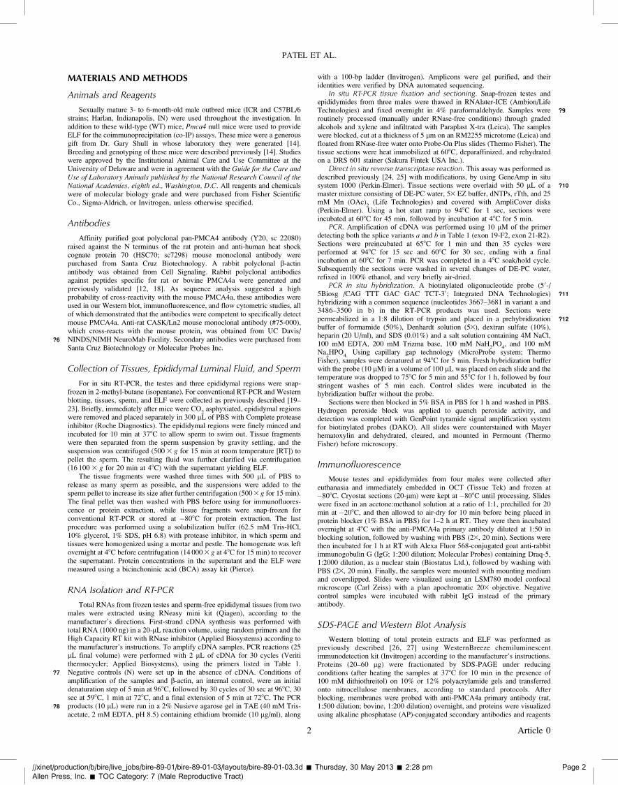
MATERIALS AND METHODS
Animals and Reagents
Sexually mature 3- to 6-month-old male outbred mice (ICR and C57BL/6strains; Harlan, Indianapolis, IN) were used throughout the investigation. Inaddition to these wild-type (WT) mice, Pmca4 null mice were used to provideELF for the coimmunoprecipitation (co-IP) assays. These mice were a generousgift from Dr. Gary Shull in whose laboratory they were generated [14].Breeding and genotyping of these mice were described previously [14]. Studieswere approved by the Institutional Animal Care and Use Committee at theUniversity of Delaware and were in agreement with the Guide for the Care andUse of Laboratory Animals published by the National Research Council of theNational Academies, eighth ed., Washington, D.C. All reagents and chemicalswere of molecular biology grade and were purchased from Fisher ScientificCo., Sigma-Aldrich, or Invitrogen, unless otherwise specified.
Antibodies
Affinity purified goat polyclonal pan-PMCA4 antibody (Y20, sc 22080)raised against the N terminus of the rat protein and anti-human heat shockcognate protein 70 (HSC70; sc7298) mouse monoclonal antibody werepurchased from Santa Cruz Biotechnology. A rabbit polyclonal b-actinantibody was obtained from Cell Signaling. Rabbit polyclonal antibodiesagainst peptides specific for rat or bovine PMCA4a were generated andpreviously validated [12, 18]. As sequence analysis suggested a highprobability of cross-reactivity with the mouse PMCA4a, these antibodies wereused in our Western blot, immunofluorescence, and flow cytometric studies, allof which demonstrated that the antibodies were competent to specifically detectmouse PMCA4a. Anti-rat CASK/Ln2 mouse monoclonal antibody (#75-000),which cross-reacts with the mouse protein, was obtained from UC Davis/NINDS/NIMH NeuroMab Facility.?6 Secondary antibodies were purchased fromSanta Cruz Biotechnology or Molecular Probes Inc.
Collection of Tissues, Epididymal Luminal Fluid, and Sperm
For in situ RT-PCR, the testes and three epididymal regions were snap-frozen in 2-methyl-butane (isopentane). For conventional RT-PCR and Westernblotting, tissues, sperm, and ELF were collected as previously described [19–23]. Briefly, immediately after mice were CO
2asphyxiated, epididymal regions
were removed and placed separately in 300 lL of PBS with Complete proteaseinhibitor (Roche Diagnostics). The epididymal regions were finely minced andincubated for 10 min at 378C to allow sperm to swim out. Tissue fragmentswere then separated from the sperm suspension by gravity settling, and thesuspension was centrifuged (500 3 g for 15 min at room temperature [RT]) topellet the sperm. The resulting fluid was further clarified via centrifugation(16 100 3 g for 20 min at 48C) with the supernatant yielding ELF.
The tissue fragments were washed three times with 500 lL of PBS torelease as many sperm as possible, and the suspensions were added to thesperm pellet to increase its size after further centrifugation (5003 g for 15 min).The final pellet was then washed with PBS before using for immunofluores-cence or protein extraction, while tissue fragments were snap-frozen forconventional RT-PCR or stored at �808C for protein extraction. The lastprocedure was performed using a solubilization buffer (62.5 mM Tris-HCl,10% glycerol, 1% SDS, pH 6.8) with protease inhibitor, in which sperm andtissues were homogenized using a mortar and pestle. The homogenate was leftovernight at 48C before centrifugation (14 000 3 g at 48C for 15 min) to recoverthe supernatant. Protein concentrations in the supernatant and the ELF weremeasured using a bicinchoninic acid (BCA) assay kit (Pierce).
RNA Isolation and RT-PCR
Total RNAs from frozen testes and sperm-free epididymal tissues from twomales were extracted using RNeasy mini kit (Qiagen), according to themanufacturer’s directions. First-strand cDNA synthesis was performed withtotal RNA (1000 ng) in a 20-lL reaction volume, using random primers and theHigh Capacity RT kit with RNase inhibitor (Applied Biosystems) according tothe manufacturer’s instructions. To amplify cDNA samples, PCR reactions (25lL final volume) were performed with 2 lL of cDNA for 30 cycles (Veritithermocycler; Applied Biosystems), using the primers listed in Table 1.Negative controls (N) were set up?7 in the absence of cDNA. Conditions ofamplification of the samples and b-actin, an internal control, were an initialdenaturation step of 5 min at 968C, followed by 30 cycles of 30 sec at 968C, 30sec at 598C, 1 min at 728C, and a final extension of 5 min at 728C. The PCRproducts (10 lL) were run in a 2% Nusieve?8 agarose gel in TAE (40 mM Tris-acetate, 2 mM EDTA, pH 8.5) containing ethidium bromide (10 lg/ml), along
with a 100-bp ladder (Invitrogen). Amplicons were gel purified, and theiridentities were verified by DNA automated sequencing.
In situ RT-PCR tissue fixation and sectioning. Snap-frozen testes andepididymides from three males were thawed in RNAlater-ICE (Ambion/LifeTechnologies) ?9and fixed overnight in 4% paraformaldehyde. Samples wereroutinely processed (manually under RNase-free conditions) through gradedalcohols and xylene and infiltrated with Paraplast X-tra (Leica). The sampleswere blocked, cut at a thickness of 5 lm on an RM2255 microtome (Leica) andfloated from RNase-free water onto Probe-On Plus slides (Thermo Fisher). Thetissue sections were heat immobilized at 608C, deparaffinized, and rehydratedon a DRS 601 stainer (Sakura Fintek USA Inc.).
Direct in situ reverse transcriptase reaction. This assay was performed asdescribed previously [24, 25] with modifications, by using GeneAmp in situsystem 1000 (Perkin-Elmer). Tissue sections were overlaid ?10with 50 lL of amaster mixture consisting of DE-PC water, 53 EZ buffer, dNTPs, rTth, and 25mM Mn (OAc)
2(Life Technologies) and covered with AmpliCover disks
(Perkin-Elmer). Using a hot start ramp to 948C for 1 sec, sections wereincubated at 608C for 45 min, followed by incubation at 48C for 5 min.
PCR. Amplification of cDNA was performed using 10 lM of the primerdetecting both the splice variants a and b in Table 1 (exon 19-F2, exon 21-R2).Sections were preincubated at 658C for 1 min and then 35 cycles wereperformed at 948C for 15 sec and 608C for 30 sec, ending with a finalincubation at 608C for 7 min. PCR was completed in a 48C soak/hold cycle.Subsequently the sections were washed in several changes of DE-PC water,refixed in 100% ethanol, and very briefly air-dried.
PCR in situ hybridization. A biotinylated oligonucleotide probe (50-/5Biosg /CAG TTT GAC GAC TCT-30; Integrated DNA Technologies) ?11
hybridizing with a common sequence (nucleotides 3667–3681 in variant a and3486–3500 in b) in the RT-PCR products was used. Sections werepermeabilized in a 1:8 dilution of trypsin ?12and placed in a prehybridizationbuffer of formamide (50%), Denhardt solution (53), dextran sulfate (10%),heparin (20 U/ml), and SDS (0.01%) and a salt solution containing 4M NaCl,100 mM EDTA, 200 mM Trizma base, 100 mM NaH
2PO
4, and 100 mM
Na2HPO
4.Using capillary gap technology (MicroProbe system; Thermo
Fisher), samples were denatured at 948C for 5 min. Fresh hybridization bufferwith the probe (10 lM) in a volume of 100 lL was placed on each slide and thetemperature was dropped to 758C for 5 min and 558C for 1 h, followed by fourstringent washes of 5 min each. Control slides were incubated in thehybridization buffer without the probe.
Sections were then blocked in 5% BSA in PBS for 1 h and washed in PBS.Hydrogen peroxide block was applied to quench peroxide activity, anddetection was completed with GenPoint tyramide signal amplification systemfor biotinylated probes (DAKO). All slides were counterstained with Mayerhematoxylin and dehydrated, cleared, and mounted in Permount (ThermoFisher) before microscopy.
Immunofluorescence
Mouse testes and epididymides from four males were collected aftereuthanasia and immediately embedded in OCT (Tissue Tek) and frozen at�808C. Cryostat sections (20-lm) were kept at�808C until processing. Slideswere fixed in an acetone:methanol solution at a ratio of 1:1, prechilled for 20min at �208C, and then allowed to air-dry for 10 min before being placed inprotein blocker (1% BSA in PBS) for 1–2 h at RT. They were then incubatedovernight at 48C with the anti-PMCA4a primary antibody diluted at 1:50 inblocking solution, followed by washing with PBS (23, 20 min). Sections werethen incubated for 1 h at RT with Alexa Fluor 568-conjugated goat anti-rabbitimmunogobulin G (IgG; 1:200 dilution; Molecular Probes) containing Draq-5,1:2000 dilution, as a nuclear stain (Biostatus Ltd.), followed by washing withPBS (23, 20 min). Finally, the samples were mounted with mounting mediumand coverslipped. Slides were visualized using an LSM780 model confocalmicroscope (Carl Zeiss) with a plan apochromatic 203 objective. Negativecontrol samples were incubated with rabbit IgG instead of the primaryantibody.
SDS-PAGE and Western Blot Analysis
Western blotting of total protein extracts and ELF was performed aspreviously described [26, 27] using WesternBreeze chemiluminescentimmunodetection kit (Invitrogen) according to the manufacturer’s instructions.Proteins (20–60 lg) were fractionated by SDS-PAGE under reducingconditions (after heating the samples at 378C for 10 min in the presence of100 mM dithiothreitol) on 10% or 12% polyacrylamide gels and transferredonto nitrocellulose membranes, according to standard protocols. Afterblocking, membranes were probed with anti-PMCA4a primary antibody (rat,1:500 dilution; bovine, 1:200 dilution) overnight, and proteins were visualizedusing alkaline phosphatase (AP)-conjugated secondary antibodies and reagents
//xinet/production/b/bire/live_jobs/bire-89-01/bire-89-01-03/layouts/bire-89-01-03.3d � Thursday, 30 May 2013 � 2:28 pm Page 2
Allen Press, Inc. � TOC Category: 7 (Male Reproductive Tract)
PATEL ET AL.
2 Article 0
dkoch
Cross-Out
close-up space [ms]
from the Western blotting detection kit. The actual amounts of proteins loadedon the gels were assessed after stripping the membrane (Restore Western blotstripping buffer; Thermo Scientific) and reprobing with b-actin antibodies(1:2000), a housekeeping protein or HSC70 (1:000), and the appropriate AP-conjugated secondary antibody. Protein bands were analyzed by densitometry(FluorChem 8800; Alpha Innotech Corp.), and the ratio of PMCA4a to theloading controls was determined and plotted.
Fractionation of Epididymal Luminal Fluid
Clarified ELF was obtained as described above from six males. Using anOptima L-70 centrifuge with a Ti60 rotor (Beckman), the ELF was subjected toultracentrifugation at 120 000 3 g for 2 h at 48C, as previously reported fromour laboratory [22], to separate the soluble (supernatant) from insolublefraction. The proteins from the supernatant were precipitated with 3 volumes ofacetone, recovered in sample buffer, and subjected to Western blotting asdescribed previously [27], along with the pellet that was also resuspended in thebuffer.
Flow Cytometric Analysis of PMCA4a in Sperm
To confirm the Western blot data for the presence of PMCA4a in sperm,immunofluorescence was performed using a modification of the procedurepublished earlier [26]. Briefly, freshly recovered caudal sperm from two tothree males per experiment were washed in PBS and fixed with 1.5%paraformaldehyde at RT for 1 h and then washed twice with PBS,permeabilized (0.1% Triton X-100 in PBS) at RT for 10 min, washed withblocking buffer (2% BSA in PBS), and incubated in blocking buffer at RT for30 min. The cells were incubated in PMCA4a primary antibody (rabbit anti-rat,1:400) or rabbit IgG (nonspecific) for the control at RT for 1 h and then washed33 with PBS and incubated in the appropriate secondary fluoresceinisothiocyanate-conjugated antibody at RT for 30 min. Sperm pellets were thenwashed 33 with PBS and resuspended in 1–1.5 mL of PBS for analysis, using aFACSCalibur unit (Becton Dickinson) flow cytometer equipped with an argonlaser at 488 nm excitation.
Sperm Uptake of PMCA4a In Vitro
Caudal sperm collected from 2–3 sexually mature mice as describedpreviously [22] were incubated in ELF recovered in PBS containing 2 lM zincacetate, pH 5.5, and protease inhibitor and clarified [22] after being subjected tocentrifugation at 3500 3 g for 20 min. This PBS vehicle for the ELF was usedfor control samples. After 30 min of incubation at 378C, control and testsamples in ELF were washed with PBS and treated with rabbit anti-ratPMCA4a antibody (1:200 dilution) at RT for 1 h. Sperm were washed 33 withPBS and incubated in secondary antibody (Alexa Fluor- 488-conjugateddonkey anti-rabbit IgG [1:200]) for 30 min). Sperm were then washed 3X withPBS and subjected to flow cytometric analysis, using a FACSCalibur (BectonDickinson) as described above.
Coimmunoprecipitation
PureProteome protein G magnetic beads (Millipore Corp.) were washedtwice in PBS with 0.1% Tween 20 and recovered by centrifugation at 2500 3 gfor 5 min. The beads were then resuspended in 100 lL of PBS with 2 lg ofanti-CASK antibodies for 2 h at 48C. After incubation, the beads werecentrifuged at 2500 3 g for 5 min and washed twice in PBS. The beads werethen resuspended in 500 lL of ELF (protein concentration of 1 mg/ml) pooledfrom all three epididymal regions of WT or Pmca4 null mice, which were usedas a control. In some experiments beads were incubated in sperm proteinextracts as previously described [26]. Following incubation in ELF for 16 h at48C, the beads were centrifuged at 2500 3 g for 5 min and washed twice withPBS. The protein bound was solubilized in sample buffer, analyzed by SDS-PAGE, and electrotransferred to nitrocellulose membranes. Coimmunoprecipi-
tated PMCA4 was detected using goat pan-PMCA4 polyclonal antibody (1:500dilution) and the Western Breeze chemiluminescent immunodetection kitmentioned above. After visualizing the PMCA4 bands, the membranes werestripped and reprobed with anti-CASK antibodies (1:1000 dilution).
Statistical Analysis
All experiments were performed at least three times. Data were analyzedusing Image J software. One-way ANOVA was performed with the means 6
SEM to detect statistical significance. Differences were considered significantat P , 0.05.
RESULTS
Pmca4 Transcript Splice Variants in Testis and EpididymisAre Differentially Expressed
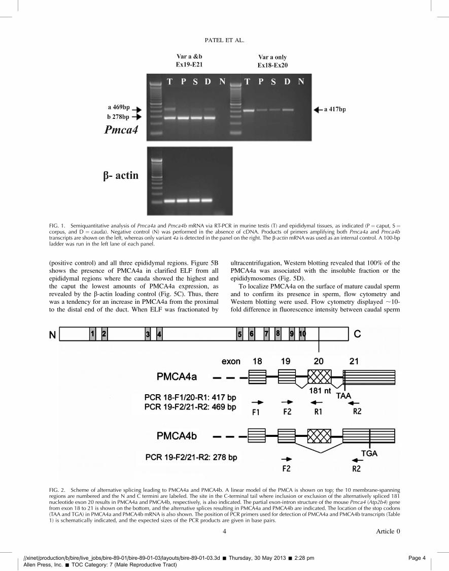
As shown in Figure 1, RT-PCR primers used to detectPmca4 transcripts (Table 1), amplified products of the expectedsizes in all three regions of the epididymis. Using b-actin as aninternal control for semiquantitative analysis, Pmca4b isexpressed evenly throughout the caput (P), corpus (S), andcauda (D) and at similar levels as in the testis (positive control). ?13
In contrast, variant 4a is differentially expressed with thestrongest expression in the testis and with increasing levelsfrom the caput to the cauda, as determined by two differentprimer pairs (Fig. 2, see scheme).
The localization of Pmca4 transcripts in the different celltypes in the testis, caput, corpus, and cauda was analyzed usingin situ RT-PCR (Fig. 3). Controls without signal are seen inFigure 3, A, C, E, and G. In the testis (Fig. 3B), expression ofthe transcripts is seen in spermatogonia, spermatocytes, andspermatids in the seminiferous tubules. In the epididymis, themRNA is localized to the epithelial lining of all three regionsand is also seen in the peritubular cells (Fig. 3, D, F, and H), aswell as in the connective tissue surrounding the tubules (Fig.3H).
Immunodetection of PMCA4a in Testis, Epididymis, ELF,Epididymosomes, and Sperm
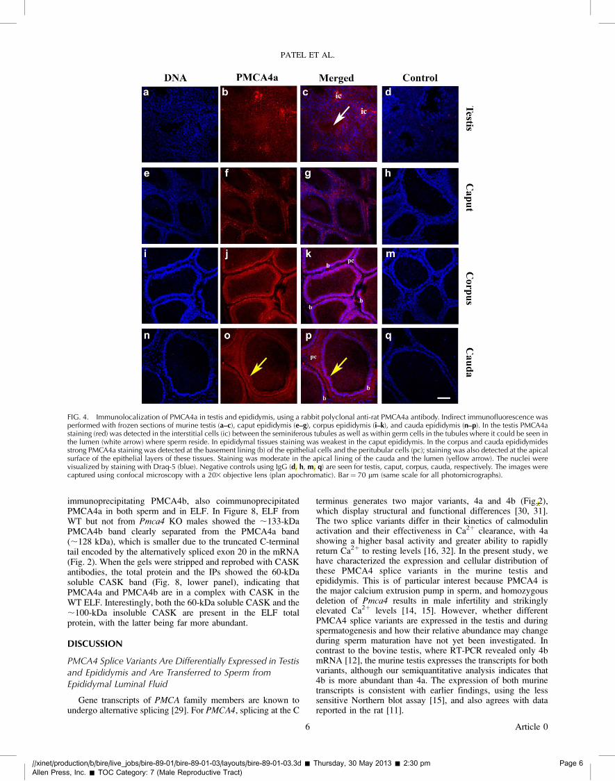
Consistent with the mRNA expression data, immunofluo-rescence revealed the presence of PMCA4a in all germ cellstages in the testis (Fig. 4, b and c). There was positiveimmunoreactivity in the lumen of the seminiferous tubules,where sperm reside (Fig. 4c, arrow). The three epididymalregions were positively stained, with the lowest intensity in thecaput (Fig. 4, f and g). Positive PMCA4a staining was detectedin the apical surface of the epithelial lining of the corpus andcauda, next to the lumen, and in the lumen (Fig. 4, j and k ando and p), . It could also be seen in the peritubular cells.Immunostaining was absent in the negative controls, whereIgG was used in place of the primary antibody (Fig. 4, d, h, m,and q).
Western blotting was performed to confirm the immuno-fluorescence findings. Figure 5A shows the presence of the;128-kDa PMCA4a band in protein extracts from the testis
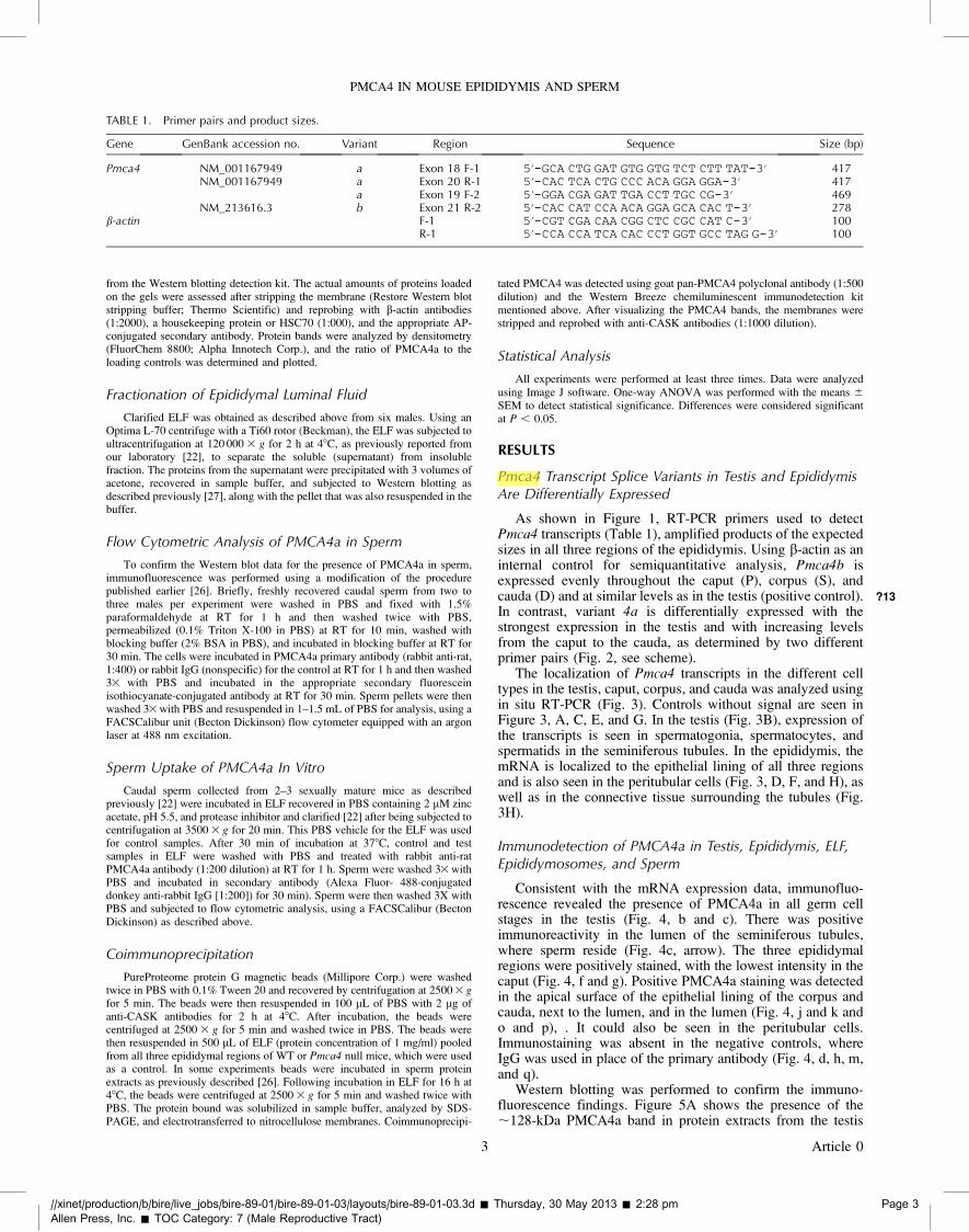
TABLE 1. Primer pairs and product sizes.
Gene GenBank accession no. Variant Region Sequence Size (bp)
Pmca4 NM_001167949 a Exon 18 F-1 50-GCA CTG GAT GTG GTG TCT CTT TAT-30 417NM_001167949 a Exon 20 R-1 50-CAC TCA CTG CCC ACA GGA GGA-30 417
a Exon 19 F-2 50-GGA CGA GAT TGA CCT TGC CG-30 469NM_213616.3 b Exon 21 R-2 50-CAC CAT CCA ACA GGA GCA CAC T-30 278
b-actin F-1 50-CGT CGA CAA CGG CTC CGC CAT C-30 100R-1 50-CCA CCA TCA CAC CCT GGT GCC TAG G-30 100
//xinet/production/b/bire/live_jobs/bire-89-01/bire-89-01-03/layouts/bire-89-01-03.3d � Thursday, 30 May 2013 � 2:28 pm Page 3
Allen Press, Inc. � TOC Category: 7 (Male Reproductive Tract)
PMCA4 IN MOUSE EPIDIDYMIS AND SPERM
3 Article 0
dkoch
Highlight
roman [ms]
dkoch
Cross-Out
delete comma and close-up space to period. [ce]
(positive control) and all three epididymal regions. Figure 5Bshows the presence of PMCA4a in clarified ELF from allepididymal regions where the cauda showed the highest andthe caput the lowest amounts of PMCA4a expression, asrevealed by the b-actin loading control (Fig. 5C). Thus, therewas a tendency for an increase in PMCA4a from the proximalto the distal end of the duct. When ELF was fractionated by
ultracentrifugation, Western blotting revealed that 100% of thePMCA4a was associated with the insoluble fraction or theepididymosomes (Fig. 5D).
To localize PMCA4a on the surface of mature caudal spermand to confirm its presence in sperm, flow cytometry andWestern blotting were used. Flow cytometry displayed ;10-fold difference in fluorescence intensity between caudal sperm
FIG. 1. Semiquantitative analysis of Pmca4a and Pmca4b mRNA via RT-PCR in murine testis (T) and epididymal tissues, as indicated (P ¼ caput, S ¼corpus, and D ¼ cauda). Negative control (N) was performed in the absence of cDNA. Products of primers amplifying both Pmca4a and Pmca4btranscripts are shown on the left, whereas only variant 4a is detected in the panel on the right. The b-actin mRNA was used as an internal control. A 100-bpladder was run in the left lane of each panel.
FIG. 2. Scheme of alternative splicing leading to PMCA4a and PMCA4b. A linear model of the PMCA is shown on top; the 10 membrane-spanningregions are numbered and the N and C termini are labeled. The site in the C-terminal tail where inclusion or exclusion of the alternatively spliced 181nucleotide exon 20 results in PMCA4a and PMCA4b, respectively, is also indicated. The partial exon-intron structure of the mouse Pmca4 (Atp2b4) genefrom exon 18 to 21 is shown on the bottom, and the alternative splices resulting in PMCA4a and PMCA4b are indicated. The location of the stop codons(TAA and TGA) in PMCA4a and PMCA4b mRNA is also shown. The position of PCR primers used for detection of PMCA4a and PMCA4b transcripts (Table1) is schematically indicated, and the expected sizes of the PCR products are given in base pairs.
//xinet/production/b/bire/live_jobs/bire-89-01/bire-89-01-03/layouts/bire-89-01-03.3d � Thursday, 30 May 2013 � 2:28 pm Page 4
Allen Press, Inc. � TOC Category: 7 (Male Reproductive Tract)
PATEL ET AL.
4 Article 0

treated with anti-PMCA4a primary antibody and that of theIgG control, confirming the specificity of the antibody and thepresence of PMCA4a on murine sperm (Fig. 6A). In addition toconfirming PMCA4a as a sperm protein, Western blotting (Fig.6B) revealed that caudal sperm have significantly (P¼ 0.003)elevated levels compared to caput sperm, where PMCA4alevels are ;5-fold lower, as revealed by densitometry (Fig.6C).
Caudal Sperm Acquire PMCA4a from the EpididymalLuminal Fluid
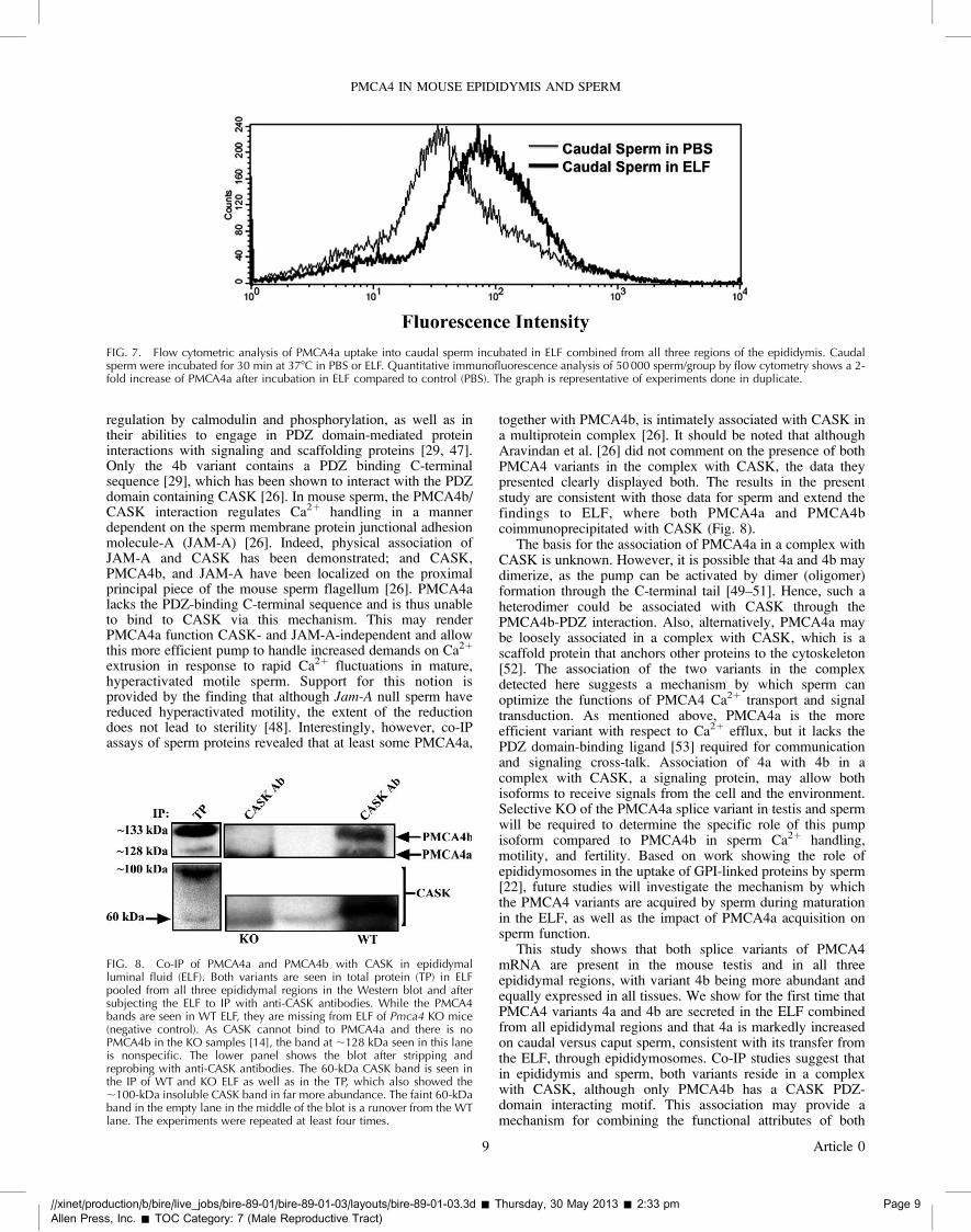
To determine whether the accumulation of PMCA4a inmature sperm might be caused by uptake from the ELF, weincubated washed caudal live sperm in purified ELF or in PBS,as a control, under physiologically relevant conditions oftemperature, pH, and the presence of zinc [28]. Quantitativeimmunofluorescence analysis of 50 000 sperm by flowcytometry showed a remarkable ;2-fold increase in PMCA4ain caudal sperm incubated for 30 min in ELF compared tosperm kept in control PBS (Fig. 7). Although the epitope for
anti-PMCA4a antibody is on the cytosolic side of themembrane, evidence that it can be accessed by the antibodyis seen from the finding that a proportion of epididymosomesretain uranyl acetate that is used for their negative staining [22].Thus, the results provide direct evidence for the uptake ofPMCA4a into mature sperm from the ELF and help explain theincreased expression of PMCA4a in caudal sperm, which areknown to be transcriptionally inactive.
Co-IP Assays Reveal PMCA4a and PMCA4b Coexist in aComplex with CASK
Recently it was shown in murine sperm that PMCA4colocalizes on the principal piece of the sperm flagellum withCASK, a signaling protein to which PMCA4b binds via itsPSD-95/Dlg/ZO-1 (PDZ) ligand at the C terminus [26]. Togain insights into the functioning of PMCA4a in relation toPMCA4b, we performed co-IP assays using anti-CASKantibodies in ELF, as well as sperm lysate. Using pan-PMCA4antibody for the Western blot in four replicated experiments,we found that PDZ domain-bearing CASK, in addition to
FIG. 3. In situ RT-PCR detects Pmca4 transcripts in testis and epididymis. Primers 19 F-2 and 21 R2 (Table) were used for PCR, and detection was by abiotinylated oligonucleotide probe recognizing both 4a and 4b transcripts, as described in Materials and Methods. Negative controls (A,C,E,G) lacking thebiotinylated detection probe showed no brown punctuate staining seen after probe hybridization. In the seminiferous tubule of the testis (B, B*),spermatogonia (sg), spermatocytes (sc), and spermatids (st) are stained in a punctuate manner, indicating expression of Pmca4 transcripts in all germ celltypes. B* shows an enlarged section of the seminiferous tubule in which germ cell types are identifiable. D, F, H) (H* and H** show enlargements) showstaining in the caput, corpus, and cauda, respectively; where the epithelial cells (e) are stained as well as the peritubular cells (pt). In the cauda transcriptsare also detected in connective tissue (ct) around the tubules and are absent from sperm in the lumen (L). Bar ¼ 25 lm.
//xinet/production/b/bire/live_jobs/bire-89-01/bire-89-01-03/layouts/bire-89-01-03.3d � Thursday, 30 May 2013 � 2:28 pm Page 5
Allen Press, Inc. � TOC Category: 7 (Male Reproductive Tract)
PMCA4 IN MOUSE EPIDIDYMIS AND SPERM
5 Article 0
dkoch
Inserted Text
insert space [ss]
dkoch
Inserted Text
insert space [ss]
dkoch
Inserted Text
insert space [ss]
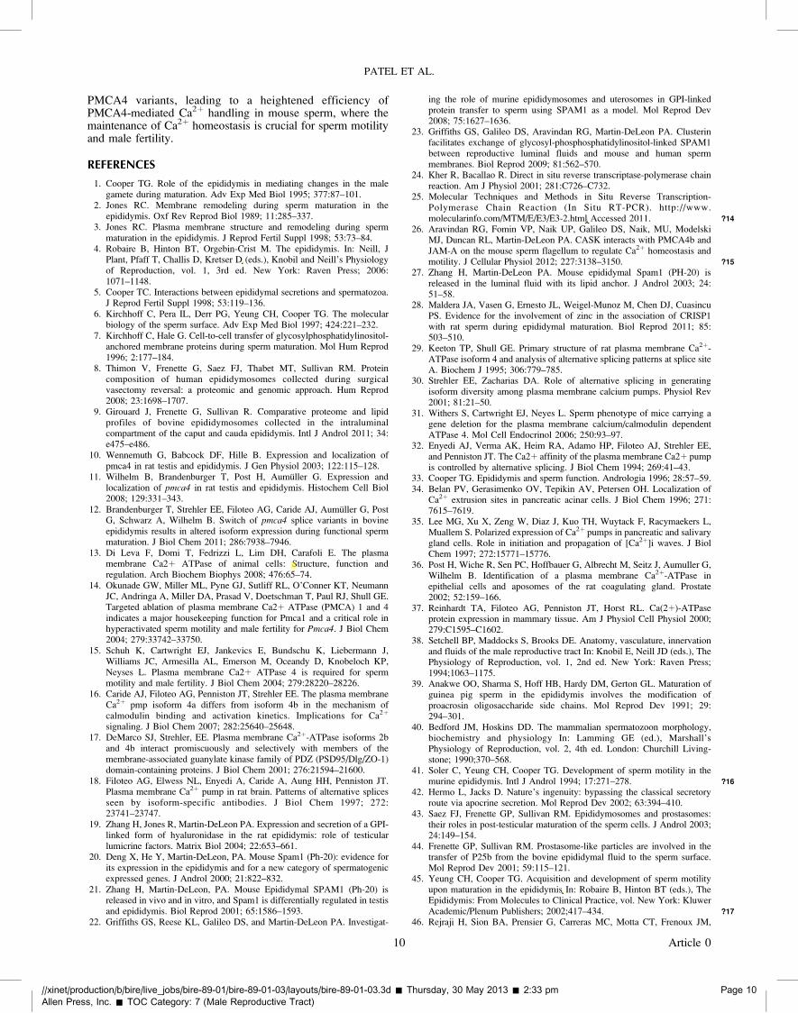
immunoprecipitating PMCA4b, also coimmunoprecipitatedPMCA4a in both sperm and in ELF. In Figure 8, ELF fromWT but not from Pmca4 KO males showed the ;133-kDaPMCA4b band clearly separated from the PMCA4a band(;128 kDa), which is smaller due to the truncated C-terminaltail encoded by the alternatively spliced exon 20 in the mRNA(Fig. 2). When the gels were stripped and reprobed with CASKantibodies, the total protein and the IPs showed the 60-kDasoluble CASK band (Fig. 8, lower panel), indicating thatPMCA4a and PMCA4b are in a complex with CASK in theWT ELF. Interestingly, both the 60-kDa soluble CASK and the;100-kDa insoluble CASK are present in the ELF totalprotein, with the latter being far more abundant.
DISCUSSION
PMCA4 Splice Variants Are Differentially Expressed in Testisand Epididymis and Are Transferred to Sperm fromEpididymal Luminal Fluid
Gene transcripts of PMCA family members are known toundergo alternative splicing [29]. For PMCA4, splicing at the C
terminus generates two major variants, 4a and 4b (Fig.2),which display structural and functional differences [30, 31].The two splice variants differ in their kinetics of calmodulinactivation and their effectiveness in Ca2þ clearance, with 4ashowing a higher basal activity and greater ability to rapidlyreturn Ca2þ to resting levels [16, 32]. In the present study, wehave characterized the expression and cellular distribution ofthese PMCA4 splice variants in the murine testis andepididymis. This is of particular interest because PMCA4 isthe major calcium extrusion pump in sperm, and homozygousdeletion of Pmca4 results in male infertility and strikinglyelevated Ca2þ levels [14, 15]. However, whether differentPMCA4 splice variants are expressed in the testis and duringspermatogenesis and how their relative abundance may changeduring sperm maturation have not yet been investigated. Incontrast to the bovine testis, where RT-PCR revealed only 4bmRNA [12], the murine testis expresses the transcripts for bothvariants, although our semiquantitative analysis indicates that4b is more abundant than 4a. The expression of both murinetranscripts is consistent with earlier findings, using the lesssensitive Northern blot assay [15], and also agrees with datareported in the rat [11].
FIG. 4. Immunolocalization of PMCA4a in testis and epididymis, using a rabbit polyclonal anti-rat PMCA4a antibody. Indirect immunofluorescence wasperformed with frozen sections of murine testis (a–c), caput epididymis (e–g), corpus epididymis (i–k), and cauda epididymis (n–p). In the testis PMCA4astaining (red) was detected in the interstitial cells (ic) between the seminiferous tubules as well as within germ cells in the tubules where it could be seen inthe lumen (white arrow) where sperm reside. In epididymal tissues staining was weakest in the caput epididymis. In the corpus and cauda epididymidesstrong PMCA4a staining was detected at the basement lining (b) of the epithelial cells and the peritubular cells (pc); staining was also detected at the apicalsurface of the epithelial layers of these tissues. Staining was moderate in the apical lining of the cauda and the lumen (yellow arrow). The nuclei werevisualized by staining with Draq-5 (blue). Negative controls using IgG (d, h, m, q) are seen for testis, caput, corpus, cauda, respectively. The images werecaptured using confocal microscopy with a 203 objective lens (plan apochromatic). Bar¼ 70 lm (same scale for all photomicrographs).
//xinet/production/b/bire/live_jobs/bire-89-01/bire-89-01-03/layouts/bire-89-01-03.3d � Thursday, 30 May 2013 � 2:30 pm Page 6
Allen Press, Inc. � TOC Category: 7 (Male Reproductive Tract)
PATEL ET AL.
6 Article 0
dkoch
Inserted Text
insert space [ss]
dkoch
Highlight
lightface [ss]
dkoch
Highlight
lightface [ss]

Importantly, transcripts for the murine PMCA4a and
PMCA4b isoforms are present in all germ cell stages
(spermatogonia, primary and secondary spermatocytes, and
spermatids), unlike those in the rat and bovine, where they are
restricted to the earlier stages [11, 12]. It is unclear if thisdiscrepancy is due to species differences or to the method oftranscript detection. In the mouse, the highly sensitive in situRT-PCR technique was used rather than the in situ transcripthybridization used in the other species [11, 12].
Our results reveal that the PMCA4 mRNA splice variants inthe murine epididymis show markedly different levels anddistribution of expression. Variant 4b was more abundant andwas equally expressed in all three epididymal regions and inthe testis. In contrast, 4a showed the highest expression in testisfollowed by the cauda and the smallest amount in the caput(Fig. 1). Although there may be species-specific differences inthe distribution of 4a in the epididymis [11, 12], generally thecauda (where sperm undergo their final maturation in the maleand where they are stored [33]) appeared to have the highestexpression of this mRNA splice variant.
The localization of PMCA4a protein in both the murinetestis and epididymal regions paralleled that of the mRNAexpression. Immunofluorescence revealed that all germ cellstages exhibit strong expression of the protein. In theepididymal regions, the protein was detected in the peritubularcells and in the epithelium where expression was seen in theapical regions lining the lumen. This finding parallels that seenin the bovine epididymis [12] but deviates from data in the rat,where PMCA4 appeared to be localized only in the basolateralmembranes of the principal and basal cells of the caputepididymal epithelium [11]. PMCA4a protein expression in theepididymis was confirmed by Western blotting that detectedthe ;128-kDa band in all tissues (Fig. 5A). The expression ofmurine PMCA4a in the apical plasma membranes of theepididymis strongly suggests that it is secreted into the lumen:PMCA concentrated in glandular epithelial cells in a variety oftissues is known to be secreted (e.g., into milk from the apicalmembrane of lactating mammary gland epithelia) [34–37].Indeed, Western blots revealed that the ELF from eachepididymal region contained the ;128-kDa PMCA4a variant(Fig. 5B) and that both 4a and 4b are present in the ELF pooledfrom all regions (Fig. 8).
As expected, the relative levels of PMCA4a in the ELF fromthe three regions mirrored those in the tissues. They increasedfrom the proximal to the distal region, with the largest amountdetected in caudal ELF where it was ;1.7-fold higher than incaput ELF. The distribution of PMCA4a in the epididymiscorrelates with that in previous studies, showing that thecorpus/cauda junction plays a major role in sperm maturation[38, 39], although other studies implicate the distal caput inmice [40, 41].
Secretion of epididymal proteins from the epididymalepithelium occurs through an apocrine pathway in which blebs(carrying membrane vesicles) dislodge from the apicalmembrane and enter the lumen, where they release membra-nous vesicles or epididymosomes [42]. These epididymosomeshave been shown to carry membrane proteins that are thentransferred to the sperm plasma membrane [43, 44]. Our studyshows for the first time that murine epididymosomes (Fig. 5D)carry PMCA4a, a 10-pass transmembrane protein. As expected,the soluble fraction of the ELF was devoid of the protein.Importantly, we found that incubation of sperm in unfraction-ated ELF increased sperm PMCA4a levels by ;2-fold after 30min of co-incubation, consistent with the role of epididymo-somes in PMCA4a transport.
FIG. 5. Western blot analysis of PMCA4a expression in murine testis andepididymal tissue (A) and ELF (B, D). Twenty and 45 lg of protein/lanewere loaded in A and B, respectively. PMCA4a was detected using a rabbitpolyclonal anti-bovine PMCA4a antibody (1:175 dilution) in A, and arabbit polyclonal anti-rat PMCA4a antibody (1:500 dilution) in B. A showsthe presence of PMCA4a in the testis and all three epididymal regions,while B shows PMCA4a in purified ELF from each of the three epididymalregions. Blots of the latter were stripped and reprobed for b-actin as aloading control, and PMCA4a band intensities were analyzed bydensitometry using Image J software. The relative expression in corpus/cauda compared to the caput is displayed in a bar graph in C. Values areexpressed as means 6 SD (n¼3), with expression highest in the cauda (*P¼ 0.014 versus caput). D shows immunoblotting of proteins from pelletand supernatant fractions of equivalent volumes of ELF, followingultracentrifugation. PMCA4a, seen in the pellet, is absent from thesupernatant. An immunopositive band (100 kDa) which is also seen in A isof unknown origin, but most likely a breakdown product as it is muchfainter in the unprocessed ELF and is not seen in B.
//xinet/production/b/bire/live_jobs/bire-89-01/bire-89-01-03/layouts/bire-89-01-03.3d � Thursday, 30 May 2013 � 2:32 pm Page 7
Allen Press, Inc. � TOC Category: 7 (Male Reproductive Tract)
PMCA4 IN MOUSE EPIDIDYMIS AND SPERM
7 Article 0
dkoch
Cross-Out
dkoch
Replacement Text
vs. [ss]

PMCA4a and PMCA4b Are Involved in Sperm Maturationand Function
Devoid of transcriptional machinery, sperm progress towardfunctional maturity in the epididymis by the acquisition ofproteins from the ELF during their transit [1–3]. Our data (Fig.6B) show that as sperm transit the epididymis, they acquirePMCA4a, resulting in an ;5-fold increase in the level ofPMCA4a in caudal sperm compared to caput sperm. This mayhelp explain the 2- to 6-times lower concentration of cytosolicCa2þ in caudal sperm than in caput sperm [45] and the gain ofmotility in the former where the potential for Ca2þ clearance ismarkedly increased. Secretion of PMCA4a in the ELF on
epididymosomes (as detected by the presence of the pump inthe ELF, specifically on the insoluble membranous vesiclefraction) provides the means by which this acquisition occurs.Epididymosomes are known to be the vehicles for proteintransfer in the ELF [43, 44] and have been previously identifiedin the mouse [22, 46]. Because both PMCA4 splice variants arepresent in the ELF, it is highly likely that the 4b variant is alsoacquired by sperm during their maturation. The combination ofPMCA4 splice variants in a certain ratio on the sperm surface islikely required to optimize the PMCA4-mediated Ca2þ
regulation in sperm.Why might the two splice variants need to be coexpressed in
mature sperm? PMCA4a and PMCA4b differ in their
FIG. 6. Flow cytometry and Western blot analyses reveal the presence of PMCA4a in murine caudal sperm. A) Quantitative immunofluorescenceanalysis of 50 000 cells treated with the rabbit polyclonal anti-bovine PMCA4a primary antibody (1:20 dilution) shows a marked right shift of the peak,indicating increased fluorescence intensity (;10-fold) compared to the controls, which were treated with rabbit IgG for flow cytometry. B) Westernblotting using anti-rat PMCA4a primary antibody (1:400 dilution) shows significantly more PMCA4a in caudal (CD) than caput (CT) sperm. The blot wasstripped and reprobed for HSC-70 (70 kDa) as a loading control. The left lane shows the presence of PMCA4a in caudal ELF. C) Densitometric analysis ofthe blots was performed, using Image J software, to determine relative expression of PMCA4a in caput and caudal sperm. The bar graph shows a ;5-foldincrease in the latter; values are expressed as means 6 SD (n¼ 3). *P¼ 0.003.
//xinet/production/b/bire/live_jobs/bire-89-01/bire-89-01-03/layouts/bire-89-01-03.3d � Thursday, 30 May 2013 � 2:32 pm Page 8
Allen Press, Inc. � TOC Category: 7 (Male Reproductive Tract)
PATEL ET AL.
8 Article 0
regulation by calmodulin and phosphorylation, as well as intheir abilities to engage in PDZ domain-mediated proteininteractions with signaling and scaffolding proteins [29, 47].Only the 4b variant contains a PDZ binding C-terminalsequence [29], which has been shown to interact with the PDZdomain containing CASK [26]. In mouse sperm, the PMCA4b/CASK interaction regulates Ca2þ handling in a mannerdependent on the sperm membrane protein junctional adhesionmolecule-A (JAM-A) [26]. Indeed, physical association ofJAM-A and CASK has been demonstrated; and CASK,PMCA4b, and JAM-A have been localized on the proximalprincipal piece of the mouse sperm flagellum [26]. PMCA4alacks the PDZ-binding C-terminal sequence and is thus unableto bind to CASK via this mechanism. This may renderPMCA4a function CASK- and JAM-A-independent and allowthis more efficient pump to handle increased demands on Ca2þ
extrusion in response to rapid Ca2þ fluctuations in mature,hyperactivated motile sperm. Support for this notion isprovided by the finding that although Jam-A null sperm havereduced hyperactivated motility, the extent of the reductiondoes not lead to sterility [48]. Interestingly, however, co-IPassays of sperm proteins revealed that at least some PMCA4a,
together with PMCA4b, is intimately associated with CASK ina multiprotein complex [26]. It should be noted that althoughAravindan et al. [26] did not comment on the presence of bothPMCA4 variants in the complex with CASK, the data theypresented clearly displayed both. The results in the presentstudy are consistent with those data for sperm and extend thefindings to ELF, where both PMCA4a and PMCA4bcoimmunoprecipitated with CASK (Fig. 8).
The basis for the association of PMCA4a in a complex withCASK is unknown. However, it is possible that 4a and 4b maydimerize, as the pump can be activated by dimer (oligomer)formation through the C-terminal tail [49–51]. Hence, such aheterodimer could be associated with CASK through thePMCA4b-PDZ interaction. Also, alternatively, PMCA4a maybe loosely associated in a complex with CASK, which is ascaffold protein that anchors other proteins to the cytoskeleton[52]. The association of the two variants in the complexdetected here suggests a mechanism by which sperm canoptimize the functions of PMCA4 Ca2þ transport and signaltransduction. As mentioned above, PMCA4a is the moreefficient variant with respect to Ca2þ efflux, but it lacks thePDZ domain-binding ligand [53] required for communicationand signaling cross-talk. Association of 4a with 4b in acomplex with CASK, a signaling protein, may allow bothisoforms to receive signals from the cell and the environment.Selective KO of the PMCA4a splice variant in testis and spermwill be required to determine the specific role of this pumpisoform compared to PMCA4b in sperm Ca2þ handling,motility, and fertility. Based on work showing the role ofepididymosomes in the uptake of GPI-linked proteins by sperm[22], future studies will investigate the mechanism by whichthe PMCA4 variants are acquired by sperm during maturationin the ELF, as well as the impact of PMCA4a acquisition onsperm function.
This study shows that both splice variants of PMCA4mRNA are present in the mouse testis and in all threeepididymal regions, with variant 4b being more abundant andequally expressed in all tissues. We show for the first time thatPMCA4 variants 4a and 4b are secreted in the ELF combinedfrom all epididymal regions and that 4a is markedly increasedon caudal versus caput sperm, consistent with its transfer fromthe ELF, through epididymosomes. Co-IP studies suggest thatin epididymis and sperm, both variants reside in a complexwith CASK, although only PMCA4b has a CASK PDZ-domain interacting motif. This association may provide amechanism for combining the functional attributes of both
FIG. 7. Flow cytometric analysis of PMCA4a uptake into caudal sperm incubated in ELF combined from all three regions of the epididymis. Caudalsperm were incubated for 30 min at 378C in PBS or ELF. Quantitative immunofluorescence analysis of 50 000 sperm/group by flow cytometry shows a 2-fold increase of PMCA4a after incubation in ELF compared to control (PBS). The graph is representative of experiments done in duplicate.
FIG. 8. Co-IP of PMCA4a and PMCA4b with CASK in epididymalluminal fluid (ELF). Both variants are seen in total protein (TP) in ELFpooled from all three epididymal regions in the Western blot and aftersubjecting the ELF to IP with anti-CASK antibodies. While the PMCA4bands are seen in WT ELF, they are missing from ELF of Pmca4 KO mice(negative control). As CASK cannot bind to PMCA4a and there is noPMCA4b in the KO samples [14], the band at ;128 kDa seen in this laneis nonspecific. The lower panel shows the blot after stripping andreprobing with anti-CASK antibodies. The 60-kDa CASK band is seen inthe IP of WT and KO ELF as well as in the TP, which also showed the;100-kDa insoluble CASK band in far more abundance. The faint 60-kDaband in the empty lane in the middle of the blot is a runover from the WTlane. The experiments were repeated at least four times.
//xinet/production/b/bire/live_jobs/bire-89-01/bire-89-01-03/layouts/bire-89-01-03.3d � Thursday, 30 May 2013 � 2:33 pm Page 9
Allen Press, Inc. � TOC Category: 7 (Male Reproductive Tract)
PMCA4 IN MOUSE EPIDIDYMIS AND SPERM
9 Article 0
PMCA4 variants, leading to a heightened efficiency ofPMCA4-mediated Ca2þ handling in mouse sperm, where themaintenance of Ca2þ homeostasis is crucial for sperm motilityand male fertility.
REFERENCES
1. Cooper TG. Role of the epididymis in mediating changes in the malegamete during maturation. Adv Exp Med Biol 1995; 377:87–101.
2. Jones RC. Membrane remodeling during sperm maturation in theepididymis. Oxf Rev Reprod Biol 1989; 11:285–337.
3. Jones RC. Plasma membrane structure and remodeling during spermmaturation in the epididymis. J Reprod Fertil Suppl 1998; 53:73–84.
4. Robaire B, Hinton BT, Orgebin-Crist M. The epididymis. In: Neill, JPlant, Pfaff T, Challis D, Kretser D (eds.), Knobil and Neill’s Physiologyof Reproduction, vol. 1, 3rd ed. New York: Raven Press; 2006:1071–1148.
5. Cooper TC. Interactions between epididymal secretions and spermatozoa.J Reprod Fertil Suppl 1998; 53:119–136.
6. Kirchhoff C, Pera IL, Derr PG, Yeung CH, Cooper TG. The molecularbiology of the sperm surface. Adv Exp Med Biol 1997; 424:221–232.
7. Kirchhoff C, Hale G. Cell-to-cell transfer of glycosylphosphatidylinositol-anchored membrane proteins during sperm maturation. Mol Hum Reprod1996; 2:177–184.
8. Thimon V, Frenette G, Saez FJ, Thabet MT, Sullivan RM. Proteincomposition of human epididymosomes collected during surgicalvasectomy reversal: a proteomic and genomic approach. Hum Reprod2008; 23:1698–1707.
9. Girouard J, Frenette G, Sullivan R. Comparative proteome and lipidprofiles of bovine epididymosomes collected in the intraluminalcompartment of the caput and cauda epididymis. Intl J Androl 2011; 34:e475–e486.
10. Wennemuth G, Babcock DF, Hille B. Expression and localization ofpmca4 in rat testis and epididymis. J Gen Physiol 2003; 122:115–128.
11. Wilhelm B, Brandenburger T, Post H, Aumuller G. Expression andlocalization of pmca4 in rat testis and epididymis. Histochem Cell Biol2008; 129:331–343.
12. Brandenburger T, Strehler EE, Filoteo AG, Caride AJ, Aumuller G, PostG, Schwarz A, Wilhelm B. Switch of pmca4 splice variants in bovineepididymis results in altered isoform expression during functional spermmaturation. J Biol Chem 2011; 286:7938–7946.
13. Di Leva F, Domi T, Fedrizzi L, Lim DH, Carafoli E. The plasmamembrane Ca2þ ATPase of animal cells: Structure, function andregulation. Arch Biochem Biophys 2008; 476:65–74.
14. Okunade GW, Miller ML, Pyne GJ, Sutliff RL, O’Conner KT, NeumannJC, Andringa A, Miller DA, Prasad V, Doetschman T, Paul RJ, Shull GE.Targeted ablation of plasma membrane Ca2þ ATPase (PMCA) 1 and 4indicates a major housekeeping function for Pmca1 and a critical role inhyperactivated sperm motility and male fertility for Pmca4. J Biol Chem2004; 279:33742–33750.
15. Schuh K, Cartwright EJ, Jankevics E, Bundschu K, Liebermann J,Williams JC, Armesilla AL, Emerson M, Oceandy D, Knobeloch KP,Neyses L. Plasma membrane Ca2þ ATPase 4 is required for spermmotility and male fertility. J Biol Chem 2004; 279:28220–28226.
16. Caride AJ, Filoteo AG, Penniston JT, Strehler EE. The plasma membraneCa2þ pmp isoform 4a differs from isoform 4b in the mechanism ofcalmodulin binding and activation kinetics. Implications for Ca2þ
signaling. J Biol Chem 2007; 282:25640–25648.17. DeMarco SJ, Strehler, EE. Plasma membrane Ca2þ-ATPase isoforms 2b
and 4b interact promiscuously and selectively with members of themembrane-associated guanylate kinase family of PDZ (PSD95/Dlg/ZO-1)domain-containing proteins. J Biol Chem 2001; 276:21594–21600.
18. Filoteo AG, Elwess NL, Enyedi A, Caride A, Aung HH, Penniston JT.Plasma membrane Ca2þ pump in rat brain. Patterns of alternative splicesseen by isoform-specific antibodies. J Biol Chem 1997; 272:23741–23747.
19. Zhang H, Jones R, Martin-DeLeon PA. Expression and secretion of a GPI-linked form of hyaluronidase in the rat epididymis: role of testicularlumicrine factors. Matrix Biol 2004; 22:653–661.
20. Deng X, He Y, Martin-DeLeon, PA. Mouse Spam1 (Ph-20): evidence forits expression in the epididymis and for a new category of spermatogenicexpressed genes. J Androl 2000; 21:822–832.
21. Zhang H, Martin-DeLeon, PA. Mouse Epididymal SPAM1 (Ph-20) isreleased in vivo and in vitro, and Spam1 is differentially regulated in testisand epididymis. Biol Reprod 2001; 65:1586–1593.
22. Griffiths GS, Reese KL, Galileo DS, and Martin-DeLeon PA. Investigat-
ing the role of murine epididymosomes and uterosomes in GPI-linkedprotein transfer to sperm using SPAM1 as a model. Mol Reprod Dev2008; 75:1627–1636.
23. Griffiths GS, Galileo DS, Aravindan RG, Martin-DeLeon PA. Clusterinfacilitates exchange of glycosyl-phosphosphatidylinositol-linked SPAM1between reproductive luminal fluids and mouse and human spermmembranes. Biol Reprod 2009; 81:562–570.
24. Kher R, Bacallao R. Direct in situ reverse transcriptase-polymerase chainreaction. Am J Physiol 2001; 281:C726–C732.
25. Molecular Techniques and Methods in Situ Reverse Transcription-Polymerase Chain Reaction (In Situ RT-PCR). http://www.molecularinfo.com/MTM/E/E3/E3-2.html Accessed 2011. ?14
26. Aravindan RG, Fomin VP, Naik UP, Galileo DS, Naik, MU, ModelskiMJ, Duncan RL, Martin-DeLeon PA. CASK interacts with PMCA4b andJAM-A on the mouse sperm flagellum to regulate Ca2þ homeostasis andmotility. J Cellular Physiol 2012; 227:3138–3150. ?15
27. Zhang H, Martin-DeLeon PA. Mouse epididymal Spam1 (PH-20) isreleased in the luminal fluid with its lipid anchor. J Androl 2003; 24:51–58.
28. Maldera JA, Vasen G, Ernesto JL, Weigel-Munoz M, Chen DJ, CuasincuPS. Evidence for the involvement of zinc in the association of CRISP1with rat sperm during epididymal maturation. Biol Reprod 2011; 85:503–510.
29. Keeton TP, Shull GE. Primary structure of rat plasma membrane Ca2þ-ATPase isoform 4 and analysis of alternative splicing patterns at splice siteA. Biochem J 1995; 306:779–785.
30. Strehler EE, Zacharias DA. Role of alternative splicing in generatingisoform diversity among plasma membrane calcium pumps. Physiol Rev2001; 81:21–50.
31. Withers S, Cartwright EJ, Neyes L. Sperm phenotype of mice carrying agene deletion for the plasma membrane calcium/calmodulin dependentATPase 4. Mol Cell Endocrinol 2006; 250:93–97.
32. Enyedi AJ, Verma AK, Heim RA, Adamo HP, Filoteo AJ, Strehler EE,and Penniston JT. The Ca2þ affinity of the plasma membrane Ca2þ pumpis controlled by alternative splicing. J Biol Chem 1994; 269:41–43.
33. Cooper TG. Epididymis and sperm function. Andrologia 1996; 28:57–59.34. Belan PV, Gerasimenko OV, Tepikin AV, Petersen OH. Localization of
Ca2þ extrusion sites in pancreatic acinar cells. J Biol Chem 1996; 271:7615–7619.
35. Lee MG, Xu X, Zeng W, Diaz J, Kuo TH, Wuytack F, Racymaekers L,Muallem S. Polarized expression of Ca2þ pumps in pancreatic and salivarygland cells. Role in initiation and propagation of [Ca2þ]i waves. J BiolChem 1997; 272:15771–15776.
36. Post H, Wiche R, Sen PC, Hoffbauer G, Albrecht M, Seitz J, Aumuller G,Wilhelm B. Identification of a plasma membrane Ca2þ-ATPase inepithelial cells and aposomes of the rat coagulating gland. Prostate2002; 52:159–166.
37. Reinhardt TA, Filoteo AG, Penniston JT, Horst RL. Ca(2þ)-ATPaseprotein expression in mammary tissue. Am J Physiol Cell Physiol 2000;279:C1595–C1602.
38. Setchell BP, Maddocks S, Brooks DE. Anatomy, vasculature, innervationand fluids of the male reproductive tract In: Knobil E, Neill JD (eds.), ThePhysiology of Reproduction, vol. 1, 2nd ed. New York: Raven Press;1994;1063–1175.
39. Anakwe OO, Sharma S, Hoff HB, Hardy DM, Gerton GL. Maturation ofguinea pig sperm in the epididymis involves the modification ofproacrosin oligosaccharide side chains. Mol Reprod Dev 1991; 29:294–301.
40. Bedford JM, Hoskins DD. The mammalian spermatozoon morphology,biochemistry and physiology In: Lamming GE (ed.), Marshall’sPhysiology of Reproduction, vol. 2, 4th ed. London: Churchill Living-stone; 1990;370–568.
41. Soler C, Yeung CH, Cooper TG. Development of sperm motility in themurine epididymis. Intl J Androl 1994; 17:271–278. ?16
42. Hermo L, Jacks D. Nature’s ingenuity: bypassing the classical secretoryroute via apocrine secretion. Mol Reprod Dev 2002; 63:394–410.
43. Saez FJ, Frenette GP, Sullivan RM. Epididymosomes and prostasomes:their roles in post-testicular maturation of the sperm cells. J Androl 2003;24:149–154.
44. Frenette GP, Sullivan RM. Prostasome-like particles are involved in thetransfer of P25b from the bovine epididymal fluid to the sperm surface.Mol Reprod Dev 2001; 59:115–121.
45. Yeung CH, Cooper TG. Acquisition and development of sperm motilityupon maturation in the epididymis In: Robaire B, Hinton BT (eds.), TheEpididymis: From Molecules to Clinical Practice, vol. New York: KluwerAcademic/Plenum Publishers; 2002;417–434. ?17
46. Rejraji H, Sion BA, Prensier G, Carreras MC, Motta CT, Frenoux JM,
//xinet/production/b/bire/live_jobs/bire-89-01/bire-89-01-03/layouts/bire-89-01-03.3d � Thursday, 30 May 2013 � 2:33 pm Page 10
Allen Press, Inc. � TOC Category: 7 (Male Reproductive Tract)
PATEL ET AL.
10 Article 0
dkoch
Highlight
s [ss]
dkoch
Cross-Out
delete comma [ss]
dkoch
Cross-Out
delete comma [ss]
dkoch
Cross-Out
delete comma [ss]
dkoch
Inserted Text
. [ss]
dkoch
Cross-Out
delete comma [ss]
dkoch
Inserted Text
. [ss]
dkoch
Cross-Out
dkoch
Replacement Text
Neill J, Plant T, Pfaff D, Challis J, Kretser D [ce]
Vericel E, Grizzard G, Vernet P, Drevet JR. Lipid remodeling of murineepididymosomes and spermatozoa during epididymal maturation. BiolReprod 2006; 74:1104–1113.
47. Strehler EE, Caride AJ, Filoteo AG, Xiong Y, Penniston JT, Enyedi A.Plasma membrane Ca2þ ATPases as dynamic regulators of cellularcalcium handling. Ann N Y Acad Sci 2007; 1099:226–236.
48. Shao M, Ghosh A, Cooke VG, Naik UP, Martin-DeLeon PA. JAM-A ispresent in mammalian spermatozoa where it is essential for normalmotility. Dev Biol 2008; 313:246–255.
49. Kosk-Kopsicka D, and Bzdega T. Activation of the erythrocyte Ca2þ-ATPase by either self-association or interaction with calmodulin. J BiolChem 1988; 263:18184–18189.
50. Vorherr T, Kessler T, Hofmann F, Carafoli E. The calmodulin-binding
domain mediates the self-association of the plasma membrane Ca2þpump. J Biol Chem 1991; 266:22–27.
51. Levi V, Rossi JP, Castello PR, Gonzalez Flecha FL. Oligomerization of
the plasma membrane calcium pump involves two regions with different
thermal stability. FEBS Lett 2003; 483:99–103.
52. Funke L, Dakoji S, Bredt DS. Membrane-associated guanylate kinases
regulate adhesion and plasticity at cell junctions. Ann Rev Biochem 2005;
74:219–245. ?18
53. Kim EY, DeMarco SJ, Marfatia SM, Chishti AH, Sheng M, Strehler EE.
Plasma membrane Ca2þ ATPase isoform 4b binds to membrane-
associated guanylate kinase (MAGUK) proteins via their PDZ (PSD-95/
Dlg/ZO-1). J Biol Chem 1998; 273:1591–1595.
//xinet/production/b/bire/live_jobs/bire-89-01/bire-89-01-03/layouts/bire-89-01-03.3d � Thursday, 30 May 2013 � 2:33 pm Page 11
Allen Press, Inc. � TOC Category: 7 (Male Reproductive Tract)
PMCA4 IN MOUSE EPIDIDYMIS AND SPERM
11 Article 0
dkoch
Cross-Out
delete [ss]

Queries for bire-89-01-03
1. Author: This article has been lightly edited for grammar, style, and usage. Please compare against your originaldocument and make changes on these pages. Please limit your corrections to substantive changes that affectmeaning. If no change is required in response to a question, please write ‘‘OK as set’’ in the margin. Copy editor.
2. Author: Your manuscript has been typeset to reflect its final print format. Typesetting programs mayinadvertently reformat text in a way that changes the meaning, e.g., genetic nomenclature italics. Therefore,please check the nomenclature in your proof carefully to make sure there have been no inadvertent formattingerrors. Copy editor.
3. Author: Support line: please spell out INBRE. Copyeditor.4. Editor: this paper has been tagged for PMC deposit; please check and confirm funding footnote.5. Editor: Please confirm wording of summary statement: PMCA4a and 4b regulate Ca2þ homeostasis and fertility,
are expressed in the epididymis, and secreted in the luminal fluid where they are acquired by sperm duringepididymal maturation. Copy editor
6. Author: In ‘‘Anti-rat CASK/Ln2’’ please spell out ‘‘UC Davis/NINDS/NIMH’’ at first mention. Copyeditor.7. Author: In ‘‘Negative controls (N) were set up’’ please explain what ‘‘(N)’’ refers to. Copyeditor.8. Author: In ‘‘The PCR products’’ if ‘‘Nusieve’’ is a commercial product, please supply mfr. and location for
consistency. Copyeditor.9. Author: In ‘‘Snap-frozen testes’’ please spell out ‘‘ICE’’ if it is an abbreviation. Copyeditor.
10. Author: In ‘‘Tissue sections were overlaid’’ please spell out DE-PC, dNTPs, and rTth at first mention. Copyeditor.11. Author: In ‘‘A biotinylated oligonucleotide probe’’ please explain the designation ‘‘50-/5Biosg’’. Copyeditor.12. Author: In ‘‘Sections were permeabilized’’ should there be some other solution in addition to trypsin? Copyeditor.13. Author: In ‘‘Using b-actin’’ correct that the reader will see letters P, S, and D in Fig. 1? Copyeditor.14. Author: Ref. 25: please supply the article pub year and Month and day URL was accessed. Copyeditor.15. Author: Ref. 26: journal ‘‘J Cellular Physiol’’ not found; please verify. Copyeditor.16. Author: Ref. 41: journal ‘‘Intl J Androl’’ not found; do you mean ‘‘Int J Androl’’? please verify. Copyeditor.17. Author: Ref. 45: please supply volume no. Copyeditor.18. Author: Ref. 52: journal ‘‘Ann Rev Biochem’’ not found; please verify. Copyeditor.
//xinet/production/b/bire/live_jobs/bire-89-01/bire-89-01-03/layouts/bire-89-01-03q.3d Thursday, 30 May 2013 2:33 pm Allen Press, Inc. Page 1