Inhibition of differentiation in myoblasts deprived of the interferon-related protein PC4
Upload
independentCategory
view
0download
0
BioMed CentralBMC Molecular Biology
ss
Open AcceResearch articleGenetic correction of splice site mutation in purified and enriched myoblasts isolated from mdx5cv miceKatie Maguire1,2, Takayuki Suzuki1, Darlise DiMatteo1, Hetal Parekh-Olmedo1 and Eric Kmiec*1Address: 1Department of Biological Sciences, University of Delaware, Newark, DE 19716, USA and 2Department of Neurology and Neurological Sciences, Stanford University School of Medicine, Stanford, CA 94305, USA
Email: Katie Maguire - [email protected]; Takayuki Suzuki - [email protected]; Darlise DiMatteo - [email protected]; Hetal Parekh-Olmedo - [email protected]; Eric Kmiec* - [email protected]
* Corresponding author
AbstractBackground: Duchenne Muscular Dystrophy (DMD) is an X-linked genetic disorder that resultsin the production of a dysfunctional form of the protein, dystrophin. The mdx5cv mouse is a modelof DMD in which a point mutation in exon 10 of the dystrophin gene creates an artificial splice site.As a result, a 53 base pair deletion of exon 10 occurs with a coincident creation of a frameshift anda premature stop codon. Using primary myoblasts from mdx5cv mice, single-stranded DNAoligonucleotides were designed to correct this DNA mutation.
Results: Single-stranded DNA oligonucleotides that were designed to repair this splice sitemutation corrected the mutation in the gene and restored expression of wild-type dystrophin. Thisrepair was validated at the DNA, RNA and protein level. We also report that the frequency ofgenetic repair of the mdx mutation can be enhanced if RNAi is used to suppress expression of therecombinase inhibitor protein Msh2 in cultures containing myoblasts but not in those heavilyenriched in myoblasts.
Conclusion: Exogenous manipulations, such as RNAi, are certainly feasible and possibly requiredto increase the successful application of gene repair in some primary or progenitor muscle cells.
BackgroundDuchenne Muscular Dystrophy (DMD) is an X-linkedrecessive disorder with an incidence of 1 in 3,500 liveborn males. It is caused by the dysfunction of dystrophin(DMD), a large 427 kDa cytoplasmic protein that forms alink between actin filaments and the dystrophin-glyco-protein (DGC) complex [1]. The loss of dystrophin isthought to affect the integrity of the cell membrane lead-ing to a disruption in cellular calcium homeostasis andeventually cell necrosis [1-3]. At the genetic level, one-
third of boys afflicted with DMD have point mutations,insertions or deletions while the remaining two-thirdshave gross deletions or rearrangements within the 2.5 Mbgene [4] [OMIM #310200 Muscular Dystrophy, Dystro-phy Type, DMD] and all generally cause a translationalframeshift.
Therapeutic strategies such as gene replacement have beenused to restore protein expression by effectively re-engi-neering dystrophin cDNA. In addition, attempts have
Published: 23 February 2009
BMC Molecular Biology 2009, 10:15 doi:10.1186/1471-2199-10-15
Received: 10 November 2008Accepted: 23 February 2009
This article is available from: http://www.biomedcentral.com/1471-2199/10/15
© 2009 Maguire et al; licensee BioMed Central Ltd. This is an Open Access article distributed under the terms of the Creative Commons Attribution License (http://creativecommons.org/licenses/by/2.0), which permits unrestricted use, distribution, and reproduction in any medium, provided the original work is properly cited.
Page 1 of 16(page number not for citation purposes)

BMC Molecular Biology 2009, 10:15 http://www.biomedcentral.com/1471-2199/10/15
been made to introduce smaller functional homologs ofDMD to cells in an attempt to reverse the muscle degrada-tion process [5-8]. Vectors, which carry these constructs,however, often elicit an immune response [8,9]. An RNA/DNA chimeric oligonucleotide (chimera) has been usedsuccessfully to correct the splice site mutation to generatea genetic alteration in the dystrophin gene, in both ex vivoand in vivo studies in mouse and dog models of DMD [10-13]. In a pioneering study, Bartlett et al. [10] showed thecorrection of the canine dystrophin gene within skeletalmuscle fibers after injecting the chimera into the cranialtibialis of Golden Retrievers. The production of wild-typedystrophin was restored and could be detected for up to48 weeks at the site of the injection. Rando et al. [11] dem-onstrated chimera-directed repair of the dystrophin genein the mouse extending the results from the dog model;once again the corrected cells were again localized to theinjection site.
Antisense oligonucleotides also have been used to alterthe splicing of the dystrophin message in a process gener-ally termed, exon skipping [14-16]. A large number ofexciting reports have demonstrated the feasibility of alter-ing the splicing patterns of the mutant DMD transcript[15,17-21]. While this approach is likely to have signifi-cant therapeutic benefit, the original DMD mutation(s)remains and thus a continuum of antisense ODN deliverywill be required [22].
When muscle fibers are damaged within the body, a pop-ulation of quiescent precursor muscle cells, or satellitecells, become activated myoblasts [23,24], rapidly divideand initiate the regeneration of the injured muscle [24]. Inthe case of DMD, the muscle is thought to be constantlygoing through cycles of degeneration and regenerationdue to the instability of the musculature and, therefore,abundantly populated with myogenic precursor cells.Thus, an important long term therapeutic approach thenwould be to isolate and genetically correct the myoblastsat the DNA level with subsequent transplantation to theanimal model in order to restore functional dystrophin tothe muscle fibers.
We have begun to develop a more defined ex vivo strategyin which modified single-stranded oligonucleotides aredesigned to repair the mutant dystrophin (ex vivo) in cellcultures enriched in myoblasts. A current challenge is toestablish a frequency of repair that leads to a level of dys-trophin expression that can be detected in targeted myob-lasts. Previously, Bertoni et al. [25] have used fluorescentlylabeled oligonucleotides to correct a point mutation incells derived from the dystrophic mdx5cv mice. We nowrepeat the correction of Dmd point mutation using unla-beled modified single stranded oligonucleotides andestablish novel quantitative assays to measure the dys-
trophin production in myoblasts. We also stimulate thecorrection reaction by using RNAi against Msh2, a knowninhibitor of the gene repair reaction [26] and observehigher levels of gene repair.
ResultsIsolation of primary cultures from dystrophic mdx5cv mice containing myoblasts with determination of isolation efficiencyIn the natural muscle environment, satellite cells rest in aquiescent state along the periphery of an intact musclefiber. When muscle fibers are damaged, muscle precursorcells, referred to as myoblasts, divide and fuse with thedamaged muscle fibers to repair them [see [27] forreview]. In order to isolate the muscle precursor cells forgene repair experiments, muscle tissue was removed fromthe limbs of 1 to 3 days old mdx5cv mice, enzymaticallydigested and plated. A pre-plating technique was used topurify these cells from fibroblasts present in the explant[27,28]. It is known that the fibroblasts adhere to the platerapidly, usually within 2 hours, and, therefore, the pre-plating technique is used them from the culture by passag-ing the supernatant at a 2 hour time point (PP1) and againat a 24 hour time point (PP2) [28]. Thus, the supernatantis passaged until PP4 at which time cells are harvested todetermine the efficiency of myoblast isolation by incuba-tion with an antibody against desmin (expressed in myob-lasts). We estimate the efficiency of myoblast isolation inthis experiment to be 53% as determined by counting thenumber of desmin positive cells and dividing this numberby the number of total cells observed in multiple fields ofview (data not shown). The remaining 47% are mostlikely fibroblasts. Thus, in the experiments outlinedbelow, we utilize cultures that contain approximately50% myoblasts and 50% fibroblasts. Primarily myoblastsare a target because only corrected myoblasts that fuse toform myotubes will be capable of expressing correcteddystrophin.
In order to determine the efficiency of gene repair of thechromosomal dystrophin gene in myoblasts, it wasimportant to determine the level of transfection of oligo-nucleotides. Oligonucleotides in varying amounts werecomplexed with Fugene 6 to observe the levels of uptakewithin the myoblasts. Fugene mediated transfection onlyshowed a 4–6% uptake of the FAM-labeled oligonucle-otides (data not shown). Therefore, we chose to use jet-PEI, which is formulated specifically for delivery ofoligonucleotides to deliver the ODN into the primary cul-tures. Myoblast cultures were seeded at increasing densi-ties and lipoplexes composed of 6 μL of jetPEI and 3 μg ofODN were added to cultures with readout of uptake byFACS analysis after 24 hours. There was no difference inthe amount of oligonucleotide uptake at different cellnumbers as shown in Figure 1 (average uptake, 36%).
Page 2 of 16(page number not for citation purposes)
BMC Molecular Biology 2009, 10:15 http://www.biomedcentral.com/1471-2199/10/15
FACS was used to quantify uptake of FAM-labeled oligo-nucleotide as well as cell death by propidium iodide.Thus, a range of cell densities can be transfected using thejetPEI complex.
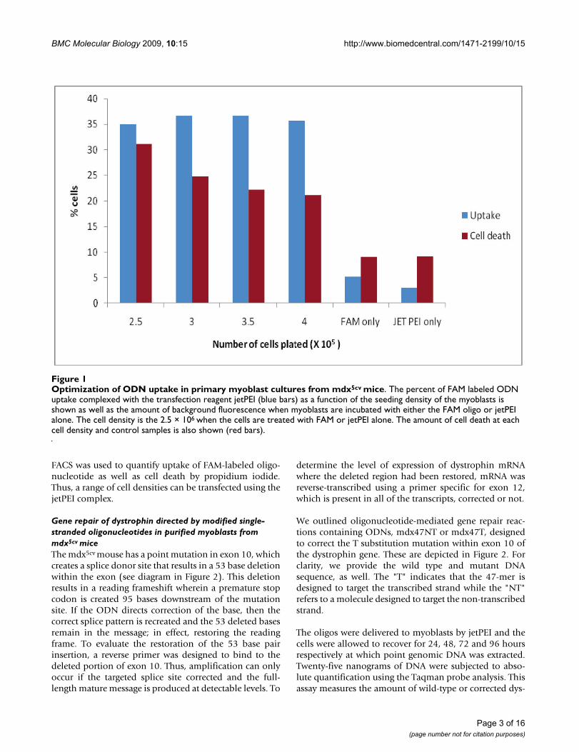
Gene repair of dystrophin directed by modified single-stranded oligonucleotides in purified myoblasts from mdx5cv miceThe mdx5cv mouse has a point mutation in exon 10, whichcreates a splice donor site that results in a 53 base deletionwithin the exon (see diagram in Figure 2). This deletionresults in a reading frameshift wherein a premature stopcodon is created 95 bases downstream of the mutationsite. If the ODN directs correction of the base, then thecorrect splice pattern is recreated and the 53 deleted basesremain in the message; in effect, restoring the readingframe. To evaluate the restoration of the 53 base pairinsertion, a reverse primer was designed to bind to thedeleted portion of exon 10. Thus, amplification can onlyoccur if the targeted splice site corrected and the full-length mature message is produced at detectable levels. To
determine the level of expression of dystrophin mRNAwhere the deleted region had been restored, mRNA wasreverse-transcribed using a primer specific for exon 12,which is present in all of the transcripts, corrected or not.
We outlined oligonucleotide-mediated gene repair reac-tions containing ODNs, mdx47NT or mdx47T, designedto correct the T substitution mutation within exon 10 ofthe dystrophin gene. These are depicted in Figure 2. Forclarity, we provide the wild type and mutant DNAsequence, as well. The "T" indicates that the 47-mer isdesigned to target the transcribed strand while the "NT"refers to a molecule designed to target the non-transcribedstrand.
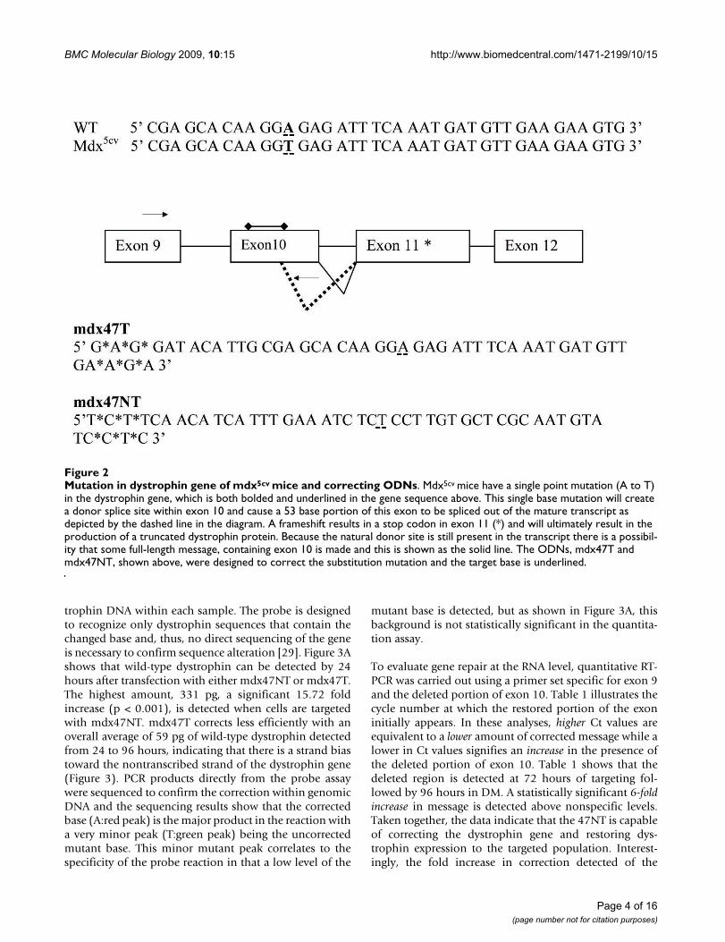
The oligos were delivered to myoblasts by jetPEI and thecells were allowed to recover for 24, 48, 72 and 96 hoursrespectively at which point genomic DNA was extracted.Twenty-five nanograms of DNA were subjected to abso-lute quantification using the Taqman probe analysis. Thisassay measures the amount of wild-type or corrected dys-
Optimization of ODN uptake in primary myoblast cultures from mdx5cv miceFigure 1Optimization of ODN uptake in primary myoblast cultures from mdx5cv mice. The percent of FAM labeled ODN uptake complexed with the transfection reagent jetPEI (blue bars) as a function of the seeding density of the myoblasts is shown as well as the amount of background fluorescence when myoblasts are incubated with either the FAM oligo or jetPEI alone. The cell density is the 2.5 × 10⁶ when the cells are treated with FAM or jetPEI alone. The amount of cell death at each cell density and control samples is also shown (red bars).
Page 3 of 16(page number not for citation purposes)
BMC Molecular Biology 2009, 10:15 http://www.biomedcentral.com/1471-2199/10/15
trophin DNA within each sample. The probe is designedto recognize only dystrophin sequences that contain thechanged base and, thus, no direct sequencing of the geneis necessary to confirm sequence alteration [29]. Figure 3Ashows that wild-type dystrophin can be detected by 24hours after transfection with either mdx47NT or mdx47T.The highest amount, 331 pg, a significant 15.72 foldincrease (p < 0.001), is detected when cells are targetedwith mdx47NT. mdx47T corrects less efficiently with anoverall average of 59 pg of wild-type dystrophin detectedfrom 24 to 96 hours, indicating that there is a strand biastoward the nontranscribed strand of the dystrophin gene(Figure 3). PCR products directly from the probe assaywere sequenced to confirm the correction within genomicDNA and the sequencing results show that the correctedbase (A:red peak) is the major product in the reaction witha very minor peak (T:green peak) being the uncorrectedmutant base. This minor mutant peak correlates to thespecificity of the probe reaction in that a low level of the
mutant base is detected, but as shown in Figure 3A, thisbackground is not statistically significant in the quantita-tion assay.
To evaluate gene repair at the RNA level, quantitative RT-PCR was carried out using a primer set specific for exon 9and the deleted portion of exon 10. Table 1 illustrates thecycle number at which the restored portion of the exoninitially appears. In these analyses, higher Ct values areequivalent to a lower amount of corrected message while alower in Ct values signifies an increase in the presence ofthe deleted portion of exon 10. Table 1 shows that thedeleted region is detected at 72 hours of targeting fol-lowed by 96 hours in DM. A statistically significant 6-foldincrease in message is detected above nonspecific levels.Taken together, the data indicate that the 47NT is capableof correcting the dystrophin gene and restoring dys-trophin expression to the targeted population. Interest-ingly, the fold increase in correction detected of the
Mutation in dystrophin gene of mdx5cv mice and correcting ODNsFigure 2Mutation in dystrophin gene of mdx5cv mice and correcting ODNs. Mdx5cv mice have a single point mutation (A to T) in the dystrophin gene, which is both bolded and underlined in the gene sequence above. This single base mutation will create a donor splice site within exon 10 and cause a 53 base portion of this exon to be spliced out of the mature transcript as depicted by the dashed line in the diagram. A frameshift results in a stop codon in exon 11 (*) and will ultimately result in the production of a truncated dystrophin protein. Because the natural donor site is still present in the transcript there is a possibil-ity that some full-length message, containing exon 10 is made and this is shown as the solid line. The ODNs, mdx47T and mdx47NT, shown above, were designed to correct the substitution mutation and the target base is underlined.
Page 4 of 16(page number not for citation purposes)
BMC Molecular Biology 2009, 10:15 http://www.biomedcentral.com/1471-2199/10/15
Page 5 of 16(page number not for citation purposes)
Quantitative measurement of dystrophin correction in primary cultures containing mdx5cv myoblasts at the DNA level and sequence analysisFigure 3Quantitative measurement of dystrophin correction in primary cultures containing mdx5cv myoblasts at the DNA level and sequence analysis. A: Total genomic DNA was isolated from targeted myoblasts at various time points, 24 to 96 hours after introduction of ODNs, mdx47NT or mdx47T. Twenty-five nanograms of this DNA was subjected to abso-lute quantification by PCR analysis with a FAM labeled probe specific only for the wild-type dystrophin sequence. A standard curve had been previously generated using C2C12 (WT) genomic DNA and the amount of wild-type dystrophin has been extrapolated from this standard and is presented as nanograms of wild-type dystrophin. These data represent the average of three experiments that were analyzed in triplicate. Significance was measured by a Student's t-test and samples were consid-ered significantly different with a p value of less than *0.05, **0.01, ***0.001. B: DNA sequence reaction of PCR products showing the chromatogram representing a mixed population at the targeted base (base 102) with adenine (green/corrected base) being the major peak and with thymine (red/mutant base) being a minor population within the reaction.

BMC Molecular Biology 2009, 10:15 http://www.biomedcentral.com/1471-2199/10/15
dystrophin gene detected with the Taqman probe(15.72×) (see Figure 3A) is not equivalent to the foldincrease in expression (6 fold). A likely explanation forthis observation indicates that some correction is occur-ring in fibroblasts, which do not express dystrophin,although such a comparison may not be possible.
The donor splice site in the DMD gene in mdx5cv micecauses a frameshift which results in a translation of trun-cated dystrophin protein (10 of the 79 exons). As shownabove, correction of the mutation at the DNA levelresulted in the restoration of intact dystrophin message.To determine whether the corrected mRNA is translatedinto normal dystrophin that contains exon 11, cells wereseeded and once again transfected with mdx47NT ormdx47T under conditions that provided the highest levelof gene correction. The entire assay was performed inmicroscope chambers so that the samples could beprobed directly with an antibody specific for the dys-trophin protein. Figure 4 reveals the presence of dys-trophin after a 72 hour recovery and 96 hourdifferentiation period, indicating dystrophin is now beingproduced within myotubes. Within myotubes that stainpositive for dystrophin, it is evident that the protein is dis-tributed throughout (see Figure 4B) These data are inagreement with the western blot analyses performed pre-viously by Bertoni et al. [25]. Altogether, the data fromDNA, mRNA and protein analyses show that singlestranded oligonucleotides without conjugated chemicalmoieties at their termini (i.e. Cy3) correct the dystrophin
gene in cultures containing an enriched population ofmyoblasts isolated from dystrophic mice.
Enhancement of dystrophin correction by knockdown of Msh2We and others have shown that a transient knockdown ofthe mismatch repair protein Msh2 in repair proficientcells can increase the overall level of gene repair [26,30].Msh2 is thought to inhibit the repair reaction through itsaction as an anti-recombinase [31,32] by precluding thebinding of the oligonucleotide. Therefore, by knockingdown the levels of this protein, we might predict that theoligonucleotide will bind more stably to the target site
Table 1: Quantitative measurement of dystrophin, GAPDH, and CKM.
Recovery TimemRNA Transfected ODN 48 hours 72 hours
(Ct value)Dystrophin mdx47NT 34.1 ± 0.15 33.8 ± 0.22
NS 34.9 ± 0.08 37.1 ± 0.35GAPDH mdx47NT 13.2 ± 0.08 13.6 ± 0.02
NS 13.3 ± 0.05 13.6 ± 0.32CKM mdx47NT 22.1 ± 0.11 13.6 ± 0.11
NS 21.5 ± 0.08 13.6 ± 0.10
A quantitative measurement of dystrophin transcripts containing the deleted portion of exon 10 was also performed. The cells were induced to differentiate with addition of media containing 2% horse serum at the indicated time points for 96 hours. Each sample was subjected to RT PCR using either dystrophin specific primers or oligo dT primers. QPCR was performed using a reverse primer specific for the deleted portion of exon 10. GAPDH was used as a loading control and CKM levels were measured to ensure similarities in the extent of differentiation between samples. Significance was measured by a Student's t-test and samples were considered significantly different with a p value of less than *0.05, **0.01, ***0.001. mdx47NT was used to correct the mutation while nonspecific ODN was used as a negative control. GAPDH and CKM were used as positive controls with the ODNs in parenthesis added to the mRNA mixtures.
Detection of dystrophin with exon specific antibodiesFigure 4Detection of dystrophin with exon specific antibod-ies. Cultures containing dystrophic myoblasts were seeded in 8 well chambers and were targeted with a 72 hour recov-ery time after the addition of the either the nonspecific ODN (A) or mdx47NT (B). These cells were then allowed to dif-ferentiate for 96 hours at which point they were fixed and incubated with antibody specific for a rabbit polyclonal anti-body that recognizes dystrophin and a secondary Alexa fluor labeled antibody (red). The cell nuclei were stained with DAPI, which is shown in blue.
Page 6 of 16(page number not for citation purposes)
BMC Molecular Biology 2009, 10:15 http://www.biomedcentral.com/1471-2199/10/15
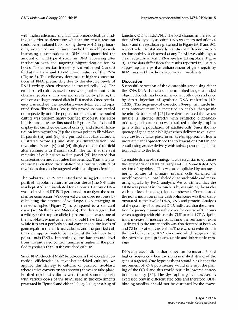
with higher efficiency and facilitate oligonucleotide bind-ing. In order to determine whether the repair reactioncould be stimulated by knocking down Msh2 in primarycells, we treated our cultures enriched in myoblasts withincreasing concentrations of RNAi and quantified theamount of wild-type dystrophin DNA appearing afterincubation with the targeting oligonucleotide for 24hours. The correction frequency was enhanced 2 and 3fold at the 1 nM and 10 nM concentrations of the RNAi(Figure 5). The efficiency decreases at higher concentra-tions of RNAi presumably due to the elevated levels ofRNAi toxicity often observed in treated cells [33]. Theenriched cell cultures used above were purified further toobtain myoblasts. This was accomplished by plating thecells on a collagen coated dish in F10 media. Once conflu-ency was reached, the myoblasts were detached and sepa-rated from fibroblasts (etc.); this procedure was carriedour repeatedly until the population of cells in the pooledculture was predominantly purified myoblast. The stepsin this procedure are illustrated in Figure 6. Panels i and iidisplay the enriched culture of cells (i) and after differen-tiation into myotubes (ii); the arrows point to fibroblasts.In panels (iii) and (iv), the purified myoblast culture isillustrated before (i) and after (ii) differentiation intomyotubes. Panels (v) and (vi) display cells in dark fieldafter staining with Desmin (red). The fact that the vastmajority of cells are stained in panel (vi) indicated thatdifferentiation into myotubes has occurred. Thus, the pro-cedure has enabled the isolation of a purified culture ofmyoblasts that can be targeted with the oligonucleotide.
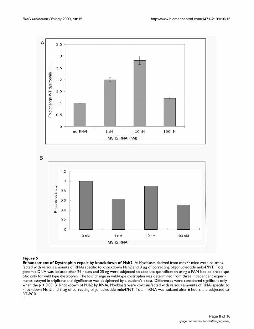
The mdx47NT ODN was introduced using jetPEI into apurified myoblast culture at various doses (the N/P ratiowas kept at 5) and incubated for 24 hours. Genomic DNAwas isolated and RT-PCR performed to analyze the sam-ples for gene repair. We observed a clear dose response bycalculating the amount of wild-type DNA emerging intreated samples (Figure 7) as compared to a standardcurve (see Methods and Materials). The data suggest thata wild type dystrophin allele is present in at least some ofthe myoblasts where gene repair should have taken place.While it is not a perfectly sound comparison, the levels ofgene repair in the enriched cultures and the purified cul-tures are approximately equivalent at the 24 hour timepoint (mdx47NT). Interestingly, the background levelfrom the untreated control samples is higher in the puri-fied myoblasts than in the enriched culture.
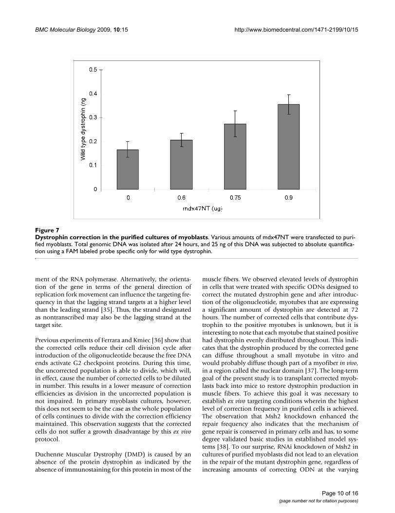
Since RNAi-directed Msh2 knockdowns had elevated cor-rection efficiencies in myoblast-enriched cultures, weapplied this strategy to cultures of purified myoblastswhere active conversion was shown (above) to take place.Purified myoblast cultures were treated simultaneouslywith various doses of the RNAi used in the experimentspresented in Figure 5 and either 0.3 μg, 0.6 μg or 0.9 μg of
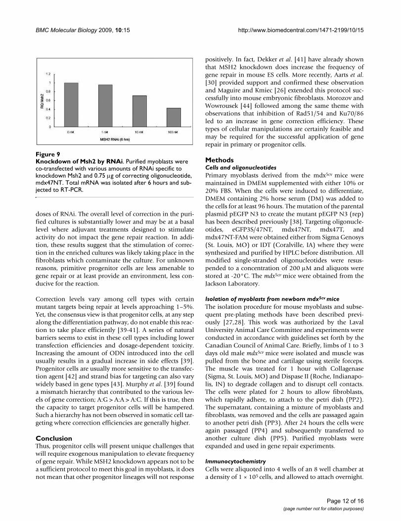
targeting ODN, mdx47NT. The fold change in the evolu-tion of wild type dystrophin DNA was measured after 24hours and the results are presented in Figure 8A, B and 8C,respectively. No statistically significant difference in cor-rection activity is observed at any RNAi level, although aclear reduction in Msh2 RNA levels is taking place (Figure9). These data differ from the results reported in Figure 5suggesting perhaps that enhancement of gene repair byRNAi may not have been occurring in myoblasts.
DiscussionSuccessful correction of the dystrophin gene using eitherthe RNA/DNA chimera or the modified single strandedoligonucleotide has been achieved in both dogs and miceby direct injection of synthetic DNA molecules [10-12,25]. The frequency of correction throughout muscle tis-sues however must be increased to enable therapeuticbenefit. Bertoni et al. [25] have demonstrated that whenmuscle is injected directly with synthetic oligonucle-otides, genetic correction was restricted to the dystrophingene within a population of satellite cells. Since the fre-quency of gene repair is higher when delivery to cells out-side the body takes place in an ex vivo approach. Thus, amore efficient approach for the treatment of DMD mightentail using ex vivo delivery with subsequent transplanta-tion back into the host.
To enable this ex vivo strategy, it was essential to optimizethe efficiency of ODN delivery and ODN-mediated cor-rection of myoblasts. This was accomplished by transfect-ing a culture of primary muscle cells enriched inmyoblasts with a FAM labeled oligonucleotide and meas-uring uptake by FACs analysis. We confirmed that theODN was present in the nucleus by examining the nucleiwith confocal imaging (data not shown). Correction ofthe point mutation in the dystrophin gene was then dem-onstrated at the level of DNA, RNA and protein. Analysisof the quantity of corrected DNA indicated that the correc-tion frequency remains stable over the course of 96 hourswhen targeting with either mdx47NT or mdx47T. A signif-icant increase in message containing the portion of exon10 deleted in the mutant cells was also detected at both 48and 72 hours after transfection. There was no reduction inthe level of repaired RNA over time which suggests thatthe corrected gene produces stable and inheritable mes-sage.
DNA analyses indicate that correction occurs at a 3 foldhigher frequency when the nontranscribed strand of thegene is targeted. One hypothesis for strand bias is that themovement of RNA polymerase would interrupt the pair-ing of the ODN and this would result in lowered correc-tion efficiency [34]. The dystrophin gene, however, isexpressed only in differentiated cells and therefore, ODNbinding stability should not be disrupted by the move-
Page 7 of 16(page number not for citation purposes)
BMC Molecular Biology 2009, 10:15 http://www.biomedcentral.com/1471-2199/10/15
Page 8 of 16(page number not for citation purposes)
Enhancement of Dystrophin repair by knockdown of Msh2Figure 5Enhancement of Dystrophin repair by knockdown of Msh2. A: Myoblasts derived from mdx5cv mice were co-trans-fected with various amounts of RNAi specific to knockdown Msh2 and 3 μg of correcting oligonucleotide mdx47NT. Total genomic DNA was isolated after 24 hours and 25 ng were subjected to absolute quantification using a FAM labeled probe spe-cific only for wild type dystrophin. The fold change in wild-type dystrophin was determined from three independent experi-ments assayed in triplicate and significance was deciphered by a student's t-test. Differences were considered significant only when the p < 0.05. B: Knockdown of Msh2 by RNAi. Myoblasts were co-transfected with various amounts of RNAi specific to knockdown Msh2 and 3 μg of correcting oligonucleotide mdx47NT. Total mRNA was isolated after 6 hours and subjected to RT-PCR.

BMC Molecular Biology 2009, 10:15 http://www.biomedcentral.com/1471-2199/10/15
Page 9 of 16(page number not for citation purposes)
Dystrophin repair in purified cultures of myoblastsFigure 6Dystrophin repair in purified cultures of myoblasts. Purification of the primary cultures of myoblasts. (i) enriched cul-ture of myoblasts. The cultures were derived from limbs of Mdx5cv mice, and population of the myoblasts was enriched using pre-plating technique (see "Materials and Methods); (ii) myotubes in the enriched culture. The enriched cultures were induced to differentiation by culturing in differentiation medium. The arrows show representative fibroblasts; (iii) purified culture of myoblasts. The enriched cultures were subjected to pre-plating repeatedly until fibroblast-like cells were not observed in cul-tures. The purified cultures were cultivated in F10-based medium; (iv) myotube in the purified culture. The cultures were induced to differentiation by culturing in differentiation medium; (v) and (vi) desmin staining of the purified culture. Cell nuclei were counter stained with DAPI (v; blue). All of the observed cells were positive for desmin staining (vi; red).
BMC Molecular Biology 2009, 10:15 http://www.biomedcentral.com/1471-2199/10/15
ment of the RNA polymerase. Alternatively, the orienta-tion of the gene in terms of the general direction ofreplication fork movement can influence the targeting fre-quency in that the lagging strand targets at a higher levelthan the leading strand [35]. Thus, the strand designatedas nontranscribed may also be the lagging strand at thetarget site.
Previous experiments of Ferrara and Kmiec [36] show thatthe corrected cells reduce their cell division cycle afterintroduction of the oligonucleotide because the free DNAends activate G2 checkpoint proteins. During this time,the uncorrected population is able to divide, which will,in effect, cause the number of corrected cells to be dilutedin number. This results in a lower measure of correctionefficiencies as division in the uncorrected population isnot impaired. In primary myoblasts cultures, however,this does not seem to be the case as the whole populationof cells continues to divide with the correction efficiencymaintained. This observation suggests that the correctedcells do not suffer a growth disadvantage by this ex vivoprotocol.
Duchenne Muscular Dystrophy (DMD) is caused by anabsence of the protein dystrophin as indicated by theabsence of immunostaining for this protein in most of the
muscle fibers. We observed elevated levels of dystrophinin cells that were treated with specific ODNs designed tocorrect the mutated dystrophin gene and after introduc-tion of the oligonucleotide, myotubes that are expressinga significant amount of dystrophin are detected at 72hours. The number of corrected cells that contribute dys-trophin to the positive myotubes is unknown, but it isinteresting to note that each myotube that stained positivehad dystrophin evenly distributed throughout. This indi-cates that the dystrophin produced by the corrected genecan diffuse throughout a small myotube in vitro andwould probably diffuse though part of a myofiber in vivo,in a region called the nuclear domain [37]. The long-termgoal of the present study is to transplant corrected myob-lasts back into mice to restore dystrophin production inmuscle fibers. To achieve this goal it was necessary toestablish ex vivo targeting conditions wherein the highestlevel of correction frequency in purified cells is achieved.The observation that Msh2 knockdown enhanced therepair frequency also indicates that the mechanism ofgene repair is conserved in primary cells and has, to somedegree validated basic studies in established model sys-tems [38]. To our surprise, RNAi knockdown of Msh2 incultures of purified myoblasts did not lead to an elevationin the repair of the mutant dystrophin gene, regardless ofincreasing amounts of correcting ODN at the varying
Dystrophin correction in the purified cultures of myoblastsFigure 7Dystrophin correction in the purified cultures of myoblasts. Various amounts of mdx47NT were transfected to puri-fied myoblasts. Total genomic DNA was isolated after 24 hours, and 25 ng of this DNA was subjected to absolute quantifica-tion using a FAM labeled probe specific only for wild type dystrophin.
Page 10 of 16(page number not for citation purposes)
BMC Molecular Biology 2009, 10:15 http://www.biomedcentral.com/1471-2199/10/15
Page 11 of 16(page number not for citation purposes)
Effect of Msh2 knockdown on dystrophin correction in purified myoblastsFigure 8Effect of Msh2 knockdown on dystrophin correction in purified myoblasts. Purified myoblasts were co-transfected with various amounts of RNAi specific to knockdown Msh2 and (A) 0.3 μg, (B) 0.6 μg or (C) 0.9 μg of correcting oligonucle-otide (mdx47NT), respectively. Total genomic DNA was isolated after 24 hours and 25 ng were subjected to absolute quanti-fication using a FAM labeled probe specific only for wild type dystrophin. The fold change in wild-type dystrophin was determined from three independent experiments assayed in triplicate.
BMC Molecular Biology 2009, 10:15 http://www.biomedcentral.com/1471-2199/10/15
doses of RNAi. The overall level of correction in the puri-fied cultures is substantially lower and may be at a basallevel where adjuvant treatments designed to stimulateactivity do not impact the gene repair reaction. In addi-tion, these results suggest that the stimulation of correc-tion in the enriched cultures was likely taking place in thefibroblasts which contaminate the culture. For unknownreasons, primitive progenitor cells are less amenable togene repair or at least provide an environment, less con-ducive for the reaction.
Correction levels vary among cell types with certainmutant targets being repair at levels approaching 1–5%.Yet, the consensus view is that progenitor cells, at any stepalong the differentiation pathway, do not enable this reac-tion to take place efficiently [39-41]. A series of naturalbarriers seems to exist in these cell types including lowertransfection efficiencies and dosage-dependent toxicity.Increasing the amount of ODN introduced into the cellusually results in a gradual increase in side effects [39].Progenitor cells are usually more sensitive to the transfec-tion agent [42] and strand bias for targeting can also varywidely based in gene types [43]. Murphy et al. [39] founda mismatch hierarchy that contributed to the various lev-els of gene correction; A:G > A:A > A:C. If this is true, thenthe capacity to target progenitor cells will be hampered.Such a hierarchy has not been observed in somatic cell tar-geting where correction efficiencies are generally higher.
ConclusionThus, progenitor cells will present unique challenges thatwill require exogenous manipulation to elevate frequencyof gene repair. While MSH2 knockdown appears not to bea sufficient protocol to meet this goal in myoblasts, it doesnot mean that other progenitor lineages will not response
positively. In fact, Dekker et al. [41] have already shownthat MSH2 knockdown does increase the frequency ofgene repair in mouse ES cells. More recently, Aarts et al.[30] provided support and confirmed these observationand Maguire and Kmiec [26] extended this protocol suc-cessfully into mouse embryonic fibroblasts. Morozov andWowrousek [44] followed among the same theme withobservations that inhibition of Rad51/54 and Ku70/86led to an increase in gene correction efficiency. Thesetypes of cellular manipulations are certainly feasible andmay be required for the successful application of generepair in primary or progenitor cells.
MethodsCells and oligonucleotidesPrimary myoblasts derived from the mdx5cv mice weremaintained in DMEM supplemented with either 10% or20% FBS. When the cells were induced to differentiate,DMEM containing 2% horse serum (DM) was added tothe cells for at least 96 hours. The mutation of the parentalplasmid pEGFP N3 to create the mutant pEGFP N3 (rep)has been described previously [38]. Targeting oligonucle-otides, eGFP3S/47NT, mdx47NT, mdx47T, andmdx47NT-FAM were obtained either from Sigma Genosys(St. Louis, MO) or IDT (Coralville, IA) where they weresynthesized and purified by HPLC before distribution. Allmodified single-stranded oligonucleotides were resus-pended to a concentration of 200 μM and aliquots werestored at -20°C. The mdx5cv mice were obtained from theJackson Laboratory.
Isolation of myoblasts from newborn mdx5cv miceThe isolation procedure for mouse myoblasts and subse-quent pre-plating methods have been described previ-ously [27,28]. This work was authorized by the LavalUniversity Animal Care Committee and experiments wereconducted in accordance with guidelines set forth by theCanadian Council of Animal Care. Briefly, limbs of 1 to 3days old male mdx5cv mice were isolated and muscle waspulled from the bone and cartilage using sterile forceps.The muscle was treated for 1 hour with Collagenase(Sigma, St. Louis, MO) and Dispase II (Roche, Indianapo-lis, IN) to degrade collagen and to disrupt cell contacts.The cells were plated for 2 hours to allow fibroblasts,which rapidly adhere, to attach to the petri dish (PP2).The supernatant, containing a mixture of myoblasts andfibroblasts, was removed and the cells are passaged againto another petri dish (PP3). After 24 hours the cells wereagain passaged (PP4) and subsequently transferred toanother culture dish (PP5). Purified myoblasts wereexpanded and used in gene repair experiments.
ImmunocytochemistryCells were aliquoted into 4 wells of an 8 well chamber ata density of 1 × 10⁵ cells, and allowed to attach overnight.
Knockdown of Msh2 by RNAiFigure 9Knockdown of Msh2 by RNAi. Purified myoblasts were co-transfected with various amounts of RNAi specific to knockdown Msh2 and 0.75 μg of correcting oligonucleotide, mdx47NT. Total mRNA was isolated after 6 hours and sub-jected to RT-PCR.
Page 12 of 16(page number not for citation purposes)
BMC Molecular Biology 2009, 10:15 http://www.biomedcentral.com/1471-2199/10/15
The cells were fixed using 100% ethanol for 5 minutes at-20°C, permeabilized with 0.1% Triton X-100, and thenincubated with either desmin (Sigma, St. Louis, MO) spe-cific or dystrophin specific antibodies raised in rabbit(Abcam, Cambridge, MA 1:1,000) at room temperature(RT) for 1 hour or overnight at 4°C. After washing thecells, a secondary goat α-rabbit antibody conjugated to aCy3 fluorophore (desmin) (Zymed Laboratories, SanFrancisco, CA) or an Alexa 546-coupled goat anti-rabbitantibody (Molecular probes 1:1500) was added to thewells and this mixture was incubated with agitation for 1hour at room temperature. After four 15 minute washes,the wells were dried and mounting media containingDAPI was added to each well (Molecular Probes, Eugene,OR). The cells were then visualized using a Zeiss™ inverted100 M Axioscope equipped with a Zeiss™ 510 LSM confo-cal microscope with a coherent krypton argon laser (30mW) containing 488 nm or 568 nm and 742 nm excita-tion lines.
Gene repair of dystrophin in purified myoblastsCells were plated in 6-well culture dishes at a density of 1× 10⁵ with media containing 20% FBS. jetPEI was used tomake oligonucleotide complexes with 3 μg of ODN at anoptimal N/P ratio (nitrogen/phosphate ratio) of 5. Thisreagent was added to 1 ml of media and the cells wereincubated for various amounts of time at which point dif-ferentiation media (DM) was added; the cells were differ-entiated for 96 hours, then harvested for FACS analysisand genomic DNA isolation. For mRNA analyses, the cellswere collected at least four days after the addition of DMcontaining 2% horse serum.
Knockdown of Msh2RNAi, designed to knock down Msh2, was purchasedfrom Ambion (Ambion, Austin, Texas); siRNA (ID#155431) is designed to anneal to the mouse MSH2 RNAat exon 3 NM_008628. During the targeting experiments,the RNAi was mixed with the correcting oligonucleotidescomplexed with jetPEI and added to the cells as describedabove.
Co-transfection ssODNs and Msh2 RNAiCells were plated in 6-well plates at a density of 1 × 10⁵
cells/well with growth media (DMEM supplemented with20% FBS, penicillin and streptomycin). Twenty four hoursafter plating, cells were co-transfected 3 μg of mdx/47NTand RNAi specific to knockdown Msh2 (0–200 nM). Sev-enty two hours after transfection, the growth media werereplaced by differentiation media (DMEM supplementedwith 2% horse serum, penicillin and streptomycin), andthe cells were maintained in differentiation media for 96h. Total RNA was isolated from the cells, and reverse tran-scription and quantitative PCR were conducted as
described in "RNA isolation and, RT PCR and quantitativePCR.
Msh2 RNAiCells were plated in 6-well plates at a density of 1 × 10⁵
cells/well with growth media. Twenty four hours afterplating, cells were co-transfected 3 μg of mdx47NT andRNAi specific to knockdown Msh2 (1–100 nM). TotalRNA was isolated 3 or 6 h after transfection Methods ofreverse transcription and quantitative PCR were same as"RNA isolation and, RT PCR and quantitative PCR",except for primers: musMSH2/257F 5' agaccctgcagagtgtt-gtgctta 3'; musMSH2/1010R 5' tgagagccagtggtgtcttcaaca 3'or mouse GAPDH4FwQ 5' tgtctcctgcgacttcaacagc 3';GAPDH5RevQ 5' atgtaggccatgaggtccacca 3'.
RNA isolation, RT PCR and quantitative PCRAfter differentiation of the myoblasts, the cells were har-vested by trypsinization and collected by centrifugation at1,500 rpm for 7 minutes. After washing with 1× PBS, theRNA was extracted using a Qiagen RNA extraction kit(Qiagen, Valencia, CA) according to the manufacturer'sinstructions. Three hundred nanograms of RNA was sub-jected to reverse transcription to generate cDNA using agene specific primer Rev-ex12 5' cag gtc caa agg gct ctt cc 3'or primer oligo dT using the first strand synthesis kit (Inv-itrogen, Carlsbad, CA). Five microliters was added to a 20μL mixture of 2× Sybrgreen Master Mix (Applied Biosys-tems, Foster City, CA) with 2.5 μM of each primer:
Forw-ex 9 5' gtt atg cct tca cag gct gc 3'; Rev-ex10 in 5'cacttcttc aac atc att tg3' or GAPDH4FwQ 5'tgtctcctgcgacttcaacagc3'; GAPDH5RevQ 5'atgtaggccatgaggtccacca 3' CKM F1 5'GCAACACCCACAACAAGTTCA 3' and CKM R1 5'TGTCTCCTTATCGCGAAGCTT 3'. Samples were added toeach well of a 96 well plate in duplicate for each primerset. Dystrophin was amplified using the cDNA generatedby the gene specific primer and GAPDH or CKM fromcDNA generated using oligo dT. QPCR was run for 1 rep-etition at 50°C for 2 minutes, 1 repetition at 95°C for 10minutes, 40 repetitions at 95°C for 15 seconds and 40repetitions at 59°C for 1 minute with data collection atstep 1 of stage 3. Upon completion of PCR, the thresholdand baseline values were set to measure Ct values at thebeginning of the exponential amplification. Ct valuesfrom 3 experiments were averaged per sample and sub-jected to a student's t-test and ANOVA to determine signif-icance.
DNA isolation and absolute quantification by PCRCells were harvested and collected by centrifugation at1500 rpm for 7 minutes and washed twice with 1× PBS orlysed directly in a tissue culture dish after washing with 1×PBS. DNA was precipitated using the genomic wizardDNA extraction kit as instructed by the manufacturer
Page 13 of 16(page number not for citation purposes)
BMC Molecular Biology 2009, 10:15 http://www.biomedcentral.com/1471-2199/10/15
(Promega, Madison WI). Twenty-five nanograms ofgenomic DNA was added to a total of 7 μL of water andthis was mixed with 13 μL of a 2× PCR master mix, 50 nMof a 6FAM labeled probe specific to bind to wild-type dys-trophin 5'FAMcac aag gag aga ttt3', 900 nM of forwardprimer mdx5cvasF 5' ttt ctg ccg agg ata cat tgc3' and 900nM of reverse primer ex 10 out 5' ctc atg agc atg aaa ctg ttc3'. Each experimental protocol was run in triplicate. Astandard curve was generated using 50 ng, 25 ng, 12.5 ng,6.25 ng, and 3.13 ng of genomic DNA isolated fromC2C12 cells, which are wild-type for dystrophin. As a sep-arate control, reaction mixtures containing 25 ng ofgenomic DNA from mdx5cv were used to determine thebackground of wild-type DNA in each experiment. PCRconditions were: 1 repetition at 50°C for 2 minutes, 1 rep-etition at 95°C for 10 minutes and 40 cycles of 95°C for15 seconds and 60°C for 1 minute. The data were col-lected at stage 3 of step 1. Threshold and baseline levelswere set to measure the Ct values at the lowest level ofexponential amplification and the average mean wasdetermined from each experiment. Statistical significancewas determined by a student's t-test and was consideredsignificant only if p was less than 0.05. Following the PCRamplification the products were purified using Qiagen'sQIAquick PCR purification kit according to the instruc-tions and submitted for sequencing using the mdx5cvasFprimer.
Primary mouse myoblast purificationThe enriched culture obtained from Tremblay lab wasplated on a collagen-coated culture dish with F10 nutrientmixture (Invitrogen, Carlsbad, CA) supplemented with10% (v/v) FBS, 2.5 ng/ml bFGF (Promega, Madison, WI),100 U/ml penicillin and 100 mg/ml streptomycin. Whenthe culture reached confluency, cells were detached fromthe dish without using trypsin. To accomplish this, thedish was washed with PBS after aspiration of medium,incubated with a small amount of PBS at room tempera-ture for 5 min, and hit firmly sideways. Predominant celltype in the detached cells was myoblast due to the fact thatmyoblasts are likely to detach from collagen-coated dishthan fibroblasts are. The detached cells were collectedwith F10 nutrient mixture, plated on a new collagen-coated dish, and expanded. The above procedures wererepeated until no fibroblasts were observed in the culture.Upon completion of purification, medium was switchedF10 growth medium to F10/DMEM (1:1) supplementedwith 10% (v/v) FBS, 2.5 ng/ml bFGF (Promega, Madison,WI), 100 U/ml penicillin and 100 mg/ml streptomycin.
Desmin stainingCells were plated on 2-well chambers at a density of 8 ×10⁴ cells/well and incubated at 37°C overnight. The cellswere fixed using ethanol for 5 minutes at -20°C, permea-bilized using 0.3% (v/v) TritonX for 10 minutes at room
temperature, blocked using 5% (v/v) heat-inactivatedgoat serum for 30 minutes at room temperature, and thenincubated with desmin specific antibody (Sigma, St.Louis, MO; dilution, 1:50) for 1 hour at room tempera-ture. After washing the cells, a secondary goat a-rabbitantibody conjugated to Cy3 (Zymed laboratories, SanFrancisco, CA; dilution, 1:750) was added to the wells andthis incubated with agitation for 1 hour at room tempera-ture. After four 15 minute washes, the wells were brieflydried and mounting media containing DAPI was added toeach well (Molecular Probes, Eugene, OR).
Oligo dose experimentPurified myoblasts were plated in 6-well plates at a den-sity of 1.5 × 10⁵ cells/well with F10/DMEM growthmedium 24 hours before transfection. jetPEI was used tomake oligonucletide complexes with various amounts ofmdx47NT at N/P (nitrogen/phosphate) ratio of 5. Thisreagent was added to 1 ml of media and the cells wereincubated. Genomic DNA was isolated 24 hours aftertransfection using masterpure DNA purification kit (Epi-centre, Madison, WI). TaqMan® probe-based real timePCR was performed to quantify wild type dystrophin.Probe specific for wild type dystrophin (5'-FAMcac aag gagaga ttt-3') were used with the primer pair: forward primer,5'-ttt ctg ccg agg ata cat tgc-3'; reverse primer, 5'-ctc atg agcatg aaa ctg ttc-3'. Real time PCR was performed with 25 nggenomic DNA, 900 nM both forward and reverse primer,and 250 nM probe in each 20 ml reaction. A standardcurve was generated using 1, 0.5, 0.25, 0.125, 0.0625 ngof genomic DNA isolated from C2C12 cells, which havewild type dystrophin gene. PCR conditions were: 1 repeti-tion at 50°C for 2 minutes, 1 repetition at 95°C for 10minutes and 40 cycles of 95°C for 15 seconds and 60°Cfor 1 minute. Each experimental protocol was run in trip-licate.
Co-transfection ssODNs and Msh2 RNAiPurified myoblasts were plated in 6-well plates at a den-sity of 1.5 × 10⁵ cells/well with F10/DMEM growthmedium 24 hours before transfection. Cells were co-trans-fected 0.75 mg of mdx47NT and siRNA for Msh2 knock-down (0–100 nM) using jetPEI. Twenty four hours aftertransfection, genomic DNA was isolated, and TaqMan®
probe-based real time PCR was performed to quantifywild type dystrophin as described above ("Oligo doseexperiment")
Msh2 RNAiPurified myoblasts were plated in 6-well plates at a den-sity of 1.5 × 10⁵ cells/well with F10/DMEM growthmedium 24 hours before transfection. Cells were co-trans-fected 0.75 mg of mdx47NT and siRNA for Msh2 knock-down (0–100 nM) using jetPEI. Total RNA was isolated 6hours after transfection. Two hundred nanograms of RNA
Page 14 of 16(page number not for citation purposes)

BMC Molecular Biology 2009, 10:15 http://www.biomedcentral.com/1471-2199/10/15
was subjected to reverse transcription to generate cDNAusing the first strand synthesis kit (Invitrogen). Fivemicroliters was added to a 20 ml mixture of 2× Sybergreenmaster mix (Applied Biosystems, Foster City, CA) with 2.5mM of each primer: musMSH2/257F 5' agaccctgcagagtgtt-gtgctta 3'; musMSH2/1010R 5' tgagagccagtggtgtcttcaaca 3'or mouse GAPDH4FwQ 5' tgtctcctgcgacttcaacagc 3';GAPDH5RevQ 5' atgtaggccatgaggtccacca 3'. Samples wereadded to each well of a 96 well plate in duplicate for eachprimer set. QPCR was run for 1 repetition at 50°C for 2minutes, 1 repetition at 95°C for 10 minutes, 40 repeti-tions at 95°C for 15 seconds and 40 repetitions at 59°Cfor 1 minute with data collection at step 1 of stage 3. Uponcompletion of PCR, the threshold and baseline valueswere set to measure Ct values at the beginning of the expo-nential amplification. Ct values from 3 experiments wereaveraged per sample.
AbbreviationsODN: Single-stranded oligonucleotides; DMD: DuchenneMuscular Dystrophy.
Authors' contributionsKM carried out the experiments in enriched myoblastsalong with the initial RNAi work; TZ purified the myob-lasts and carried out the experiments in the purified cells.KM and HPO in conjunction with EK conceived the study;HPO and EK drafted the manuscript. All authors read andapproved the final manuscript.
AcknowledgementsWe would like to thank Dr. J. Tremblay's lab for providing the enriched myoblasts and for critical reading of the manuscript (Unité de recherche en Génétique Humaine, Centre de Recherche du Centre Hospitalier de Uni-versité Laval, CHUL du CHUQ, Ste-Foy, QC, Canada). This project was supported by NIH R21 NS053608.
References1. Rando TA: The dystrophin-glycoprotein complex, cellular sig-
naling, and the regulation of cell survival in the muscular dys-trophies. Muscle Nerve 2001, 24:1575-1594.
2. Tutdibi O, Brinkmeier H, Rudel R, Fohr KJ: Increased calciumentry into dystrophin-deficient muscle fibres of MDX andADR-MDX mice is reduced by ion channel blockers. J Physiol1999, 515(Pt 3):859-868.
3. Bertorini TE, Bhattacharya SK, Palmieri GM, Chesney CM, Pifer D,Baker B: Muscle calcium and magnesium content in Duch-enne muscular dystrophy. Neurology 1982, 32:1088-1092.
4. Strober JB: Therapeutics in duchenne muscular dystrophy.NeuroRx 2006, 3:225-234.
5. Harper SQ, Hauser MA, DelloRusso C, Duan D, Crawford RW,Phelps SF, et al.: Modular flexibility of dystrophin: implicationsfor gene therapy of Duchenne muscular dystrophy. Nat Med2002, 8:253-261.
6. Tinsley JM, Potter AC, Phelps SR, Fisher R, Trickett JI, Davies KE:Amelioration of the dystrophic phenotype of mdx miceusing a truncated utrophin transgene. Nature 1996,384:349-353.
7. Deconinck N, Tinsley J, De Backer F, Fisher R, Kahn D, Phelps S, etal.: Expression of truncated utrophin leads to major func-tional improvements in dystrophin-deficient muscles ofmice. Nat Med 1997, 3:1216-1221.
8. Wang Z, Allen JM, Riddell SR, Gregorevic P, Storb R, Tapscott SJ, etal.: Immunity to adeno-associated virus-mediated genetransfer in a random-bred canine model of Duchenne mus-cular dystrophy. Hum Gene Ther 2007, 18:18-26.
9. Wang B, Li J, Fu FH, Xiao X: Systemic human minidystrophingene transfer improves functions and life span of dystrophinand dystrophin/utrophin-deficient mice. J Orthop Res 2008 inpress.
10. Bartlett RJ, Stockinger S, Denis MM, Bartlett WT, Inverardi L, Le TT,et al.: In vivo targeted repair of a point mutation in the caninedystrophin gene by a chimeric RNA/DNA oligonucleotide.Nat Biotechnol 2000, 18:615-622.
11. Rando TA, Disatnik MH, Zhou LZ: Rescue of dystrophin expres-sion in mdx mouse muscle by RNA/DNA oligonucleotides.Proc Natl Acad Sci USA 2000, 97:5363-5368.
12. Bertoni C, Rando TA: Dystrophin Gene Repair in mdx MusclePrecursor Cells In Vitro and In Vivo Mediated by RNA-DNAChimeric Oligonucleotides. Hum Gene Ther 2002, 13:707-718.
13. Bertoni C, Lau C, Rando TA: Restoration of dystrophin expres-sion in mdx muscle cells by chimeraplast-mediated exonskipping. Hum Mol Genet 2003, 12:1087-1099.
14. Pramono ZA, Takeshima Y, Alimsardjono H, Ishii A, Takeda S, MatsuoM: Induction of exon skipping of the dystrophin transcript inlymphoblastoid cells by transfecting an antisense oligodeox-ynucleotide complementary to an exon recognitionsequence. Biochem Biophys Res Commun 1996, 226:445-449.
15. Wilton SD, Lloyd F, Carville K, Fletcher S, Honeyman K, Agrawal S,et al.: Specific removal of the nonsense mutation from themdx dystrophin mRNA using antisense oligonucleotides.Neuromuscul Disord 1999, 9:330-338.
16. Kole R, Sazani P: Antisense effects in the cell nucleus: modifi-cation of splicing. Curr Opin Mol Ther 2001, 3:229-234.
17. Dunckley MG, Manoharan M, Villiet P, Eperon IC, Dickson G: Modi-fication of splicing in the dystrophin gene in cultured Mdxmuscle cells by antisense oligoribonucleotides. Hum Mol Genet1998, 7:1083-1090.
18. Mann CJ, Honeyman K, Cheng AJ, Ly T, Lloyd F, Fletcher S, et al.:Antisense-induced exon skipping and synthesis of dystrophinin the mdx mouse. Proc Natl Acad Sci USA 2001, 98:42-47.
19. van Deutekom JC, Bremmer-Bout M, Janson AA, Ginjaar IB, Baas F,den Dunnen JT, et al.: Antisense-induced exon skipping restoresdystrophin expression in DMD patient derived muscle cells.Hum Mol Genet 2001, 10:1547-1554.
20. Aartsma-Rus A, Bremmer-Bout M, Janson AA, den Dunnen JT, vanOmmen GJ, van Deutekom JC: Targeted exon skipping as apotential gene correction therapy for Duchenne musculardystrophy. Neuromuscul Disord 2002, 12(Suppl 1):S71-S77.
21. Aartsma-Rus A, Janson AA, Kaman WE, Bremmer-Bout M, den Dun-nen JT, Baas F, et al.: Therapeutic antisense-induced exon skip-ping in cultured muscle cells from six different DMDpatients. Hum Mol Genet 2003, 12:907-914.
22. Vacek M, Sazani P, Kole R: Antisense-mediated redirection ofmRNA splicing. Cell Mol Life Sci 2003, 60:825-833.
23. Morgan JE, Partridge TA: Muscle satellite cells. Int J Biochem CellBiol 2003, 35:1151-1156.
24. Hawke TJ, Garry DJ: Myogenic satellite cells: physiology tomolecular biology. J Appl Physiol 2001, 91:534-551.
25. Bertoni C, Morris GE, Rando TA: Strand bias in oligonucleotide-mediated dystrophin gene editing. Hum Mol Genet 2005,14:221-233.
26. Maguire KK, Kmiec EB: Multiple roles for MSH2 in the repair ofa deletion mutation directed by modified single-stranded oli-gonucleotides. Gene 2007, 386:107-114.
27. Qu Z, Huard J: Matching host muscle and donor myoblasts formyosin heavy chain improves myoblast transfer therapy.Gene Ther 2000, 7:428-437.
28. Rando TA, Blau HM: Primary mouse myoblast purification,characterization, and transplantation for cell-mediated genetherapy. J Cell Biol 1994, 125:1275-1287.
29. Im WB, Phelps SF, Copen EH, Adams EG, Slightom JL, ChamberlainJS: Differential expression of dystrophin isoforms in strains ofmdx mice with different mutations. Hum Mol Genet 1996,5:1149-1153.
30. Aarts M, Dekker M, de Vries S, van der WA, Te RH: Generation ofa mouse mutant by oligonucleotide-mediated gene modifi-cation in ES cells. Nucleic Acids Res 2006, 34:e147.
Page 15 of 16(page number not for citation purposes)
BMC Molecular Biology 2009, 10:15 http://www.biomedcentral.com/1471-2199/10/15
Publish with BioMed Central and every scientist can read your work free of charge
"BioMed Central will be the most significant development for disseminating the results of biomedical research in our lifetime."
Sir Paul Nurse, Cancer Research UK
Your research papers will be:
available free of charge to the entire biomedical community
peer reviewed and published immediately upon acceptance
cited in PubMed and archived on PubMed Central
yours — you keep the copyright
Submit your manuscript here:http://www.biomedcentral.com/info/publishing_adv.asp
BioMedcentral
31. Datta A, Adjiri A, New L, Crouse GF, Jinks RS: Mitotic crossoversbetween diverged sequences are regulated by mismatchrepair proteins in Saccaromyces cerevisiae. Mol Cell Biol 1996,16:1085-1093.
32. Selva EM, New L, Crouse GF, Lahue RS: Mismatch correction actsas a barrier to homeologous recombination in Saccharomy-ces cerevisiae. Genetics 1995, 139:1175-1188.
33. Hough SR, Wiederholt KA, Burrier AC, Woolf TM, Taylor MF: WhyRNAi makes sense. Nat Biotechnol 2003, 21:731-732.
34. Liu L, Rice MC, Drury M, Cheng S, Gamper H, Kmiec EB: Strandbias in targeted gene repair is influenced by transcriptionalactivity. Mol Cell Biol 2002, 22:3852-3863.
35. Brachman EE, Kmiec EB: DNA replication and transcriptiondirect a DNA strand bias in the process of targeted generepair in mammalian cells. J Cell Sci 2004, 117:3867-3874.
36. Ferrara L, Kmiec EB: Targeted gene repair activates Chk1 andChk2 and stalls replication in corrected cells. DNA Repair(Amst) 2006, 5:422-431.
37. Kinoshita I, Vilquin JT, Asselin I, Chamberlain J, Tremblay JP: Trans-plantation of myoblasts from a transgenic mouse overex-pressing dystrophin prduced only a relatively small increaseof dystrophin-positive membrane. Muscle Nerve 1998,21:91-103.
38. Hu Y, Parekh-Olmedo H, Drury M, Skogen M, Kmiec EB: Reactionparameters of targeted gene repair in Mammalian cells. MolBiotechnol 2005, 29:197-210.
39. Murphy BR, Moayedpardazi HS, Gewirtz AM, Diamond SL, Pierce EA:Delivery and mechanistic considerations for the productionof knock-in mice by single-stranded oligonucleotide genetargeting. Gene Ther 2007, 14:304-315.
40. Pierce EA, Liu Q, Igoucheva O, Omarrudin R, Ma H, Diamond SL, etal.: Oligonucleotide-directed single-base DNA alterations inmouse embryonic stem cells. Gene Ther 2003, 10:24-33.
41. Dekker M, Brouwers C, Te RH: Targeted gene modification inmismatch-repair-deficient embryonic stem cells by single-stranded DNA oligonucleotides. Nucleic Acids Res 2003, 31:E27.
42. Almofti MR, Harashima H, Shinohara Y, Almofti A, Li W, Kiwada H:Lipoplex size determines lipofection efficiency with or with-out serum. Mol Membr Biol 2003, 20:35-43.
43. Yamamoto T, Moerschell RP, Wakem LP, Komar-Panicucci S, Sher-man F: Strand-specificity in the transformation of yeast withsynthetic oligonucleotides. Genetics 1992, 131:811-819.
44. Morozov V, Wawrousek EF: Single-strand DNA-mediated tar-geted mutagenesis of genomic DNA in early mouse embryosis stimulated by Rad51/54 and by Ku70/86 inhibition. GeneTher 2008, 15:468-472.
Page 16 of 16(page number not for citation purposes)
Copyright © 2022 FDOKUMEN