Development of a novel splice array platform and its application in the identification of...
15
Pio et al. BMC Genomics 2010, 11:352 http://www.biomedcentral.com/1471-2164/11/352 Open Access METHODOLOGY ARTICLE © 2010 Pio et al; licensee BioMed Central Ltd. This is an Open Access article distributed under the terms of the Creative Commons At- tribution License (http://creativecommons.org/licenses/by/2.0), which permits unrestricted use, distribution, and reproduction in any medium, provided the original work is properly cited. Methodology article Development of a novel splice array platform and its application in the identification of alternative splice variants in lung cancer Ruben Pio* 1,2 , David Blanco 1,3 , Maria Jose Pajares 1,3 , Elena Aibar 4 , Olga Durany 4 , Teresa Ezponda 1 , Jackeline Agorreta 1,3 , Javier Gomez-Roman 5 , Miguel Angel Anton 6 , Angel Rubio 6 , Maria D Lozano 7 , Jose M López- Picazo 8 , Francesc Subirada 4 , Tamara Maes 4 and Luis M Montuenga 1,3 Abstract Background: Microarrays strategies, which allow for the characterization of thousands of alternative splice forms in a single test, can be applied to identify differential alternative splicing events. In this study, a novel splice array approach was developed, including the design of a high-density oligonucleotide array, a labeling procedure, and an algorithm to identify splice events. Results: The array consisted of exon probes and thermodynamically balanced junction probes. Suboptimal probes were tagged and considered in the final analysis. An unbiased labeling protocol was developed using random primers. The algorithm used to distinguish changes in expression from changes in splicing was calibrated using internal non- spliced control sequences. The performance of this splice array was validated with artificial constructs for CDC6, VEGF, and PCBP4 isoforms. The platform was then applied to the analysis of differential splice forms in lung cancer samples compared to matched normal lung tissue. Overexpression of splice isoforms was identified for genes encoding CEACAM1, FHL-1, MLPH, and SUSD2. None of these splicing isoforms had been previously associated with lung cancer. Conclusions: This methodology enables the detection of alternative splicing events in complex biological samples, providing a powerful tool to identify novel diagnostic and prognostic biomarkers for cancer and other pathologies. Background Alternative splicing of pre-mRNA is a post-transcrip- tional modification essential for the regulation of gene expression and function. Through alternative splicing, multiple transcripts are produced from a single mRNA precursor, widely expanding proteome diversity. Deep sequencing applied to diverse human tissues and epithe- lial cell lines has recently revealed that more than 90% of human genes undergo alternative splicing [1]. A global analysis of alternative splicing in the human transcrip- tome suggested that exon skipping is the most prevalent form of alternative splicing [2]. Alternative splicing is a tightly regulated process influenced by cell type, develop- mental stage, external conditions, etc; however, it is also associated with multiple disease conditions, including cancer [3]. For example, cancer-related aberrantly spliced variants have been shown to be actively involved in the initiation and/or progression of some types of cancer [4]. Splicing alterations are the consequence of splice-site mutations, deregulation of splicing regulatory factors, or both [5]. Tumor-specific variations in splicing may gener- ate new epitopes that can serve as a starting point for immune therapy or targeted delivery, as well as for the development of new diagnostic or prognostic tools [6]. Thus, the identification and molecular characterization of alternative splicing variants associated with cancer is currently a very active area of research [7]. In recent years, powerful techniques for genome-wide identifica- tion and analysis of alternative splicing isoforms have been developed. These large-scale high-throughput ana- lytical methods have been applied to the identification of differential splicing events in cancer tissues [8]. Exon microarrays, which contain both known and predicted * Correspondence: [email protected] 1 Division of Oncology, Center for Applied Medical Research, Pamplona, Spain Full list of author information is available at the end of the article
-
Upload
independent -
Category
Documents
-
view
4 -
download
0
Transcript of Development of a novel splice array platform and its application in the identification of...
Pio et al. BMC Genomics 2010, 11:352http://www.biomedcentral.com/1471-2164/11/352
Open AccessM E T H O D O L O G Y A R T I C L E
Methodology articleDevelopment of a novel splice array platform and its application in the identification of alternative splice variants in lung cancerRuben Pio*1,2, David Blanco1,3, Maria Jose Pajares1,3, Elena Aibar4, Olga Durany4, Teresa Ezponda1, Jackeline Agorreta1,3, Javier Gomez-Roman5, Miguel Angel Anton6, Angel Rubio6, Maria D Lozano7, Jose M López-Picazo8, Francesc Subirada4, Tamara Maes4 and Luis M Montuenga1,3
AbstractBackground: Microarrays strategies, which allow for the characterization of thousands of alternative splice forms in a single test, can be applied to identify differential alternative splicing events. In this study, a novel splice array approach was developed, including the design of a high-density oligonucleotide array, a labeling procedure, and an algorithm to identify splice events.
Results: The array consisted of exon probes and thermodynamically balanced junction probes. Suboptimal probes were tagged and considered in the final analysis. An unbiased labeling protocol was developed using random primers. The algorithm used to distinguish changes in expression from changes in splicing was calibrated using internal non-spliced control sequences. The performance of this splice array was validated with artificial constructs for CDC6, VEGF, and PCBP4 isoforms. The platform was then applied to the analysis of differential splice forms in lung cancer samples compared to matched normal lung tissue. Overexpression of splice isoforms was identified for genes encoding CEACAM1, FHL-1, MLPH, and SUSD2. None of these splicing isoforms had been previously associated with lung cancer.
Conclusions: This methodology enables the detection of alternative splicing events in complex biological samples, providing a powerful tool to identify novel diagnostic and prognostic biomarkers for cancer and other pathologies.
BackgroundAlternative splicing of pre-mRNA is a post-transcrip-tional modification essential for the regulation of geneexpression and function. Through alternative splicing,multiple transcripts are produced from a single mRNAprecursor, widely expanding proteome diversity. Deepsequencing applied to diverse human tissues and epithe-lial cell lines has recently revealed that more than 90% ofhuman genes undergo alternative splicing [1]. A globalanalysis of alternative splicing in the human transcrip-tome suggested that exon skipping is the most prevalentform of alternative splicing [2]. Alternative splicing is atightly regulated process influenced by cell type, develop-mental stage, external conditions, etc; however, it is alsoassociated with multiple disease conditions, including
cancer [3]. For example, cancer-related aberrantly splicedvariants have been shown to be actively involved in theinitiation and/or progression of some types of cancer [4].Splicing alterations are the consequence of splice-sitemutations, deregulation of splicing regulatory factors, orboth [5]. Tumor-specific variations in splicing may gener-ate new epitopes that can serve as a starting point forimmune therapy or targeted delivery, as well as for thedevelopment of new diagnostic or prognostic tools [6].Thus, the identification and molecular characterizationof alternative splicing variants associated with cancer iscurrently a very active area of research [7]. In recentyears, powerful techniques for genome-wide identifica-tion and analysis of alternative splicing isoforms havebeen developed. These large-scale high-throughput ana-lytical methods have been applied to the identification ofdifferential splicing events in cancer tissues [8]. Exonmicroarrays, which contain both known and predicted
* Correspondence: [email protected] Division of Oncology, Center for Applied Medical Research, Pamplona, SpainFull list of author information is available at the end of the article
© 2010 Pio et al; licensee BioMed Central Ltd. This is an Open Access article distributed under the terms of the Creative Commons At-tribution License (http://creativecommons.org/licenses/by/2.0), which permits unrestricted use, distribution, and reproduction in anymedium, provided the original work is properly cited.
Pio et al. BMC Genomics 2010, 11:352http://www.biomedcentral.com/1471-2164/11/352
Page 2 of 15
exons, have been recently used for this purpose [9-13].However, since they are not specifically designed toexamine alternative splicing, they fail to detect eventssuch as the alternative use of 5' or 3' splice sites, intronretention, or the insertion of cryptic exons. Other splic-ing-specific microarrays have been developed to covermost alternative splicing events. These arrays contain oli-gonucleotide probes that span exon-exon junctions, andprobes positioned within exons to determine individualexon levels and overall transcript expression. The use ofsplice-junction oligonucleotides to analyze splice eventswas proposed as early as 1986, when Morgan and Wardused them to identify differential splice forms of minutevirus in mice cDNA [14]. In 1996, Lockhart et al. reportedone of the first genome-wide microarray studies and sug-gested the potential of microarrays for the analysis ofalternative splicing [15], but it was not until 2002 thatClark et al. developed the first microarray containingsplice-junction oligonucleotides to analyze splice eventsin yeast [16]. In 2003, Johnson et al. used microarrayscontaining oligonucleotide probes complementary toexon-exon junction sequences to discover new alternativesplice variants in human tissues [17,18]. Also in 2003,Wang et al. designed an algorithm that aimed to deconvo-lute the absolute concentrations of each alternative tran-script present in a complex mixture starting from thehybridization intensities detected on splice chips [19]. Anew algorithm, called SPACE, has recently been devel-oped for estimating the number of different splicing iso-forms (known and unknown), and determining theirstructures and relative concentrations [20].
Nonetheless, currently available splice arrays still havemany limitations, mainly due to problems in the design ofthe array, the labeling protocol, and data analysis. Thedevelopment of robust and efficient splice microarraysand data-analysis methods will facilitate progress in thediagnosis, prognosis, and therapy of cancer and otherpathologies. In the present work, we describe a novelcomprehensive methodology for high-throughput profil-ing of alternative splicing in complex biological samples.In this methodology, processing of results is based on thearray specific design, which is original and thought spe-cifically for alternative splicing-discovery. The strategyconsists of optimization of probe design, development ofan unbiased amplification protocol that avoids inappro-priate transcript coverage due to 3'-biased labeling, andimplementation of detailed data processing. Oligonucle-otides for the splice array were designed using the Tethysmodule (Oryzon Genomics, Barcelona, Spain), aninhouse oligo design program, complemented with a newsplice-analysis specific module (AltTethys). The algo-rithm targets the best possible oligonucleotide for eachsequence, rather than imposing a strict oligonucleotidequality cutoff. A new labeling protocol was developed to
ensure optimal all-length transcript coverage and linealamplification, working with small amounts of humanmaterial. To analyze the data, we developed a novel algo-rithm for the analysis of two-color arrays that allows for astatistically robust identification of candidate splicedgenes in absence of a prior hypothesis about the contrib-uting isoforms. We have applied this technology to theidentification of lung cancer-associated splicing variants.Lung cancer is a devastating disease with few therapeuticoptions or suitable molecular biomarkers for early diag-nosis. The results obtained in this study validated theutility of the platform, allowing the identification of newcancer-associated splicing variants with potential utilityin the management of lung cancer.
ResultsDevelopment and validation of the unbiased labeling protocolThe basis for the reliable detection of splice forms in amicroarray format is a high-quality, non-biased labelingprocedure. Different labeling protocols have been appliedin splicing analyses: (a) Castle et al. developed a PCR +T7 amplification-based protocol [17]; (b) a commercialkit is available from Stratagene (Fairplay II) to produce alabeled first-strand cDNA using random hexamers whichworks well if a sufficient amount of starting material isavailable; (c) some authors have applied standard 3'-biased RNA labeling protocols, ignoring the loss of 5'events.
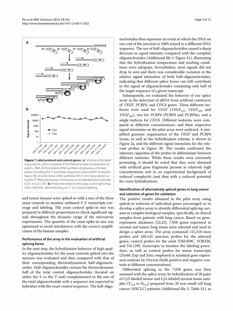
Considering the valuable and limited starting materialof human clinical samples (total RNA amounts were <100 ng), as well as ours and others previous experience inexpression microarray studies with cRNA lineal-ampli-fied material by in vitro transcription [21], we developeda novel protocol for the detection of alternative spliceevents consisting of the following steps (Figure 1a): (a)extraction of polyA + RNA; (b) first-strand cDNA synthe-sis employing a chimeric primer including the T7 pro-moter sequences and a stretch of 6 random bases startingfrom 50 ng of polyA + RNA; (c) second-strand cDNAsynthesis; and (d) in vitro transcription using the T7 RNApolymerase in the presence of a labeled ribonucleotide(Cy3- or Cy5-CTP). For a first validation of performance,three different yeast sequences were tested, rangingbetween 4500 and 7000 bp to cover any outlier isoformlength. PCR-amplified yeast control sequences and thecorresponding high-quality yeast control oligonucle-otides were used on the microarray surface to assesswhether a bias with respect to oligonucleotide positionwithin the transcript had occurred. While some varia-tions in signal intensity of the different oligonucleotideswere identified, there was no evidence of 3'- or 5'-end bias(Figure 1b). The same controls were included later in thelung array design. All human samples (both from normal
Pio et al. BMC Genomics 2010, 11:352http://www.biomedcentral.com/1471-2164/11/352
Page 3 of 15
and tumor tissues) were spiked-in with a mix of the threeyeast controls to monitor unbiased 3'-5' transcripts cov-erage and labeling. The yeast control spike-in mix wasprepared in different proportions to check significant sig-nals throughout the dynamic range of the microarrayexperiments. The quantity of the yeast spike-in mix wasoptimized to avoid interference with the correct amplifi-cation of the human samples.
Performance of the array in the evaluation of artificial splicing formsIn the next step, the hybridization behavior of high-qual-ity oligonucleotides for the yeast controls spiked into themixture was evaluated and then compared with that oftheir corresponding thermodynamical half-oligonucle-otides. Half-oligonucleotides contain the thermodynamichalf of the total control oligonucleotides (located oneither the 5' or the 3' end) complemented to the size ofthe total oligonucleotide with a sequence not expected tohybridize with the yeast control sequence. The half-oligo-
nucleotides thus represent an event in which the DNA onone end of the junction is 100% joined to a different DNAsequence. The use of half-oligonucleotides caused a sharpdecrease in signal intensity compared with the completeoligonucleotides (Additional file 1: Figure S1), illustratingthat the hybridization temperature and washing condi-tions were adequate. Nevertheless, most signals did notdrop to zero and there was considerable variation in therelative signal intensities of both half-oligonucleotides,indicating that different splice forms can still contributeto the signal of oligonucleotides containing only half ofthe target sequence of a given transcript.
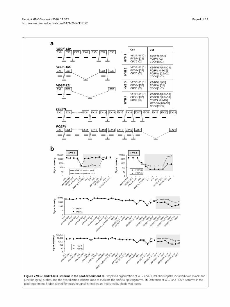
Subsequently, we evaluated the behavior of our splicearray in the detection of aRNA from artificial constructsof VEGF, PCBP4, and CDC6 genes. Three different iso-forms were used for VEGF (VEGF121, VEGF165, andVEGF185), two for PCBP4 (PCBP4 and PCBP4a), and asingle isoform for CDC6. Different isoforms were com-pared at different concentrations, and their respectivesignal intensities on the pilot array were analyzed. A sim-plified genomic organization of the VEGF and PCBP4exons, as well as the hybridization scheme, is shown inFigure 2a, and the different signal intensities for the rele-vant probes in Figure 2b. The results confirmed theinherent capacities of the probes to differentiate betweendifferent isoforms. While these results were extremelypromising, it should be noted that they were obtainedwith artificial gene fragments present at relatively highconcentrations and in an experimental background ofreduced complexity (and thus with a reduced potentialfor cross-hybridization).
Identification of alternatively spliced genes in lung cancer and selection of genes for validationThe positive results obtained in the pilot array usingspiked-in isoforms of individual genes encouraged us todevelop a splice array to identify differential splicing vari-ants in complex biological samples, specifically, in clinicalsamples from patients with lung cancer. Based on gene-expression databases [22,23], 7,958 genes expressed innormal and tumor lung tissue were selected and used todesign a splice array. The array contained 115,318 exonprobes and 105,141 junction probes for the selectedgenes, control probes for the yeast YML059C, YOR328,and YIL129C transcripts to monitor the labeling proce-dure, as well as control probes for maize transcripts(Zm48, Exp and Xet), employed in standard gene expres-sion analyses by Oryzon (both positive and negative con-trols at different concentrations).
Differential splicing in the 7,958 genes was thenassessed with the splice array, by hybridization of 20 pairsof Cy5 labeled tumor and Cy3 labeled normal tissue sam-ples (TCy5 vs NCy3) prepared from 20 non-small cell lungcancer (NSCLC) patients (Additional file 2: Table S1), as
Figure 1 Label protocol and control genes. (a) Scheme of the label-ing protocol, which consisted of the following steps: (I) extraction of polyA + RNA, (II) first-strand cDNA synthesis employing a chimeric primer including the T7 promoter sequences and a stretch of random bases, (III) second-strand cDNA synthesis, (IV) in vitro transcription us-ing the T7 RNA polymerase in the presence of a labeled ribonucleotide (Cy3- or Cy5-CTP). (b) Probe intensities for the yeast control gene frag-ment YML059C, demonstrating no 3'- or 5'-biased labeling.
AAAAAA
T7
T7
T7
T7T7
T7
T7
a
b
1
10
100
1000
10000
100000
ORY_C1-
126
ORY_C1-
506
ORY_C1-
798
ORY_C1-
1221
ORY_C1-
1680
ORY_C1-
1932
ORY_C1-
2184
ORY_C1-
2562
ORY_C1-
2940
ORY_C1-
3403
ORY_C1-
3781
ORY_C1-
4158
ORY_C1-
4578
Sig
nal
In
ten
sity
Cy3
Cy5
T7
Pio et al. BMC Genomics 2010, 11:352http://www.biomedcentral.com/1471-2164/11/352
Page 4 of 15
Figure 2 VEGF and PCBP4 isoforms in the pilot experiment. (a) Simplified organization of VEGF and PCBP4, showing the included exon (black) and junction (gray) probes, and the hybridization scheme used to evaluate the artificial splicing forms. (b) Detection of VEGF and PCBP4 isoforms in the pilot experiment. Probes with differences in signal intensities are indicated by shadowed boxes.
a
b
EX9 EX8 EX7 EX6 EX5 EX4 EX3VEGF-189
EX9 EX8 EX4 EX3
VEGF-165
EX9 EX8 EX3
VEGF-121
EX3 EX4 EX11 EX12 EX13 EX14PCBP4
EX15 EX16 EX17 EX18 EX19 EX20 EX21
EX3 EX4 EX11 EX12 EX13 EX14PCBP4
EX15 EX16 EX17 EX21
Cy3
VEGF165 [C1]PCBP4 [C2]CDC6 [C3]
VEGF165 [C1]PCBP4 [C2]CDC6 [C3]
VEGF165 [C1]PCBP4 [C2]CDC6 [C3]
VEGF165 [C1]PCBP4 [C2]CDC6 [C3]
Cy5
VEGF185 [C1]PCBP4 [C2]CDC6 [3xC3]
VEGF185 [0.5xC1]PCBP4 [0.5xC2]PCBP4a [0.5xC2]CDC6 [3xC3]
VEGF121 [C1]PCBP4a [C2]CDC6 [3xC3]
VEGF185 [0.5xC1] VEGF121 [0.5xC1] PCBP4 [0.5xC2]������ [0.5xC2]CDC6 [3xC3]
HY
B1
HY
B2
HY
B3
HY
B4
1
10
100
1000
10000
100000
EX9
JN4-E
X9_EX8
JN5-E
X9_EX8
JN6-E
X9_EX8-
EX8
JN8-E
X8_EX5(
6)(7)
EX7EX6
EX5
JN11-E
X6_EX4
JN7-E
X8_EX4
EX4
JN10-E
X4_EX2(
3)
Sig
nal
In
ten
sity
VEGF165 pool1 vs. pool2
VEGF-185 pool1 vs. pool2
HYB 1
1
10
100
1000
10000
100000
JN4-E
X9_EX8
JN5-E
X9_EX8
JN6-E
X9_EX8-
EX8
JN8-E
X8_EX5(
6)(7)
EX7EX6
EX5
JN11-E
X6_EX4
JN7-E
X8_EX4
EX4
Sig
nal
In
ten
sity
VEGF165
VEGF121
HYB 3
1
10
100
1,000
10,000
EX3
JN3-E
X3_EX4
EX4
JN4-E
X4_EX5
EX5
JN5-E
X4_EX6
EX6
JN6-E
X5_EX7
EX7
JN7-E
X6_EX8(
9)EX8
JN8-E
X8_EX10
EX9
JN9-E
X10_E
X11-E
X9_EX11
EX10EX11
JN10-E
X11_E
X13
JN11-E
X11_E
X12
EX12EX13
JN12-E
X13_E
X15
EX14EX15
JN14-E
X15_E
X16
EX16
JN16-E
X16_E
X17
EX17
JN17-E
X17_E
X18
EX18
JN18-E
X18_E
X19
EX19
JN19-E
X19_E
X20
EX20
Sig
nal
In
ten
sity
PCBP4
PCBP4a
1
10
100
1,000
10,000
100,000
EX3
JN3-E
X3_EX4
EX4
JN4-E
X4_EX5
EX5
JN5-E
X4_EX6
EX6
JN6-E
X5_EX7
EX7
JN7-E
X6_EX8(
9)EX8
JN8-E
X8_EX10
EX9
JN9-E
X10_E
X11-E
X9_EX11
EX10EX11
JN10-E
X11_E
X13
JN11-E
X11_E
X12
EX12EX13
JN12-E
X13_E
X15
EX14EX15
JN14-E
X15_E
X16
EX16
JN16-E
X16_E
X17
EX17
JN17-E
X17_E
X18
EX18
JN18-E
X18_E
X19
EX19
JN19-E
X19_E
X20
EX20
Sig
nal
In
ten
sity
PCBP4
PCBP4a
Pio et al. BMC Genomics 2010, 11:352http://www.biomedcentral.com/1471-2164/11/352
Page 5 of 15
well as three self to self comparisons (NCy5 vs NCy3). Thearray data from this study have been submitted to GeneExpression Omnibus http://www.ncbi.nlm.nih.gov/geounder accession no. GSE18346. The results were analyzedwith AltPolyphemus (Additional file 2: SupplementaryMethods), which allowed intensity changes of all the oli-gonucleotides for a given gene to be analyzed withrespect to whether these changes reflected gene expres-sion changes or isoform changes. Essentially, after pre-processing (data filtering and normalization) the algo-rithm first estimates the experimental variability of themicroarray analysis platform using the data of the stan-dard deviation of the gene probe data on the replicates ofthe self to self array.
To identify differences in expression level or spliceforms, "change" is first assessed in the self to self hybrid-ization. Any change that is not clearly greater than theinherent variability of the measurement system is consid-ered "no-change". In the self to self hybridization thestandard deviation (σs,g) can be calculated from the totalgene probe dataset and correlated to the standard devia-tion of the control probe dataset (σs,g = CF·σc). The dataspreading for the total gene probe set was always a littlehigher than that for the control probes (Additional file 2:Supplementary Methods), which means that the controlprobes slightly underestimate the experimental variation.In tumor vs normal tissue experiments, it is not possibleto measure directly the standard deviation for the no-change situation for the total gene probe data set (as dif-ferent samples are compared), but the standard deviationon the control dataset (σc) can be measured and the stan-dard deviation for the no-change situation for the totalgene probe dataset for the tumor vs normal array can beestimated (σ*s,g = CF·σc). Robust change can then bedefined as change below or above the threshold TH = ±3σ*s,g, although more stringent cut-off can be applied ifdesired. Once robust change is defined, the algorithmexamines whether the ratios of the signal in the Cy5 andCy3 channels for the exon and junction probes for a givengene (G) fall within the variability of the experiment(reflecting genes with regular differential gene expres-sion) or outside of that variability (potential splice formvariation); i.e. below or above μG ± 3σ*s,g, μG being themean value for the ratios of the signal in the Cy5 and Cy3channels for all the oligos for gene G. Note that μG for agene with differential expression will be clearly over thethreshold for the detection of change (the individualprobes are differentially expressed) but the variationamong the different probes will be below that threshold(Additional file 1: Figure S2c). For a gene to be selected asa candidate for differential splicing, at least one probe hasto fall outside of the limits of the marked threshold. Thealgorithm considers two hypotheses: the observed
hybridization can be explained by a differential mixtureof isoforms or by a whole gene expression change, andcalculates the error of both hypotheses. If there is a possi-ble isoform change, the algorithm establishes the "FormChange", an arbitrary and empirically defined figure torelatively rank the candidates. Figure S3 in Additional file1 shows a typical output from the AltPolyphemus soft-ware for a candidate gene susceptible to alternative splic-ing in lung cancer.
AltPolyphemus can identify splice candidates on datafrom individual tumor vs. normal tissue comparisons oron data from replicate analysis; i.e. on the mean changefor every probe in a given dataset. In performing the pro-cess on data from a biological replica analysis on a set oftumor vs. normal tissue comparisons, the aim is not onlyto identify individual splice variants but rather to identifythose events that, because of their prevalence, appear tobe more relevant. A strict biological replicate analysis wasperformed on the entire lung cancer set in which: (a) allvalues for a given oligonucleotide were considered (nooutlier elimination of samples was allowed, as this coulddistort our impression of prevalence of events); (b) thesignal cut-off for the oligonucleotide detection was set atthe mean signal + 3σ*s,g of a negative control and the Agi-lent background control oligonucleotides, thus selectingfor genes expressed clearly above the signal detectionlevel; (c) the fold change cut-off to make the selection wasset at the mean fold change of the total array data set ±3σ*s,g, selecting the most relevant changes. Internalassessment of the performance of the algorithm withthree self-to-self hybridizations was done for experimen-tal variability. Then AltPolyphemus analyzed the averagevalue from the biological replicate analysis and generateda list of genes ranked by the value of their "form change".This analysis generated 260 potential candidates. Table 1shows the top 10 genes identified in the analysis.
Validation of splice variants differentially expressed in lung cancerDifferences in alternative splicing between primaryNSCLC tissue and normal lung tissue in the ten selectedgenes were validated by PCR and sequencing. Validationwas performed with samples from a group of patientsincluded in the array (Additional file 2: Table S1) and anindependent series of NSCLC patients (Additional file 2:Table S2). IPO8 was used as the reference gene [24].Alterations in alternative splicing were confirmed in 4 outof the 10 genes: CEACAM1, FHL1, MLPH, and SUSD2.CEACAM1Ceacam1 (carcinoembryonic antigen-related cell adhe-sion molecule 1) is a transmembrane protein involved inintercellular binding and related to several normal andpathological processes [25]. Alternatively spliced formshave been identified for CEACAM1 [25]. Our analysis
Pio et al. BMC Genomics 2010, 11:352http://www.biomedcentral.com/1471-2164/11/352
Page 6 of 15
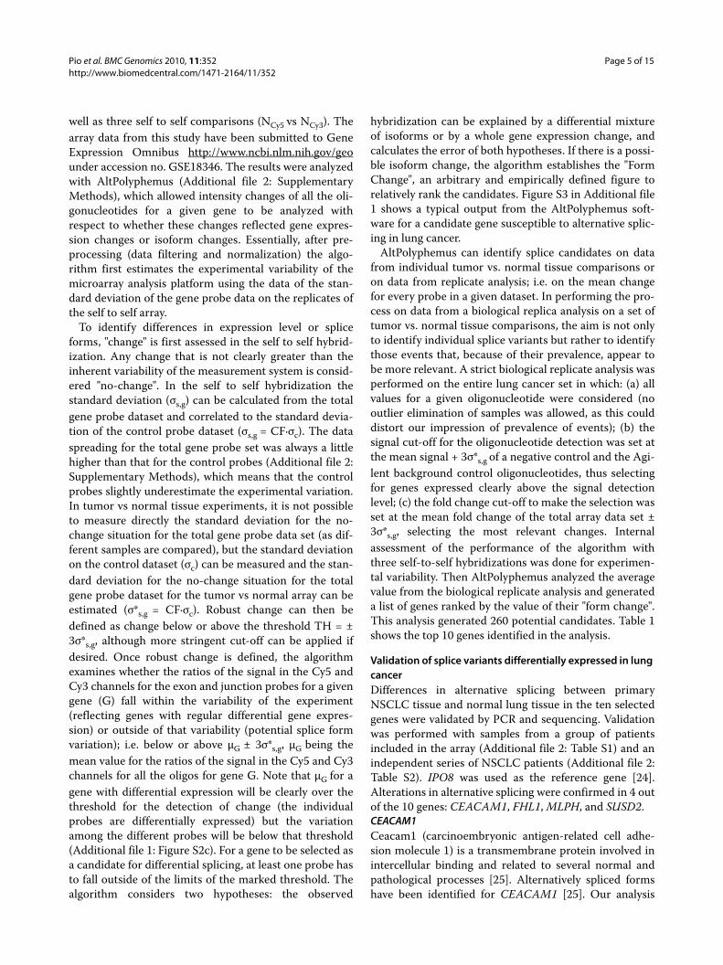
predicted changes in the splicing of this gene aroundexons 2 and 5. The isoforms generated by alternativeusage of these exons are (Figure 3a): CEACAM1-1 (lack-ing exons 3-5), CEACAM1-3 (without exon 5), andCEACAM1-3A (lacking exon 5 and including an addi-tional exon, hereafter designated as exon Y). To validatethe results obtained in the splice array, PCRs were per-formed with primers specifically designed to identifythese three different splice forms (Figure 3a and Addi-tional file 2: Table S3). Samples from primary tumors andtheir corresponding normal lung tissue were use to evalu-ate the expression of these isoforms in samples from 24NSCLC patients, 11 previously included in the splicearray (Figure 3b) and 13 from an independent series. Inall cases, the identity of the amplified variants was con-firmed by sequencing. These results clearly showed thatalternative splicing was extensive in lung cancer tissues atthe predicted CEACAM1 region. CEACAM1-1 andCEACAM1-3 were upregulated in tumors comparedwith their corresponding normal samples (p = 0.029 andp = 0.011, respectively; Additional file 1: Figure S4a).Thus, 92% (22 of 24) of the tumor samples expressedCEACAM1-1 whereas only 12 (50%) of the normal lungsamples were positive for this isoform. More interest-ingly, CEACAM1-1 expression was upregulated in 18(75%) of the tumors compared with their respective nor-mal-tissue counterparts. Nineteen tumors (79%) and 12normal lung samples (50%) expressed CEACAM1-3,which was upregulated in 17 tumors (71%). The predomi-nance of the CEACAM1-3A splice variant in cancer tis-sues was confirmed by conventional PCR (data notshown) and quantified by real-time PCR in 23 cases (Fig-ure 3c). CEACAM1-3A was significantly upregulated intumors compared with their corresponding normal sam-ples (p = 0.002). This isoform was upregulated in 19tumors (83%), with an increase in expression ranging
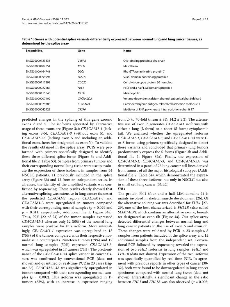
from 2- to 70-fold (mean ± SD: 14.2 ± 3.3). The alterna-tive use of exon 7 generates CEACAM1 isoforms witheither a long (L-form) or a short (S-form) cytoplasmictail. We analyzed whether the upregulated isoformsCEACAM1-1, CEACAM1-3, and CEACAM1-3A were L-or S-forms using primers specifically designed to detectthese variants and concluded that primary lung tumorspredominantly express the S-forms (Figure 3b and Addi-tional file 1: Figure S4a). Finally, the expression ofCEACAM1-1, CEACAM1-3, and CEACAM1-3A wasdetermined in a panel of 23 lung cancer cell lines derivedfrom tumors of all the major histological subtypes (Addi-tional file 2: Table S4), which demonstrated the expres-sion of these three isoforms not only in NSCLC but alsoin small cell lung cancer (SCLC).FHL1The protein Fhl1 (four and a half LIM domains 1) ismainly involved in skeletal muscle development [26]. Ofthe alternative splicing variants described for FHL1 [27-29], one of the best characterized is FHL1B (also calledSLIMMER), which contains an alternative exon 6, hereaf-ter designated as exon 6b (Figure 4a). Our splice arraydetected differential changes between normal lung andlung cancer patients in the use of exon 6 and exon 6b.These changes were validated by PCR in 23 samples, 8samples from patients included in the splice array and 15additional samples from the independent set. Conven-tional PCR followed by sequencing revealed the expres-sion of two FHL1 isoforms in lung samples: FHL1 andFHL1B (data not shown). Expression of the two isoformswas specifically quantified by real-time PCR. In agree-ment with previous reports in other types of cancer [30-32], both were found to be downregulated in lung cancerspecimens compared with normal lung tissue (data notshown). Interestingly, a significant change in the ratiobetween FHL1 and FHL1B was also observed (p = 0.003;
Table 1: Genes with potential splice variants differentially expressed between normal lung and lung cancer tissues, as determined by the splice array
Ensembl No. Gene Name
ENSG00000123838 C4BPA C4b-binding protein alpha chain
ENSG00000102854 MSLN Mesothelin
ENSG00000164741 DLC1 Rho GTPase-activating protein 7
ENSG00000099994 SUSD2 Sushi domain-containing protein 2
ENSG00000117399 CDC20 Cell division cycle protein 20 homolog
ENSG00000022267 FHL1 Four and a half LIM domains protein 1
ENSG00000115648 MLPH Melanophilin
ENSG00000007402 CACNA2D2 Voltage-dependent calcium channel subunit alpha-2/delta-2
ENSG00000079385 CEACAM1 Carcinoembryonic antigen-related cell adhesion molecule 1
ENSG00000042429 CRSP6 Mediator of RNA polymerase II transcription subunit 17
Pio et al. BMC Genomics 2010, 11:352http://www.biomedcentral.com/1471-2164/11/352
Page 7 of 15
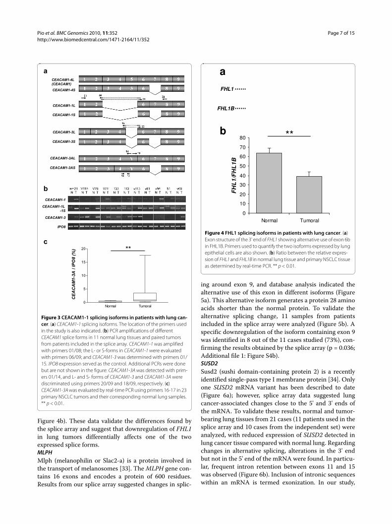
Figure 4b). These data validate the differences found bythe splice array and suggest that downregulation of FHL1in lung tumors differentially affects one of the twoexpressed splice forms.MLPHMlph (melanophilin or Slac2-a) is a protein involved inthe transport of melanosomes [33]. The MLPH gene con-tains 16 exons and encodes a protein of 600 residues.Results from our splice array suggested changes in splic-
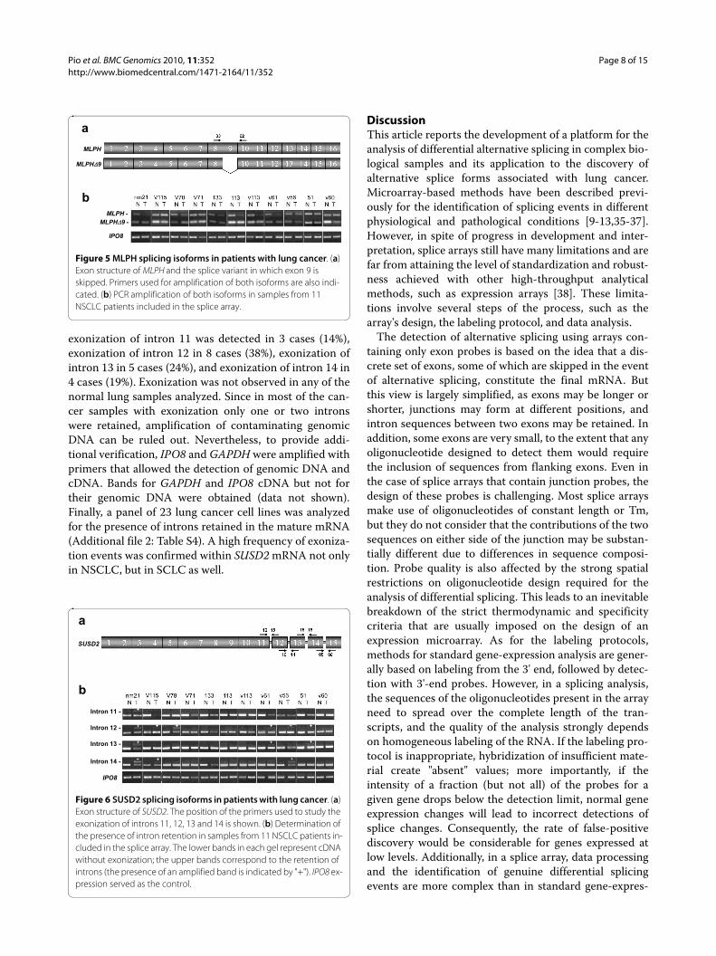
ing around exon 9, and database analysis indicated thealternative use of this exon in different isoforms (Figure5a). This alternative isoform generates a protein 28 aminoacids shorter than the normal protein. To validate thealternative splicing change, 11 samples from patientsincluded in the splice array were analyzed (Figure 5b). Aspecific downregulation of the isoform containing exon 9was identified in 8 out of the 11 cases studied (73%), con-firming the results obtained by the splice array (p = 0.036;Additional file 1: Figure S4b).SUSD2Susd2 (sushi domain-containing protein 2) is a recentlyidentified single-pass type I membrane protein [34]. Onlyone SUSD2 mRNA variant has been described to date(Figure 6a); however, splice array data suggested lungcancer-associated changes close to the 5' and 3' ends ofthe mRNA. To validate these results, normal and tumor-bearing lung tissues from 21 cases (11 patients used in thesplice array and 10 cases from the independent set) wereanalyzed, with reduced expression of SUSD2 detected inlung cancer tissue compared with normal lung. Regardingchanges in alternative splicing, alterations in the 3' endbut not in the 5' end of the mRNA were found. In particu-lar, frequent intron retention between exons 11 and 15was observed (Figure 6b). Inclusion of intronic sequenceswithin an mRNA is termed exonization. In our study,
Figure 3 CEACAM1-1 splicing isoforms in patients with lung can-cer. (a) CEACAM1-1 splicing isoforms. The location of the primers used in the study is also indicated. (b) PCR amplifications of different CEACAM1 splice forms in 11 normal lung tissues and paired tumors from patients included in the splice array. CEACAM1-1 was amplified with primers 01/08; the L- or S-forms in CEACAM1-1 were evaluated with primers 06/09; and CEACAM1-3 was determined with primers 01/15. IPO8 expression served as the control. Additional PCRs were done but are not shown in the figure: CEACAM1-3A was detected with prim-ers 01/14, and L- and S- forms of CEACAM1-3 and CEACAM1-3A were discriminated using primers 20/09 and 18/09, respectively. (c) CEACAM1-3A was evaluated by real-time PCR using primers 16-17 in 23 primary NSCLC tumors and their corresponding normal lung samples. ** p < 0.01.
a
b
c
***
CEACAM1-4L (CEACAM1)
CEACAM1-4S
CEACAM1-3L
CEACAM1-3S
CEACAM1-1L
CEACAM1-1S
CEACAM1-3AL
CEACAM1-3AS
CEACAM1-1
CEACAM1-1L-1S
CEACAM1-3
IPO8
**
Normal Tumoral0
5
10
15
20
CE
AC
AM
1-3A
/ IP
O8
(%)
CE
AC
AM
1-3A
/ IP
O8
(%)
Figure 4 FHL1 splicing isoforms in patients with lung cancer. (a) Exon structure of the 3' end of FHL1 showing alternative use of exon 6b in FHL1B. Primers used to quantify the two isoforms expressed by lung epithelial cells are also shown. (b) Ratio between the relative expres-sion of FHL1 and FHL1B in normal lung tissue and primary NSCLC tissue as determined by real-time PCR. ** p < 0.01.
a
FHL1
FHL1B
b **
0
10
20
30
40
50
60
70
80
Normal Tumoral
FH
L1/
FH
L1B
Pio et al. BMC Genomics 2010, 11:352http://www.biomedcentral.com/1471-2164/11/352
Page 8 of 15
exonization of intron 11 was detected in 3 cases (14%),exonization of intron 12 in 8 cases (38%), exonization ofintron 13 in 5 cases (24%), and exonization of intron 14 in4 cases (19%). Exonization was not observed in any of thenormal lung samples analyzed. Since in most of the can-cer samples with exonization only one or two intronswere retained, amplification of contaminating genomicDNA can be ruled out. Nevertheless, to provide addi-tional verification, IPO8 and GAPDH were amplified withprimers that allowed the detection of genomic DNA andcDNA. Bands for GAPDH and IPO8 cDNA but not fortheir genomic DNA were obtained (data not shown).Finally, a panel of 23 lung cancer cell lines was analyzedfor the presence of introns retained in the mature mRNA(Additional file 2: Table S4). A high frequency of exoniza-tion events was confirmed within SUSD2 mRNA not onlyin NSCLC, but in SCLC as well.
DiscussionThis article reports the development of a platform for theanalysis of differential alternative splicing in complex bio-logical samples and its application to the discovery ofalternative splice forms associated with lung cancer.Microarray-based methods have been described previ-ously for the identification of splicing events in differentphysiological and pathological conditions [9-13,35-37].However, in spite of progress in development and inter-pretation, splice arrays still have many limitations and arefar from attaining the level of standardization and robust-ness achieved with other high-throughput analyticalmethods, such as expression arrays [38]. These limita-tions involve several steps of the process, such as thearray's design, the labeling protocol, and data analysis.
The detection of alternative splicing using arrays con-taining only exon probes is based on the idea that a dis-crete set of exons, some of which are skipped in the eventof alternative splicing, constitute the final mRNA. Butthis view is largely simplified, as exons may be longer orshorter, junctions may form at different positions, andintron sequences between two exons may be retained. Inaddition, some exons are very small, to the extent that anyoligonucleotide designed to detect them would requirethe inclusion of sequences from flanking exons. Even inthe case of splice arrays that contain junction probes, thedesign of these probes is challenging. Most splice arraysmake use of oligonucleotides of constant length or Tm,but they do not consider that the contributions of the twosequences on either side of the junction may be substan-tially different due to differences in sequence composi-tion. Probe quality is also affected by the strong spatialrestrictions on oligonucleotide design required for theanalysis of differential splicing. This leads to an inevitablebreakdown of the strict thermodynamic and specificitycriteria that are usually imposed on the design of anexpression microarray. As for the labeling protocols,methods for standard gene-expression analysis are gener-ally based on labeling from the 3' end, followed by detec-tion with 3'-end probes. However, in a splicing analysis,the sequences of the oligonucleotides present in the arrayneed to spread over the complete length of the tran-scripts, and the quality of the analysis strongly dependson homogeneous labeling of the RNA. If the labeling pro-tocol is inappropriate, hybridization of insufficient mate-rial create "absent" values; more importantly, if theintensity of a fraction (but not all) of the probes for agiven gene drops below the detection limit, normal geneexpression changes will lead to incorrect detections ofsplice changes. Consequently, the rate of false-positivediscovery would be considerable for genes expressed atlow levels. Additionally, in a splice array, data processingand the identification of genuine differential splicingevents are more complex than in standard gene-expres-
Figure 5 MLPH splicing isoforms in patients with lung cancer. (a) Exon structure of MLPH and the splice variant in which exon 9 is skipped. Primers used for amplification of both isoforms are also indi-cated. (b) PCR amplification of both isoforms in samples from 11 NSCLC patients included in the splice array.
a
bMLPH -
IPO8
MLPH
MLPH��9
MLPH�9 -
Figure 6 SUSD2 splicing isoforms in patients with lung cancer. (a) Exon structure of SUSD2. The position of the primers used to study the exonization of introns 11, 12, 13 and 14 is shown. (b) Determination of the presence of intron retention in samples from 11 NSCLC patients in-cluded in the splice array. The lower bands in each gel represent cDNA without exonization; the upper bands correspond to the retention of introns (the presence of an amplified band is indicated by "+"). IPO8 ex-pression served as the control.
a
b
SUSD2
Intron 12 -
Intron 13 -
Intron 14 -
IPO8
Intron 11 -
Pio et al. BMC Genomics 2010, 11:352http://www.biomedcentral.com/1471-2164/11/352
Page 9 of 15
sion arrays and require specific analytical algorithms.This is due to the larger variation in thermodynamic con-ditions and possible cross-hybridization or folding ofsub-optimally designed probes. Finally, an additionalrequirement for the analytical algorithm is the need todistinguish differential splicing from changes in geneexpression levels. The methodology described in thepresent work has addressed all these limitations.
In designing the probes, we applied an oligonucleotidedesign algorithm that performs an in silico thermody-namic simulation of the hybridization procedure. Thealgorithm targets the best possible oligonucleotide foreach sequence, rather than imposing a strict oligonucle-otide quality cutoff. Several control oligonucleotides werealso designed and included in the array to control labelingand hybridization processes. The proper design andinclusion of control oligonucleotides, as well as appropri-ate use of the data generated by these controls in dataprocessing, are especially relevant considering that tech-nical variability can be introduced by the addition of stepsin the labeling protocol necessary to avoid labeling biases.To analyze the data, we developed new software to inter-pret the intensity changes of all the oligonucleotides for agiven gene and to decide whether they reflected expres-sion changes or isoform changes.
The efficacy of our new methodology and its potentialusefulness in a clinical setting were tested in an applica-tion designed to identify genes differentially spliced inprimary lung tumors. Lung cancer is the leading cause ofcancer deaths worldwide [39], with the major form,NSCLC, accounting for about 80% of all lung cancers. Inspite of advances in early detection and treatment, overall5-year survival rates for NSCLC remain at about 15%[40], underlining the need for a better understanding ofthe molecular pathogenesis of NSCLC. It has been pro-posed that modifications in the concentration, localiza-tion, composition, or activity of RNA-binding proteinsacting as splicing regulatory factors induce the splicingalterations characteristic of lung cancer [41]. In thissense, the abnormal expression of heterogeneous nuclearribonucleoproteins (hnRNP) in NSCLC clinical samplesand animal models suggests that tumors develop specifichnRNP profiles [42-44]. This alteration would generateclinically relevant alternative splice forms contributing tolung carcinogenesis. A recent report presented agenome-wide analysis of alternative splicing events inlung adenocarcinoma [13]. In that study, the authorsobtained a list of cancer-related candidate genes showingalternative splicing events and implicated in cancer.
In the present study, the presence of differentiallyexpressed splice variants in NSCLC was evaluated using asplice array designed to detect near 8000 genes known tobe expressed in lung tissue. Analysis of the splice arraydata generated a list of candidates, from which 10 genes
were selected for validation. Since one of the main pur-poses for this selection was to validate the quality of thedetection process, no biological criteria were consideredin the selection of the candidate genes at this point. RT-PCR experiments, followed by sequencing, were used tovalidate the results from the array, with changes in alter-native splicing confirmed in four genes. As expected, thevalidation success was below the rates obtained in geneexpression studies and was comparable to the ratesreported in previous splicing studies [13,18]. Regardlessof the platform and algorithm used to detect differentialsplicing, by microarray or other hybridization-basedanalysis, it is important to realize that the technology isinherently sensitive to a number of errors that can lead tothe incorrect identification of alternative splicing. Forexample, low-level expression can lead to the erroneousidentification of splice events, due to the fact that not alloligonucleotides generate the same level of signal, and thesignal of low-responsive oligonucleotides can drop belowthe detection limit thereby generating false "formchanges" when the overall expression level differsbetween Cy3 and Cy5 channels. Cross-hybridization,obviously, is another potential cause of the detection offalse "form changes". While cross-hybridization cansometimes be suspected when the higher signal of oneoligonucleotide compared to the others cannot be justi-fied by a much higher Tm or a sub-optimal design, it willgenerally go unnoticed until further detailed analysis isperformed. Moreover, there is no guarantee that all possi-ble gene-structure changes are analyzed in the validationprocess, unless a very extensive validation approach isapplied for any gene of interest (which may be hamperedby the availability of clinical material). The four geneswith lung cancer-associated alternative splice formsnewly identified in this study were: CEACAM1, FHL1,MLPH, and SUSD2.
Ceacam1 is a CEA-related cell adhesion moleculedownregulated in several human cancer types, includingprostate, breast, and colorectal cancers [25]. CEACAM1has been described as a lung tumor marker, and itsexpression has been associated with the prognosis of lungadenocarcinoma [45-47]. Two major CEACAM1 iso-forms have been described: a long (L-) form and a short(S-) form, which, respectively, include or exclude exon 7.The exclusion of exon 7 generates a proximal stop codonthat translates into a shorter cytoplasmic domain. Tumorcells transfected with CEACAM1-1L are less tumori-genic, suggesting that the L-form functions as a tumorsuppressor gene [48]. Wang et al. reported thatCEACAM1-4S is the predominant isoform in NSCLC tis-sues, whereas in normal lung tissues the main isoform isCEACAM1-4L [49]. This splice pattern was recently con-firmed [13]. In addition to confirming previous data, ouranalysis predicted other changes in the splicing of
Pio et al. BMC Genomics 2010, 11:352http://www.biomedcentral.com/1471-2164/11/352
Page 10 of 15
CEACAM1 around exons 2 and 5, which were validatedby PCR. For the first time, it was demonstrated that lungtumors frequently overexpress three splice isoforms:CEACAM1-1, CEACAM1-3, and CEACAM1-3A. Thealternative use of these exons affects different Ig-likestructural domains in the extracellular portion of therespective proteins.
The family of four and a half LIM (FHL) proteins, alsoknown as skeletal muscle LIM proteins (SLIM), is charac-terized by four complete LIM domains preceded by an N-terminal half LIM domain [50]. LIM domains arecysteine-rich, double zinc-finger motifs involved in pro-tein-protein interactions. FHL has been shown to regu-late tissue differentiation, proliferation, adhesion,migration, cytoskeletal organization [51,52], and recently,to play a role in carcinogenesis through a TGF-β-like sig-naling pathway [53]. Four and a half LIM domains 1 (Fhl1or Slim1) is a member of this family and has likewise beenimplicated in skeletal muscle development [26] as well asin the pulmonary vascular remodeling underlying pulmo-nary hypertension [54]. Interestingly, Fhl1 is downregu-lated in many types of solid malignancies and it exhibitstumor suppressor activity [30-32]. Among the splice vari-ants described for FHL1, in our study the expression oftwo of them, FHL1 and FHL1B, was identified in lungsamples. In agreement with previous reports, clear down-regulation in the expression of the FHL1 gene wasdetected in lung cancer specimens. More importantly, wedetermined that the downregulation of FHL1 is signifi-cantly higher than that of FHL1B. The two proteins areidentical over the first three LIM domains but FHL1Bcontains a distinct C-terminus (96 amino acids) withthree potential bipartite nuclear localization signals, aputative nuclear export sequence, and a binding motif forthe transcription factor RBP-J [27,28]. Whereas FHL1 ismainly located at focal adhesions, FHL1B is predomi-nantly a nuclear protein and has unique physiologicalfunctions, including the regulation of Notch signalingthrough its association with RBP-J [55]. Notch signalingprofoundly influences the regulation of tumor progres-sion, specifically, tumor cell proliferation, differentiation,apoptosis, and angiogenesis [56].
Mlph is a member of the synaptotagmin-like proteinfamily and is involved in the transport of melanosomes[33]. These lysosome-related organelles are specialized inthe synthesis and distribution of melanin. Mlph is anessential member of the melanosome trafficking com-plex, acting as a link between Rab27a and myosin Va[57,58]. It may also be involved in the trafficking of epi-thelial Na + channel in cells of the collecting duct of thekidney [59]. Mlph contains an N-terminal Slp homologydomain (SHD) involved in binding to Rab27a, a myosin-binding domain (MBD) in its middle region, and a C-ter-minal actin-binding domain (ABD). Here, we demon-strated that lung tissue expresses at least two isoforms of
MLPH, one with and one without exon 9. Skipping of thisexon generates a protein 28 amino acids shorter than thenormal protein, without affecting any of the three charac-terized functional domains. In lung tumors, there is spe-cific downregulation of the isoform containing exon 9.
The recently identified Susd2 is a single-pass type Imembrane protein with an extracellular portion that con-tains somatomedin B, AMOP, von Willebrand factor typeD, and sushi/CCP/SCR domains [34]. Although its physi-ological function is still unknown, overexpression ofSusd2 is thought to suppress tumorigenicity [34,60]. Inagreement with this postulated role for the protein, weobserved reduced expression of SUSD2 in lung cancer tis-sues. Interestingly, intron retention was frequentlydetected between the last exons of the mRNA. Inclusionof intronic sequences within an mRNA is termedexonization. Although this modification is the rarest typeof alternative splicing found in normal cells, exonizationevents in cancer cells are frequent and may be associatedwith impairments in splicing regulatory factors [61]. Theexonization of introns affects the extracellular portion ofSUSD2 in that the translation of intron 11 introduces apremature stop codon which disrupts the von Willebrandfactor type D domain at amino acid 631. Exonization ofintron 12 generates a protein whose last 41 amino acidsare substituted by 68 new amino acids (38 coded byintron 12 and 30 new amino acids translated as conse-quence of a frame-shift in the reading frame of exon 13).Translation of intron 13 generates a protein with a newsequence of 23 amino acids from position 781 (withoutaffecting any known functional domain), while retentionof intron 14 introduces a premature stop codon, eliminat-ing seven amino acids at the C-terminal end.
ConclusionsWe have developed and tested a novel platform for high-throughput analysis of alternative splicing events in bio-logical samples. The application of this methodology willaid in understanding the functional relevance of splicevariants in pathological conditions and facilitate the iden-tification of new biomarkers and targets for therapy. Toprove the usefulness of this platform, this methodologywas used to identify cancer-associated splice variants inlung cancer. Differentially expressed splice variants offour genes were identified, with potential utility in thediagnosis of lung cancer. Additional work is in progress toanalyze the relevance of these newly characterized can-cer-associated isoforms as well as to validate additionalcandidates from data obtained in the splice array.
MethodsClinical samplesPrimary tumors and their corresponding normal lung tis-sues were obtained from patients with non-small cell lungcancer (NSCLC) treated with curative resectional surgery
Pio et al. BMC Genomics 2010, 11:352http://www.biomedcentral.com/1471-2164/11/352
Page 11 of 15
at the Clínica Universidad de Navarra (Pamplona, Spain)or at the Hospital Marqués de Valdecilla (Santander,Spain). None of the patients received chemo- or radio-therapy prior to surgery. The study was approved by theethics committees of the participating institutions andinformed consent was obtained from each patient. Surgi-cally removed samples were immediately frozen in liquidnitrogen and stored at -80°C until use. A portion of eachsample was sectioned in a cryostat and mounted ontoslides. After fixation, these samples were stained withhematoxylin and eosin, and then carefully examined bytwo experienced researchers. Samples containing lessthan 70% tumor cells were discarded. The 42 specimensselected for the study were divided into two groups: onefor discovery (n = 20; Additional file 2: Table S1) and onefor validation (n = 22; Additional file 2: Table S2).
Lung cancer cell linesAll lung cancer cell lines were obtained from the Ameri-can Type Culture Collection (ATCC), except HCC44,HCC827, EPLC-272H, and HCC15, which were obtainedfrom the German Collection of Microorganisms and CellCultures (DSMZ). Cells were grown in RPMI supple-mented with 2 mM glutamine, 10% fetal bovine serum(FBS), 100 U/ml penicillin, and 100 μg/ml streptomycin.
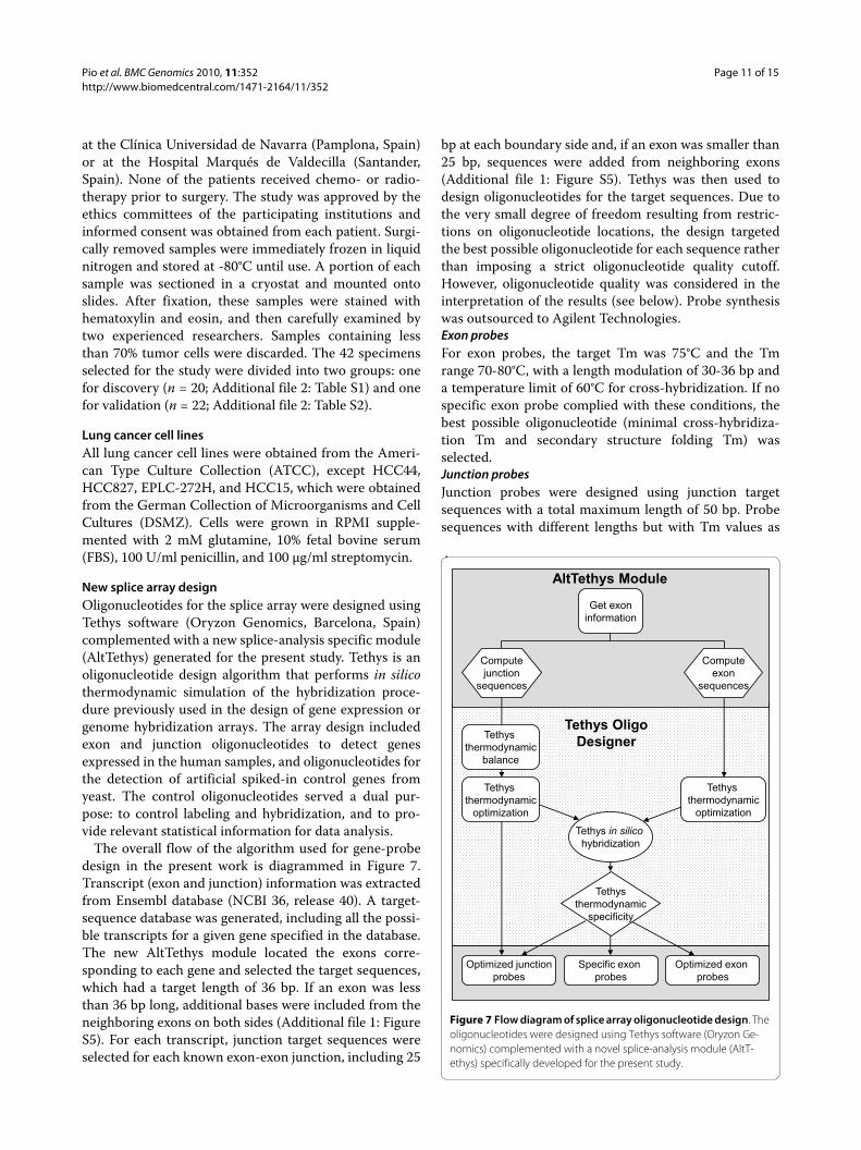
New splice array designOligonucleotides for the splice array were designed usingTethys software (Oryzon Genomics, Barcelona, Spain)complemented with a new splice-analysis specific module(AltTethys) generated for the present study. Tethys is anoligonucleotide design algorithm that performs in silicothermodynamic simulation of the hybridization proce-dure previously used in the design of gene expression orgenome hybridization arrays. The array design includedexon and junction oligonucleotides to detect genesexpressed in the human samples, and oligonucleotides forthe detection of artificial spiked-in control genes fromyeast. The control oligonucleotides served a dual pur-pose: to control labeling and hybridization, and to pro-vide relevant statistical information for data analysis.
The overall flow of the algorithm used for gene-probedesign in the present work is diagrammed in Figure 7.Transcript (exon and junction) information was extractedfrom Ensembl database (NCBI 36, release 40). A target-sequence database was generated, including all the possi-ble transcripts for a given gene specified in the database.The new AltTethys module located the exons corre-sponding to each gene and selected the target sequences,which had a target length of 36 bp. If an exon was lessthan 36 bp long, additional bases were included from theneighboring exons on both sides (Additional file 1: FigureS5). For each transcript, junction target sequences wereselected for each known exon-exon junction, including 25
bp at each boundary side and, if an exon was smaller than25 bp, sequences were added from neighboring exons(Additional file 1: Figure S5). Tethys was then used todesign oligonucleotides for the target sequences. Due tothe very small degree of freedom resulting from restric-tions on oligonucleotide locations, the design targetedthe best possible oligonucleotide for each sequence ratherthan imposing a strict oligonucleotide quality cutoff.However, oligonucleotide quality was considered in theinterpretation of the results (see below). Probe synthesiswas outsourced to Agilent Technologies.Exon probesFor exon probes, the target Tm was 75°C and the Tmrange 70-80°C, with a length modulation of 30-36 bp anda temperature limit of 60°C for cross-hybridization. If nospecific exon probe complied with these conditions, thebest possible oligonucleotide (minimal cross-hybridiza-tion Tm and secondary structure folding Tm) wasselected.Junction probesJunction probes were designed using junction targetsequences with a total maximum length of 50 bp. Probesequences with different lengths but with Tm values as
Figure 7 Flow diagram of splice array oligonucleotide design. The oligonucleotides were designed using Tethys software (Oryzon Ge-nomics) complemented with a novel splice-analysis module (AltT-ethys) specifically developed for the present study.
Tethys in silicohybridization
AltTethys Module
Specific exon probes
Optimized junctionprobes
Optimized exon probes
Tethys thermodynamic
balance
Tethys thermodynamic
optimization
Get exoninformation
Computejunction
sequences
Computeexon
sequences
Tethys thermodynamic
optimization
Tethys OligoDesigner
Tethysthermodynamic
specificity
Pio et al. BMC Genomics 2010, 11:352http://www.biomedcentral.com/1471-2164/11/352
Page 12 of 15
similar as possible for the two sections of the oligonucle-otide flanking both sides of the junction were selected.The total length of the junction probes ranged from 25 to42 bp. Among all possible sequences, the oligonucleotidewith a Tm as close as possible to the target temperature of75°C was selected. Finally, the oligonucleotide waschecked for Secondary Structure Folding Tm and Maxi-mum Cross-Hybridization Tm using the Tethys oligonu-cleotide design backend, taking note of the potential forcross-hybridization.Control probesAn extensive battery of probes was included to controllabeling and hybridization processes and to provide rele-vant statistical information for data analysis: (a) threeyeast artificial target sequences were spiked into thehybridization mixture at three different concentrationlevels but balanced in the Cy3 and Cy5 channels; (b) opti-mum oligonucleotides (in terms of specificity and ther-modynamics) distributed over the length of the targetsequences to assess labeling bias; (c) half oligonucleotides(generated by splitting optimum oligonucleotides intotwo thermodynamically equivalent halves comple-mented with stretches of AT at the 5' or 3' end) to simu-late differential splicing at the splice-donor or splice-acceptor site. These probes were included more than tentimes in the array in order to determine intra-array vari-ability.
In addition, a higher number of positive and negativecontrol probes were distributed over the array surface(maize expansin, ZmMYB42, and xyloglucan endo-trans-glycosylase). These probes were used to assess detectionlimits and range, to verify spatial homogeneity, and todetermine experimental within-array variation.
Cloning of artificial constructs for VEGF, PCBP4, and CDC6The performance of the splice array was tested using apilot array designed to identify different transcripts ofthree genes: VEGF, PCBP4, and CDC6. Artificial tran-scripts were generated for three VEGF isoforms(VEGF121, VEGF165, and VEGF185) [62], two PCBP4 iso-forms (PCBP4 and PCBP4a) [44], and the only knownCDC6 isoform.
Preparation of yeast controlsThree yeast sequences of DNA were amplified by PCRfrom genomic DNA of Saccharomyces cerevisae strainS288C using two chimeric primers, where a primer con-sisted of a T7 promoter and the gene specific sequence,and the other primer consisted of a tail of 20 timidinesand the gene specific sequence. These three genes were:YIL129C (7100 bp), YML059C (4900 bp) and YOR328W(4600 bp). They were amplified by PCR with 32 ng ofgenomic DNA from yeast and using a combination of twopolymerase TaqI:Pfu (20:1). cRNA was generated using
an in vitro transcription system (T7 Megascript kit;Ambion) getting the final artificial unique splice forms.The sequences were spiked into the samples prior tolabeling.
RNA extraction and labelingTotal RNA from paired normal and tumor samples fromlung tissues was extracted using the RNeasy ExtractionKit (Qiagen) according to the manufacturer's instruc-tions, with minor modifications. RNA quality wasassessed using an Agilent Bioanalyzer 2100 and quanti-fied using a Nanodrop ND-1000 spectrophotometer.Samples with an RNA integrity number (RIN) below 7were excluded from further analysis. PolyA + RNA wasextracted using Dynabead magnetic particles. To obtainhomogeneous labeling of the RNA across the entirelength of the transcript, a novel labeling procedure,described in the Results section, was developed. Fiftynanograms of PolyA + RNA from normal tissue waslabeled with Cy3 and the same amount of PolyA + RNAfrom tumor samples with Cy5. In addition, 50 ng of PolyA+ RNA from normal tissue was labeled with Cy5. Prior tolabeling, artificial yeast transcipts were spiked into allpolyA + samples mixtures. The quality of the labeledsamples was verified using the Agilent 2100 Bioanalyzerand sample concentration was determined using theNanodrop ND-1000 spectrophotometer.
Array hybridization and data acquisitionLabeled normal tissue cRNA (4.5 μg) was mixed with thesame amount of labeled tumor cRNA from the samepatient, and equal quantities of Cy3 and Cy5 labeled Xetand Zm42 cRNA controls were spiked in to serve ashybridization controls. The cRNA was mixed with 25 ×fragmentation buffer (Agilent) and incubated at 60°C for30 min to fragment RNA. Afterwards, 250 μl of 2 ×hybridization buffer (Agilent) was added to stop the frag-mentation reaction and the mixture was hybridized onthe array. Slides were incubated for 17 h at 60°C in anAgilent DNA Hybridization Oven (G2545A) with therotation setting at 4 rpm. A total of twenty Cy3 labelednormal and Cy5 labeled tumor lung cancer samples, werecohybridized pairwise on the splice array as well as threeCy3 labeled normal and Cy5 labeled normal samples. Rawdata were acquired using an Agilent DNA MicroarrayScanner and Agilent Feature Extraction Software (V.9.1).The general reproducibility of the hybridization platform(labeling procedure, hybridization, and detection) wasassessed by means of self to self hybridization; the stan-dard deviation of the fold change of all oligonucleotideswas 0.093.
Data processingFor data processing, a novel algorithm that distinguishedbetween changes in gene expression and splicing varia-
Pio et al. BMC Genomics 2010, 11:352http://www.biomedcentral.com/1471-2164/11/352
Page 13 of 15
tion was developed. The analysis of differential splice iso-forms is more complex than the analysis of differentialgene expression, due to a higher variation in thermody-namic conditions and possible cross-hybridization andfolding of sub-optimally designed oligonucleotides. Inaddition, compared with regular gene expression analy-ses, additional variation can be introduced due to theincorporation of extra steps in the labeling protocol. Thisgives a special importance to the incorporation of spiked-in controls in the array design and their use in data pro-cessing. The data processing procedure (which is detailedin Additional file 2: Supplementary Methods) was dividedinto four steps: data filtering and normalization, probespot calibration, gene probe statistical analysis, and iso-form analysis. Data processing is discussed more exten-sively in Additional file 2: Supplementary Methods.
Validation of cancer-associated splice variants by PCRResults obtained in the splice array were validated byPCR. Two micrograms of RNA from the clinical sampleswere reverse transcribed. Genomic DNA contaminationwas controlled in each RNA sample using a reaction mixlacking reverse transcriptase. One microliter of cDNAdiluted 1:10 was used for PCR amplification, and the PCRproducts were electrophoresed in agarose gels. Forsequencing, the amplified bands were purified using theQiagen MinElute PCR Purification Kit and sequenced inan ABI377 sequencer (Perkin-Elmer Applied Biosystems).Real-time PCR was performed using SYBR Green PCRMaster Mix (Applied Biosystems) in the Applied Biosys-tems 7300 Real-Time PCR System. The reactions werecarried out according to the manufacturer's instructions.Each sample was analyzed in triplicate. Relative levels ofexpression were determined by the Ct method usingIPO8 as the reference [24]. Primers used for validationare shown in Additional file 2: Table S3.
Additional material
AbbreviationsNSCLC: non-small cell lung cancer; Ceacam1: carcinoembryonic antigen-related cell adhesion molecule 1; SCLC: small cell lung cancer; Fhl1: four and ahalf LIM domains 1; Mlph: melanophilin or Slac2-a; Susd2: sushi domain-con-taining protein 2; hnRNP: heterogeneous nuclear ribonucleoproteins.
Authors' contributionsRP, OD, TM and LMM conceived and designed the experiments. DB, MJP, TE, JAand MAA performed the lung cancer molecular biology experiments. OD, EA,FS and TM carried out the bioinformatics, platform design and hybridization.RP, DB, EA, OD, TE, JA, MAA, AR, FS, TM and LMM analyzed the data. JGR, MJP,MDL, JMLP and FS contributed reagents/materials/analysis tools. RP, DB, EA,OD, FS, TM and LLM wrote the paper. All authors read and approved the finalmanuscript.
AcknowledgementsThis work was supported by "UTE project CIMA" and grants from Spanish Minis-try of Industry [Programa Ingenio 2010, CENIT Ref. Oncnosis], Fundación MMA, and Red Temática de Investigación Cooperativa en Cáncer [RTICC, RD06/0020/0066], Instituto de Salud Carlos III (ISCIII), Spanish Ministry of Science and Inno-vation & European Regional Development Fund (ERDF) "Una manera de hacer Europa". We greatly appreciate Cristina Sainz, Amaya Lavin and Ana Remirez for their technical assistance and Uxua Montes for her help in the collection of clinical samples.
Author Details1Division of Oncology, Center for Applied Medical Research, Pamplona, Spain, 2Department of Biochemistry, School of Medicine, University of Navarra, Pamplona, Spain, 3Department of Histology and Pathology, School of Medicine, University of Navarra, Pamplona, Spain, 4Oryzon Genomics, Scientific Parc University of Barcelona, Barcelona, Spain, 5Department of Pathology, Marques de Valdecilla University Hospital, School of Medicine, University of Cantabria, Santander, Spain, 6CEIT and TECNUN, University of Navarra, San Sebastian, Spain, 7Department of Pathology, Clínica Universidad de Navarra, Pamplona, Spain and 8Department of Oncology, Clínica Universidad de Navarra, Pamplona, Spain
References1. Wang ET, Sandberg R, Luo S, Khrebtukova I, Zhang L, Mayr C, Kingsmore
SF, Schroth GP, Burge CB: Alternative isoform regulation in human tissue transcriptomes. Nature 2008, 456(7221):470-476.
2. Sultan M, Schulz MH, Richard H, Magen A, Klingenhoff A, Scherf M, Seifert M, Borodina T, Soldatov A, Parkhomchuk D, et al.: A global view of gene activity and alternative splicing by deep sequencing of the human transcriptome. Science 2008, 321(5891):956-960.
3. Venables JP: Unbalanced alternative splicing and its significance in cancer. Bioessays 2006, 28(4):378-386.
4. Kalnina Z, Zayakin P, Silina K, Line A: Alterations of pre-mRNA splicing in cancer. Genes Chromosomes Cancer 2005, 42(4):342-357.
5. Grosso AR, Martins S, Carmo-Fonseca M: The emerging role of splicing factors in cancer. EMBO Rep 2008, 9(11):1087-1093.
6. Pajares MJ, Ezponda T, Catena R, Calvo A, Pio R, Montuenga LM: Alternative splicing: an emerging topic in molecular and clinical oncology. Lancet Oncol 2007, 8(4):349-357.
7. Gattenlohner S, Stuhmer T, Leich E, Reinhard M, Etschmann B, Volker HU, Rosenwald A, Serfling E, Bargou RC, Ertl G, et al.: Specific Detection of CD56 (NCAM) Isoforms for the Identification of Aggressive Malignant Neoplasms with Progressive Development. Am J Pathol 2009, 174(4):1160-1171.
8. Calarco JA, Saltzman AL, Ip JY, Blencowe BJ: Technologies for the global discovery and analysis of alternative splicing. Adv Exp Med Biol 2007, 623:64-84.
9. Gardina PJ, Clark TA, Shimada B, Staples MK, Yang Q, Veitch J, Schweitzer A, Awad T, Sugnet C, Dee S, et al.: Alternative splicing and differential gene expression in colon cancer detected by a whole genome exon array. BMC Genomics 2006, 7:325.
10. French PJ, Peeters J, Horsman S, Duijm E, Siccama I, van den Bent MJ, Luider TM, Kros JM, van der Spek P, Sillevis Smitt PA: Identification of differentially regulated splice variants and novel exons in glial brain tumors using exon expression arrays. Cancer Res 2007, 67(12):5635-5642.
11. Cheung HC, Baggerly KA, Tsavachidis S, Bachinski LL, Neubauer VL, Nixon TJ, Aldape KD, Cote GJ, Krahe R: Global analysis of aberrant pre-mRNA splicing in glioblastoma using exon expression arrays. BMC Genomics 2008, 9:216.
12. Thorsen K, Sorensen KD, Brems-Eskildsen AS, Modin C, Gaustadnes M, Hein AM, Kruhoffer M, Laurberg S, Borre M, Wang K, et al.: Alternative splicing in colon, bladder, and prostate cancer identified by exon array analysis. Mol Cell Proteomics 2008, 7(7):1214-1224.
13. Xi L, Feber A, Gupta V, Wu M, Bergemann AD, Landreneau RJ, Litle VR, Pennathur A, Luketich JD, Godfrey TE: Whole genome exon arrays identify differential expression of alternatively spliced, cancer-related genes in lung cancer. Nucleic Acids Res 2008, 36(20):6535-6547.
Additional file 1 Supplementary Figures S1 to S9.Additional file 2 Supplementary Methods and Tables S1 to S4.
Received: 21 December 2009 Accepted: 3 June 2010 Published: 3 June 2010This article is available from: http://www.biomedcentral.com/1471-2164/11/352© 2010 Pio et al; licensee BioMed Central Ltd. This is an Open Access article distributed under the terms of the Creative Commons Attribution License (http://creativecommons.org/licenses/by/2.0), which permits unrestricted use, distribution, and reproduction in any medium, provided the original work is properly cited.BMC Genomics 2010, 11:352
Pio et al. BMC Genomics 2010, 11:352http://www.biomedcentral.com/1471-2164/11/352
Page 14 of 15
14. Morgan WR, Ward DC: Three splicing patterns are used to excise the small intron common to all minute virus of mice RNAs. J Virol 1986, 60(3):1170-1174.
15. Lockhart DJ, Dong H, Byrne MC, Follettie MT, Gallo MV, Chee MS, Mittmann M, Wang C, Kobayashi M, Horton H, et al.: Expression monitoring by hybridization to high-density oligonucleotide arrays. Nat Biotechnol 1996, 14(13):1675-1680.
16. Clark TA, Sugnet CW, Ares MJ: Genomewide analysis of mRNA processing in yeast using splicing-specific microarrays. Science 2002, 296(5569):907-910.
17. Castle J, Garrett-Engele P, Armour CD, Duenwald SJ, Loerch PM, Meyer MR, Schadt EE, Stoughton R, Parrish ML, Shoemaker DD, et al.: Optimization of oligonucleotide arrays and RNA amplification protocols for analysis of transcript structure and alternative splicing. Genome Biol 2003, 4(10):R66.
18. Johnson JM, Castle J, Garrett-Engele P, Kan Z, Loerch PM, Armour CD, Santos R, Schadt EE, Stoughton R, Shoemaker DD: Genome-wide survey of human alternative pre-mRNA splicing with exon junction microarrays. Science 2003, 302(5653):2141-2144.
19. Wang H, Hubbell E, Hu JS, Mei G, Cline M, Lu G, Clark T, Siani-Rose MA, Ares M, Kulp DC, et al.: Gene structure-based splice variant deconvolution using a microarray platform. Bioinformatics 2003, 19(Suppl 1):i315-322.
20. Anton MA, Gorostiaga D, Guruceaga E, Segura V, Carmona-Saez P, Pascual-Montano A, Pio R, Montuenga LM, Rubio A: SPACE: an algorithm to predict and quantify alternatively spliced isoforms using microarrays. Genome Biol 2008, 9(2):R46.
21. Van Gelder RN, von Zastrow ME, Yool A, Dement WC, Barchas JD, Eberwine JH: Amplified RNA synthesized from limited quantities of heterogeneous cDNA. Proc Natl Acad Sci USA 1990, 87(5):1663-1667.
22. Bhattacharjee A, Richards WG, Staunton J, Li C, Monti S, Vasa P, Ladd C, Beheshti J, Bueno R, Gillette M, et al.: Classification of human lung carcinomas by mRNA expression profiling reveals distinct adenocarcinoma subclasses. Proc Natl Acad Sci USA 2001, 98(24):13790-13795.
23. Beer DG, Kardia SL, Huang CC, Giordano TJ, Levin AM, Misek DE, Lin L, Chen G, Gharib TG, Thomas DG, et al.: Gene-expression profiles predict survival of patients with lung adenocarcinoma. Nat Med 2002, 8(8):816-824.
24. Nguewa PA, Agorreta J, Blanco D, Lozano MD, Gomez-Roman J, Sanchez BA, Valles I, Pajares MJ, Pio R, Rodriguez MJ, et al.: Identification of Importin 8 (IPO8) as the most accurate reference gene for the clinicopathological analysis of lung specimens. BMC Mol Biol 2008, 9:103.
25. Gray-Owen SD, Blumberg RS: CEACAM1: contact-dependent control of immunity. Nat Rev Immunol 2006, 6(6):433-446.
26. Cowling BS, McGrath MJ, Nguyen MA, Cottle DL, Kee AJ, Brown S, Schessl J, Zou Y, Joya J, Bonnemann CG, et al.: Identification of FHL1 as a regulator of skeletal muscle mass: implications for human myopathy. J Cell Biol 2008, 183(6):1033-1048.
27. Brown S, McGrath MJ, Ooms LM, Gurung R, Maimone MM, Mitchell CA: Characterization of two isoforms of the skeletal muscle LIM protein 1, SLIM1. Localization of SLIM1 at focal adhesions and the isoform slimmer in the nucleus of myoblasts and cytoplasm of myotubes suggests distinct roles in the cytoskeleton and in nuclear-cytoplasmic communication. J Biol Chem 1999, 274(38):27083-27091.
28. Lee SM, Li HY, Ng EK, Or SM, Chan KK, Kotaka M, Chim SS, Tsui SK, Waye MM, Fung KP, et al.: Characterization of a brain-specific nuclear LIM domain protein (FHL1B) which is an alternatively spliced variant of FHL1. Gene 1999, 237(1):253-263.
29. Ng EK, Lee SM, Li HY, Ngai SM, Tsui SK, Waye MM, Lee CY, Fung KP: Characterization of tissue-specific LIM domain protein (FHL1C) which is an alternatively spliced isoform of a human LIM-only protein (FHL1). J Cell Biochem 2001, 82(1):1-10.
30. Fryknas M, Wickenberg-Bolin U, Goransson H, Gustafsson MG, Foukakis T, Lee JJ, Landegren U, Hoog A, Larsson C, Grimelius L, et al.: Molecular markers for discrimination of benign and malignant follicular thyroid tumors. Tumour Biol 2006, 27(4):211-220.
31. Li X, Jia Z, Shen Y, Ichikawa H, Jarvik J, Nagele RG, Goldberg GS: Coordinate suppression of Sdpr and Fhl1 expression in tumors of the breast, kidney, and prostate. Cancer Sci 2008, 99(7):1326-1333.
32. Sakashita K, Mimori K, Tanaka F, Kamohara Y, Inoue H, Sawada T, Hirakawa K, Mori M: Clinical significance of loss of Fhl1 expression in human gastric cancer. Ann Surg Oncol 2008, 15(8):2293-2300.
33. Matesic LE, Yip R, Reuss AE, Swing DA, O'Sullivan TN, Fletcher CF, Copeland NG, Jenkins NA: Mutations in Mlph, encoding a member of the Rab effector family, cause the melanosome transport defects observed in leaden mice. Proc Natl Acad Sci USA 2001, 98(18):10238-10243.
34. Sugahara T, Yamashita Y, Shinomi M, Yamanoha B, Iseki H, Takeda A, Okazaki Y, Hayashizaki Y, Kawai K, Suemizu H, et al.: Isolation of a novel mouse gene, mSVS-1/SUSD2, reversing tumorigenic phenotypes of cancer cells in vitro. Cancer Sci 2007, 98(6):900-908.
35. Fehlbaum P, Guihal C, Bracco L, Cochet O: A microarray configuration to quantify expression levels and relative abundance of splice variants. Nucleic Acids Res 2005, 33(5):e47.
36. Hartmann B, Castelo R, Blanchette M, Boue S, Rio DC, Valcarcel J: Global analysis of alternative splicing regulation by insulin and wingless signalling in Drosophila cells. Genome Biol 2009, 10(1):R11.
37. Castle JC, Zhang C, Shah JK, Kulkarni AV, Kalsotra A, Cooper TA, Johnson JM: Expression of 24,426 human alternative splicing events and predicted cis regulation in 48 tissues and cell lines. Nat Genet 2008, 40(12):1416-1425.
38. Rogers S, Cambrosio A: Making a new technology work: the standardization and regulation of microarrays. Yale J Biol Med 2007, 80(4):165-178.
39. Parkin DM, Bray F, Ferlay J, Pisani P: Global cancer statistics, 2002. CA Cancer J Clin 2005, 55(2):74-108.
40. Ries LAG, Melbert D, Krapcho M, Stinchcomb DG, Howlader N, Horner MJ, Mariotto A, Miller BA, Feuer EJ, Altekruse SF, et al.: SEER Cancer Statistics Review, 1975-2005. National Cancer Institute Bethesda, MD; 2008.
41. Pio R, Montuenga LM: Alternative splicing in lung cancer. J Thorac Oncol 2009, 4(6):674-678.
42. Pino I, Pio R, Toledo G, Zabalegui N, Vicent S, Rey N, Lozano MD, Torre W, Garcia-Foncillas J, Montuenga LM: Altered patterns of expression of members of the heterogeneous nuclear ribonucleoprotein (hnRNP) family in lung cancer. Lung Cancer 2003, 41(2):131-143.
43. Zerbe LK, Pino I, Pio R, Cosper PF, Dwyer-Nield LD, Meyer AM, Port JD, Montuenga LM, Malkinson AM: Relative amounts of antagonistic splicing factors, hnRNP A1 and ASF/SF2, change during neoplastic lung growth: implications for pre-mRNA processing. Mol Carcinog 2004, 41(4):187-196.
44. Pio R, Zudaire I, Pino I, Castano Z, Zabalegui N, Vicent S, Garcia-Amigot F, Odero MD, Lozano MD, Garcia-Foncillas J, et al.: Alpha CP-4, encoded by a putative tumor suppressor gene at 3p21, but not its alternative splice variant alpha CP-4a, is underexpressed in lung cancer. Cancer Res 2004, 64(12):4171-4179.
45. Laack E, Nikbakht H, Peters A, Kugler C, Jasiewicz Y, Edler L, Brummer J, Schumacher U, Hossfeld DK: Expression of CEACAM1 in adenocarcinoma of the lung: a factor of independent prognostic significance. J Clin Oncol 2002, 20(21):4279-4284.
46. Sienel W, Dango S, Woelfle U, Morresi-Hauf A, Wagener C, Brummer J, Mutschler W, Passlick B, Pantel K: Elevated expression of carcinoembryonic antigen-related cell adhesion molecule 1 promotes progression of non-small cell lung cancer. Clin Cancer Res 2003, 9(6):2260-2266.
47. Lee MK, Kim JH, Lee CH, Kim JM, Kang CD, Kim YD, Choi KU, Kim HW, Kim JY, Park do Y, et al.: Clinicopathological significance of BGP expression in non-small-cell lung carcinoma: relationship with histological type, microvessel density and patients' survival. Pathology 2006, 38(6):555-560.
48. Estrera VT, Luo W, Phan D, Earley K, Hixson DC, Lin SH: The cytoplasmic domain of C-CAM1 tumor suppressor is necessary and sufficient for suppressing the tumorigenicity of prostate cancer cells. Biochem Biophys Res Commun 1999, 263(3):797-803.
49. Wang L, Lin SH, Wu WG, Kemp BL, Walsh GL, Hong WK, Mao L: C-CAM1, a candidate tumor suppressor gene, is abnormally expressed in primary lung cancers. Clin Cancer Res 2000, 6(8):2988-2993.
50. Fimia GM, De Cesare D, Sassone-Corsi P: A family of LIM-only transcriptional coactivators: tissue-specific expression and selective activation of CREB and CREM. Mol Cell Biol 2000, 20(22):8613-8622.
http://www.ncbi.nlm.nih.gov/entrez/query.fcgi?cmd=Retrieve&db=PubMed&dopt=Abstract&list_uids=3783817
http://www.ncbi.nlm.nih.gov/entrez/query.fcgi?cmd=Retrieve&db=PubMed&dopt=Abstract&list_uids=9634850
Pio et al. BMC Genomics 2010, 11:352http://www.biomedcentral.com/1471-2164/11/352
Page 15 of 15
51. Arber S, Halder G, Caroni P: Muscle LIM protein, a novel essential regulator of myogenesis, promotes myogenic differentiation. Cell 1994, 79(2):221-231.
52. Kadrmas JL, Beckerle MC: The LIM domain: from the cytoskeleton to the nucleus. Nat Rev Mol Cell Biol 2004, 5(11):920-931.
53. Ding L, Wang Z, Yan J, Yang X, Liu A, Qiu W, Zhu J, Han J, Zhang H, Lin J, et al.: Human four-and-a-half LIM family members suppress tumor cell growth through a TGF-beta-like signaling pathway. J Clin Invest 2009, 119(2):349-361.
54. Kwapiszewska G, Wygrecka M, Marsh LM, Schmitt S, Trosser R, Wilhelm J, Helmus K, Eul B, Zakrzewicz A, Ghofrani HA, et al.: Fhl-1, a new key protein in pulmonary hypertension. Circulation 2008, 118(11):1183-1194.
55. Liang L, Zhang HW, Liang J, Niu XL, Zhang SZ, Feng L, Liang YM, Han H: KyoT3, an isoform of murine FHL1, associates with the transcription factor RBP-J and represses the RBP-J-mediated transactivation. Biochim Biophys Acta 2008, 1779(12):805-810.
56. Hu XB, Feng F, Wang YC, Wang L, He F, Dou GR, Liang L, Zhang HW, Liang YM, Han H: Blockade of Notch signaling in tumor-bearing mice may lead to tumor regression, progression, or metastasis, depending on tumor cell types. Neoplasia 2009, 11(1):32-38.
57. Fukuda M, Kuroda TS, Mikoshiba K: Slac2-a/melanophilin, the missing link between Rab27 and myosin Va: implications of a tripartite protein complex for melanosome transport. J Biol Chem 2002, 277(14):12432-12436.
58. Strom M, Hume AN, Tarafder AK, Barkagianni E, Seabra MC: A family of Rab27-binding proteins. Melanophilin links Rab27a and myosin Va function in melanosome transport. J Biol Chem 2002, 277(28):25423-25430.
59. Martel JA, Michael D, Fejes-Toth G, Naray-Fejes-Toth A: Melanophilin, a novel aldosterone-induced gene in mouse cortical collecting duct cells. Am J Physiol Renal Physiol 2007, 293(3):F904-913.
60. Sugahara T, Yamashita Y, Shinomi M, Isobe Y, Yamanoha B, Iseki H, Takeda A, Okazaki Y, Kawai K, Suemizu H, et al.: von Willebrand factor type D domain mutant of SVS-1/SUSD2, vWD(m), induces apoptosis in HeLa cells. Cancer Sci 2007, 98(6):909-915.
61. Kim E, Goren A, Ast G: Insights into the connection between cancer and alternative splicing. Trends Genet 2008, 24(1):7-10.
62. Tischer E, Mitchell R, Hartman T, Silva M, Gospodarowicz D, Fiddes JC, Abraham JA: The human gene for vascular endothelial growth factor. Multiple protein forms are encoded through alternative exon splicing. J Biol Chem 1991, 266(18):11947-11954.
doi: 10.1186/1471-2164-11-352Cite this article as: Pio et al., Development of a novel splice array platform and its application in the identification of alternative splice variants in lung cancer BMC Genomics 2010, 11:352