Netrins guide migration of distinct glial cells in the Drosophila embryo
Upload
independentCategory
view
2download
0
Available online at www.sciencedirect.com
11 (2007) 408–422www.elsevier.com/developmentalbiology
Developmental Biology 3
Differential expression and functions of neuronal and glial neurofascinisoforms and splice variants during PNS development
Sayantani Basak, Karthik Raju, Joanne Babiarz, Noriko Kane-Goldsmith,Darshan Koticha 1, Martin Grumet ⁎
W. M. Keck Center for Collaborative Neuroscience and Dept. of Cell Biology and Neuroscience, Rutgers University, 604 Allison Rd.,Piscataway, NJ 08854-8082, USA
Received for publication 6 February 2007; revised 22 August 2007; accepted 23 August 2007Available online 31 August 2007
Abstract
The cell adhesion molecule neurofascin (NF) has a major neuronal isoform (NF186) containing a mucin-like domain followed by a fifthfibronectin type III repeat while these domains are absent from glial NF155. Neuronal NF isoforms lacking one or both of these domains areexpressed transiently in embryonic dorsal root ganglia (DRG). These two domains are co-expressed in mature NF186, which peaks in expressionprior to birth and then persists almost exclusively at nodes of Ranvier on myelinated axons. In contrast, glial NF155 is only detected postnatally withthe onset of myelination. All these forms of NF bound homophilically and to Schwann cells but only the mature NF186 isoform inhibits celladhesion, and this activity may be important in formation of the node of Ranvier. Schwann cells deficient in NF155 myelinated DRG axons in adelayed manner and they showed significantly decreased clustering of both NF and Caspr in regions where paranodes normally form. The combinedresults suggest that NF186 is expressed prenatally on DRG neurons and it may modulate their adhesive interactions with Schwann cells, whichexpress NF155 postnatally and require it for development of axon–glial paranodal junctions.© 2007 Elsevier Inc. All rights reserved.
Keywords: Ig CAM; Myelin; Glia; Nerve development; Node of Ranvier; Paranode; Schwann cell; PNS
Introduction
Neurofascin (NF) is a member of the L1 subgroup of theIg superfamily that is involved in neurite outgrowth, neuritefasciculation and the organization of the node of Ranvier atthe earliest stages of development (Brummendorf andRathjen, 1995; Koticha et al., 2006; Lambert et al., 1997;Pruss et al., 2004). NF has domain organization resemblingother members of the Ig superfamily, consisting of six Igdomains, several fibronectin type III (FnIII) repeats, a singletransmembrane region and a cytoplasmic domain. An unusualfeature of NF among various Ig CAMs is its manyalternatively spliced isoforms that exhibit differential spatio-
⁎ Corresponding author. Fax: +1 732 445 2063.E-mail address: [email protected] (M. Grumet).
1 Present address: Protein Research Applications, Biosciences Division, Milli-pore Corp., Danvers, MA 01923, USA.
0012-1606/$ - see front matter © 2007 Elsevier Inc. All rights reserved.doi:10.1016/j.ydbio.2007.08.045
temporal patterns of expression during development (Hasselet al., 1997). Two of the major splice variants that displaydistinctive patterns of expression during development areneuronal NF186 containing a mucin-like region followed by afifth FnIII repeat and glial NF155 containing a third FnIIIrepeat with an integrin binding motif (Davis et al., 1996; Taitet al., 2000). Despite recent progress in elucidating differ-ential splicing of NF mRNA during development (Hassel etal., 1997; Tait et al., 2000), the functions of the differentisoforms and their temporal expression patterns duringdevelopment are still poorly understood.
Characterization of the chicken NF gene and its multiplealternatively spliced isoforms (Hassel et al., 1997) provided amolecular basis for expression of many differentially expressedNF isoforms during development. Differential expression of tenalternatively spliced exons suggested that at least fifty differentNF isoforms could be expressed in chick (Hassel et al., 1997).Cell type specific regulation in mammals is exemplified by
409S. Basak et al. / Developmental Biology 311 (2007) 408–422
expression of NF186 on neurons and NF155 on myelinatingSchwann cells and oligodendrocytes (Collinson et al., 1998;Tait et al., 2000). NF splicing has already been recognized tofunction in modulating heterophilic binding to contactin, axoninand tenascin-R (Pruss et al., 2006, 2004; Volkmer et al., 1998)and the mucin-like region of NF186 inhibits cell adhesion (Ko-ticha et al., 2005). Considering the complexity of NF splicing inthe chicken, it is of interest to explore whether similar patternsof NF isoforms are expressed during mammalian developmentand to understand their functions.
NF155 is expressed by myelinating cells in PNS and itsexpression is transiently upregulated at the onset of myelino-genesis in the CNS (Collinson et al., 1998), suggesting that itmay play a role in node formation by oligodendrocytes in CNS(Tait et al., 2000). NF186 and NrCAM, which are expressed onneurons (Davis et al., 1996; Tait et al., 2000), bind to ankyrinGand are clustered at developing nodes of Ranvier in peripheralaxons (Custer et al., 2003; Lambert et al., 1997; Salzer, 2003;Schafer et al., 2006). NF is involved in early stages of nodeformation in peripheral axons and node formation is perturbedby binding of NF-Fc to gliomedin on Schwann cells in vitro(Eshed et al., 2005; Koticha et al., 2006). NF knockout studiesin mice indicate that it plays critical roles in axo-glial signalingand organization of proteins at the node of Ranvier (Sherman etal., 2005). However, the functions of the various NF isoformsare still unclear.
In this study, we investigated developmentally regulatedexpression patterns and functions of various exons of neuronaland glial NF in rat PNS. The results indicate complex changesin neuronal NF mRNA isoforms before birth, and dramaticupregulation of glial NF mRNA isoforms as myelinationbegins postnatally. Structure–function analyses of neuronal NFisoforms suggests that maximal inhibition of cell adhesionrequires the mucin-like domain, which localizes specifically tothe node of Ranvier and has been proposed to be involved innode formation. Finally, glial NF has been implicated inparanodal clustering in co-cultures of Schwann cell withaxons.
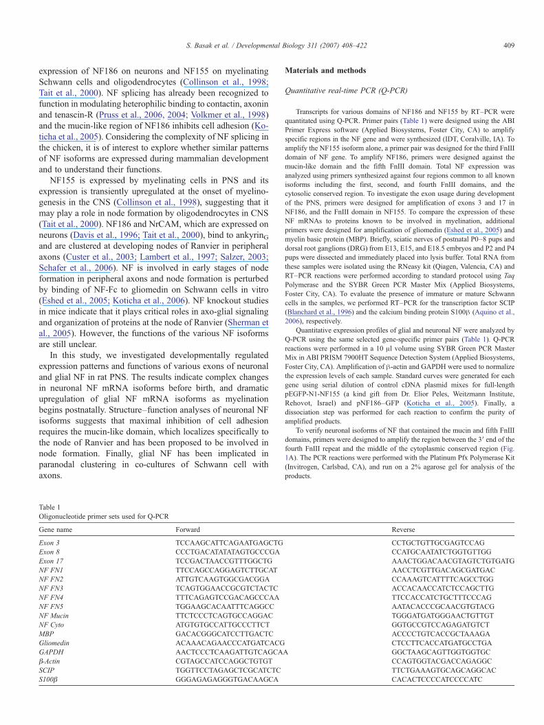
Table 1Oligonucleotide primer sets used for Q-PCR
Gene name Forward
Exon 3 TCCAAGCATTCAGAATGAGCTGExon 8 CCCTGACATATATAGTGCCCGAExon 17 TCCGACTAACCGTTTGGCTGNF FN1 TTCCAGCCAGGAGTCTTGCATNF FN2 ATTGTCAAGTGGCGACGGANF FN3 TCAGTGGAACCGCGTCTACTCNF FN4 TTTCAGAGTCCGACAGCCCAANF FN5 TGGAAGCACAATTTCAGGCCNF Mucin TTCTCCCTCAGTGCCAGGACNF Cyto ATGTGTGCCATTGCCCTTCTMBP GACACGGGCATCCTTGACTCGliomedin ACAAACAGAACCCATGATCACGGAPDH AACTCCCTCAAGATTGTCAGCAβ-Actin CGTAGCCATCCAGGCTGTGTSCIP TGGTTCCTAGAGCTCGCATCTCS100β GGGAGAGAGGGTGACAAGCA
Materials and methods
Quantitative real-time PCR (Q-PCR)
Transcripts for various domains of NF186 and NF155 by RT–PCR werequantitated using Q-PCR. Primer pairs (Table 1) were designed using the ABIPrimer Express software (Applied Biosystems, Foster City, CA) to amplifyspecific regions in the NF gene and were synthesized (IDT, Coralville, IA). Toamplify the NF155 isoform alone, a primer pair was designed for the third FnIIIdomain of NF gene. To amplify NF186, primers were designed against themucin-like domain and the fifth FnIII domain. Total NF expression wasanalyzed using primers synthesized against four regions common to all knownisoforms including the first, second, and fourth FnIII domains, and thecytosolic conserved region. To investigate the exon usage during developmentof the PNS, primers were designed for amplification of exons 3 and 17 inNF186, and the FnIII domain in NF155. To compare the expression of theseNF mRNAs to proteins known to be involved in myelination, additionalprimers were designed for amplification of gliomedin (Eshed et al., 2005) andmyelin basic protein (MBP). Briefly, sciatic nerves of postnatal P0–8 pups anddorsal root ganglions (DRG) from E13, E15, and E18.5 embryos and P2 and P4pups were dissected and immediately placed into lysis buffer. Total RNA fromthese samples were isolated using the RNeasy kit (Qiagen, Valencia, CA) andRT–PCR reactions were performed according to standard protocol using TaqPolymerase and the SYBR Green PCR Master Mix (Applied Biosystems,Foster City, CA). To evaluate the presence of immature or mature Schwanncells in the samples, we performed RT–PCR for the transcription factor SCIP(Blanchard et al., 1996) and the calcium binding protein S100β (Aquino et al.,2006), respectively.
Quantitative expression profiles of glial and neuronal NF were analyzed byQ-PCR using the same selected gene-specific primer pairs (Table 1). Q-PCRreactions were performed in a 10 μl volume using SYBR Green PCR MasterMix in ABI PRISM 7900HT Sequence Detection System (Applied Biosystems,Foster City, CA). Amplification of β-actin and GAPDH were used to normalizethe expression levels of each sample. Standard curves were generated for eachgene using serial dilution of control cDNA plasmid mixes for full-lengthpEGFP-N1-NF155 (a kind gift from Dr. Elior Peles, Weitzmann Institute,Rehovot, Israel) and pNF186–GFP (Koticha et al., 2005). Finally, adissociation step was performed for each reaction to confirm the purity ofamplified products.
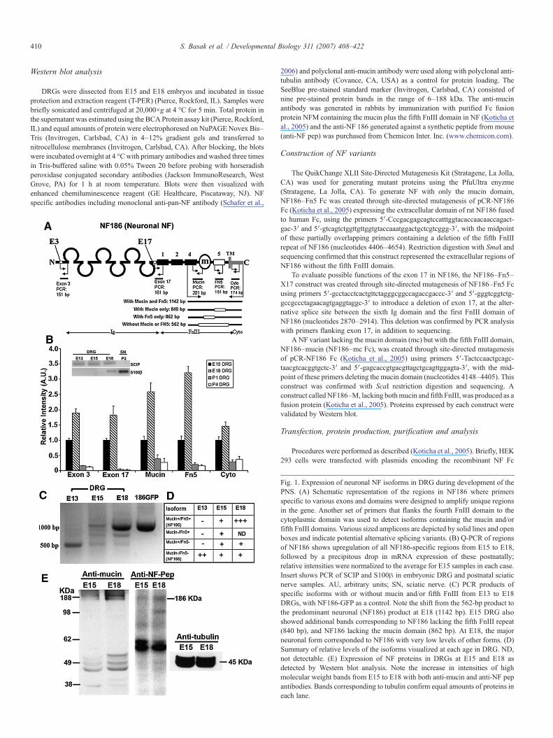
To verify neuronal isoforms of NF that contained the mucin and fifth FnIIIdomains, primers were designed to amplify the region between the 3′ end of thefourth FnIII repeat and the middle of the cytoplasmic conserved region (Fig.1A). The PCR reactions were performed with the Platinum Pfx Polymerase Kit(Invitrogen, Carlsbad, CA), and run on a 2% agarose gel for analysis of theproducts.
Reverse
CCTGCTGTTGCGAGTCCAGCCATGCAATATCTGGTGTTGGAAACTGGACAACGTAGTCTGTGATGAACCTCGTTGACAGCGATGACCCAAAGTCATTTTCAGCCTGGACCACAACCATCTCCAGCTTGTTCCACCATCTGCTTTCCCAGAATACACCCGCAACGTGTACGTGGGATGATGGGAACTGTTGTGGTGCCGTCCAGAGATGTCTACCCCTGTCACCGCTAAAGACTCCTTCACCATGATGCCTGA
A GGCTAAGCAGTTGGTGGTGCCCAGTGGTACGACCAGAGGCTTCTGAAAGTGCAGCAGGCACCACACTCCCCATCCCCATC
410 S. Basak et al. / Developmental Biology 311 (2007) 408–422
Western blot analysis
DRGs were dissected from E15 and E18 embryos and incubated in tissueprotection and extraction reagent (T-PER) (Pierce, Rockford, IL). Samples werebriefly sonicated and centrifuged at 20,000×g at 4 °C for 5 min. Total protein inthe supernatant was estimated using the BCAProtein assay kit (Pierce, Rockford,IL) and equal amounts of protein were electrophoresed on NuPAGENovex Bis–Tris (Invitrogen, Carlsbad, CA) in 4–12% gradient gels and transferred tonitrocellulose membranes (Invitrogen, Carlsbad, CA). After blocking, the blotswere incubated overnight at 4 °Cwith primary antibodies and washed three timesin Tris-buffered saline with 0.05% Tween 20 before probing with horseradishperoxidase conjugated secondary antibodies (Jackson ImmunoResearch, WestGrove, PA) for 1 h at room temperature. Blots were then visualized withenhanced chemiluminescence reagent (GE Healthcare, Piscataway, NJ). NFspecific antibodies including monoclonal anti-pan-NF antibody (Schafer et al.,
2006) and polyclonal anti-mucin antibody were used along with polyclonal anti-tubulin antibody (Covance, CA, USA) as a control for protein loading. TheSeeBlue pre-stained standard marker (Invitrogen, Carlsbad, CA) consisted ofnine pre-stained protein bands in the range of 6–188 kDa. The anti-mucinantibody was generated in rabbits by immunization with purified Fc fusionprotein NFM containing the mucin plus the fifth FnIII domain in NF (Koticha etal., 2005) and the anti-NF 186 generated against a synthetic peptide from mouse(anti-NF pep) was purchased from Chemicon Inter. Inc. (www.chemicon.com).
Construction of NF variants
The QuikChange XLII Site-Directed Mutagenesis Kit (Stratagene, La Jolla,CA) was used for generating mutant proteins using the PfuUltra enyzme(Stratagene, La Jolla, CA). To generate NF with only the mucin domain,NF186–Fn5 Fc was created through site-directed mutagenesis of pCR-NF186Fc (Koticha et al., 2005) expressing the extracellular domain of rat NF186 fusedto human Fc, using the primers 5′-Cccgacgagcagtccatttggtacaccaacaaccagact-gac-3′ and 5′-gtcagtctggttgttggtgtaccaaatggactgctcgtcggg-3′, with the midpointof these partially overlapping primers containing a deletion of the fifth FnIIIrepeat of NF186 (nucleotides 4406–4654). Restriction digestion with SmaI andsequencing confirmed that this construct represented the extracellular regions ofNF186 without the fifth FnIII domain.
To evaluate possible functions of the exon 17 in NF186, the NF186–Fn5–X17 construct was created through site-directed mutagenesis of NF186–Fn5 Fcusing primers 5′-gcctacctcactgttctagggcggccagaccgaccc-3′ and 5′-gggtcggtctg-gccgccctagaacagtgaggtaggc-3′ to introduce a deletion of exon 17, at the alter-native splice site between the sixth Ig domain and the first FnIII domain ofNF186 (nucleotides 2870–2914). This deletion was confirmed by PCR analysiswith primers flanking exon 17, in addition to sequencing.
A NF variant lacking the mucin domain (mc) but with the fifth FnIII domain,NF186–mucin (NF186–mc Fc), was created through site-directed mutagenesisof pCR-NF186 Fc (Koticha et al., 2005) using primers 5′-Tactccaactgcagc-taacgtcacggtgctc-3′ and 5′-gagcaccgtgacgttagctgcagttggagta-3′, with the mid-point of these primers deleting the mucin domain (nucleotides 4148–4405). Thisconstruct was confirmed with ScaI restriction digestion and sequencing. Aconstruct calledNF186–M, lacking bothmucin and fifth FnIII, was produced as afusion protein (Koticha et al., 2005). Proteins expressed by each construct werevalidated by Western blot.
Transfection, protein production, purification and analysis
Procedures were performed as described (Koticha et al., 2005). Briefly, HEK293 cells were transfected with plasmids encoding the recombinant NF Fc
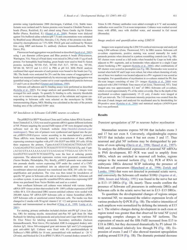
Fig. 1. Expression of neuronal NF isoforms in DRG during development of thePNS. (A) Schematic representation of the regions in NF186 where primersspecific to various exons and domains were designed to amplify unique regionsin the gene. Another set of primers that flanks the fourth FnIII domain to thecytoplasmic domain was used to detect isoforms containing the mucin and/orfifth FnIII domains. Various sized amplicons are depicted by solid lines and openboxes and indicate potential alternative splicing variants. (B) Q-PCR of regionsof NF186 shows upregulation of all NF186-specific regions from E15 to E18,followed by a precipitous drop in mRNA expression of these postnatally;relative intensities were normalized to the average for E15 samples in each case.Insert shows PCR of SCIP and S100β in embryonic DRG and postnatal sciaticnerve samples. AU, arbitrary units; SN, sciatic nerve. (C) PCR products ofspecific isoforms with or without mucin and/or fifth FnIII from E13 to E18DRGs, with NF186-GFP as a control. Note the shift from the 562-bp product tothe predominant neuronal (NF186) product at E18 (1142 bp). E15 DRG alsoshowed additional bands corresponding to NF186 lacking the fifth FnIII repeat(840 bp), and NF186 lacking the mucin domain (862 bp). At E18, the majorneuronal form corresponded to NF186 with very low levels of other forms. (D)Summary of relative levels of the isoforms visualized at each age in DRG. ND,not detectable. (E) Expression of NF proteins in DRGs at E15 and E18 asdetected by Western blot analysis. Note the increase in intensities of highmolecular weight bands from E15 to E18 with both anti-mucin and anti-NF pepantibodies. Bands corresponding to tubulin confirm equal amounts of proteins ineach lane.
411S. Basak et al. / Developmental Biology 311 (2007) 408–422
proteins using Lipofectamine 2000 (Invitrogen, Carlsbad, CA). Stable trans-fectants were isolated and Fc fusion proteins were bound to Ultralink Protein-Abeads (Pierce, Rockford, IL) and eluted using Pierce Gentle Ag/Ab ElutionBuffer (Pierce, Rockford, IL) (Haspel et al., 2000). Proteins were dialyzedagainst Tris-buffered saline solution (pH 7.5) and concentrations were estimatedby Bradford assay (Biorad Kit, Hercules, CA). Purity of the proteins was deter-mined by electrophoresis on 7.5% SDS–PAGE gels and confirmed by Westernblot using HRP–anti-human Fc antibody (Jackson ImmunoResearch, WestGrove, PA).
Fluorescent bead aggregation was performed as described (Jacob et al., 2002)using 1 μm diameter yellow/green and Blue protein-A beads (Polysciences,Warrington, VA). Nine μl of bead slurry were mixed in 200 μl with 15 μg of eachprotein. For homophilic bead binding, green beads were used to bind Fc fusionproteins including NF186–Fn5, NF186–Fn5–X17, NF186–mc, NF186,NF186–M, NF155 and L1-1212 in PBS at pH 8.0. SDS–PAGE analysisconfirmed that equal amounts of each protein were bound to the beads (see Fig.3B). The beads were sonicated for 20 s and the time course of reaggregation ofbeads was measured semiquantitatively by microscopy and then aggregation wasquantified using a Coulter Counter set to count superthreshold particles between2.5 and 6 μm as described (Grumet and Edelman, 1988).
Schwann cell adhesion and Fc binding assays were performed as described(Koticha et al., 2005). For image analysis and quantification, 6 images wereaveraged for each sample. To quantify Fc binding to cells, intensity thresholdswere set to remove non-specific fluorescence of the substrate and regions cor-responding to Schwann cells were selected on the monolayers by S100βimmunostaining (Sigma, MO). Binding was calculated as the ratio of the proteinbinding area of the cell/total S100+ area.
NF knockdown in Schwann Cell-DRG neuron co-cultures
The pSIREN kit BD™Knockout Clone and Confirm Adeno RNAi System 2fromClontech (CA,USA)was used to generate siRNA against different isoformsof NF. Primers targeting the second FnIII repeat of NF were designed using thesoftware tool on the Clontech website (http://bioinfo2.clontech.com/rnaidesigner/). Three sets of primers were synthesized and ligated into the pre-cut pSIREN-DNR-Express vector according to the manufacturer's protocol.These siRNAs were functionally tested by co-transfection of these siRNAexpressing constructs together with pNF-186-EGFP into HEK 293 cells. Of thethree sequences the primers, 5′gatccAAACCCGAGAGACTTGGAACATT-CAAGAGATGTTCCAAGTCTCTCGGGTTTTTTTTTACGCGTg and 5′aatt-cACGCGTAAAAAAAAACCCGAGAGACTTGGAACATCTCTT-GAATGTTCCAAGTCTCTCGGGTTTg were the best at reducing proteinexpression. The adenoviral expression vectors were generated commercially(Vector Biolabs, Philadelphia, PA). Briefly, pSIREN plasmids were subclonedinto premade shuttle vectors expressing GFP under the control of a CMVpromoter and were recombined into an adenoviral genome vector. Therecombinant adenoviral plasmid was transfected into HEK 293 cells for virusamplification and production. The virus was then tested for knockdown ofspecific NF genes in Schwann cells and on myelination in DRG–Schwann cellco-culture system. A non-specific scrambled RNAi adenovirus expressing GFPalone was purchased from Vector Biolabs and used as a control.
Near confluent Schwann cell cultures were infected with various AdenosiRNAGFP viruses at titers that resulted in 90–100% cellular expression of GFPwithin 48 h. E16 dissociated DRG neuron-enriched cultures were seeded with200,000 Adeno NF siRNA-GFP Schwann cells or Adeno Scrambled siRNAGFP Schwann cells or uninfected control Schwann cells. The media was thenchanged to C-media with 50 ng/ml vitamin C (C+C) and grown in myelinationmedium and immunostained as described (Ching et al., 1999; Lustig et al.,2001).
For staining, primary antibodies included chicken anti-P0 IgY (Aves Labs.Inc, OR) for staining myelin, monoclonal anti-Pan NF (gift from Dr. MattRasband) for labeling node/paranode and polyclonal anti-Caspr 6061(Gift fromDr. Elior Peles) for labeling paranode. Secondary antibodies (JacksonImmunoResearch, West Grove, PA) included AMCA-conjugated donkey anti-chicken IgY, Alexa 568-conjugated goat anti-mouse IgG and Cy5-conjugatedgoat anti-rabbit IgG. Cultures were fixed with 4% paraformaldehyde inDulbecco's PBS (dPBS) for 10 min, permeabilized with methanol at −20 °C(20 min), and blocked for 2 h in dPBS/1% BSA/1% normal donkey serum/0.2%
Triton X-100. Primary antibodies were added overnight at 4 °C and secondaryantibodies were used for 2 h at room temperature. Cultures were washed severaltimes with dPBS, once with distilled water, and mounted in Gel mount(Biomedia).
Image analysis and quantification using LSM510
Images were acquired using the LSM 510 confocal microscope and analyzedusing LSM software (Zeiss, Thornwood, NY). In DRG neuron–Schwann cellco-culture experiments, positive staining was scored manually for node/paranode proteins when detected in a pattern resembling controls (see Fig. 8B).NF clusters were scored as a full nodes when bounded by Caspr on both sidesadjacent to P0+ segments, and as heminodes when adjacent to a single Casprcluster bordering a P0+ segment (Koticha et al., 2006); other combinations ofNF clusters were scored as incomplete (see Fig. 8B). Heminodes were scored asnormal when both NF and Caspr were robust, but when robust clustering of onlyone of these two markers was located adjacent to a P0+ segment it was scored asincomplete. For quantification of myelination in co-cultures stained for P0, fourtile-scan images consisting of nine 25× images (Koticha et al., 2006) wereanalyzed with LSM 510/Multi Time macro (Carl Zeiss, Thornwood, NY). Totalimaged area was approximately 4.2 mm2 of DRG–Schwann cell co-culture,which covered approximately 5% of the entire coverslip. Due to the depth of themyelinated co-culture (approximately 15–22 μm), Z-stacks were imaged at0.85 μm intervals. The 3-D maximum intensity projection was created from thetile-scan Z-stack images and analyzed for myelinated area by thresholding forP0-positive areas (Koticha et al., 2006) and statistical analysis (ANOVA/posthoc) was performed.
Results
Transient upregulation of NF in neurons before myelination
Mammalian neurons express NF186 that includes exons 3and 17 but not exon 8. Conversely, oligodendroglia expressNF155 that includes exon 8 but not exons 3 and 17. Thusneuronal and glial NF isoforms may be distinguished by pat-terns of exon splicing (Davis et al., 1996; Hassel et al., 1997).To analyze the differential expression of neuronal NF mRNAsin PNS development, RT–PCR was used to amplify fromDRGs, which are enriched in neuronal cell bodies, regionsunique to the neuronal isoform (Fig. 1A). PCR of RNA inembryonic DRGs detected SCIP indicating the presence ofSchwann cell precursors (Blanchard et al., 1996; Zorick andLemke, 1996) that were not detected in postnatal sciatic nerve,and conversely the Schwann cell marker S100β (Aquino et al.,2006; Jessen and Mirsky, 1991) was found in the nerve but notin E13–15 DRGs (Fig. 1B, insert). These results confirm thepresence of Schwann cell precursors in embryonic DRGs andSchwann cells in the sciatic nerve but not in E13–E15 DRGs.
To quantitate the developmental changes of regions uniqueto neuronal NF, we measured the relative expression levels ofvarious products by Q-PCR (Fig. 1B). The relative intensities ofeach amplicon were normalized by defining the intensity at E15as 1. The relative changes during development in each exon orregion tested was greater than that observed for total NF (cyto)suggesting complex changes in various NF isoforms. Theexpression of mucin and the fifth FnIII increased from E15 toE18 in DRG, but they decreased dramatically by P1 (about 10-fold) and remained relatively low through P4 (Fig. 1B). Ex-pression of exons 3 and 17 also showed transient upregulationpeaking around E18 (Fig. 1B). The dramatic postnatal decrease
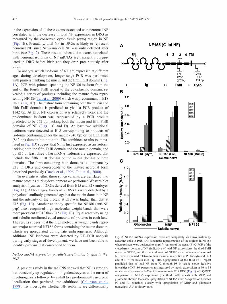
Fig. 2. NF155 mRNA expression correlates temporally with myelination bySchwann cells in PNS. (A) Schematic representation of the regions in NF155where primers were designed to amplify regions of the gene. (B) Q-PCR of thecytoplasmic domain of NF (indicative of total NF expression), the third FnIIIrepeat in NF155, and the mucin domain of NF186 as an indicator of neuronalNF, were expressed relative to their maximal intensities at P8 for cyto and FN3and at E18 for mucin (see Fig. 1B). Upregulation of the third FnIII repeatparalleled that of total NF from P2 through P8 in sciatic nerve. Relativeintensities of NF186 expression (as measured by mucin expression) in P0 to P8sciatic nerve were only 1–2% of its maximum in E18 DRG (Fig. 1). (C) Q-PCRcomparison of NF155 expression (the third FnIII repeat) with MBP andgliomedin showed that early upregulation of NF155 mRNA expression betweenP0 and P3 coincided closely with upregulation of MBP and gliomedintranscripts. AU, arbitrary units.
412 S. Basak et al. / Developmental Biology 311 (2007) 408–422
in the expression of all these exons associated with neuronal NFcorrelated with the decrease in total NF expression in DRG asmeasured by the conserved cytoplasmic (cyto) region in NF(Fig. 1B). Prenatally, total NF in DRGs is likely to representneuronal NF since Schwann cell NF was only detected afterbirth (see Fig. 2). These results indicate that exons associatedwith neuronal isoforms of NF mRNAs are transiently upregu-lated in DRG before birth and they drop precipitously afterbirth.
To analyze which isoforms of NF are expressed at differentages during development, longer-range PCR was performedwith primers flanking the mucin and the fifth FnIII domain (Fig.1A). PCR with primers spanning the NF186 isoform from theend of the fourth FnIII repeat to the cytoplasmic domain, re-vealed a series of products including the mature form repre-senting NF186 (Tait et al., 2000) which was predominant in E18DRG (Fig. 1C). The mature form containing both the mucin andfifth FnIII domains is predicted to yield a PCR product of1142 bp. At E13, NF expression was relatively weak and thepredominant isoform was represented by a PCR productpredicted to be 562 bp, lacking both the mucin and fifth FnIIIdomains of NF (Figs. 1C and D). At least two additionalisoforms were detected at E15 corresponding to products ofisoforms containing either the mucin (840 bp) or the fifth FnIII(862 bp) domain but not both. The combined results (summa-rized in Fig. 1D) suggest that NF is first expressed as an isoformlacking both the fifth FnIII domain and the mucin domain, andby E15 at least three other mRNA isoforms are expressed thatinclude the fifth FnIII domain or the mucin domain or bothdomains. The form containing both domains is dominant byE18 in DRG and corresponds to the mature neuronal formdescribed previously (Davis et al., 1996; Tait et al., 2000).
To evaluate whether these splice variants are translated intomature proteins during development we performed Western blotanalysis of lysates of DRGs derived from E15 and E18 embryos(Fig. 1E). At both ages, bands at ∼186 kDa were detected by apolyclonal antibody generated against the mucin domain in NFand the intensity of the protein at E18 was higher than that atE15 (Fig. 1E). Another antibody specific for NF186 (anti-NFpep) also recognized high molecular weight bands that weremore prevalent at E18 than E15 (Fig. 1E). Equal reactivity usinganti-tubulin confirmed equal amounts of proteins in each lane.The results suggest that the high molecular weight bands repre-sent major neuronal NF186 forms containing the mucin domain,which are upregulated during late embryogenesis. Althoughadditional NF isoforms were detected by RT–PCR analysisduring early stages of development, we have not been able toidentify proteins that correspond to them.
NF155 mRNA expression parallels myelination by glia in thePNS
A previous study in the rat CNS showed that NF is stronglybut transiently up-regulated in oligodendrocytes at the onset ofmyelinogenesis followed by a shift to a predominantly neuronallocalization that persisted into adulthood (Collinson et al.,1998). To investigate whether NF isoforms are differentially
413S. Basak et al. / Developmental Biology 311 (2007) 408–422
expressed in PNS glia as well, we analyzed the timing and theexpression profiles of different NF isoforms in developing ratsciatic nerve that is rich in Schwann cells but not neuronal cellbodies. Q-PCR analysis using a primer set to target the FN3domains specific to NF155 indicated that NF155 mRNA ex-pression was dramatically upregulated from P2 through P8 insciatic nerve (Fig. 2). Similarly, PCR products representing totalNF rose dramatically from P2 through P8 in sciatic nerve (Fig.2B). In contrast, the major neuronal NF form including themucin domain was detected in embryonic DRGs (Fig. 1), butnot in early postnatal sciatic nerve (Fig. 2B). These resultssuggest that NF155 expression coincides with the onset ofmyelination in the PNS early postnatally. The very low levels oftotal NF expression in P0 and P1 sciatic nerve suggests thatSchwann cell precursors express little or no NF and it is upre-gulated as they mature into Schwann cells. We confirmed thatthe early upregulation of NF155 mRNA between P0 and P3correlated closely with the expression of mRNAs for MBP andgliomedin (Fig. 2C), proteins that are associated with earlystages of myelination and node formation (Eshed et al., 2005).The combined results indicate that only glial NF mRNAs iso-forms are found in postnatal sciatic nerve and suggest thatSchwann cells upregulate NF155 mRNA as they begin to mye-linate axons.
Effects of NF exon splicing on binding
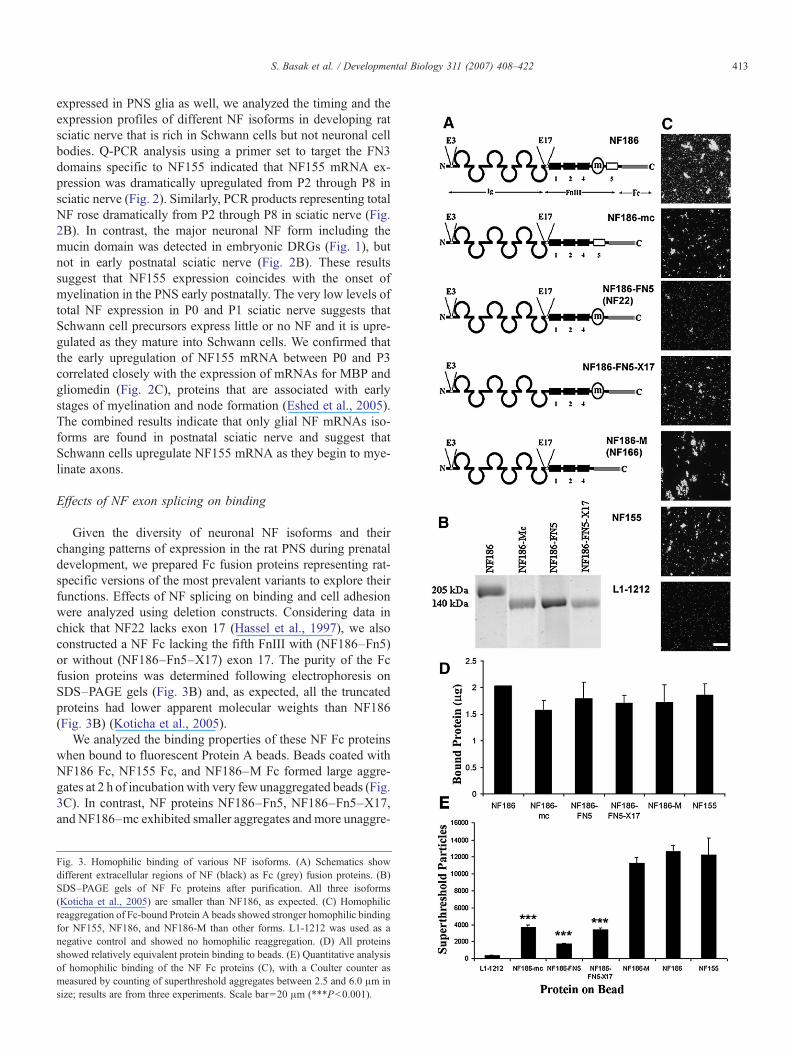
Given the diversity of neuronal NF isoforms and theirchanging patterns of expression in the rat PNS during prenataldevelopment, we prepared Fc fusion proteins representing rat-specific versions of the most prevalent variants to explore theirfunctions. Effects of NF splicing on binding and cell adhesionwere analyzed using deletion constructs. Considering data inchick that NF22 lacks exon 17 (Hassel et al., 1997), we alsoconstructed a NF Fc lacking the fifth FnIII with (NF186–Fn5)or without (NF186–Fn5–X17) exon 17. The purity of the Fcfusion proteins was determined following electrophoresis onSDS–PAGE gels (Fig. 3B) and, as expected, all the truncatedproteins had lower apparent molecular weights than NF186(Fig. 3B) (Koticha et al., 2005).
We analyzed the binding properties of these NF Fc proteinswhen bound to fluorescent Protein A beads. Beads coated withNF186 Fc, NF155 Fc, and NF186–M Fc formed large aggre-gates at 2 h of incubation with very few unaggregated beads (Fig.3C). In contrast, NF proteins NF186–Fn5, NF186–Fn5–X17,and NF186–mc exhibited smaller aggregates and more unaggre-
Fig. 3. Homophilic binding of various NF isoforms. (A) Schematics showdifferent extracellular regions of NF (black) as Fc (grey) fusion proteins. (B)SDS–PAGE gels of NF Fc proteins after purification. All three isoforms(Koticha et al., 2005) are smaller than NF186, as expected. (C) Homophilicreaggregation of Fc-bound Protein A beads showed stronger homophilic bindingfor NF155, NF186, and NF186-M than other forms. L1-1212 was used as anegative control and showed no homophilic reaggregation. (D) All proteinsshowed relatively equivalent protein binding to beads. (E) Quantitative analysisof homophilic binding of the NF Fc proteins (C), with a Coulter counter asmeasured by counting of superthreshold aggregates between 2.5 and 6.0 μm insize; results are from three experiments. Scale bar=20 μm (***Pb0.001).

414 S. Basak et al. / Developmental Biology 311 (2007) 408–422
gated beads, suggesting that they have weaker homophilicbinding. SDS–PAGE analysis confirmed that all the proteinsbound equally well to Fluorosbrite Protein A-coated beads (Fig.3D) indicating that differences in aggregation were not due tovariations in the amounts of bound Fc fusion proteins. All the NFproteins, however, exhibited significantly greater levels of ag-gregation than the negative control (L1-1212 Fc; Fig. 3D), whichhad no detectable L1 binding activity (Haspel et al., 2000).
Quantification of numbers of superthreshold aggregatessupported visual analysis (Fig. 3E). The results indicate that theearliest expressed form of NF in neurons (NF186–M) as well asthe mature forms (NF186, NF155) show stronger homophilicbinding than the transiently expressed forms (NF186–Fn5,NF186–Fn5–X17 and NF186–mc) (Fig. 3E). Interestingly,NF186–Fn5, which clearly showed some aggregation by com-parison to the negative control L1-1212 Fc did not aggregate aswell as the same protein without exon 17 (NF186–Fn5–X17;Fig. 3E), suggesting that this exon located at the hinge betweenthe Ig domains and the FnIII repeats in Ig/FnIII CAMs maymodulate homophilic binding in NF perhaps by regulating thefolding of the molecule (Haspel and Grumet, 2003). However,we detected only weak levels of exon 17 expression at E15DRG in the rat (Fig. 1B), thus, its function remains unclear.Insofar as both early and later expressed NF isoforms exhibitedrobust homophilic binding, the developmental changes in neu-ronal NFs does not predict dramatic changes in NF homophilicbinding during this period of development.
We then analyzed binding of these NF variants to live cells asdescribed previously (Koticha et al., 2005). NF155 and NF186–M Fc bound strongly to Schwann cells while NF186 exhibitedrelatively little binding (Fig. 4) (Koticha et al., 2005). BothNF186–Fn5 and NF186–Fn5–X17 (Fig. 4) exhibited weakbinding to Schwann cells that was similar to the level observedfor NF186 (Figs. 4B, E and F). However, binding of NF186–mclacking the mucin domain was strong and comparable to thebest NFs (Figs. 4G and H). Quantification of NF-Fc binding andnormalization to areas of the dish covered by Schwann cellsconfirmed that NFs without mucin bind more robustly toSchwann cell than NFs with mucin and that the mucin domainitself had no detectable binding (Fig. 4H). The results confirmthat multiple NF variants bind to Schwann cells and suggest thatthe presence of the mucin domain reduces the binding affinity oravidity of neuronal NFs for glia. The results suggest that deve-lopmental upregulation of NF186 may modulate Schwann cellinteractions with axons.
Maximal inhibition of adhesion by the mucin domain requiresthe fifth FnIII repeat
Our previous studies (Koticha et al., 2005) showed thatNF186 inhibits Schwann cell adhesion and the inhibition in-volves the M-region consisting of the mucin domain and thefifth FnIII repeat. To localize this inhibitory activity further, wetested adhesion of Schwann cells to the various recombinant NFmutant proteins (Fig. 5A) (Koticha et al., 2005). Cells adhered(Figs. 5B, C, G) and spread (Figs. 5J, K, O) within 2 h mostextensively on NF155, NF186–M and NF186–mc. Adhesion
was inhibited on NF186 (Figs. 5A, I) and NFM (Figs. 5D, L)with reduced numbers of cells bound within the protein coatedspots (circles) and cells spread poorly on substrates coated withNF186–Fn5 (Figs. 5E, M) or NF186–Fn5–X17 (Figs. 5F, N).Quantitation of the adherent cells per field of view confirmedthat significantly greater numbers of cells adhered to NF186–mc substrate (comparable to NF155, NF186–M, or L1) than toNF186–Fn5 and NF186–Fn5–X17 substrates (Fig. 5Q). Veryfew cells bound to NFM or to NF186 (Fig. 5Q). The combinedresults indicate that the mucin along with fifth FnIII domain ofNF is required for maximal inhibition of glial adhesion and thatthe mucin domain plays a critical role in this process. In add-ition, the presence of the fifth FnIII repeat reduces cell spread-ing even in the absence of the mucin domain but exon 17 did nothave a detectable affect. The developmental upregulation ofneuronal NF186 with its ability to inhibit cell adhesion suggeststhat it may modulate interactions among axons as well as withSchwann cells.
Knockdown of NF155 in Schwann cell delays myelinationslightly and inhibits Caspr clustering when co-cultured withDRG neurons
Knockout of all NF isoforms in mice indicated that NFs arenecessary for clustering of specific proteins at nodes and para-nodes, and rescue of NF155 in glia in knockout mice restoredformation of paranodal junctions but it did not rescue nodalclustering (Sherman et al., 2005). Given one major form of NFin glia, we decided to analyze its function specifically in thesecells by depletion using specific siRNAs introduced intoSchwann cells. Four siRNA sequences targeted to various re-gions of NF were screened and the most effective one waswithin the conserved second FnIII repeat (Supplementary Fig.1). Adenoviral vectors encoding for expression of GFP as amarker were prepared with this siRNA sequence and ascrambled sequence as a control, and were used to infectSchwann cells.
Six days after adding Schwann cells to DRG neurons, therewere few weakly P0+ myelin segments in both the siRNAcontrol and uninfected control co-cultures but none were ob-served in co-cultures with the NF siRNA-treated Schwann cells(Fig. 6A). These isolated myelin segments were bordered byvariably sized clusters of NF but Caspr remained diffuselylocalized along axons at these early times. NF colocalized withmyelin segments only in controls but not in the NF siRNA-treated Schwann cell co-cultures, suggesting that this NF is onthe Schwann cells (Fig. 6A). These results suggest that NFRNAi inhibits induction of NF155 in Schwann cells despitetheir close association with axons.
Both control and NF siRNA-infected GFP+ Schwann cellselongated and associated closely with the axons although asso-ciation of NF deficient Schwann cells was less distinct. Thus,initial recognition between Schwann cells and axons does notappear to be dependent on NF. Higher-magnification imagingconfirmed NF clustering adjacent to myelin segments en-sheathed by GFP+ Schwann cells when the scrambled RNAi-treated Schwann cells or control uninfected Schwann cells were

Fig. 4. Binding of NF isoforms to Schwann cell surface. (A–G) Binding of various NF Fc proteins to the surface of Schwann cells was detected after fixation andimmunofluorescence with S100β, which is shown as inserts in the upper right of each panel. (H) Quantification of protein-binding normalized to total S100β+ cellareas confirmed the qualitative results and the absence of binding for NFM. NF155, NF186-M, and NF186-mc exhibited higher levels of protein binding to the surfaceof Schwann cells relative to the other proteins. NF186, NF186-FN5, and NF186-FN5-X17 exhibited similar levels of protein binding to cell surfaces that was weaker,while NFM shows almost no binding. Scale bar=10 μm. AU, arbitrary units.
415S. Basak et al. / Developmental Biology 311 (2007) 408–422
used but not with the NF siRNA-infected GFP+ Schwann cells(Fig. 6B). These NF clusters are likely to represent neuronal NFrather than glial NF since they were not accompanied byclustering of Caspr. This is consistent with the observations thatneuronal NF can cluster without Caspr but glial NF colocalizedwith Caspr (Schafer et al., 2006). In all three conditions, Casprserved as a marker for axons at this early time since it remaineddiffusely distributed along axons.
Confocal microscopy revealed close association of GFP+Schwann cells with axons but this was particularly difficult tovisualize since GFP was in the cytoplasm and was most pre-valent around cell bodies but sparse in the extended processes.Given that myelin contains very little cytoplasm, the GFPsignals are likely to be more faint in regions of Schwann cellswhere they ensheath and myelinate axons. Nevertheless, wehave detected weak GFP+ tubular profiles overlapping P0+
regions in myelination co-cultures at 6 and 12 days (Fig. 6B andsee Fig. 8B). In the older 19-day cultures, it was extremelydifficult to visualize GFP in myelinated processes and wetherefore focused on the younger cultures.
After incubation for 12 days, there was extensive myelina-tion in all groups including the NF siRNA-infected Schwanncells co-cultured with DRG neurons (Fig. 7A). Althoughslightly less myelinated areas was measured in 12 daysknockdown cultures, the percentage of myelinated areas werenot statistically different among control uninfected Schwanncells, control siRNA-infected Schwann cells, and the NFsiRNA-infected Schwann cells at 12 days or 19 days (Figs. 7Aand B). Quantitation was not done on the 6-day cultures becauseP0 signals were weak and variable, and no myelin was detectedwith the NF siRNA-treated Schwann cells (Fig. 6). Thecombined results suggest that knockdown of NF results in a
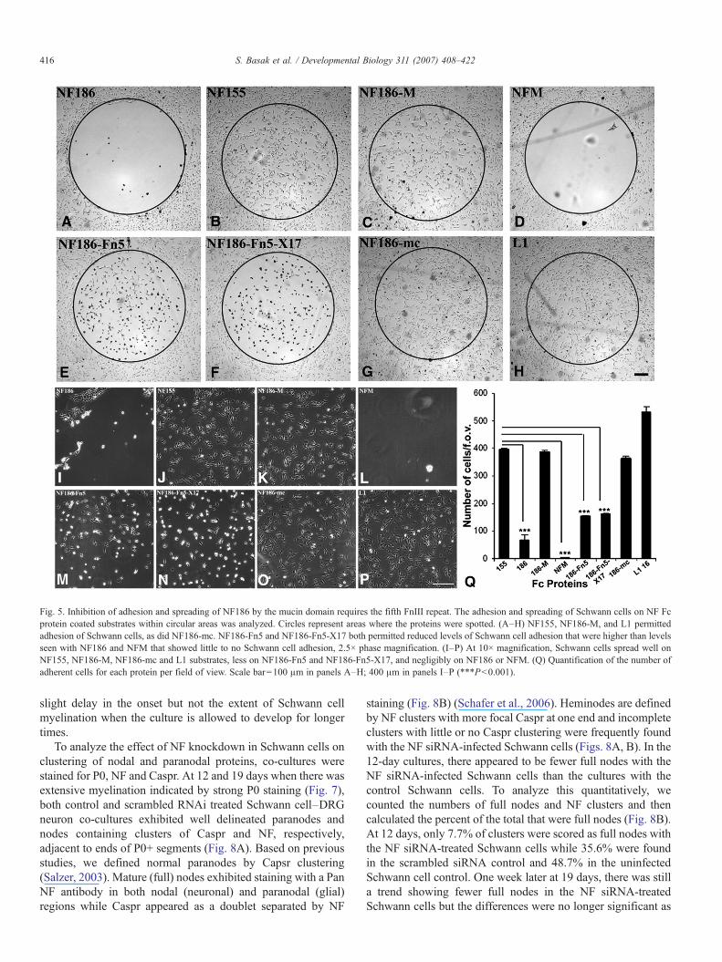
Fig. 5. Inhibition of adhesion and spreading of NF186 by the mucin domain requires the fifth FnIII repeat. The adhesion and spreading of Schwann cells on NF Fcprotein coated substrates within circular areas was analyzed. Circles represent areas where the proteins were spotted. (A–H) NF155, NF186-M, and L1 permittedadhesion of Schwann cells, as did NF186-mc. NF186-Fn5 and NF186-Fn5-X17 both permitted reduced levels of Schwann cell adhesion that were higher than levelsseen with NF186 and NFM that showed little to no Schwann cell adhesion, 2.5× phase magnification. (I–P) At 10× magnification, Schwann cells spread well onNF155, NF186-M, NF186-mc and L1 substrates, less on NF186-Fn5 and NF186-Fn5-X17, and negligibly on NF186 or NFM. (Q) Quantification of the number ofadherent cells for each protein per field of view. Scale bar=100 μm in panels A–H; 400 μm in panels I–P (***Pb0.001).
416 S. Basak et al. / Developmental Biology 311 (2007) 408–422
slight delay in the onset but not the extent of Schwann cellmyelination when the culture is allowed to develop for longertimes.
To analyze the effect of NF knockdown in Schwann cells onclustering of nodal and paranodal proteins, co-cultures werestained for P0, NF and Caspr. At 12 and 19 days when there wasextensive myelination indicated by strong P0 staining (Fig. 7),both control and scrambled RNAi treated Schwann cell–DRGneuron co-cultures exhibited well delineated paranodes andnodes containing clusters of Caspr and NF, respectively,adjacent to ends of P0+ segments (Fig. 8A). Based on previousstudies, we defined normal paranodes by Capsr clustering(Salzer, 2003). Mature (full) nodes exhibited staining with a PanNF antibody in both nodal (neuronal) and paranodal (glial)regions while Caspr appeared as a doublet separated by NF
staining (Fig. 8B) (Schafer et al., 2006). Heminodes are definedby NF clusters with more focal Caspr at one end and incompleteclusters with little or no Caspr clustering were frequently foundwith the NF siRNA-infected Schwann cells (Figs. 8A, B). In the12-day cultures, there appeared to be fewer full nodes with theNF siRNA-infected Schwann cells than the cultures with thecontrol Schwann cells. To analyze this quantitatively, wecounted the numbers of full nodes and NF clusters and thencalculated the percent of the total that were full nodes (Fig. 8B).At 12 days, only 7.7% of clusters were scored as full nodes withthe NF siRNA-treated Schwann cells while 35.6% were foundin the scrambled siRNA control and 48.7% in the uninfectedSchwann cell control. One week later at 19 days, there was stilla trend showing fewer full nodes in the NF siRNA-treatedSchwann cells but the differences were no longer significant as
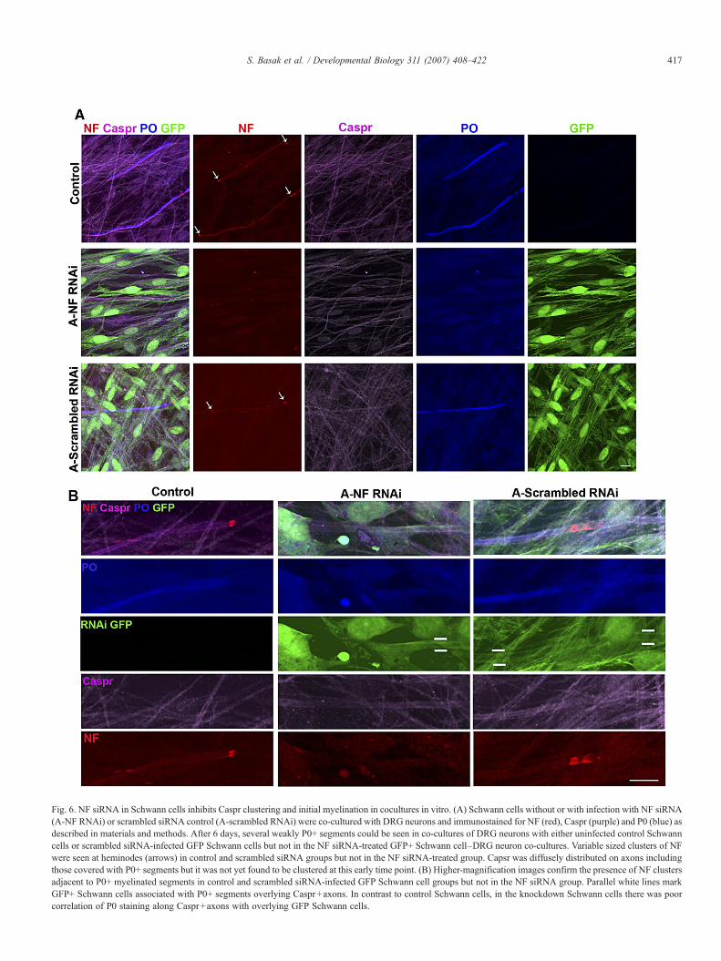
Fig. 6. NF siRNA in Schwann cells inhibits Caspr clustering and initial myelination in cocultures in vitro. (A) Schwann cells without or with infection with NF siRNA(A-NF RNAi) or scrambled siRNA control (A-scrambled RNAi) were co-cultured with DRG neurons and immunostained for NF (red), Caspr (purple) and P0 (blue) asdescribed in materials and methods. After 6 days, several weakly P0+ segments could be seen in co-cultures of DRG neurons with either uninfected control Schwanncells or scrambled siRNA-infected GFP Schwann cells but not in the NF siRNA-treated GFP+ Schwann cell–DRG neuron co-cultures. Variable sized clusters of NFwere seen at heminodes (arrows) in control and scrambled siRNA groups but not in the NF siRNA-treated group. Capsr was diffusely distributed on axons includingthose covered with P0+ segments but it was not yet found to be clustered at this early time point. (B) Higher-magnification images confirm the presence of NF clustersadjacent to P0+ myelinated segments in control and scrambled siRNA-infected GFP Schwann cell groups but not in the NF siRNA group. Parallel white lines markGFP+ Schwann cells associated with P0+ segments overlying Caspr+axons. In contrast to control Schwann cells, in the knockdown Schwann cells there was poorcorrelation of P0 staining along Caspr+axons with overlying GFP Schwann cells.
417S. Basak et al. / Developmental Biology 311 (2007) 408–422
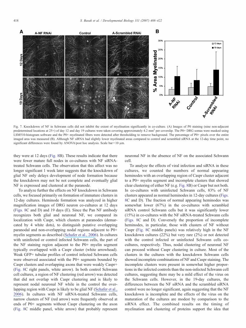
Fig. 7. Knockdown of NF in Schwann cells did not inhibit the extent of myelination significantly in co-culture. (A) Images of P0 staining (nine non-adjacentpredetermined locations at 25×) of day 12 and day 19 cultures were taken covering approximately 4.2 mm2 per coverslip. The P0+ DRG somas were masked usingLSM510-histogram software and the P0+ myelinated fibers were detected after thresholding to remove background. The percentage of P0+ pixels over the entireimaged area was measured (B). Although NF siRNA had slightly lower myelinated areas compared to control and scrambled siRNA at the 12-day time point, nosignificant differences were found by ANOVA/post hoc analysis. Scale bar=10 μm.
418 S. Basak et al. / Developmental Biology 311 (2007) 408–422
they were at 12 days (Fig. 8B). These results indicate that therewere fewer mature full nodes in co-cultures with NF siRNA-treated Schwann cells. The observation that this affect was nolonger significant 1 week later suggests that the knockdown ofglial NF only delays development of node formation becausethe knockdown may not be not complete and eventually glialNF is expressed and clustered at the paranode.
To analyze further the effects on NF knockdown in Schwanncells, we focused primarily on formation of immature clusters in12-day cultures. Heminode formation was analyzed in highermagnification images of DRG neuron co-cultures at 12 days(Figs. 8C and D) and 19 days (E). Since the Pan NF antibodyrecognizes both glial and neuronal NF, we compared itslocalization with Caspr, which clusters at paranodes (demar-cated by 4 white dots), to distinguish putative overlappingparanodal and non-overlapping nodal regions adjacent to P0+myelin segments as described (Schafer et al., 2006). In cultureswith uninfected or control infected Schwann cells, the part ofthe NF staining region adjacent to the P0+ myelin segmenttypically overlapped with a Caspr cluster (white arrowhead).Weak GFP+ tubular profiles of control infected Schwann cellswere observed associated with the P0+ segments bounded byCaspr clusters and overlapping axons that were weakly Caspr+(Fig. 8C right panels, white arrow). In both control Schwanncell cultures, a region of NF clustering (red arrow) was detectedthat did not overlap with Caspr clustering and is likely torepresent nodal neuronal NF while in the control the over-lapping region with Caspr is likely to be glial NF (Schafer et al.,2006). In cultures with NF siRNA-treated Schwann cells,narrow clusters of NF (red arrow) were frequently observed atends of P0+ segments without Caspr clustering on the axon(Fig. 8C middle panel, white arrow) that probably represent
neuronal NF in the absence of NF on the associated Schwanncell.
To analyze the effects of viral infection and siRNA in thesecultures, we counted the numbers of normal appearingheminodes with an overlapping region of Caspr cluster adjacentto a P0+ myelin segment and incomplete clusters that showedclear clustering of either NF (e.g. Fig. 8B) or Caspr but not both.In co-cultures with uninfected Schwann cells, 83% of NFclusters appeared as normal heminodes in 12-day cultures (Figs.8C and D). The fraction of normal appearing heminodes wassomewhat lower (67%) in the co-cultures with scrambledsiRNA control Schwann cells but it was significantly lower(15%) in co-cultures with the NF siRNA-treated Schwann cells(Figs. 8C and D). Conversely the proportion of incompleteheminodes, in particular, those with clusters of NF withoutCaspr (Fig. 8C middle panels) was relatively high in the NFknockdown cultures (22%) but very rare (2%) or not detectedwith the control infected or uninfected Schwann cells co-cultures, respectively. Thus, nodal clustering of neuronal NFcan proceed without Caspr clustering in culture. Most of theclusters in the cultures with the knockdown Schwann cellsshowed incomplete combinations of NF and Caspr staining. Theincomplete clusters were present in somewhat higher propor-tions in the infected controls than the non-infected Schwann cellcultures, suggesting there may be a mild effect of the virus onthe Schwann cells. However, in the 19-day cultures, thedifferences between the NF siRNA and the scrambled siRNAcontrol were no longer significant, again suggesting that the NFknockdown is incomplete and the effects of the virus on thematuration of the cultures are modest by comparison to thesiRNA effect. The combined results on the timing ofmyelination and clustering of proteins support the idea that
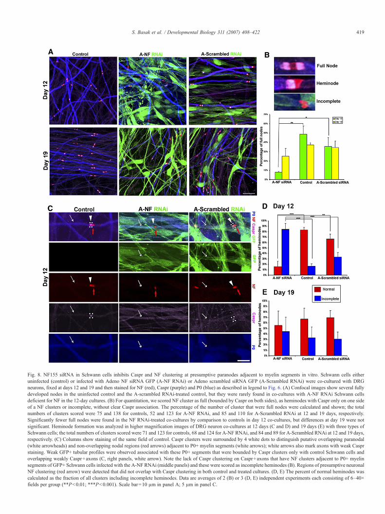
Fig. 8. NF155 siRNA in Schwann cells inhibits Caspr and NF clustering at presumptive paranodes adjacent to myelin segments in vitro. Schwann cells eitheruninfected (control) or infected with Adeno NF siRNA GFP (A-NF RNAi) or Adeno scrambled siRNA GFP (A-Scrambled RNAi) were co-cultured with DRGneurons, fixed at days 12 and 19 and then stained for NF (red), Caspr (purple) and P0 (blue) as described in legend to Fig. 6. (A) Confocal images show several fullydeveloped nodes in the uninfected control and the A-scrambled RNAi-treated control, but they were rarely found in co-cultures with A-NF RNAi Schwann cellsdeficient for NF in the 12-day cultures. (B) For quantitation, we scored NF cluster as full (bounded by Caspr on both sides), as heminodes with Caspr only on one sideof a NF clusters or incomplete, without clear Caspr association. The percentage of the number of cluster that were full nodes were calculated and shown; the totalnumbers of clusters scored were 75 and 138 for controls, 52 and 123 for A-NF RNAi, and 85 and 110 for A-Scrambled RNAi at 12 and 19 days, respectively.Significantly fewer full nodes were found in the NF RNAi-treated co-cultures by comparison to controls in day 12 co-cultures, but differences at day 19 were notsignificant. Heminode formation was analyzed in higher magnification images of DRG neuron co-cultures at 12 days (C and D) and 19 days (E) with three types ofSchwann cells; the total numbers of clusters scored were 71 and 123 for controls, 68 and 124 for A-NF RNAi, and 84 and 89 for A-Scrambled RNAi at 12 and 19 days,respectively. (C) Columns show staining of the same field of control. Caspr clusters were surrounded by 4 white dots to distinguish putative overlapping paranodal(white arrowheads) and non-overlapping nodal regions (red arrows) adjacent to P0+ myelin segments (white arrows); white arrows also mark axons with weak Casprstaining. Weak GFP+ tubular profiles were observed associated with these P0+ segments that were bounded by Caspr clusters only with control Schwann cells andoverlapping weakly Caspr+axons (C, right panels, white arrow). Note the lack of Caspr clustering on Caspr+axons that have NF clusters adjacent to P0+ myelinsegments of GFP+ Schwann cells infected with the A-NF RNAi (middle panels) and these were scored as incomplete heminodes (B). Regions of presumptive neuronalNF clustering (red arrow) were detected that did not overlap with Caspr clustering in both control and treated cultures. (D, E) The percent of normal heminodes wascalculated as the fraction of all clusters including incomplete heminodes. Data are averages of 2 (B) or 3 (D, E) independent experiments each consisting of 6–40×fields per group (∗∗Pb0.01; ∗∗∗Pb0.001). Scale bar=10 μm in panel A; 5 μm in panel C.
419S. Basak et al. / Developmental Biology 311 (2007) 408–422

420 S. Basak et al. / Developmental Biology 311 (2007) 408–422
knocking down NF expression selectively in Schwann cellsresults in a slight delay in myelination and a significant delay inparanodal formation however nodal NF clustering appeared toproceed even in the absence of Caspr clustering.
Discussion
The results show dramatic differences in expression andfunctions of several major NF isoforms that are expressed onneurons and glia during PNS development. We found thatmammals express multiple alternatively spliced forms of NF aspredicted from a study in chicken (Hassel et al., 1997). Threeneuronal isoforms may be transiently upregulated in DRG be-fore total neuronal NF mRNAs are dramatically down regulatedprior to birth. Glial NF is not expressed until after birth when itis dramatically upregulated coincident with the onset of mye-lination in the sciatic nerve. Thus, expression of neuronal andglial NF mRNAs peak transiently before and after birth, res-pectively, in a complementary pattern (Fig. 9). Our knockdownstudies of NF specifically in rat Schwann cells demonstrate thatit is critical for formation of paranodes confirming knockoutstudies in mice that eliminated all NF isoforms (Sherman et al.,2005). They further suggest that glial NF is not necessary forclustering of receptors including neuronal NF at the node ofRanvier. These studies combined with previous studies (Daviset al., 1996; Lambert et al., 1997; Lustig et al., 2001; Sherman etal., 2005; Tait et al., 2000) further underscore that neuronal andglial NF isoforms play different roles in development of thenode of Ranvier and the paranode, respectively.
Changes in NF isoforms during development suggest dra-matic differences in the potential functions of the various neu-ronal isoforms expressed prior to myelination. Neuronal iso-forms of NF were first detected at E13 in rat and they werefound to lack the mucin and fifth FnIII domains similar to theNF166 isoform detected early in chick development (Hassel etal., 1997). By E15, at least two more isoforms are detected,one similar to chicken NF22 containing the fifth FnIII domain
Fig. 9. Model summarizing expression patterns of major NF isoforms duringdevelopment. The mature neuronal form (NF186, blue) is dramatically upre-gulated as axons are ensheathed and then it decreases to persist at low levelspostnatally. In contrast, the glial form (NF155, green) is only expressed post-natally at the onset of myelination.
(Pruss et al., 2004) and the other contains the mucin but not thefifth FnIII domain. Functional analyses indicate that all three ofthese early embryonic forms bind homophilically (Fig. 3) andNF also binds heterophilically with axonin-1, contactin and Nr-CAM (Pruss et al., 2004; Volkmer et al., 1998), thus NF mayinitially promote axonal fasciculation by multiple mechanisms(Rathjen et al., 1987). The mature neuronal isoform containingboth the mucin and the fifth FnIII domains predominates by E18in rat. This isoform of NF also binds homophilically but it is astrong inhibitor of cell adhesion (Fig. 5), suggesting that it maybe involved in defasciculation of axons at later times (Koticha etal., 2005). Regulated switching from NF166 to NF185 wasfound in chick DRG, consistent with the observation that thelatter form containing the mucin and the fifth FnIII domains ispoorly adhesive for cells (Pruss et al., 2006). Changes in otherexons such as 17 were also observed but no evidence for theirmodulation of NF function has been found yet. However, thefunction of the immature neuronal NF isoforms is uncertainsince evidence for expression of the predicted proteins has yet tobe obtained.
Although neuronal NF mRNA drops to relatively low levelspostnatally, NF186 persists in mature nerves and is highlyrestricted to the node of Ranvier. Neuronal NF may be criticalfor co-clustering of sodium channels via cis interaction betweenthese proteins on axons (Ratcliffe et al., 2001). Recent studies(Dzhashiashvili et al., 2007) indicate that it may be targeteddirectly to the node which could explain how relatively lowlevels of mRNA expression postnatally are sufficient to main-tain its high expression specifically at the node. In NF null mice,adjacent Schwann cells contacted each other in presumptivenodal regions lacking NF186 (Sherman et al., 2005), consistentwith the proposal that the inhibition of cell adhesion by NF186plays a critical role in limiting cellular contacts at nodes ofRanvier (Koticha et al., 2005). Thus, the inhibition of celladhesion by NF186 may explain why Schwann cell processesdo not form stable contacts in the nodal region. However, glio-medin on Schwann cells mediates binding to axons via NF andNrCAM (Eshed et al., 2005), and may provide a mechanism foraxo-glial interactions that allows Schwann cells to associatewith axons despite the inhibition by NF186. Although NF doesnot appear to be directly involved in myelination (Sherman etal., 2005) (Koticha et al., 2006) (Fig. 7), glial NF plays a criticalrole in paranodal formation (Sherman et al., 2005) (Fig. 8). Inthe absence of glial NF, Caspr does not cluster, but neuronal NFcan still form clusters adjacent to incomplete paranodal junc-tions (Fig. 8B) that border myelin sheaths (Sherman et al.,2005). Thus, neuronal and glial NFs play different functionsthat are likely to be related to their differential spatiotemporalpatterns of expression and perhaps their compositions due tosplicing.
The different forms of NF described here were clearly dis-tinguishable based on their inclusion of specific exons that ex-hibited complementary patterns of expression in cell bodies ofDRG neurons (Fig. 1) and Schwann cells in sciatic nerves (Fig.2), consistent with previous studies that detected them in neu-rons and glia (Collinson et al., 1998; Davis et al., 1996; Tait etal., 2000). The Pan NF monoclonal antibody (Schafer et al.,
421S. Basak et al. / Developmental Biology 311 (2007) 408–422
2004) was critical for detecting clusters of NF during myeli-nation in co-cultures and showing that they were disrupted byspecific NF siRNAs in Schwann cells. Although study of nativeNF proteins is extremely complicated by the large possiblenumber of isoforms expressed in different cells and their mul-tiple post-translational modifications including glycosylationand cleavage (Maier et al., 2006; Pruss et al., 2006; Volkmer etal., 1992), the analysis of the mRNAs by Q-PCR revealeddramatic changes in neurons and glia. We have detected NFproteins that are upregulated between E15 and E18 in DRGscorresponding to mature NF with mucin, suggesting that theyare predominant in DRG neurons before myelination.
Mammalian studies demonstrated localization of Nr-CAMand NF at nascent nodes of Ranvier as early as P2 in sciaticnerves, prior to clustering of sodium channels and ankyrinG(Custer et al., 2003; Lambert et al., 1997). Although the Pan NFmonoclonal antibody (Schafer et al., 2004) does not distinguishamong NF isoforms, it may allow detection of NF at earliertimes than other anti-NF antibodies that are available (Schafer etal., 2004). Comparison of NF with the paranodal protein Casprshowed that early postnatally NF clusters partially overlap withCaspr at heminodes and NF clusters were occasionally detectedwithout Caspr clustering (Schafer et al., 2006). We found thatknockdown of glial NF in Schwann cells increased the fre-quency of finding NF clusters (presumably neuronal) withoutadjacent Caspr clusters, confirming that NF on axons can clusterindependent of Caspr. In early phases of co-cultures, myelina-tion was only detected with control Schwann cells, on which P0colocalized with NF staining, suggesting this is glial NF thatwas not expressed on knockdown Schwann cells. Thus NF isnot essential for myelination but neuronal NF plays a criticalrole in nodal clustering (Sherman et al., 2005).
In conclusion, NF mRNAs that are expressed first on DRGneurons during development including NF186–M, NF186–mc,NF186–Fn5, encode proteins that can bind homophilically andmay contribute to initial axonal fasciculation along with otherCAMs (Chang et al., 1987). By comparison to other prevalentneural CAMs such as L1 that mediate homophilic binding(Haspel and Grumet, 2003), NF exhibits robust homophilicbinding on beads in vitro (Fig. 3). Subsequent upregulation ofNF186 may favor defasciculation of DRG axons due to itsinhibition of cell adhesion mediated by neural CAMs includingNF and L1 (Koticha et al., 2005). However, NF186 does notinhibit laminin-mediated adhesion (Koticha et al., 2005),consistent with the observed increase in extracellular matrixand association of Schwann cells in the nerve by E18 (Wanneret al., 2006). At least at early times in DRG–Schwann cellcultures, we detected NF on neuron-associated Schwann cellsand Caspr distributed along axons before these proteins co-cluster at paranodes along with contactin on the axon (Giraultand Peles, 2002). It is also appears by electron microscopy thatboth NF155 (Chang et al., 2000) and gliomedin (Eshed et al.,2005) are expressed on paranodal loops in addition to theirconcentration at axo-glial junctions. Thus interactions betweenadjacent closely spaced paranodal loops may also be mediatedby NF either homophilically (Maier et al., 2006) or heterophili-cally by binding to gliomedin. Interestingly, some paranodal
loops in NF null mice appear to have irregularly large inter-loopspacing (Sherman et al., 2005), consistent with a role for NF155in this interaction.
Acknowledgments
We thank Dr. Steve Lambert for providing neurofascincDNAs, Drs. Peter Brophy, Ori Peles and Mat Rasband forantibodies, and Dr. Ori Peles for helpful discussions. This workwas supported by grants from NIH and NMSS.
Appendix A. Supplementary data
Supplementary data associated with this article can be found,in the online version, at doi:10.1016/j.ydbio.2007.08.045.
References
Aquino, J.B., Hjerling-Leffler, J., Koltzenburg, M., Edlund, T., Villar, M.J.,Ernfors, P., 2006. In vitro and in vivo differentiation of boundary cap neuralcrest stem cells into mature Schwann cells. Exp. Neurol. 198, 438–449.
Blanchard, A.D., Sinanan, A., Parmantier, E., Zwart, R., Broos, L., Meijer, D.,Meier, C., Jessen, M.R., Mirsky, R., 1996. Oct-6 (SCIP/Tst-1) is expressedin Schwann cell precursors, embryonic Schwann cells, and postnatalmyelinating Schwann cells: comparison with Oct-1, Krox-20, and Pax-3.J. Neurosci. Res. 46, 630–640.
Brummendorf, T., Rathjen, F.G., 1995. Cell adhesion molecules 1: immunoglo-bulin superfamily. Protein Profile 2, 963–1108.
Chang, S., Rathjen, F.G., Raper, J.A., 1987. Extension of neurites on axons isimpaired by antibodies against specific neural cell surface glycoproteins.J. Cell Biol. 104, 355–362.
Chang, B.J., Cho, I.J., Brophy, P.J., 2000. A study on the immunocytochemicallocalization of neurofascin in rat sciatic nerve. J. Vet. Sci. 1, 67–71.
Ching, W., Zanazzi, G., Levinson, S.R., Salzer, J.L., 1999. Clustering ofneuronal sodium channels requires contact with myelinating Schwann cells.J. Neurocytol. 28, 295–301.
Collinson, J.M., Marshall, D., Gillespie, C.S., Brophy, P.J., 1998. Transientexpression of neurofascin by oligodendrocytes at the onset of myelinogen-esis: implications for mechanisms of axon–glial interaction. Glia 23, 11–23.
Custer, A.W., Kazarinova-Noyes, K., Sakurai, T., Xu, X., Simon, W., Grumet,M., Shrager, P., 2003. The role of the ankyrin-binding protein NrCAM innode of Ranvier formation. J. Neurosci. 23, 10032–10039.
Davis, J.Q., Lambert, S., Bennett, V., 1996. Molecular composition of the nodeof Ranvier: identification of ankyrin-binding cell adhesion molecules neuro-fascin (mucin+/third FNIII domain−) and NrCAM at nodal axon segments.J. Cell Biol. 135, 1355–1367.
Dzhashiashvili, Y., Zhang, Y., Galinska, J., Grumet, M., 2007. Nodes of ranvierand initial segments assemble by distinct mechanisms. J. Cell Biol. 177,857–870.
Eshed, Y., Feinberg, K., Poliak, S., Sabanay, H., Sarig-Nadir, O., Spiegel, I.,Bermingham Jr., J.R., Peles, E., 2005. Gliomedin mediates Schwann cell–axon interaction and the molecular assembly of the nodes of Ranvier.Neuron 47, 215–229.
Girault, J.A., Peles, E., 2002. Development of nodes of Ranvier. Curr. Opin.Neurobiol. 12, 476–485.
Grumet, M., Edelman, G.M., 1988. Neuron–glia cell adhesion molecule inter-acts with neurons and astroglia via different binding mechanisms. J. CellBiol. 106, 487–503.
Haspel, J., Grumet, M., 2003. L1CAM: A multi-domain cell adhesion moleculewith modular and cooperative binding modes. Frontiers in Bioscience 8,s1210–s1225.
Haspel, J., Friedlander, D.R., Ivgy-May, N., Chickramane, S., Roonprapunt, C.,Chen, S., Schachner, M., Grumet, M., 2000. Critical and optimal Ig domainsfor promotion of neurite outgrowth by L1/Ng-CAM. J. Neurobiol. 42,287–302.
422 S. Basak et al. / Developmental Biology 311 (2007) 408–422
Hassel, B., Rathjen, F.G., Volkmer, H., 1997. Organization of the neurofascingene and analysis of developmentally regulated alternative splicing. J. Biol.Chem. 272, 28742–28749.
Jacob, J., Haspel, J., Kane-Goldsmith, N., Grumet, M., 2002. L1 mediatedhomophilic binding and neurite outgrowth are modulated by alternativesplicing of exon 2. J. Neurobiol. 51, 177–189.
Jessen, M.R., Mirsky, R., 1991. Schwann cell precursors and their development.Glia 4, 185–194.
Koticha, D., Babiarz, J., Kane-Goldsmith, N., Jacob, J., Raju, K., Grumet,M., 2005. Cell adhesion and neurite outgrowth are promoted byneurofascin NF155 and inhibited by NF186. Mol. Cell. Neurosci. 30,137–148.
Koticha, D., Maurel, P., Zanazzi, G., Kane-Goldsmith, N., Basak, S., Babiarz, J.,Salzer, J., Grumet, M., 2006. Neurofascin interactions play a critical role inclustering sodium channels, ankyrin G and beta IV spectrin at peripheralnodes of Ranvier. Dev. Biol. 293, 1–12.
Lambert, S., Davis, J.Q., Bennett, V., 1997. Morphogenesis of the node ofRanvier: co-clusters of ankyrin and ankyrin-binding integral proteins defineearly developmental intermediates. J. Neurosci. 17, 7025–7036.
Lustig, M., Zanazzi, G., Sakurai, T., Blanco, C., Levinson, S.R., Lambert, S.,Grumet, M., Salzer, J.L., 2001. Nr-CAM and neurofascin interactionsregulate ankyrin G and sodium channel clustering at the node of Ranvier.Curr. Biol. 11, 1864–1869.
Maier, O., van der Heide, T., Johnson, R., de Vries, H., Baron, W., Hoekstra, D.,2006. The function of neurofascin155 in oligodendrocytes is regulated bymetalloprotease-mediated cleavage and ectodomain shedding. Exp. CellRes. 312, 500–511.
Pruss, T., Niere, M., Kranz, E.U., Volkmer, H., 2004. Homophilic interactions ofchick neurofascin in trans are important for neurite induction. Eur. J.Neurosci. 20, 3184–3188.
Pruss, T., Kranz, E.U., Niere, M., Volkmer, H., 2006. A regulated switch ofchick neurofascin isoforms modulates ligand recognition and neuriteextension. Mol. Cell. Neurosci. 31, 354–365.
Ratcliffe, C.F., Westenbroek, R.E., Curtis, R., Catterall, W.A., 2001. Sodium
channel beta1 and beta3 subunits associate with neurofascin through theirextracellular immunoglobulin-like domain. J. Cell Biol. 154, 427–434.
Rathjen, F.G., Wolff, J.M., Chang, S., Bonhoeffer, F., Raper, J.A., 1987.Neurofascin: a novel chick cell-surface glycoprotein involved in neurite–neurite interactions. Cell 51, 841–849.
Salzer, J.L., 2003. Polarized domains of myelinated axons. Neuron 40,297–318.
Schafer, D.P., Bansal, R., Hedstrom, K.L., Pfeiffer, S.E., Rasband, M.N., 2004.Does paranode formation and maintenance require partitioning of neuro-fascin 155 into lipid rafts? J. Neurosci. 24, 3176–3185.
Schafer, D.P., Custer, A.W., Shrager, P., Rasband, M.N., 2006. Early events innode of Ranvier formation during myelination and remyelination in thePNS. Neuron Glia Biol. 2, 69–79.
Sherman, D.L., Tait, S., Melrose, S., Johnson, R., Zonta, B., Court, F.A.,Macklin, W.B., Meek, S., Smith, A.J., Cottrell, D.F., Brophy, P.J., 2005.Neurofascins are required to establish axonal domains for saltatoryconduction. Neuron 48, 737–742.
Tait, S., Gunn-Moore, F., Collinson, J.M., Huang, J., Lubetzki, C., Pedraza, L.,Sherman, D.L., Colman, D.R., Brophy, P.J., 2000. An oligodendrocyte celladhesion molecule at the site of assembly of the paranodal axo-glialjunction. J. Cell Biol. 150, 657–666.
Volkmer, H., Hassel, B., Wolff, J.M., Frank, R., Rathjen, F.G., 1992. Structure ofthe axonal surface recognition molecule neurofascin and its relationship to aneural subgroup of the immunoglobulin superfamily. J. Cell Biol. 118,149–161.
Volkmer, H., Zacharias, U., Norenberg, U., Rathjen, F.G., 1998. Dissection ofcomplex molecular interactions of neurofascin with axonin-1, F11, andtenascin-R, which promote attachment and neurite formation of tectal cells.J. Cell Biol. 142, 1083–1093.
Wanner, I.B., Guerra, N.K., Mahoney, J., Kumar, A., Wood, P.M., Mirsky, R.,Jessen, K.R., 2006. Role of N-cadherin in Schwann cell precursors ofgrowing nerves. Glia 54, 439–459.
Zorick, T.S., Lemke, G., 1996. Schwann cell differentiation. Curr. Opin. CellBiol. 8, 870–876.
Copyright © 2022 FDOKUMEN