column splice fracture effects on the seismic performance of ...
Upload
independentCategory
view
2download
0
rXXXX American Chemical Society A dx.doi.org/10.1021/bi2001278 | Biochemistry XXXX, XXX, 000–000
ARTICLE
pubs.acs.org/biochemistry
The Splice Variant of the V2 Vasopressin ReceptorAdopts Alternative TopologiesAlexis Gonzalez,† Mauricio Borquez,† Cesar A. Trigo,† Marianne Brenet,† Jos�e M. Sarmiento,†
Carlos D. Figueroa,‡ Javier Navarro,§ and Carlos B. Gonzalez*,†,§
†Department of Physiology, Universidad Austral de Chile, Valdivia, Chile‡Department of Anatomy, Histology and Pathology, Universidad Austral de Chile, Valdivia, Chile§Department of Neuroscience and Cell Biology, University of Texas Medical Branch, Galveston, Texas 77555, United States
bS Supporting Information
Alternative splicing of the RNAs encoding GPCRs promotesthe functional and structural diversity of this class of
receptors. Splice variants of the corticotropin-releasing hor-mone receptor1 and the calcitonin receptor2 containing diverseextracellular N-terminal sequences give rise to different ligandbinding profiles. Alternative splicing also generates truncationsof GPCRs, including deletions of all transmembrane domainsand generation of soluble N-terminal domains of metabotropicglutamate receptors,3 corticotropin-releasing hormone receptors,4
and luteinizing hormone receptors.5 Of particular importance arethose splice variants such as dopamine D2,6 histamine H3,7 andthe cholescytokinin receptors,8 which contain diverse intracellularloop sequences, thus giving rise to changes in the coupling toG-protein subtypes. Splice variants with distinct extracellular loopsequences are less frequent.9 The most frequent splice variants arethose containing distinct C-terminal sequences, including trans-membrane segment 7 (TM7) and the C-terminal tails.10�15
Previously, we identified a splice variant (V2b) of the canonicalV2a vasopressin receptor.16 V2b has an amino acid sequenceidentical to that of the V2a receptor up to residue 303, close to theend of the sixth transmembrane domain, but the V2b sequencescorresponding to the seventh transmembrane domain and thecarboxyl terminus are distinct from those of V2a. The V2b variant
is expressed in the distal tubule and collecting duct of the kidney,but in contrast to V2a, V2b is retained in the endoplasmicreticulum�Golgi complex and does not bind vasopressin. Im-portantly, the V2b variant forms oligomers with the V2a receptorand behaves as a dominant negative mutant by retaining the V2ainside the cell. On this basis, we have suggested that the expressionof V2a on the cell surface is determined by the relative expressionsof V2b to V2a receptors. Because the V2b variant containsnoncanonical sequences corresponding to the putative seventhtransmembrane segment and C-terminal tail we have investigatedthe topology adopted by V2b and its subcellular distribution.We found that in contrast to the canonical V2a, V2b adoptedtwo topologies, one with six transmembrane segments (TM6)with the C-terminus on the extracellular side of the membrane andanother with seven transmembrane segments (TM7) with theC-terminus on the intracellular side. Furthermore, we observedthat both topological isoforms oligomerized with the V2a canonicalreceptor.
Received: January 25, 2011Revised: April 15, 2011
ABSTRACT: The V2 receptor gene encodes two receptorvariants by alternative splicing, the canonical V2 receptor (V2areceptor) and V2b. The V2b variant has an amino acid sequenceidentical to that of the V2a receptor up to the sixth transmem-brane domain, but the V2b sequences corresponding to theputative seventh transmembrane domain and the carboxylterminus are different from those of the V2a receptor. Here weinvestigate the topology and subcellular distribution of the V2bvariant. We found that, in contrast to the V2a receptor, the V2badopted two topologies: one with six transmembrane segmentswith the C-terminus on the extracellular side of the membraneand another with seven transmembrane segments with theC-terminus on the intracellular side, similar to typical G-protein-coupled receptors. Furthermore, we observed that both topological isoforms oligomerized with the V2a canonical receptor. Unlike theV2a receptor, V2b did notmove to the plasmamembrane, but it is retained in the ER�Golgi compartments. These findings indicate thatthe C-terminal sequence beyond the sixth transmembrane of the V2a is required for the stabilization of the seven-transmembranetopology of the receptor and is also essential for the trafficking of the receptor to the plasma membrane.
B dx.doi.org/10.1021/bi2001278 |Biochemistry XXXX, XXX, 000–000
Biochemistry ARTICLE
’EXPERIMENTAL PROCEDURES
Plasmid Construction. The construction of the expressionvectors (pECFP-N1) encoding V2b�CFP and V2a�CFP fusionproteins has been described previously.16,17 The V2a and V2bmutants without native glycosylation sites (N22Q and N185Q)and with an engineered glycosylation site in the linker sequencebetween the receptors and CFP were constructed by PCR-basedmutagenesis,18 which produced the V2b(m)�CFP and V2a-(m)�CFP constructs, respectively. Similarly, we generated aC-terminally truncated V2a mutant containing only transmembranesegments 1�6 (TM1�6), lacking the native glycosylation sites andcontaining the engineered glycosylation site in the linker sequence asdescribed above [V2a(m)-TM1�6�CFP]. In addition, we insertedan HA tag at the amino terminus of all mutants [HA-V2b-(m)�CFP,HA-V2a(m)�CFP, andHA-V2a(m)-TM1�6�CFP].Cell Culture and Transfections. CHO-K1 cells were transi-
ently transfected with the expression plasmids encoding thereceptor mutants, using Fugene 6 as described previously.16,17
Cell Surface Biotinylation Assay. CHO-K1 cells transfectedwith plasmids encoding the V2a�CFP or the V2b�CFP werewashed three times with PBS (pH 8.0), and incubated withN-hydroxysulfosuccinimide esters of biotin (Sulfo-biotin) inbiotinylation buffer (2 mM CaCl2, 150 mM NaCl, and 10 mMtriethanolamine) for 30 min at 4 �C. The biotinylation reactionwas stopped with three washes of PBS containing 100 mMglycine and three more washes with PBS. Cells were lysed withRIPA buffer [50 nM Tris-HCl (pH 7.4), 150 mM NaCl, 1%Nonidet P-40, 0.5% sodium deoxycholate, 0.1% SDS, 1 μg/mLleupeptin, 5 μg/mL soybean trypsin inhibitor, 0.1 μg/mL PMSF,and 10 mM N-methylmaleimide]. Cell extracts were centrifugedat 16000g for 1 h. Supernatants were incubated with 50 μL ofneutravidine-agarose. The precipitates were collected by centrifuga-tion, washed with 100 and 300 mMNaCl, resuspended in Laemmlibuffer, and subjected to Western blotting.Western Blotting. Cell extracts were fractionated via sodium
dodecyl sulfate�polyacrylamide gel electrophoresis and blottedonto nitrocellulose filters as described previously.16 The CFP-tagged proteins were detected with a polyclonal anti-GFP anti-body (Invitrogen) that reacts against all the variants of Aequoreavictoria such as EGFP, EYFP, and ECFP.Co-Immunoprecipitation.CHO-K1 cells were cotransfected
with two plasmids, one containing the cDNA encoding the V2areceptor tagged with HA and the other containing the cDNAencoding V2b tagged with CFP. Transfected cells were treatedwith and without 5 μg/mL tunicamycin. After being incubatedfor 48 h, the cells were washed with phosphate-buffered salineand homogenized with a Dounce homogenizer in a 5 mM Tris-HCl (pH 7.4) buffer containing 15 mM EDTA, 1 mM phenyl-methanesulfonyl fluoride, 10 μg/mL leupeptin, and 10 μg/mLaprotinin. The homogenate was centrifuged for 10 min at 6000g,and the postnuclear supernatant was centrifuged for 45 min at150000g. The membrane pellet was solubilized in RIPA buffer[50 mM Tris-HCl (pH 7.4), 150 mM NaCl, 0.1% SDS, 0.5%sodium deoxycholate, 1% Nonidet P-40, 10 mM N-ethylmalei-mide, 0.1 mM phenylmethanesulfonyl fluoride, 5 mg/mL soy-bean trypsin inhibitor, and 1 μg/mL leupeptin] and centrifugedfor 45 min at 16000g. The supernatant was incubated with apolyclonal anti-GFP antibody (2.5 μg, Invitrogen) and 20 μL ofagarose-protein A (50% slurry) at 4 �C for 16 h. The immu-noabsorbent was washed three times with RIPA buffer andcollected by centrifugation for 2 min at 1000g. The immune
complexes were resuspended in sample buffer and subjected tosodium dodecyl sulfate�polyacrylamide gel electrophoresis andWestern blotting. The V2a receptor was detected by using amouse monoclonal anti-HA antibody (Roche) and a peroxidase-labeled goat anti-mouse antibody (Jackson Laboratories).Immunocytochemistry.CHO-K1 cells cultured on coverslips
were transfected with expression plasmids encoding HA-V2b-(m)�CFP, HA-V2a(m)�CFP, or HA-V2a(m)-TM1�6�CFP.After 48 h, the cells were fixed with methanol or 4% paraformal-dehyde and permeabilized with digitonin as describedpreviously.19,20 Cells were stained with anti-HA monoclonal orpolyclonal anti-GFP, anti-calnexin, anti-ERGIC-53, or anti-58Kantibodies using Alexa 488- and Alexa 633-labeled secondaryantibodies and visualized with an Olympus Fluoview 1000confocal microscope. To detect CFP, cells were excited with a440 nm diode laser and the emitted light was detected using a BP465�495 nm filter. To detect the Alexa 488-labeled antibody, thecells were excited with a 488 nm argon laser and the emitted lightwas detected by using a BP 505�525 nm filter, whereas the Alexa633-labeled antibody was detected via excitation with a 633 nmHe�Ne laser and the emitted light detected using a LP 650 nmfilter.
’RESULTS
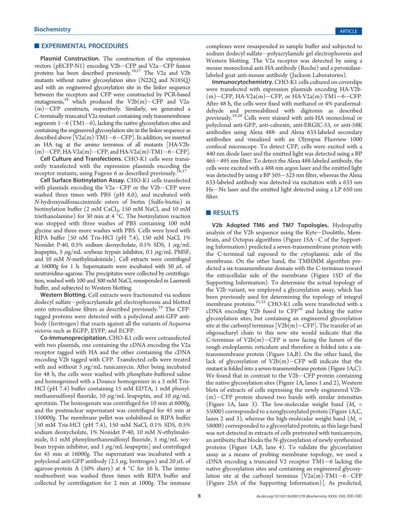
V2b Adopted TM6 and TM7 Topologies. Hydropathyanalysis of the V2b sequence using the Kyte�Doolittle, Mem-brain, and Octopus algorithms (Figure 1SA�C of the Support-ing Information) predicted a seven-transmembrane protein withthe C-terminal tail exposed to the cytoplasmic side of themembrane. On the other hand, the TMHMM algorithm pre-dicted a six-transmembrane domain with the C-terminus towardthe extracellular side of the membrane (Figure 1SD of theSupporting Information). To determine the actual topology ofthe V2b variant, we employed a glycosylation assay, which hasbeen previously used for determining the topology of integralmembrane proteins.21,22 CHO-K1 cells were transfected with acDNA encoding V2b fused to CFP16 and lacking the nativeglycosylation sites, but containing an engineered glycosylationsite at the carboxyl terminus [V2b(m)�CFP]. The transfer of anoligosacharyl chain to this new site would indicate that theC-terminus of V2b(m)�CFP is now facing the lumen of therough endoplasmic reticulum and therefore is folded into a six-transmembrane protein (Figure 1A,B). On the other hand, thelack of glycosylation of V2b(m)�CFP will indicate that themutant is folded into a seven-transmembrane protein (Figure 1A,C).We found that in contrast to the V2b�CFP protein containingthe native glycosylation sites (Figure 1A, lanes 1 and 2), Westernblots of extracts of cells expressing the newly engineered V2b-(m)�CFP protein showed two bands with similar intensities(Figure 1A, lane 3). The low-molecular weight band (Mr =55000) corresponded to a nonglycosylated protein (Figure 1A,C,lanes 2 and 3), whereas the high-molecular weight band (Mr =58000) corresponded to a glycosylated protein, as this large bandwas not detected in extracts of cells pretreated with tunicamycin,an antibiotic that blocks theN-glycosylation of newly synthesizedproteins (Figure 1A,B, lane 4). To validate the glycosylationassay as a means of probing membrane topology, we used acDNA encoding a truncated V2 receptor TM1�6 lacking thenative glycosylation sites and containing an engineered glycosy-lation site at the carboxyl terminus [V2a(m)-TM1�6�CFP(Figure 2SA of the Supporting Information)]. As predicted,
C dx.doi.org/10.1021/bi2001278 |Biochemistry XXXX, XXX, 000–000
Biochemistry ARTICLE
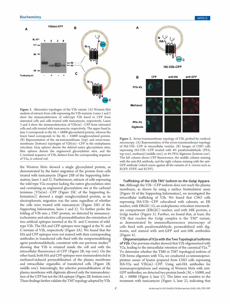
the Western blots showed a single glycosylated protein, asdemonstrated by the faster migration of the protein from cellstreated with tunicamycin (Figure 2SB of the Supporting Infor-mation, lanes 1 and 2). Furthermore, extracts of cells expressingthe wild-type V2a receptor lacking the native glycosylation sitesand containing an engineered glycosylation site at the carboxylterminus [V2a(m)�CFP (Figure 2SC of the Supporting In-formation)] showed a single nonglycosylated protein, as itselectrophoretic migration was the same regardless of whetherthe cells were treated with tunicamycin (Figure 2SD of theSupporting Information, lanes 1 and 2). To further probe thefolding of V2b into a TM7 protein, we detected by immunocy-tochemistry and selective cell permeabilization the orientation oftwo artificial epitopes inserted at the N- and C-termini of wild-type V2b. The HA and CFP epitopes were tagged at the N- andC-termini of V2b, respectively (Figure 2A). We found that theHA and CFP epitopes were not stained with their correspondingantibodies in transfected cells fixed with the nonpermeabilizingagent paraformaldehyde, consistent with our previous studies16
showing that V2b is retained inside the cell and with theintracellular fluorescence of CFP (Figure 2B, top row). On theother hand, both HA and CFP epitopes were immunodetected inmethanol-induced permeabilization of the plasma membraneand intracellular organelles of transfected cells (Figure 2B,middle row). Interestingly, the selective permeabilization of theplasma membrane with digitonin allowed only the immunodetec-tion of the CFP but not the HA epitope (Figure 2B, bottom row).These findings further validate the TM7 topology adopted byV2b.
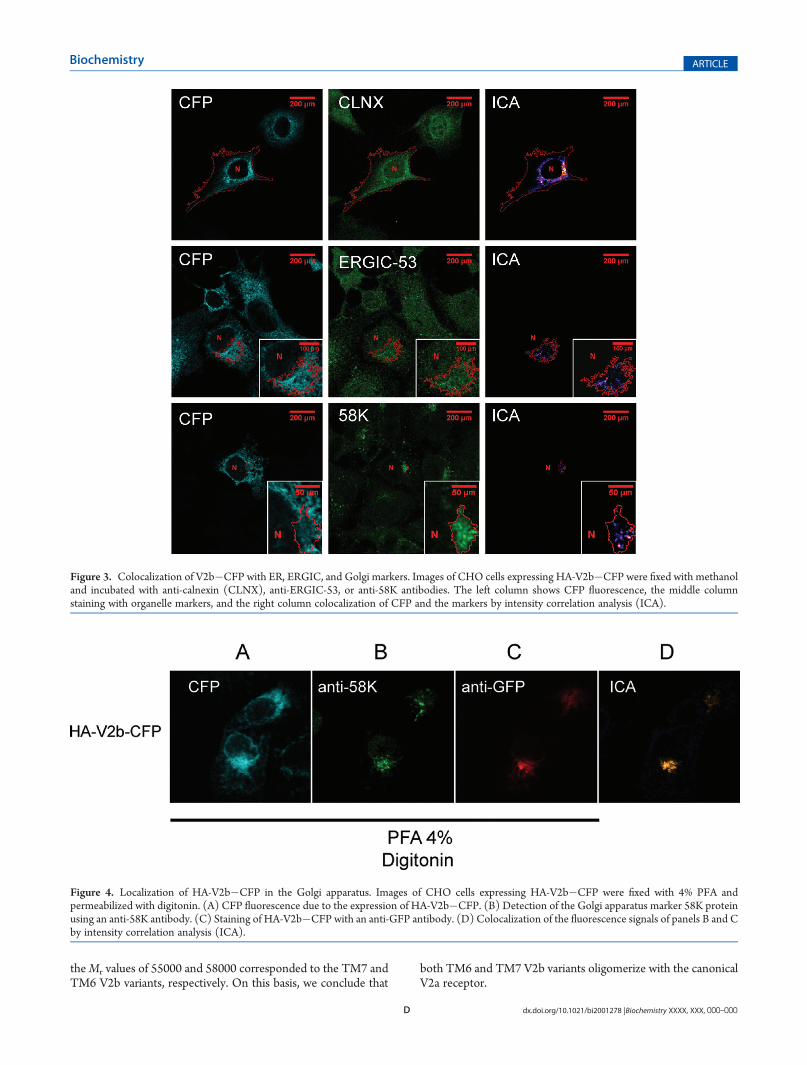
Trafficking of the V2b TM7 Isoform to the Golgi Appara-tus. Although the V2b�CFP isoform does not reach the plasmamembrane, as shown by using a surface biotinylation assay(Figure 3S of the Supporting Information), we investigated theintracellular trafficking of V2b. We found that CHO cellsexpressing HA-V2b�CFP colocalized with calnexin, an ERmarker, with ERGIC-53, an endoplasmic reticulum intermedi-ate compartment (ERGIC) marker, and with 58K protein, aGolgi marker (Figure 3). Further, we found that, at least, theV2b that reaches the Golgi complex is the TM7 variant,as demonstrated by immunofluorescence microscopy ofcells fixed with paraformaldehyde, permeabilized with dig-itonin, and stained with anti-GFP and anti-58K antibodies(Figure 4).OligomerizationofV2awith the TwoTopological Isoforms
of V2b.Our previous studies showed that V2b oligomerized withV2a, leading to the intracellular retention of the canonical V2a.16
To determine whether the TM6 or TM7 topological isoform ofV2b forms oligomers with V2a, we conducted co-immunopreci-pitation assays of lysates prepared from CHO cells expressingHA-V2a and V2b(m)�CFP. Using anti-HA antibodies forimmunoprecipitations and staining of Western blots with anti-GFP antibodies, we detected two protein bands [Mr = 55000, andMr = 58000 (Figure 5, lane 1)]. The latter was sensitive to thetreatment with tunicamycin (Figure 5, lane 2), indicating that
Figure 1. Alternative topologies of the V2b variant. (A) Western blotanalysis of extracts from cells expressing the V2b mutants. Lanes 1 and 2show the immunodetection of wild-type V2b fused to CFP fromuntreated cells and cells treated with tunicamycin, respectively. Lanes3 and 4 show the immunodetection of V2b(m)�CFP from untreatedcells and cells treated with tunicamycin, respectively. The upper band inlane 3 corresponds to theMr = 58000 glycosylated protein, whereas thelower band corresponds to the Mr = 55000 nonglycosylated protein.(B) Representation of the six-transmembrane (top) and seven-trans-membrane (bottom) topologies of V2b(m)�CFP in the endoplasmicreticulum. Gray spheres denote the deleted native glycosylation sites;blue spheres denote the engineered glycosylation sites, and theC-terminal sequence of V2b, distinct from the corresponding sequenceof V2a, is colored red.
Figure 2. Seven-transmembrane topology of V2b, probed by confocalmicroscopy. (A) Representation of the seven-transmembrane topologyof HA-V2b�CFP in intracellular vesicles. (B) Images of CHO cellsexpressing HA-V2b�CFP treated with 4% paraformaldehyde (PFA,top row), methanol (middle row), or 4% PFA/digitonin (bottom row).The left column shows CFP fluorescence, the middle column stainingwith the anti-HA antibody, and the right column staining with the anti-GFP antibody (which reacts against all the variants of A. victoria such asEGFP, EYFP, and ECFP).
D dx.doi.org/10.1021/bi2001278 |Biochemistry XXXX, XXX, 000–000
Biochemistry ARTICLE
theMr values of 55000 and 58000 corresponded to the TM7 andTM6 V2b variants, respectively. On this basis, we conclude that
both TM6 and TM7 V2b variants oligomerize with the canonicalV2a receptor.
Figure 3. Colocalization of V2b�CFP with ER, ERGIC, and Golgi markers. Images of CHO cells expressing HA-V2b�CFP were fixed with methanoland incubated with anti-calnexin (CLNX), anti-ERGIC-53, or anti-58K antibodies. The left column shows CFP fluorescence, the middle columnstaining with organelle markers, and the right column colocalization of CFP and the markers by intensity correlation analysis (ICA).
Figure 4. Localization of HA-V2b�CFP in the Golgi apparatus. Images of CHO cells expressing HA-V2b�CFP were fixed with 4% PFA andpermeabilized with digitonin. (A) CFP fluorescence due to the expression of HA-V2b�CFP. (B) Detection of the Golgi apparatus marker 58K proteinusing an anti-58K antibody. (C) Staining of HA-V2b�CFP with an anti-GFP antibody. (D) Colocalization of the fluorescence signals of panels B and Cby intensity correlation analysis (ICA).

E dx.doi.org/10.1021/bi2001278 |Biochemistry XXXX, XXX, 000–000
Biochemistry ARTICLE
’DISCUSSION
On the basis of glycosylation assays and epitope mapping ofV2b, we have demonstrated that this splice variant, in contrast tothe canonical V2a receptor, is assembled into TM6 and TM7topologies. The mechanisms underlying the generation of TM6or TM7 are unknown. Previous studies have suggested that theassembly of membrane domains into the membrane is deter-mined by partitioning of the protein domains between thetraslocon (Sec61) and the surrounding lipid bilayer.23,24 Theefficiency of insertion of a protein domain into the membraneseems to depend on the number of hydrophobic residues andthe length of the helix but also on the relative position of theamino acid within the helix.25,26 Consequently, short hydropho-bic segments or the presence of charged residues may affect theprobability of the insertion. We suggest that the two topologiesadopted by V2b are due to the fact that the noncanonical seventhtransmembrane segment of V2b is not efficiently inserted intothe membrane. Alternatively, it is possible that the seventhtransmembrane segment and the C-terminus of V2a stabilizethe receptor into a seven-transmembrane protein topology,whereas the noncanonical seventh transmembrane and the shortC-terminus in V2b most likely destabilize the seven-transmem-brane topology. This idea is supported by the analysis of therecent crystal structures of GPCRs indicating multiple interac-tions between residues of the seventh transmembrane helix andresidues from helices 1�6,27 and with the poor assembly ofreceptor chimeras derived from heterologous GPCRs,28 whichexhibit sequence diversity in their transmembrane segments,including the seventh transmembrane segment. Furthermore,helix 8 in the C-terminus of the receptor, identified in the crystalstructures of GPCRs, may further stabilize the seven-transmem-brane protein topology, via interaction with lipids on thecytoplasmic side of the plasma membrane. Indeed, recent studieshave shown that helix 8 of GPCRs binds to the plasmamembrane.29 This is consistent with previous observationsindicating that the interaction of specific lipids with membraneproteins dictates the transmembrane topology.30
We also demonstrated that the V2b isoform reaches the Golgiapparatus but fails to traffic to the plasma membrane, as
suggested by our previous binding studies.16 This is most likelydue to the lack of trafficking motifs in the C-terminal domains, asV2a receptor mutant R337X, a truncated V2 mutant lackingthe cytoplasmic C-terminus, failed to traffic to the plasmamembrane.31 Alternatively, the C-terminus could confer stabilityto the V2a receptor, an important requirement for trafficking tothe plasma membrane.32,33 In fact, unstable mutants of CFTRfailed to traffic to the plasma membrane.34,35
Interestingly, both the TM6 and the TM7 V2b topologicalisoforms form oligomers with the V2a receptor, indicating thatthe first six transmembrane segments of the receptor are suffi-cient to drive the oligomerization between V2a and V2b, which isconsistent with our findings that the truncated V2a mutantcontaining only transmembrane segments 1�6 is also retainedinside the cell and also oligomerizes with the canonical V2areceptor (unpublished observations).
In conclusion, we have demonstrated that the V2b splicevariant is folded into two alternative topological isoforms ofsix and seven transmembrane segments, and that the seven-transmembrane segment isoform traffics to the Golgi apparatus.
’ASSOCIATED CONTENT
bS Supporting Information. Hydrophobicity profiles forthe V2 receptor isoform and seven-transmembrane topology ofthe V2a receptor. This material is available free of charge via theInternet at http://pubs.acs.org.
’AUTHOR INFORMATION
Corresponding Author*Department of Physiology, Edif. Ciencias Biom�edicas, Of. 406,Universidad Austral de Chile, Campus Isla Teja, Valdivia 509-9200, Chile. E-mail: [email protected]. Telephone: 56-63-221419.Fax: 56-63-221513.
Funding SourcesThis work was supported by Grant 1060158 from Fondecyt.
’ABBREVIATIONS
ECFP, enhanced cyan fluorescence protein; V2a, vasopressinreceptor type 2a isoform;V2b, vasopressin receptor type 2b isoform;GPCR, G-protein-coupled receptor;TM, transmembrane segment;RIPA, radio-immunoprecipitation assay; PMSF, phenylmethanesul-fonyl fluoride; EDTA, ethylenediaminetetraacetic acid; SDS, so-dium dodecyl sulfate; TMHMM, transmembrane hidden Markovmodel; V2b(m)�CFP, CFP fused to the V2b mutant;V2a(m)�CFP, CFP fused to the V2a mutant;CHO-K1, Chinese hamsterovary; PFA, paraformaldehyde;HA, hemagglutinin antigen;TM6,six-transmembrane segments;TM7, seven-transmembrane segments;PCR, polymerase chain reaction.
’REFERENCES
(1) Pisarchik, A., and Slominski, A. T. (2001) Alternative splicing ofCRH-R1 receptors in human and mouse skin: Identification of newvariants and their differential expression. FASEB J. 15, 2754–2756.
(2) Nag, K., Sultana, N., Kato, A., and Hirose, S. (2007) Headlesssplice variant acting as dominant negative calcitonin receptor. Biochem.Biophys. Res. Commun. 362, 1037–1043.
(3) Ferraguti, F., and Shigemoto, R. (2006) Metabotropic glutamatereceptors. Cell Tissue Res. 326, 483–504.
Figure 5. Oligomerization of V2a with both V2b topological isoforms.CHO cells coexpressing HA-V2a and V2b(m)�CFP were subjected toimmunoprecipitation (IP) using an anti-HA antibody and proteinA-agarose.Western blots (WB) of the immunoprecipitates were detectedwith anti-GFP antibody. Lanes 1 and 2 show the immunodetection ofV2b(m)�CFP of the immunoprecipitate from untreated cells or cellstreated with tunicamycin.
F dx.doi.org/10.1021/bi2001278 |Biochemistry XXXX, XXX, 000–000
Biochemistry ARTICLE
(4) Chen, A. M., Perrin, M. H., Digruccio, M. R., Vaughan, J. M.,Brar, B. K., Arias, C. M., Lewis, K. A., Rivier, J. E., Sawchenko, P. E., andVale, W. W. (2005) A soluble mouse brain splice variant of type 2Rcorticotropin-releasing factor (CRF) receptor binds ligands and mod-ulates their activity. Proc. Natl. Acad. Sci. U.S.A. 102, 2620–2625.(5) Apaja, P. M., Tuusa, J. T., Pietila, E. M., Rajaniemi, H. J., and
Petaja-Repo, U. E. (2006) Luteinizing hormone receptor ectodomainsplice variant misroutes the full-length receptor into a subcompartmentof the endoplasmic reticulum. Mol. Biol. Cell 17, 2243–2255.(6) Monsma, F. J., Jr., McVittie, L. D., Gerfen, C. R., Mahan, L. C.,
and Sibley, D. R. (1989) Multiple D2 dopamine receptors produced byalternative RNA splicing. Nature 342, 926–929.(7) Leurs, R., Bakker, R. A., Timmerman, H., and de Esch, I. J.
(2005) The histamine H3 receptor: From gene cloning to H3 receptordrugs. Nat. Rev. 4, 107–120.(8) Hellmich,M. R., Rui, X. L., Hellmich, H. L., Fleming, R. Y., Evers,
B. M., and Townsend, C. M., Jr. (2000) Human colorectal cancersexpress a constitutively active cholecystokinin-B/gastrin receptor thatstimulates cell growth. J. Biol. Chem. 275, 32122–32128.(9) Kilpatrick, G. J., Dautzenberg, F. M., Martin, G. R., and Eglen,
R. M. (1999) 7TM receptors: The splicing on the cake. TrendsPharmacol. Sci. 20, 294–301.(10) Breyer, M. D., and Breyer, R. M. (2001) G protein-coupled
prostanoid receptors and the kidney. Annu. Rev. Physiol. 63, 579–605.(11) Hawrylyshyn, K. A., Michelotti, G. A., Coge, F., Guenin, S. P.,
and Schwinn, D. A. (2004) Update on human R1-adrenoceptor subtypesignaling and genomic organization. Trends Pharmacol. Sci. 25, 449–455.(12) Doyle, G. A., Rebecca Sheng, X., Lin, S. S., Press, D. M., Grice,
D. E., Buono, R. J., Ferraro, T. N., and Berrettini, W. H. (2007)Identification of three mouse μ-opioid receptor (MOR) gene(Oprm1) splice variants containing a newly identified alternativelyspliced exon. Gene 388, 135–147.(13) Moller, L. N., Stidsen, C. E., Hartmann, B., and Holst, J. J.
(2003) Somatostatin receptors. Biochim. Biophys. Acta 1616, 1–84.(14) Lai, J. P., Ho, W. Z., Kilpatrick, L. E., Wang, X., Tuluc, F.,
Korchak, H. M., and Douglas, S. D. (2006) Full-length and truncatedneurokinin-1 receptor expression and function during monocyte/macrophage differentiation. Proc. Natl. Acad. Sci. U.S.A. 103, 7771–7776.(15) Bockaert, J., Claeysen, S., Becamel, C., Dumuis, A., and Marin,
P. (2006) Neuronal 5-HT metabotropic receptors: Fine-tuning of theirstructure, signaling, and roles in synaptic modulation. Cell Tissue Res.326, 553–572.(16) Sarmiento, J. M., Anazco, C. C., Campos, D. M., Prado, G. N.,
Navarro, J., andGonzalez, C. B. (2004)Novel down-regulatorymechanismof the surface expression of the vasopressin V2 receptor by an alternativesplice receptor variant. J. Biol. Chem. 279, 47017–47023.(17) Vargas, K. J., Sarmiento, J. M., Ehrenfeld, P., Anazco, C. C.,
Villanueva, C. I., Carmona, P. L., Brenet, M., Navarro, J., Muller-Esterl,W., and Gonzalez, C. B. (2009) Postnatal expression of V2 vasopressinreceptor splice variants in the rat cerebellum.Differentiation 77, 377–385.(18) Lorenzo, M. M., and Blasco, R. (1998) PCR-based method for
the introduction of mutations in genes cloned and expressed in vacciniavirus. BioTechniques 24, 308–313.(19) Jung, S. K., Morimoto, R., Otsuka, M., and Omote, H. (2006)
Transmembrane topology of vesicular glutamate transporter 2. Biol.Pharm. Bull. 29, 547–549.(20) Zhang, F. F., and Pajor, A. M. (2001) Topology of the Naþ/
dicarboxylate cotransporter: The N-terminus and hydrophilic loop 4 arelocated intracellularly. Biochim. Biophys. Acta 1511, 80–89.(21) van Geest, M., and Lolkema, J. S. (2000) Membrane topology
and insertion of membrane proteins: Search for topogenic signals.Microbiol. Mol. Biol. Rev. 64, 13–33.(22) Cheung, J. C., and Reithmeier, R. A. (2007) Scanning N-gly-
cosylation mutagenesis of membrane proteins. Methods 41, 451–459.(23) Hessa, T., Meindl-Beinker, N. M., Bernsel, A., Kim, H., Sato, Y.,
Lerch-Bader, M., Nilsson, I., White, S. H., and von Heijne, G. (2007)Molecular code for transmembrane-helix recognition by the Sec61translocon. Nature 450, 1026–1030.
(24) Xie, K., Hessa, T., Seppala, S., Rapp, M., von Heijne, G., andDalbey, R. E. (2007) Features of transmembrane segments that promotethe lateral release from the translocase into the lipid phase. Biochemistry46, 15153–15161.
(25) Jaud, S., Fernandez-Vidal, M., Nilsson, I., Meindl-Beinker,N. M., Hubner, N. C., Tobias, D. J., von Heijne, G., and White, S. H.(2009) Insertion of short transmembrane helices by the Sec61 translocon.Proc. Natl. Acad. Sci. U.S.A. 106, 11588–11593.
(26) Hessa, T., Kim, H., Bihlmaier, K., Lundin, C., Boekel, J.,Andersson, H., Nilsson, I., White, S. H., and von Heijne, G. (2005)Recognition of transmembrane helices by the endoplasmic reticulumtranslocon. Nature 433, 377–381.
(27) Park, J. H., Scheerer, P., Hofmann, K. P., Choe, H. W., andErnst, O. P. (2008) Crystal structure of the ligand-free G-protein-coupled receptor opsin. Nature 454, 183–187.
(28) Kobilka, B. K., Kobilka, T. S., Daniel, K., Regan, J. W., Caron,M. G., and Lefkowitz, R. J. (1988) Chimeric R2-, β2-adrenergicreceptors: Delineation of domains involved in effector coupling andligand binding specificity. Science 240, 1310–1316.
(29) Jaakola, V. P., Griffith, M. T., Hanson, M. A., Cherezov, V.,Chien, E. Y., Lane, J. R., Ijzerman, A. P., and Stevens, R. C. (2008) The2.6 angstrom crystal structure of a human A2A adenosine receptorbound to an antagonist. Science 322, 1211–1217.
(30) Wang, X., Bogdanov, M., and Dowhan, W. (2002) Topology ofpolytopic membrane protein subdomains is dictated by membranephospholipid composition. EMBO J. 21, 5673–5681.
(31) Hermosilla, R., Oueslati, M., Donalies, U., Schonenberger, E.,Krause, E., Oksche, A., Rosenthal, W., and Schulein, R. (2004) Disease-causing V2 vasopressin receptors are retained in different compartmentsof the early secretory pathway. Traffic 5, 993–1005.
(32) Thielen, A., Oueslati, M., Hermosilla, R., Krause, G., Oksche, A.,Rosenthal, W., and Schulein, R. (2005) The hydrophobic amino acidresidues in the membrane-proximal C tail of the G protein-coupledvasopressin V2 receptor are necessary for transport-competent receptorfolding. FEBS Lett. 579, 5227–5235.
(33) Krause, G., Hermosilla, R., Oksche, A., Rutz, C., Rosenthal, W.,and Schulein, R. (2000) Molecular and conformational features of atransport-relevant domain in the C-terminal tail of the vasopressin V2receptor. Mol. Pharmacol. 57, 232–242.
(34) Smit, L. S., Strong, T. V., Wilkinson, D. J., Macek, M., Jr.,Mansoura, M. K.,Wood, D. L., Cole, J. L., Cutting, G. R., Cohn, J. A., andDawson, D. C. et al. (1995) Missense mutation (G480C) in the CFTRgene associated with protein mislocalization but normal chloridechannel activity. Hum. Mol. Genet. 4, 269–273.
(35) Cheng, S. H., Fang, S. L., Zabner, J., Marshall, J., Piraino, S.,Schiavi, S. C., Jefferson, D. M., Welsh, M. J., and Smith, A. E. (1995)Functional activation of the cystic fibrosis trafficking mutant ΔF508-CFTR by overexpression. Am. J. Physiol. 268, L615–L624.
Copyright © 2022 FDOKUMEN