Functional characterization of alternative splice variants of the ...
203
HAL Id: tel-03162582 https://tel.archives-ouvertes.fr/tel-03162582 Submitted on 8 Mar 2021 HAL is a multi-disciplinary open access archive for the deposit and dissemination of sci- entific research documents, whether they are pub- lished or not. The documents may come from teaching and research institutions in France or abroad, or from public or private research centers. L’archive ouverte pluridisciplinaire HAL, est destinée au dépôt et à la diffusion de documents scientifiques de niveau recherche, publiés ou non, émanant des établissements d’enseignement et de recherche français ou étrangers, des laboratoires publics ou privés. Functional characterization of alternative splice variants of the Drosophila GATA transcription factor serpent containing either one or two zinc finger domains Douaa Moussalem To cite this version: Douaa Moussalem. Functional characterization of alternative splice variants of the Drosophila GATA transcription factor serpent containing either one or two zinc finger domains. Genetics. Université Paul Sabatier - Toulouse III, 2020. English. NNT : 2020TOU30138. tel-03162582
-
Upload
khangminh22 -
Category
Documents
-
view
1 -
download
0
Transcript of Functional characterization of alternative splice variants of the ...
HAL Id: tel-03162582https://tel.archives-ouvertes.fr/tel-03162582
Submitted on 8 Mar 2021
HAL is a multi-disciplinary open accessarchive for the deposit and dissemination of sci-entific research documents, whether they are pub-lished or not. The documents may come fromteaching and research institutions in France orabroad, or from public or private research centers.
L’archive ouverte pluridisciplinaire HAL, estdestinée au dépôt et à la diffusion de documentsscientifiques de niveau recherche, publiés ou non,émanant des établissements d’enseignement et derecherche français ou étrangers, des laboratoirespublics ou privés.
Functional characterization of alternative splice variantsof the Drosophila GATA transcription factor serpent
containing either one or two zinc finger domainsDouaa Moussalem
To cite this version:Douaa Moussalem. Functional characterization of alternative splice variants of the Drosophila GATAtranscription factor serpent containing either one or two zinc finger domains. Genetics. UniversitéPaul Sabatier - Toulouse III, 2020. English. �NNT : 2020TOU30138�. �tel-03162582�

THÈSE En vue de l’obtention du
DOCTORAT DE L’UNIVERSITÉ DE TOULOUSE
Délivré par l'Université Toulouse 3 - Paul Sabatier
Présentée et soutenue par
Douaa MOUSSALEM
Le 12 octobre 2020
Titre : Functional characterization of alternative splice variants of the Drosophila GATA
transcription factor Serpent containing either one or two zinc finger domains.
Ecole doctorale : BSB - Biologie, Santé, Biotechnologies
Spécialité : GENETIQUE MOLECULAIRE
Unité de recherche :
CBD - Centre de Biologie du Développement
Thèse dirigée par
Marc HAENLIN et Dani OSMAN
Jury
M. David CRIBBS, Président
Mme Annarita MICCIO, Rapporteure
Mme Kyra CAMPBELL, Rapporteure
M. Samir MERABET, Rapporteur
M. Marc HAENLIN, Directeur de thèse
M. Dani OSMAN, Co-directeur de thèse

1
“I dedicate this work to my parents, who have always supported
and encouraged me to achieve my goals. This work is also
dedicated to my adorable sisters: Amani, Mariam and Aya, who
are an inexhaustible source of motivation, love and strength.”

2
Acknowledgments
First, I would like to thank Drs. Annarita Miccio, Kyra Campbell, Samir Merabet and David
Cribbs, for accepting to be part of my thesis jury. I also profoundly thank my supervisors Dani
Osman and Marc Haenlin, the CBD (Centre de Biologie du développement), its director
Fabienne Pituello, the doctoral school BSB (Biologie-Santé-Biotechnologies) and its director
Philippe Valet, for giving me the opportunity to do my PhD.
I would like to express my sincere gratitude to Dr. Dani Osman, who returned to Lebanon from
Switzerland five years ago. When Dani arrived at the Lebanese University with his tiny flies,
he sparked my curiosity to know everything about what was happening in this wonderful
organism. I was impressed by his positivity, his intelligence and his supportive soul. One of the
best days of my life was when Dani accepted to be my thesis co-director. He subsequently
helped me to get my Lebanese fellowship, and then he introduced me via skype to Marc and
Lucas. But he didn’t stop there, his support and care are endless.
I would now like to thank Dr. Marc Haenlin for accepting me in his group and for helping me
get third and fourth year fellowships from the UPS (Université Paul Sabatier). Thank you, Marc,
for helping me to get settled in France once I had arrived, and thank you for being around every
day. Thank you for your support and guidance throughout my work, the scientific discussions
that we had, and also for correcting my thesis manuscript.
I am also grateful to the greatest researcher and the queen of Genetics, Dr. Vanessa Gobert.
Thank you, Vanessa, for always being so active and dynamic. Thank you for staying around
and for listening to all of the scientific questions from the PhD students. You definitely
transmitted your passion for science. And thank you for reading and correcting my thesis
manuscript and for helping me every single time I needed help.

3
I also thank the GATA factor enthusiast Benoit Auge for his technical help in the lab, the
awesome colleague Thomas Genais for being so kind and active throughout my entire thesis
period, Delhia Gigan for her energy and sense of humour, and the graduated PhD student
Aichun Chen for sending us every year greeting cards from China.
I thank our neighbours, the Zebrafish people, Patrick’s group members, for the joy and the sense
of humour that they spread. Special thanks to Patrick Blader and Pascale Dufourcq who have
reminded me several times that I should take a break and not only think about work. And thanks
to Pascale again, for giving me a ride home each time we finished late in lab.
I am also thankful to Alain Vincent and Michele Crozatier’s group members for the useful
discussions that we had during all these years, for sharing materials, and also for the group
spirit. Special thanks to Caroline Monod for reading and correcting my thesis manuscript and
for always being a gentle person.
My deep gratitude to François Payre for his kindness and care. My sincere thanks to Philippe
Valenti for all the useful advice that he gave me. I absolutely thank the entire Payre group for
being so friendly and active.
Of course, I thank Alice Davy and Cathy Soula’s group. Thanks also to the dearest funniest
colleague Mohammad Fawal, and the kindest David Ohayon.
I would like to express my deep gratitude to all the members of the CBD, including the groups
of Fabienne Pituello, Bertrand Bénazéraf, Muriel Boube, Andreas Merdes and Eric Théveneau.
Special thanks to Gwenaelle Juffroy and Marie Pelletier and everyone working in the
secretariat, you are the sweetest and the kindest. Also, thanks to Julien Favier and Amélie
Destenabes for fly medium preparation. Thanks again to Julien for always finding solutions to
my requests. Thanks to the service informatique, Laurent Sanchou and Franck Cressent,

4
especially for providing me with a PC. I used it throughout my entire thesis period. And thank
you Franck, for always helping me.
Finally, thank you all for being part of my four years of thesis. Seeing you was a constant source
of pleasure and encouragement. Your kind words and your passion for science helped generate
an uplifting environment every day. I will always remember the CBD outings, which were full
of learning and funny times. I wish all of you, years of exciting research, interesting results,
health and happiness.
5
Content
Content ................................................................................................................................................... 5
Abstract .................................................................................................................................................. 9
Titre/Résumé ........................................................................................................................................ 11
Foreword .............................................................................................................................................. 14
List of abbreviations ............................................................................................................................ 16
List of figures ....................................................................................................................................... 21
Chapter I. Introduction ...................................................................................................................... 24
(A) Introduction to GATA transcription factors................................................................................ 25
1- Molecular structure of GATA transcription factors .................................................................. 25
2- GATA factors interact with other transcriptional regulators..................................................... 29
a- GATA factors interact with different partners ...................................................................... 29
b- GATA factors interact with cofactors of the Friend of GATA family .................................. 32
3- GATA mutations are associated to human diseases .................................................................. 37
a- Mutations in GATA factors induce the formation of numerous pathologies ........................ 37
b- Point mutations in GATA zinc fingers provoke human diseases whose severity depends on
the position and the nature of the mutation ............................................................................... 38
(B) GATA factors mode of action ..................................................................................................... 41
1- Mechanistic functions of GATA factors ................................................................................... 41
a- GATA and transcriptional activation .................................................................................... 41
b- GATA and transcriptional repression ................................................................................... 42
c- GATA and chromatin looping ............................................................................................... 44
2- Dynamic functions of GATA factors ........................................................................................ 45
a- GATA as pioneer factors ....................................................................................................... 45
b- The GATA switch ................................................................................................................. 46
c- GATA antagonism with other transcriptional regulators ...................................................... 46
(C) Role of GATA factors during mammalian and Drosophila development .................................. 48
1- GATA transcription factors are expressed in numerous mammalian and Drosophila tissues .. 48
6
2- GATA transcription factors have essential functions in mammalian and Drosophila organs .. 50
a- The hematopoietic system ..................................................................................................... 50
1- Mammalian and Drosophila hematopoiesis ...................................................................... 50
2- Role of GATA factors during mammalian and Drosophila hematopoiesis ...................... 57
b- The cardiac system ................................................................................................................ 59
1- Mammalian and Drosophila cardiogenesis ....................................................................... 59
2- Role of GATA factors in mammalian and Drosophila cardiogenesis ............................... 62
c- The gastro-intestinal system .................................................................................................. 64
1- Mammalian and Drosophila intestinal development ......................................................... 64
2- Role of GATA factors in mammalian and Drosophila intestine ....................................... 68
d- The ovaries ............................................................................................................................ 72
1- Mammalian and Drosophila oogenesis ............................................................................. 72
2- Role of GATA factors in mammalian and Drosophila ovaries ......................................... 73
e- The liver and the fat body...................................................................................................... 75
1- Mammalian liver and Drosophila fat body formation ....................................................... 76
2- Role of GATA factors in the mammalian liver and the Drosophila fat body ................... 76
(D) Drosophila as a model system to study GATA functions ......................................................... 78
Chapter II. Results .............................................................................................................................. 84
(A) GATA factor Srp is produced as two isoforms, containing either one or two Zinc-Finger
domains ............................................................................................................................................. 85
(B) Both isoforms of GATA factor Srp are produced at postembryonic stages ................................ 89
(C) Generation of srp mutant alleles specifically deprived of either SrpNC or SrpC isoform .......... 91
(D) Generation of a srp mutant allele that specifically abolishes interaction of Srp with its FOG
cofactor Ush ...................................................................................................................................... 94
(E) SrpC, but not SrpNC, is required for Drosophila viability ......................................................... 96
(F) Either SrpNC or SrpC is sufficient for normal gut development .............................................. 100
(G) SrpC, but not SrpNC, is required for plasmatocyte differentiation and crystal cell development
......................................................................................................................................................... 100
(H) SrpC, but not SrpNC, is required for fat body development ..................................................... 107
7
(I) SrpNC/Ush complex maintains larval lymph gland integrity .................................................... 115
(J) SrpNC/Ush complex regulates lymph gland prohemocyte proliferation and differentiation ..... 117
(K) SrpNC/Ush interaction is required for lymph gland crystal cell formation .............................. 121
(L) SrpNC/Ush is required for inhibition of lamellocyte formation................................................ 125
(M) SrpNC/Ush, but not SrpC, has a dosage sensitive effect on lamellocyte formation ................ 125
(N) SrpNC/Ush complex is dispensable for ovogenesis ................................................................. 127
(O) SrpNC/Ush complex, but not SrpC, is required for normal oogenesis ..................................... 132
Chapter III. Discussion ..................................................................................................................... 140
(A) Using CRISPR/Cas9 to generate flies with Srp isoform loss-of-function ................................ 141
(B) Similarities and differences between the mammalian and Drosophila GATA zinc finger
associated functions ......................................................................................................................... 143
(C) Common and different functions for Srp zinc finger domains .................................................. 144
(D) The Srp N-terminal zinc finger domain is dispensable for Drosophila development .............. 147
(E) Role of Srp N-zinc finger domain during fly development ....................................................... 148
1) The Srp N-zinc finger is required for regulation of hematopoietic homeostasis .................... 148
2) The Srp N-terminal zinc finger is required for egg maturation ............................................... 150
(F) The functions of the Srp N-terminal zinc finger depend mostly on its interaction with the FOG
factor U-shaped ............................................................................................................................... 150
(G) Why has nature selected a Srp isoform with only one zinc finger domain? ............................. 152
(H) Subfunctionnalization in Serpent isoforms ............................................................................... 154
Chapter IV. Materials and Methods ................................................................................................ 156
1- Generation of srp∆srpNC and srp∆srpC mutant fly lines ........................................................... 157
2- Generation of srpV735G mutant fly line ..................................................................................... 158
3- Generating transgenic RNAi fly lines specific to srp isoforms .................................................. 159
4- Fly strains .................................................................................................................................... 160
5- Embryonic RNA extraction and RT-PCR ................................................................................... 161
6- Survival analysis ......................................................................................................................... 161
7- Bioinformatic tools ..................................................................................................................... 161

8
8- RNA in situ hybridization ........................................................................................................... 162
9- Lymph gland and circulating hemocytes immunostaining ......................................................... 163
10- Quantification of lamellocyte phenotype penetrance ................................................................ 164
11- Analysis of the fertility phenotype ............................................................................................ 165
Chapter V. References ...................................................................................................................... 166
9
Abstract
GATA transcription factors play crucial roles in various developmental processes throughout
bilaterian animals. In mammals, six GATA factors are present and they play essential functions
in different tissues such as the blood, the gut, the liver and the gonads. GATA proteins have
two highly conserved domains, the N-terminal and the C-terminal zinc fingers. The C-terminal
finger recognizes GATA DNA-binding consensus motif, while the N-terminal finger stabilizes
fixation to DNA palindromic sequences and allows their interaction with cofactors of the Friend
Of GATA (FOG) family. GATA zinc finger mutations are associated to a vast panel of human
diseases whose severity depends on the affected GATA gene and on the position of the mutation
in the zinc fingers.
Numerous studies have demonstrated the high level of molecular and functional similarities
existing between flies and humans. Drosophila melanogaster has five GATA factors containing
either one or two zinc fingers, whose sequences are almost identical to those of the canonical
zinc fingers of vertebrates. Among them, the Drosophila GATA factor Serpent (Srp) is required
for the formation of blood cells, gut and fat body as well as during oogenesis. In all these tissues,
two isoforms of Srp are generated through an alternative splicing event giving rise to proteins
containing either both zinc fingers (N- and C-terminal, hence the name of this isoform: SrpNC)
or only the C-terminal zinc finger (SrpC). In a previous work, our team has shown that SrpC
and SrpNC activate some genes in a similar manner but also they regulate others differently.
Moreover, interaction between SrpNC and its cofactor FOG, U-shaped, is responsible for some
but not all aspects of the distinct activities of SrpC and SrpNC. The purpose of this study is to
provide a deep genetic investigation of possible differential functional roles of Srp isoforms
10
during Drosophila development. Using CRISPR/Cas9 technology, we generated two mutant
fly lines deleted either of SrpC or of SrpNC. In addition, we produced a third mutant fly line in
which we specifically introduced into the N-terminal zinc finger of Srp a single point mutation
that alters its interaction with U-shaped.
Analysis of these mutants revealed that both isoforms regulate redundantly the transcription of
a common set of genes during gut development as well as few genes involved during early
hematopoiesis. Surprisingly, flies devoid of SrpNC (isoform containing two-zinc fingers as the
mammalian GATA factors) are viable, showing that this isoform is dispensable for most of the
developmental processes controlled by Srp. Nonetheless, SrpNC appears to be specifically
required in the maintenance of blood cell homeostasis and for fly fertility. Furthermore,
disrupting the interaction of Srp and its FOG cofactor U-shaped is equivalent to the complete
loss of the isoform SrpNC, showing that SrpNC forms a complex with U-shaped to ensure its
functions. In contrast, our genetic approach unraveled that SrpC isoform is essential for viability
and fat body development, suggesting that this isoform regulate different developmental
programs compared to SrpNC. Altogether, our results reveal a greater functional flexibility
played by the GATA zinc fingers to fulfil their many roles throughout development. Also, this
work illustrates that, like genome duplication in vertebrates, alternative splicing provides an
efficient strategy to promote subfunctionalization and generate GATA functional diversity in
invertebrates.
Key words
GATA, Friend of GATA, Drosophila, Zinc finger, Development, Hematopoiesis.
11
Titre/Résumé
Caractérisation fonctionnelle des variants d'épissage alternatifs du
facteur de transcription GATA de la drosophile Serpent contenant un
ou deux domaines de doigt de zinc
Les facteurs de transcription GATA jouent un rôle crucial dans divers processus de
développement chez les animaux bilatéraux. Chez les mammifères, six facteurs GATA sont
présents et ils jouent des rôles essentiels dans différents tissus tels que le sang, l'intestin, le foie
et les gonades. Les protéines GATA possèdent deux domaines hautement conservés, les doigts
de zinc N-terminal et C-terminal. Le doigt C-terminal reconnaît le motif consensus de liaison à
l'ADN GATA, tandis que le doigt N-terminal stabilise la fixation aux séquences palindromiques
d'ADN et permet leur interaction avec les cofacteurs de la famille Friend Of GATA (FOG). Les
mutations des doigts de zinc GATA sont associées à un vaste éventail de maladies humaines
dont la gravité dépend du gène GATA affecté et de la position de la mutation dans les doigts de
zinc.
De nombreuses études ont démontré le haut niveau de similarités moléculaires et fonctionnelles
existant entre les mouches et les humains. La drosophile possède cinq facteurs GATA contenant
un ou deux doigts de zinc, dont les séquences sont presque identiques à celles des doigts de zinc
canoniques des vertébrés. Parmi eux, le facteur GATA de la drosophile Serpent (Srp) est requis
pour la formation des cellules sanguines, de l’intestin et du corps gras ainsi que pendant
l'ovogenèse. Dans tous ces tissus, deux isoformes de Srp sont générées par un événement
d'épissage alternatif donnant naissance à des protéines contenant soit les deux doigts de zinc

12
(N- et C-terminal, d'où le nom de cette isoforme: SrpNC) ou uniquement le doigt de zinc C-
terminal (SrpC). Dans un travail précédent, notre équipe a montré que SrpC et SrpNC activent
certains gènes cibles de manière similaire mais aussi elles en régulent d'autres différemment.
En plus, l'interaction entre SrpNC et son cofacteur FOG, U-shaped, est responsable de certaines
mais pas de toutes les activités distinctes de SrpC et SrpNC. Le but de cette étude est de fournir
une investigation génétique approfondie des rôles fonctionnels différentiels possibles des
isoformes Srp au cours du développement de la drosophile. En utilisant la technologie
CRISPR/Cas9, nous avons généré deux lignées de mouches mutantes invalidées soit pour SrpC
ou pour SrpNC. En outre, nous avons produit une troisième lignée de mouche mutante dans
laquelle nous avons spécifiquement introduit dans le doigt de zinc N-terminal de Srp une
mutation ponctuelle qui modifie son interaction avec U-shaped.
L'analyse de ces mutants a révélé que les deux isoformes régulent d’une manière redondante la
transcription d'un ensemble commun de gènes au cours du développement intestinal ainsi que
de quelques gènes impliqués dans l'hématopoïèse précoce. Étonnamment, les mouches
dépourvues de SrpNC (isoforme contenant deux doigts de zinc comme les facteurs GATA des
mammifères) sont viables, montrant que cette isoforme est dispensable pour la plupart des
processus de développement contrôlés par Srp. Néanmoins, SrpNC semble être spécifiquement
nécessaire pour le maintien de l'homéostasie des cellules sanguines et pour la fertilité des
mouches. En outre, la perturbation de l'interaction de Srp et de son cofacteur FOG U-shaped
équivaut à la perte complète de l'isoforme SrpNC, montrant que SrpNC forme un complexe
avec U-shaped pour assurer ses fonctions. En revanche, notre approche génétique a révélé que
l'isoforme SrpC est essentielle pour la viabilité et le développement du corps gras, suggérant
que cette isoforme régule différents programmes développementaux par rapport à SrpNC. Dans
l'ensemble, nos résultats révèlent une plus grande flexibilité fonctionnelle jouée par les doigts
13
de zinc GATA pour remplir leurs nombreux rôles tout au long du développement. En outre, ce
travail illustre que, comme la duplication du génome chez les vertébrés, l'épissage alternatif
fournit une stratégie efficace pour promouvoir la sous-fonctionnalisation et générer la diversité
fonctionnelle des facteurs GATA chez les invertébrés.
Mots clés
GATA, Friend of GATA, Drosophile, Doigt de zinc, Développement, Hématopoïèse.

14
Foreword
Cells are the units that constitute all living organisms. The human body is composed of trillions
of cells that are organized into at least 200 different cell types. For every single type, the cells
are tensely programmed in order to acquire specific shape and to accomplish particular
functions. Cells of one type constitute a tissue, and different tissues coordinate together in order
to form multifunctional organs. Although all the cells possess the same genetic material, the
diversity of physical and functional properties between the different cell types depends on the
activation or the repression of different set of genes in these cells. The determination of the
gene expression state in every cell and at every specific time is under the precise control of
thousands of transcription factors and cofactors. Mutations affecting these transcriptional
regulators have been widely associated to a broad range of human diseases including cancer.
Therefore, over the years, a huge number of studies were focusing on deciphering the functions
of multitude of transcription factors and their cofactors, in order to understand how these
proteins act, in normal and/or pathologic sistuations. Herein, I will focus on the transcription
factors belonging to the GATA family and their cofactors of the Friend of GATA (FOG) family.
The fruit fly Drosophila melanogaster has been introduced into the genetic research world in
1910 by the American scientist Thomas Hunt Morgan who discovered the white-eye mutation
in the fly and its linkage to the X-chromosome. Throughout the twentieth century, lots of efforts
have been dedicated to accumulating knowledge on Drosophila genes and their role in different
vital processes such as the development, homeostasis maintenance, adaptive behaviors and
response to stress. The major outcome of these works is the unexpected level of similarity
between flies and humans at the molecular and physiological levels. Indeed, it has been
15
estimated that about 77% of genes associated to human diseases have a functional homolog in
the fly (Reiter et al., 2001). In addition, flies are easy to raise in laboratory conditions, they
have many offspring and they have a short life cycle. All of these advantages make Drosophila
a powerful model organism to study and to understand the mode of action of different proteins
and regulators, notably those associated to human diseases.
During my PhD work, I was interested in studying the roles played in different organs and at
different stages of development by two proteins of the GATA and of the FOG families
respectively, called Serpent and U-shaped.
Through this thesis, I will start by a short description of the state of the art concerning
transcription factors of the GATA family and their FOG cofactors, at the molecular and
functional levels, both in mammals and in Drosophila. Next, I will present the experimental
results I obtained during my PhD internship. Finally, I will briefly present and discuss the
conclusions dawned from these results.
16
List of abbreviations
α-Spec: alpha spectrin
Adh: Alcohol dehydrogenase
AEL: Acute erythroid leukemia
AF: Atrial fibrillation
AGM: Aorta-gonad-mesonephros
ALL/LBL: Acute lymphoblastic leukemia/lymphoma
Amel: Apis mellifera
AML: Acute myeloid leukemia
ASD: Atrial septal defects
BAV: Bicuspid aortic valve
BC: blast crisis
BR-C: Broad-Complex
BRG1: Brahma-related gene-1
C.elegans: Caenorabditis elegans
C-ZnF: C-terminal zinc finger
Cas9: CRISPR-associated protein 9
CBP: CREB-binding protein
CML: chronic myeloid leukemia
Col: Collier
Crb2: Crumbs Cell Polarity Complex Component 2
CRISPR: Clustered regularly interspaced short palindromic repeats
Crq: Croquemort
17
CtBP: C-terminal binding protein
CYP11A1: Cytochrome P450 Family 11 Subfamily A Member 1
CYP19: Cytochrome P450 aromatase
DCM: Dilated cardiomyopathy
DCML: Dentritic cell, monocyte, B and NK lymphoid deficiency
Dmel: Drosophila melanogaster
dsRNA: double stranded RNA
E2A: E2-alpha
EKLF: Erythroid Krüppel-like factor
EMT: Epithelial-to-mesenchymal transition
EPP: erythropoietic porphyria
ETP: Early T-cell precursor
Ets: E26 transformation-specific
Fas: Fasciclin
FOG: Friend of GATA
FOXA: Forkhead box subfamily A
Glt: Glutactin
Grn: Grain
H3K9: Histone H3 lysine 9
H3K27: Histone H3 lysine 27
HDR: Hypoparathyroidism, sensorineural deafness and renal insufficiency
Hsap: Homo sapiens
HSC: Hematopoietic stem cell
Kit: Tyrosine-protein kinase
18
KLF: Krüppel-like family factor
Lcup: Lucilia cuprina
LDB1: LIM domain binding 1
LGL: large granular lymphocytic leukemia
LMO2: LIM-only protein 2
Lz: Lozenge
MADS : MCM1- AGAMOUS-DEFICIENS-SRF
Mdom: Musca domestica
MDS: myelodysplastic syndromes
MED1: Mediator Complex Subunit 1
MEF2: Myocyte enhancer factor 2
msn: mishhapen
MTA3: Metastasis-associated protein
N-ZnF: N-terminal zinc finger
NCBI: National Center for Biotechnology and Information
Nkx2-5: NK2 homeobox 5
NuRD: Nucleosome remodeling and histone deacetylase
Orb: Oo18 RNA-binding protein
Pnr: Pannier
PPH3: Phosphohistone H3
PPO: Prophenoloxidase
PSC: Posterior signaling center
PTA: Persistent truncus arteriosus
PU.1: PU box binding-1
19
Pxn: Peroxidasin
Refseq: Reference Sequence
RNAi: RNA interference
RT-PCR: Reverse-transcription polymerase chain reaction
RUNX1: Runt-related transcription factor 1
sgRNA: single guide RNA
shRNA: Short-hairpin RNA
SP1: Specificity protein 1
SRF: Serum response factor
Srp: Serpent
SrpC: Serpent containing C-terminal zinc finger domain
SrpNC: Serpent containing N and C-terminal zinc finger domains
ssODN: Single-stranded donor oligonucleotides
STAR: Steroidogenic acute regulatory protein
SWI/SNF: Switch/sucrose nonfermenting
TAL1: T-cell acute leukemia 1
Tcas: Tribolium castaneum
Tig: Tiggrin
Tin: Tinman
Tj: Traffic-jam
TOF: Tetralogy of Fallot
Ush: U-shaped
VDRC: Vienna Drosophila Resource Center
Vkg: Viking
VSD: Ventricular septal defects

20
VT: Vienna tile
Wdr77: WD Repeat Domain 77
ZFPM: Zinc Finger Protein, FOG Family

21
List of figures
Figure 1. Functional domains and essential amino acids in the mammalian GATA3 transcription factor.
............................................................................................................................................................... 27
Figure 2. Conservation of the zinc finger domains in the six mammalian GATA. .............................. 28
Figure 3. Molecular conservation of the region constituting and surrounding the GATA zinc finger
domains between Drosophila and mammals. ........................................................................................ 30
Figure 4. Molecular conservation of mammalian and Drosophila GATA cofactors of the Friend of
GATA family. ....................................................................................................................................... 33
Figure 5. Key amino acid residues identified in GATA1 N-terminal zinc finger domain as required for
GATA/FOG interaction. ........................................................................................................................ 35
Figure 6. Point mutations inside and surrounding the mammalian GATA zinc finger domains are
associated to human diseases................................................................................................................. 39
Figure 7. GATA transcription factors mode of action. ......................................................................... 43
Figure 8. GATA factors expression in various organs during mammalian development. ................... 49
Figure 9. Mammalian and Drosophila hematopoietic waves. .............................................................. 51
Figure 10. Drosophila embryonic hematopoiesis. ................................................................................ 53
Figure 11. Structure of the Drosophila larval lymph gland. ................................................................. 54
Figure 12. Mammalian and Drosophila blood cells. ............................................................................ 56
Figure 13. Mammalian and Drosophila heart. ..................................................................................... 61
Figure 14. The Drosophila heart development. .................................................................................... 63
Figure 15. Mammalian and Drosophila gut. ........................................................................................ 66
Figure 16. The Drosophila gut formation. ........................................................................................... 67
Figure 17. The Drosophila oogenesis. .................................................................................................. 74
Figure 18. The Drosophila fat body formation. ................................................................................... 77
Figure 19. Different isoforms of srp are produced by alternative splicing process. ............................. 82
Figure 20. Generation of mutants with SrpNC or SrpC loss-of-function. ............................................ 86
22
Figure 21. Alignment of SrpC protein sequence with GATA protein sequences of other arthropods. 87
Figure 22. Alignment of SrpC protein sequence with mammalian GATA protein sequences. ............ 88
Figure 23. Two isoforms of Serpent are produced in Drosophila. ....................................................... 90
Figure 24. Validation by sequencing of the expected mutations in newly created mutant fly lines. ... 92
Figure 25. Validation of the expected loss-of-function of srp isoforms in newly created mutant fly lines
by RT-PCR. ........................................................................................................................................... 93
Figure 26. Alignment of SrpNC protein sequence with mammalian GATA protein sequences of other
arthropods. ............................................................................................................................................. 95
Figure 27. Survival analysis of different generated srp mutants. ......................................................... 97
Figure 28. Validation of the survival analysis data. ............................................................................. 98
Figure 29. Both SrpNC and SrpC are sufficient for the Drosophila embryonic intestine development.
............................................................................................................................................................. 101
Figure 30. Both SrpNC and SrpC are sufficient for Drosophila embryonic plasmatocyte development.
............................................................................................................................................................. 103
Figure 31. SrpC, but not SrpNC, is requiered for the expression of pxn and lz in Drosophila embryonic
hemocytes. ........................................................................................................................................... 104
Figure 32. Validation of the role of SrpC in the expression of pxn and lz in Drosophila embryonic
hemocytes. ........................................................................................................................................... 105
Figure 33. Loss-of-function of SrpC in Drosophila embryonic hemocytes has no effect on the embryos
ability to develop until adult stage....................................................................................................... 108
Figure 34. SrpC, but not SrpNC, is required for normal fat body development and/or fat body marker
expression in Drosophila embryos. ..................................................................................................... 109
Figure 35. Validation of the association of fat body development and/or fat body marker expression
alteration to Srp isoforms loss-of-function. ......................................................................................... 110
Figure 36. Validation of the dependence of normal fat body development and/or fat body marker
expression on the presence of normal expression of Srp isoforms. ..................................................... 112
Figure 37. The double strand RNAs interfering with Srp expression available in databases recognize
srp cDNA regions common to both SrpNC and SrpC. ........................................................................ 113
23
Figure 38. The down-regulation of SrpC in fat body alters the ability of the fly to pass through pupal
stage. .................................................................................................................................................... 114
Figure 39. Mutations in SrpNC isoform affect lymph gland integrity. .............................................. 116
Figure 40. SrpNC is dispensable for lymph gland posterior signaling center formation. .................. 118
Figure 41. SrpNC is required for lymph gland prohemocyte maintenance. ....................................... 119
Figure 42. SrpNC is required for regulation of lymph gland prohemocyte proliferation and
differentiation. ..................................................................................................................................... 120
Figure 43. The isoform SrpNC is essential to inhibit lamellocyte formation. .................................... 122
Figure 44. SrpNC/Ush complex is required for regulation of crystal cell formation. ........................ 123
Figure 45. The complex SrpNC/Ush is essential to inhibit lamellocyte formation. ........................... 124
Figure 46. SrpNC/Ush complex, but not SrpC, has a dominant effect on lamellocyte formation. .... 126
Figure 47. SrpC might regulate, but slightly, lamellocyte formation. ................................................ 128
Figure 48. SrpNC is dispensable for ovarian germline cell formation. .............................................. 129
Figure 49. SrpNC is dispensable for ovarian early stages follicle cell formation. ............................. 130
Figure 50. SrpNC is dispensable for ovarian oocyte formation. ........................................................ 131
Figure 51. SrpNC/Ush complex is required for normal egg formation and maturation. .................... 134
Figure 52. Ush is required for normal egg maturation. ...................................................................... 137
Figure 53. SrpC is dispensable for fly fertility. .................................................................................. 139
Figure 54. Summary of the processes that depend on Srp function and their division into categories
according to the contribution of Srp isoforms, SrpC and SrpNC. ....................................................... 145

24
Chapter I. Introduction
25
(A) Introduction to GATA transcription factors
GATA proteins belong to a well characterized and widely studied family of transcription
factors, whose founding member was identified in 1988 in chicken erythroid nuclear extract,
for its ability to bind two distinct sites within an enhancer region of the β-globin gene and thus
activate β-globin expression. The two DNA sequences bound by this factor contain a common
motif WGATAR, in which W corresponds to an A or T nucleotide and R to an A or G (Evans
et al., 1988). This is why this factor is named GATA1 (Orkin, 1990). After GATA1’s discovery,
screening of cDNA libraries using a murine GATA1 cDNA clone as a probe led to the
identification of two other members of GATA family: GATA2 and GATA3 (Orkin, 1990;
Yamamoto et al., 1990). Later, a similar technique allowed the discovery of GATA4, GATA5
and GATA6 factors (Arceci et al., 1993; Laverriere et al., 1994). The particularity of all these
identified GATA proteins is their ability to recognize and bind GATA motif-containing DNA
sequences (Morimoto et al., 1999; Romano & Miccio, 2020).
1- Molecular structure of GATA transcription factors
GATA transcription factors are highly conserved proteins that are present in organisms ranging
from flies to humans. In mammals, the six identified GATA factors (GATA1 to GATA6)
contain two zinc finger domains, referred to as N-terminal and C-terminal zinc fingers (M. H.
Lentjes et al., 2016; M. Tremblay et al., 2018). Each of the two zinc finger domains is formed
of 4 cysteine residues that coordinate a single zinc ion. These cysteine residues are positioned
into a sequence with the characteristic Cys-X2-Cys-X17-Cys-X2-Cys spacing, and the two zinc
26
finger domains are separated by a linker of 29 amino acids (Figure 1). At the C-terminal side
of each zinc finger domain, a basic amino acid-containing region is found, which is necessary
for the binding of GATA proteins to DNA (Omichinski et al., 1993; Pedone et al., 1997). The
binding of GATA to DNA is mainly established by the C-terminal zinc finger domain (C-ZnF)
and its adjacent basic C-terminal region (Martin & Orkin, 1990; Omichinski et al., 1993; Yang
& Evans, 1992). Although dispensable for binding to the GATA-containing DNA motif, the N-
terminal zinc finger domain (N-ZnF) contributes to stabilization of Protein/DNA interaction,
predominantly on palindromic GATA sequences (Martin & Orkin, 1990; Trainor et al., 1996;
Yang & Evans, 1992). In addition, it was shown that the N-ZnF of GATA2 and GATA3 proteins
is able to bind to DNA sites containing a GATC sequence, in a manner dependent on its adjacent
basic C-terminal region (Pedone et al., 1997). Furthermore, some basic residues in this adjacent
region can regulate GATA transcriptional activity without affecting the Protein/DNA
interaction. For example, it was found that mutating the KRR basic amino acids located between
GATA3’s zinc finger domains, abolishes GATA3-mediated activation of minimal T cell
receptor alpha and beta enhancers in vitro, but has no effect on the protein’s ability to associate
with DNA (V. M. Smith et al., 1995). Finally, the GATA N-terminal and C-terminal zinc finger
domains also play a role during GATA’s interaction with other transcriptional regulators (Jason
A. Lowry & Mackay, 2006). Mammalian GATA factors contain a nuclear localization signal
and transcriptional trans-activating domains located in the C-terminal and the N-terminal
domain of the protein respectively (E. E. Morrisey, Ip, Tang, & Parmacek, 1997). However,
contrary to the zinc finger domains that are highly conserved among all six mammalian GATA
factors, the GATA N-terminal and C-terminal regions display only a low level of amino acid
similarity (Figure 2) (M. Tremblay et al., 2018).

27
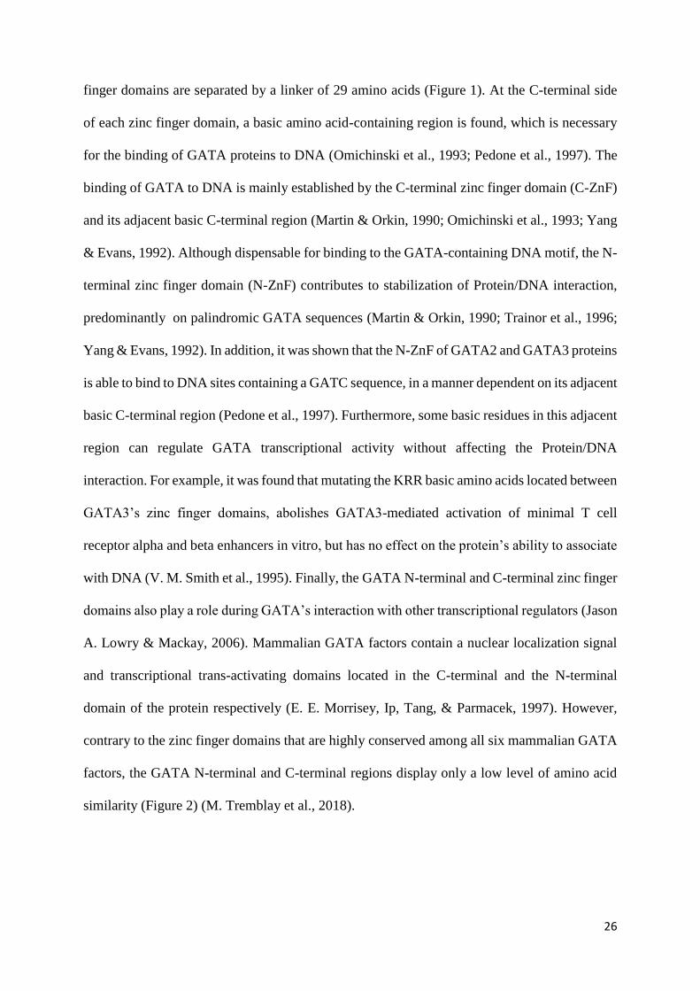
Figure 1. Functional domains and essential amino acids in the mammalian GATA3
transcription factor.
GATA3 protein length is 443 amino acids. GATA3 has two transactivation domains (TA1 and
TA2) in the N-terminal region of the protein (N-) and two zinc finger domains (N-terminal zinc
finger and C-terminal zinc finger) followed each by a conserved basic region. Each zinc finger
domain has the characteristic Cys-X2-Cys-X17-Cys-X2-Cys spacing where the four cysteine
residues coordinate a single zinc ion (Zn2+). The two zinc finger domains are separated by a
linker of 29 amino acids. The amino acid residues marked in blue were shown in a crystal
structure of the C-terminal zinc finger to make direct contact with DNA while residues marked
in red have been shown to be involved in normal GATA3 function (adapted from (Ho et al.,
2009).

28
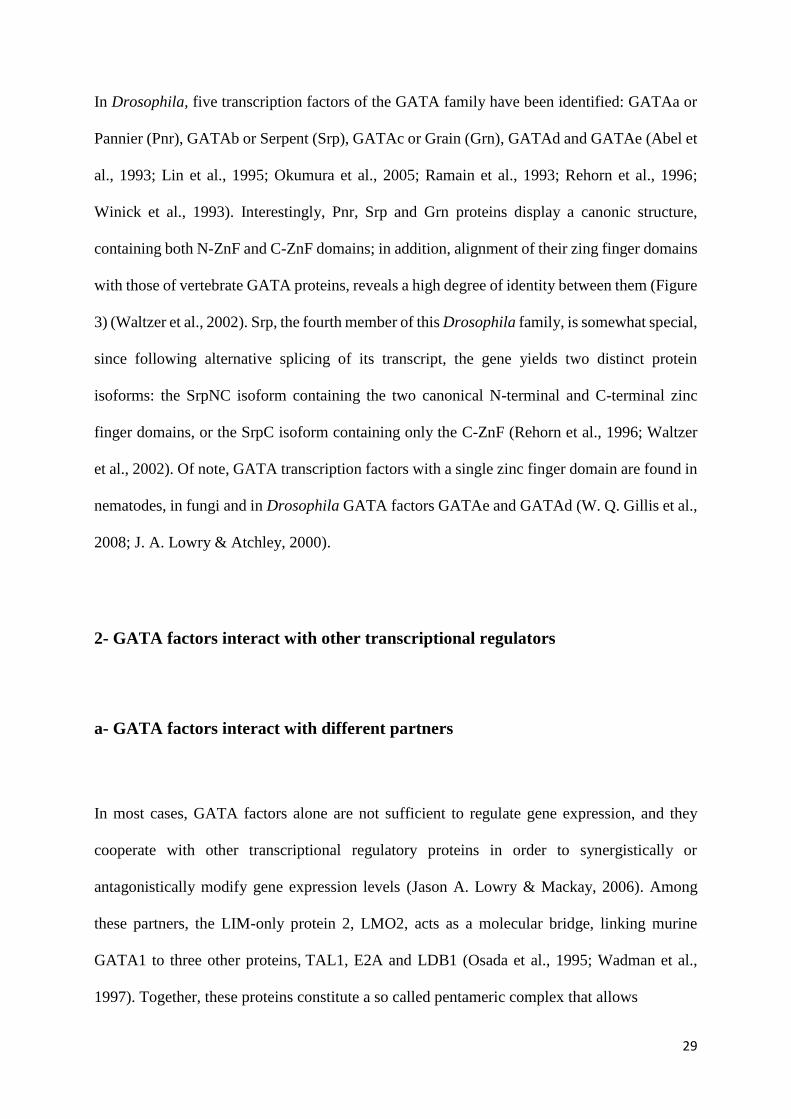
Figure 2. Conservation of the zinc finger domains in the six mammalian GATA.
The six mammalian GATA transcription factors contain two highly conserved zinc finger
domains (Zn), a nuclear localization signal (NLS) and two less conserved C-terminal and N-
terminal regions. The N-terminal region contains transcriptional activation domains (AD). Each
percentage represents the similarity level of the protein sequence of one part of the
corresponding protein in comparison to its equivalent part in GATA3 protein (M. Tremblay et
al., 2018).
29
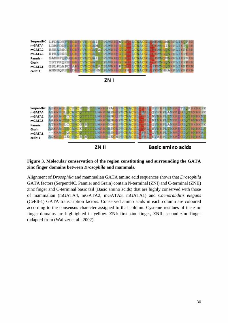
In Drosophila, five transcription factors of the GATA family have been identified: GATAa or
Pannier (Pnr), GATAb or Serpent (Srp), GATAc or Grain (Grn), GATAd and GATAe (Abel et
al., 1993; Lin et al., 1995; Okumura et al., 2005; Ramain et al., 1993; Rehorn et al., 1996;
Winick et al., 1993). Interestingly, Pnr, Srp and Grn proteins display a canonic structure,
containing both N-ZnF and C-ZnF domains; in addition, alignment of their zing finger domains
with those of vertebrate GATA proteins, reveals a high degree of identity between them (Figure
3) (Waltzer et al., 2002). Srp, the fourth member of this Drosophila family, is somewhat special,
since following alternative splicing of its transcript, the gene yields two distinct protein
isoforms: the SrpNC isoform containing the two canonical N-terminal and C-terminal zinc
finger domains, or the SrpC isoform containing only the C-ZnF (Rehorn et al., 1996; Waltzer
et al., 2002). Of note, GATA transcription factors with a single zinc finger domain are found in
nematodes, in fungi and in Drosophila GATA factors GATAe and GATAd (W. Q. Gillis et al.,
2008; J. A. Lowry & Atchley, 2000).
2- GATA factors interact with other transcriptional regulators
a- GATA factors interact with different partners
In most cases, GATA factors alone are not sufficient to regulate gene expression, and they
cooperate with other transcriptional regulatory proteins in order to synergistically or
antagonistically modify gene expression levels (Jason A. Lowry & Mackay, 2006). Among
these partners, the LIM-only protein 2, LMO2, acts as a molecular bridge, linking murine
GATA1 to three other proteins, TAL1, E2A and LDB1 (Osada et al., 1995; Wadman et al.,
1997). Together, these proteins constitute a so called pentameric complex that allows
30
Figure 3. Molecular conservation of the region constituting and surrounding the GATA
zinc finger domains between Drosophila and mammals.
Alignment of Drosophila and mammalian GATA amino acid sequences shows that Drosophila
GATA factors (SerpentNC, Pannier and Grain) contain N-terminal (ZNI) and C-terminal (ZNII)
zinc finger and C-terminal basic tail (Basic amino acids) that are highly conserved with those
of mammalian (mGATA4, mGATA2, mGATA3, mGATA1) and Caenorabditis elegans
(CeElt-1) GATA transcription factors. Conserved amino acids in each column are coloured
according to the consensus character assigned to that column. Cysteine residues of the zinc
finger domains are highlighted in yellow. ZNI: first zinc finger, ZNII: second zinc finger
(adapted from (Waltzer et al., 2002).
31
simultaneous recognition of the GATA DNA binding site (WGATAR) by GATA1, and of the
E-box motif (CANNTG) by TAL1 and E2A. Once bound to DNA, this complex activates
reporter gene expression (Osada et al., 1995; Wadman et al., 1997). Murine GATA1 is also able
to interact with factors of the Krüppel-like family, Sp1 and EKLF, themselves zinc finger
containing proteins. SP1 recognizes both consensus DNA sequences GC and CACC, while
EKLF binds a subset of extended CACC sequences. Both of these factors associate with
GATA1 in order to synergistically promote erythroid cell-specific gene expression (Lavallée et
al., 2006; Merika & Orkin, 1995). In addition, it was demonstrated that human and rat GATA4
proteins interact with Nkx factors, such as Nkx2-5. Nkx factors are homeodomain-containing
proteins that recognize a TNAAGTG DNA sequence and that interact with GATA4 to
synergistically activate cardiac target genes (Durocher et al., 1997; Liu et al., 2002; Zhu et al.,
2000). Similarly, members of the MADS box family of transcription factors, SRF and MEF2,
interact with rat GATA4 proteins to promote transcriptional activation of their target genes. The
MADS box motif is a DNA-binding and dimerization domain, and proteins of this family
recognize A/T-rich DNA regions (Belaguli et al., 2000; S. Morin et al., 2000; Steves Morin et
al., 2001). It is noteworthy that depending on the cellular context, interaction with the same
partner can yield different outcomes; for instance, in mammals, PU.1 transcription factors of
the Ets family interact with GATA1 factors in order to trigger gene expression in eosinophil
cells, while they functionally antagonize each other during the differentiation of hematopoietic
myeloid progenitors into erythroid versus myeloid cells (Du et al., 2002; Nerlov et al., 2000;
Rekhtman et al., 1999). Ets family members contain an Ets domain, a winged helix-turn-
helix structure recognizing the DNA sequence harboring the core GGAA motif. Finally, factors
of the RUNX family contain a Runt domain that binds the TGYGGTY DNA sequence. The
RUNX proteins RUNX1 and Lozenge (Lz), interact with the mammalian GATA1 and the
Drosophila Srp, respectively, during mouse megakaryocyte differentiation and fly crystal cell
32
(megakaryocyte-like) development (Elagib et al., 2003; Waltzer et al., 2003). In conclusion,
proteins of different families such as LIM-only protein 2, Krüppel-like factors, Nkx
homeodomain-containing proteins, MADS box containing factors, transcription factors of the
Ets family and RUNX family proteins, interact with GATA proteins to regulate their
transcriptional activity in a large number of cell types.
b- GATA factors interact with cofactors of the Friend of GATA family
The most widely studied co-factors of GATA factors belong to the Friend of GATA (FOG)
family. To date, two members of the FOG family have been identified in mouse, FOG1 and
FOG2, also known as ZFPM1 and ZFPM2, respectively (Lu et al., 1999; Svensson et al., 1999;
Tevosian et al., 1999; Tsang et al., 1997). FOG1 proteins contain nine zinc finger domains (1
to 9), distributed throughout the protein. Four of these zinc fingers are of the C2H2 type, while
the five other zinc fingers are of the C2HC type. FOG2 proteins have eight zinc finger domains
that are highly conserved with their eight equivalent zinc finger domains in FOG1 protein
(Chlon & Crispino, 2012). Structural comparison between FOG1 and FOG2 shows that the
equivalent of the eighth FOG1 zinc finger, a C2H2-type zinc finger, is absent in FOG2,
therefore giving rise to a protein with five C2HC type fingers and only three C2H2 type finger
domains (Figure 4A). None of the FOG zinc fingers are able to bind DNA, and among the nine
FOG1 zinc fingers, only the fingers 1, 5, 6 and 9, which are of the C2HC type, are able to
interact with GATA factors. In Drosophila, only one FOG cofactor called U-shaped (Ush) has
been identified (Cubadda et al., 1997; Haenlin et al., 1997). Ush shares 20% homology with
mammalian FOG1 and contains nine zinc finger domains. Similar to FOG1, four of these zinc
finger domains are of the C2H2 type, while the five others are of the C2HC type (Figure 4B).
33
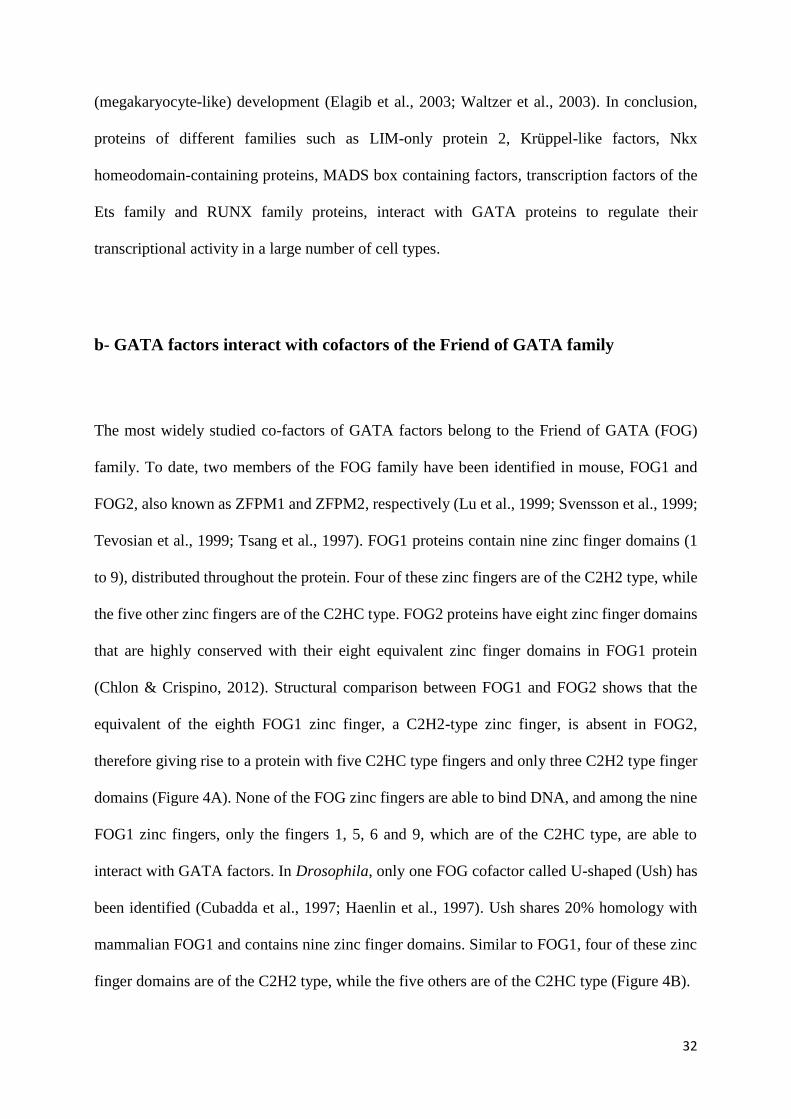
Figure 4. Molecular conservation of mammalian and Drosophila GATA cofactors of the
Friend of GATA family.
(A) Two members of the Friend of GATA (FOG) family are present in the mouse: FOG1 and
FOG2, formed of 995 and 1151 amino acids, respectively. Both cofactors contain zinc finger
domains of the C2HC (red vertical bars) and of C2H2 (blue vertical bars) type and also two co-
repressor interaction motifs that are colored in black (NuRD or CtBP). (B) In Drosophila, U-
shaped (Ush) protein of the FOG family is constituted of 1191 amino acid and it contains 8 zinc
finger dmains conserved with those of mammalian FOG, and contains also a CtBP interaction
motif (adapted from (Chlon & Crispino, 2012)).
34
Amino acid sequence alignment of FOG1, FOG2 and Ush zinc fingers, which are able to
interact with GATA factors, revealed a number of conserved residues. The resulting consensus
sequence for FOG binding to GATA factors was predicted to be X3-Phe-X-Cys-X2-Cys-X-Ile-
X2-Arg/Ser-X3-Thr/Asn-X3-His-X2-Tyr-Tyr-Cys-X3, where X stands for any amino acid
residue (A. H. Fox et al., 1999). Mutation of key residues in zinc finger 1 of FOG1 protein was
shown to interfere with the FOG1/GATA1 interaction, demonstrating their importance during
FOG cofactor binding to GATA proteins (A. H. Fox et al., 1999).
FOG zinc fingers interact with GATA proteins by specifically binding the core of the GATA
N-ZnF (A. Fox et al., 1999; A. H. Fox et al., 1999; Tsang et al., 1997). Interestingly, the ability
of FOG to recognize GATA N-ZnF, and not C-ZnF, is due to the presence in the N-ZnF of key
residues that are important for the interaction with FOG, and which are not found in the GATA
C-ZnF. These residues (referring to the murine GATA1 protein) are Glu203, Val205, Gly208,
Ala209, His222 and Tyr223 (Figure 5) (A. Fox et al., 1999). Substitution of the murine GATA1
Val205 residue into glycine (V205G) impairs the interaction with FOG1 and modulates the
chromatin occupancy of GATA1 during hematopoietic cell lineages specification (Chlon et al.,
2012; J. D. Crispino et al., 1999). Mapping of the key FOG interacting residues onto the solved
three-dimensional structure of the GATA1 N-ZnF domain, shows that they form a single
contiguous surface, essentially located outside of the DNA binding region (A. H. Fox et al.,
1999; Kowalski et al., 1999). Interestingly, these identified residues are highly conserved
among the N-ZnF of all six mammalian GATA transcription factors (M. H. Lentjes et al., 2016).
In agreement with the fact that FOG proteins recognize the GATA N-terminal, but not C-
terminal, zinc finger domain, it was demonstrated that the Drosophila FOG cofactor Ush binds
GATA factors Pnr and SrpNC, which both contain the canonical N-ZnF domain. However, Ush

35
Figure 5. Key amino acid residues identified in GATA1 N-terminal zinc finger domain as
required for GATA/FOG interaction.
(A) Representation of 3D structure of GATA1 N-terminal zinc finger domain. The six amino
acid residues identified as critical for the interaction with FOG are shown in yellow and their
position in the protein is added. The cysteine residues colored in orange attache the zinc ion of
the finger. (B) The key amino acid residues (marked in grey) are conserved among the N-
terminal zinc finger of mammalian GATA factors (mGATA-1, hGATA-2, hGATA-3) but they
are absent from the C-terminal zinc finger domain (adapted from (A. Fox et al., 1999; Kowalski
et al., 1999).

36
is unable to interact with the SrpC isoform that is devoid of the N-ZnF (Haenlin et al., 1997;
Waltzer et al., 2002). Finally, the SrpNCV421G protein variant harboring a valine to glycine
substitution equivalent to the mammalian GATA1 V205G, displays altered interaction with Ush
and behaves as the SrpC isoform rather than as the SrpNC isoform (Nancy Fossett et al., 2003).
These results indicate a conserved mode of interaction of GATA and FOG proteins between
Drosophila and mammals (Nancy Fossett et al., 2003; A. Fox et al., 1999; Waltzer et al., 2002).
Depending on the cellular context and the promoter bound by the GATA/FOG complexes, FOG
may acts as a co-activator or as a co-repressor of the GATA-dependent transcription (Chlon &
Crispino, 2012). Supporting the idea of their co-repressor function, two co-repressor binding
motifs have been identified in both mammalian FOG1 and FOG2 (Figure 4A). The first one,
the PIDLS motif, allows the fixation of the C-terminal binding protein (CtBP), while the second
is known to recruit the nucleosome remodeling and histone deacetylase (NuRD) complex (A.
H. Fox et al., 1999; Hong et al., 2005). Similarly, in Drosophila, Ush acts as negative regulator
for GATA factors activity (N. Fossett et al., 2000, 2001; Haenlin et al., 1997) and contains a
co-repressor binding motif allowing interaction with CtBP. However, unlike FOG1 and FOG2,
Ush does not contain any NuRD complex binding site (Chlon & Crispino, 2012).
Altogether, GATA proteins have the capacity to interact with different partners belonging to
the Friend of GATA family, as well as with members of other protein families, in order to
modulate their own functions. This ability to contrastingly interact with a variety of proteins,
opens up the functional range of GATA proteins in mammals and other GATA producing
organisms.

37
3- GATA mutations are associated to human diseases
a- Mutations in GATA factors induce the formation of numerous pathologies
The relation between GATA gene mutations and human diseases has been widely studied.
Genome, exome and transcriptome sequencing have led to the identification of a huge number
of GATA mutations in patients with different types of biological disorders (M. H. Lentjes et
al., 2016). The type of disease depends on the affected GATA gene and its expression pattern.
For example, GATA1, GATA2 and GATA3 proteins are expressed in hematopoietic cell
lineages, and mutations affecting these factors are related to numerous hematological disorders:
acute myeloid leukemia (AML), dendritic cell, monocyte, B and NK lymphoid deficiency
(DCML), myelodysplastic syndromes (MDS), large granular lymphocytic leukemia (LGL),
chronic myeloid leukemia (CML) with blast crisis (BC), Emberger syndrome, neutropenia,
thrombocytopenia, dyserythropoietic anemia, β-thalassemia, erythropoietic porphyria (EPP),
acute erythroid leukemia (AEL) and early T-cell precursor (ETP) acute lymphoblastic
leukemia/lymphoma (ALL/LBL) (John D. Crispino & Horwitz, 2017; Ping et al., 2017; Spinner
et al., 2014; J. Zhang et al., 2012; S.-J. Zhang et al., 2008).
Furthermore, GATA3 is also expressed in developing and differentiated mammary glands
(Asselin-Labat et al., 2007; Kouros-Mehr et al., 2006), as well as in embryonic kidney, inner
ear and parathyroid glands (Debacker et al., 1999; Labastie et al., 1995; Rivolta & Holley,
1998). Accordingly, GATA3 mutations are found associated to breast cancer,
hypoparathyroidism, sensorineural deafness and renal insufficiency (HDR) syndrome (Ellis et
al., 2012; Muroya et al., 2001; Okawa et al., 2015; Usary et al., 2004).
38
Similarly, the factors GATA4, GATA5 and GATA6 that are expressed in the mammalian
developing heart (Pikkarainen et al., 2004), are associated to cardiac diseases, such as atrial
septal defects (ASD), ventricular septal defects (VSD), bicuspid aortic valve (BAV), dilated
cardiomyopathy (DCM), tetralogy of Fallot (TOF), familial atrial fibrillation (AF) and
persistent truncus arteriosus (PTA). Additionally, alteration of these factors provokes other
diseases such as neonatal diabetes, adult-onset diabetes and congenital diaphragmatic hernia
(Allen et al., 2011; De Franco et al., 2013; L. Yu et al., 2013). Finally, loss-of-function
mutations in GATA4 are also associated to the 46, XY disorder of sex development (Lourenço
et al., 2011; Martinez de LaPiscina et al., 2018).
b- Point mutations in GATA zinc fingers provoke human diseases whose severity
depends on the position and the nature of the mutation
Interestingly, most GATA associated human diseases were identified in patients carrying point
mutations affecting residues located in and around the two GATA zinc finger domains (Figure
6) (M. H. Lentjes et al., 2016). The severity of the diseases associated to GATA zinc finger
point mutations varies depending on the affected zinc finger domain, on the residue altered in
the zinc finger domain, but also on the nature of the substitution, as the same residue can be the
target of more than one type of substitution, resulting in a different clinical outcome.
In GATA2 protein, many identified point mutations in the N-ZnF domain are associated to
AML, while point mutations found in the GATA2 C-terminal zinc finger lead to the
development of other hematopoietic disorders, such as MDS, AML and CML with BC
formation (M. H. Lentjes et al., 2016).
39
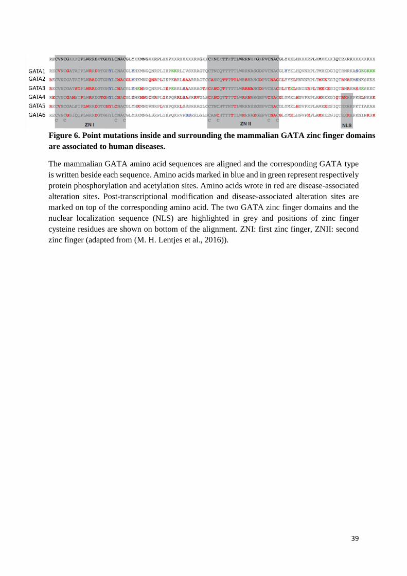
The mammalian GATA amino acid sequences are aligned and the corresponding GATA type
is written beside each sequence. Amino acids marked in blue and in green represent respectively
protein phosphorylation and acetylation sites. Amino acids wrote in red are disease-associated
alteration sites. Post-transcriptional modification and disease-associated alteration sites are
marked on top of the corresponding amino acid. The two GATA zinc finger domains and the
nuclear localization sequence (NLS) are highlighted in grey and positions of zinc finger
cysteine residues are shown on bottom of the alignment. ZNI: first zinc finger, ZNII: second
zinc finger (adapted from (M. H. Lentjes et al., 2016)).
Figure 6. Point mutations inside and surrounding the mammalian GATA zinc finger domains
are associated to human diseases.
40
Also, in the same N-ZnF domain of GATA1, mutating amino acids that are required for the
GATA1/FOG1 interaction, such as the residues V205, G208 and D218, alters erythrocyte and
thrombocyte formation, leading to hematological disorders including anemia and
thrombocytopenia (K. Freson et al., 2001; Kathleen Freson et al., 2002; Mehaffey et al., 2001;
Nichols et al., 2000). Meanwhile, mutation of the R218 arginine residue, located in the same
GATA1 N-ZnF, affects the ability of the factor to bind to DNA palindromic sequences, without
affecting the interaction GATA1/FOG1, and leads to thrombocytopenia and β-thalassemia,
associated to gray platelet syndrome and porphyria, respectively (Balduini et al., 2004; Phillips
et al., 2007; Tubman et al., 2007; C. Yu et al., 2002).
Finally, at the same amino acid position, the nature of the substitution affects the severity of the
outcome. For instance, two mutations affect the GATA1 Gly208 residue, G208S and G208R,
but the former is associated to macrothrombocytopenia and mild dyserythropoietic anemia,
while the latter is associated to severe macrothrombocytopenia and severe dyserythroipoietic
anemia (Mehaffey et al., 2001; Vecchio et al., 2005). Analyzing the effect of these two
mutations on GATA1’s ability to interact with FOG1, showed that G208R generates a stronger
disruption of GATA1/FOG1 interaction as compared to G208S, which might explain the more
severe phenotypes developed by patients with the G208R substitution as compared to those
carrying the G208S mutation (A. E. Campbell et al., 2013). Similar results were observed in
the case of the D218G and D218Y mutations. The D218G mutation is associated to
thrombocytopenia without anemia, while the D218Y mutation provokes thrombocytopenia and
severe dyserythropoietic anemia (K. Freson et al., 2001; Kathleen Freson et al., 2002). The
more severe phenotype obtained in patients with the D218Y mutation mirrors the fact that the
substitution of the D218 residue into a tyrosine alters more extensively the affinity of GATA1
for FOG1 than its substitution into a glycine (A. E. Campbell et al., 2013; Kathleen Freson et

41
al., 2002). All these results show that the GATA zinc finger domain amino acids have critical
and sophisticated roles during the establishment of GATA functions.
In conclusion, altering the ability of GATA proteins to function properly is associated to several
human disorders, where the type and the severity of the disease vary depending on the nature
and on the position of the mutation. Given the important and critical roles played by GATA
factors in humans, understanding their mode of action is of definite interest.
(B) GATA factors mode of action
1- Mechanistic functions of GATA factors
a- GATA and transcriptional activation
GATA1, the first member of GATA factors was identified by its ability to bind the β-globin
gene enhancer and to activate its expression in chicken red blood cell precursors (Evans et al.,
1988). Two transactivation domains are present in GATA1. They are located in the N-terminal
and the C-terminal regions of the protein and they play redundant as well as specific functions
during regulation of hematopoiesis in mice (Kaneko et al., 2012). Although poorly conserved,
those transactivation domains have been identified in the six mammalian GATA factors (Chlon
& Crispino, 2012).
42
Studies in mice and isolated human cells revealed several target genes that are positively
regulated by GATA factors (Cheng et al., 2009; Kurek et al., 2007; Martynova et al., 2019;
Welch et al., 2004; Ming Yu et al., 2009). As many other transcriptional factors, GATA proteins
recruit co-activators with histone acetyl- and methyl-transferase activities, in order to render
the DNA more accessible to transcription and thus regulate gene expression (Figure 7D).
Accordingly, both histone acetyltransferase proteins p300 and CBP have been shown to interact
with various GATA factors and to act as transcriptional co-activators (Blobel et al., 1998; Dai
& Markham, 2001; Kakita et al., 1999; Wada et al., 2000). Moreover, by recruiting H3K79
monomethyltransferase protein, GATA1 allows the methylation of their bound elements within
the murine β-globin enhancer, prior to the activation of β-globin transcription (Steger et al.,
2008).
In Drosophila, like in mammals, GATA factors act as transcriptional activators. For example,
Pnr and Srp interact with components of the mediator transcriptional co-activator complex in
order to promote expression of the pro-neural genes achaete and scute, and of the anti-microbial
peptide coding gene Metchnikowin, respectively (Garcia-Garcia et al., 1999; Immarigeon et al.,
2019; Kuuluvainen et al., 2014).
b- GATA and transcriptional repression
In addition to their functions as transcriptional activators, GATA factors act as transcriptional
repressors (Figure 7E). For example, GATA3 directly interacts with transcriptional co-
repressors such as the NuRD complex member MTA3, or the H3K9 mono- and
dimethyltransferase G9A protein in human breast adenocarcinoma cells.
43
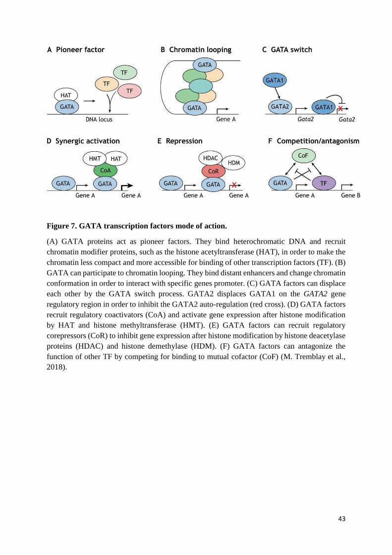
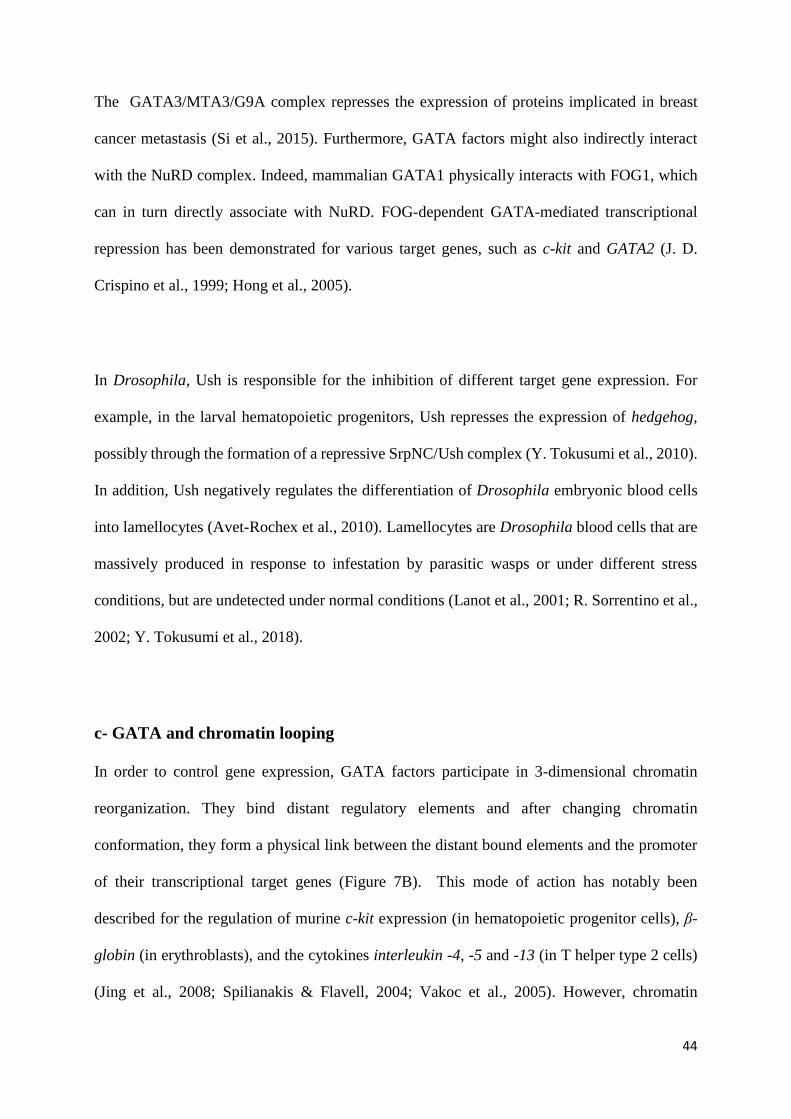
Figure 7. GATA transcription factors mode of action.
(A) GATA proteins act as pioneer factors. They bind heterochromatic DNA and recruit
chromatin modifier proteins, such as the histone acetyltransferase (HAT), in order to make the
chromatin less compact and more accessible for binding of other transcription factors (TF). (B)
GATA can participate to chromatin looping. They bind distant enhancers and change chromatin
conformation in order to interact with specific genes promoter. (C) GATA factors can displace
each other by the GATA switch process. GATA2 displaces GATA1 on the GATA2 gene
regulatory region in order to inhibit the GATA2 auto-regulation (red cross). (D) GATA factors
recruit regulatory coactivators (CoA) and activate gene expression after histone modification
by HAT and histone methyltransferase (HMT). (E) GATA factors can recruit regulatory
corepressors (CoR) to inhibit gene expression after histone modification by histone deacetylase
proteins (HDAC) and histone demethylase (HDM). (F) GATA factors can antagonize the
function of other TF by competing for binding to mutual cofactor (CoF) (M. Tremblay et al.,
2018).
44
The GATA3/MTA3/G9A complex represses the expression of proteins implicated in breast
cancer metastasis (Si et al., 2015). Furthermore, GATA factors might also indirectly interact
with the NuRD complex. Indeed, mammalian GATA1 physically interacts with FOG1, which
can in turn directly associate with NuRD. FOG-dependent GATA-mediated transcriptional
repression has been demonstrated for various target genes, such as c-kit and GATA2 (J. D.
Crispino et al., 1999; Hong et al., 2005).
In Drosophila, Ush is responsible for the inhibition of different target gene expression. For
example, in the larval hematopoietic progenitors, Ush represses the expression of hedgehog,
possibly through the formation of a repressive SrpNC/Ush complex (Y. Tokusumi et al., 2010).
In addition, Ush negatively regulates the differentiation of Drosophila embryonic blood cells
into lamellocytes (Avet-Rochex et al., 2010). Lamellocytes are Drosophila blood cells that are
massively produced in response to infestation by parasitic wasps or under different stress
conditions, but are undetected under normal conditions (Lanot et al., 2001; R. Sorrentino et al.,
2002; Y. Tokusumi et al., 2018).
c- GATA and chromatin looping
In order to control gene expression, GATA factors participate in 3-dimensional chromatin
reorganization. They bind distant regulatory elements and after changing chromatin
conformation, they form a physical link between the distant bound elements and the promoter
of their transcriptional target genes (Figure 7B). This mode of action has notably been
described for the regulation of murine c-kit expression (in hematopoietic progenitor cells), β-
globin (in erythroblasts), and the cytokines interleukin -4, -5 and -13 (in T helper type 2 cells)
(Jing et al., 2008; Spilianakis & Flavell, 2004; Vakoc et al., 2005). However, chromatin
45
conformation alteration by GATA proteins might also require other gene expression regulators,
such as the mediator protein Med1, the chromatin remodeler BRG1, the bridging molecule
LDB1 and the cofactor FOG1 (Kim et al., 2009; Song et al., 2007; M. Stumpf et al., 2006;
Vakoc et al., 2005).
2- Dynamic functions of GATA factors
a- GATA as pioneer factors
In addition to their classical function of binding GATA motifs in DNA and regulating gene
transcription, GATA proteins also contribute to the remodeling of DNA packaging. For
instance, GATA1 physically interacts through its zinc finger domains with the mammalian
chromatin remodeling complex SWI/SNF, in order to efficiently activate transcription from
nucleosome assembled promoters in vitro (Kadam et al., 2000). Also, murine GATA4
associates to heterochromatic DNA, in order to decompact chromatin and promote DNA
accessibility for other transcription factors (Cirillo et al., 2002). The ability to affect chromatin
conformation prior to gene regulation by other factors, is a characteristic feature of ‘‘pioneer
factors’’ (Figure 7A) (Zaret & Carroll, 2011). The role of GATA proteins as pioneer factors
was discovered in 2002, when GATA4 and FOXA factors were both found to bind the albumin
gene enhancer, in order to decompact chromatin and promote hepatocyte specification (Bossard
& Zaret, 1998; Cirillo et al., 2002).
46
b- The GATA switch
Different members of the GATA family were shown to bind the same chromatin sites
sequentially, in order to yield different transcriptional outputs in a dynamic fashion (Bresnick
et al., 2010; Doré et al., 2012; Huang et al., 2016). This process of GATA factor displacement
by another member of the family is called the ‘‘GATA switch’’ (Figure 7C). The most studied
GATA-switch context is the displacement of murine GATA2 by GATA1 at the GATA2 locus
upon erythroid differentiation. This displacement is responsible for switching off the feed-
forward autoregulatory loop of GATA2, by removing the histone acetyltransferase CBP and
altering chromatin looping conformation (Grass et al., 2003, 2006; Martowicz et al., 2005).
Many other genes are known to be the target of GATA switch events. For example, expression
of Wdr77 and kit genes is restricted after GATA switches, in order to inhibit proliferation of the
murine developing blood cells (Rylski et al., 2003; Min Yu et al., 2016).
In Drosophila, regulation of the same gene by two different GATA proteins has been illustrated
in the developing embryonic gut, where Srp and GATAe act sequentially in the endoderm to
control expression of the ectodermal protein coding gene brachyenteron. Srp inhibits
brachyenteron expression during early stages of embryogenesis, and GATAe acts during later
stages of embryogenesis, when srp expression has ceased (R. Murakami et al., 1999; Okumura
et al., 2005; Reuter, 1994).
c- GATA antagonism with other transcriptional regulators
47
GATA and other transcription factors have been shown to antagonize the function of each other
in some contexts (Figure 7F). For instance, mouse GATA1 promotes common myeloid
progenitor differentiation towards an erythrocytic fate, while PU.1 directs their differentiation
into myeloid cells (Hoppe et al., 2016). PU.1 in developing erythrocytes inhibits GATA1-
mediated erythropoiesis, and reciprocally GATA1 expression inhibits PU.1-mediated
transcription of myeloid specific genes (Nerlov et al., 2000; Rekhtman et al., 1999; P. Zhang et
al., 1999). However, although GATA1 and PU.1 ensure antagonistic functions during
determination of erythroid versus myeloid cell fates, they act synergistically during
differentiation of myeloid cell progenitors into mast cells (Du et al., 2002). However, during
this process, FOG1 has to be down-regulated. In the case of FOG1 ectopic expression in mast
cells progenitors, FOG competes with PU.1 for interaction with GATA1 and hence affects
GATA1/PU.1-dependent mast cell differentiation (Cantor et al., 2008; Sugiyama et al., 2008).
Moreover, competition for binding to GATA proteins has been identified in Drosophila, as Srp
interacts with the transcription factor of the RUNX family Lozenge in order to trigger
differentiation of crystal cells, which are reminiscent of mammalian megakaryocytes. However,
Ush competes with Lz for the association with Srp, and overexpression of ush in crystal cell
progenitors alters Srp/Lz-mediated crystal cell differentiation (N. Fossett et al., 2001; Nancy
Fossett et al., 2003; Muratoglu et al., 2007).
Taken together, these results show that GATA factors have many different modes of action.
They cooperate with or antagonize other transcriptional regulators to control gene expression,
and they play additional roles in facilitating the activity of other transcriptional factors.
Depending on the context (cell type and developmental stage), GATA proteins display an
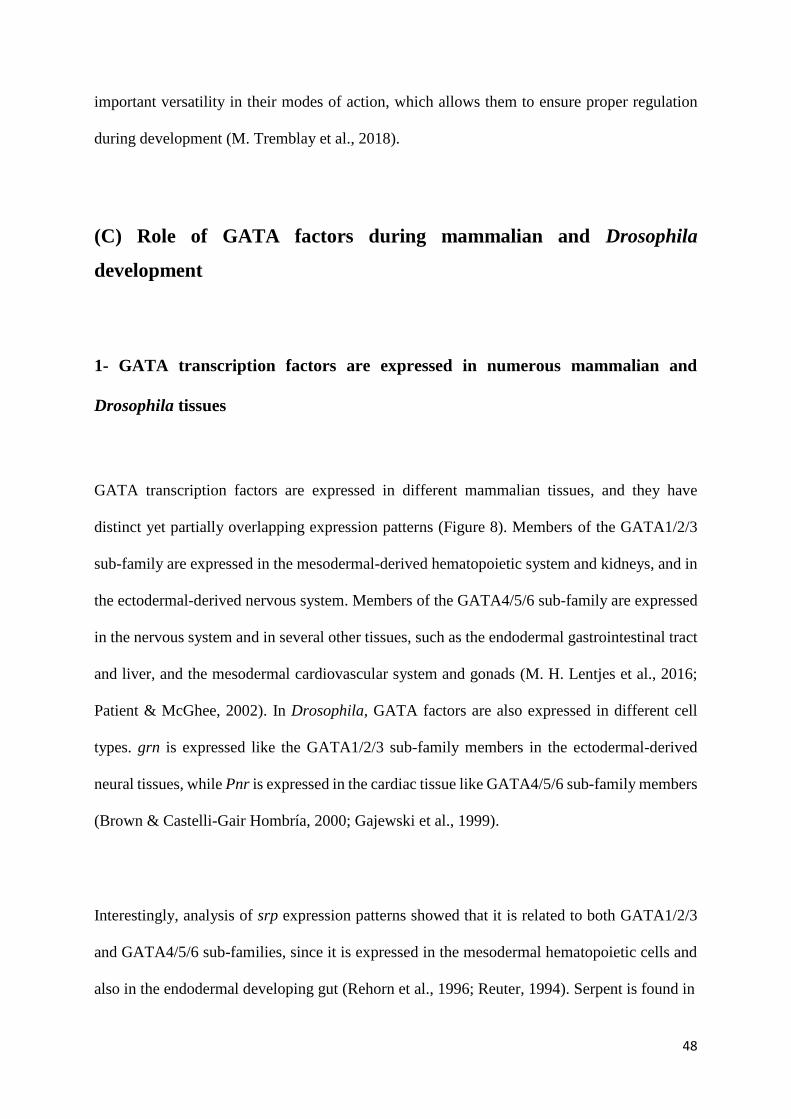
48
important versatility in their modes of action, which allows them to ensure proper regulation
during development (M. Tremblay et al., 2018).
(C) Role of GATA factors during mammalian and Drosophila
development
1- GATA transcription factors are expressed in numerous mammalian and
Drosophila tissues
GATA transcription factors are expressed in different mammalian tissues, and they have
distinct yet partially overlapping expression patterns (Figure 8). Members of the GATA1/2/3
sub-family are expressed in the mesodermal-derived hematopoietic system and kidneys, and in
the ectodermal-derived nervous system. Members of the GATA4/5/6 sub-family are expressed
in the nervous system and in several other tissues, such as the endodermal gastrointestinal tract
and liver, and the mesodermal cardiovascular system and gonads (M. H. Lentjes et al., 2016;
Patient & McGhee, 2002). In Drosophila, GATA factors are also expressed in different cell
types. grn is expressed like the GATA1/2/3 sub-family members in the ectodermal-derived
neural tissues, while Pnr is expressed in the cardiac tissue like GATA4/5/6 sub-family members
(Brown & Castelli-Gair Hombría, 2000; Gajewski et al., 1999).
Interestingly, analysis of srp expression patterns showed that it is related to both GATA1/2/3
and GATA4/5/6 sub-families, since it is expressed in the mesodermal hematopoietic cells and
also in the endodermal developing gut (Rehorn et al., 1996; Reuter, 1994). Serpent is found in
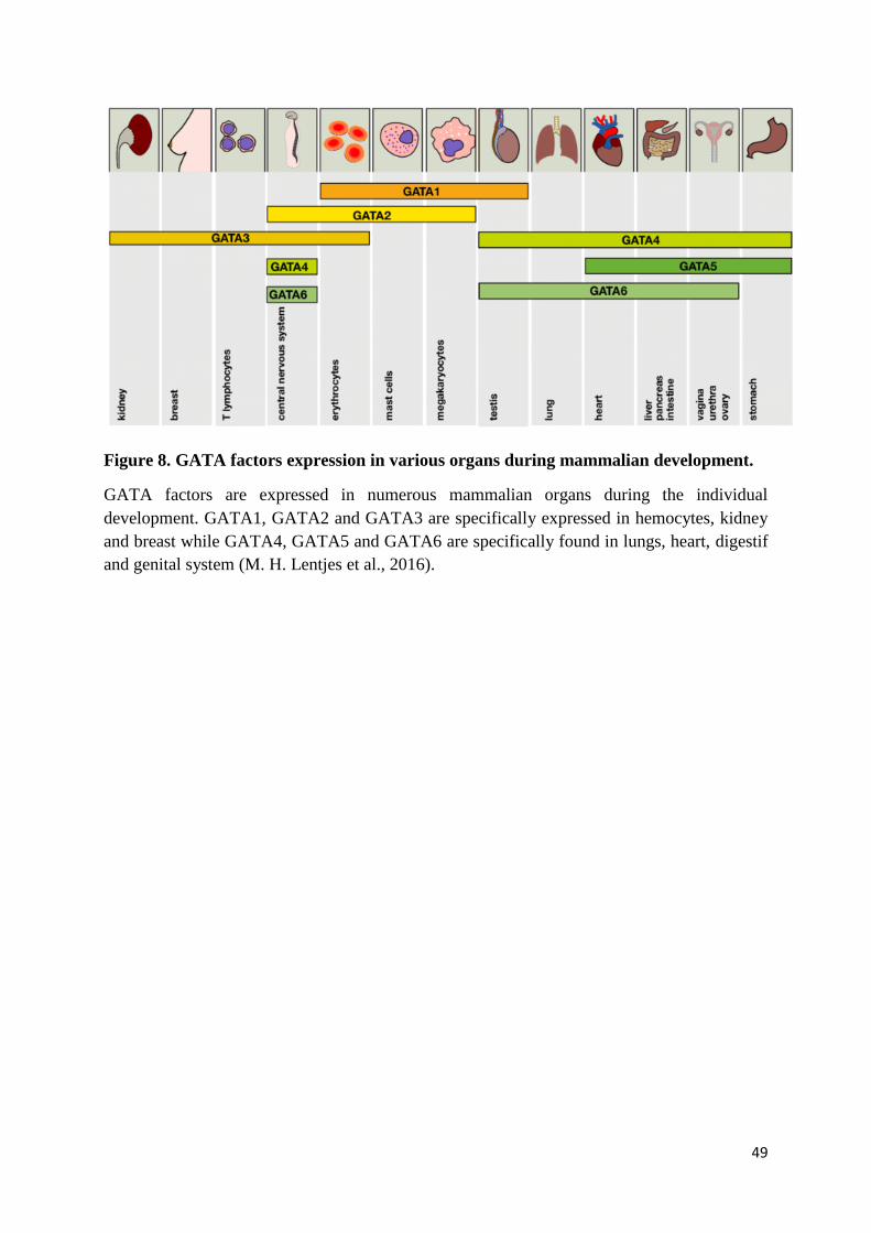
49
Figure 8. GATA factors expression in various organs during mammalian development.
GATA factors are expressed in numerous mammalian organs during the individual
development. GATA1, GATA2 and GATA3 are specifically expressed in hemocytes, kidney
and breast while GATA4, GATA5 and GATA6 are specifically found in lungs, heart, digestif
and genital system (M. H. Lentjes et al., 2016).
50
other tissues, namely the fat body (the functional homologue of mammalian liver and adipose
tissue) and the ovaries (Lepesant et al., 2020; Rehorn et al., 1996). GATAe is expressed in the
endodermal derived gut and Malpighian tubules (the equivalent of mammalian kidney)
(Martínez-Corrales et al., 2019; Okumura et al., 2005). However, no expression pattern is found
for GATAd (Okumura et al., 2005). Importantly, the GATA factors cited above are not just
expressed in the mentioned tissues, they also ensure essential roles in those tissues.
2- GATA transcription factors have essential functions in mammalian and
Drosophila organs
a- The hematopoietic system
1- Mammalian and Drosophila hematopoiesis
In mammals, distinct waves of hematopoiesis take place (Figure 9B). The first wave, called
primitive hematopoiesis, occurs in the extra-embryonic yolk sac, and gives rise to mainly
erythrocytes. The second wave, known as definitive hematopoiesis, happens in the embryo
proper in the aorta-gonad-mesonephros (AGM) region, as well as in the extra-embryonic
placenta. During definitive hematopoiesis appear self-renewing hematopoietic stem cells
(HSCs) that later give rise to all blood cell types. The HSCs colonize and expand in other
hematopoietic organs: fetal liver, spleen and thymus and subsequently in the adult bone
marrow, the major site of postnatal hematopoiesis (Ivanovs et al., 2017).
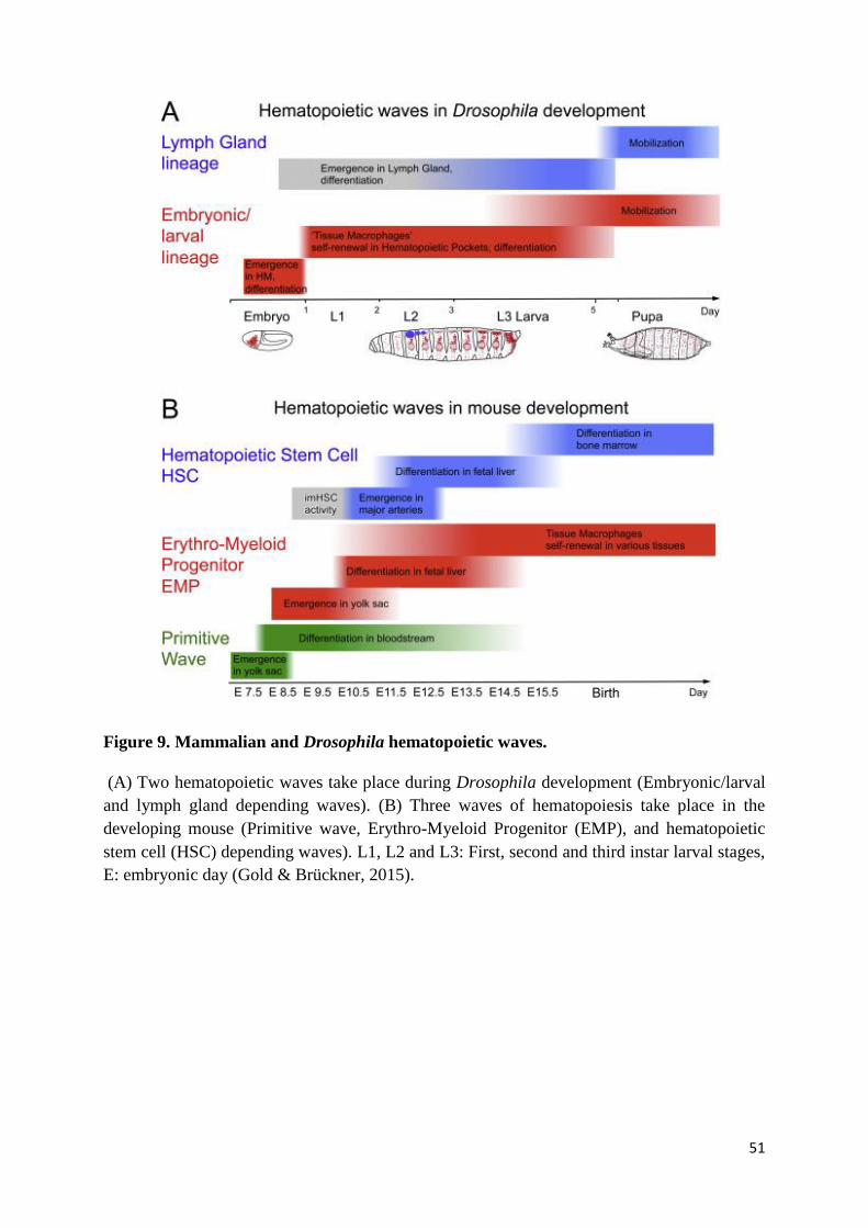
51
Figure 9. Mammalian and Drosophila hematopoietic waves.
(A) Two hematopoietic waves take place during Drosophila development (Embryonic/larval
and lymph gland depending waves). (B) Three waves of hematopoiesis take place in the
developing mouse (Primitive wave, Erythro-Myeloid Progenitor (EMP), and hematopoietic
stem cell (HSC) depending waves). L1, L2 and L3: First, second and third instar larval stages,
E: embryonic day (Gold & Brückner, 2015).
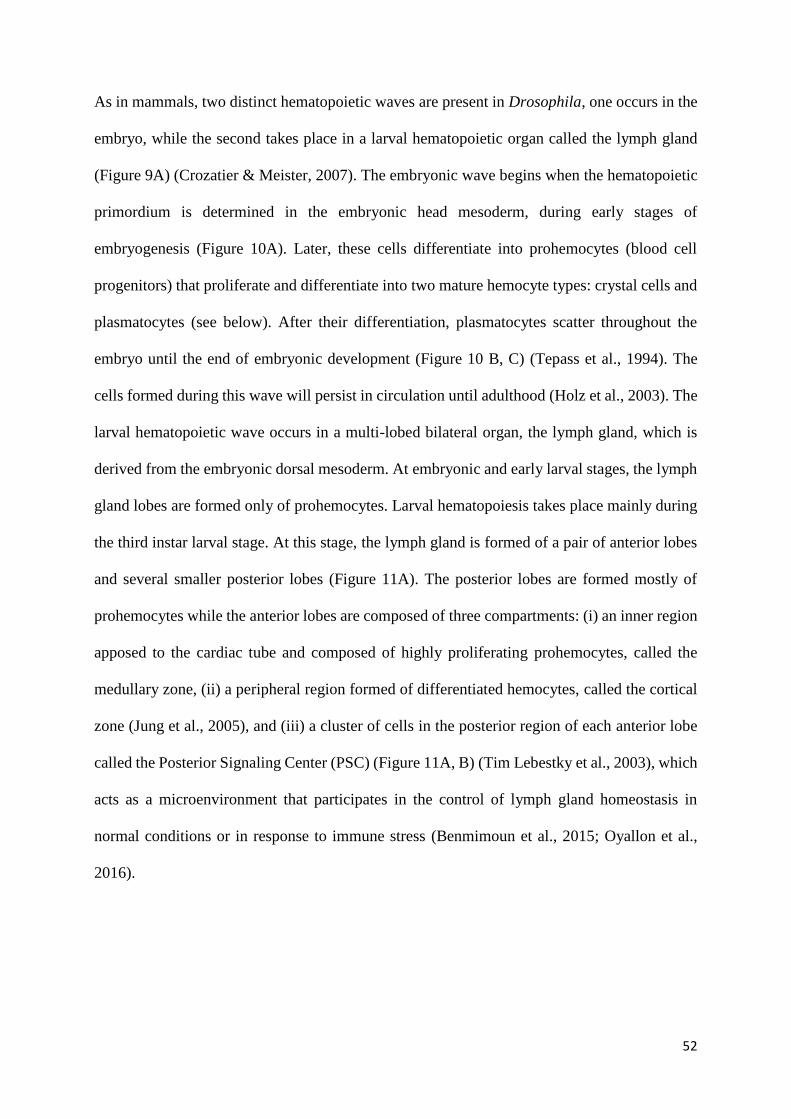
52
As in mammals, two distinct hematopoietic waves are present in Drosophila, one occurs in the
embryo, while the second takes place in a larval hematopoietic organ called the lymph gland
(Figure 9A) (Crozatier & Meister, 2007). The embryonic wave begins when the hematopoietic
primordium is determined in the embryonic head mesoderm, during early stages of
embryogenesis (Figure 10A). Later, these cells differentiate into prohemocytes (blood cell
progenitors) that proliferate and differentiate into two mature hemocyte types: crystal cells and
plasmatocytes (see below). After their differentiation, plasmatocytes scatter throughout the
embryo until the end of embryonic development (Figure 10 B, C) (Tepass et al., 1994). The
cells formed during this wave will persist in circulation until adulthood (Holz et al., 2003). The
larval hematopoietic wave occurs in a multi-lobed bilateral organ, the lymph gland, which is
derived from the embryonic dorsal mesoderm. At embryonic and early larval stages, the lymph
gland lobes are formed only of prohemocytes. Larval hematopoiesis takes place mainly during
the third instar larval stage. At this stage, the lymph gland is formed of a pair of anterior lobes
and several smaller posterior lobes (Figure 11A). The posterior lobes are formed mostly of
prohemocytes while the anterior lobes are composed of three compartments: (i) an inner region
apposed to the cardiac tube and composed of highly proliferating prohemocytes, called the
medullary zone, (ii) a peripheral region formed of differentiated hemocytes, called the cortical
zone (Jung et al., 2005), and (iii) a cluster of cells in the posterior region of each anterior lobe
called the Posterior Signaling Center (PSC) (Figure 11A, B) (Tim Lebestky et al., 2003), which
acts as a microenvironment that participates in the control of lymph gland homeostasis in
normal conditions or in response to immune stress (Benmimoun et al., 2015; Oyallon et al.,
2016).
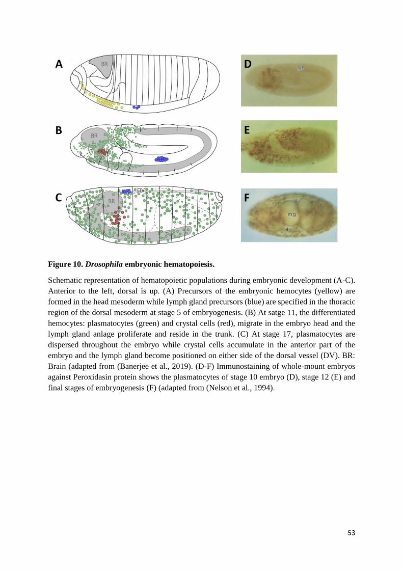
53
Figure 10. Drosophila embryonic hematopoiesis.
Schematic representation of hematopoietic populations during embryonic development (A-C).
Anterior to the left, dorsal is up. (A) Precursors of the embryonic hemocytes (yellow) are
formed in the head mesoderm while lymph gland precursors (blue) are specified in the thoracic
region of the dorsal mesoderm at stage 5 of embryogenesis. (B) At satge 11, the differentiated
hemocytes: plasmatocytes (green) and crystal cells (red), migrate in the embryo head and the
lymph gland anlage proliferate and reside in the trunk. (C) At stage 17, plasmatocytes are
dispersed throughout the embryo while crystal cells accumulate in the anterior part of the
embryo and the lymph gland become positioned on either side of the dorsal vessel (DV). BR:
Brain (adapted from (Banerjee et al., 2019). (D-F) Immunostaining of whole-mount embryos
against Peroxidasin protein shows the plasmatocytes of stage 10 embryo (D), stage 12 (E) and
final stages of embryogenesis (F) (adapted from (Nelson et al., 1994).
54
Figure 11. Structure of the Drosophila larval lymph gland.
(A) Schematic diagram of the third instar larval lymph gland. The lymph gland flanks the heart
tube and it is formed of two large lobes called anterior lobes and several (2 to 4 pairs) smaller
lobes called posterior lobes formed of progenitor cells (blue). At each side of the heart tube the
lobes are separated by pericardial cells. Each anterior lobe contains a medullar zone (MZ)
formed of progenitor cells (blue), of cortical zone (CZ) harboring the differentiated hemocytes
(red) and the posterior signaling center (PSC) (green). The green arrows indicate the
presumptive PSC-dependent prohemocyte maintenance signals and the yellow arrows represent
the main axes of differentiation. (B) Confocal section of third instar lymph gland anterior lobes
stained for the PSC (green), blood cell progenitors (blue) and differentiated hemocytes (red)
(adapted from (Benmimoun et al., 2015).
55
Differentiation of the mammalian hematopoietic progenitor cells gives rise to multiple cell
types (Figure 12). The erythroid lineage consists of erythrocytes, or red blood cells responsible
for transport of oxygen to tissues and for recovery of carbon dioxide produced as waste. The
megakaryocytic lineage produces the platelets implicated in blood clotting. The myeloid cell
lineages are composed of granulocytes (eosinophils, mast cells and neutrophils), macrophages
and dendritic cells, all of which are responsible for innate immune defense. Finally, lymphoid
cell lineages regroup the lymphocytes (B-cells, T-cells and natural killer cells) that are the major
players of adaptive immunity (Hartenstein, 2006; C. Smith, 2003).
Contrary to mammals, Drosophila has neither red blood cells nor lymphoid cells. Only three
types of blood cells are produced in the fly (Figure 12). Plasmatocytes are the equivalent of
mammalian macrophages and ensure phagocytic functions (apoptotic body removal and micro-
organism clearance) (Gold & Brückner, 2015). Crystal cells are the equivalent of granulocytes
and owe their name to the presence of crystalline inclusions in their cytoplasm. These inclusions
contain melanin precursors that participate in Drosophila melanization reactions that allow
sequestration and destruction of invading pathogens, as well as wound healing. Drosophila
crystal cells are also regarded as the equivalent of mammalian megakaryocytes, due to their
role in the wound healing (Rizki & Rizki, 1959; Tang, 2009). Finally, under stress conditions
(such as after wasp infestation, mechanical stress challenge or genetic deregulation) the fly can
produce a third blood cell type: these large flat cells with extended filaments are called
lamellocytes (Lanot et al., 2001; R. Sorrentino et al., 2002; Y. Tokusumi et al., 2018). They
are found only at larval stages and they surround and destroy particles that are too big to be
phagocytosed by plasmatocytes (Wood & Jacinto, 2007).

56
Figure 12. Mammalian and Drosophila blood cells.
Different blood cell lineages are produced; they all emerge from hematopoietic stem cells that
differentiate later into myeloid or lymphoid stem cells. The lymphoid stem cells give rise to
lymphocytes and lymphoid dendritic cell that play a role during the adaptive immunity and
antigen presentation, respectively. The myeloid stem cells give rise to more restraint
hematopoietic precursors (myeloblast, monoblast, pro-erythroblast) that will be at the origin of
several types of mature blood cells: myeloid dendritic cell, macrophage, neutriophil, eosinophil,
basophil and erythrocyte. Megakaryocytes are also derived of myeloid stem cells and are the
origin of thrombocytes or platelets. The function of each cell lineage in human is added beside
the corresponding cell type. In Drosophila, three blood cell types are produced: plasmatocyte
represents macrophage-like phagocyte, crystal-cell is megacaryocytes-like cell that participate
in blood clotting and lamellocyte constitutes an encapsulating hemocyte that participate in the
parasites’ attack (« Organs », 2015).

57
2- Role of GATA factors during mammalian and Drosophila hematopoiesis
GATA1 and GATA2 play essential functions during mammalian primitive hematopoiesis.
Although not required for erythroid precursor cell formation in the murine embryonic yolk sac,
GATA1 is crucial for the differentiation of these precursors into mature erythrocytes (McDevitt
et al., 1997; Pevny et al., 1991, 1995; Takahashi et al., 1997). GATA1 loss-of-function causes
a blockage of the yolk sac erythropoiesis at the pro-erythroblast stage, followed by apoptosis
of the pro-erythroblasts (Y. Fujiwara et al., 1996; Pevny et al., 1995). Contrary to GATA1 null
mice that display altered mature erythrocyte formation, mice with GATA2 loss-of-function
have somewhat normal maturation of red blood cells, but the number of mature erythrocytes is
reduced two- to seven-fold (F.-Y. Tsai et al., 1994). Thus, GATA2 and GATA1 proteins
regulate proliferation and viability of embryonic developing blood cells.
Contrary to the primitive hematopoiesis, the mammalian definitive hematopoiesis relies on the
presence of pluripotent HSCs that give rise not only to erythrocytes but also to megakaryocytes,
myeloid cells and lymphoid cells. Interestingly, GATA1, GATA2 and GATA3 play essential
functions throughout the development of the hematopoietic lineages of this post-embryonic
hematopoiesis (T. Fujiwara, 2017; Ho et al., 2009; Katsumura & Bresnick, 2017). First, GATA3
participates in the maintenance of the number of long-term repopulating HSCs, their entry into
the cell cycle, and their subsequent proliferation (Frelin et al., 2013; Ku et al., 2012). Second,
GATA1 and GATA2 are essential for differentiation of HSCs into myeloid cells. In the
granulocyte-monocyte progenitors, GATA2 determines the mast cells fate, while in the
megakaryocyte-erythrocyte progenitors, GATA2 promotes megakaryocyte differentiation at
the expense of erythrocytes (Ikonomi et al., 2000; F. Y. Tsai & Orkin, 1997). In parallel,
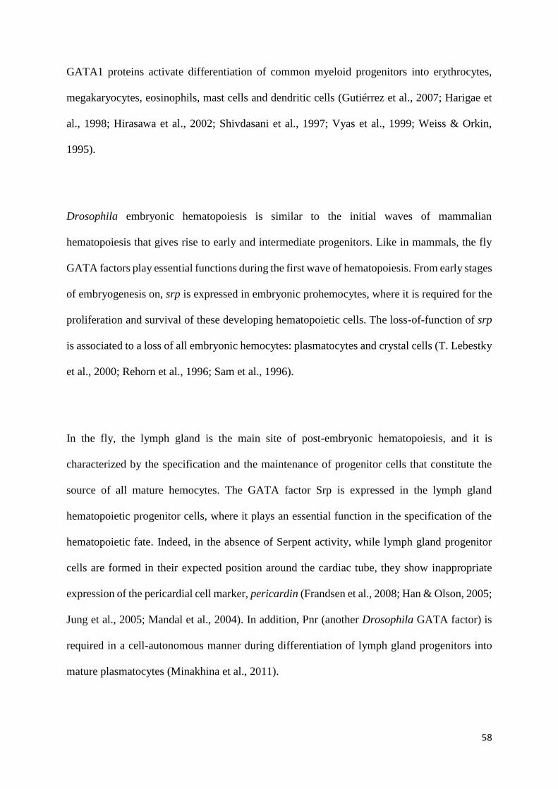
58
GATA1 proteins activate differentiation of common myeloid progenitors into erythrocytes,
megakaryocytes, eosinophils, mast cells and dendritic cells (Gutiérrez et al., 2007; Harigae et
al., 1998; Hirasawa et al., 2002; Shivdasani et al., 1997; Vyas et al., 1999; Weiss & Orkin,
1995).
Drosophila embryonic hematopoiesis is similar to the initial waves of mammalian
hematopoiesis that gives rise to early and intermediate progenitors. Like in mammals, the fly
GATA factors play essential functions during the first wave of hematopoiesis. From early stages
of embryogenesis on, srp is expressed in embryonic prohemocytes, where it is required for the
proliferation and survival of these developing hematopoietic cells. The loss-of-function of srp
is associated to a loss of all embryonic hemocytes: plasmatocytes and crystal cells (T. Lebestky
et al., 2000; Rehorn et al., 1996; Sam et al., 1996).
In the fly, the lymph gland is the main site of post-embryonic hematopoiesis, and it is
characterized by the specification and the maintenance of progenitor cells that constitute the
source of all mature hemocytes. The GATA factor Srp is expressed in the lymph gland
hematopoietic progenitor cells, where it plays an essential function in the specification of the
hematopoietic fate. Indeed, in the absence of Serpent activity, while lymph gland progenitor
cells are formed in their expected position around the cardiac tube, they show inappropriate
expression of the pericardial cell marker, pericardin (Frandsen et al., 2008; Han & Olson, 2005;
Jung et al., 2005; Mandal et al., 2004). In addition, Pnr (another Drosophila GATA factor) is
required in a cell-autonomous manner during differentiation of lymph gland progenitors into
mature plasmatocytes (Minakhina et al., 2011).
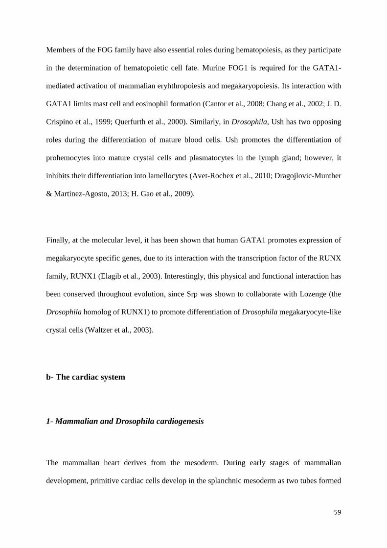
59
Members of the FOG family have also essential roles during hematopoiesis, as they participate
in the determination of hematopoietic cell fate. Murine FOG1 is required for the GATA1-
mediated activation of mammalian eryhthropoiesis and megakaryopoiesis. Its interaction with
GATA1 limits mast cell and eosinophil formation (Cantor et al., 2008; Chang et al., 2002; J. D.
Crispino et al., 1999; Querfurth et al., 2000). Similarly, in Drosophila, Ush has two opposing
roles during the differentiation of mature blood cells. Ush promotes the differentiation of
prohemocytes into mature crystal cells and plasmatocytes in the lymph gland; however, it
inhibits their differentiation into lamellocytes (Avet-Rochex et al., 2010; Dragojlovic-Munther
& Martinez-Agosto, 2013; H. Gao et al., 2009).
Finally, at the molecular level, it has been shown that human GATA1 promotes expression of
megakaryocyte specific genes, due to its interaction with the transcription factor of the RUNX
family, RUNX1 (Elagib et al., 2003). Interestingly, this physical and functional interaction has
been conserved throughout evolution, since Srp was shown to collaborate with Lozenge (the
Drosophila homolog of RUNX1) to promote differentiation of Drosophila megakaryocyte-like
crystal cells (Waltzer et al., 2003).
b- The cardiac system
1- Mammalian and Drosophila cardiogenesis
The mammalian heart derives from the mesoderm. During early stages of mammalian
development, primitive cardiac cells develop in the splanchnic mesoderm as two tubes formed

60
from different cardiac precursors, called primary and secondary heart fields. The primitive heart
tubes then merge into one single tube called the linear heart tube (Brade et al., 2013). This linear
heart tube swells and forms various anatomic features of the heart. Later, the cardiac tube twists
and turns on itself by a process called heart looping, to form a heart with four chambers: right
ventricle, left ventricle, right atrium and left atrium (Figure 13A) (Bruneau, 2013; Schleich et
al., 2013).
The mammalian heart is composed of three tissue layers. The first layer (the endocardium)
consists of endothelial cells underlying connective tissue, and it represents the innermost layer
of the heart. The second layer (the myocardium) is composed of cardiomyocytes that are
responsible for heart contractions. The third layer (the epicardium) comprises epithelial cells
and constitutes the outermost layer of the heart. The epicardium receives the nervous and blood
vessels that supply the heart, and it surrounds a pericardial cavity containing a serous fluid that
prevents friction during heart contractions (Gavaghan, 1998).
In order to establish a directional blood flow, the heart chambers are separated by septa and
cardiac valves (Figure 13A). The atrial septum separates the right and left atria, while the
ventricular septum separates the right and left ventricles. The atrium and ventricle of each side
of the heart are separated by atrioventricular valves (Krishnan et al., 2014; Lamers Wouter H.
& Moorman Antoon F.M., 2002).
Compared to the vertebrate heart, the Drosophila heart is quite simple: it is basically a simple
tube of bilateral cardiomyocytes (Figure 13B). Fly cardiac cell precursors form 11 clusters of
cells located along the most dorsal part of the embryonic mesoderm (Gajewski et al., 1999). By

61
Figure 13. Mammalian and Drosophila heart.
(A) The mammalian heart is formed of four chambers: right and left ventricle plus right and left
atrium. Chambers are separated by septa and cardiac valves (white arrows) that control blood
circulation direction into the heart. The transport of the blood from the heart to the other body
organs and vice versa is occurred through heart vessels. The aorta is the vessel that transport
oxygenated blood from the heart towards the upper and lower body organs. (B) The fly heart is
formed of simple tube of bilateral cardiomyocytes located at the dorsal midline. The regulation
of the Drosophila hemolymph flow direction in the heart depends on ostia and valves. The
anterior portion of the heart is named conical chamber from which is attached the fly aorta. The
alary mucles are associated with the heart likely to give it lateral stability and support (adapted
from (« Organs », 2015)).
62
the end of embryogenesis, the bilateral heart progenitor cells have migrated to the dorsal
midline, where they form a linear heart tube (Figure 14A-C).
The fly cardiac tube consists of only two cell types: cardioblasts that differentiate into
contractile cardiomyocytes, and pericardial cells that flank the cardioblasts and act as
nephrocytes (Vogler & Bodmer, 2015). In Drosophila, the function of the cardiac tube is to
pump the hemolymph containing essential components such as nutrients, signaling molecules
and hemocytes, from the posterior part of the body towards the anterior part. Like in mammals,
regulation of the flow direction in the fly heart is achieved by ostia and valves present in the
heart tube (Figure 13B). At the adult stage, the ostia form 5 pairs of openings distributed along
the heart which allow the hemolymph to enter but not to exit the cardiac tube, while the valves
are formed of intracardiac specialized cardiomyocytes that can close off the heart tube at five
different locations (Rotstein & Paululat, 2016).
2- Role of GATA factors in mammalian and Drosophila cardiogenesis
In mammals, the transcription factors of the GATA family including GATA4 and GATA6 play
essential functions during heart formation (Clowes et al., 2014; Peterkin et al., 2005). Although
the loss-of-function of either GATA4 or GATA6 alone has no effect on differentiation of
mammalian cardiomyocytes, mutant mice with simultaneous loss-of-function of GATA4 and
GATA6 are devoid of a heart, which results from defects in cardiomyocyte differentiation (Kuo
et al., 1997; Molkentin et al., 1997; Zhao et al., 2008). Therefore, GATA4 and GATA6 factors

63
Figure 14. The Drosophila heart development.
(A-C) Schematic representation of heart development. Anterior is to the left and the dorsal is
up. (A) The heart is formed from two lines of cardioblasts migrating toward the dorsal midline
of stage 13 embryo. (B) At the stage 16, the embryonic dorsal vessel is located in the dorsal
midline. (C) The adult fly circulatory system is formed of the aorta located in the thoracic part
of adult fly and of the heart present in the abdomen. The adult abdominal segments (A1 to A8)
are shown (adapted from (Wolf, 2012).

64
have redundant roles during heart development. Similar to the common role of GATA4 and
GATA6 in mammalian heart development, Pnr has critical functions during Drosophila cardiac
cell specification, and its loss-of-function is associated to an almost complete absence of
cardiomyocytes (Klinedinst & Bodmer, 2003).
Interestingly, it was shown that activation of gene expression in cardiac cells is under the control
of rat GATA4 and the homeotic box transcription factor, Nkx2-5, which both interact physically
to promote the cardiomyocyte program (Durocher et al., 1997). Similarly, in Drosophila, it was
found that Pnr activates the expression of cardiac cell markers such as myocyte enhancer factor
2 (mef2), which also depends on the presence of the Nkx2-5 protein homologous Tinman (Tin);
both Pnr and Tin are able to physically interact in cultured cells (Gajewski et al., 1999, 2001).
c- The gastro-intestinal system
1- Mammalian and Drosophila intestinal development
In mammals, gut morphogenesis begins when the anterior and posterior edges of the
endodermal epithelial sheet fold off to form the foregut and the hindgut pockets. Once folded,
both pockets continue to migrate towards each other in order to meet in the center of the embryo
and to form a closed tube, the primitive gut tube (Spence et al., 2011). The digestive tube is
subdivided into three regions: foregut, midgut and hindgut, which will give rise to different
adult intestinal structures. The foregut gives rise to structures including the esophagus, liver
and pancreas. The midgut yields the stomach and the small intestine, and the hindgut forms the
65
colon. The small intestine is in turn divided into three regions: duodenum, jejunum, and ileum
(Figure 15A) (Spence et al., 2011).
The regions of the mammalian intestine differ in their functions. The jejunum’s role is to digest
and absorb important nutrients such as fatty acids, amino acids and sugars. The other small
intestine regions also participate in the absorption of these nutrients, however, they have
additional specific functions, like the absorption of iron in the duodenum and the absorption of
vitamin B12 and bile salts in the ileum (Aronson et al., 2014). In contrast, in the colon, the
epithelial cells absorb only water and mineral ions. The colon also contains bacteria that allow
the fermentation of indigestible materials (Greenwood-Van Meerveld et al., 2017). The
intestinal epithelium is formed of repeated crypt-villus units that contain different types of
intestinal cells, such as the enterocytes (that play a role in nutrient absorption) and the secretory
cells (enteroendocrine, goblet, tuft and Paneth cells). In addition, in the intestinal crypt, there
are quiescent intestinal stem cells, rapidly dividing stem cells and proliferating transit-
amplifying cells that differentiate into mature intestinal cells as they migrate to the base of or
outside the crypt (Figure 15A) (Noah et al., 2011).
Similar to the mammalian digestive tube, the Drosophila gut is composed of three regions: two
ectodermal-derived regions, namely the anterior gut (foregut) and the posterior gut (hindgut),
and an endodermal-derived region called the midgut (Figure 15B). The embryonic midgut is
formed from two different primordia, the anterior midgut primordium (that appears at early
stages of embryogenesis at the anterior-ventral side of embryonic blastoderm) and the posterior
midgut primordium (that is formed at the posterior side of embryonic blastoderm) closely
related to the hindgut ectodermal cells (Figure 16A). During embryonic germ band elongation,

66
Figure 15. Mammalian and Drosophila gut.
(A) The mammalian intestine is divided into four regions: esophagus, stomach, small intestine
and colon. The intestinal epithelium is formed of series of crypt-villus structure that are
composed of different intestinal cell types: absorptive erythrocytes (EC), secretory cells
(enteroendocrine (EE), goblet cells, Paneth cells), intestinal stem cells (ISC) and transit-
amplifying cells. The epithelium is surrounded by visceral muscles and also by mucus that
prevent direct contact between intestinal epithelial cells and intestinal bacteria located in the
lumen. Neutrophils circulate in proximity of the intestinal epithelium (B) The fly gut is divided
into foregut, midgut (anterior and posterior midgut) and hindgut. The intestinal epithelium is
formed of EC, EE, ISC and erythroblasts (EB). The epithelium is surrounded by visceral
muscles and by peritrophic matrix mucus, which is the equivalent of the human mucus.
Hemocytes can be found close to the intestinal epithelium. The Malpighian tubules in the fly
are the equivalent of the mammalian kidney (Capo et al., 2019).

67
Figure 16. The Drosophila gut formation.
(A-C) Schematic representation of embryonic gut formation. The embryonic anterior side is to
the left while the dorsal is up. (A) At stage 7 of embryogenesis, the endodermal (red) anterior
midgut primordium (amp) and posterior midgut primordium (pmg) are located in the embryonic
anterior and posterior parts, respectively. The ectodermal (blue) anlagen that surround the amp
and the pmg are at the origin of the foregut and hindgut respectively. (B) At stage 12, the amg
and pmg are migrating one toward the other. (C) At stage 13, the amp and the pmg cells fuse to
form a continuous gut tube. ph: pharynx, es: esophagus, pv: proventriculus, mp: malpighian
tubules, sg: salivary glands (adapted from (Hartenstein, 1993). (D-F) In situ hybridization of
whole-mount embryos against GATAe transcript shows the amg and the pmg of stage 8 embryo
(D), the migrating amp and pmg and developing Malpighian tubues (mt) of stage 12 embryo
(E) and the closed multi-chambered midgut at the stage 17 of embryogenesis (F) (adapted from
(Okumura et al., 2005).

68
the anterior and posterior midgut primordia move towards each other, and once they meet in
the middle of the embryo, undergo mesenchymal to epithelial transition and fuse to one another,
giving rise to the gut tube at the end of embryogenesis (Figure 16B, C). Although constituted
of a simple monolayer epithelium, the midgut can be divided, as the mammalian small intestine,
into different regions that differ from each other by histological and anatomical characteristics
and by gene expression patterning (Buchon et al., 2013; Ryutaro Murakami et al., 1994).
As in vertebrates, the Drosophila gastrointestinal tract plays a major role in nutrient digestion
and molecule absorption (Lemaitre & Miguel-Aliaga, 2013; Miguel-Aliaga et al., 2018). The
adult midgut epithelium contains four cell types, two of them representing mature gut cells,
called enterocytes and enteroendocrine cells. Enterocytes have a major role during nutrient
absorption and digestion, while enteroendocrine cells are responsible for secretion of peptide
hormones to regulate gut physiology and homeostasis (Miguel-Aliaga et al., 2018). The other
two cell types are the intestinal stem cells and their daughters (the enteroblasts) that are able to
constantly regenerate midgut epithelium and to repair tissue damage (Figure 15B) (Jiang et al.,
2011).
2- Role of GATA factors in mammalian and Drosophila intestine
Members of the GATA family of both Drosophila and mammals have similar roles during
intestine development. In mice, GATA4, GATA5 and GATA6 are expressed in embryonic
intestinal cells (Arceci et al., 1993; Fang et al., 2006; E. E. Morrisey, Ip, Tang, Lu, et al., 1997;
Edward E. Morrisey et al., 1996). Among them, GATA5 has no essential functions during gut
morphogenesis and differentiation (Molkentin et al., 2000). In contrast, both GATA4 and
69
GATA6 have redundant and crucial roles during gut development (Kuo et al., 1997; Molkentin
et al., 1997; Walker et al., 2014). Mice with simultaneous loss-of-function of both GATA
factors have altered developing intestinal epithelium and intestinal villus architecture (Walker
et al., 2014). Similarly, in Drosophila, three GATA factors Srp, Grn and GATAe are exrepssed
in developing embryonic midgut primordia. Among these factors, only Srp is required for
midgut primordia specification and subsequently for gut formation (Hernández de Madrid &
Casanova, 2018; Lin et al., 1995; Okumura et al., 2005; Reuter, 1994; Sam et al., 1996).
In adult mouse small intestine, GATA4 is expressed in the duodenum and in the jejunum but
not in the ileum. GATA4 maintains jejunum identity, as loss-of-function of GATA4 in the
jejunum causes a loss of jejunal specific gene expression and inadequate expression of ileal
specific genes in jujenal enterocytes (Bosse et al., 2006; Fang et al., 2006). This altered gene
expression affects the ability of the intestine to absorb cholesterol and dietary fat, which are
two jejunal-specific functions, and causes an increase of bile acid absorption, which is an ileal-
specific function (Battle et al., 2008; Bosse et al., 2006). Contrary to GATA4, whose expression
profile is restricted to the proximal intestine, GATA6 is expressed throughout the small intestine
including the jejunum (Fang et al., 2006). Mice with inducible disruption of GATA6 in the
small intestine, display reduced expression in the ileal enterocytes of some genes normally
expressed in the ileum, and mainly of those involved in lipid metabolism, such as
apolipoprotein CIII, apolipoprotein A-I, and fatty acid binding protein 6. In parallel, GATA6
inactivation in the ileum induces the expression of genes normally expressed in the colon
(Beuling et al., 2011). Thus, GATA6 is required for activation of ileum specific gene expression
and inhibition of colon specific genes transcription in the ileal enterocytes. The role of members
of GATA family in regulation of the intestinal regionalization has also been identified in
Drosophila. Srp acts as a homeotic gene during early stages of embryogenesis in order to
70
prevent endodermal anterior midgut primordium from adopting ectodermal foregut fate, and to
specify endodermal posterior midgut versus ectodermal hindgut fate (Reuter, 1994). Similarly,
GATAe maintains endodermal identity of the midgut by inhibiting expression of the
prospective hindgut determinant brachyenteron in the posterior midgut primordium (Okumura
et al., 2005). Later, in embryonic developed intestine, GATAe plays important functions in the
expression of terminally differentiated pan-midgut markers, namely innexin and CG4781, and
regionalized midgut markers such as integrin-βν, midgut expression 1, Tetraspanin 29Fa,
CG5077, CG10300, lambda-Trypsin, CG17633 and CG18493 (Okumura et al., 2007). In a
similar manner, in the Drosophila adult midgut, GATAe regulates most of the genes expressed
in a regionalized manner in the midgut, including genes encoding proteins involved in digestion
such as the Amylase distal, beta-galactosidase and the trypsin family enzymes (theta-Trypsin,
iota-Trypsin and CG31269) (Buchon et al., 2013). All these results show that Drosophila Srp
and GATAe proteins are required, as are the mammalian GATA4 and GATA6, for determining
intestinal region identities in the gut, and for regulating expression levels of essential genes in
gut enterocytes.
Furthermore, murine GATA6 proteins regulate the proliferation of cells at the bottom of the
colonial crypt and their subsequent migration from the bottom to the villus surface. The
proliferation of the crypt intestinal stem cells and their subsequent migration and differentiation
into mature intestinal cells is essential for colonic epithelial renewal (Beuling et al., 2012).
Similarly, GATAe is required for the maintenance of intestinal stem cells, by regulating their
proliferation and other stem cells properties, such as their small nuclei size and diploid state
(Okumura et al., 2016). By regulating proliferation and stemness of intestinal stem cells and
consequently their differentiation into intestinal mature cells, GATAe is identified, like
71
GATA6, as a crucial factor for the continuous regeneration of the adult midgut during normal
fly development, but also during stress conditions (Dutta et al., 2015; Okumura et al., 2016).
Differentiation of mature intestinal cells depends on activation of the Notch signaling pathway
in intestinal stem cells. GATAe contributes to Notch signaling pathway activation in
Drosophila adult intestine by maintaining expression of the Notch receptor ligand Delta in
intestinal stem cells. Alteration of GATAe-mediated Notch activation in intestinal stem cells is
related to a high reduction rate of both enterocytes and enteroendocrine differentiation
(Okumura et al., 2016). Strikingly, the double knockout of GATA4 and GATA6 in mammalian
developing intestine is also associated to reduced expression of Delta and of Delta mediated
activation of the Notch signaling pathway in intestinal cells. This dis-regulation of the GATA4
and GATA6-mediated Notch activation in mammalian intestine, alters the intestinal epithelial
cell populations and provokes a significant reduction in the differentiation rate of enterocytes
and enteroendocrine cells (Walker et al., 2014).
Finally, members of GATA family have essential roles in the epithelial-to-mesenchymal
transition (EMT), which is a process required for cell migration. It occurs during formation of
the fly embryonic intestine, where the anterior and posterior midgut primordia gradually lose
their epithelial properties to become more rounded and irregular in shape. They finally adopt
mesenchymal properties, allowing them to migrate towards the center of the embryo, where
they form a closed gut tube (K. Campbell et al., 2011; Leptin, 1995; Reuter, 1994). Srp is
required for fly intestinal EMT (K. Campbell et al., 2011; Reuter, 1994). This role depends on
direct repression by Srp of crumbs expression in these cells, which results in relocalization of
junctional protein dE-cadherin without affecting its expression. Interestingly, it was found that

72
vertebrate GATA6 factor promotes EMT of mammalian cells in a similar manner to that of Srp,
and this by inhibiting expression of the mammalian crumbs ortholog crb2, and by provoking a
downregulation of junctional dE-cadherin proteins without blocking their expression (K.
Campbell et al., 2011). Thus, Srp and GATA6 factors acts similarly during EMT in mammalian
and Drosophila cells.
d- The ovaries
1- Mammalian and Drosophila oogenesis
Mammalian gonads develop from mesonephros when the epithelium overlaying the
mesonephros coelomic surface starts to swell and to form the presumptive gonads, the genital
ridge. Primordial germ cells are specified at the base of the extra-embryonic allantois. These
cells migrate towards the genital ridge to later populate the primordial gonads (DeFalco &
Capel, 2009).
Mammalian ovaries contain germ cells representing all the cells that will undergo
gametogenesis, ranging from oogonia to eggs, as well as somatic cells that form the structures
harboring and helping the primordial germ cells during their development, namely the ovarian
follicles. Mammalian ovarian follicles are constituted of granulosa cells, surrounded by
steroidogenic cells called theca cells that form a part of the ovarian stroma (J. Wu et al., 2016).
73
In Drosophila as well, the gonads develop from two different cell types: the primordial germ
cells that form one cell cluster, and the somatic gonadal precursors that form three cell clusters
in the mesoderm of developing embryo (Brookman et al., 1992; Mahowald, 1962). During
embryonic germ band elongation, primordial germ cells are passively moved from the most
posterior end of the embryo to the dorso-posterior part, and the cells are later pulled in an
embryonic dorsal invagination. Germ cells then pass through the invaginating epithelium
towards the mesoderm in the direction of the somatic gonadal precursors, which migrate and
coalesce to then ensheath the primordial germ cells. Finally, both germline and somatic cells
compact together to form a round embryonic gonad (Santos & Lehmann, 2004).
Fly ovaries are composed of egg chambers (Figure 17) that each contain 15 small nurse cells
and one larger oocyte. Nurse cells and oocyte all derive from division of ovarian germline stem
cells. The egg chamber is surrounded by an epithelial monolayer of follicle cells that provide
the oocyte with yolk proteins, a source of nutrients essential for oocyte growth (DiMario &
Mahowald, 1987; X. Wu et al., 2008). Yolk proteins are either expressed in the female fly fat
body and transported by the hemolymph to the follicle cells, or directly expressed in the ovarian
follicle cells (Barnett et al., 1980; Hames & Bownes, 1978).
2- Role of GATA factors in mammalian and Drosophila ovaries
GATA4 is highly expressed in the anterior part of mammalian mesonephros coelomic
epithelium, and to a lesser degree in the posterior coelomic epithelium. GATA4 is required for
coelomic epithelium thickening that occurs mostly in the anterior part of the mesonephros, in
order to constitute the genital ridge and hence, the gonadal primordium (Hu et al., 2013). In
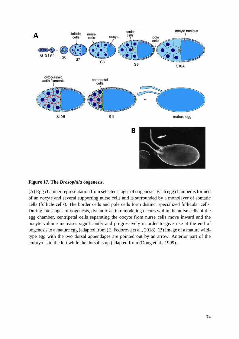
74
Figure 17. The Drosophila oogenesis.
(A) Egg chamber representation from selected stages of oogenesis. Each egg chamber is formed
of an oocyte and several supporting nurse cells and is surrounded by a monolayer of somatic
cells (follicle cells). The border cells and pole cells form distinct specialized follicular cells.
During late stages of oogenesis, dynamic actin remodeling occurs within the nurse cells of the
egg chamber, centripetal cells separating the oocyte from nurse cells move inward and the
oocyte volume increases significantly and progressively in order to give rise at the end of
oogenesis to a mature egg (adapted from (E. Fedorova et al., 2018). (B) Image of a mature wild-
type egg with the two dorsal appendages are pointed out by an arrow. Anterior part of the
embryo is to the left while the dorsal is up (adapted from (Dong et al., 1999).
75
contrast, no GATA factors have been identified as being expressed in developing Drosophila
germline and somatic cells (Lin et al., 1995; Okumura et al., 2005; Rehorn et al., 1996; Winick
et al., 1993).
In the developing murine ovaries, GATA4 is expressed in somatic granulosa cells, where it
promotes granulosa cell proliferation and ovarian follicle formation (Efimenko et al., 2013;
Viger et al., 1998). In addition, loss of GATA4 expression in the ovary leads to a reduction of
ovary size, of the number of oocytes released, of the level of estrogen produced, and of the
expression of the GATA4 ovarian target genes Star, Cyp11a1 and Cyp19 (Kyrönlahti et al.,
2011). Similar or even stronger phenotypes have been observed in the ovary of mice mutants,
with simultaneous loss of GATA4 and GATA6 functions (Bennett et al., 2012; Padua et al.,
2014). Thus, GATA4 and GATA6 play essential functions during mammalian gonadogenesis,
folliculogenesis and oocyte release. In Drosophila, GATA factors Srp and GATAd are
expressed in Drosophila adult ovaries (Lepesant et al., 2020). Srp plays essential functions in
ovarian somatic cells that ensure Drosophila oogenesis, as female flies with downregulated Srp
in somatic cells lay almost no eggs, and their ovarioles contain degenerating mid-stage egg
chambers, revealed by the pycnotic morphology of their nuclei (Lepesant et al., 2020). In
addition, it has been suggested that Srp is responsible, in the ovarian follicle cells, for the
expression of yolk proteins that are required for oocyte nutrition (Lossky & Wensink, 1995).
Thus, Drosophila Srp has, as vertebrate GATA4 and GATA6, essential functions during fly
oogenesis.
e- The liver and the fat body
76
1- Mammalian liver and Drosophila fat body formation
The liver derives from the foregut endoderm, where progenitor cells destined to adopt the
hepatic cell fate are specified. These cells later converge to generate the epithelial cells of the
liver bud (K. D. Tremblay & Zaret, 2005). During gestation, the liver develops into a
hematopoietic organ. However, after birth, the liver shifts from a hematopoietic role to being
the primary site controlling levels of many metabolites and serum proteins, in the bloodstream
and for endotoxin detoxification (Zaret, 2002).
The Drosophila fat body is often referred to as the equivalent of mammalian adipose cells and
liver. It works as an energy reservoir and nutrient sensor, in order to regulate proper fly
development and lifespan (Y. Zhang & Xi, 2015). Drosophila fat body development starts when
clusters of fat body precursors are specified in the different embryonic mesodermal segments
(Figure 18A). Later during development, fat body precursors start to proliferate, and fat body
primordia extend in size (Figure 18B). These primordia then coalesce to form a continuous
sheet of fat body cells, along the lateral wall of the embryo at the end of embryogenesis (Figure
18C) (Abel et al., 1993; Rehorn et al., 1996; Riechmann et al., 1998; Sam et al., 1996).
2- Role of GATA factors in the mammalian liver and the Drosophila fat body
GATA4 and FOXA proteins bind the regulatory regions of liver-specific genes, such as
albumin, where they act as pioneer factors, thus facilitating the accession of other transcription
factors to regulatory regions, in order to promote liver-specific gene expression. Binding of
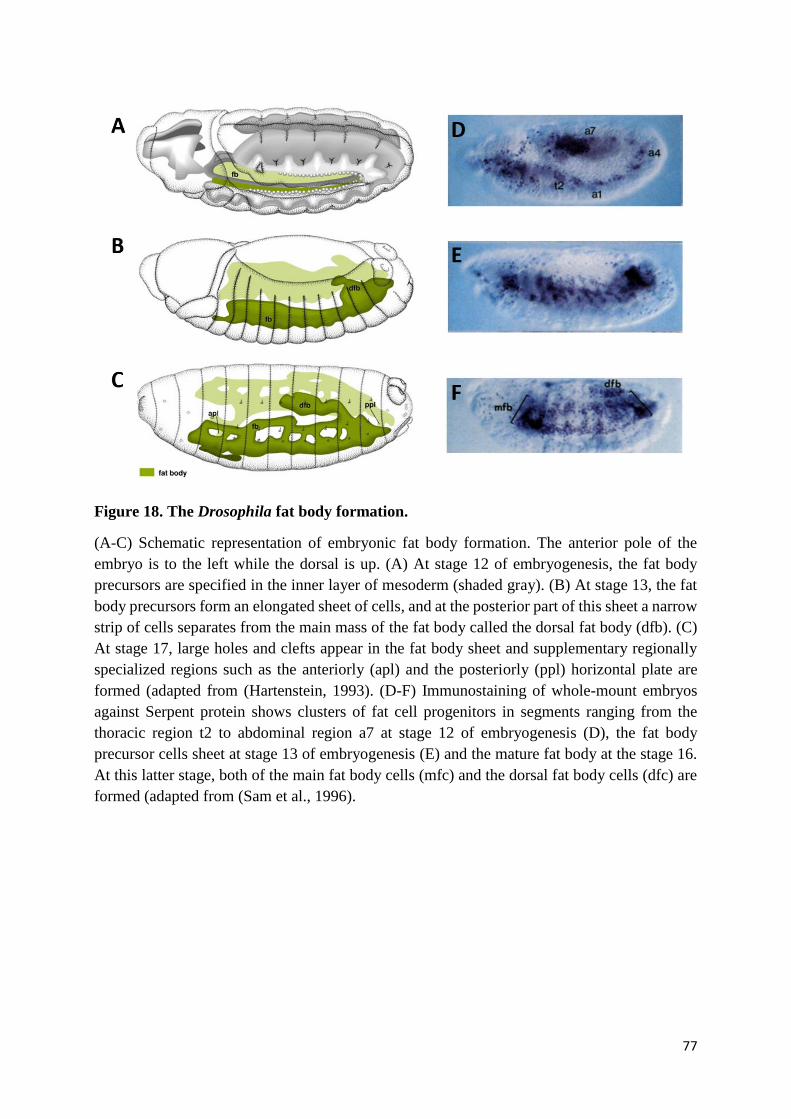
77
Figure 18. The Drosophila fat body formation.
(A-C) Schematic representation of embryonic fat body formation. The anterior pole of the
embryo is to the left while the dorsal is up. (A) At stage 12 of embryogenesis, the fat body
precursors are specified in the inner layer of mesoderm (shaded gray). (B) At stage 13, the fat
body precursors form an elongated sheet of cells, and at the posterior part of this sheet a narrow
strip of cells separates from the main mass of the fat body called the dorsal fat body (dfb). (C)
At stage 17, large holes and clefts appear in the fat body sheet and supplementary regionally
specialized regions such as the anteriorly (apl) and the posteriorly (ppl) horizontal plate are
formed (adapted from (Hartenstein, 1993). (D-F) Immunostaining of whole-mount embryos
against Serpent protein shows clusters of fat cell progenitors in segments ranging from the
thoracic region t2 to abdominal region a7 at stage 12 of embryogenesis (D), the fat body
precursor cells sheet at stage 13 of embryogenesis (E) and the mature fat body at the stage 16.
At this latter stage, both of the main fat body cells (mfc) and the dorsal fat body cells (dfc) are
formed (adapted from (Sam et al., 1996).

78
GATA4 to liver-specific regulatory regions occurs prior to commitment of the pluripotent
endodermal cells to hepatic cell fate, suggesting that GATA4 has a role in liver specification.
However, in GATA4 loss-of-function mice, the hepatic cells are still specified in the endoderm.
The same results are obtained in mice with GATA6 loss-of-function, suggesting that GATA4
and GATA6 are redundant during liver specification (Watt et al., 2007; Zhao et al., 2005).
Although mutant mice with loss-of-function of GATA4 or GATA6 do not alter liver
specification, expansion of the primary hepatic rudiment is altered in both mutants,
demonstrating the essential roles played by GATA4 and GATA6 factors during liver bud
growth and thus liver development (Watt et al., 2007; Zhao et al., 2005). In Drosophila, Srp is
expressed in fat body precursors since their specification and remains expressed in fat body
cells throughout the entire fat body development. Although some fat body precursors are
specified at early stages in srp loss of function mutant embryos, the number of fat body
precursors produced is reduced as compared to wild-type flies. In addition, these fat body
precursors are not able to proliferate and die. Premature apoptosis of fat body precursors
provokes a subsequent loss of differentiated fat body cells. These results show that GATA4,
GATA6 and Srp are required for the expansion and development of both the mammalian liver
and the fly fat body.
(D) Drosophila as a model system to study GATA functions
GATA proteins are versatile transcription factors. They act as either activators or repressors
of gene expression. They modulate chromatin conformation and the DNA packaging in order
to facilitate activation of gene transcription (M. Tremblay et al., 2018). In addition, they interact
with several other proteins in order to widely regulate their own functions (Jason A. Lowry &
79
Mackay, 2006). Importantly, during mammalian development, GATA factors are critical for
the formation and physiological functions of numerous tissues and organs, such as blood cells,
heart, lungs, intestine, liver, pancreas, kidneys, testis, ovaries, breast and nervous system (M.
H. Lentjes et al., 2016). Despite the variable modes of action and roles identified for the six
mammalian GATA factors, they all have in common the presence of two highly conserved zinc
finger domains that are crucial for GATA protein functions (Martin & Orkin, 1990; Trainor et
al., 1996; Trainor et al., 2000; Tsang et al., 1997; Yang & Evans, 1992). Accordingly, these two
domains have been strictly conserved across evolution. In the fruit fly Drosophila
melanogaster, five GATA factors are produced, three of them having the canonical two zinc
finger domains (Lin et al., 1995; Ramain et al., 1993; Waltzer et al., 2002). The amino acid
sequences of these domains are almost identical to those of the mammalian GATA zinc finger
domains. Among these amino acids, we mention the sole valine residue of the N-terminal zinc
finger domain that is responsible for the interaction of the mammalian GATA1 and GATA4
factors with FOG proteins. This residue is also present in the N-ZnF of Drosophila GATA Srp
and plays, like in mammals, an important role during the interaction of Srp with the Drosophila
FOG Ush (J. D. Crispino et al., 1999, 2001; Nancy Fossett et al., 2003).
In addition to this sequence conservation between mammalian and Drosophila GATA zinc
fingers, it has been shown that GATA proteins display an incredible level of similarity, as they
are implicated in the regulation of the same developmental processes, including blood cell
differentiation (Rehorn et al., 1996; Takahashi et al., 1997), hematopoietic precursor
proliferation and maintenance (Frelin et al., 2013; H. Gao et al., 2016), cardiomyocyte
differentiation (Klinedinst & Bodmer, 2003; Zhao et al., 2008), gut formation and regeneration
(Reuter, 1994; Walker et al., 2014), gut regionalization (Battle et al., 2008; Beuling et al., 2011;
Okumura et al., 2005, 2007), oocyte maintenance (Kyrönlahti et al., 2011; Lepesant et al., 2020)
80
and finally mammalian liver / Drosophila fat body development (Rehorn et al., 1996; Watt et
al., 2007; Zhao et al., 2005). They also share similarities at the mechanistic level (same
molecular interactors, antagonism mechanisms, transcriptional regulation and GATA switch
mode of action, as described above), and they even regulate the same target genes during similar
developmental processes, e.g., GATA4 and GATA6 proteins promote Delta expression during
gut formation (Walker et al., 2014), while Drosophila GATAe is implicated in the expression
of Delta in intestinal stem cells (Dutta et al., 2015; Okumura et al., 2016), and both Srp and
mammalian GATA6 repress the expression of Crumbs transmembrane proteins in order to
promote EMT (K. Campbell et al., 2011).
Throughout the years, Drosophila has emerged as a powerful genetic model organism.
Numerous studies have been designed in Drosophila in order to analyze the relation between
human genetic alterations and illnesses, including morphological defects, abnormal organ
functioning and cancer (Bier, 2005; Mirzoyan et al., 2019). Furthermore, basic developmental
regulatory pathways and processes are conserved between vertebrates and invertebrates, thus
establishing Drosophila as an ideal model in which to elucidate the mode of action and the role
of numerous factors implicated in various developmental processes (Gold & Brückner, 2015;
Pitsouli & Perrimon, 2008; Saffman & Lasko, 1999; Zaffran, 2003). Among them, we have
described above the mammalian and Drosophila hematopoietic development (Gold &
Brückner, 2015), cardiac developmental processes (Brade et al., 2013; Vogler & Bodmer,
2015), intestine morphogenesis, ovary formation, and finally mammalian liver and Drosophila
fat body development (Sam et al., 1996; Zaret, 2002; Y. Zhang & Xi, 2015).
81
Conservation of developmental processes between mammals and fly, as well as the structural
and functional conservation of GATA factors across evolution, led us to consider the fly as an
ideal organism model in which to study the roles of GATA zinc finger domains. Among the
Drosophila GATA factors, Srp provides a unique opportunity to study the functions of the
GATA zinc finger domains as, like I have already stated, since Srp proteins are produced as
two different isoforms containing either the two zinc finger domains (SrpNC) or only the C-
terminal zinc finger domain (SrpC) (Figure 19) (Waltzer et al., 2002). Interestingly, the
alternative splicing process at the origin of these two isoforms, occurs in all cells expressing
srp gene and at the same developmental time (Waltzer et al., 2002). The simultaneous presence
of the two Srp isoforms, makes Srp protein very interesting to study in order to understand the
role of GATA zinc finger domains in vivo.
In order to analyze the role of Srp zinc finger domains during the development of the fly, I took
advantage of the CRISPR/Cas9 technique to generate two mutant fly lines (Gratz et al., 2015).
The first fly line is characterized by the loss-of-function of SrpNC isoform but not of SrpC, thus
allowing us to investigate the role of the N-ZnF domain function during fly development. The
second fly line is unable to produce the SrpC isoform but still expresses the SrpNC isoform, a
situation similar to all vertebrate contexts. The study of this mutant fly line should help us in
understanding of the role played by this single zinc finger-containing SrpC isoform during
development. Finally, a mutant fly line with a substitution mimicking the V205G mutation in
the N-terminal zinc finger domain, has been engineered in order to identify the functions
depending on the interaction of SrpNC with its cofactor of the Friend of GATA family, Ush.
Srp proteins are produced in several tissues, where they play essential functions (hemocytes,
gut, fat body and the ovaries) (Lepesant et al., 2020; Rehorn et al., 1996; Reuter, 1994; Sam et
al., 1996). Although several studies have been carried out to determine the functions of Srp in
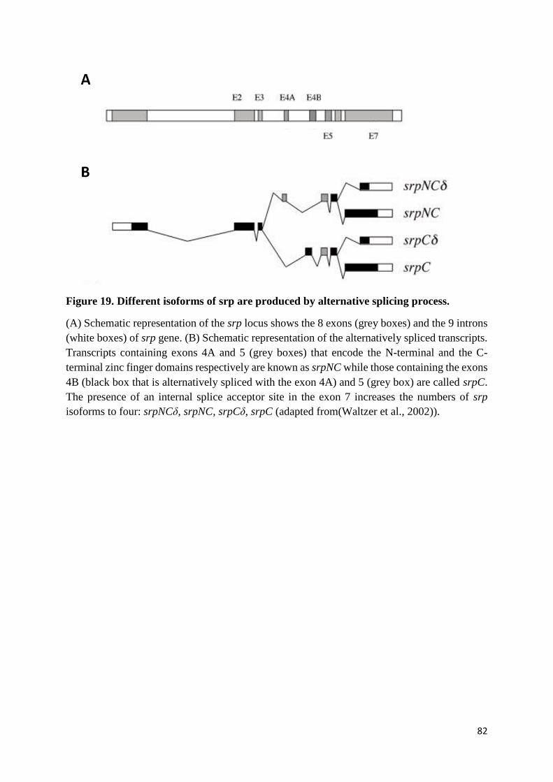
82
Figure 19. Different isoforms of srp are produced by alternative splicing process.
(A) Schematic representation of the srp locus shows the 8 exons (grey boxes) and the 9 introns
(white boxes) of srp gene. (B) Schematic representation of the alternatively spliced transcripts.
Transcripts containing exons 4A and 5 (grey boxes) that encode the N-terminal and the C-
terminal zinc finger domains respectively are known as srpNC while those containing the exons
4B (black box that is alternatively spliced with the exon 4A) and 5 (grey box) are called srpC.
The presence of an internal splice acceptor site in the exon 7 increases the numbers of srp
isoforms to four: srpNCδ, srpNC, srpCδ, srpC (adapted from(Waltzer et al., 2002)).

83
vivo, no study has ever assessed the roles of the zinc finger domains during the establishment
of these functions. Characterization of the different mutants I generated, reveals an alternative
splicing-mediated sub-functionalization of the srp gene. The results of my work are described
in the next chapter.

84
Chapter II. Results
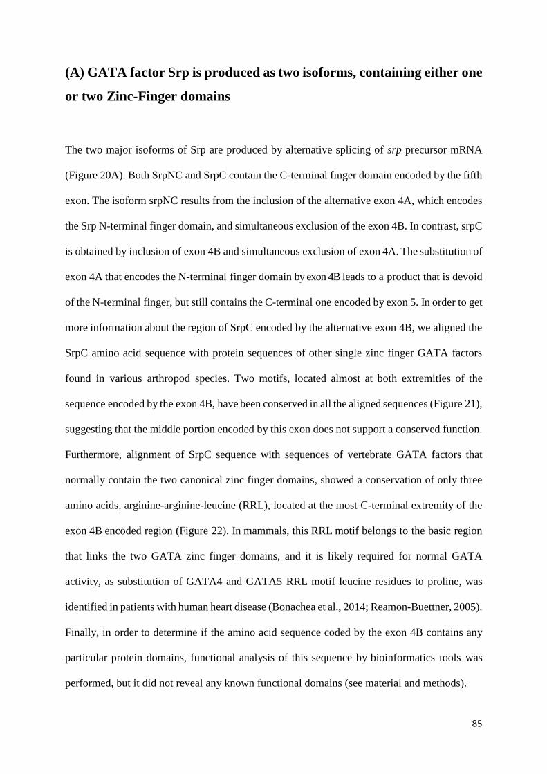
85
(A) GATA factor Srp is produced as two isoforms, containing either one
or two Zinc-Finger domains
The two major isoforms of Srp are produced by alternative splicing of srp precursor mRNA
(Figure 20A). Both SrpNC and SrpC contain the C-terminal finger domain encoded by the fifth
exon. The isoform srpNC results from the inclusion of the alternative exon 4A, which encodes
the Srp N-terminal finger domain, and simultaneous exclusion of the exon 4B. In contrast, srpC
is obtained by inclusion of exon 4B and simultaneous exclusion of exon 4A. The substitution of
exon 4A that encodes the N-terminal finger domain by exon 4B leads to a product that is devoid
of the N-terminal finger, but still contains the C-terminal one encoded by exon 5. In order to get
more information about the region of SrpC encoded by the alternative exon 4B, we aligned the
SrpC amino acid sequence with protein sequences of other single zinc finger GATA factors
found in various arthropod species. Two motifs, located almost at both extremities of the
sequence encoded by the exon 4B, have been conserved in all the aligned sequences (Figure 21),
suggesting that the middle portion encoded by this exon does not support a conserved function.
Furthermore, alignment of SrpC sequence with sequences of vertebrate GATA factors that
normally contain the two canonical zinc finger domains, showed a conservation of only three
amino acids, arginine-arginine-leucine (RRL), located at the most C-terminal extremity of the
exon 4B encoded region (Figure 22). In mammals, this RRL motif belongs to the basic region
that links the two GATA zinc finger domains, and it is likely required for normal GATA
activity, as substitution of GATA4 and GATA5 RRL motif leucine residues to proline, was
identified in patients with human heart disease (Bonachea et al., 2014; Reamon-Buettner, 2005).
Finally, in order to determine if the amino acid sequence coded by the exon 4B contains any
particular protein domains, functional analysis of this sequence by bioinformatics tools was
performed, but it did not reveal any known functional domains (see material and methods).
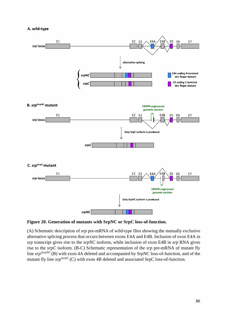
86
Figure 20. Generation of mutants with SrpNC or SrpC loss-of-function.
(A) Schematic description of srp pre-mRNA of wild-type flies showing the mutually exclusive
alternative splicing process that occurs between exons E4A and E4B. Inclusion of exon E4A in
srp transcript gives rise to the srpNC isoform, while inclusion of exon E4B in srp RNA gives
rise to the srpC isoform. (B-C) Schematic representation of the srp pre-mRNA of mutant fly
line srp∆srpNC (B) with exon 4A deleted and accompanied by SrpNC loss-of-function, and of the
mutant fly line srp∆srpC (C) with exon 4B deleted and associated SrpC loss-of-function.
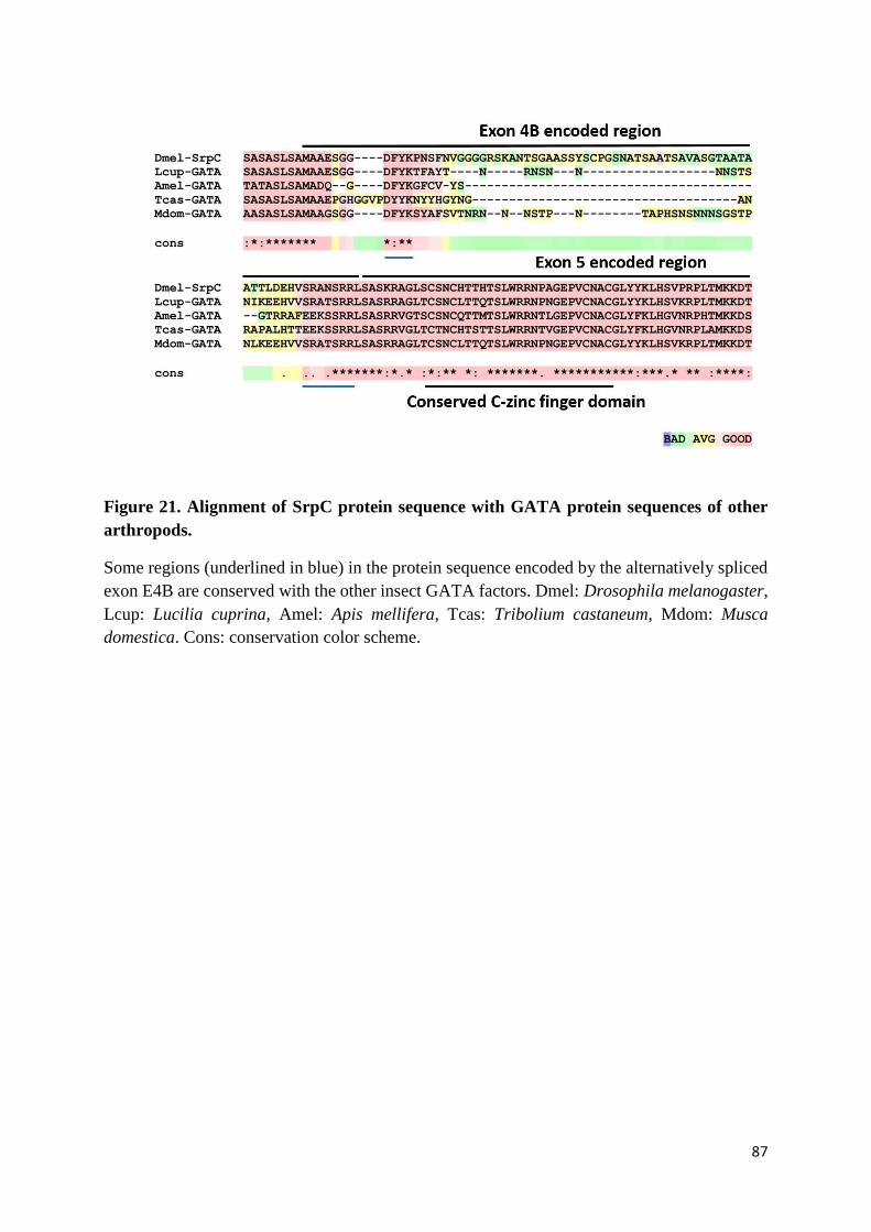
87
Figure 21. Alignment of SrpC protein sequence with GATA protein sequences of other
arthropods.
Some regions (underlined in blue) in the protein sequence encoded by the alternatively spliced
exon E4B are conserved with the other insect GATA factors. Dmel: Drosophila melanogaster,
Lcup: Lucilia cuprina, Amel: Apis mellifera, Tcas: Tribolium castaneum, Mdom: Musca
domestica. Cons: conservation color scheme.
88
Figure 22. Alignment of SrpC protein sequence with mammalian GATA protein
sequences.
RRL motif of the sequence encoded by the alternatively spliced exon E4B, juxtaposed to the C
zinc finger domain of SrpC, is conserved with mammalian GATA factors. Dmel: Drosophila
melanogaster, Hsap: Homo sapiens. Cons: conservation color scheme.
89
Altogether, these data suggest that substitution of exon 4A by exon 4B results in the production
of a single zinc finger GATA Srp isoform.
(B) Both isoforms of GATA factor Srp are produced at postembryonic
stages
As was already demonstrated at embryonic stage (Waltzer et al., 2002), we found that Srp
proteins are produced as two alternatively spliced variants during the Drosophila post-
embryonic stages. Total RNA of white-eye flies at third instar larval stage was extracted and
used as matrix for reverse-transcription polymerase chain reaction (RT-PCR) experiments, in
order to identify the nature of srp isoforms present in the extracts. To do so, forward and reverse
primers that recognize exons of srp gene located at either side of the srp mutually exclusive
exons 4A and 4B were used. Migration of RT-PCR products on agarose gel led to the detection
of two amplicons corresponding to both srp mRNA isoforms: srpNC and srpC (Figure 23A).
Similar results were obtained after analysis of transcripts extracted specifically from fat body
larvae (Figure 23B, lane 1), an organ already known to express high srp levels (Senger et al.,
2006). Furthermore, analyses of mRNAs produced in samples containing adult fly digestive
system and Malpighian tubules also showed the presence of the two alternatively spliced srp
isoforms (Figure 23B, lanes 2, 3). All these results indicate that at different Drosophila
developmental stages and in different tissues expressing srp, similar alternative splicing
mechanisms occur, giving rise to two variants of Srp proteins. This prompted us to analyze the
role of each of these two isoforms in the fly, by generating mutant fly lines carrying SrpNC or
SrpC loss-of-function.

90
Figure 23. Two isoforms of Serpent are produced in Drosophila.
(A) Agarose gel showing the RT-PCR results from total RNA of third instar larvae, indicating
the presence of two cDNA bands, corresponding to both isoforms SrpC and SrpNC. Note that
the size of the exon included in SrpC is larger than the alternatively spliced exon found in
SrpNC, which explains the slower migration of the SrpC corresponding band in comparison to
the SrpNC band. (B) Agarose gel showing the RT-PCR results from the RNA extracted from
adult proventriculus and midgut (left lane of gel), adult malpighian tubules and hindgut (middle
lane) and larval fat body (right lane of gel). Two bands corresponding to SrpNC and SrpC have
been identified in these tissues.
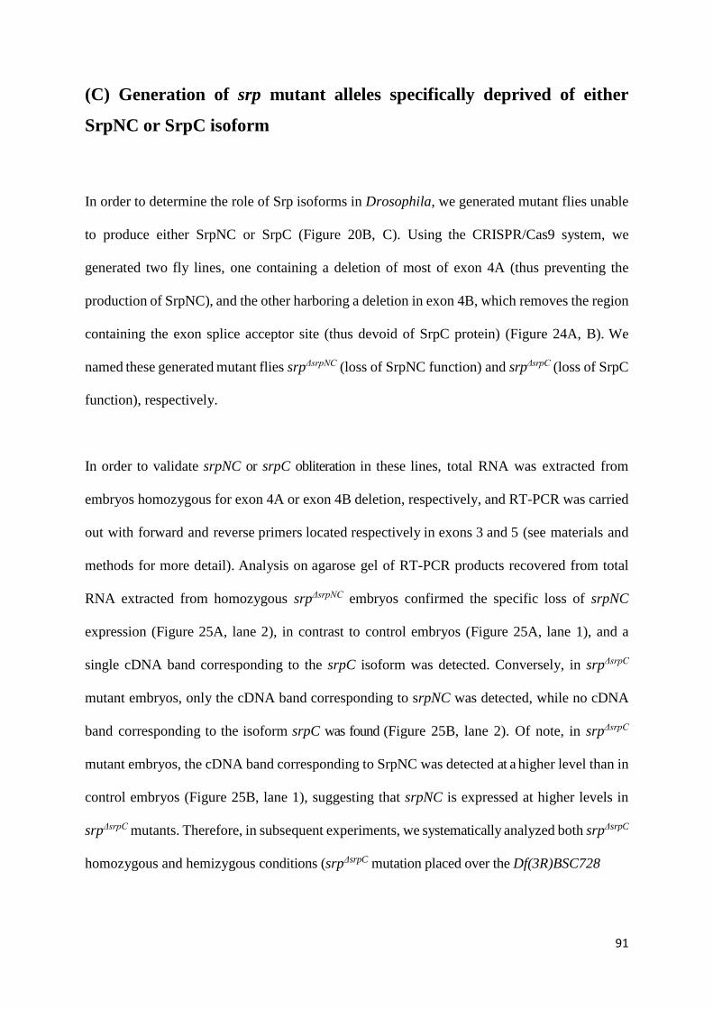
91
(C) Generation of srp mutant alleles specifically deprived of either
SrpNC or SrpC isoform
In order to determine the role of Srp isoforms in Drosophila, we generated mutant flies unable
to produce either SrpNC or SrpC (Figure 20B, C). Using the CRISPR/Cas9 system, we
generated two fly lines, one containing a deletion of most of exon 4A (thus preventing the
production of SrpNC), and the other harboring a deletion in exon 4B, which removes the region
containing the exon splice acceptor site (thus devoid of SrpC protein) (Figure 24A, B). We
named these generated mutant flies srpΔsrpNC (loss of SrpNC function) and srpΔsrpC (loss of SrpC
function), respectively.
In order to validate srpNC or srpC obliteration in these lines, total RNA was extracted from
embryos homozygous for exon 4A or exon 4B deletion, respectively, and RT-PCR was carried
out with forward and reverse primers located respectively in exons 3 and 5 (see materials and
methods for more detail). Analysis on agarose gel of RT-PCR products recovered from total
RNA extracted from homozygous srpΔsrpNC embryos confirmed the specific loss of srpNC
expression (Figure 25A, lane 2), in contrast to control embryos (Figure 25A, lane 1), and a
single cDNA band corresponding to the srpC isoform was detected. Conversely, in srpΔsrpC
mutant embryos, only the cDNA band corresponding to srpNC was detected, while no cDNA
band corresponding to the isoform srpC was found (Figure 25B, lane 2). Of note, in srpΔsrpC
mutant embryos, the cDNA band corresponding to SrpNC was detected at a higher level than in
control embryos (Figure 25B, lane 1), suggesting that srpNC is expressed at higher levels in
srpΔsrpC mutants. Therefore, in subsequent experiments, we systematically analyzed both srpΔsrpC
homozygous and hemizygous conditions (srpΔsrpC mutation placed over the Df(3R)BSC728
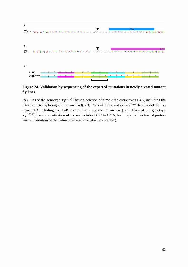
92
Figure 24. Validation by sequencing of the expected mutations in newly created mutant
fly lines.
(A) Flies of the genotype srpΔsrpNC have a deletion of almost the entire exon E4A, including the
E4A acceptor splicing site (arrowhead). (B) Flies of the genotype srpΔsrpC have a deletion in
exon E4B including the E4B acceptor splicing site (arrowhead). (C) Flies of the genotype
srpV735G, have a substitution of the nucleotides GTC to GGA, leading to production of protein
with substitution of the valine amino acid to glycine (bracket).
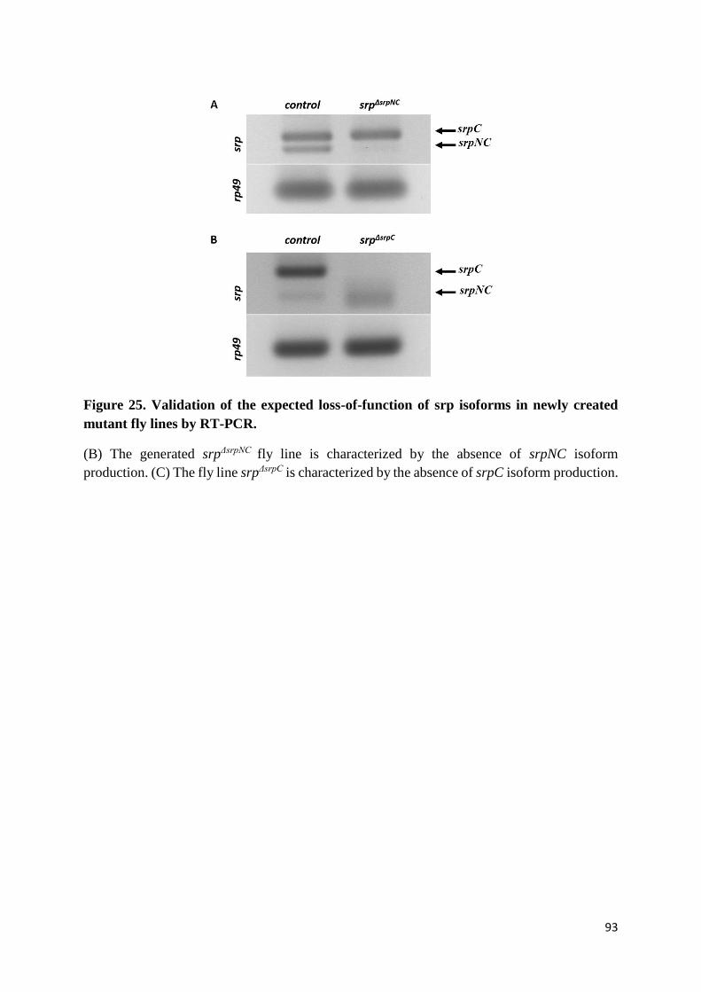
93
Figure 25. Validation of the expected loss-of-function of srp isoforms in newly created
mutant fly lines by RT-PCR.
(B) The generated srpΔsrpNC fly line is characterized by the absence of srpNC isoform
production. (C) The fly line srpΔsrpC is characterized by the absence of srpC isoform production.

94
deficiency that uncovers the srp locus), in order to detect any phenotypes that might be associated
to srpNC overexpression in these mutants. In addition, we also tested allelic combinations with
the amorphic allele srp3 and srp6G, or the hemocyte specific allele srpAS. The srp6G allele
corresponds to a mutation inducing a premature stop codon in the protein region encoded by
srp second exon, thus leading to the loss of almost the entire Srp protein, including the two zinc
finger domains. The srp3 allele carries a missense mutation in srp exon encoding the C-terminal
zinc finger domain, which most likely inhibits the interaction Srp/DNA. The srpAS allele
contains an insertion of a transposable element in a regulatory region that controls srp
expression specifically in embryonic hematopoietic cells, thus altering srp expression in these
cells and leading to a complete loss of blood cell development (Rehorn et al., 1996).
(D) Generation of a srp mutant allele that specifically abolishes
interaction of Srp with its FOG cofactor Ush
Important regulators of GATA factor functions are the FOG cofactors (Chlon & Crispino,
2012). The valine residue in GATA N-terminal zinc finger is required for interaction with FOG,
and substitution of this valine to glycine alters the association GATA/FOG (J. D. Crispino et
al., 1999). Interestingly, the Srp N-terminal zinc finger amino acid sequence is highly conserved
with those of the mammalian GATA factors (Figure 26), and the knock-in replacement of valine
to glycine alters the interaction of SrpNC with Ush (Nancy Fossett et al., 2003). In order to
study the role of SrpNC/Ush interaction in Drosophila, we generated a mutant fly line harboring
a substitution of this valine to glycine (V735G) (Figure 24C). The position of this valine in the
N-finger was referred to the srp transcript variant B, annotated by the National Center for
Biotechnology and Information (NCBI) Reference Sequence (Refseq) database (protein-id:
NP_732098.1). Flies of this line are named srpV735G.
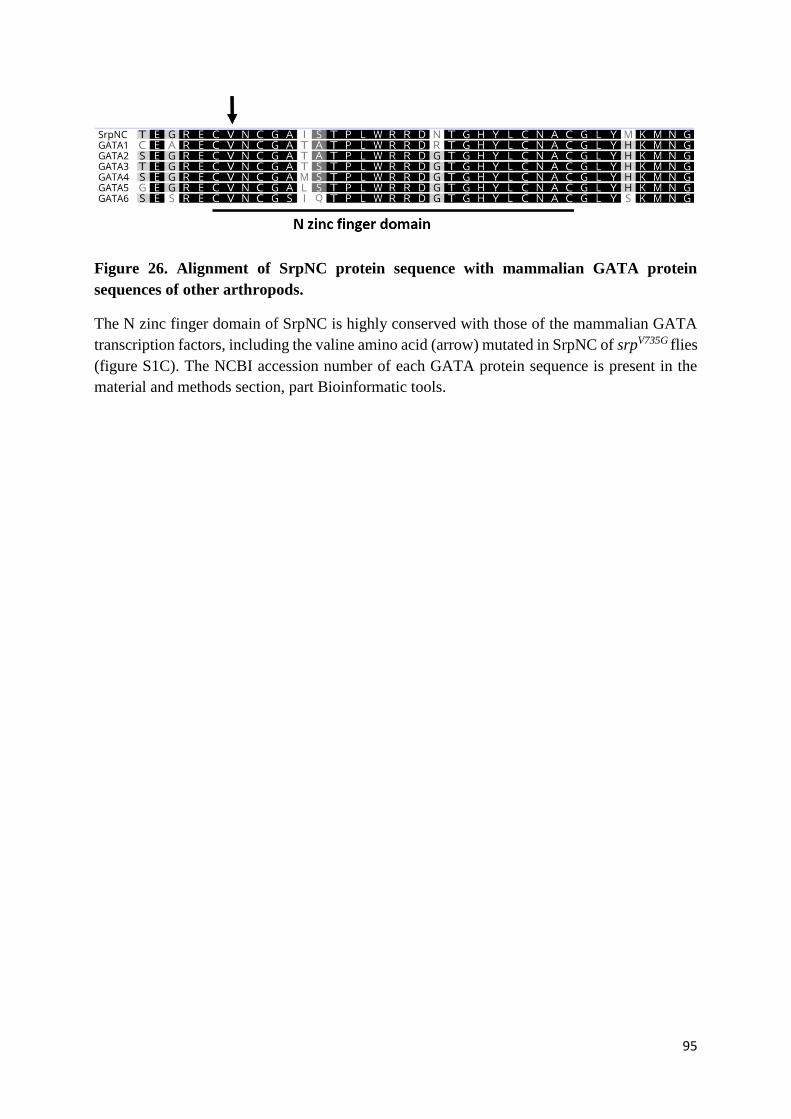
95
Figure 26. Alignment of SrpNC protein sequence with mammalian GATA protein
sequences of other arthropods.
The N zinc finger domain of SrpNC is highly conserved with those of the mammalian GATA
transcription factors, including the valine amino acid (arrow) mutated in SrpNC of srpV735G flies
(figure S1C). The NCBI accession number of each GATA protein sequence is present in the
material and methods section, part Bioinformatic tools.

96
(E) SrpC, but not SrpNC, is required for Drosophila viability
Loss of srp function affects the ability of the fly to reach adulthood (Rehorn et al., 1996). All
embryos homozygous for the amorphic allele srp6G die before hatching (Figure 27). In order to
determine whether this lethality is due to the absence of SrpC, of SrpNC, or to the simultaneous
loss of both isoforms, the viability of srpΔsrpC and srpΔsrpNC embryos was assessed.
About 60% of srpΔsrpC homozygous embryos were able to develop until the first instar larval
stage (Figure 27), then most of them died before reaching third instar larval stage (only 13.88%
of these larvae develop into third instar larvae). The surviving larvae were able to form pupae,
but only a few escapers emerged as adults and then died immediately. As a consequence, no
srpΔsrpC adult flies were observed, indicating that the SrpC isoform is required for fly viability.
In order to confirm that the lethality observed in srpΔsrpC mutant flies is due to the loss of SrpC
function, and not to the genetic background of srpΔsrpC flies, or to the overexpression of SrpNC
isoform in these mutants, we analyzed the lethality of flies with the srpΔsrpC mutation placed over
the Df(3R)BSC728 deficiency, which removes several genes including srp. Interestingly,
hemizygous embryos of (srpΔsrpC/Df(3R)BSC728) also failed to reach larval stage (Figure 28B),
indicating that the lethality of srpΔsrpC mutants is not a consequence of their genetic background.
Similarly, srpΔsrpC/srp6G trans-heterozygous individuals died before reaching third instar larval
stage (Figure 28A,B). All these results show that developmental defects found in mutant
srpΔsrpC are specifically due to the loss-of-function of Srp, and that SrpC is essential for fly
development. In addition, the fact that the lethality phenotype is more severe in embryos
hemizygous for srpΔsrpC than in homozygous embryos indicates that srpΔsrpC is an hypomorphic
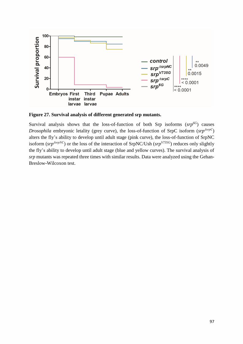
97
Figure 27. Survival analysis of different generated srp mutants.
Survival analysis shows that the loss-of-function of both Srp isoforms (srp6G) causes
Drosophila embryonic letality (grey curve), the loss-of-function of SrpC isoform (srpΔsrpC)
alters the fly’s ability to develop until adult stage (pink curve), the loss-of-function of SrpNC
isoform (srpΔsrpNC) or the loss of the interaction of SrpNC/Ush (srpV735G) reduces only slightly
the fly’s ability to develop until adult stage (blue and yellow curves). The survival analysis of
srp mutants was repeated three times with similar results. Data were analyzed using the Gehan-
Breslow-Wilcoxon test.
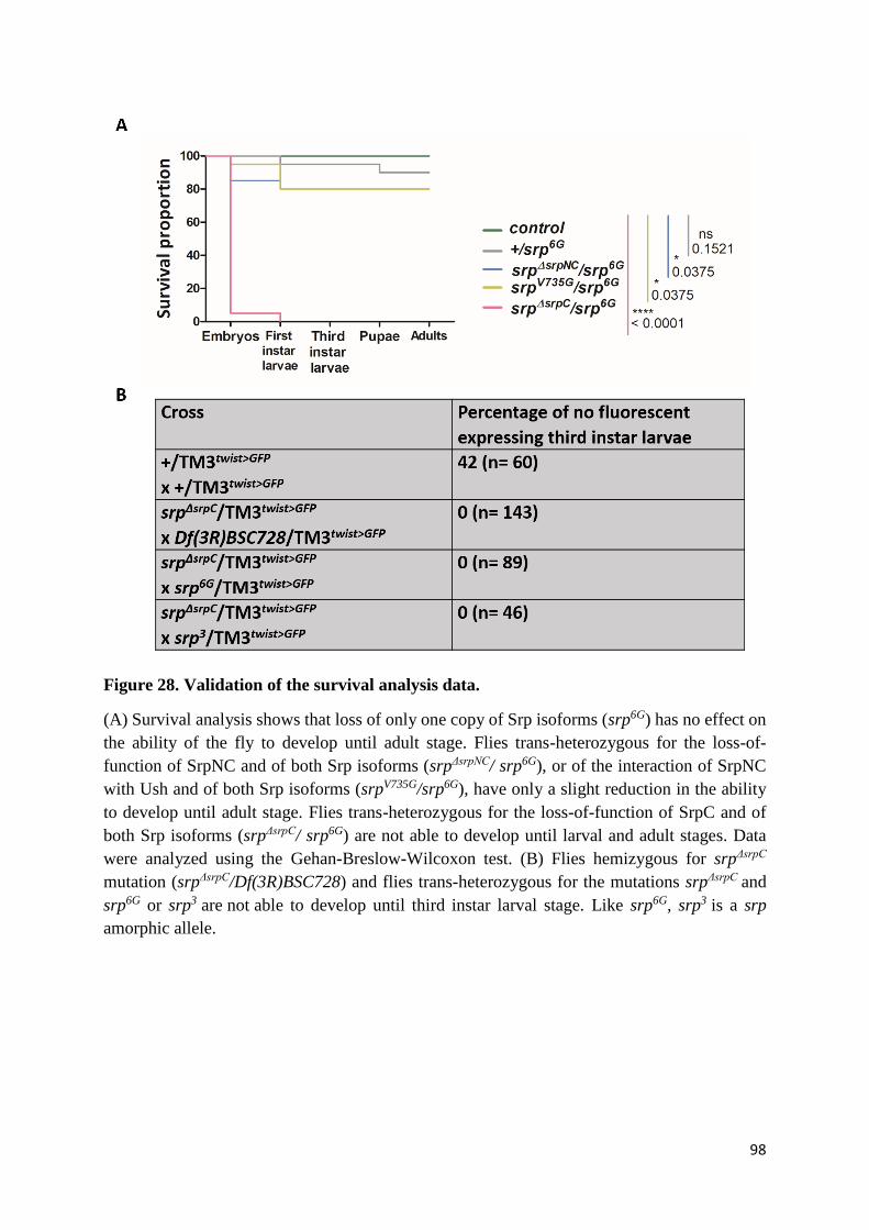
98
Figure 28. Validation of the survival analysis data.
(A) Survival analysis shows that loss of only one copy of Srp isoforms (srp6G) has no effect on
the ability of the fly to develop until adult stage. Flies trans-heterozygous for the loss-of-
function of SrpNC and of both Srp isoforms (srpΔsrpNC/ srp6G), or of the interaction of SrpNC
with Ush and of both Srp isoforms (srpV735G/srp6G), have only a slight reduction in the ability
to develop until adult stage. Flies trans-heterozygous for the loss-of-function of SrpC and of
both Srp isoforms (srpΔsrpC/ srp6G) are not able to develop until larval and adult stages. Data
were analyzed using the Gehan-Breslow-Wilcoxon test. (B) Flies hemizygous for srpΔsrpC
mutation (srpΔsrpC/Df(3R)BSC728) and flies trans-heterozygous for the mutations srpΔsrpC and
srp6G or srp3 are not able to develop until third instar larval stage. Like srp6G, srp3 is a srp
amorphic allele.
99
allele and that the presence of SrpNC in srpΔsrpC embryos slightly compensate SrpC loss-of-
function. Thus, SrpNC and SrpC have partially redundant functions during the fly development.
Furthermore, GATA zinc finger domains interact with DNA but also with other proteins. We
analyzed thus the viability of embryos having srpΔsrpC allele placed over the DNA binding
mutant allele srp3 in order to understand how SrpC acts. Interestingly, embryos of the genotype
srpΔsrpC/srp3 die at embryonic stage (Figure 28B). In embryos srpΔsrpC/srp3 , srp3 allele produces
mutant SrpC and SrpNC proteins that are unable to bind DNA while srpΔsrpC allele will generate
normal SrpNC protein that will bind and regulate the expression of its proper target genes and
of target genes that are common to both SrpC and SrpNC. As srpΔsrpC/srp3 embryos are not able
to pass embryogenesis, we conclude that there are some SrpC specific target genes that are not
recognized by SrpNC and that are required for Drosophila viability.
Contrarily to srpΔsrpC mutants, flies with a specific loss of SrpNC function were able to develop
until the adult stage (Figure 27). About 95% of srpΔsrpNC homozygous embryos developed into
first instar larvae. The majority of those developed and reached the third instar larval and pupal
stages, and about 94.34% of srpΔsrpNC pupae gave rise to adult flies. Likewise, almost 80% of
srpΔsrpNC/srp6G flies reached adulthood (Figure 28A), showing that SrpNC is mostly dispensable
for Drosophila viability (although the observed survival rate was slightly, yet significantly,
lower than that in control conditions).
Similar to the results obtained with SrpNC loss-of-function, srpV735G homozygous embryos
were able to develop until adult stage, as about 96.67% of these embryos were able to hatch,
and 77.58% of hatched larvae developed into adult flies (Figure 27). Accordingly, about 80%
of embryos of the genotype srpV735G/srp6G were able to develop until adult stage (Figure 28A).
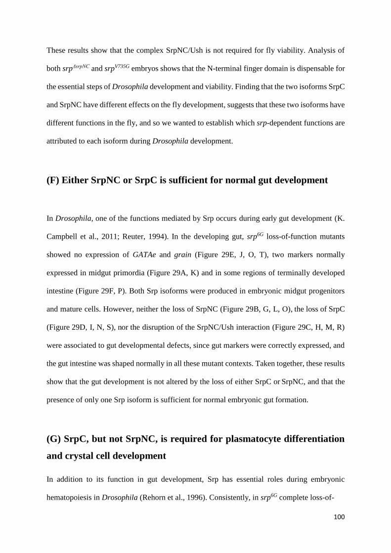
100
These results show that the complex SrpNC/Ush is not required for fly viability. Analysis of
both srpΔsrpNC and srpV735G embryos shows that the N-terminal finger domain is dispensable for
the essential steps of Drosophila development and viability. Finding that the two isoforms SrpC
and SrpNC have different effects on the fly development, suggests that these two isoforms have
different functions in the fly, and so we wanted to establish which srp-dependent functions are
attributed to each isoform during Drosophila development.
(F) Either SrpNC or SrpC is sufficient for normal gut development
In Drosophila, one of the functions mediated by Srp occurs during early gut development (K.
Campbell et al., 2011; Reuter, 1994). In the developing gut, srp6G loss-of-function mutants
showed no expression of GATAe and grain (Figure 29E, J, O, T), two markers normally
expressed in midgut primordia (Figure 29A, K) and in some regions of terminally developed
intestine (Figure 29F, P). Both Srp isoforms were produced in embryonic midgut progenitors
and mature cells. However, neither the loss of SrpNC (Figure 29B, G, L, O), the loss of SrpC
(Figure 29D, I, N, S), nor the disruption of the SrpNC/Ush interaction (Figure 29C, H, M, R)
were associated to gut developmental defects, since gut markers were correctly expressed, and
the gut intestine was shaped normally in all these mutant contexts. Taken together, these results
show that the gut development is not altered by the loss of either SrpC or SrpNC, and that the
presence of only one Srp isoform is sufficient for normal embryonic gut formation.
(G) SrpC, but not SrpNC, is required for plasmatocyte differentiation
and crystal cell development
In addition to its function in gut development, Srp has essential roles during embryonic
hematopoiesis in Drosophila (Rehorn et al., 1996). Consistently, in srp6G complete loss-of-
101
Figure 29. Both SrpNC and SrpC are sufficient for the Drosophila embryonic intestine
development.
The loss-of-function of Srp (srp6G) alters embryonic intestine development, as the embryonic
intestine markers, grn and GATAe, normally expressed in the anterior and posterior midgut
primordia (A and K) or in the completely developed intestine (F and P), are not expressed in
srp6G mutants (E, J, O, T). The loss-of-function of only SrpNC (B, G, L, Q) or only SrpC (D, I,
N, S) has no effect on intestine development or grn and GATAe expression. Also, loss of the
interaction SrpNC/Ush (C, H, M, R) has no effect on this process. grn: grain. Each experiment
was repeated three times with similar results.
102
function mutants, no plasmatocytes expressing the croquemort (crq) or viking (vkg) markers
are detected (Figure 30E, J), in contrast to wild-type embryos (Figure 30A, F). Interestingly,
plasmatocytes expressing crq and vkg are visible in both srpΔsrpC (Figure 30D, I) and srpΔsrpNC
(Figure 30B, G) specific loss-of-function mutants, as well as in srpV735G mutants (Figure 30C,
H). These results show that plasmatocytes are formed even when only one isoform of Serpent
is present. However, we found that the staining for the plasmatocyte marker Peroxidasin (Pxn)
is almost abolished in srpΔsrpC embryos (Figure 31D, I) contrary to wild-type embryos (Figure
31A, F), srpΔsrpNC embryos (Figure 31B, G) and srpV735G embryos (Figure 31C, H), indicating
that the loss-of-function of SrpC, but not that of SrpNC or disrupted SrpNC/Ush interaction,
impairs Pxn expression in plasmatocytes and that SrpNC is unable to compensate, even slightly,
SrpC loss-of-function during pxn expression.
In order to confirm the role of SrpC in pxn expression, we analyzed srpΔsrpC/srpAS trans-
heterozygous embryos. Interestingly, they display a reduction in Pxn expression (Figure 32B) in
comparison to plasmatocytes of wild-type embryos (Figure 32A). This phenotype is similar to
that obtained in srpΔsrpC homozygous embryos, indicating that the loss of Pxn expression in the
mutant srpΔsrpC is due to the loss-of-function of SrpC in the hemocytes. Taken together, these
results demonstrate that the SrpC isoform is required for Peroxidasin expression in the
embryonic plasmatocytes, while the SrpNC isoform is dispensable in this process.
In addition to plasmatocytes that constitute 95% of the total hemocytes, the crystal cells, which
are the equivalent of mammalian granulocytes, form the remaining 5% of the hemocytes. No
expression of the crystal cell fate determinant coding gene lozenge (lz) or of the crystal cell-
specific differentiation marker Prophenoloxidase 2 (PPO2) is detected in srp6G null mutants
(Figure 31O, T) as compared to wild type embryos (Figure 31K, P). Interestingly, we found that

103
Figure 30. Both SrpNC and SrpC are sufficient for Drosophila embryonic plasmatocyte
development.
Analysis of embryonic plasmatocyte marker expression, crq (A-E) and vkg (F-J), shows that
Srp loss-of-function alters embryonic plasmatocyte formation, compare (E, J) to (A, F), where
expression of crq and vkg in plasmatocytes normally migrating in head mesoderm during stage
11 of embryogenesis (A) and circulating throughout the embryo during stage 16 of
embryogenesis (B) is absent in srp6G mutants (E, J). The loss-of-function of only SrpNC (B, G)
or only SrpC (D, I) has no effect on plasmatocyte formation. Loss of SrpNC/Ush interaction
(C, H) has no effect on this process. crq: croquemort, vkg: viking. Each experiment was repeated
three times with similar results.
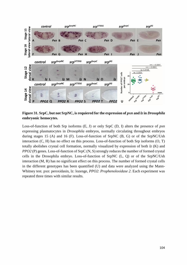
104
Figure 31. SrpC, but not SrpNC, is requiered for the expression of pxn and lz in Drosophila
embryonic hemocytes.
Loss-of-function of both Srp isoforms (E, J) or only SrpC (D, I) alters the presence of pxn
expressing plasmatocytes in Drosophila embryos, normally circulating throughout embryos
during stages 15 (A) and 16 (F). Loss-of-function of SrpNC (B, G) or of the SrpNC/Ush
interaction (C, H) has no effect on this process. Loss-of-function of both Srp isoforms (O, T)
totally abolishes crystal cell formation, normally visualized by expression of both lz (K) and
PPO2 (P) genes. Loss-of-function of SrpC (N, S) strongly reduces the number of formed crystal
cells in the Drosophila embryo. Loss-of-function of SrpNC (L, Q) or of the SrpNC/Ush
interaction (M, R) has no significant effect on this process. The number of formed crystal cells
in the different genotypes has been quantified (U) and data were analyzed using the Mann-
Whitney test. pxn: peroxidasin, lz: lozenge, PPO2: Prophenoloxidase 2. Each experiment was
repeated three times with similar results.
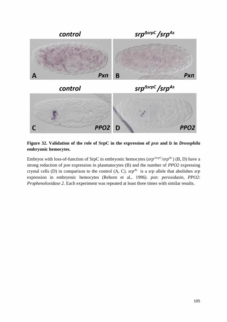
105
Figure 32. Validation of the role of SrpC in the expression of pxn and lz in Drosophila
embryonic hemocytes.
Embryos with loss-of-function of SrpC in embryonic hemocytes (srpΔsrpC/srpAs ) (B, D) have a
strong reduction of pxn expression in plasmatocytes (B) and the number of PPO2 expressing
crystal cells (D) in comparison to the control (A, C). srpAs is a srp allele that abolishes srp
expression in embryonic hemocytes (Rehorn et al., 1996). pxn: peroxidasin, PPO2:
Prophenoloxidase 2. Each experiment was repeated at least three times with similar results.
106
mutants with loss-of-function of SrpC display a significant reduction in the number of
progenitor crystal cells expressing lz (Figure 31N, S) and consequently of mature crystal cells
(Figure 31S) in comparison to wild-type embryos (Figure 31K, P), srpΔsrpNC embryos (Figure
31L, Q) and srpV735G embryos (Figure 31M, R). Quantification of crystal cells in each mutant is
represented (Figure 31U). The fact that the absence of both srp isoforms in srp6G embryos
(Figure 31O, T, U) has more severe effect on crystal cell development than the absence of only
SrpC isoform (Figure 31N, S, U) indicates that SrpNC can partially compensate SrpC loss-of-
function during crystal cell formation.
To corroborate that alteration of crystal cell formation in srpΔsrpC mutants is due to the loss-of-
function of SrpC and not to the mutant genetic background, we analyzed crystal cell formation
in srpΔsrpC/srpAS trans-heterozygous embryos. As expected, these embryos display a strong
reduction in crystal cells numbers (Figure 32D) in comparison to wild-type embryos (Figure
32C). This phenotype is very similar to what is seen in srpΔsrpC mutants, demonstrating that
alteration of crystal cells formation in these mutants is specifically associated to the loss of SrpC
function.
Altogether, these results show that SrpC, but neither SrpNC nor the SrpNC/Ush complex, is
required for crystal cell formation.
Interestingly, although these srpΔsrpC/srpAs embryos exhibit a notable reduction of pxn expression
in plasmatocytes and a strong reduction in the number of crystal cells formed, we found that
these embryos are able to develop until the adult stage, contrary to srpΔsrpC mutants that die
before reaching adult stage (Figure 33). These results indicate that the loss-of-function of SrpC
in embryonic hemocytes is not sufficient to cause fly lethality and that SrpC might have essential
functions in other cell types.
107
(H) SrpC, but not SrpNC, is required for fat body development
As already published, mutants with srp loss-of-function are unable to develop mature fat body
cells (Rehorn et al., 1996; Sam et al., 1996) that express the markers Alcohol dehydrogenase
(Adh) (Figure 34E, J), Glutactin (Glt) (Figure 34O, T) and Tiggrin (Tig) (Figure 34Y, D’)
contrary to wild-type embryos whose fat body cells are expressing those markers (Figure 34A,
F, K, P, U, Z). Interestingly, the srpΔsrpC mutation alters expression of all three fat body markers
(Figure 34D, I, N, S, X, C’), which might result from defective fat body cell maturation. In
contrast, srpΔsrpNC (Figure 34B, G, L, Q, V, A’) or srpV735G (Figure 34C, H, M, R, W, B’)
embryos have shown fat body formation, since all markers are expressed normally. These results
demonstrate that SrpC, but not SrpNC or the complex SrpNC/Ush, is required for fat body
formation.
In order to confirm that the fat body phenotypes observed in srpΔsrpC mutants are due to the loss
of SrpC, we analyzed Glt expression in srpΔsrpC/srp6G trans-heterozygous embryos.
Interestingly, we found that srpΔsrpC/srp6G embryos (Figure 35C, G) display a lower level of Glt
expression than srpΔsrpC mutants (Figure 35B, F), which is more like the phenotype observed in
srp6G mutants (Figure 35D, H). The lower Glt expression level found in srpΔsrpC/srp6G trans-
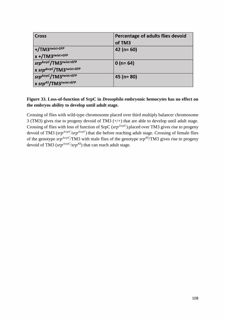
108
Figure 33. Loss-of-function of SrpC in Drosophila embryonic hemocytes has no effect on
the embryos ability to develop until adult stage.
Crossing of flies with wild-type chromosome placed over third multiply balancer chromosome
3 (TM3) gives rise to progeny devoid of TM3 (+/+) that are able to develop until adult stage.
Crossing of flies with loss of function of SrpC (srpΔsrpC) placed over TM3 gives rise to progeny
devoid of TM3 (srpΔsrpC/srpΔsrpC) that die before reaching adult stage. Crossing of female flies
of the genotype srpΔsrpC/TM3 with male flies of the genotype srpAS/TM3 gives rise to progeny
devoid of TM3 (srpΔsrpC/srpAS) that can reach adult stage.
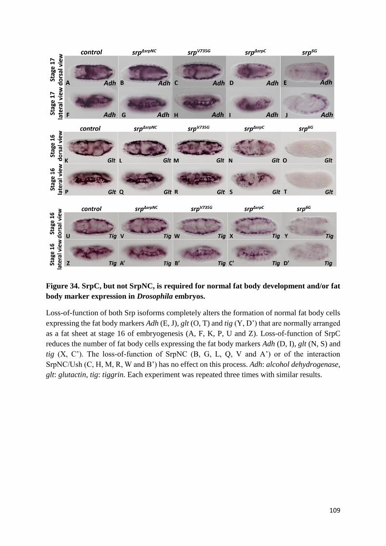
109
Figure 34. SrpC, but not SrpNC, is required for normal fat body development and/or fat
body marker expression in Drosophila embryos.
Loss-of-function of both Srp isoforms completely alters the formation of normal fat body cells
expressing the fat body markers Adh (E, J), glt (O, T) and tig (Y, D’) that are normally arranged
as a fat sheet at stage 16 of embryogenesis (A, F, K, P, U and Z). Loss-of-function of SrpC
reduces the number of fat body cells expressing the fat body markers Adh (D, I), glt (N, S) and
tig (X, C’). The loss-of-function of SrpNC (B, G, L, Q, V and A’) or of the interaction
SrpNC/Ush (C, H, M, R, W and B’) has no effect on this process. Adh: alcohol dehydrogenase,
glt: glutactin, tig: tiggrin. Each experiment was repeated three times with similar results.
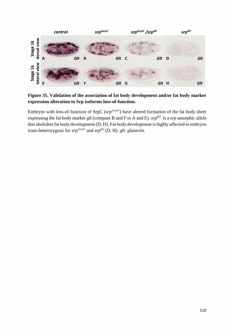
110
Figure 35. Validation of the association of fat body development and/or fat body marker
expression alteration to Srp isoforms loss-of-function.
Embryos with loss-of-function of SrpC (srpΔsrpC) have altered formation of the fat body sheet
expressing the fat body marker glt (compare B and F to A and E). srp6G is a srp amorphic allele
that abolishes fat body development (D, H). Fat body development is highly affected in embryos
trans-heterozygous for srpΔsrpC and srp6G (D, H). glt: glutactin.
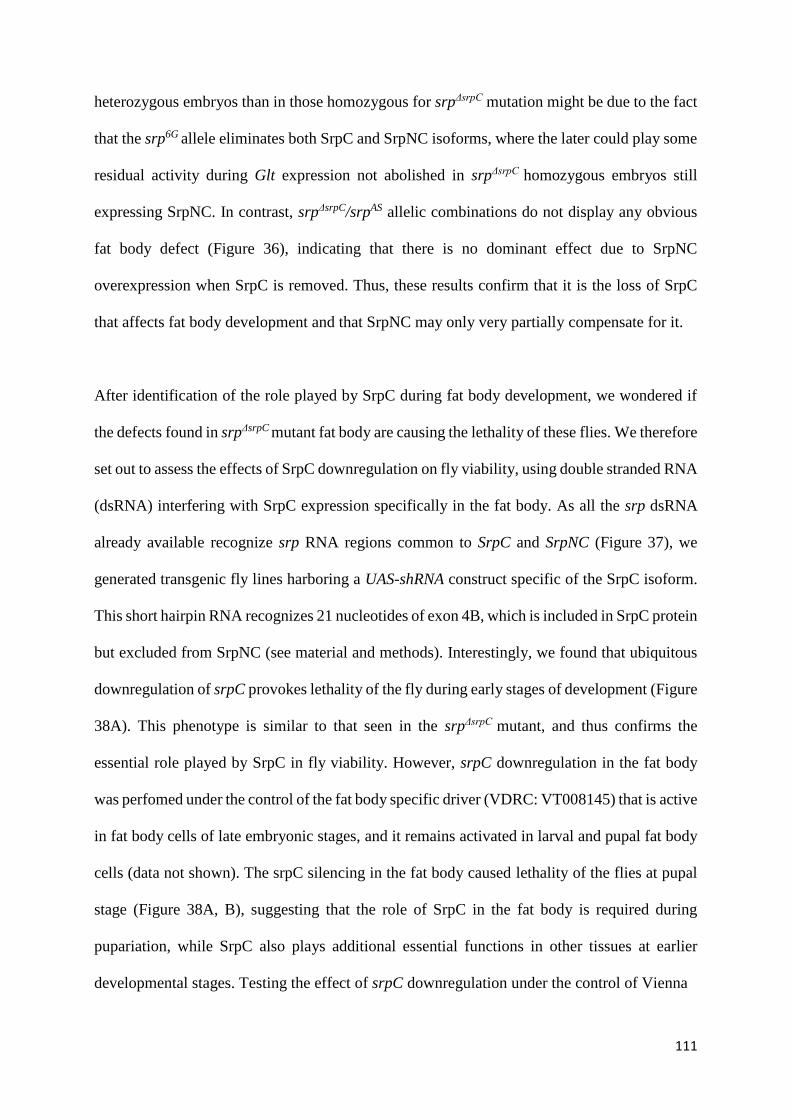
111
heterozygous embryos than in those homozygous for srpΔsrpC mutation might be due to the fact
that the srp6G allele eliminates both SrpC and SrpNC isoforms, where the later could play some
residual activity during Glt expression not abolished in srpΔsrpC homozygous embryos still
expressing SrpNC. In contrast, srpΔsrpC/srpAS allelic combinations do not display any obvious
fat body defect (Figure 36), indicating that there is no dominant effect due to SrpNC
overexpression when SrpC is removed. Thus, these results confirm that it is the loss of SrpC
that affects fat body development and that SrpNC may only very partially compensate for it.
After identification of the role played by SrpC during fat body development, we wondered if
the defects found in srpΔsrpC mutant fat body are causing the lethality of these flies. We therefore
set out to assess the effects of SrpC downregulation on fly viability, using double stranded RNA
(dsRNA) interfering with SrpC expression specifically in the fat body. As all the srp dsRNA
already available recognize srp RNA regions common to SrpC and SrpNC (Figure 37), we
generated transgenic fly lines harboring a UAS-shRNA construct specific of the SrpC isoform.
This short hairpin RNA recognizes 21 nucleotides of exon 4B, which is included in SrpC protein
but excluded from SrpNC (see material and methods). Interestingly, we found that ubiquitous
downregulation of srpC provokes lethality of the fly during early stages of development (Figure
38A). This phenotype is similar to that seen in the srpΔsrpC mutant, and thus confirms the
essential role played by SrpC in fly viability. However, srpC downregulation in the fat body
was perfomed under the control of the fat body specific driver (VDRC: VT008145) that is active
in fat body cells of late embryonic stages, and it remains activated in larval and pupal fat body
cells (data not shown). The srpC silencing in the fat body caused lethality of the flies at pupal
stage (Figure 38A, B), suggesting that the role of SrpC in the fat body is required during
pupariation, while SrpC also plays additional essential functions in other tissues at earlier
developmental stages. Testing the effect of srpC downregulation under the control of Vienna
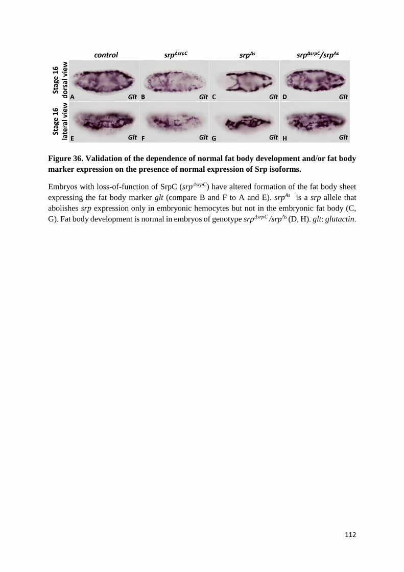
112
Figure 36. Validation of the dependence of normal fat body development and/or fat body
marker expression on the presence of normal expression of Srp isoforms.
Embryos with loss-of-function of SrpC (srpΔsrpC) have altered formation of the fat body sheet
expressing the fat body marker glt (compare B and F to A and E). srpAs is a srp allele that
abolishes srp expression only in embryonic hemocytes but not in the embryonic fat body (C,
G). Fat body development is normal in embryos of genotype srpΔsrpC /srpAs (D, H). glt: glutactin.
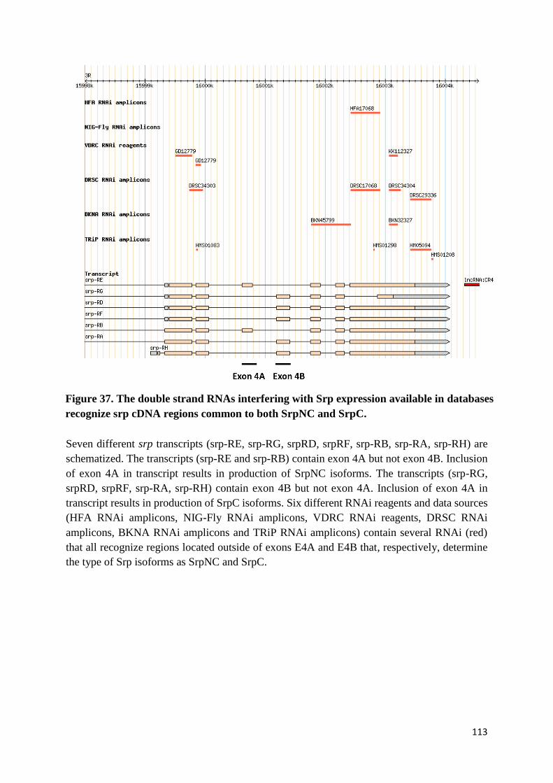
113
Seven different srp transcripts (srp-RE, srp-RG, srpRD, srpRF, srp-RB, srp-RA, srp-RH) are
schematized. The transcripts (srp-RE and srp-RB) contain exon 4A but not exon 4B. Inclusion
of exon 4A in transcript results in production of SrpNC isoforms. The transcripts (srp-RG,
srpRD, srpRF, srp-RA, srp-RH) contain exon 4B but not exon 4A. Inclusion of exon 4A in
transcript results in production of SrpC isoforms. Six different RNAi reagents and data sources
(HFA RNAi amplicons, NIG-Fly RNAi amplicons, VDRC RNAi reagents, DRSC RNAi
amplicons, BKNA RNAi amplicons and TRiP RNAi amplicons) contain several RNAi (red)
that all recognize regions located outside of exons E4A and E4B that, respectively, determine
the type of Srp isoforms as SrpNC and SrpC.
Figure 37. The double strand RNAs interfering with Srp expression available in databases
recognize srp cDNA regions common to both SrpNC and SrpC.

114
Figure 38. The down-regulation of SrpC in fat body alters the ability of the fly to pass
through pupal stage.
Quantification of the percentage of pupae (A) and of adults (B) raised after crossing of flies of
the genotypes dsSrpC/TM6 with ubiquitous driver Tub-Gal4 and fat body specific driver
VT008145-Gal4. The downregulation of SrpC by Tub-Gal4 (Tub-Gal4;;UAS-dsSrpC/+)
affects the ability of the fly to develop until both pupal and adult stages, while its
downregulation in the fat body (VT008145-Gal4/UAS-dsSrpC) alters its ability to pass through
pupal stage.
115
Tiles drivers corresponding to different transcriptional regulatory regions located upstream of
srp locus (and thus in tissues more likely expressing srp gene), on fly viability could help in
identifying of additional territories that are responsible for SrpC dependent fly development.
(I) SrpNC/Ush complex maintains larval lymph gland integrity
Although no role for the isoform SrpNC has been identified in embryonic tissues expressing
srp or regarding fly viability, we wondered if there was any role for SrpNC in srp expressing
tissues at post-embryonic stages. srp is expressed at different stages of larval development in
cells that constitute the lymph gland, which is the main site of post-embryonic hematopoiesis
(Jung, 2005). This prompted us to determine whether SrpNC or the SrpNC/Ush complex has
a role in this hematopoietic organ, and we quantified the integrity of the lymph glands in
various conditions, by defining phenotypic categories (lymph glands are either intact, Figure
39A, B, partially dispersed Figure 39C, or completely dispersed, Figure 39D). Interestingly,
while lymph glands of control larvae are mostly intact (Figure 39E), those of srpΔsrpNC larvae
are globally more dispersed, and the lymph gland bursting phenotype is even more
pronounced in srpV735G mutants with a disrupted SrpNC/Ush interaction, as almost 30% of the
analyzed lymph glands are completely ruptured (Figure 39E). Note that all the larvae analyzed
during this work are mid third instar larval stage females; finding intact lymph glands in
srpΔsrpNC and srpV735G mutants at this stage allows us to analyze the effect of the loss-of-
function of SrpNC and of the SrpNC/Ush interaction, respectively, on the formation of all of
the lymph gland hematopoietic cell types, including the posterior signaling center (PSC) cells,
progenitor cells, plasmatocytes and crystal cells.
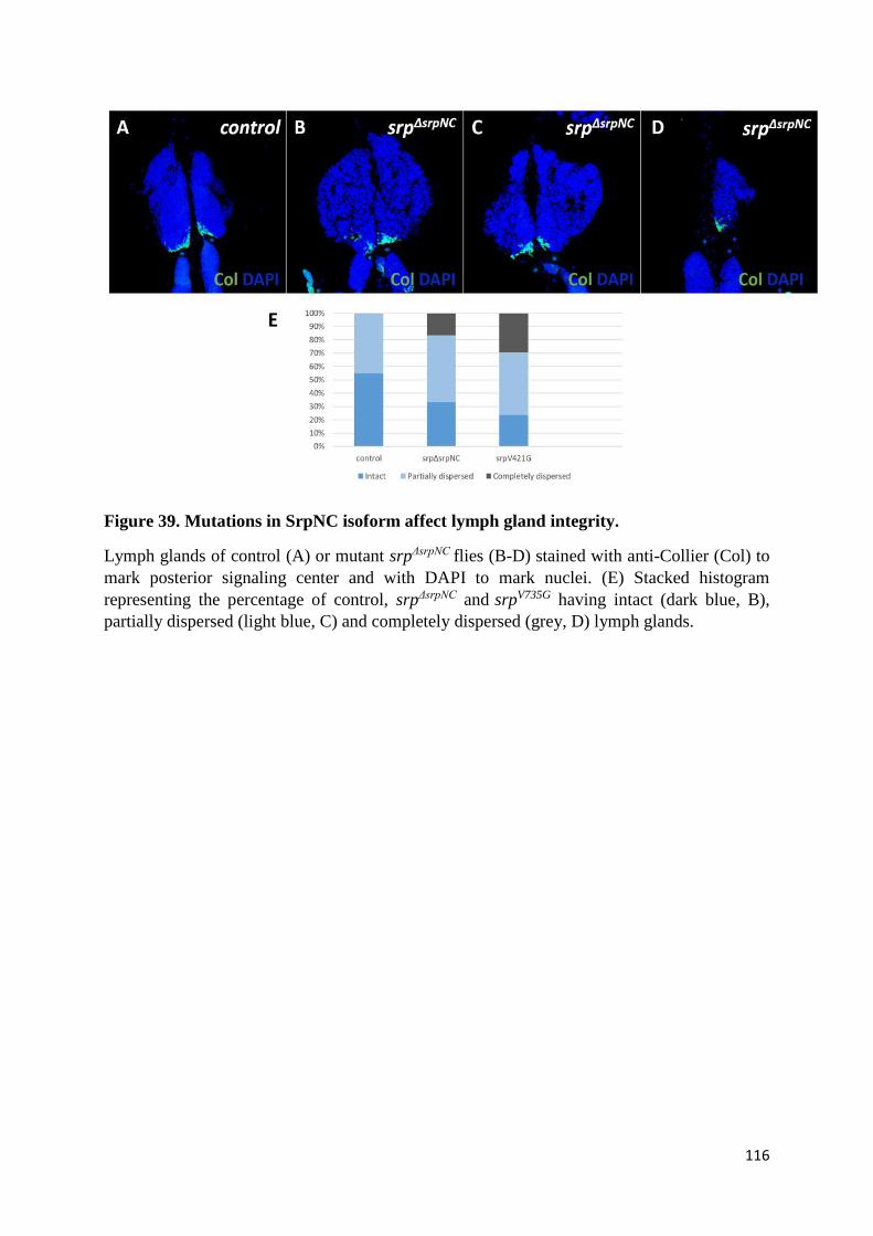
116
Figure 39. Mutations in SrpNC isoform affect lymph gland integrity.
Lymph glands of control (A) or mutant srpΔsrpNC flies (B-D) stained with anti-Collier (Col) to
mark posterior signaling center and with DAPI to mark nuclei. (E) Stacked histogram
representing the percentage of control, srpΔsrpNC and srpV735G having intact (dark blue, B),
partially dispersed (light blue, C) and completely dispersed (grey, D) lymph glands.
117
(J) SrpNC/Ush complex regulates lymph gland prohemocyte
proliferation and differentiation
One of the markers used during lymph gland constituent characterization is Collier (Col). Col
is expressed in lymph gland progenitor cells as well as in the PSC (Figure 41C) (Benmimoun
et al., 2015; Crozatier et al., 2004). In agreement with the dispensability of Srp during the PSC
formation already described (Crozatier et al., 2004), we found that in srpΔsrpNC (Figure 40B) as
well as in srpV735G mutants (Figure 40C), PSC cells expressing Col are specified and maintained
as in wild-type lymph glands (Figure 40A).
The progenitor cells are located in the lymph gland anterior lobe medullary zone, as well as in
the lymph gland posterior lobes (Jung et al., 2005). Staining of the medullar zone in the srpΔsrpNC
mutant with an -Col antibody shows a strong reduction of the medullary zone size (Figure
41D) in comparison to the control (Figure 41C). In addition, in lymph gland posterior lobes, we
observed some progenitor cell differentiation into lamellocytes, which specifically initiate
expression of the misshapen-mCherry transgene (msn-mCherry) (Figure 42H). However, in
addition to developing a lamellocyte fate, posterior lobe progenitor cells also differentiate into
crystal cells, as in about 83% of srpΔsrpNC larvae, the crystal cell marker Prophenoloxidase 1
(PPO1) is also detected (Figure 42I). Taken together, these results show that SrpNC is required
for the maintenance of hematopoietic progenitor cell identity and for inhibition of their
differentiation into mature hemocytes. Interestingly, similar results have been observed in the
posterior lobes of the mutant srpV735G (Figure 42M), indicating that the interaction SrpNC/Ush
is required to inhibit differentiation of lymph gland progenitor cells in the posterior lobes.

118
Figure 40. SrpNC is dispensable for lymph gland posterior signaling center formation.
Lymph glands of control (A), srpΔsrpNC (B) and srpV735G larvae (C) stained with anti-Collier
(Col) (green) to mark posterior signaling center and with DAPI (blue) to mark nuclei. Collier
is produced in the posterior signaling center in all contexts.
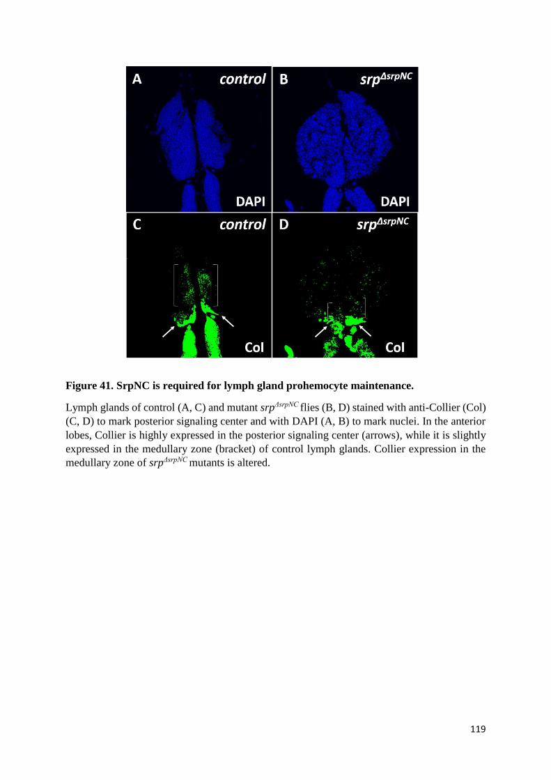
119
Figure 41. SrpNC is required for lymph gland prohemocyte maintenance.
Lymph glands of control (A, C) and mutant srpΔsrpNC flies (B, D) stained with anti-Collier (Col)
(C, D) to mark posterior signaling center and with DAPI (A, B) to mark nuclei. In the anterior
lobes, Collier is highly expressed in the posterior signaling center (arrows), while it is slightly
expressed in the medullary zone (bracket) of control lymph glands. Collier expression in the
medullary zone of srpΔsrpNC mutants is altered.
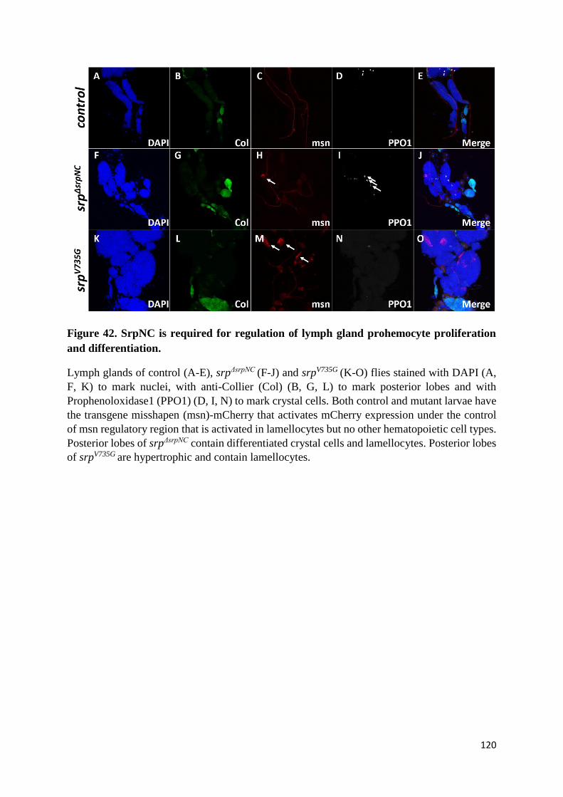
120
Figure 42. SrpNC is required for regulation of lymph gland prohemocyte proliferation
and differentiation.
Lymph glands of control (A-E), srpΔsrpNC (F-J) and srpV735G (K-O) flies stained with DAPI (A,
F, K) to mark nuclei, with anti-Collier (Col) (B, G, L) to mark posterior lobes and with
Prophenoloxidase1 (PPO1) (D, I, N) to mark crystal cells. Both control and mutant larvae have
the transgene misshapen (msn)-mCherry that activates mCherry expression under the control
of msn regulatory region that is activated in lamellocytes but no other hematopoietic cell types.
Posterior lobes of srpΔsrpNC contain differentiated crystal cells and lamellocytes. Posterior lobes
of srpV735G are hypertrophic and contain lamellocytes.
121
In addition to regulating posterior lobe hematopoietic progenitor cell differentiation, we found
that the SrpNC/Ush complex also regulates the size of the posterior lobes, as lobes of srpV735G
larvae are significantly hypertrophic in comparison to those of control larvae (Figure 42O, E).
This result indicates that the SrpNC/Ush interaction is either required for regulation of
progenitor cell proliferation or to control their survival rate. Analysis of the number of
progenitor cells expressing the cellular division marker PHH3, or the anti-apoptotic marker p53,
should give more information about this phenotype. Accordingly, a role for Ush in the control
of lymph gland progenitor cell proliferation and lymph gland size has already been reported (H.
Gao et al., 2009, 2016; R. P. Sorrentino et al., 2007). Note that the lymph gland hypertrophic
phenotype seen in the mutant srpΔsrpNC (Figure 42J) is not as pronounced as in the mutant
srpV735G (Figure 42O), suggesting that SrpNC and SrpNC/Ush complex ensure different
functions in lymph gland progenitor cell regulation.
(K) SrpNC/Ush interaction is required for lymph gland crystal cell
formation
According to what we found at embryonic stages, SrpNC is dispensable for larval lymph gland
plasmatocytes and crystal cell formation, as in srpΔsrpNC mutants both plasmatocytes expressing
the P1 marker (Figure 43B) and crystal cells expressing the PPO1 marker (Figure 43D) are
detected at mid-third instar larvae. However, in the lymph gland of the srpV735G mutant, both
the crystal cell progenitors expressing Lz (Figure 44A) and the mature crystal cells expressing
PPO1 (Figure 44B) are not detected, suggesting that although SrpNC is not essential for the
activation of the lymph gland crystal cell differentiation program, its presence free of the
interaction with Ush blocks this process. In contrary, formation of plasmatocytes is not affected
by loss of the interaction SrpNC/Ush (Figure 45B). These results show that the SrpNC/Ush
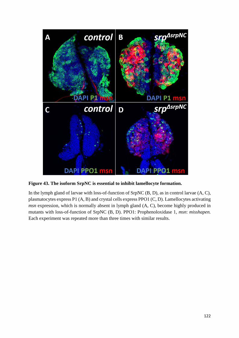
122
Figure 43. The isoform SrpNC is essential to inhibit lamellocyte formation.
In the lymph gland of larvae with loss-of-function of SrpNC (B, D), as in control larvae (A, C),
plasmatocytes express P1 (A, B) and crystal cells express PPO1 (C, D). Lamellocytes activating
msn expression, which is normally absent in lymph gland (A, C), become highly produced in
mutants with loss-of-function of SrpNC (B, D). PPO1: Prophenoloxidase 1, msn: misshapen.
Each experiment was repeated more than three times with similar results.
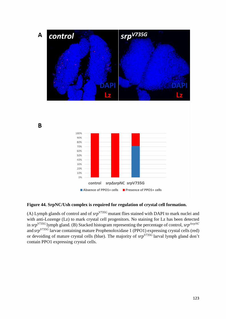
123
Figure 44. SrpNC/Ush complex is required for regulation of crystal cell formation.
(A) Lymph glands of control and of srpV735G mutant flies stained with DAPI to mark nuclei and
with anti-Lozenge (Lz) to mark crystal cell progenitors. No staining for Lz has been detected
in srpV735G lymph gland. (B) Stacked histogram representing the percentage of control, srpΔsrpNC
and srpV735G larvae containing mature Prophenoloxidase 1 (PPO1) expressing crystal cells (red)
or devoiding of mature crystal cells (blue). The majority of srpV735G larval lymph gland don’t
contain PPO1 expressing crystal cells.

124
Figure 45. The complex SrpNC/Ush is essential to inhibit lamellocyte formation.
In the lymph gland of larvae with loss of the SrpNC/Ush interaction (B, C), as in control larvae
(A), plasmatocytes expressing P1 (A, B) are formed. Lamellocytes activating msn expression
are not detected in the control lymph gland (A) but they are produced in mutants with loss of
SrpNC/Ush interaction. msn: misshapen. This experiment was repeated three times with similar
results.

125
complex is required for crystal cell but not plasmatocyte formation. The different effects
promoted by SrpNC alone and the SrpNC/Ush complex on crystal cell formation suggest an
antagonistic role played by Ush and SrpNC during this process.
(L) SrpNC/Ush is required for inhibition of lamellocyte formation
Although the loss of SrpNC function has no effect on plasmatocyte and crystal cell formation,
srpΔsrpNC mutant lymph glands reveal an inappropriate production of lamellocytes (as shown by
their expression of the msn-mCherry reporter gene) at mid-third instar larval stage (Figure 43B,
D), while these cells are normally absent in wild-type lymph glands (Figure 43A, C). Thus,
SrpNC is required for inhibition of lamellocyte formation. Consistent with premature
differentiation of lamellocytes and loss of integrity in the srpΔsrpNC mutant lymph glands,
lamellocytes were also detected in the hemolymph of srpΔsrpNC larvae by the end of the mid-third
instar larval stage (Figure 46B, G) and not in control larvae (Figure 46A, G). Interestingly,
similar results were obtained in the srpV735G mutant with loss of the interaction SrpNC/Ush, as
lamellocytes were detected both in lymph glands and in hemolymph of homozygous srpV735G
(Figure 45C and 46D, G) indicating that the interaction SrpNC/Ush is required for inhibition of
lamellocyte production. These results are consistent with the already published role for Ush in
the inhibition of lamellocyte formation (Avet-Rochex et al., 2010; R. P. Sorrentino et al., 2007)
(Figure 46F, G).
(M) SrpNC/Ush, but not SrpC, has a dosage sensitive effect on
lamellocyte formation
Following identification of a role for the SrpNC/Ush complex in the inhibition of lamellocyte
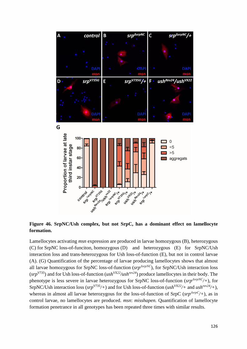
126
Figure 46. SrpNC/Ush complex, but not SrpC, has a dominant effect on lamellocyte
formation.
Lamellocytes activating msn expression are produced in larvae homozygous (B), heterozygous
(C) for SrpNC loss-of-funciton, homozygous (D) and heterozygous (E) for SrpNC/Ush
interaction loss and trans-heterozygous for Ush loss-of-function (E), but not in control larvae
(A). (G) Quantification of the percentage of larvae producing lamellocytes shows that almost
all larvae homozygous for SrpNC loss-of-function (srpΔsrpNC), for SrpNC/Ush interaction loss
(srpV735) and for Ush loss-of-function (ushVX22/ushrev24) produce lamellocytes in their body. The
phenotype is less severe in larvae heterozygous for SrpNC loss-of-function (srpΔsrpNC/+), for
SrpNC/Ush interaction loss (srpV735/+) and for Ush loss-of-function (ushVX22/+ and ushrev24/+),
whereas in almost all larvae heterozygous for the loss-of-function of SrpC (srpΔsrpC/+), as in
control larvae, no lamellocytes are produced. msn: misshapen. Quantification of lamellocyte
formation penetrance in all genotypes has been repeated three times with similar results.
127
formation, we analyzed the effect of the loss-of-function of only one copy of SrpNC or of Ush.
Lamellocytes are detected in the hemolymph of at least 70% of flies in the different
heterozygous background for the srpΔsrpNC, srpV735G, ushRev24, ushVX22 mutations (Figure 46G),
indicating that control of lamellocyte production depends on the dose of SrpNC and Ush. Of
note, more than 80% of srpΔsrpC/+ heterozygous larvae are completely devoid of lamellocytes
like wild-type larvae (Figure 46G), indicating that, contrary to the complex SrpNC/Ush, the
isoform SrpC doesn’t display a dosage sensitive effect on lamellocyte formation. In order to
more analyze the role of SrpC during lamellocyte formation, I analyzed the hemolymph of
larvae having downregulation of SrpC expression in all hemocytes using the Collagen-Gal4
(Cg-Gal4) driver. Only few of these larvae have lamellocytes activating misshapen (msn)
expression in their hemolymph indicating that SrpC might contribute but slightly in the
inhibition of lamellocyte differentiation (Figure 47).
(N) SrpNC/Ush complex is dispensable for ovogenesis
A recent study demonstrated that srp is expressed and plays essential functions in adult ovaries
also (Lepesant et al., 2020). Interestingly, we found that although srpΔsrpNC and srpV735G flies
develop apparently normally until adult stage, adult female mutants are sterile. Thus, the
SrpNC/Ush complex is required for female fertility. In order to identify the defects responsible
for the srpΔsrpNC and srpV735G female sterility, we checked for ovary formation in both mutant
fly lines, but no noticeable defect in general morphology of the ovaries was seen, as in both
wild-type and srpΔsrpNC female flies ovarioles are formed and the linear sequences of egg
chambers that constitute the ovariole are also formed (Figures 48B, C, 49B, C, 50B, C).
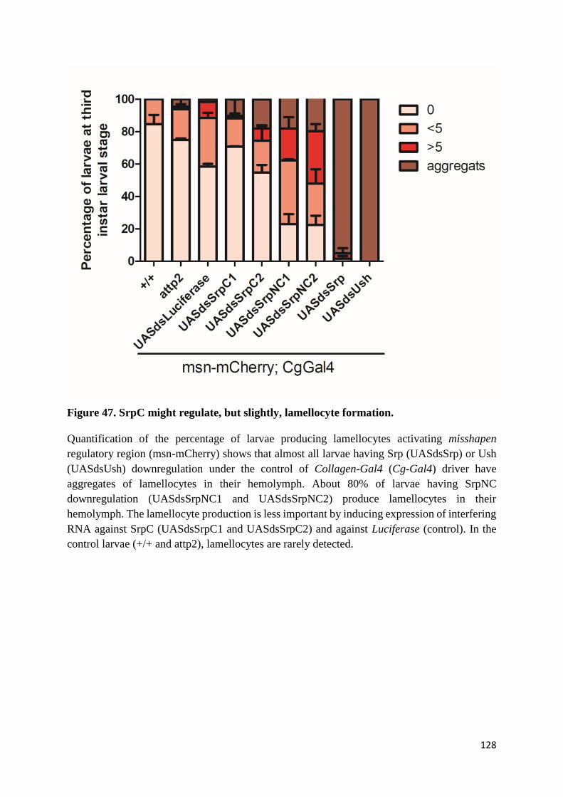
128
Figure 47. SrpC might regulate, but slightly, lamellocyte formation.
Quantification of the percentage of larvae producing lamellocytes activating misshapen
regulatory region (msn-mCherry) shows that almost all larvae having Srp (UASdsSrp) or Ush
(UASdsUsh) downregulation under the control of Collagen-Gal4 (Cg-Gal4) driver have
aggregates of lamellocytes in their hemolymph. About 80% of larvae having SrpNC
downregulation (UASdsSrpNC1 and UASdsSrpNC2) produce lamellocytes in their
hemolymph. The lamellocyte production is less important by inducing expression of interfering
RNA against SrpC (UASdsSrpC1 and UASdsSrpC2) and against Luciferase (control). In the
control larvae (+/+ and attp2), lamellocytes are rarely detected.
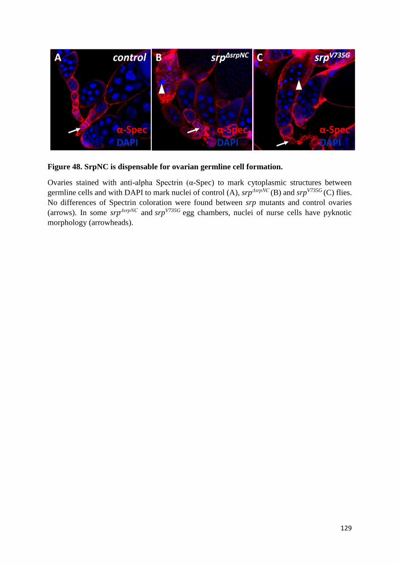
129
Figure 48. SrpNC is dispensable for ovarian germline cell formation.
Ovaries stained with anti-alpha Spectrin (α-Spec) to mark cytoplasmic structures between
germline cells and with DAPI to mark nuclei of control (A), srpΔsrpNC (B) and srpV735G (C) flies.
No differences of Spectrin coloration were found between srp mutants and control ovaries
(arrows). In some srpΔsrpNC and srpV735G egg chambers, nuclei of nurse cells have pyknotic
morphology (arrowheads).
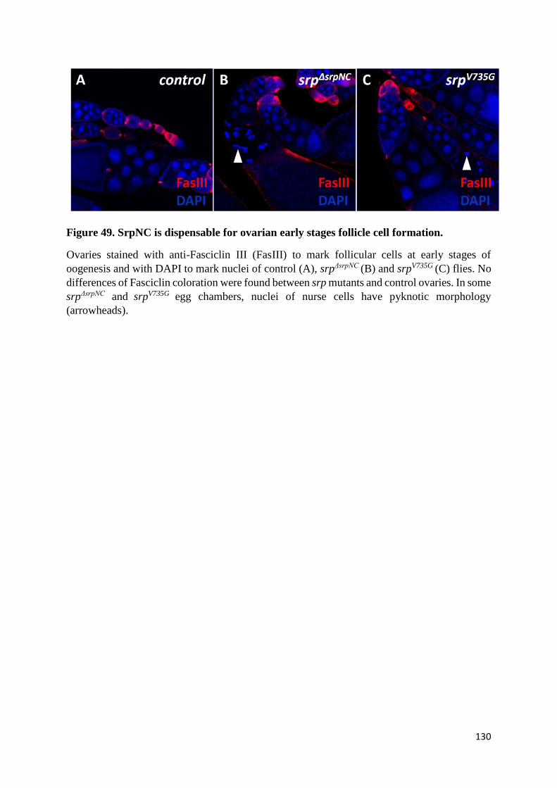
130
Figure 49. SrpNC is dispensable for ovarian early stages follicle cell formation.
Ovaries stained with anti-Fasciclin III (FasIII) to mark follicular cells at early stages of
oogenesis and with DAPI to mark nuclei of control (A), srpΔsrpNC (B) and srpV735G (C) flies. No
differences of Fasciclin coloration were found between srp mutants and control ovaries. In some
srpΔsrpNC and srpV735G egg chambers, nuclei of nurse cells have pyknotic morphology
(arrowheads).

131
Figure 50. SrpNC is dispensable for ovarian oocyte formation.
Ovaries stained with anti-oo-RNA binding protein (Orb) to mark oocytes and with Phalloidin
(Actin) to mark filamentous actin, and with DAPI to mark nuclei of control (A), srpΔsrpNC (B)
and srpV735G (C) flies. No differences of actin or of Orb coloration were found between srp
mutants and control ovaries.
132
We next analyzed the formation of different ovarian components in the ovaries of these mutants.
The unit in the ovaries is the egg chamber. Each ovarian egg chamber contains 15 nurse cells
and one oocyte, surrounded by a monolayer of epithelial follicle cells (E. Fedorova et al., 2019).
Developing germline cells that are at the origin of nurse cells and oocyte are spanned by large
cytoplasmic structures that can be visualized by different membrane skeletal proteins including
α-spectrin (de Cuevas et al., 1996) (Figure 48A). The follicle cells express cell adhesion
molecule fasciclin III during early stages of oogenesis, but later Fasciclin III expression
becomes restricted to polar follicle cells (St. Johnston, 2001) (Figure 49A). Finally, the
developing oocyte is characterized by the expression of the RNA binding protein Orb (Lantz et
al., 1994) (Figure 50A). Staining for α-spectrin, Fasciclin III and Orb proteins in srpΔsrpNC
mutants (Figure 48B, 49B, 50B) and in srpV735G ovaries (Figure 48C, 49C, 50C) showed no
significant differences of expression in comparison to control ovaries (Figure 48A, 49A, 50A).
These results indicate that the germline cells, the follicle cells and the oocyte are formed
normally in the absence of SrpNC isoform and of SrpNC/Ush interaction in the early egg
chambers, and this suggest that the sterility of srpΔsrpNC and srpV735G mutants is more likely due
to defects during the later stages of oogenesis.
(O) SrpNC/Ush complex, but not SrpC, is required for normal oogenesis
In some cases, in srpΔsrpNC (Figures 48B, 49B) and srpV735G (Figures 48C, 49C) mutants, we
observed pycnotic morphology of the DAPI-stained nuclei of the nurse cells. This phenotype
of degeneration of the mid-stage egg chambers in some Drosophila egg chambers has been
already detected in ovaries of flies with downregulation of both Srp isoforms, and analyzed as
the consequence of alteration of the ovarian mid-oogenesis checkpoint function, which detects
if an egg chamber has to die or to survive and produce an egg (Lepesant et al., 2020). However,

133
in srpΔsrpNC mutants, this phenotype was not very pronounced, indicating that fertility problems
in these mutants are not solely due to alteration of the mid-oogenesis checkpoint and that other
defects could be responsible.
Recently, it was shown that srp is required for Drosophila oogenesis and/or egg laying
(Lepesant et al., 2020). Similarly, we found that, while wild-type flies lay eggs abundantly,
srpΔsrpNC or srpV735G female flies lay only very few eggs (Figure 51A). This indicates that a
SrpNC/Ush interaction is required for normal oocyte release.
In addition, embryos laid by srpΔsrpNC (Figure 51C, F) and srpV735G (Figure 51D, G) flies show
clear physical defects, caused by an absence of the Drosophila eggshell, of the egg respiratory
appendages (the dorsal appendages), and of the egg sperm entry point (the micropyle) that are
normally formed in eggs laid by wild-type females (Figure 51B, E). This indicates that the
SrpNC isoform is required to interact with Ush, for normal Drosophila egg maturation. In
agreement with the role of Ush in oogenesis proposed here, analysis of the Fly atlas expression
data shows that Ush is highly expressed in the Drosophila ovary (Table 1), which supports the
idea of a function for Ush during oogenesis. In order to validate the role of Ush during egg
maturation, we induced the expression of Ush interfering RNA (VDRC: 5712) in ovarian
follicle cells of normal unmutated flies using the insertion traffic-jam-Gal4 (Tj-Gal4) (Lepesant
et al., 2020) that is activated in the ovarian follicle cells, and we found that contrarily to embryos
laid by control flies (Figure 52A), embryos laid by flies having Ush downregulation (Figure
52B) have morphological defects identical to those of embryos laid by srpΔsrpNC and srpV735G
mutants including an absence of dorsal appendages, micropyle and eggshell.
In order to determine the role of the SrpC isoform in fly oogenesis, we analyzed the effect on
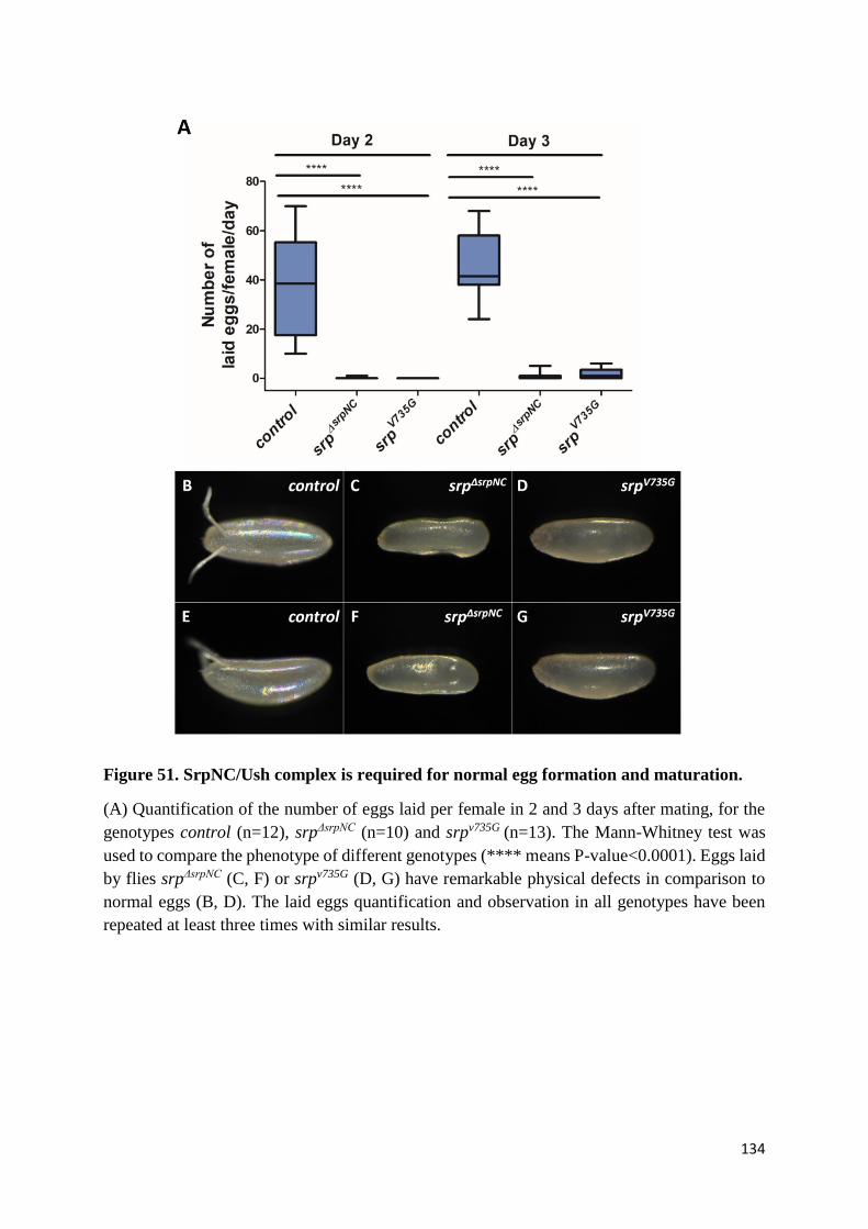
134
Figure 51. SrpNC/Ush complex is required for normal egg formation and maturation.
(A) Quantification of the number of eggs laid per female in 2 and 3 days after mating, for the
genotypes control (n=12), srpΔsrpNC (n=10) and srpv735G (n=13). The Mann-Whitney test was
used to compare the phenotype of different genotypes (**** means P-value<0.0001). Eggs laid
by flies srpΔsrpNC (C, F) or srpv735G (D, G) have remarkable physical defects in comparison to
normal eggs (B, D). The laid eggs quantification and observation in all genotypes have been
repeated at least three times with similar results.

135
Tissue
mRNA Signal Present Call Enrichment Affy Call
Brain 46 ± 1 4 of 4 0.90 None
Head 41 ± 2 4 of 4 0.80 None
Eye 23 ± 4 3 of 4 0.47 Down
Thoracicoabdominal ganglion 60 ± 4 4 of 4 1.20 None
Salivary gland 41 ± 4 4 of 4 0.84 None
Crop 15 ± 2 4 of 4 0.30 Down
Midgut 6 ± 2 0 of 4 0.10 Down
Tubule 17 ± 1 3 of 4 0.40 Down
Hindgut 20 ± 1 4 of 4 0.40 Down
Heart 158 ± 13 4 of 4 3.23 Up
Fat body 20 ± 6 4 of 4 0.42 Down
Ovary 100 ± 3 4 of 4 2.00 Up
Testis 21 ± 2 4 of 4 0.40 Down
Male accessory glands 115 ± 3 4 of 4 2.40 Up
Virgin spermatheca 78 ± 2 4 of 4 1.61 Up
Mated spermatheca 96 ± 9 4 of 4 1.98 Up
Adult carcass 32 ± 3 4 of 4 0.70 Down
Larval CNS 105 ± 7 4 of 4 2.16 Up
Larval Salivary gland 13 ± 1 0 of 4 0.28 Down
Larval midgut 78 ± 4 4 of 4 1.60 Up
Larval tubule 20 ± 2 4 of 4 0.40 Down
Larval hindgut 27 ± 0 4 of 4 0.56 Down
Larval fat body 82 ± 9 4 of 4 1.70 Up
Larval trachea 54 ± 6 4 of 4 1.12 None
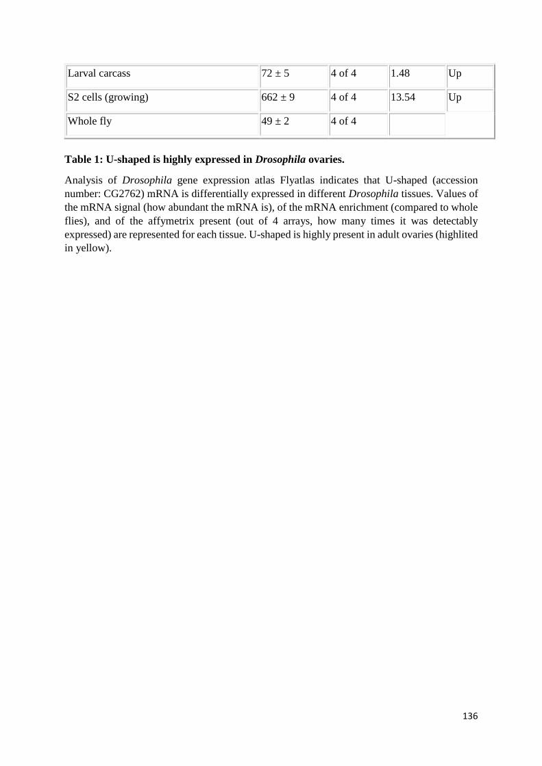
136
Larval carcass 72 ± 5 4 of 4 1.48 Up
S2 cells (growing) 662 ± 9 4 of 4 13.54 Up
Whole fly 49 ± 2 4 of 4
Table 1: U-shaped is highly expressed in Drosophila ovaries.
Analysis of Drosophila gene expression atlas Flyatlas indicates that U-shaped (accession
number: CG2762) mRNA is differentially expressed in different Drosophila tissues. Values of
the mRNA signal (how abundant the mRNA is), of the mRNA enrichment (compared to whole
flies), and of the affymetrix present (out of 4 arrays, how many times it was detectably
expressed) are represented for each tissue. U-shaped is highly present in adult ovaries (highlited
in yellow).
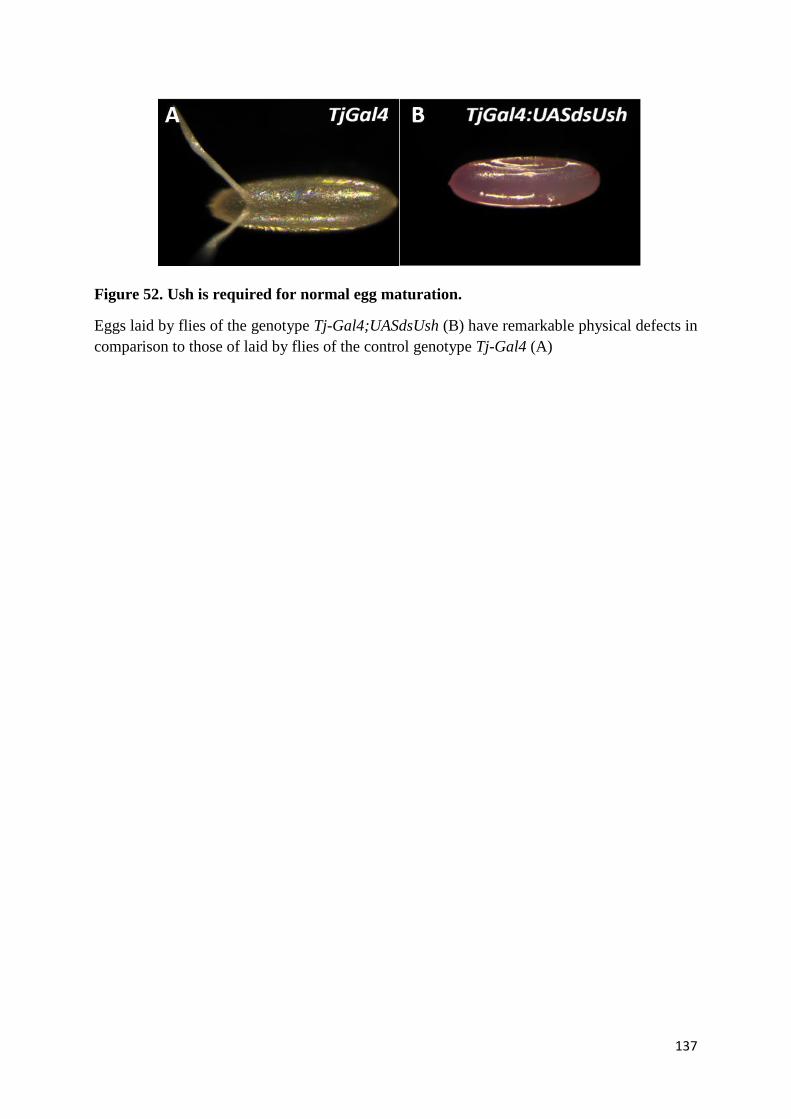
137
Figure 52. Ush is required for normal egg maturation.
Eggs laid by flies of the genotype Tj-Gal4;UASdsUsh (B) have remarkable physical defects in
comparison to those of laid by flies of the control genotype Tj-Gal4 (A)
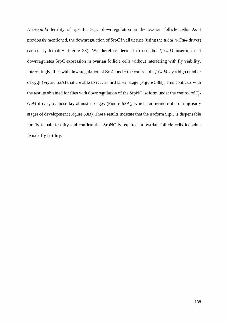
138
Drosophila fertility of specific SrpC downregulation in the ovarian follicle cells. As I
previously mentioned, the downregulation of SrpC in all tissues (using the tubulin-Gal4 driver)
causes fly lethality (Figure 38). We therefore decided to use the Tj-Gal4 insertion that
downregulates SrpC expression in ovarian follicle cells without interfering with fly viability.
Interestingly, flies with downregulation of SrpC under the control of Tj-Gal4 lay a high number
of eggs (Figure 53A) that are able to reach third larval stage (Figure 53B). This contrasts with
the results obtained for flies with downregulation of the SrpNC isoform under the control of Tj-
Gal4 driver, as those lay almost no eggs (Figure 53A), which furthermore die during early
stages of development (Figure 53B). These results indicate that the isoform SrpC is dispensable
for fly female fertility and confirm that SrpNC is required in ovarian follicle cells for adult
female fly fertility.
139
Figure 53. SrpC is dispensable for fly fertility.
(A) Quantification of the number of eggs laid by flies of the genotypes Tj-Gal4;+, Tj-
Gal4;dsSrpNC(1), Tj-Gal4;dsSrpNC(2), Tj-Gal4;dsSrpC(1) and Tj-Gal4;dsSrpC(2) crossed
with fertile males of the genotype white-eye. Downregulation of SrpNC, but not SrpC, alters
egg formation or release. (B) The number of progeny at third instar larval stage (L1) raised
from female flies of the genotypes the genotypes Tj-Gal4;+, Tj-Gal4;dsSrpNC(1), Tj-
Gal4;dsSrpNC(2), Tj-Gal4;dsSrpC(1) and Tj-Gal4;dsSrpC(2). Flies with downregulation of
SrpNC but not SrpC are sterile.

140
Chapter III. Discussion
141
Over the years, Drosophila melanogaster has emerged as a valuable model to study biological
processes implicated in normal development (Gold & Brückner, 2015; Pitsouli & Perrimon,
2008; Saffman & Lasko, 1999; Zaffran, 2003). Genes of the GATA transcription factor family
encode zinc finger-containing proteins that are essential for mammalian development (M.
Tremblay et al., 2018). Different functions are attributed to each of the two GATA zinc fingers
(Martin & Orkin, 1990; Trainor et al., 1996; Yang & Evans, 1992), and mutation in each of
them can provoke redundant but also specific developmental defects in many mammalian
tissues and at different stages of development (M. H. F. M. Lentjes et al., 2016). During my
PhD work, I studied the function of both zinc finger domains of the Drosophila GATA factor
Serpent (Srp), which is expressed in different organs, where it plays essential functions at
specific developmental stages (Rehorn et al., 1996; Reuter, 1994). The particularity of this
factor is that it exists as two different isoforms, containing either the canonical N-terminal and
C-terminal GATA zinc finger domains (SrpNC), or only the C-zinc finger (SrpC). Both
isoforms are simultaneously produced in all srp-expressing cells (Waltzer et al., 2002). This
characteristic allowed us to investigate in vivo the specific roles of each zinc finger domain
during fly development.
(A) Using CRISPR/Cas9 to generate flies with Srp isoform loss-of-
function
In order to study the functions of SrpNC and SrpC isoforms in Drosophila, we used
CRISPR/Cas9 technology to generate mutant fly lines devoid of either the SrpNC or SrpC
isoform. Using CRISPR/Cas9 has several advantages: it is a simple and efficient method to edit
genomic DNA in the fly (Lino et al., 2018), and the modification introduced in the genome can
142
be easily detected by PCR; CRISPR/Cas9 is the first method that efficiently allows precise
control of DNA editing (Lino et al., 2018); it is particularly well adapted in Drosophila
melanogaster, a model with a profusion of genetic tools, ensuring fast selection and
stabilization of the events of interest in the genome; excision of particular genomic region
ensures complete elimination of the mRNA of interest; CRISPR/Cas9 has fewer chances to
produce off-target effects in comparison to other methods (Boettcher & McManus, 2015).
Moreover, there are improved bioinformatics tools (e.g. CRISPR optimal target finder tool) to
help identify of the most appropriate guide RNAs targeting the desired genomic region with
minimal off-target binding. Coupling this technique with RNA interference is an excellent way
to validate the obtained phenotypes, as both methods are unlikely to yield the same off-target
effects. Additionally, other approaches can be applied to confirm that the observed phenotypes
result from the editing event, and not from off-target modifications, such as increasing the
number of independent mutation events and comparing the resulting phenotypes. In our case,
screening for mutant flies devoid of SrpNC yielded at least nine different founders positive for
the desired mutation. Analysis of these mutants’ viability revealed that they are all able to
develop until adult stage and that all female flies are sterile, unable to produce offspring. This
shows that all the obtained mutants act similarly. In parallel, the screen for mutants with SrpC
loss-of-function allowed the recovery of three different founders of the desired mutation.
Analysis of their viability showed that they are all unable to reach adulthood. This indicates
that mutant lethality is caused by the loss-of-function of SrpC, rather than by another off-target
mutation. Finally, for both types of mutation (srpΔsrpNC and srpΔsrpC mutants), complementation
analyses (performed either with other known srp alleles or with a deficiency uncovering the srp
locus) confirmed that the characterized phenotypes are not due to the genetic background in
which our mutants were generated, or to off-target secondary mutations.
143
(B) Similarities and differences between the mammalian and Drosophila
GATA zinc finger associated functions
In Drosophila, Srp ensures essential functions during several processes and in different organs.
In the intestinal system
In the developing intestine, Srp allows the epithelial-to-mesenchymal transition (EMT) of
endodermal cells (K. Campbell et al., 2011; Reuter, 1994). A previous study found that GATA
factors in both mammals and Drosophila use similar molecular target genes during the
induction of the EMT (K. Campbell et al., 2011). Our results show that the GATA C-ZnF
domain is sufficient, while the N-ZnF is dispensable for Srp-mediated EMT. Similarly, in
vertebrates, it was shown that GATA4 allows EMT of endocardial cells independently of its N-
terminal zinc finger-mediated interaction with FOG (Rivera-Feliciano et al., 2006).
In the hematopoietic system
During Drosophila hematopoiesis, we found that the C-ZnF is sufficient for proliferation and
maintenance of embryonic hemocytes. Similarly, studies on GATA vertebrates showed that
GATA1 is required for maintenance of pro-erythroblasts and their differentiation into mature
hemocytes during mammalian primitive hematopoiesis (Y. Fujiwara et al., 1996), and that
GATA1 C-terminal zinc finger is sufficient to carry out this function (Shimizu et al., 2001).
Also, the GATA/FOG interaction is required during mammalian definitive hematopoiesis, in
order to regulate specific hematopoietic lineage differentiation. For example, GATA1/FOG1
inhibits differentiation of mast cells progenitors into mast cells, by activating the neutrophil cell
fate program in these progenitors (Sugiyama et al., 2008). Likewise, we found that in
Drosophila, the SrpNC/Ush complex is required for regulation of normal hematopoietic cell
144
differentiation, mainly by preventing spurious lamellocyte formation in hematopoietic
territories.
In the ovaries
During oogenesis, SrpNC/Ush plays essential functions in egg formation and release. This
situation can be paralleled in mammals, since GATA4 is required for the expression of essential
proteins in ovary follicular cells, which control egg maintenance and release (Kyrönlahti et al.,
2011). The role of FOG in the adult mammalian ovary, however, has not been studied yet.
Unlike the mammalian GATA4/FOG2 complex, which plays essential functions during
development of both ovary and testis (Manuylov et al., 2008; Tevosian et al., 2002), we found
that the SrpNC/Ush complex is dispensable for Drosophila sex-specific gonad formation.
In summary, our work provides a great deal of new insights concerning the roles and different
modes of action of Drosophila GATA zinc finger domains in various developmental contexts.
Although not all the functions of GATA zinc fingers have been conserved throughout evolution,
a striking number of similarities have emerged between mammals and Drosophila. Future
challenges imply the exploitation of the similarities identified between Drosophila and
mammalian GATA zinc finger activities, in order to modulate the human GATA zinc finger
domains associated with diseases.
(C) Common and different functions for Srp zinc finger domains
Our analysis leads us to divide the functions of Srp into three categories (Figure 53).
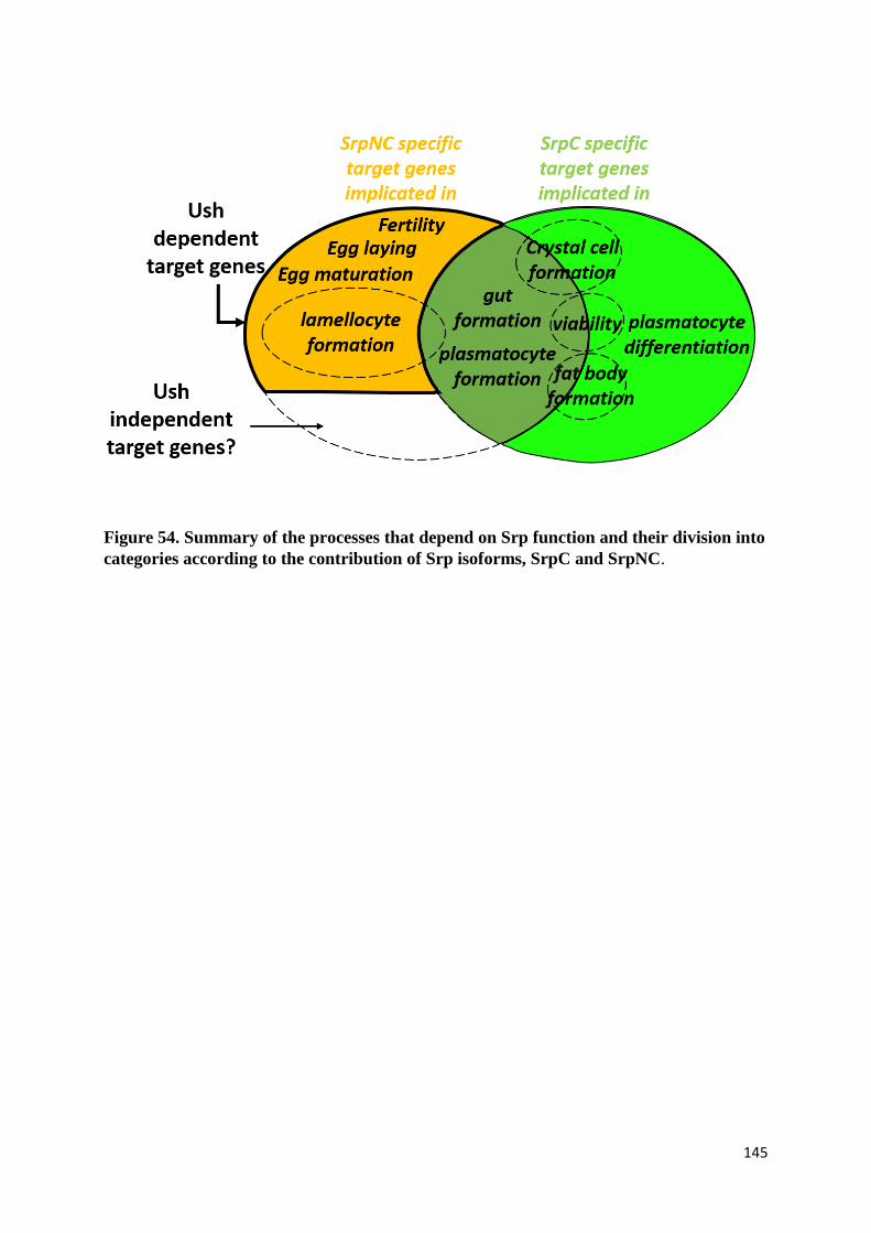
145
Figure 54. Summary of the processes that depend on Srp function and their division into
categories according to the contribution of Srp isoforms, SrpC and SrpNC.
146
(i) The functions for which the presence of C-terminal zinc finger is sufficient: SrpNC and SrpC
are therefore redundant. This is the case for instance during gut and plasmatocyte formation.
(ii) The functions specifically assigned to the SrpC isoform: these are the functions for which
SrpNC, despite the fact that it also contains the C-ZnF, cannot compensate for the loss of SrpC.
This was observed for fat body and crystal cell formation, as well as for plasmatocyte
maturation. These results have several possible explanations. For example, SrpC contains a
region encoded by the mutually exclusive exon E4B that can be implicated in the regulation of
SrpC specific functions. However, analysis of this sequence by bioinformatics tools did not
reveal any known functional or structural motifs. It could be interesting to assess the
dispensability of this region by creating a mutant containing both Srp isoforms but without the
non-conserved E4B-encoded part of SrpC. 3D structural characterization of GATA zinc finger
domains shows that each GATA zinc finger can recognize specific DNA motifs. In fact, both
zinc finger domains have a homologous core that interacts with the first three bases of the
GATA binding site, while a QTRNRK motif in the C-terminal basic tail of the GATA C-
terminal zinc finger allows the recognition of the fourth base pair of the GATA site,
adenine/thymine (Bates et al., 2008; Ghirlando & Trainor, 2003). The C-terminal basic tail of
the N-terminal zinc finger doesn’t have the QTRNRK motif, and it displays more affinity for
the GATC, GATG and GATT sites than for the GATA sites (Ghirlando & Trainor, 2003; Pedone
et al., 1997). According to this difference in DNA binding site recognition between the two zinc
finger domains, the N-ZnF and its adjacent basic region are able to modulate the binding
specificity of the C-ZnF, preventing it from recognizing the consensus GATA site (Trainor et
al., 2000). Thus, the presence of an isoform devoid of the N-ZnF could be required to recognize
specific target genes. A third explanation for the incapability of SrpNC to compensate the loss-
of-function of SrpC may depend on the presence of regulatory proteins, such as the FOG
cofactor Ush, which interacts with the N-ZnF of SrpNC and interferes with its ability to activate

147
SrpC target genes. This type of negative regulation has been shown during mast cell progenitor
differentiation, where FOG interacts with GATA1 in order to recruit the NuRD repressor
complex and inhibit the GATA1-mediated mast cell formation (Cantor et al., 2008; Z. Gao et
al., 2010; Gregory et al., 2010). It was also demonstrated by our group that Ush antagonizes
SrpNC- but not SrpC-mediated gene transcription in Drosophila embryonic mesoderm and in
the mammalian COS-7 cell line (Waltzer et al., 2002, 2003).
(iii) Finally, the third category regroups functions that require the presence of both N- and C-
terminal zinc fingers, as the downregulation of SrpNC in the follicle cells of Drosophila ovaries
causes sterility, but the downregulation of SrpC in the presence of SrpNC has no such effect.
Additionally, we found that the loss of one copy of SrpNC isoform alters Drosophila
hematopoietic homeostasis, but the loss of one copy of SrpC in the presence of SrpNC has no
effect on this context. However, effects on hematopoietic homeostasis of SrpNC or SrpC
downregulation in the lymph gland have to be characterized, in order to consolidate the
hypothesis of the existence of SrpNC-dependent specific functions in this context.
To summarize, we envision three modes of action of Srp through the differential use of its two
alternatively spliced isoforms: Srp either relies only on its C-terminal zinc finger, or it must be
devoid of the N-ZnF, or finally in a third scenario, it would require both zinc finger domains
simultaneously in order to properly fulfill its role.
(D) The Srp N-terminal zinc finger domain is dispensable for Drosophila
development
The most intriguing result from the analysis of srp isoform-specific mutants is the ability of the
fly to develop until adult stage in the absence of the GATA N-ZnF domain. This domain plays
148
a role during GATA binding to DNA palindromic sequences and during the interaction of
GATA with other proteins (Jason A. Lowry & Mackay, 2006; Trainor et al., 1996; Yang &
Evans, 1992). The in vivo requirement for this domain has been shown in mammals by analysis
of mutant mice harboring in the N-terminal zinc finger the V205G substitution, which abolishes
GATA interaction with FOG cofactors. The introduction of this mutation in GATA1 or in both
GATA1 and GATA2, as well as in GATA4, causes mice embryonic lethality associated to
defects during hematopoiesis or heart formation (J. D. Crispino et al., 2001; Shimizu et al.,
2004). Our results show that neither the loss-of-function of SrpNC nor the loss of the interaction
of SrpNC with Ush affect the fly development, demonstrating that the essential functions of Srp
are not supported by the N-ZnF domain.
(E) Role of Srp N-zinc finger domain during fly development
1) The Srp N-zinc finger is required for regulation of hematopoietic homeostasis
We found that SrpNC and SrpNC/Ush interaction regulates many aspects of larval
hematopoiesis. In the lymph gland, mutant analysis showed that crystal cell formation is
abolished after loss of SrpNC/Ush interaction, but not in the context of loss-of-function of
SrpNC protein, indicating that without a proper interaction with Ush, SrpNC proteins act as
negative regulator of crystal cell formation. This suggests that in this specific situation, the
cofactor Ush is required to promote crystal cells formation, which contradicts what was already
observed in other contexts. That different functions are played by GATA and the GATA/FOG
complex has already been seen in the fly, since the induction of larval circulating crystal cells
149
by overexpression of SrpNC, or of another GATA factor Pnr, is antagonized by Ush (Chatterjee
et al., 2019; Nancy Fossett et al., 2003). In addition, we show that during embryonic crystal cell
formation, the presence of a SrpNC variant unable to interact with Ush does not block crystal
cell formation at the embryonic stage, indicating that SrpNC/Ush interaction is not essential for
crystal cell development at this stage. The differences observed between formation of crystal
cells in the embryo and in the lymph gland suggest that GATA factors act differently during
the two hematopoietic waves.
Interestingly, it was already demonstrated that lymph glands with loss of expression of ush
expression, display a reduced number of both plasmatocytes and crystal cells (Dragojlovic-
Munther & Martinez-Agosto, 2013; H. Gao et al., 2009). Accordingly, we show that the role of
Ush during crystal cell formation depends on its interaction with SrpNC. However,
plasmatocyte formation is not affected in srpNCV735G mutant, suggesting that the SrpNC/Ush
interaction is not required in this process, and that Ush-mediated plasmatocyte formation is
likely to depend on its interaction with another GATA factor expressed in the lymph gland.
Apart from crystal cells and plasmatocytes, the fly produces a third type of hemocytes called
lamellocytes, in response to various stress situations (Letourneau et al., 2016). Interestingly, we
found that both SrpNC and SrpNC/Ush interaction inhibits lamellocyte formation under normal
conditions. Analysis of lamellocyte formation in larval hemolymph showed that the phenotype
is more severe in larvae with loss of SrpNC/Ush interaction, than in those with SrpNC loss-of-
function. In contrast, srp∆srpNC mutants display, a lamellocyte production, whose level is more
important in the lymph gland than in the larval hemolymph. This discrepancy might reflect the
existence of compartment-specific hematopoietic regulatory programs for lamellocyte
induction in the lymph gland or in circulation.
150
2) The Srp N-terminal zinc finger is required for egg maturation
We found that SrpNC and the SrpNC/Ush complex are required for egg dorsal respiratory
appendages, micropyle and eggshell formation. The lack of dorsal appendages can be the
consequence of an alteration of appendage precursor cell determination, or of subsequent
appendage morphogenesis events. To discriminate between these two possibilities, staining of
mutant ovaries with Broad-Complex (BR-C) antibody that mark the appendage progenitors
would certainly be useful. Additionally, the formation of functional micropyle requires the
migration of specialized ovarian follicle cells, called border cells and centripetal cells (Montell
et al., 1992; Suzanne et al., 2001; Twombly et al., 1996). Hence, analysis of the border cell and
centripetal cell formation in srpΔsrpNC and srpNCV735G mutant ovaries, would help determine if
the loss of micropyle is due to a hampered determination or migration of these specialized cells,
or due to deregulation of genes implicated in other steps of micropyle morphogenesis. Finally,
synthesis of eggshell components requires the expression of different vitelline membrane and
chorion genes, which have to be finely regulated both spatially and temporally (Cavaliere et al.,
2008). Identifying SrpNC and SrpNC/Ush transcriptional targets participating in eggshell
secretion would therefore be of great interest. As we didn’t find any defects in srpΔsrpNC and
srpNCV735G mutant ovaries morphogenesis, it would be imaginable to carry on transcriptomic
analysis by RNA sequencing and compare gene expression in wild type, srpΔsrpNC mutant and
srpNCV735G mutant ovaries, in order to identify the cause of sterility at the transcriptional level.
(F) The functions of the Srp N-terminal zinc finger depend mostly on its
interaction with the FOG factor U-shaped
151
Although dispensable for Drosophila development, we found that the Srp N-ZnF is required
for maintenance of hematopoietic homeostasis and for female fertility. The N-ZnF plays a role
during GATA binding to DNA palindromic sequences, as well as during the interaction of
GATA with other proteins, such as FOG proteins, the Krüppel-like family factor KLF13, and
the LIM domain containing protein 2 (LMO2) that bridges GATA to transcription factors of the
bHLH family (Lavallée et al., 2006; Jason A. Lowry & Mackay, 2006; Trainor et al., 1996;
Yang & Evans, 1992). The most studied regulator of GATA function is FOG (Chlon & Crispino,
2012). During GATA/FOG interaction, the only valine present in the N-ZnF domain is required
for the interaction with FOG, and its substitution into glycine alters GATA/FOG association
without affecting GATA binding to DNA, or to KLF13 and LMO2 (J. D. Crispino et al., 1999;
Lavallée et al., 2006; Wilkinson-White et al., 2011). Accordingly, only SrpNC has the ability
to interact with Ush (Waltzer et al., 2002). Importantly, the N-ZnF amino acid sequence of
SrpNC is highly conserved, with the equivalent mammalian sequence including the valine
residue (Figure 7) (Waltzer et al., 2002), and the valine to glycine substitution of SrpNC alters
the SrpNC/Ush interaction (Nancy Fossett et al., 2003). Interestingly, during this work we
showed that mutants with the V735G substitution have the same defects as those observed in
mutants with specific SrpNC loss-of-function, indicating that the interaction of SrpNC with
Ush is required for Drosophila hematopoietic homeostasis and female fertility (Figure 54).
Thus, the functions played here by the N-ZnF domain depend on its ability to interact with its
FOG cofactor, rather than on its binding to specific DNA sequences, or its interaction with the
other aforementioned regulators.
The role of Ush in hematopoietic homeostasis regulation has been already described (Avet-
Rochex et al., 2010; Dragojlovic-Munther & Martinez-Agosto, 2013; H. Gao et al., 2009; R. P.
Sorrentino et al., 2007), and we determine here that most of its role in this context is due to its
152
interaction with SrpNC. Also, during this work, a role for Ush during the Drosophila oogenesis
was detected for the first time. The role of SrpNC/Ush interaction in oogenesis could be
confirmed by restoring fly fertility in the srpNCV735G mutant, by expressing in srpV735G ovaries
a mutant form of Ush protein harboring the compensatory substitution serine to arginine,
identified in the mammalian FOG zinc finger domain as responsible for restoration of the
interaction of FOG with vertebrate GATA proteins that contain the substitution valine to
glycine mimicked in SrpNCV735G (J. D. Crispino et al., 1999). Of note, among the five
Drosophila GATA factors, only Srp and GATAd are expressed in Drosophila ovaries (Lepesant
et al., 2020). GATAd being devoid of the zinc finger domains that allows the interaction
GATA/FOG (Okumura et al., 2005), the expression of a form of Ush with this compensatory
mutation in the ovaries should not interfere with the binding of Ush to GATA proteins other
than SrpNC in this organ.
However, although all the identified SrpNC mediated functions are dependent of the interaction
with Ush, we cannot exclude the possibility of presence of some no identified functions for
SrpNC that are independent of Ush (Figure 54).
(G) Why has nature selected a Srp isoform with only one zinc finger
domain?
The ancestor of GATA transcription factors of triploblastic animals is the diploblastic
cnidarians that have a single GATA factor, which like SrpNC, contains two conserved zinc
finger domains. During evolution, some GATA genes in the fly have lost DNA sequence coding
for the N-zinc finger, giving rise to factors with only one zinc finger, such as SrpC (W. J. Gillis
et al., 2007; W. Q. Gillis et al., 2008). This type of GATA factor with only one zinc finger
domain is also found in other triploblastic protostomes, such as C.elegans, which have eleven
153
GATA factors, yet only one of them (elt-1) containing double zinc finger domains (all the others
contain only the C-ZnF) (W. Q. Gillis et al., 2008). In the fly, we found that the N-ZnF domain
is dispensable for development. Similarly, in C.elegans, a high number of GATA-mediated
functions are endorsed by the single zinc finger GATA factors (Block & Shapira, 2015), and
analysis of elt-1 mode of action revealed that deletion of elt-1 N-ZnF domain has no effect on
the elt-1-dependant gene activation (Shim et al., 1995). It thus appears that dispensability of
GATA N-ZnF during development is a common feature of invertebrates. This striking
difference with mammals may reflect the different biological complexity of these organisms,
as it was shown that in Human the estimated number of protein/protein interactions is
significantly higher than in organisms of lesser complexity, such as Drosophila and
Caenorhabditis (M. P. H. Stumpf et al., 2008). Thus, mammals might have a bigger need for
protein/protein interactions mediated by the GATA N-ZnF, in order to control their
development.
In addition, we found that SrpC has essential functions during fly development. Accordingly,
under normal conditions, flies produce about 5 times more SrpC than SrpNC isoform (Waltzer
et al., 2002). Single zing finger containing versions exhibit a smaller surface available for
interactions with other factors than a protein with two zinc finger domains. The ability of SrpC
to only mildly interact with other proteins could make it more rapid and efficient in DNA-
binding, prior to transcription activation of target genes. Conversely, SrpNC could interact with
other proteins that interfere with, or reduce the speed of, its activity during transcriptional
regulation.
Finally, although dispensable for fly development, we found that the N-ZnF domain plays
essential functions during the hematopoietic homeostasis and fly oogenesis (discussed above).
This is hypothesized to be why the fly gene has kept production of this isoform with two zinc
154
finger domains in srp expressing cells (Waltzer et al., 2002). Processes regulated by the N-zinc
finger domain are highly dynamic and renewable, which suggests that these processes require
more complex regulation programs during their realization than in other stable developmental
processes, such as gut and fat body formation, for which the presence of the C-zinc finger
domain is sufficient.
(H) Subfunctionnalization in Serpent isoforms
Gene duplication and alternative splicing are dominant factors in the evolution of eukaryote
complexity and diversity. After gene duplication, two major fates can be adopted by the
generated gene copies, namely neofunctionalisation (acquisition of novel functional properties
in comparison to the ancestral gene) and subfunctionalisation (performing of complementary
functions that jointly match that of the ancestral gene) (Sandve et al., 2017). We found that the
majority of the functions played by Srp are portioned between the isoforms SrpC and SrpNC,
suggesting that a subfunctionalization process occurs with the srp gene. However, we also
found that both isoforms still have redundant functions during embryonic gut and plasmatocyte
formation, suggesting that degeneration and complementation occuring before
subfunctionalisation (Force et al., 1999) are not completely achieved.
Individual gene duplication is common in all organisms (Taylor & Raes, 2004). In the fly, srp
gene was obtained after duplication of its ancestral gene, which gave rise to srp and to other
gene copies, including the GATA factor Pnr-encoding gene (W. Q. Gillis et al., 2008), which
has a distinct expression pattern and produces only canonic GATA factor isoforms with two
zinc finger domains (Fromental-Ramain et al., 2008; Heitzler et al., 1996; Ramain et al., 1993).
155
Intriguingly, production of SrpC factor, relies on alternative splicing of the srp transcript, rather
than on gene duplication and subsequent degenaration of the N-ZnF coding sequence in one of
the two duplicates. Production of both Srp isoforms by alternative splicing keeps the expression
of both srp isoforms under the control of the same regulatory region. It is unknown, however,
why exactly the fly has chosen the subfunctionnalization process by alternative splicing instead
of gene duplication. One possibility is that in some territories, during fly development, the
simultaneous presence of both Srp isoforms is splicing required, therefore exerting an
evolutionary pressure on this alternative splicing event. Thus, in the end, this work illustrates
that, like genome duplication in vertebrates, alternative splicing provides an efficient strategy
to promote subfunctionalization and generate GATA functional diversity in invertebrates.
Identification and analysis of more genes sharing this particularity would be of high interest for
a better understanding of the complex functional diversity in the fly and other organisms.

156
Chapter IV. Materials and Methods
157
1- Generation of srp∆srpNC and srp∆srpC mutant fly lines
The srp∆srpNC and srp∆srpC mutant fly lines were created by generating deletions in the srp locus
using the CRISPR/Cas9 genome editing system, with tools and reagents published in (Port et
al., 2014). For each fly line, two different single guide RNAs (sgRNAs) were used, which
determine the limits of the genomic DNA region to be removed. The sgRNAs were designed
using the “CRISPR optimal target finder tool”, and sgRNAs that recognize zero off-target sites
were selected. sgRNAs sense sequences were as follows:
- srp∆srpNC: 5’-ATAAGTCATAAGGTTTTGCT and AATTAAACAGCCTAGAAGAT
- srp∆srpC: 5’-CACTTTTCGATTAAACTAGT and CGTAGTAAGGCTAACACGAG
For each sgRNA, sense and anti-sense oligonucleotides were annealed and inserted into the
pCFD3 plasmid (addgene #49410) following the specific protocol in the CRISPR fly design
website (https://www.crisprflydesign.org/). After validation of cloning efficiency by
sequencing, recombinant plasmids were purified by the Qiagen plasmid maxiprep purification
kit, checked by sequencing and injected into embryos expressing the nuclease Cas9 in the
germline cells under the control of the vasa promoter (Bloomington stock 51323). The
injection mix contains a specific pair of sgRNA recombinant plasmids at a concentration of
250 ng/µl each. For injections, embryos at early stages of embryogenesis were covered by
Voltalef 10S oil (VWR Chemicals, # 24627.188) in order to make the embryo clearly visible
under the chorion. The mix was injected at the embryonic posterior pole, using the Leica
mechanical micromanipulator and a needle pulled from a capillary glass (Harvard Apparatus,
#30-0019) by a Sutter instruments brand horizontal puller. Injected embryos were allowed to
develop until adulthood. These F0 adult flies were crossed in order to yield F1 progeny. F1
males were subsequently crossed for 4-5 days, then removed from the cross and their DNA
extracted as described in (Gloor et al., 1993).
158
The screening PCR was performed using the following primers:
- srp∆srpNCF: 5’-AATTCAAACCAACAAAAAGACACCT
- srp∆srpNCR: 5’- GCAGATGCAGATTGATCAGATTTTC
- srp∆srpCF: 5’- CCCCTGAAAAGCTCCAATGC
- srp∆srpCR: 5’- CTCAGTGGCCAAGGAGGTTT
These primers bind regions located outside of the expected deletions. Flies positive for the
deletion will give an amplicon of size smaller than that obtained from the control chromosome.
Upon detection, presence of the deletion was validated by Sanger sequencing.
2- Generation of srpV735G mutant fly line
The srpV735G mutant fly line was created by introducing a missense mutation (a GGA codon
replacing the wild-type GTC codon) in srp, using the CRISPR/Cas9 genome editing system. A
sgRNA was used to target the nuclease Cas9 to a 5’-ATGCCGATTATTTCACTGAG sequence
in the vicinity of the nucleotides intended to be modified. A single-strand DNA donor (ssODN)
of 200 base pairs harboring the mutated nucleotides was used as a repair template. The
recombinant sgRNA-containing pCFD3 plasmid was prepared as described above. The ssODN
was manufactured by Integrated DNA Technologies. The injection mix constituted of
250 ng/µl of the recombinant plasmid and 100 ng/µl of the ssDNA. ssODN sequence:
CGTTCATTTT CATGTACAAG CCGCAGGCAT TGCACAAATA GTGTCCCGTG
TTATCGCGTC GCCATAATGG GGTTGAAATC GCACCACAGT TTCCACACTC
ACGTCCTTCT GTAAAATAAT CGGCATCGAA TAATGCTTCA AAGATTGACA
TAGAAATAT CGGAATAAGT CATAAGGTTT TGCTAGGTGT TTGTTTGATT.
The screening PCR of F1 males’ genomic DNA involved the following primers:
- srpV735GF: 5’-ACACAATACGCAAATGC,

159
- srpV735GR: 5’-ACTGAGAGAGGATGTTGC
- srpV735GRm: 5’-CAGTTTCCACACTCACG.
F and R primers bind outside of the modified DNA region, while Rm is a reverse primer that
specifically recognizes the modified nucleotides incorporated from the ssODN-directed repair
event. Note that some silent mutations have been inserted in the ssODN (Figure 5), in order to
allow the design of a Rm primer efficient at discriminating the modified DNA from the
unmodified DNA. Upon detection, events of interest were confirmed by sequencing.
3- Generating transgenic RNAi fly lines specific to srp isoforms
RNAi constructs were designed using the E-RNAi web service
(https://www.dkfz.de/signaling/e-rnai3//). The 21-nucleotide sequences correspond to specific
regions of srp gene exon 4A or 4B, in order to specifically interfere with expression of srpNC
and srpC, respectively. Sequences with the lowest off-target score were selected and blasted
using the National Center for Biotechnology and Information (NCBI) website against the
D. melanogaster RNA-sequences available at the NCBI Reference RNA Sequences
(Refseq_rna) database, in order to validate the absence of matches with off-target sites. Short-
hairpin RNA (shRNA) have been designed as described as described in (Jian-Quan Ni, et al,
2011) and synthetized by the Integrated DNA Technologies (IDT) platform.
- Exon 4A specific shRNA (1) sense strand: CTAGCAGTCGATAACACGGGACACTATTT
TAGTTATATTCAAGCATAAAATAGTGTCCCGTGTTATCGGCG
- Exon 4A specific shRNA (1) anti-sense strand: AATTCGCCGATAACACGGGACACTATTT
TATGCTTGAATATAACTAAAATAGTGTCCCGTGTTATCGACTG
- Exon 4A specific shRNA (2) sense strand: CTAGCAGTGCATGAATCGACCCCTAATTA
TAGTTATATTCAAGCATATAATTAGGGGTCGATTCATGCGCG
160
- Exon 4A specific shRNA (2) anti-sense strand: AATTCGCGCATGAATCGACCCCTAATTA
TATGCTTGAATATAACTATAATTAGGGGTCGATTCATGCACTG
- Exon 4B specific shRNA (1) sense strand: CTAGCAGTCGCTGAATCAGGCGGGGATTT
TAGTTATATTCAAGCATAAAATCCCCGCCTGATTCAGCGGCG
- Exon 4B specific shRNA (1) anti-sense strand: AATTCGCCGCTGAATCAGGCGGGGATTT
TATGCTTGAATATAACTAAAATCCCCGCCTGATTCAGCGACTG
- Exon 4B specific shRNA (2) sense strand: CTAGCAGTCTATAAACCCAACTCATTTAA
TAGTTATATTCAAGCATATTAAATGAGTTGGGTTTATAGGCG
- Exon 4B specific shRNA (2) anti-sense strand: AATTCGCCTATAAACCCAACTCATTTAA
TATGCTTGAATATAACTATTAAATGAGTTGGGTTTATAGACTG
For each shRNA construct, the synthetized sense and anti-sense strands were annealed and
cloned into pWalium20 plasmid (DGRC: 1472) following a protocol adapted from the cloning
in pCFD3 vector protocol (https://www.crisprflydesign.org/). The recombinant plasmids were
individually injected into flies containing AttP2 sites that allow insertion of a plasmid into the
AttP2 platform in genomic DNA.
4- Fly strains
The fly strains srp3, srpAs, Df(3R)BSC728 were obtained from the Bloomington Drosophila
Stock Center. The fly strains lz-Gal4 and srp6G were already available in our group (Waltzer et
al., 2002, 2003). The fly strains ushVX22, ushRev24 were kindly provided by P.Heitzler’s team.
The fly strain msnF9-mCherry was kindly provided by R. Schulz’s group.
161
5- Embryonic RNA extraction and RT-PCR
Embryos of the genotypes w1118, srpΔsrpNC, srpΔsrpC were allowed to develop on agar plates, at
25◦C until stages 14 to 16 of embryogenesis. Total embryonic RNA was extracted using the
RNeasy Plus Mini kit (Qiagen). Reverse transcription was performed using random primers
(Invitrogen, P/N 58875), and PCR was performed using the following primer pairs: SrpNC-F:
5’-GATACCTGGTTCGATCC / SrpNC-R: 5’-TGGTGTCCTTTTTCATG, and SrpC-F: 5’-
CTCGGCATCGTTGTC / SrpC-R: 5’-TCCGGCTCGCTTTGAGG.
6- Survival analysis
Throughout the survival analysis period, flies were raised at 25°C. For each analyzed genotype,
embryos were collected on agar plates at stages 14 to 16 of embryogenesis, and their ability to
hatch was recorded. 48 hours later, the ability of the hatched first instar larvae to reach the third
instar larval stage was quantified, and the third instar larvae L3 were transferred to tubes
containing standard media, where analysis of their ability to develop into pupal and adult flies
was recorded 48 hours and 5 days after transfer.
For the table in figure 28, crosses were performed in tubes containing standard media, and the
progeny was allowed to develop at 25°C until the third instar larval or adult stage.
7- Bioinformatic tools
Multiple alignment of the SrpNC amino acid sequence with the mammalian GATA protein
sequences was done by using the bioinformatics software Geneious. SrpNC (NP_732098.1),

162
GATA1 (AAH09797.1), GATA2 (AAH51272.1), GATA3 (NP_001002295.1), GATA4
(AAI43480.1), GATA5 (AAH47790.1) and GATA6 (NP_005248.2).
Multiple alignment of the SrpC amino acid sequence with GATA protein sequences of other
species was done by using the combine popular aligners M-Coffee of the multiple sequence
alignment server T-coffee. Dmel-SrpC (NP_732100.2), Lcup-GATA (XP_023301929), Amel-
GATA (XP_016769881.2), Tcas-GATA (XP_008200496.1), Mdom- GATA
(XP_005187504.3), Hsap-GATA2 (AAH51272.1), Hsap-GATA3 (NP_001002295.1) and
Hsap-GATA4 (AAI43480.1).
Analysis of the presence of known functional domains in the amino acid sequence encoded by
the exon E4B was done by the bioinformatics tools InterPro, ExPASY and NCBI Conserved
Domain Search.
8- RNA in situ hybridization
Anti-sense probes specific for alcohol dehydrogenase, croquemort, GATAe, glutactin, grain,
lozenge, Prophenoloxidase 2, Peroxidasin, tiggrin and viking transcripts were synthesized after
linearization of plasmids containing the cDNA sequences of each of the mentioned target
(Drosophila Golden Collection website). Probe transcription was performed for 2 hours at 37◦C
in the presence of the appropriate T7, T3 or SP6 RNA polymerase (Promega), RNasin
ribonuclease inhibitors (Promega) and a mixture of dATP, dCTP, dGTP and dioxigenin (Dig)-
coupled dUTP (Roche). The synthetized probe was precipitated in the presence of EDTA PH8,
LiCl, torula RNA and ethanol and conserved at -20◦C in Hybridization Buffer (HB; 50ml of
HB: 25 ml of deionized formamide, 10 ml of 10% Roche blocking reagent, 5 ml of saline-
sodium citrate buffer 20x (x), 1 ml of torula RNA (50 mg/ml), 0,5 ml CHAPS (10%), 0,5 ml
EDTA (0,5M), 50 µl heparin (0,05 g/ml) and 50 µl of 100% TweenTM 20).

163
Embryos were collected on agar plate at 25◦C. They were dechorionated by soaking the eggs in
a 50% bleach solution for 5 minutes at room temperature, rinsed under tap-water and then fixed
in a fixation solution (1.8 ml of 37% formaldehyde solution, 200 µl of EGTA 0.5M and 2 ml
of heptane). Fixation was performed by shaking embryos on a rotomax at room temperature for
20 minutes. Fixed embryos were washed several times with 100% methanol, then with 1%
phosphate buffered saline containing 0.1% of TweenTM 20 (PBSTw), and incubated for 1h at
65◦C in HB. Probe hybridization was performed overnight at 65◦C in HB containing probe at the
appropriate dilution. The following day, embryos were incubated for one hour at 65◦C in HB
solution and then for 20 minutes at the same temperature, after adding equivalent volumes of
HB solution and PBSTw to the embryos. Samples were then washed at room temperature in
PBSTw several times and in PBSTw containing 1% of bovine serum albumin (PBSTw-BSA)
for 30 minutes. Embryos were then incubated overnight at 4°C in PBSTw-BSA containing α-
DIG antibodies conjugated with alkaline phosphatase (Roche). On the third day, embryos were
washed 5 times with PBSTw for 10 minutes, and later once for 10 minutes with the Staining
Buffer solution (SB) containing MgCl2, NaCl, Tris-HCl PH 9.5 and Tween TM 20. Finally, the
coloration solution formed by the NBT/BCIP substrate diluted in the SB solution was added to
the embryos in order to reveal the anti-DIG binding sites on the embryonic tissues. Images were
acquired by the Nikon Eclipse 80i microscope (NIS-Element software).
9- Lymph gland and circulating hemocytes immunostaining
Larvae were grown on standard medium at 25◦C. For lymph gland analysis, we selected mid-
third larval instar larvae, and for circulating hemocytes analysis we selected wandering larvae
prior to spiracle eversion. All the analyzed larvae having the X-linked transgenes msnF9-
mCherry are heterozygous females for the transgene. Samples were fixed with 4%
164
paraformaldehyde for 20 minutes at room temperature. They were washed several times with
1X phosphate buffered saline (PBS) and then with 1X PBS solution containing 0.3% Triton X-
100 (PBSTr). All samples were then blocked for 30 minutes in PBSTr containing 1% of bovine
serum albumin (PBSTr-BSA). Incubation lasted one night (α-P1, α-L1, Phalloidin) or 36 hours
(α-Collier, α-PPO1), in PBSTr-BSA. Antibodies used were: Mouse α-P1 (kindly provided by
I. Ando, 1/30), Mouse α-Col (kindly provided by M. Crozatier, 1/50) and Rabbit α- PPO1 ((Li
et al., 2012), 1/10 000). Samples were washed several times with PBSTr-BSA and incubated
with Alexa Fluor-labeled secondary antibodies (Molecular Probes, 1/400). DAPI (Sigma-
Aldrich) was adjusted to 1 mg/L in blocking reagent and added either to the primary antibodies
solution for lymph gland staining, or incubated alone for 20 minutes at room temperature for
circulating hemocyte staining. Samples were mounted in Vectashield medium (Vector
laboratories). Slides were then analyzed using the Leica SP8 inverted-based confocal
microscope. The optimized system option was chosen to collect Z-series of images by the Leica
LAS-X Life Science software. Maximum intensity projection of Z-stack of each image was
created by the image processing software ImageJ. For each experiment, images of different
genotypes were taken with the same signal intensity.
10- Quantification of lamellocyte phenotype penetrance
Larvae were raised on standard medium at 25◦C. Live larvae were observed on a Leica
fluorescence detecting macroscope and were separated into 4 categories, depending on the
number of msnF9-mCherry expressing circulating cells detected in each larva. Activation of
reporter expression in the hemolymph is restricted to lamellocytes (T. Tokusumi et al., 2009).
Larvae with no positive cells in their hemolymph were placed in the “0 lamellocytes” category.
Larvae with 1-5 cells expressing the reporter in their hemolymph were placed in the “<5
165
lamellocytes category”. Larvae with more than 5 cherry-positive circulating cells were placed
in the “>5 lamellocytes category”. Finally, larvae with large clusters of mCherry fluorescent
cells were placed in the category of larvae with “lamellocyte aggregates”. Only the percentage
of larvae with extreme phenotypes (0 lamellocytes and aggregates of lamellocytes) were taken
into consideration during the analysis of the ability of different genotypes to produce
lamellocytes.
11- Analysis of the fertility phenotype
Virgin female w1118, srpΔsrpNC, srpV735G flies were mated with w1118 males and allowed to lay
eggs on agar plates, at 25◦C. The number of laid eggs was quantified 2 and 3 days after the
beginning of the crosses. Photos of the eggs laid were aquired with a Leica macroscope.

166
Chapter V. References
167
Abel, T., Michelson, A. M., & Maniatis, T. (1993). A Drosophila GATA family member that
binds to Adh regulatory sequences is expressed in the developing fat body.
Development, 119(3), 623‑633.
Allen, H. L., Flanagan, S. E., Shaw-Smith, C., De Franco, E., Akerman, I., Caswell, R.,
International Pancreatic Agenesis Consortium, Ferrer, J., Hattersley, A. T., & Ellard, S.
(2011). GATA6 haploinsufficiency causes pancreatic agenesis in humans. Nature
Genetics, 44(1), 20‑22. https://doi.org/10.1038/ng.1035
Arceci, R. J., King, A. A., Simon, M. C., Orkin, S. H., & Wilson, D. B. (1993). Mouse GATA-
4 : A retinoic acid-inducible GATA-binding transcription factor expressed in
endodermally derived tissues and heart. Molecular and Cellular Biology, 13(4),
2235‑2246. https://doi.org/10.1128/mcb.13.4.2235
Aronson, B. E., Stapleton, K. A., & Krasinski, S. D. (2014). Role of GATA factors in
development, differentiation, and homeostasis of the small intestinal epithelium.
American Journal of Physiology - Gastrointestinal and Liver Physiology, 306(6),
G474‑G490. https://doi.org/10.1152/ajpgi.00119.2013
Asselin-Labat, M.-L., Sutherland, K. D., Barker, H., Thomas, R., Shackleton, M., Forrest, N.
C., Hartley, L., Robb, L., Grosveld, F. G., van der Wees, J., Lindeman, G. J., &
Visvader, J. E. (2007). Gata-3 is an essential regulator of mammary-gland
morphogenesis and luminal-cell differentiation. Nature Cell Biology, 9(2), 201‑209.
https://doi.org/10.1038/ncb1530
Avet-Rochex, A., Boyer, K., Polesello, C., Gobert, V., Osman, D., Roch, F., Augé, B., Zanet,
J., Haenlin, M., & Waltzer, L. (2010). An in vivo RNA interference screen identifies
gene networks controlling Drosophila melanogaster blood cell homeostasis. BMC
Developmental Biology, 10, 65. https://doi.org/10.1186/1471-213X-10-65
Balduini, C. L., Pecci, A., Loffredo, G., Izzo, P., Noris, P., Grosso, M., Bergamaschi, G., Rosti,
V., Magrini, U., Ceresa, I. F., Conti, V., Poggi, V., & Savoia, A. (2004). Effects of the
R216Q mutation of GATA-1 on erythropoiesis and megakaryocytopoiesis. Thrombosis
and Haemostasis, 91(1), 129‑140. https://doi.org/10.1160/TH03-05-0290
Banerjee, U., Girard, J. R., Goins, L. M., & Spratford, C. M. (2019). Drosophila as a Genetic
Model for Hematopoiesis. Genetics, 211(2), 367‑417.
https://doi.org/10.1534/genetics.118.300223
168
Barnett, T., Pachl, C., Gergen, J. P., & Wensink, P. C. (1980). The isolation and characterization
of Drosophila yolk protein genes. Cell, 21(3), 729‑738. https://doi.org/10.1016/0092-
8674(80)90436-5
Bates, D. L., Chen, Y., Kim, G., Guo, L., & Chen, L. (2008). Crystal structures of multiple
GATA zinc fingers bound to DNA reveal new insights into DNA recognition and self-
association by GATA. Journal of molecular biology, 381(5), 1292‑1306.
https://doi.org/10.1016/j.jmb.2008.06.072
Battle, M. A., Bondow, B. J., Iverson, M. A., Adams, S. J., Jandacek, R. J., Tso, P., & Duncan,
S. A. (2008). GATA4 is essential for jejunal function in mice. Gastroenterology, 135(5),
1676-1686.e1. https://doi.org/10.1053/j.gastro.2008.07.074
Belaguli, N. S., Sepulveda, J. L., Nigam, V., Charron, F., Nemer, M., & Schwartz, R. J. (2000).
Cardiac Tissue Enriched Factors Serum Response Factor and GATA-4 Are Mutual
Coregulators. Molecular and Cellular Biology, 20(20), 7550‑7558.
Benmimoun, B., Haenlin, M., & Waltzer, L. (2015). Blood cell progenitor maintenance : Collier
barks out of the niche. Fly, 9(4), 160‑164.
https://doi.org/10.1080/19336934.2016.1151130
Bennett, J., Wu, Y.-G., Gossen, J., Zhou, P., & Stocco, C. (2012). Loss of GATA-6 and GATA-
4 in Granulosa Cells Blocks Folliculogenesis, Ovulation, and Follicle Stimulating
Hormone Receptor Expression Leading to Female Infertility. Endocrinology, 153(5),
2474‑2485. https://doi.org/10.1210/en.2011-1969
Beuling, E., Aronson, B. E., Tran, L. M. D., Stapleton, K. A., ter Horst, E. N., Vissers, L. A. T.
M., Verzi, M. P., & Krasinski, S. D. (2012). GATA6 Is Required for Proliferation,
Migration, Secretory Cell Maturation, and Gene Expression in the Mature Mouse
Colon. Molecular and Cellular Biology, 32(17), 3392‑3402.
https://doi.org/10.1128/MCB.00070-12
Beuling, E., Baffour-Awuah, N. Y. A., Stapleton, K. A., Aronson, B. E., Noah, T. K., Shroyer,
N. F., Duncan, S. A., Fleet, J. C., & Krasinski, S. D. (2011). GATA factors regulate
proliferation, differentiation, and gene expression in small intestine of mature mice.
Gastroenterology, 140(4), 1219-1229.e1-2.
https://doi.org/10.1053/j.gastro.2011.01.033
169
Bier, E. (2005). Drosophila , the golden bug, emerges as a tool for human genetics. Nature
Reviews Genetics, 6(1), 9‑23. https://doi.org/10.1038/nrg1503
Blobel, G. A., Nakajima, T., Eckner, R., Montminy, M., & Orkin, S. H. (1998). CREB-binding
protein cooperates with transcription factor GATA-1 and is required for erythroid
differentiation. Proceedings of the National Academy of Sciences of the United States
of America, 95(5), 2061‑2066. https://doi.org/10.1073/pnas.95.5.2061
Block, D. H., & Shapira, M. (2015). GATA transcription factors as tissue-specific master
regulators for induced responses. Worm, 4(4).
https://doi.org/10.1080/21624054.2015.1118607
Boettcher, M., & McManus, M. T. (2015). Choosing the Right Tool for the Job : RNAi, TALEN
or CRISPR. Molecular cell, 58(4), 575‑585.
https://doi.org/10.1016/j.molcel.2015.04.028
Bonachea, E. M., Chang, S.-W., Zender, G., LaHaye, S., Fitzgerald-Butt, S., McBride, K. L.,
& Garg, V. (2014). Rare GATA5 sequence variants identified in individuals with
bicuspid aortic valve. Pediatric Research, 76(2), 211‑216.
https://doi.org/10.1038/pr.2014.67
Bossard, P., & Zaret, K. S. (1998). GATA transcription factors as potentiators of gut endoderm
differentiation. Development (Cambridge, England), 125(24), 4909‑4917.
Bosse, T., Piaseckyj, C. M., Burghard, E., Fialkovich, J. J., Rajagopal, S., Pu, W. T., &
Krasinski, S. D. (2006). Gata4 Is Essential for the Maintenance of Jejunal-Ileal Identities
in the Adult Mouse Small Intestine. Molecular and Cellular Biology, 26(23),
9060‑9070. https://doi.org/10.1128/MCB.00124-06
Brade, T., Pane, L. S., Moretti, A., Chien, K. R., & Laugwitz, K.-L. (2013). Embryonic Heart
Progenitors and Cardiogenesis. Cold Spring Harbor Perspectives in Medicine, 3(10).
https://doi.org/10.1101/cshperspect.a013847
Bresnick, E. H., Lee, H.-Y., Fujiwara, T., Johnson, K. D., & Keles, S. (2010). GATA switches
as developmental drivers. The Journal of Biological Chemistry, 285(41), 31087‑31093.
https://doi.org/10.1074/jbc.R110.159079
Brookman, J. J., Toosy, A. T., Shashidhara, L. S., & White, R. A. (1992). The 412
retrotransposon and the development of gonadal mesoderm in Drosophila. Development
(Cambridge, England), 116(4), 1185‑1192.
170
Brown, S., & Castelli-Gair Hombría, J. (2000). Drosophila grain encodes a GATA transcription
factor required for cell rearrangement during morphogenesis. Development
(Cambridge, England), 127(22), 4867‑4876.
Bruneau, B. G. (2013). Signaling and transcriptional networks in heart development and
regeneration. Cold Spring Harbor Perspectives in Biology, 5(3), a008292.
https://doi.org/10.1101/cshperspect.a008292
Buchon, N., Osman, D., David, F. P. A., Fang, H. Y., Boquete, J.-P., Deplancke, B., &
Lemaitre, B. (2013). Morphological and molecular characterization of adult midgut
compartmentalization in Drosophila. Cell Reports, 3(5), 1725‑1738.
https://doi.org/10.1016/j.celrep.2013.04.001
Campbell, A. E., Wilkinson-White, L., Mackay, J. P., Matthews, J. M., & Blobel, G. A. (2013).
Analysis of disease-causing GATA1 mutations in murine gene complementation
systems. Blood, 121(26), 5218‑5227. https://doi.org/10.1182/blood-2013-03-488080
Campbell, K., Whissell, G., Franch-Marro, X., Batlle, E., & Casanova, J. (2011). Specific
GATA Factors Act as Conserved Inducers of an Endodermal-EMT. Developmental
Cell, 21(6), 1051‑1061. https://doi.org/10.1016/j.devcel.2011.10.005
Cantor, A. B., Iwasaki, H., Arinobu, Y., Moran, T. B., Shigematsu, H., Sullivan, M. R., Akashi,
K., & Orkin, S. H. (2008). Antagonism of FOG-1 and GATA factors in fate choice for
the mast cell lineage. The Journal of Experimental Medicine, 205(3), 611‑624.
https://doi.org/10.1084/jem.20070544
Capo, F., Wilson, A., & Di Cara, F. (2019). The Intestine of Drosophila melanogaster : An
Emerging Versatile Model System to Study Intestinal Epithelial Homeostasis and Host-
Microbial Interactions in Humans. Microorganisms, 7(9).
https://doi.org/10.3390/microorganisms7090336
Cavaliere, V., Bernardi, F., Romani, P., Duchi, S., & Gargiulo, G. (2008). Building up the
Drosophila eggshell : First of all the eggshell genes must be transcribed. Developmental
Dynamics: An Official Publication of the American Association of Anatomists, 237(8),
2061‑2072. https://doi.org/10.1002/dvdy.21625
Chang, A. N., Cantor, A. B., Fujiwara, Y., Lodish, M. B., Droho, S., Crispino, J. D., & Orkin,
S. H. (2002). GATA-factor dependence of the multitype zinc-finger protein FOG-1 for
its essential role in megakaryopoiesis. Proceedings of the National Academy of Sciences
171
of the United States of America, 99(14), 9237‑9242.
https://doi.org/10.1073/pnas.142302099
Chatterjee, A., Aavula, K., & Nongthomba, U. (2019). Beadex, a homologue of the vertebrate
LIM domain only protein, is a novel regulator of crystal cell development in Drosophila
melanogaster. Journal of Genetics, 98.
Cheng, Y., Wu, W., Ashok Kumar, S., Yu, D., Deng, W., Tripic, T., King, D. C., Chen, K.-B.,
Zhang, Y., Drautz, D., Giardine, B., Schuster, S. C., Miller, W., Chiaromonte, F., Zhang,
Y., Blobel, G. A., Weiss, M. J., & Hardison, R. C. (2009). Erythroid GATA1 function
revealed by genome-wide analysis of transcription factor occupancy, histone
modifications, and mRNA expression. Genome Research, 19(12), 2172‑2184.
https://doi.org/10.1101/gr.098921.109
Chlon, T. M., & Crispino, J. D. (2012). Combinatorial regulation of tissue specification by
GATA and FOG factors. Development (Cambridge, England), 139(21), 3905‑3916.
https://doi.org/10.1242/dev.080440
Cirillo, L. A., Lin, F. R., Cuesta, I., Friedman, D., Jarnik, M., & Zaret, K. S. (2002). Opening
of compacted chromatin by early developmental transcription factors HNF3 (FoxA) and
GATA-4. Molecular Cell, 9(2), 279‑289. https://doi.org/10.1016/s1097-
2765(02)00459-8
Clowes, C., Boylan, M. G., Ridge, L. A., Barnes, E., Wright, J. A., & Hentges, K. E. (2014).
The functional diversity of essential genes required for mammalian cardiac
development. Genesis (New York, N.y. : 2000), 52(8), 713‑737.
https://doi.org/10.1002/dvg.22794
Crispino, J. D., Lodish, M. B., MacKay, J. P., & Orkin, S. H. (1999). Use of altered specificity
mutants to probe a specific protein-protein interaction in differentiation : The GATA-
1:FOG complex. Molecular Cell, 3(2), 219‑228. https://doi.org/10.1016/s1097-
2765(00)80312-3
Crispino, J. D., Lodish, M. B., Thurberg, B. L., Litovsky, S. H., Collins, T., Molkentin, J. D.,
& Orkin, S. H. (2001). Proper coronary vascular development and heart morphogenesis
depend on interaction of GATA-4 with FOG cofactors. Genes & Development, 15(7),
839‑844. https://doi.org/10.1101/gad.875201
172
Crispino, John D., & Horwitz, M. S. (2017). GATA factor mutations in hematologic disease.
Blood, 129(15), 2103. https://doi.org/10.1182/blood-2016-09-687889
Crozatier, M., & Meister, M. (2007). Drosophila haematopoiesis. Cellular Microbiology, 9(5),
1117‑1126. https://doi.org/10.1111/j.1462-5822.2007.00930.x
Crozatier, M., Ubeda, J.-M., Vincent, A., & Meister, M. (2004). Cellular immune response to
parasitization in Drosophila requires the EBF orthologue collier. PLoS Biology, 2(8),
E196. https://doi.org/10.1371/journal.pbio.0020196
Cubadda, Y., Heitzler, P., Ray, R. P., Bourouis, M., Ramain, P., Gelbart, W., Simpson, P., &
Haenlin, M. (1997). U-shaped encodes a zinc finger protein that regulates the proneural
genes achaete and scute during the formation of bristles in Drosophila. Genes &
Development, 11(22), 3083‑3095. https://doi.org/10.1101/gad.11.22.3083
Dai, Y.-S., & Markham, B. E. (2001). P300 Functions as a Coactivator of Transcription Factor
GATA-4. Journal of Biological Chemistry, 276(40), 37178‑37185.
https://doi.org/10.1074/jbc.M103731200
de Cuevas, M., Lee, J. K., & Spradling, A. C. (1996). Alpha-spectrin is required for germline
cell division and differentiation in the Drosophila ovary. Development (Cambridge,
England), 122(12), 3959‑3968.
De Franco, E., Shaw-Smith, C., Flanagan, S. E., Shepherd, M. H., Hattersley, A. T., & Ellard,
S. (2013). GATA6 Mutations Cause a Broad Phenotypic Spectrum of Diabetes From
Pancreatic Agenesis to Adult-Onset Diabetes Without Exocrine Insufficiency. Diabetes,
62(3), 993‑997. https://doi.org/10.2337/db12-0885
Debacker, C., Catala, M., & Labastie, M. C. (1999). Embryonic expression of the human
GATA-3 gene. Mechanisms of Development, 85(1‑2), 183‑187.
https://doi.org/10.1016/s0925-4773(99)00088-x
DeFalco, T., & Capel, B. (2009). GONAD MORPHOGENESIS IN VERTEBRATES :
DIVERGENT MEANS TO A CONVERGENT END. Annual review of cell and
developmental biology, 25, 457‑482.
https://doi.org/10.1146/annurev.cellbio.042308.13350
DiMario, P. J., & Mahowald, A. P. (1987). Female sterile (1) yolkless : A recessive female
sterile mutation in Drosophila melanogaster with depressed numbers of coated pits and

173
coated vesicles within the developing oocytes. The Journal of Cell Biology, 105(1),
199‑206. https://doi.org/10.1083/jcb.105.1.199
Dong, X., Tsuda, L., Zavitz, K. H., Lin, M., Li, S., Carthew, R. W., & Zipursky, S. L. (1999).
Ebi regulates epidermal growth factor receptor signaling pathways in Drosophila. Genes
& Development, 13(8), 954‑965.
Doré, L. C., Chlon, T. M., Brown, C. D., White, K. P., & Crispino, J. D. (2012). Chromatin
occupancy analysis reveals genome-wide GATA factor switching during
hematopoiesis. Blood, 119(16), 3724‑3733. https://doi.org/10.1182/blood-2011-09-
380634
Dragojlovic-Munther, M., & Martinez-Agosto, J. A. (2013). Extracellular matrix-modulated
Heartless signaling in Drosophila blood progenitors regulates their differentiation via a
Ras/ETS/FOG pathway and target of rapamycin function. Developmental Biology,
384(2), 313‑330. https://doi.org/10.1016/j.ydbio.2013.04.004
Du, J., Stankiewicz, M. J., Liu, Y., Xi, Q., Schmitz, J. E., Lekstrom-Himes, J. A., & Ackerman,
S. J. (2002). Novel Combinatorial Interactions of GATA-1, PU.1, and C/EBPε Isoforms
Regulate Transcription of the Gene Encoding Eosinophil Granule Major Basic Protein.
Journal of Biological Chemistry, 277(45), 43481‑43494.
https://doi.org/10.1074/jbc.M204777200
Durocher, D., Charron, F., Warren, R., Schwartz, R. J., & Nemer, M. (1997). The cardiac
transcription factors Nkx2-5 and GATA-4 are mutual cofactors. The EMBO Journal,
16(18), 5687‑5696. https://doi.org/10.1093/emboj/16.18.5687
Dutta, D., Dobson, A. J., Houtz, P. L., Gläßer, C., Revah, J., Korzelius, J., Patel, P. H., Edgar,
B. A., & Buchon, N. (2015). Regional Cell-Specific Transcriptome Mapping Reveals
Regulatory Complexity in the Adult Drosophila Midgut. Cell Reports, 12(2), 346‑358.
https://doi.org/10.1016/j.celrep.2015.06.009
Efimenko, E., Padua, M. B., Manuylov, N. L., Fox, S. C., Morse, D. A., & Tevosian, S. G.
(2013). The transcription factor GATA4 is required for follicular development and
normal ovarian function. Developmental Biology, 381(1), 144‑158.
https://doi.org/10.1016/j.ydbio.2013.06.004
Elagib, K. E., Racke, F. K., Mogass, M., Khetawat, R., Delehanty, L. L., & Goldfarb, A. N.
(2003). RUNX1 and GATA-1 coexpression and cooperation in megakaryocytic
174
differentiation. Blood, 101(11), 4333‑4341. https://doi.org/10.1182/blood-2002-09-
2708
Ellis, M. J., Ding, L., Shen, D., Luo, J., Suman, V. J., Wallis, J. W., Van Tine, B. A., Hoog, J.,
Goiffon, R. J., Goldstein, T. C., Ng, S., Lin, L., Crowder, R., Snider, J., Ballman, K.,
Weber, J., Chen, K., Koboldt, D. C., Kandoth, C., … Mardis, E. R. (2012). Whole
Genome Analysis Informs Breast Cancer Response to Aromatase Inhibition. Nature,
486(7403), 353‑360. https://doi.org/10.1038/nature11143
Evans, T., Reitman, M., & Felsenfeld, G. (1988). An erythrocyte-specific DNA-binding factor
recognizes a regulatory sequence common to all chicken globin genes. Proceedings of
the National Academy of Sciences of the United States of America, 85(16), 5976‑5980.
https://doi.org/10.1073/pnas.85.16.5976
Fang, R., Olds, L. C., & Sibley, E. (2006). Spatio-temporal patterns of intestine-specific
transcription factor expression during postnatal mouse gut development. Gene
Expression Patterns: GEP, 6(4), 426‑432.
https://doi.org/10.1016/j.modgep.2005.09.003
Fedorova, E., Dorogova, N., Bolobolova, E., Fedorova, S., Karagodin, D., Ogienko, A.,
Khruscheva, A., & Baricheva, E. (2018). GAGA protein is required for multiple aspects
of Drosophila oogenesis and female fertility. genesis, 57, e23269.
https://doi.org/10.1002/dvg.23269
Force, A., Lynch, M., Pickett, F. B., Amores, A., Yan, Y. L., & Postlethwait, J. (1999).
Preservation of duplicate genes by complementary, degenerative mutations. Genetics,
151(4), 1531‑1545.
Fossett, N., Tevosian, S. G., Gajewski, K., Zhang, Q., Orkin, S. H., & Schulz, R. A. (2001).
The Friend of GATA proteins U-shaped, FOG-1, and FOG-2 function as negative
regulators of blood, heart, and eye development in Drosophila. Proceedings of the
National Academy of Sciences of the United States of America, 98(13), 7342‑7347.
https://doi.org/10.1073/pnas.131215798
Fossett, N., Zhang, Q., Gajewski, K., Choi, C. Y., Kim, Y., & Schulz, R. A. (2000). The
multitype zinc-finger protein U-shaped functions in heart cell specification in the
Drosophila embryo. Proceedings of the National Academy of Sciences of the United
States of America, 97(13), 7348‑7353. https://doi.org/10.1073/pnas.97.13.7348
175
Fossett, Nancy, Hyman, K., Gajewski, K., Orkin, S. H., & Schulz, R. A. (2003). Combinatorial
interactions of Serpent, Lozenge, and U-shaped regulate crystal cell lineage
commitment during Drosophila hematopoiesis. Proceedings of the National Academy
of Sciences, 100(20), 11451‑11456. https://doi.org/10.1073/pnas.1635050100
Fox, A. H., Liew, C., Holmes, M., Kowalski, K., Mackay, J., & Crossley, M. (1999).
Transcriptional cofactors of the FOG family interact with GATA proteins by means of
multiple zinc fingers. The EMBO Journal, 18(10), 2812‑2822.
https://doi.org/10.1093/emboj/18.10.2812
Fox, A., Kowalski, K., King, G., Mackay, J., & Crossley, M. (1999). Key residues characteristic
of GATA N-fingers are recognized by FOG. The Journal of biological chemistry, 273,
33595‑33603. https://doi.org/10.1074/jbc.273.50.33595
Frandsen, J. L., Gunn, B., Muratoglu, S., Fossett, N., & Newfeld, S. J. (2008). Salmonella
pathogenesis reveals that BMP signaling regulates blood cell homeostasis and immune
responses in Drosophila. Proceedings of the National Academy of Sciences of the United
States of America, 105(39), 14952‑14957. https://doi.org/10.1073/pnas.0808208105
Frelin, C., Herrington, R., Janmohamed, S., Barbara, M., Tran, G., Paige, C. J., Benveniste, P.,
Zuñiga-Pflücker, J.-C., Souabni, A., Busslinger, M., & Iscove, N. N. (2013). GATA-3
REGULATES THE SELF-RENEWAL OF LONG-TERM HEMATOPOIETIC STEM
CELLS. Nature immunology, 14(10), 1037‑1044. https://doi.org/10.1038/ni.2692
Freson, K., Devriendt, K., Matthijs, G., Van Hoof, A., De Vos, R., Thys, C., Minner, K.,
Hoylaerts, M. F., Vermylen, J., & Van Geet, C. (2001). Platelet characteristics in
patients with X-linked macrothrombocytopenia because of a novel GATA1 mutation.
Blood, 98(1), 85‑92. https://doi.org/10.1182/blood.v98.1.85
Freson, Kathleen, Matthijs, G., Thys, C., Mariën, P., Hoylaerts, M. F., Vermylen, J., & Van
Geet, C. (2002). Different substitutions at residue D218 of the X-linked transcription
factor GATA1 lead to altered clinical severity of macrothrombocytopenia and anemia
and are associated with variable skewed X inactivation. Human Molecular Genetics,
11(2), 147‑152. https://doi.org/10.1093/hmg/11.2.147
Fromental-Ramain, C., Vanolst, L., Delaporte, C., & Ramain, P. (2008). Pannier encodes two
structurally related isoforms that are differentially expressed during Drosophila
development and display distinct functions during thorax patterning. Mechanisms of
Development, 125(1‑2), 43‑57. https://doi.org/10.1016/j.mod.2007.10.008
176
Fujiwara, T. (2017). GATA Transcription Factors : Basic Principles and Related Human
Disorders. The Tohoku Journal of Experimental Medicine, 242(2), 83‑91.
https://doi.org/10.1620/tjem.242.83
Fujiwara, Y., Browne, C. P., Cunniff, K., Goff, S. C., & Orkin, S. H. (1996). Arrested
development of embryonic red cell precursors in mouse embryos lacking transcription
factor GATA-1. Proceedings of the National Academy of Sciences of the United States
of America, 93(22), 12355‑12358.
Gajewski, K., Fossett, N., Molkentin, J. D., & Schulz, R. A. (1999). The zinc finger proteins
Pannier and GATA4 function as cardiogenic factors in Drosophila. Development
(Cambridge, England), 126(24), 5679‑5688.
Gajewski, K., Zhang, Q., Choi, C. Y., Fossett, N., Dang, A., Kim, Y. H., Kim, Y., & Schulz, R.
A. (2001). Pannier is a transcriptional target and partner of Tinman during Drosophila
cardiogenesis. Developmental Biology, 233(2), 425‑436.
https://doi.org/10.1006/dbio.2001.0220
Gao, H., Baldeosingh, R., Wu, X., & Fossett, N. (2016). The Friend of GATA Transcriptional
Co-Regulator, U-Shaped, Is a Downstream Antagonist of Dorsal-Driven Prohemocyte
Differentiation in Drosophila. PloS One, 11(5), e0155372.
https://doi.org/10.1371/journal.pone.0155372
Gao, H., Wu, X., & Fossett, N. (2009). Upregulation of the Drosophila Friend of GATA gene
U-shaped by JAK/STAT signaling maintains lymph gland prohemocyte potency.
Molecular and Cellular Biology, 29(22), 6086‑6096.
https://doi.org/10.1128/MCB.00244-09
Gao, Z., Huang, Z., Olivey, H. E., Gurbuxani, S., Crispino, J. D., & Svensson, E. C. (2010).
FOG-1-mediated recruitment of NuRD is required for cell lineage re-enforcement
during haematopoiesis. The EMBO Journal, 29(2), 457‑468.
https://doi.org/10.1038/emboj.2009.368
Garcia-Garcia, M. J., Ramain, P., Simpson, P., & Modolell, J. (1999). Different contributions
of pannier and wingless to the patterning of the dorsal mesothorax of Drosophila.
Development, 126(16), 3523‑3532.
Gavaghan, M. (1998). Cardiac anatomy and physiology : A review. AORN Journal, 67(4),
802‑822; quiz 824‑828. https://doi.org/10.1016/s0001-2092(06)62644-6
177
Ghirlando, R., & Trainor, C. D. (2003). Determinants of GATA-1 Binding to DNA THE ROLE
OF NON-FINGER RESIDUES. Journal of Biological Chemistry, 278(46),
45620‑45628. https://doi.org/10.1074/jbc.M306410200
Gillis, W. J., Bowerman, B., & Schneider, S. Q. (2007). Ectoderm- and endomesoderm-specific
GATA transcription factors in the marine annelid Platynereis dumerilli. Evolution &
Development, 9(1), 39‑50. https://doi.org/10.1111/j.1525-142X.2006.00136.x
Gillis, W. Q., Bowerman, B. A., & Schneider, S. Q. (2008). The evolution of protostome GATA
factors : Molecular phylogenetics, synteny, and intron/exon structure reveal orthologous
relationships. BMC Evolutionary Biology, 8, 112. https://doi.org/10.1186/1471-2148-8-
112
Gloor, G. B., Preston, C. R., Johnson-Schlitz, D. M., Nassif, N. A., Phillis, R. W., Benz, W. K.,
Robertson, H. M., & Engels, W. R. (1993). Type I Repressors of P Element Mobility.
Genetics, 135(1), 81‑95.
Gold, K. S., & Brückner, K. (2015). Macrophages and cellular immunity in Drosophila
melanogaster. Seminars in Immunology, 27(6), 357‑368.
https://doi.org/10.1016/j.smim.2016.03.010
Grass, J. A., Boyer, M. E., Pal, S., Wu, J., Weiss, M. J., & Bresnick, E. H. (2003). GATA-1-
dependent transcriptional repression of GATA-2 via disruption of positive
autoregulation and domain-wide chromatin remodeling. Proceedings of the National
Academy of Sciences of the United States of America, 100(15), 8811‑8816.
https://doi.org/10.1073/pnas.1432147100
Grass, J. A., Jing, H., Kim, S.-I., Martowicz, M. L., Pal, S., Blobel, G. A., & Bresnick, E. H.
(2006). Distinct functions of dispersed GATA factor complexes at an endogenous gene
locus. Molecular and Cellular Biology, 26(19), 7056‑7067.
https://doi.org/10.1128/MCB.01033-06
Gratz, S. J., Rubinstein, C. D., Harrison, M. M., Wildonger, J., & O’Connor-Giles, K. M.
(2015). CRISPR-Cas9 Genome Editing in Drosophila. Current Protocols in Molecular
Biology, 111, 31.2.1-31.2.20. https://doi.org/10.1002/0471142727.mb3102s111
Greenwood-Van Meerveld, B., Johnson, A. C., & Grundy, D. (2017). Gastrointestinal
Physiology and Function. Handbook of Experimental Pharmacology, 239, 1‑16.
https://doi.org/10.1007/164_2016_118
178
Gregory, G. D., Miccio, A., Bersenev, A., Wang, Y., Hong, W., Zhang, Z., Poncz, M., Tong,
W., & Blobel, G. A. (2010). FOG1 requires NuRD to promote hematopoiesis and
maintain lineage fidelity within the megakaryocytic-erythroid compartment. Blood,
115(11), 2156‑2166. https://doi.org/10.1182/blood-2009-10-251280
Gutiérrez, L., Nikolic, T., van Dijk, T. B., Hammad, H., Vos, N., Willart, M., Grosveld, F.,
Philipsen, S., & Lambrecht, B. N. (2007). Gata1 regulates dendritic-cell development
and survival. Blood, 110(6), 1933‑1941. https://doi.org/10.1182/blood-2006-09-048322
Haenlin, M., Cubadda, Y., Blondeau, F., Heitzler, P., Lutz, Y., Simpson, P., & Ramain, P.
(1997). Transcriptional activity of pannier is regulated negatively by heterodimerization
of the GATA DNA-binding domain with a cofactor encoded by the u-shaped gene of
Drosophila. Genes & Development, 11(22), 3096‑3108.
https://doi.org/10.1101/gad.11.22.3096
Hames, B. D., & Bownes, M. (1978). Synthesis of yolk proteins in Drosophila melanogaster.
Insect Biochemistry, 8(5), 319‑328. https://doi.org/10.1016/0020-1790(78)90016-1
Han, Z., & Olson, E. N. (2005). Hand is a direct target of Tinman and GATA factors during
Drosophila cardiogenesis and hematopoiesis. Development (Cambridge, England),
132(15), 3525‑3536. https://doi.org/10.1242/dev.01899
Harigae, H., Takahashi, S., Suwabe, N., Ohtsu, H., Gu, L., Yang, Z., Tsai, F. Y., Kitamura, Y.,
Engel, J. D., & Yamamoto, M. (1998). Differential roles of GATA-1 and GATA-2 in
growth and differentiation of mast cells. Genes to Cells: Devoted to Molecular &
Cellular Mechanisms, 3(1), 39‑50. https://doi.org/10.1046/j.1365-2443.1998.00166.x
Hartenstein, V. (1993). Atlas of Drosophila Development. Cold Spring Harbor Laboratory
Press.
Hartenstein, V. (2006). Blood cells and blood cell development in the animal kingdom. Annual
Review of Cell and Developmental Biology, 22, 677‑712.
https://doi.org/10.1146/annurev.cellbio.22.010605.093317
Heitzler, P., Haenlin, M., Ramain, P., Calleja, M., & Simpson, P. (1996). A genetic analysis of
pannier, a gene necessary for viability of dorsal tissues and bristle positioning in
Drosophila. Genetics, 143(3), 1271‑1286.
Hernández de Madrid, B., & Casanova, J. (2018). GATA factor genes in the Drosophila midgut
embryo. PLoS ONE, 13(3). https://doi.org/10.1371/journal.pone.0193612
179
Hirasawa, R., Shimizu, R., Takahashi, S., Osawa, M., Takayanagi, S., Kato, Y., Onodera, M.,
Minegishi, N., Yamamoto, M., Fukao, K., Taniguchi, H., Nakauchi, H., & Iwama, A.
(2002). Essential and instructive roles of GATA factors in eosinophil development. The
Journal of Experimental Medicine, 195(11), 1379‑1386.
https://doi.org/10.1084/jem.20020170
Ho, I.-C., Tai, T.-S., & Pai, S.-Y. (2009). GATA3 and the T-cell lineage : Essential functions
before and after T-helper-2-cell differentiation. Nature Reviews Immunology, 9(2),
125‑135. https://doi.org/10.1038/nri2476
Holz, A., Bossinger, B., Strasser, T., Janning, W., & Klapper, R. (2003). The two origins of
hemocytes in Drosophila. Development (Cambridge, England), 130(20), 4955‑4962.
https://doi.org/10.1242/dev.00702
Hong, W., Nakazawa, M., Chen, Y.-Y., Kori, R., Vakoc, C. R., Rakowski, C., & Blobel, G. A.
(2005). FOG-1 recruits the NuRD repressor complex to mediate transcriptional
repression by GATA-1. The EMBO Journal, 24(13), 2367‑2378.
https://doi.org/10.1038/sj.emboj.7600703
Hoppe, P. S., Schwarzfischer, M., Loeffler, D., Kokkaliaris, K. D., Hilsenbeck, O., Moritz, N.,
Endele, M., Filipczyk, A., Gambardella, A., Ahmed, N., Etzrodt, M., Coutu, D. L.,
Rieger, M. A., Marr, C., Strasser, M. K., Schauberger, B., Burtscher, I., Ermakova, O.,
Bürger, A., … Schroeder, T. (2016). Early myeloid lineage choice is not initiated by
random PU.1 to GATA1 protein ratios. Nature, 535(7611), 299‑302.
https://doi.org/10.1038/nature18320
Hu, Y.-C., Okumura, L. M., & Page, D. C. (2013). Gata4 is required for formation of the genital
ridge in mice. PLoS Genetics, 9(7), e1003629.
https://doi.org/10.1371/journal.pgen.1003629
Huang, J., Liu, X., Li, D., Shao, Z., Cao, H., Zhang, Y., Trompouki, E., Bowman, T. V., Zon,
L. I., Yuan, G.-C., Orkin, S. H., & Xu, J. (2016). Dynamic Control of Enhancer
Repertoires Drives Lineage and Stage-Specific Transcription during Hematopoiesis.
Developmental Cell, 36(1), 9‑23. https://doi.org/10.1016/j.devcel.2015.12.014
Ikonomi, P., Rivera, C. E., Riordan, M., Washington, G., Schechter, A. N., & Noguchi, C. T.
(2000). Overexpression of GATA-2 inhibits erythroid and promotes megakaryocyte
differentiation. Experimental Hematology, 28(12), 1423‑1431.
https://doi.org/10.1016/S0301-472X(00)00553-1
180
Immarigeon, C., Bernat-Fabre, S., Augé, B., Faucher, C., Gobert, V., Haenlin, M., Waltzer, L.,
Payet, A., Cribbs, D. L., Bourbon, H.-M. G., & Boube, M. (2019). Drosophila Mediator
Subunit Med1 Is Required for GATA-Dependent Developmental Processes : Divergent
Binding Interfaces for Conserved Coactivator Functions. Molecular and Cellular
Biology, 39(7). https://doi.org/10.1128/MCB.00477-18
Ivanovs, A., Rybtsov, S., Ng, E. S., Stanley, E. G., Elefanty, A. G., & Medvinsky, A. (2017).
Human haematopoietic stem cell development : From the embryo to the dish.
Development (Cambridge, England), 144(13), 2323‑2337.
https://doi.org/10.1242/dev.134866
Jiang, H., Grenley, M. O., Bravo, M.-J., Blumhagen, R. Z., & Edgar, B. A. (2011).
EGFR/Ras/MAPK Signaling Mediates Adult Midgut Epithelial Homeostasis and
Regeneration in Drosophila. Cell Stem Cell, 8(1), 84‑95.
https://doi.org/10.1016/j.stem.2010.11.026
Jing, H., Vakoc, C. R., Ying, L., Mandat, S., Wang, H., Zheng, X., & Blobel, G. A. (2008).
Exchange of GATA factors mediates transitions in looped chromatin organization at a
developmentally regulated gene locus. Molecular Cell, 29(2), 232‑242.
https://doi.org/10.1016/j.molcel.2007.11.020
Jung, S.-H., Evans, C. J., Uemura, C., & Banerjee, U. (2005). The Drosophila lymph gland as
a developmental model of hematopoiesis. Development (Cambridge, England),
132(11), 2521‑2533. https://doi.org/10.1242/dev.01837
Kadam, S., McAlpine, G. S., Phelan, M. L., Kingston, R. E., Jones, K. A., & Emerson, B. M.
(2000). Functional selectivity of recombinant mammalian SWI/SNF subunits. Genes &
Development, 14(19), 2441‑2451. https://doi.org/10.1101/gad.828000
Kakita, T., Hasegawa, K., Morimoto, T., Kaburagi, S., Wada, H., & Sasayama, S. (1999). P300
Protein as a Coactivator of GATA-5 in the Transcription of Cardiac-restricted Atrial
Natriuretic Factor Gene. Journal of Biological Chemistry, 274(48), 34096‑34102.
https://doi.org/10.1074/jbc.274.48.34096
Kaneko, H., Kobayashi, E., Yamamoto, M., & Shimizu, R. (2012). N- and C-terminal
Transactivation Domains of GATA1 Protein Coordinate Hematopoietic Program. The
Journal of Biological Chemistry, 287(25), 21439‑21449.
https://doi.org/10.1074/jbc.M112.370437
181
Katsumura, K. R., & Bresnick, E. H. (2017). The GATA factor revolution in hematology.
Blood, 129(15), 2092‑2102. https://doi.org/10.1182/blood-2016-09-687871
Kim, S.-I., Bultman, S. J., Kiefer, C. M., Dean, A., & Bresnick, E. H. (2009). BRG1
requirement for long-range interaction of a locus control region with a downstream
promoter. Proceedings of the National Academy of Sciences of the United States of
America, 106(7), 2259‑2264. https://doi.org/10.1073/pnas.0806420106
Klinedinst, S. L., & Bodmer, R. (2003). Gata factor Pannier is required to establish competence
for heart progenitor formation. Development (Cambridge, England), 130(13),
3027‑3038. https://doi.org/10.1242/dev.00517
Kouros-Mehr, H., Slorach, E. M., Sternlicht, M. D., & Werb, Z. (2006). GATA-3 maintains the
differentiation of the luminal cell fate in the mammary gland. Cell, 127(5), 1041‑1055.
https://doi.org/10.1016/j.cell.2006.09.048
Kowalski, K., Czolij, R., King, G. F., Crossley, M., & Mackay, J. P. (1999). The solution
structure of the N-terminal zinc finger of GATA-1 reveals a specific binding face for
the transcriptional co-factor FOG. Journal of Biomolecular NMR, 13(3), 249‑262.
https://doi.org/10.1023/a:1008309602929
Krishnan, A., Samtani, R., Dhanantwari, P., Lee, E., Yamada, S., Shiota, K., Donofrio, M. T.,
Leatherbury, L., & Lo, C. W. (2014). A Detailed Comparison of Mouse and Human
Cardiac Development. Pediatric research, 76(6), 500‑507.
https://doi.org/10.1038/pr.2014.128
Ku, C.-J., Hosoya, T., Maillard, I., & Engel, J. D. (2012). GATA-3 regulates hematopoietic
stem cell maintenance and cell-cycle entry. Blood, 119(10), 2242‑2251.
https://doi.org/10.1182/blood-2011-07-366070
Kuo, C. T., Morrisey, E. E., Anandappa, R., Sigrist, K., Lu, M. M., Parmacek, M. S., Soudais,
C., & Leiden, J. M. (1997). GATA4 transcription factor is required for ventral
morphogenesis and heart tube formation. Genes & Development, 11(8), 1048‑1060.
https://doi.org/10.1101/gad.11.8.1048
Kurek, D., Garinis, G. A., van Doorninck, J. H., van der Wees, J., & Grosveld, F. G. (2007).
Transcriptome and phenotypic analysis reveals Gata3-dependent signalling pathways in
murine hair follicles. Development (Cambridge, England), 134(2), 261‑272.
https://doi.org/10.1242/dev.02721
182
Kuuluvainen, E., Hakala, H., Havula, E., Sahal Estimé, M., Rämet, M., Hietakangas, V., &
Mäkelä, T. P. (2014). Cyclin-dependent kinase 8 module expression profiling reveals
requirement of mediator subunits 12 and 13 for transcription of Serpent-dependent
innate immunity genes in Drosophila. The Journal of Biological Chemistry, 289(23),
16252‑16261. https://doi.org/10.1074/jbc.M113.541904
Kyrönlahti, A., Vetter, M., Euler, R., Bielinska, M., Jay, P. Y., Anttonen, M., Heikinheimo, M.,
& Wilson, D. B. (2011). GATA4 deficiency impairs ovarian function in adult mice.
Biology of Reproduction, 84(5), 1033‑1044.
https://doi.org/10.1095/biolreprod.110.086850
Labastie, M.-C., Catala, M., Gregoire, J.-M., & Peault, B. (1995). The GATA-3 gene is
expressed during human kidney embryogenesis. Kidney International, 47(6),
1597‑1603. https://doi.org/10.1038/ki.1995.223
Lamers Wouter H., & Moorman Antoon F.M. (2002). Cardiac Septation. Circulation Research,
91(2), 93‑103. https://doi.org/10.1161/01.RES.0000027135.63141.89
Lanot, R., Zachary, D., Holder, F., & Meister, M. (2001). Postembryonic hematopoiesis in
Drosophila. Developmental Biology, 230(2), 243‑257.
https://doi.org/10.1006/dbio.2000.0123
Lantz, V., Chang, J. S., Horabin, J. I., Bopp, D., & Schedl, P. (1994). The Drosophila orb RNA-
binding protein is required for the formation of the egg chamber and establishment of
polarity. Genes & Development, 8(5), 598‑613. https://doi.org/10.1101/gad.8.5.598
Lavallée, G., Andelfinger, G., Nadeau, M., Lefebvre, C., Nemer, G., Horb, M. E., & Nemer,
M. (2006). The Kruppel-like transcription factor KLF13 is a novel regulator of heart
development. The EMBO Journal, 25(21), 5201‑5213.
https://doi.org/10.1038/sj.emboj.7601379
Laverriere, A. C., MacNeill, C., Mueller, C., Poelmann, R. E., Burch, J. B., & Evans, T. (1994).
GATA-4/5/6, a subfamily of three transcription factors transcribed in developing heart
and gut. The Journal of Biological Chemistry, 269(37), 23177‑23184.
Lebestky, T., Chang, T., Hartenstein, V., & Banerjee, U. (2000). Specification of Drosophila
hematopoietic lineage by conserved transcription factors. Science (New York, N.Y.),
288(5463), 146‑149. https://doi.org/10.1126/science.288.5463.146
183
Lebestky, Tim, Jung, S.-H., & Banerjee, U. (2003). A Serrate-expressing signaling center
controls Drosophila hematopoiesis. Genes & Development, 17(3), 348‑353.
https://doi.org/10.1101/gad.1052803
Lemaitre, B., & Miguel-Aliaga, I. (2013). The digestive tract of Drosophila melanogaster.
Annual Review of Genetics, 47, 377‑404. https://doi.org/10.1146/annurev-genet-
111212-133343
Lentjes, M. H. F. M., Niessen, H. E. C., Akiyama, Y., de Bruïne, A. P., Melotte, V., & van
Engeland, M. (2016). The emerging role of GATA transcription factors in development
and disease. Expert Reviews in Molecular Medicine, 18, e3.
https://doi.org/10.1017/erm.2016.2
Lepesant, J. M. J., Iampietro, C., Galeota, E., Augé, B., Aguirrenbengoa, M., Mercé, C.,
Chaubet, C., Rocher, V., Haenlin, M., Waltzer, L., Pelizzola, M., & Di Stefano, L.
(2020). A dual role of dLsd1 in oogenesis : Regulating developmental genes and
repressing transposons. Nucleic Acids Research, 48(3), 1206‑1224.
https://doi.org/10.1093/nar/gkz1142
Leptin, M. (1995). Drosophila gastrulation : From pattern formation to morphogenesis. Annual
Review of Cell and Developmental Biology, 11, 189‑212.
https://doi.org/10.1146/annurev.cb.11.110195.001201
Letourneau, M., Lapraz, F., Sharma, A., Vanzo, N., Waltzer, L., & Crozatier, M. (2016).
Drosophila hematopoiesis under normal conditions and in response to immune stress.
FEBS Letters, 590(22), 4034‑4051. https://doi.org/10.1002/1873-3468.12327
Li, X., Ma, M., Liu, F., Chen, Y., Lu, A., Ling, Q.-Z., Li, J., Beerntsen, B. T., Yu, X.-Q., Liu,
C., & Ling, E. (2012). Properties of Drosophila melanogaster prophenoloxidases
expressed in Escherichia coli. Developmental & Comparative Immunology, 36(4),
648‑656. https://doi.org/10.1016/j.dci.2011.11.005
Lin, W.-H., Huang, L.-H., Yeh, J.-Y., Hoheisel, J., Lehrach, H., Sun, Y. H., & Tsai, S.-F.
(1995). Expression of a Drosophila GATA Transcription Factor in Multiple Tissues in
the Developing Embryos IDENTIFICATION OF HOMOZYGOUS LETHAL
MUTANTS WITH P-ELEMENT INSERTION AT THE PROMOTER REGION.
Journal of Biological Chemistry, 270(42), 25150‑25158.
https://doi.org/10.1074/jbc.270.42.25150
184
Lino, C. A., Harper, J. C., Carney, J. P., & Timlin, J. A. (2018). Delivering CRISPR : A review
of the challenges and approaches. Drug Delivery, 25(1), 1234‑1257.
https://doi.org/10.1080/10717544.2018.1474964
Liu, C., Glasser, S. W., Wan, H., & Whitsett, J. A. (2002). GATA-6 and thyroid transcription
factor-1 directly interact and regulate surfactant protein-C gene expression. The Journal
of Biological Chemistry, 277(6), 4519‑4525. https://doi.org/10.1074/jbc.M107585200
Lossky, M., & Wensink, P. C. (1995). Regulation of Drosophila yolk protein genes by an ovary-
specific GATA factor. Molecular and Cellular Biology, 15(12), 6943‑6952.
https://doi.org/10.1128/MCB.15.12.6943
Lourenço, D., Brauner, R., Rybczyńska, M., Nihoul-Fékété, C., McElreavey, K., &
Bashamboo, A. (2011). Loss-of-function mutation in GATA4 causes anomalies of
human testicular development. Proceedings of the National Academy of Sciences of the
United States of America, 108(4), 1597‑1602. https://doi.org/10.1073/pnas.1010257108
Lowry, J. A., & Atchley, W. R. (2000). Molecular evolution of the GATA family of
transcription factors : Conservation within the DNA-binding domain. Journal of
Molecular Evolution, 50(2), 103‑115. https://doi.org/10.1007/s002399910012
Lowry, Jason A., & Mackay, J. P. (2006). GATA-1 : One protein, many partners. The
International Journal of Biochemistry & Cell Biology, 38(1), 6‑11.
https://doi.org/10.1016/j.biocel.2005.06.017
Lu, J., McKinsey, T. A., Xu, H., Wang, D., Richardson, J. A., & Olson, E. N. (1999). FOG-2,
a Heart- and Brain-Enriched Cofactor for GATA Transcription Factors. Molecular and
Cellular Biology, 19(6), 4495‑4502.
Mahowald, A. P. (1962). Fine structure of pole cells and polar granules in Drosophila
melanogaster. Journal of Experimental Zoology, 151(3), 201‑215.
https://doi.org/10.1002/jez.1401510302
Mandal, L., Banerjee, U., & Hartenstein, V. (2004). Evidence for a fruit fly hemangioblast and
similarities between lymph-gland hematopoiesis in fruit fly and mammal aorta-gonadal-
mesonephros mesoderm. Nature Genetics, 36(9), 1019‑1023.
https://doi.org/10.1038/ng1404
185
Manuylov, N. L., Smagulova, F. O., Leach, L., & Tevosian, S. G. (2008). Ovarian development
in mice requires the GATA4-FOG2 transcription complex. Development, 135(22),
3731‑3743. https://doi.org/10.1242/dev.024653
Martin, D. I., & Orkin, S. H. (1990). Transcriptional activation and DNA binding by the
erythroid factor GF-1/NF-E1/Eryf 1. Genes & Development, 4(11), 1886‑1898.
https://doi.org/10.1101/gad.4.11.1886
Martinez de LaPiscina, I., de Mingo, C., Riedl, S., Rodriguez, A., Pandey, A. V., Fernández-
Cancio, M., Camats, N., Sinclair, A., Castaño, L., Audi, L., & Flück, C. E. (2018).
GATA4 Variants in Individuals With a 46,XY Disorder of Sex Development (DSD)
May or May Not Be Associated With Cardiac Defects Depending on Second Hits in
Other DSD Genes. Frontiers in Endocrinology, 9.
https://doi.org/10.3389/fendo.2018.00142
Martínez-Corrales, G., Cabrero, P., Dow, J. A. T., Terhzaz, S., & Davies, S.-A. (2019). Novel
roles for GATAe in growth, maintenance and proliferation of cell populations in the
Drosophila renal tubule. Development (Cambridge, England), 146(9).
https://doi.org/10.1242/dev.178087
Martowicz, M. L., Grass, J. A., Boyer, M. E., Guend, H., & Bresnick, E. H. (2005). Dynamic
GATA factor interplay at a multicomponent regulatory region of the GATA-2 locus.
The Journal of Biological Chemistry, 280(3), 1724‑1732.
https://doi.org/10.1074/jbc.M406038200
Martynova, E., Zhao, Y., Xie, Q., Zheng, D., & Cvekl, A. (2019). Transcriptomic analysis and
novel insights into lens fibre cell differentiation regulated by Gata3. Open Biology, 9,
190220. https://doi.org/10.1098/rsob.190220
McDevitt, M. A., Shivdasani, R. A., Fujiwara, Y., Yang, H., & Orkin, S. H. (1997). A
« knockdown » mutation created by cis-element gene targeting reveals the dependence
of erythroid cell maturation on the level of transcription factor GATA-1. Proceedings
of the National Academy of Sciences of the United States of America, 94(13),
6781‑6785. https://doi.org/10.1073/pnas.94.13.6781
Mehaffey, M. G., Newton, A. L., Gandhi, M. J., Crossley, M., & Drachman, J. G. (2001). X-
linked thrombocytopenia caused by a novel mutation of GATA-1. Blood, 98(9),
2681‑2688. https://doi.org/10.1182/blood.v98.9.2681
186
Merika, M., & Orkin, S. H. (1995). Functional synergy and physical interactions of the
erythroid transcription factor GATA-1 with the Krüppel family proteins Sp1 and EKLF.
Molecular and Cellular Biology, 15(5), 2437‑2447.
https://doi.org/10.1128/mcb.15.5.2437
Miguel-Aliaga, I., Jasper, H., & Lemaitre, B. (2018). Anatomy and Physiology of the Digestive
Tract of Drosophila melanogaster. Genetics, 210(2), 357‑396.
https://doi.org/10.1534/genetics.118.300224
Minakhina, S., Tan, W., & Steward, R. (2011). JAK/STAT and the GATA factor Pannier
control hemocyte maturation and differentiation in Drosophila. Developmental Biology,
352(2), 308‑316. https://doi.org/10.1016/j.ydbio.2011.01.035
Mirzoyan, Z., Sollazzo, M., Allocca, M., Valenza, A. M., Grifoni, D., & Bellosta, P. (2019).
Drosophila melanogaster : A Model Organism to Study Cancer. Frontiers in Genetics,
10. https://doi.org/10.3389/fgene.2019.00051
Molkentin, J. D., Lin, Q., Duncan, S. A., & Olson, E. N. (1997). Requirement of the
transcription factor GATA4 for heart tube formation and ventral morphogenesis. Genes
& Development, 11(8), 1061‑1072. https://doi.org/10.1101/gad.11.8.1061
Molkentin, J. D., Tymitz, K. M., Richardson, J. A., & Olson, E. N. (2000). Abnormalities of
the genitourinary tract in female mice lacking GATA5. Molecular and Cellular Biology,
20(14), 5256‑5260. https://doi.org/10.1128/mcb.20.14.5256-5260.2000
Montell, D. J., Rorth, P., & Spradling, A. C. (1992). Slow border cells, a locus required for a
developmentally regulated cell migration during oogenesis, encodes Drosophila C/EBP.
Cell, 71(1), 51‑62. https://doi.org/10.1016/0092-8674(92)90265-e
Morimoto, T., Hasegawa, K., Kaburagi, S., Kakita, T., Masutani, H., Kitsis, R. N., Matsumori,
A., & Sasayama, S. (1999). GATA-5 is involved in leukemia inhibitory factor-
responsive transcription of the beta-myosin heavy chain gene in cardiac myocytes. The
Journal of Biological Chemistry, 274(18), 12811‑12818.
https://doi.org/10.1074/jbc.274.18.12811
Morin, S., Charron, F., Robitaille, L., & Nemer, M. (2000). GATA-dependent recruitment of
MEF2 proteins to target promoters. The EMBO Journal, 19(9), 2046‑2055.
https://doi.org/10.1093/emboj/19.9.2046
187
Morin, Steves, Paradis, P., Aries, A., & Nemer, M. (2001). Serum Response Factor-GATA
Ternary Complex Required for Nuclear Signaling by a G-Protein-Coupled Receptor.
Molecular and Cellular Biology, 21(4), 1036‑1044.
https://doi.org/10.1128/MCB.21.4.1036-1044.2001
Morrisey, E. E., Ip, H. S., Tang, Z., Lu, M. M., & Parmacek, M. S. (1997). GATA-5 : A
transcriptional activator expressed in a novel temporally and spatially-restricted pattern
during embryonic development. Developmental Biology, 183(1), 21‑36.
https://doi.org/10.1006/dbio.1996.8485
Morrisey, E. E., Ip, H. S., Tang, Z., & Parmacek, M. S. (1997). GATA-4 activates transcription
via two novel domains that are conserved within the GATA-4/5/6 subfamily. The
Journal of Biological Chemistry, 272(13), 8515‑8524.
https://doi.org/10.1074/jbc.272.13.8515
Morrisey, Edward E., Ip, H. S., Lu, M. M., & Parmacek, M. S. (1996). GATA-6 : A Zinc Finger
Transcription Factor That Is Expressed in Multiple Cell Lineages Derived from Lateral
Mesoderm. Developmental Biology, 177(1), 309‑322.
https://doi.org/10.1006/dbio.1996.0165
Murakami, R., Takashima, S., & Hamaguchi, T. (1999). Developmental genetics of the
Drosophila gut : Specification of primordia, subdivision and overt-differentiation.
Cellular and Molecular Biology (Noisy-Le-Grand, France), 45(5), 661‑676.
Murakami, Ryutaro, Shigenaga, A., Matsumoto, A., Yamaoka, I., & Tanimura, T. (1994).
Novel tissue units of regional differentiation in the gut epithelium of Drosopbila, as
revealed by P-element-mediated detection of enhancer. Roux’s Archives of
Developmental Biology: The Official Organ of the EDBO, 203(5), 243‑249.
https://doi.org/10.1007/BF00360519
Muratoglu, S., Hough, B., Mon, S. T., & Fossett, N. (2007). The GATA factor Serpent cross-
regulates lozenge and u-shaped expression during Drosophila blood cell development.
Developmental biology, 311(2), 636‑649. https://doi.org/10.1016/j.ydbio.2007.08.015
Muroya, K., Hasegawa, T., Ito, Y., Nagai, T., Isotani, H., Iwata, Y., Yamamoto, K., Fujimoto,
S., Seishu, S., Fukushima, Y., Hasegawa, Y., & Ogata, T. (2001). GATA3 abnormalities
and the phenotypic spectrum of HDR syndrome. Journal of Medical Genetics, 38(6),
374‑380. https://doi.org/10.1136/jmg.38.6.374
188
Nelson, R. E., Fessler, L. I., Takagi, Y., Blumberg, B., Keene, D. R., Olson, P. F., Parker, C.
G., & Fessler, J. H. (1994). Peroxidasin : A novel enzyme-matrix protein of Drosophila
development. The EMBO Journal, 13(15), 3438‑3447.
Nerlov, C., Querfurth, E., Kulessa, H., & Graf, T. (2000). GATA-1 interacts with the myeloid
PU.1 transcription factor and represses PU.1-dependent transcription. Blood, 95(8),
2543‑2551.
Nichols, K. E., Crispino, J. D., Poncz, M., White, J. G., Orkin, S. H., Maris, J. M., & Weiss, M.
J. (2000). Familial dyserythropoietic anaemia and thrombocytopenia due to an inherited
mutation in GATA1. Nature Genetics, 24(3), 266‑270. https://doi.org/10.1038/73480
Noah, T. K., Donahue, B., & Shroyer, N. F. (2011). Intestinal development and differentiation.
Experimental cell research, 317(19), 2702‑2710.
https://doi.org/10.1016/j.yexcr.2011.09.006
Okawa, T., Yoshida, M., Usui, T., Kudou, T., Iwasaki, Y., Fukuoka, K., Takahashi, N., Uehara,
Y., & Oiso, Y. (2015). A novel loss-of-function mutation of GATA3 (p.R299Q) in a
Japanese family with Hypoparathyroidism, Deafness, and Renal Dysplasia (HDR)
syndrome. BMC Endocrine Disorders, 15. https://doi.org/10.1186/s12902-015-0065-7
Okumura, T., Matsumoto, A., Tanimura, T., & Murakami, R. (2005). An endoderm-specific
GATA factor gene, dGATAe, is required for the terminal differentiation of the
Drosophila endoderm. Developmental Biology, 278(2), 576‑586.
https://doi.org/10.1016/j.ydbio.2004.11.021
Okumura, T., Tajiri, R., Kojima, T., Saigo, K., & Murakami, R. (2007). GATAe-dependent and
-independent expressions of genes in the differentiated endodermal midgut of
Drosophila. Gene Expression Patterns, 7(1), 178‑186.
https://doi.org/10.1016/j.modgep.2006.07.001
Okumura, T., Takeda, K., Kuchiki, M., Akaishi, M., Taniguchi, K., & Adachi-Yamada, T.
(2016). GATAe regulates intestinal stem cell maintenance and differentiation in
Drosophila adult midgut. Developmental Biology, 410(1), 24‑35.
https://doi.org/10.1016/j.ydbio.2015.12.017
Omichinski, J. G., Clore, G. M., Schaad, O., Felsenfeld, G., Trainor, C., Appella, E., Stahl, S.
J., & Gronenborn, A. M. (1993). NMR structure of a specific DNA complex of Zn-

189
containing DNA binding domain of GATA-1. Science (New York, N.Y.), 261(5120),
438‑446. https://doi.org/10.1126/science.8332909
Organs. (2015, juin 7). Droso4schools. https://droso4schools.wordpress.com/organs/
Orkin, S. H. (1990). Globin gene regulation and switching : Circa 1990. Cell, 63(4), 665‑672.
https://doi.org/10.1016/0092-8674(90)90133-y
Osada, H., Grutz, G., Axelson, H., Forster, A., & Rabbitts, T. H. (1995). Association of
erythroid transcription factors : Complexes involving the LIM protein RBTN2 and the
zinc-finger protein GATA1. Proceedings of the National Academy of Sciences of the
United States of America, 92(21), 9585‑9589.
Oyallon, J., Vanzo, N., Krzemień, J., Morin-Poulard, I., Vincent, A., & Crozatier, M. (2016).
Two Independent Functions of Collier/Early B Cell Factor in the Control of Drosophila
Blood Cell Homeostasis. PloS One, 11(2), e0148978.
https://doi.org/10.1371/journal.pone.0148978
Padua, M. B., Fox, S. C., Jiang, T., Morse, D. A., & Tevosian, S. G. (2014). Simultaneous gene
deletion of gata4 and gata6 leads to early disruption of follicular development and germ
cell loss in the murine ovary. Biology of Reproduction, 91(1), 24.
https://doi.org/10.1095/biolreprod.113.117002
Patient, R. K., & McGhee, J. D. (2002). The GATA family (vertebrates and invertebrates).
Current Opinion in Genetics & Development, 12(4), 416‑422.
https://doi.org/10.1016/s0959-437x(02)00319-2
Pedone, P. V., Omichinski, J. G., Nony, P., Trainor, C., Gronenborn, A. M., Clore, G. M., &
Felsenfeld, G. (1997). The N-terminal fingers of chicken GATA-2 and GATA-3 are
independent sequence-specific DNA binding domains. The EMBO Journal, 16(10),
2874‑2882. https://doi.org/10.1093/emboj/16.10.2874
Peterkin, T., Gibson, A., Loose, M., & Patient, R. (2005). The roles of GATA-4, -5 and -6 in
vertebrate heart development. Seminars in Cell & Developmental Biology, 16(1), 83‑94.
https://doi.org/10.1016/j.semcdb.2004.10.003
Pevny, L., Lin, C. S., D’Agati, V., Simon, M. C., Orkin, S. H., & Costantini, F. (1995).
Development of hematopoietic cells lacking transcription factor GATA-1. Development
(Cambridge, England), 121(1), 163‑172.
190
Pevny, L., Simon, M. C., Robertson, E., Klein, W. H., Tsai, S. F., D’Agati, V., Orkin, S. H., &
Costantini, F. (1991). Erythroid differentiation in chimaeric mice blocked by a targeted
mutation in the gene for transcription factor GATA-1. Nature, 349(6306), 257‑260.
https://doi.org/10.1038/349257a0
Phillips, J. D., Steensma, D. P., Pulsipher, M. A., Spangrude, G. J., & Kushner, J. P. (2007).
Congenital erythropoietic porphyria due to a mutation in GATA1 : The first trans-acting
mutation causative for a human porphyria. Blood, 109(6), 2618‑2621.
https://doi.org/10.1182/blood-2006-06-022848
Pikkarainen, S., Tokola, H., Kerkelä, R., & Ruskoaho, H. (2004). GATA transcription factors
in the developing and adult heart. Cardiovascular Research, 63(2), 196‑207.
https://doi.org/10.1016/j.cardiores.2004.03.025
Ping, N., Sun, A., Song, Y., Wang, Q., Yin, J., Cheng, W., Xu, Y., Wen, L., Yao, H., Ma, L.,
Qiu, H., Ruan, C., Wu, D., & Chen, S. (2017). Exome sequencing identifies highly
recurrent somatic GATA2 and CEBPA mutations in acute erythroid leukemia.
Leukemia, 31(1), 195‑202. https://doi.org/10.1038/leu.2016.162
Pitsouli, C., & Perrimon, N. (2008). Developmental biology : Our fly cousins’ gut. Nature,
454(7204), 592‑593. https://doi.org/10.1038/454592a
Port, F., Chen, H.-M., Lee, T., & Bullock, S. L. (2014). Optimized CRISPR/Cas tools for
efficient germline and somatic genome engineering in Drosophila. Proceedings of the
National Academy of Sciences of the United States of America, 111(29), E2967‑E2976.
https://doi.org/10.1073/pnas.1405500111
Querfurth, E., Schuster, M., Kulessa, H., Crispino, J. D., Döderlein, G., Orkin, S. H., Graf, T.,
& Nerlov, C. (2000). Antagonism between C/EBPβ and FOG in eosinophil lineage
commitment of multipotent hematopoietic progenitors. Genes & Development, 14(19),
2515‑2525. https://doi.org/10.1101/gad.177200
Ramain, P., Heitzler, P., Haenlin, M., & Simpson, P. (1993). Pannier, a negative regulator of
achaete and scute in Drosophila, encodes a zinc finger protein with homology to the
vertebrate transcription factor GATA-1. Development (Cambridge, England), 119(4),
1277‑1291.
191
Reamon-Buettner, S. M. (2005). GATA4 zinc finger mutations as a molecular rationale for
septation defects of the human heart. Journal of Medical Genetics, 42(5), e32‑e32.
https://doi.org/10.1136/jmg.2004.025395
Rehorn, K. P., Thelen, H., Michelson, A. M., & Reuter, R. (1996). A molecular aspect of
hematopoiesis and endoderm development common to vertebrates and Drosophila.
Development (Cambridge, England), 122(12), 4023‑4031.
Reiter, L. T., Potocki, L., Chien, S., Gribskov, M., & Bier, E. (2001). A Systematic Analysis of
Human Disease-Associated Gene Sequences In Drosophila melanogaster. Genome
Research, 11(6), 1114‑1125. https://doi.org/10.1101/gr.169101
Rekhtman, N., Radparvar, F., Evans, T., & Skoultchi, A. I. (1999). Direct interaction of
hematopoietic transcription factors PU.1 and GATA-1 : Functional antagonism in
erythroid cells. Genes & Development, 13(11), 1398‑1411.
https://doi.org/10.1101/gad.13.11.1398
Reuter, R. (1994). The gene serpent has homeotic properties and specifies endoderm versus
ectoderm within the Drosophila gut. Development (Cambridge, England), 120(5),
1123‑1135.
Riechmann, V., Rehorn, K. P., Reuter, R., & Leptin, M. (1998). The genetic control of the
distinction between fat body and gonadal mesoderm in Drosophila. Development
(Cambridge, England), 125(4), 713‑723.
Rivera-Feliciano, J., Lee, K.-H., Kong, S. W., Rajagopal, S., Ma, Q., Springer, Z., Izumo, S.,
Tabin, C. J., & Pu, W. T. (2006). Development of heart valves requires Gata4 expression
in endothelial-derived cells. Development (Cambridge, England), 133(18), 3607‑3618.
https://doi.org/10.1242/dev.02519
Rivolta, M. N., & Holley, M. C. (1998). GATA3 is downregulated during hair cell
differentiation in the mouse cochlea. Journal of Neurocytology, 27(9), 637‑647.
https://doi.org/10.1023/A:1006951813063
Rizki, M. T. M., & Rizki, R. M. (1959). Functional Significance of the Crystal Cells in the
Larva of Drosophila melanogaster. The Journal of biophysical and biochemical
cytology. https://doi.org/10.1083/jcb.5.2.235
192
Romano, O., & Miccio, A. (2020). GATA factor transcriptional activity : Insights from
genome-wide binding profiles. IUBMB Life, 72(1), 10‑26.
https://doi.org/10.1002/iub.2169
Rotstein, B., & Paululat, A. (2016). On the Morphology of the Drosophila Heart. Journal of
Cardiovascular Development and Disease, 3(2). https://doi.org/10.3390/jcdd3020015
Rylski, M., Welch, J. J., Chen, Y.-Y., Letting, D. L., Diehl, J. A., Chodosh, L. A., Blobel, G.
A., & Weiss, M. J. (2003). GATA-1-Mediated Proliferation Arrest during Erythroid
Maturation. Molecular and Cellular Biology, 23(14), 5031‑5042.
https://doi.org/10.1128/MCB.23.14.5031-5042.2003
Saffman, E. E., & Lasko, P. (1999). Germline development in vertebrates and invertebrates.
Cellular and Molecular Life Sciences: CMLS, 55(8‑9), 1141‑1163.
https://doi.org/10.1007/s000180050363
Sam, S., Leise, W., & Keiko Hoshizaki, D. (1996). The serpent gene is necessary for
progression through the early stages of fat-body development. Mechanisms of
Development, 60(2), 197‑205. https://doi.org/10.1016/S0925-4773(96)00615-6
Sandve, S. R., Rohlfs, R. V., & Hvidsten, T. R. (2017). Subfunctionalization versus
neofunctionalization after whole-genome duplication. BioRxiv, 190090.
https://doi.org/10.1101/190090
Santos, A. C., & Lehmann, R. (2004). Germ Cell Specification and Migration in Drosophila
and beyond. Current Biology, 14(14), R578‑R589.
https://doi.org/10.1016/j.cub.2004.07.018
Schleich, J.-M., Abdulla, T., Summers, R., & Houyel, L. (2013). An overview of cardiac
morphogenesis. Archives of Cardiovascular Diseases, 106(11), 612‑623.
https://doi.org/10.1016/j.acvd.2013.07.001
Senger, K., Harris, K., & Levine, M. (2006). GATA factors participate in tissue-specific
immune responses in Drosphila larvae. Proceedings of the National Academy of
Sciences of the United States of America, 103, 15957‑15962.
https://doi.org/10.1073/pnas.0607608103
Shim, Y. H., Bonner, J. J., & Blumenthal, T. (1995). Activity of a C. elegans GATA
transcription factor, ELT-1, expressed in yeast. Journal of Molecular Biology, 253(5),
665‑676. https://doi.org/10.1006/jmbi.1995.0581
193
Shimizu, R., Ohneda, K., Engel, J. D., Trainor, C. D., & Yamamoto, M. (2004). Transgenic
rescue of GATA-1-deficient mice with GATA-1 lacking a FOG-1 association site
phenocopies patients with X-linked thrombocytopenia. Blood, 103(7), 2560‑2567.
https://doi.org/10.1182/blood-2003-07-2514
Shimizu, R., Takahashi, S., Ohneda, K., Engel, J. D., & Yamamoto, M. (2001). In vivo
requirements for GATA-1 functional domains during primitive and definitive
erythropoiesis. The EMBO Journal, 20(18), 5250‑5260.
https://doi.org/10.1093/emboj/20.18.5250
Shivdasani, R. A., Fujiwara, Y., McDevitt, M. A., & Orkin, S. H. (1997). A lineage-selective
knockout establishes the critical role of transcription factor GATA-1 in megakaryocyte
growth and platelet development. The EMBO Journal, 16(13), 3965‑3973.
https://doi.org/10.1093/emboj/16.13.3965
Si, W., Huang, W., Zheng, Y., Yang, Y., Liu, X., Shan, L., Zhou, X., Wang, Y., Su, D., Gao,
J., Yan, R., Han, X., Li, W., He, L., Shi, L., Xuan, C., Liang, J., Sun, L., Wang, Y., &
Shang, Y. (2015). Dysfunction of the Reciprocal Feedback Loop between GATA3- and
ZEB2-Nucleated Repression Programs Contributes to Breast Cancer Metastasis. Cancer
Cell, 27(6), 822‑836. https://doi.org/10.1016/j.ccell.2015.04.011
Smith, C. (2003). Hematopoietic stem cells and hematopoiesis. Cancer Control: Journal of the
Moffitt Cancer Center, 10(1), 9‑16. https://doi.org/10.1177/107327480301000103
Smith, V. M., Lee, P. P., Szychowski, S., & Winoto, A. (1995). GATA-3 Dominant Negative
Mutant FUNCTIONAL REDUNDANCY OF THE T CELL RECEPTOR α AND β
ENHANCERS. Journal of Biological Chemistry, 270(4), 1515‑1520.
https://doi.org/10.1074/jbc.270.4.1515
Song, S.-H., Hou, C., & Dean, A. (2007). A positive role for NLI/Ldb1 in long-range beta-
globin locus control region function. Molecular Cell, 28(5), 810‑822.
https://doi.org/10.1016/j.molcel.2007.09.025
Sorrentino, R., Carton, Y., & Govind, S. (2002). Cellular Immune Response to Parasite
Infection in the Drosophila Lymph Gland Is Developmentally Regulated.
Developmental biology, 243, 65‑80. https://doi.org/10.1006/dbio.2001.0542

194
Sorrentino, R. P., Tokusumi, T., & Schulz, R. A. (2007). The Friend of GATA protein U-shaped
functions as a hematopoietic tumor suppressor in Drosophila. Developmental Biology,
311(2), 311‑323. https://doi.org/10.1016/j.ydbio.2007.08.011
Spence, J. R., Lauf, R., & Shroyer, N. F. (2011). Vertebrate Intestinal Endoderm Development.
Developmental dynamics : an official publication of the American Association of
Anatomists, 240(3), 501‑520. https://doi.org/10.1002/dvdy.22540
Spilianakis, C. G., & Flavell, R. A. (2004). Long-range intrachromosomal interactions in the T
helper type 2 cytokine locus. Nature Immunology, 5(10), 1017‑1027.
https://doi.org/10.1038/ni1115
Spinner, M. A., Sanchez, L. A., Hsu, A. P., Shaw, P. A., Zerbe, C. S., Calvo, K. R., Arthur, D.
C., Gu, W., Gould, C. M., Brewer, C. C., Cowen, E. W., Freeman, A. F., Olivier, K. N.,
Uzel, G., Zelazny, A. M., Daub, J. R., Spalding, C. D., Claypool, R. J., Giri, N. K., …
Holland, S. M. (2014). GATA2 deficiency : A protean disorder of hematopoiesis,
lymphatics, and immunity. Blood, 123(6), 809‑821. https://doi.org/10.1182/blood-
2013-07-515528
St. Johnston, H. L.-S. and D. (2001). Delta signaling from the germ line controls the
proliferation and differentiation of the somatic follicle cells during Drosophila
oogenesis. Genes & Development, 15(11), 1393‑1405.
https://doi.org/10.1101/gad.200901
Steger, D. J., Lefterova, M. I., Ying, L., Stonestrom, A. J., Schupp, M., Zhuo, D., Vakoc, A. L.,
Kim, J.-E., Chen, J., Lazar, M. A., Blobel, G. A., & Vakoc, C. R. (2008). DOT1L/KMT4
recruitment and H3K79 methylation are ubiquitously coupled with gene transcription in
mammalian cells. Molecular and Cellular Biology, 28(8), 2825‑2839.
https://doi.org/10.1128/MCB.02076-07
Stumpf, M. P. H., Thorne, T., de Silva, E., Stewart, R., An, H. J., Lappe, M., & Wiuf, C. (2008).
Estimating the size of the human interactome. Proceedings of the National Academy of
Sciences of the United States of America, 105(19), 6959‑6964.
https://doi.org/10.1073/pnas.0708078105
Stumpf, M., Waskow, C., Krötschel, M., van Essen, D., Rodriguez, P., Zhang, X., Guyot, B.,
Roeder, R. G., & Borggrefe, T. (2006). The mediator complex functions as a coactivator
for GATA-1 in erythropoiesis via subunit Med1/TRAP220. Proceedings of the National
195
Academy of Sciences of the United States of America, 103(49), 18504‑18509.
https://doi.org/10.1073/pnas.0604494103
Sugiyama, D., Tanaka, M., Kitajima, K., Zheng, J., Yen, H., Murotani, T., Yamatodani, A., &
Nakano, T. (2008). Differential context-dependent effects of friend of GATA-1 (FOG-
1) on mast-cell development and differentiation. Blood, 111(4), 1924‑1932.
https://doi.org/10.1182/blood-2007-08-104489
Suzanne, M., Perrimon, N., & Noselli, S. (2001). The Drosophila JNK pathway controls the
morphogenesis of the egg dorsal appendages and micropyle. Developmental Biology,
237(2), 282‑294. https://doi.org/10.1006/dbio.2001.0384
Svensson, E. C., Tufts, R. L., Polk, C. E., & Leiden, J. M. (1999). Molecular cloning of FOG-
2 : A modulator of transcription factor GATA-4 in cardiomyocytes. Proceedings of the
National Academy of Sciences of the United States of America, 96(3), 956‑961.
Takahashi, S., Onodera, K., Motohashi, H., Suwabe, N., Hayashi, N., Yanai, N., Nabesima, Y.,
& Yamamoto, M. (1997). Arrest in primitive erythroid cell development caused by
promoter-specific disruption of the GATA-1 gene. The Journal of Biological Chemistry,
272(19), 12611‑12615. https://doi.org/10.1074/jbc.272.19.12611
Tang, H. (2009). Regulation and function of the melanization reaction in Drosophila. Fly, 3(1),
105‑111. https://doi.org/10.4161/fly.3.1.7747
Taylor, J. S., & Raes, J. (2004). Duplication and divergence : The evolution of new genes and
old ideas. Annual Review of Genetics, 38, 615‑643.
https://doi.org/10.1146/annurev.genet.38.072902.092831
Tepass, U., Fessler, L. I., Aziz, A., & Hartenstein, V. (1994). Embryonic origin of hemocytes
and their relationship to cell death in Drosophila. Development (Cambridge, England),
120(7), 1829‑1837.
Tevosian, S. G., Albrecht, K. H., Crispino, J. D., Fujiwara, Y., Eicher, E. M., & Orkin, S. H.
(2002). Gonadal differentiation, sex determination and normal Sry expression in mice
require direct interaction between transcription partners GATA4 and FOG2.
Development (Cambridge, England), 129(19), 4627‑4634.
Tevosian, S. G., Deconinck, A. E., Cantor, A. B., Rieff, H. I., Fujiwara, Y., Corfas, G., & Orkin,
S. H. (1999). FOG-2 : A novel GATA-family cofactor related to multitype zinc-finger
196
proteins Friend of GATA-1 and U-shaped. Proceedings of the National Academy of
Sciences, 96(3), 950‑955. https://doi.org/10.1073/pnas.96.3.950
Tokusumi, T., Sorrentino, R. P., Russell, M., Ferrarese, R., Govind, S., & Schulz, R. A. (2009).
Characterization of a Lamellocyte Transcriptional Enhancer Located within the
misshapen Gene of Drosophila melanogaster. PLoS ONE, 4(7).
https://doi.org/10.1371/journal.pone.0006429
Tokusumi, Y., Tokusumi, T., & Schulz, R. A. (2018). Mechanical stress to Drosophila larvae
stimulates a cellular immune response through the JAK/STAT signaling pathway.
Biochemical and Biophysical Research Communications, 502(3), 415‑421.
https://doi.org/10.1016/j.bbrc.2018.05.192
Tokusumi, Y., Tokusumi, T., Stoller-Conrad, J., & Schulz, R. A. (2010). Serpent, Suppressor
of Hairless and U-shaped are crucial regulators of hedgehog niche expression and
prohemocyte maintenance during Drosophila larval hematopoiesis. Development
(Cambridge, England), 137(21), 3561‑3568. https://doi.org/10.1242/dev.053728
Trainor, C. D., Ghirlando, R., & Simpson, M. A. (2000). GATA zinc finger interactions
modulate DNA binding and transactivation. The Journal of Biological Chemistry,
275(36), 28157‑28166. https://doi.org/10.1074/jbc.M000020200
Trainor, C D, Omichinski, J. G., Vandergon, T. L., Gronenborn, A. M., Clore, G. M., &
Felsenfeld, G. (1996). A palindromic regulatory site within vertebrate GATA-1
promoters requires both zinc fingers of the GATA-1 DNA-binding domain for high-
affinity interaction. Molecular and Cellular Biology, 16(5), 2238‑2247.
Tremblay, K. D., & Zaret, K. S. (2005). Distinct populations of endoderm cells converge to
generate the embryonic liver bud and ventral foregut tissues. Developmental Biology,
280(1), 87‑99. https://doi.org/10.1016/j.ydbio.2005.01.003
Tremblay, M., Sanchez-Ferras, O., & Bouchard, M. (2018). GATA transcription factors in
development and disease. Development, 145(20), dev164384.
https://doi.org/10.1242/dev.164384
Tsai, F. Y., & Orkin, S. H. (1997). Transcription factor GATA-2 is required for
proliferation/survival of early hematopoietic cells and mast cell formation, but not for
erythroid and myeloid terminal differentiation. Blood, 89(10), 3636‑3643.

197
Tsai, F.-Y., Keller, G., Kuo, F. C., Weiss, M., Chen, J., Rosenblatt, M., Alt, F. W., & Orkin, S.
H. (1994). An early haematopoietic defect in mice lacking the transcription factor
GATA-2. Nature, 371(6494), 221‑226. https://doi.org/10.1038/371221a0
Tsang, A. P., Visvader, J. E., Turner, C. A., Fujiwara, Y., Yu, C., Weiss, M. J., Crossley, M.,
& Orkin, S. H. (1997). FOG, a multitype zinc finger protein, acts as a cofactor for
transcription factor GATA-1 in erythroid and megakaryocytic differentiation. Cell,
90(1), 109‑119. https://doi.org/10.1016/s0092-8674(00)80318-9
Tubman, V. N., Levine, J. E., Campagna, D. R., Monahan-Earley, R., Dvorak, A. M., Neufeld,
E. J., & Fleming, M. D. (2007). X-linked gray platelet syndrome due to a GATA1
Arg216Gln mutation. Blood, 109(8), 3297‑3299. https://doi.org/10.1182/blood-2006-
02-004101
Twombly, V., Blackman, R. K., Jin, H., Graff, J. M., Padgett, R. W., & Gelbart, W. M. (1996).
The TGF-beta signaling pathway is essential for Drosophila oogenesis. Development
(Cambridge, England), 122(5), 1555‑1565.
Usary, J., Llaca, V., Karaca, G., Presswala, S., Karaca, M., He, X., Langerød, A., Kåresen, R.,
Oh, D. S., Dressler, L. G., Lønning, P. E., Strausberg, R. L., Chanock, S., Børresen-
Dale, A.-L., & Perou, C. M. (2004). Mutation of GATA3 in human breast tumors.
Oncogene, 23(46), 7669‑7678. https://doi.org/10.1038/sj.onc.1207966
Vakoc, C. R., Letting, D. L., Gheldof, N., Sawado, T., Bender, M. A., Groudine, M., Weiss, M.
J., Dekker, J., & Blobel, G. A. (2005). Proximity among distant regulatory elements at
the beta-globin locus requires GATA-1 and FOG-1. Molecular Cell, 17(3), 453‑462.
https://doi.org/10.1016/j.molcel.2004.12.028
Vecchio, G. C. D., Giordani, L., Santis, A. D., & Mattia, D. D. (2005). Dyserythropoietic
Anemia and Thrombocytopenia due to a Novel Mutation in GATA-1. Acta
Haematologica, 114(2), 113‑116. https://doi.org/10.1159/000086586
Viger, R. S., Mertineit, C., Trasler, J. M., & Nemer, M. (1998). Transcription factor GATA-4
is expressed in a sexually dimorphic pattern during mouse gonadal development and is
a potent activator of the Müllerian inhibiting substance promoter. Development
(Cambridge, England), 125(14), 2665‑2675.
198
Vogler, G., & Bodmer, R. (2015). Cellular Mechanisms of Drosophila Heart Morphogenesis.
Journal of Cardiovascular Development and Disease, 2(1), 2‑16.
https://doi.org/10.3390/jcdd2010002
Vyas, P., Ault, K., Jackson, C. W., Orkin, S. H., & Shivdasani, R. A. (1999). Consequences of
GATA-1 Deficiency in Megakaryocytes and Platelets. Blood, 93(9), 2867‑2875.
https://doi.org/10.1182/blood.V93.9.2867
Wada, H., Hasegawa, K., Morimoto, T., Kakita, T., Yanazume, T., & Sasayama, S. (2000). A
p300 protein as a coactivator of GATA-6 in the transcription of the smooth muscle-
myosin heavy chain gene. The Journal of Biological Chemistry, 275(33), 25330‑25335.
https://doi.org/10.1074/jbc.M000828200
Wadman, I. A., Osada, H., Grütz, G. G., Agulnick, A. D., Westphal, H., Forster, A., & Rabbitts,
T. H. (1997). The LIM-only protein Lmo2 is a bridging molecule assembling an
erythroid, DNA-binding complex which includes the TAL1, E47, GATA-1 and
Ldb1/NLI proteins. The EMBO Journal, 16(11), 3145‑3157.
https://doi.org/10.1093/emboj/16.11.3145
Walker, E. M., Thompson, C. A., & Battle, M. A. (2014). GATA4 and GATA6 regulate
intestinal epithelial cytodifferentiation during development. Developmental Biology,
392(2), 283‑294. https://doi.org/10.1016/j.ydbio.2014.05.017
Waltzer, L., Bataillé, L., Peyrefitte, S., & Haenlin, M. (2002). Two isoforms of Serpent
containing either one or two GATA zinc fingers have different roles in Drosophila
haematopoiesis. The EMBO Journal, 21(20), 5477‑5486.
https://doi.org/10.1093/emboj/cdf545
Waltzer, L., Ferjoux, G., Bataillé, L., & Haenlin, M. (2003). Cooperation between the GATA
and RUNX factors Serpent and Lozenge during Drosophila hematopoiesis. The EMBO
Journal, 22(24), 6516‑6525. https://doi.org/10.1093/emboj/cdg622
Watt, A. J., Zhao, R., Li, J., & Duncan, S. A. (2007). Development of the mammalian liver and
ventral pancreas is dependent on GATA4. BMC Developmental Biology, 7, 37.
https://doi.org/10.1186/1471-213X-7-37
Weiss, M. J., & Orkin, S. H. (1995). Transcription factor GATA-1 permits survival and
maturation of erythroid precursors by preventing apoptosis. Proceedings of the National
Academy of Sciences of the United States of America, 92(21), 9623‑9627.
199
Welch, J. J., Watts, J. A., Vakoc, C. R., Yao, Y., Wang, H., Hardison, R. C., Blobel, G. A.,
Chodosh, L. A., & Weiss, M. J. (2004). Global regulation of erythroid gene expression
by transcription factor GATA-1. Blood, 104(10), 3136‑3147.
https://doi.org/10.1182/blood-2004-04-1603
Wilkinson-White, L., Gamsjaeger, R., Dastmalchi, S., Wienert, B., Stokes, P. H., Crossley, M.,
Mackay, J. P., & Matthews, J. M. (2011). Structural basis of simultaneous recruitment
of the transcriptional regulators LMO2 and FOG1/ZFPM1 by the transcription factor
GATA1. Proceedings of the National Academy of Sciences of the United States of
America, 108(35), 14443‑14448. https://doi.org/10.1073/pnas.1105898108
Winick, J., Abel, T., Leonard, M. W., Michelson, A. M., Chardon-Loriaux, I., Holmgren, R.
A., Maniatis, T., & Engel, J. D. (1993). A GATA family transcription factor is expressed
along the embryonic dorsoventral axis in Drosophila melanogaster. Development
(Cambridge, England), 119(4), 1055‑1065.
Wolf, M. J. (2012). Modeling Dilated Cardiomyopathies in Drosophila. Trends in
cardiovascular medicine, 22(3), 55‑61. https://doi.org/10.1016/j.tcm.2012.06.012
Wood, W., & Jacinto, A. (2007). Drosophila melanogaster embryonic haemocytes : Masters of
multitasking. Nature Reviews Molecular Cell Biology, 8(7), 542‑551.
https://doi.org/10.1038/nrm2202
Wu, J., Ding, X., & Wang, J. (2016). Stem Cells in Mammalian Gonads. In R. P. Piprek (Éd.),
Molecular Mechanisms of Cell Differentiation in Gonad Development (p. 289‑307).
Springer International Publishing. https://doi.org/10.1007/978-3-319-31973-5_11
Wu, X., Tanwar, P. S., & Raftery, L. A. (2008). Drosophila follicle cells : Morphogenesis in an
eggshell. Seminars in cell & developmental biology, 19(3), 271‑282.
https://doi.org/10.1016/j.semcdb.2008.01.004
Yamamoto, M., Ko, L. J., Leonard, M. W., Beug, H., Orkin, S. H., & Engel, J. D. (1990).
Activity and tissue-specific expression of the transcription factor NF-E1 multigene
family. Genes & Development, 4(10), 1650‑1662.
https://doi.org/10.1101/gad.4.10.1650
Yang, H. Y., & Evans, T. (1992). Distinct roles for the two cGATA-1 finger domains.
Molecular and Cellular Biology, 12(10), 4562‑4570.
https://doi.org/10.1128/mcb.12.10.4562
200
Yu, C., Niakan, K. K., Matsushita, M., Stamatoyannopoulos, G., Orkin, S. H., & Raskind, W.
H. (2002). X-linked thrombocytopenia with thalassemia from a mutation in the amino
finger of GATA-1 affecting DNA binding rather than FOG-1 interaction. Blood, 100(6),
2040‑2045. https://doi.org/10.1182/blood-2002-02-0387
Yu, L., Wynn, J., Cheung, Y. H., Shen, Y., Mychaliska, G. B., Crombleholme, T. M., Azarow,
K. S., Lim, F. Y., Chung, D. H., Potoka, D., Warner, B. W., Bucher, B., Stolar, C.,
Aspelund, G., Arkovitz, M. S., & Chung, W. K. (2013). Variants in GATA4 Are a Rare
Cause of Familial and Sporadic Congenital Diaphragmatic Hernia. Human genetics,
132(3), 285‑292. https://doi.org/10.1007/s00439-012-1249-0
Yu, Min, Wang, U., & Wang, Z. (2016). E2F and GATA switches turn off WD repeat domain
77 expression in differentiating cells. The Biochemical Journal, 473(15), 2331‑2343.
https://doi.org/10.1042/BCJ20160130
Yu, Ming, Riva, L., Xie, H., Schindler, Y., Moran, T. B., Cheng, Y., Yu, D., Hardison, R.,
Weiss, M. J., Orkin, S. H., Bernstein, B. E., Fraenkel, E., & Cantor, A. B. (2009).
Insights into GATA-1-mediated gene activation versus repression via genome-wide
chromatin occupancy analysis. Molecular Cell, 36(4), 682‑695.
https://doi.org/10.1016/j.molcel.2009.11.002
Zaffran, S. (2003). Early Cardiac Development in Vertebrates and Invertebrates : Transcription
Factors and Signaling Processes. Proceedings of Indian National Science Academy, 5,
703‑718.
Zaret, K. S. (2002). Regulatory phases of early liver development : Paradigms of organogenesis.
Nature Reviews Genetics, 3(7), 499‑512. https://doi.org/10.1038/nrg837
Zaret, K. S., & Carroll, J. S. (2011). Pioneer transcription factors : Establishing competence for
gene expression. Genes & Development, 25(21), 2227‑2241.
https://doi.org/10.1101/gad.176826.111
Zhang, J., Ding, L., Holmfeldt, L., Wu, G., Heatley, S. L., Payne-Turner, D., Easton, J., Chen,
X., Wang, J., Rusch, M., Lu, C., Chen, S.-C., Wei, L., Collins-Underwood, J. R., Ma,
J., Roberts, K. G., Pounds, S. B., Ulyanov, A., Becksfort, J., … Mullighan, C. G. (2012).
The genetic basis of early T-cell precursor acute lymphoblastic leukaemia. Nature,
481(7380), 157‑163. https://doi.org/10.1038/nature10725
201
Zhang, P., Behre, G., Pan, J., Iwama, A., Wara-Aswapati, N., Radomska, H. S., Auron, P. E.,
Tenen, D. G., & Sun, Z. (1999). Negative cross-talk between hematopoietic regulators :
GATA proteins repress PU.1. Proceedings of the National Academy of Sciences of the
United States of America, 96(15), 8705‑8710. https://doi.org/10.1073/pnas.96.15.8705
Zhang, S.-J., Ma, L.-Y., Huang, Q.-H., Li, G., Gu, B.-W., Gao, X.-D., Shi, J.-Y., Wang, Y.-Y.,
Gao, L., Cai, X., Ren, R.-B., Zhu, J., Chen, Z., & Chen, S.-J. (2008). Gain-of-function
mutation of GATA-2 in acute myeloid transformation of chronic myeloid leukemia.
Proceedings of the National Academy of Sciences of the United States of America,
105(6), 2076‑2081. https://doi.org/10.1073/pnas.0711824105
Zhang, Y., & Xi, Y. (2015). Fat Body Development and its Function in Energy Storage and
Nutrient Sensing in Drosophila melanogaster. Journal of Tissue Science and
Engineering, 6(1), 1‑8. https://doi.org/10.4172/2157-7552.1000141
Zhao, R., Watt, A. J., Battle, M. A., Li, J., Bondow, B. J., & Duncan, S. A. (2008). Loss of both
GATA4 and GATA6 blocks cardiac myocyte differentiation and results in acardia in
mice. Developmental Biology, 317(2), 614‑619.
https://doi.org/10.1016/j.ydbio.2008.03.013
Zhao, R., Watt, A. J., Li, J., Luebke-Wheeler, J., Morrisey, E. E., & Duncan, S. A. (2005).
GATA6 is essential for embryonic development of the liver but dispensable for early
heart formation. Molecular and Cellular Biology, 25(7), 2622‑2631.
https://doi.org/10.1128/MCB.25.7.2622-2631.2005
Zhu, W., Shiojima, I., Hiroi, Y., Zou, Y., Akazawa, H., Mizukami, M., Toko, H., Yazaki, Y.,
Nagai, R., & Komuro, I. (2000). Functional analyses of three Csx/Nkx-2.5 mutations
that cause human congenital heart disease. The Journal of Biological Chemistry,
275(45), 35291‑35296. https://doi.org/10.1074/jbc.M000525200