Functionally relevant polymorphisms in the human nuclear ...
Upload
independentCategory
view
3download
0
Molecular Cloning and Expression of a Functional Snake Venom Serine Proteinase,with Platelet Aggregating Activity, from theCerastes cerastesViper†,‡
Hafedh Dekhil,§ Anne Wisner,| Naziha Marrakchi,§ Mohamed El Ayeb,§ Cassian Bon,| and Habib Karoui*,§
Laboratoire des Venins et Toxines, Institut Pasteur de Tunis, 13 place Pasteur, 1002 Tunis BelVedere, Tunisia, andUnite desVenins, Institut Pasteur, 25 rue du Docteur Roux, 75724 Paris Cedex 15, France
ReceiVed May 14, 2003; ReVised Manuscript ReceiVed July 2, 2003
ABSTRACT: The venoms of Viperidae snakes contain numerous serine proteinases that have been recognizedto possess one or more of the essential activities of thrombin on fibrinogen and platelets. Among them,a platelet proaggregant protein, cerastocytin, has been isolated from the venom of the Tunisian viperCerastes cerastes. Using the RACE-PCR technique, we isolated and identified the complete nucleotidesequence of a cDNA serine proteinase precursor. The recombinant protein was designated rCC-PPP (forC. cerastesplatelet proaggregant protein), since its deduced amino acid sequence is more than 96% identicalto the partial polypeptide sequences that have been determined for natural cerastocytin. The structure ofthe rCC-PPP cDNA is similar to that of snake venom serine proteinases. The expression of rCC-PPP inEscherichia colisystem allowed, for the first time, the preparation and purification of an active proteinfrom snake venom with platelet proaggregant and fibrinogenolytic activities. Purified rCC-PPP efficientlyactivates blood platelets at nanomolar (8 nM) concentrations, as do natural cerastocytin (5 nM) and thrombin(1 nM). It is able to clot purified fibrinogen and to hydrolyzeR-chains. Thus, rCC-PPP could be thereforeconsidered a cerastocytin isoform. By comparison with other snake venom serine proteinases, a Gly replacesthe conserved Cys42. This implies that rCC-PPP lacks the conserved Cys42-Cys58 disulfide bridge. Astructural analysis performed by molecular modeling indicated that the segment of residues Tyr67-Arg80
of rCC-PPP corresponds to anion-binding exosite 1 of thrombin that is involved in its capacity to induceplatelet aggregation. Furthermore, the surface of the rCC-PPP molecule is characterized by a hydrophobicpocket, comprising the 90 loop (Phe90-Val99), Tyr172, and Trp215 residues, which might be involved inthe fibrinogen clotting activity of rCC-PPP.
Snake venom glands contain a large variety of enzymesand other toxic polypeptides. These venom components aredirectly, or indirectly, involved in the various symptomsobserved during envenomations. Thus, they are largelyresponsible for snakebite morbidity and mortality thatrepresent a significant human, medical, and economic toll,particularly in the rural tropic (1). It is well-established thatnumerous snake venom components belong to the family ofserine proteinases and that they act on different steps of theblood coagulation cascade. These serine proteinases areresponsible for disorder in blood coagulation and hemor-rhagic behavior of the prey (2). Up to now, venom serineproteinases were characterized mostly in the snake venomsof the Viperidae and Crotalidae families. Among the variousserine proteinases from snake venom, some of them resemblethrombin in their ability to trigger the clotting of fibrinogen
through fibrinopeptide release, while other ones lack fibrino-gen clotting activity but can directly aggregate platelets inplatelet-rich plasma and washed platelet suspensions (3).Several serine proteinases with platelet proaggregant activi-ties were described, in particular, crotalocytin (4), MSP11
(5), thrombocytin (6), PA-BJ (3), and cerastocytin (7). Inaddition, platelet activation by PA-BJ and thrombocytin hasbeen proven to be mediated through PAR 1 and PAR 4 (8),as in the case of thrombin effects on platelets. Further, ithas been shown that PA-BJ and thrombocytin activate PAR1, splitting its Arg41-Ser42 peptide bond likeR-thrombin (8).
Cerastocytin has been identified and purified from thevenom of the desert viperCerastes cerastes. Cerastocytin isa serine proteinase made of a single polypeptide chain of 38kDa, which is a thrombin-like enzyme (7). Like thrombin,cerastocytin is a potent inducer of platelet aggregation, witha half-effective concentration of 2-5 nM, and its mechanism
† This work was supported by Secretariat d’Etat a` la RechercheScientifique et Technique PNM Grant P95/SP27GH and by Comite´Mixte Franco-Tunisien pour la Coope´ration Universitaire Grant 96/F09023.
‡ The nucleotide sequence reported in this paper has been submittedto the EMBL Nucleotide Sequence Database as entry AJ553977.
* To whom correspondence should be addressed: Laboratoire desVenins et Toxines, Institut Pasteur de Tunis, 13 place Pasteur, 1002Tunis Belvedere, Tunisia. Phone: 00 216 71 843 755 (ext. 420). Fax:00 216 71 791 833. E-mail: [email protected].
§ Institut Pasteur de Tunis.| Institut Pasteur.
1 Abbreviations: cDNA, complementary deoxyribonucleic acid;HEPES, 4-(2-hydroxyethyl)-1-piperazineethanesulfonic acid; KN-BJ,kininogenase fromBothrops jararaca; MSP1,moojeniserine proteinase1; mRNA, messenger RNA; PA-BJ, platelet aggregating enzyme fromBothrops jararaca; PAR, protein activator receptor of thrombin; PCR,polymerase chain reaction; RACE, rapid amplification of cDNA ends;rCC-PPP, recombinantC. cerastesplatelet proaggregant protein; RT-PCR, reverse transcriptase PCR; SDS-PAGE, sodium dodecyl sulfate-polyacrylamide gel electrophoresis; TLf1,Trimeresurus flaVoViridisserine proteinase 1; TSV-PA,Trimeriserus stejnegerivenom plasmi-nogen activator.
10609Biochemistry2003,42, 10609-10618
10.1021/bi034790b CCC: $25.00 © 2003 American Chemical SocietyPublished on Web 08/15/2003
of platelet activation appears to be similar to that of thrombin(7). However, the two enzymes have different sensitivitiesto selective inhibitors; for example, hirudin or the combina-tion of antithrombin III and heparin inhibited plateletaggregation induced by thrombin but not the platelet ag-gregation induced by cerastocytin (7).
In this report, we describe the molecular cloning of acDNA encoding a cerastocytin-like protein that has beensuccessfully expressed inEscherichia coliand characterizedas a recombinant and active protein. This is the firstpreparation of an active recombinant serine proteinase fromsnake venom that possesses platelet proaggregant and fi-brinogenolytic activities. In addition, the biochemical char-acteristics, the enzymatic properties, and the structure-function relationships of this recombinant serine proteinaseaffecting hemostasis were investigated.
EXPERIMENTAL PROCEDURES
Materials.Venom was collected from matureC. cerastessnakes of both sexes in the Serpentarium of Institut Pasteurde Tunis. MonoS (HR 5/5) columns for fast performanceliquid chromatography (FPLC) were from Pharmacia (Upp-sala, Sweden). Chromogenic synthetic substrates H-D-Phe-Pip-Arg-p-nitroanilide (S-2238) and H-D-Val-Leu-Lys-p-nitroanilide (S-2251) were from Kabi-Vitrum (Stockholm,Sweden). All oligonucleotide primers were synthesized byGenset Laboratory (Paris, France).
Amplification of a cDNA Internal Fragment by PCR.Onthe basis of the determined N-terminal and internal polypep-tide sequences of cerastocytin (7) and Marrakchi et al.(unpublished data), four oligonucleotide primers (S1, P2, P5r,and P6r) were designed for the amplification of a cDNAinternal fragment by PCR, based on the analysis of geneticcode usage for the published cDNA sequences, correspondingto venom proteinases batroxobin (9), ancrod (10), and TSV-PA (11). Primer S1 (5′-GTCTTTGGAGGTGATGAATGT-3′) is oriented in the sense direction and corresponds toN-terminal residues 1-7 of cerastocytin. Primer P2 (5′-ACTTTGATCAACAARGAATGGGT-3′) is oriented in thesense direction and corresponds to residues 26-37 ofcerastocytin. Primer P5r (5′-ACATGTATCTATGCCTC-CYTT-3′) is oriented in the antisense direction and corre-sponds to residues 173-180 of cerastocytin. Primer P6r (5′-ATCCAGTVTGTATAATCGAAGACCTT-3′) is oriented inthe antisense direction and corresponds to residues 214-222 of cerastocytin. Total RNA was prepared fromC.cerastesvenom glands according to the method of Chomezyn-ski and Sacchi (12). Single-stranded cDNAs were preparedfrom the mRNAs contained in the total RNA preparation (5µg) by reverse transcriptase (Life Technologies, Inc.), usingoligo-d(T)18 primers. A first amplification by PCR with theprimers S1 and P6r was carried out using pfu polymerase(Stratagene), under the following conditions: a first denatur-ing step of 3 min at 94°C followed by 30 cycles of 1 minat 94 °C, 1 min at 60°C, and 1 min at 72°C. A secondinternal amplification with P2 and P5r primers was carriedout under the same conditions. From the sequences of thecDNA fragments obtained from the second PCR amplifica-tion, rCC-PPP specific primers were defined to be used inRACE-RT-PCR and designated P9 (5′-ACTGTGTGAAA-GAGCTTACAATGATCT-3′), oriented in the sense direc-
tion, and P9r (5′-AGATCATTGTAAGCTCTTTCACACAGT-3′), oriented in the antisense direction.
RACE-RT-PCR.Based on the very conserved sequencesof the untranslated cDNA regions and signal peptides ofTSV-PA (11), KN-BJ (13) and TLf1 (14) two oligonucleotideprimers, PS (5′-ATGGTGCTGATCAGCGTGCTAGCAAGC-CTTCTG-3′), an oligonucleotide oriented in the sensedirection that corresponds to the 33 first nucleotides of thesignal peptide, and Ncr2 (5′-AACTGCCTGGGAATATAT-TCCACTGCAGTTGAA-3′), an oligonucleotide oriented inthe antisense direction that corresponds to the sequence ofthe 3′-UTR. PS and Ncr2 were used in combination with P9
and P9r for 5′ and 3′ RACE-RT-PCR, performed essentiallyas described by Frohman (15). These amplifications allowedus to obtain the full-length cDNA sequence encoding rCC-PPP. In the case of 5′ RACE-RT-PCR amplification, thereverse transcription was performed by using primer P9r.Then PCR amplification was carried out by using P9r andPS. For the amplification of the 3′ end, the reverse transcrip-tion was performed by using oligonucleotide primer Ncr2.Then PCR amplification was carried out by using P9 andNcr2. The whole cDNA sequence was obtained by perform-ing a third PCR overlapping the sequences of 3′ and 5′ RACEproducts using PS and Ncr2. The amplified product was firstsubcloned into the pGEM vector (Promega) and thensequenced on both strands. DNA sequencing was performedby the dideoxy terminal method using a T7 sequencing kit(LKB Pharmacia).
Construction of an rCC-PPP Expression Plasmid.ThepET expression system (Novagen) was used for expressionof rCC-PPP cDNA inE. coli. To subclone the rCC-PPP openreading frame from the cDNA into the pET-17b vector, apolymerase chain reaction was conducted with a forwardprimer, Penn, and a reverse primer, Petor. Primer Penncontains aHindIII site to facilitate subcloning (italic), thecoding sequence for a tetrapeptide recognition site of factorXa (bold), and five N-terminal residues of the rCC-PPPcoding sequence (underlined) (5′-AAGCTTATCGAAG-GTCGTGTCATTGGAGGTGCT-3′). Primer Petor (5′-CTCGAGTCATTGGGGGCAAGTCG-3′) is an antisenseoligonucleotide that contains anXhoI site to facilitatesubcloning (italic), a stop codon (bold), and the codingsequence for the five C-terminal residues of a cerastocytin-like protein (underlined). The products, amplified with Pennand Petor, were first subcloned into the pGEM vector(Promega). After digestion withHindIII and XhoI, therecovered rCC-PPP open reading frame was ligated into thepET-17b vector atHindIII and XhoI sites, to generate theexpression plasmid. The constructed plasmid was sequencedon both strands to ensure that the coding sequence of acerastocytin-like protein was correct.
Expression and Folding of rCC-PPP.The expression andfolding of rCC-PPP were performed according to the methodof Zhang et al. (16), with some modifications. The samplewas dialyzed, at 4°C for 150 min, against 20 mM Tris-HCl(pH 8.0) containing 0.25 M NaCl, 2 mM EDTA, and 2 Murea. Bovine factor Xa was added at an enzyme:substrateratio of 1:100, and digestion was conducted overnight at 37°C. The protein was then fully denatured by incubation for120 min at room temperature in 20 mM Tris-HCl (pH 9.5)containing 8 M urea, 2 mM EDTA, and 50 mMâ-mercap-toethanol. The refolding was started by diluting the sample
10610 Biochemistry, Vol. 42, No. 36, 2003 Dekhil et al.
50-fold in 20 mM Tris-HCl (pH 9.5) containing 2 mMEDTA. Then the refolding process was allowed to proceedat room temperature under constant agitation. It was moni-tored by assaying the amidolytic activity of rCC-PPP withthe chromogenic substrate S-2238. When the enzyme activityreached a maximal level, usually after 30-48 h, the refoldingmixture was stored at 4°C for subsequent purification.
Purification of rCC-PPP.The purification of rCC-PPP wasperformed according to the last purification step used fornatural cerastocytin fromC. cerastesvenom (7). RefoldedrCC-PPP was first concentrated and dialyzed against 20 mMHEPES (pH 7.5). Then the sample was loaded on a FPLCMonoS HR5/5 column pre-equilibrated with dialysis buffer.Elution was carried out at a flow rate of 1 mL/min with alinear gradient from 0 to 1 M NaCl.
Measurement of Protein Concentrations.Protein concen-trations were determined with the Bio-Rad protein assay.
SDS-Polyacrylamide Gel Electrophoresis.SDS-PAGE(20%) was performed with a Phast system apparatus (LKBPharmacia). Samples were pretreated in 2% SDS and 5%â-mercaptoethanol at 100°C for 5 min. Gels were stainedwith 0.1% Coomassie Brilliant Blue R in a methanol/aceticacid/water mixture (3:1:6) and destained in the same solution.
Chromogenic Assays and Kinetic Analysis.The amidolyticactivity of the enzymes was measured with Beckmanspectrophotometer in 1 cm path length plastic cuvettes asdescribed by Zhang et al. (11). The concentration ofchromogenic substrate S-2238 varied from 0.01 to 0.3 mM.The enzyme quantities were 0.3µg.
Platelet Aggregation.Rabbit platelets were prepared fromwhole blood obtained from a 3-4 kg male (Hy/CR), withaddition of 5 mM EDTA. Platelets were then separated fromblood and washed as previously described by Ardlie et al.(17). Platelet aggregation was assessed by the turbidimetricmethod (18), using a chronolog aggregometer. Platelets (0.4mL) were incubated withR-thrombin or rCC-PPP at 37°Cfor 5 min.
Computer Analysis.Multiple alignments of protein se-quence and nucleotide sequences were performed usingClustal X (19) and edited with GeneDoc (www.psc.edu/biomed/genedoc/). The sequence was also analyzed for thepresence of mucin-type galactose N-acetyl-O-glycosylationand N-acetyl-N-glycosylation, using NetOGlyc 2.0(www.cbs.dtu.dk/services).
Molecular Modeling and Structure Prediction.The proteinsequences used and their accession numbers are as follows:rCC-PPP (this work), PA-BJ (SwissProt entry P81824),LM-TL (SwissProt entry P33589), TSV-PA (SwissProt entryQ91516), and humanR-thrombin heavy chain (PDB entry1PPB) (20). Molecular modeling was performed starting fromthe experimentally determined three-dimensional (3D) struc-ture of TSV-PA resolved at 2.50 Å (PDB entry 1BQYA)(21) and used as a template. Automated homology modelswere generated by the Expert Protein Analysis SystemProteomics server running at the Swiss Institute of Bioin-formatics (Geneva, Switzerland), using SWISS-MODEL-ProModIII. The SWISS-PDB VIEWER software from theEXPASY server was used to visualize and compare the effectof the different substitutions on the structure and electrostaticpotential of various molecular structures.
RESULTS
Molecular Cloning and Sequence Analysis.The purifiedcerastocytin fromC. cerastesvenom (7) was subjected toamino acid sequencing. The N-terminal amino acid sequenceand the sequences of cerastocytin internal peptides generatedby a trypsin treatment were obtained by Edman degradation.These polypeptide sequences are very similar to the sequenceof snake venom serine proteinases such as PA-BJ, a plateletproaggregant from the venom of the snakeBothrops jararaca(3); LM-TL, a fibrinogenolytic enzyme from the venom ofthe snakeLachesis muta(1); and TSV-PA, a plasminogenactivator from the venom of the snakeTrimeresurus stej-negeri (11).
Specific oligonucleotide primers P2 (in the sense directionand corresponding to cerastocytin N-terminal residues 26-37) and P5R (in the antisense direction and corresponding toresidues 173-180) were designed in regions of the polypep-tide sequences that are very conserved among the above-mentioned snake venom serine proteinases. They were usedto amplify by PCR partial cDNA sequences. Twenty-fivedifferent clones of∼450 bases were sequenced, and theirdeduced amino acid sequences were compared to the partialpolypeptide sequence determined for natural cerastocytin.The cDNA sequences that were most similar with that ofnatural cerastocytin were used to design specific cerastocytinprimers, which were used to perform the 5′ and 3′ RACE-RT-PCR (15) that allowed us to obtain the complete cDNAsequence encoding a cerastocytin-like protein. These primerswere designated P9 and P9r, and correspond to cerastocytinresidues 132-161. In practice, the total RNA prepared fromindividual C. cerastesvenom glands served as a templatefor the first-strand cDNA synthesis performed with Ncr2 andP9r primers. Then 3′ RACE-RT-PCR amplification wasperformed using an Ncr2 cDNA preparation as a templatewith P9 and Ncr2 primers. Similarly, 5′ RACE-RT-PCRamplification was performed with the P9r preparation as atemplate with PS and P9r as specific primers. The wholecDNA sequence was obtained by performing a third PCRoverlapping the sequences of the two 3′ and 5′ RACEproducts. This PCR-amplified product has the expected sizefor a cDNA encoding a cerastocytin-like protein. It waspurified and cloned into the pGEM vector. A positive clone(0.87 kb insert) was identified and isolated. Both strands ofthe inserts were sequenced to confirm the primary structureof the cloned product. The complete nucleotide sequence ofthis cDNA clone and the deduced amino acid sequence areshown in Figure 1. The polypeptide sequence deduced fromthe cDNA clone was compared with the N-terminal aminoacid sequence of natural cerastocytin, as well as with theamino acid sequences of internal peptides generated bytrypsin digestion. Altogether, these polypeptide sequencesrepresent almost 50% of the total sequence of cerastocytin.Interestingly, these alignments show a level of identitybetween the deduced sequence of rCC-PPP and that ofnatural cerastocytin of>96%. In fact, the cDNA-deducedamino acid sequence of rCC-PPP matched those determinedfor the sequenced fragments of natural cerastocytin apartfrom five differences: Asn35, Cys58, His72, Arg80, and Lys192
(R-chymotrypsinogen numbering scheme) (21) (Figure 2).The cDNA structure of rCC-PPP is similar to that of other
serine proteinases from snake venom. It contains a coding
Platelet-Aggregating Serine Proteinase fromC. cerastes Biochemistry, Vol. 42, No. 36, 200310611
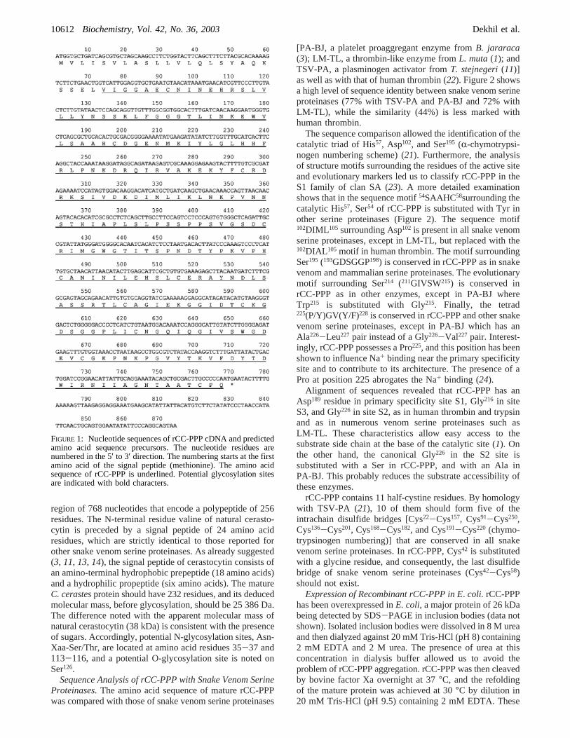
region of 768 nucleotides that encode a polypeptide of 256residues. The N-terminal residue valine of natural cerasto-cytin is preceded by a signal peptide of 24 amino acidresidues, which are strictly identical to those reported forother snake venom serine proteinases. As already suggested(3, 11, 13, 14), the signal peptide of cerastocytin consists ofan amino-terminal hydrophobic prepeptide (18 amino acids)and a hydrophilic propeptide (six amino acids). The matureC. cerastesprotein should have 232 residues, and its deducedmolecular mass, before glycosylation, should be 25 386 Da.The difference noted with the apparent molecular mass ofnatural cerastocytin (38 kDa) is consistent with the presenceof sugars. Accordingly, potential N-glycosylation sites, Asn-Xaa-Ser/Thr, are located at amino acid residues 35-37 and113-116, and a potential O-glycosylation site is noted onSer126.
Sequence Analysis of rCC-PPP with Snake Venom SerineProteinases.The amino acid sequence of mature rCC-PPPwas compared with those of snake venom serine proteinases
[PA-BJ, a platelet proaggregant enzyme fromB. jararaca(3); LM-TL, a thrombin-like enzyme fromL. muta(1); andTSV-PA, a plasminogen activator fromT. stejnegeri(11)]as well as with that of human thrombin (22). Figure 2 showsa high level of sequence identity between snake venom serineproteinases (77% with TSV-PA and PA-BJ and 72% withLM-TL), while the similarity (44%) is less marked withhuman thrombin.
The sequence comparison allowed the identification of thecatalytic triad of His57, Asp102, and Ser195 (R-chymotrypsi-nogen numbering scheme) (21). Furthermore, the analysisof structure motifs surrounding the residues of the active siteand evolutionary markers led us to classify rCC-PPP in theS1 family of clan SA (23). A more detailed examinationshows that in the sequence motif54SAAHC58surrounding thecatalytic His57, Ser54 of rCC-PPP is substituted with Tyr inother serine proteinases (Figure 2). The sequence motif102DIML 105 surrounding Asp102 is present in all snake venomserine proteinases, except in LM-TL, but replaced with the102DIAL 105 motif in human thrombin. The motif surroundingSer195 (193GDSGGP198) is conserved in rCC-PPP as in snakevenom and mammalian serine proteinases. The evolutionarymotif surrounding Ser214 (211GIVSW215) is conserved inrCC-PPP as in other enzymes, except in PA-BJ whereTrp215 is substituted with Gly215. Finally, the tetrad225(P/Y)GV(Y/F)228 is conserved in rCC-PPP and other snakevenom serine proteinases, except in PA-BJ which has anAla226-Leu227 pair instead of a Gly226-Val227 pair. Interest-ingly, rCC-PPP possesses a Pro225, and this position has beenshown to influence Na+ binding near the primary specificitysite and to contribute to its architecture. The presence of aPro at position 225 abrogates the Na+ binding (24).
Alignment of sequences revealed that rCC-PPP has anAsp189 residue in primary specificity site S1, Gly216 in siteS3, and Gly226 in site S2, as in human thrombin and trypsinand as in numerous venom serine proteinases such asLM-TL. These characteristics allow easy access to thesubstrate side chain at the base of the catalytic site (1). Onthe other hand, the canonical Gly226 in the S2 site issubstituted with a Ser in rCC-PPP, and with an Ala inPA-BJ. This probably reduces the substrate accessibility ofthese enzymes.
rCC-PPP contains 11 half-cystine residues. By homologywith TSV-PA (21), 10 of them should form five of theintrachain disulfide bridges [Cys22-Cys157, Cys91-Cys250,Cys136-Cys201, Cys168-Cys182, and Cys191-Cys220 (chymo-trypsinogen numbering)] that are conserved in all snakevenom serine proteinases. In rCC-PPP, Cys42 is substitutedwith a glycine residue, and consequently, the last disulfidebridge of snake venom serine proteinases (Cys42-Cys58)should not exist.
Expression of Recombinant rCC-PPP in E. coli. rCC-PPPhas been overexpressed inE. coli, a major protein of 26 kDabeing detected by SDS-PAGE in inclusion bodies (data notshown). Isolated inclusion bodies were dissolved in 8 M ureaand then dialyzed against 20 mM Tris-HCl (pH 8) containing2 mM EDTA and 2 M urea. The presence of urea at thisconcentration in dialysis buffer allowed us to avoid theproblem of rCC-PPP aggregation. rCC-PPP was then cleavedby bovine factor Xa overnight at 37°C, and the refoldingof the mature protein was achieved at 30°C by dilution in20 mM Tris-HCl (pH 9.5) containing 2 mM EDTA. These
FIGURE 1: Nucleotide sequences of rCC-PPP cDNA and predictedamino acid sequence precursors. The nucleotide residues arenumbered in the 5′ to 3′ direction. The numbering starts at the firstamino acid of the signal peptide (methionine). The amino acidsequence of rCC-PPP is underlined. Potential glycosylation sitesare indicated with bold characters.
10612 Biochemistry, Vol. 42, No. 36, 2003 Dekhil et al.
refolding conditions allowed us to obtain an average of 1.5mg of active protein from 10 mg of digested factor Xaprotein. At this stage, active proteinase represents only 15%of the total proteins. Thus, active rCC-PPP was purified frommisfolded and contaminant proteins by chromatography ina Mono S cation exchange column (Figure 3). Protein ofpeak 3, which is able to hydrolyze chromogenic substratesand which possesses platelet aggregating activity, representspurified rCC-PPP.
Analysis of Enzymatic and of Biological ActiVities ofRecombinant and Purified rCC-PPP.The enzymatic activityof recombinant and purified rCC-PPP was examined byassessing the hydrolysis of synthetic and chromogenicsubstrates S-2238 and S-2251. A significant catalytic activitywas noticed, and Michaelis constantsKm and kcat weredetermined from Lineweaver-Burk plots. They are com-pared in Table 1 with those determined, under the sameexperimental conditions, for natural cerastocytin and rCC-PPP. As shown in Table 1, the values of the kineticparameters of rCC-PPP and of natural cerastocytin are
similar. In addition, theKm values determined with S-2238for rCC-PPP and cerastocytin are close to that reported forPA-BJ (320µM) (3).
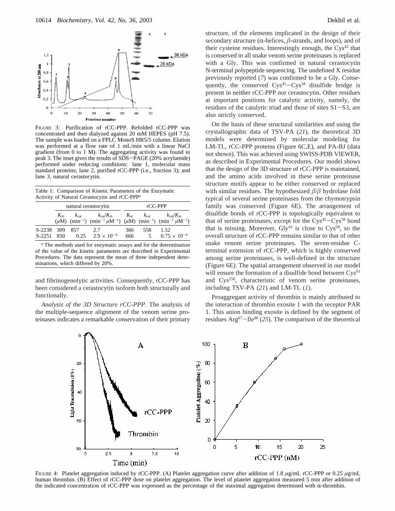
Like natural cerastocytin, purified rCC-PPP is a potentplatelet proaggregant activator for washed rabbit platelets,as shown in Figure 4A. The proaggregant activity of purifiedrCC-PPP is dose-dependent, and a half-effective dose of 8nM was determined (Figure 4B). This value is similar tothe one measured for purified natural cerastocytin [5 nM (7)]and slightly higher than that calculated for thrombin [1 nM(24)].
Natural cerastocytin was shown to clot purified fibrinogen,hydrolyzing theR-chain only (7). Similarly, rCC-PPP is ableto clot purified fibrinogen (not shown) and to hydrolyze theR-chain (Figure 5). The hydrolysis of theR-chain is completewithin <0.5 h. However, Figure 5 further shows that rCC-PPP degrades also more slowlyâ- and γ-chains, since asignificant hydrolysis of these chains can be noticed afterincubation for only 3 h. So far, natural cerastocytin and rCC-PPP are serine proteinases that have both platelet aggregating
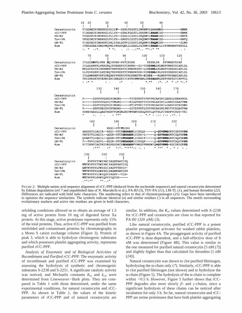
FIGURE 2: Multiple-amino acid sequence alignment of rCC-PPP (deduced from the nucleotide sequence) and natural cerastocytin determinedby Edman degradation (ref7 and unpublished data of N. Marrakchi et al.), PA-BJ (3), TSV-PA (11), LM-TL (1), and human thrombin (22).Differences are indicated with bold italic characters. The numbering refers to that of chymotrypsinogen (21). Gaps have been introducedto optimize the sequence similarities. The symbols indicate identical (/) and similar residues (:) in all sequences. The motifs surroundingevolutionary markers and active site residues are given in bold characters.
Platelet-Aggregating Serine Proteinase fromC. cerastes Biochemistry, Vol. 42, No. 36, 200310613
and fibrinogenolytic activities. Consequently, rCC-PPP hasbeen considered a cerastocytin isoform both structurally andfunctionally.
Analysis of the 3D Structure rCC-PPP.The analysis ofthe multiple-sequence alignment of the venom serine pro-teinases indicates a remarkable conservation of their primary
structure, of the elements implicated in the design of theirsecondary structure (R-helices,â-strands, and loops), and oftheir cysteine residues. Interestingly enough, the Cys42 thatis conserved in all snake venom serine proteinases is replacedwith a Gly. This was confirmed in natural cerastocytinN-terminal polypeptide sequencing. The undefined X residuepreviously reported (7) was confirmed to be a Gly. Conse-quently, the conserved Cys42-Cys58 disulfide bridge ispresent in neither rCC-PPP nor cerastocytin. Other residuesat important positions for catalytic activity, namely, theresidues of the catalytic triad and those of sites S1-S3, arealso strictly conserved.
On the basis of these structural similarities and using thecrystallographic data of TSV-PA (21), the theoretical 3Dmodels were determined by molecular modeling forLM-TL, rCC-PPP proteins (Figure 6C,E), and PA-BJ (datanot shown). This was achieved using SWISS-PDB VIEWER,as described in Experimental Procedures. Our model showsthat the design of the 3D structure of rCC-PPP is maintained,and the amino acids involved in these serine proteinasestructure motifs appear to be either conserved or replacedwith similar residues. The hypothesizedâ/â hydrolase foldtypical of several serine proteinases from the chymotrypsinfamily was conserved (Figure 6E). The arrangement ofdisulfide bonds of rCC-PPP is topologically equivalent tothat of serine proteinases, except for the Cys42-Cys58 bondthat is missing. Moreover, Gly42 is close to Cys58, so theoverall structure of rCC-PPP remains similar to that of othersnake venom serine proteinases. The seven-residue C-terminal extension of rCC-PPP, which is highly conservedamong serine proteinases, is well-defined in the structure(Figure 6E). The spatial arrangement observed in our modelwill ensure the formation of a disulfide bond between Cys91
and Cys250, characteristic of venom serine proteinases,including TSV-PA (21) and LM-TL (1).
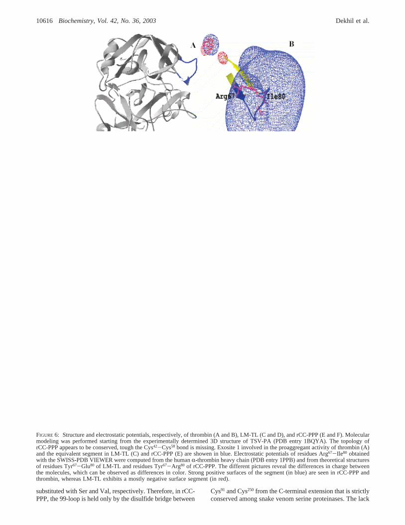
Proaggregant activity of thrombin is mainly attributed tothe interaction of thrombin exosite 1 with the receptor PAR1. This anion binding exosite is defined by the segment ofresidues Arg67-Ile80 (25). The comparison of the theoretical
FIGURE 3: Purification of rCC-PPP. Refolded rCC-PPP wasconcentrated and then dialyzed against 20 mM HEPES (pH 7.5).The sample was loaded on a FPLC MonoS HR5/5 column. Elutionwas performed at a flow rate of 1 mL/min with a linear NaClgradient (from 0 to 1 M). The aggregating activity was found inpeak 3. The inset gives the results of SDS-PAGE (20% acrylamide)performed under reducing conditions: lane 1, molecular massstandard proteins; lane 2, purified rCC-PPP (i.e., fraction 3); andlane 3, natural cerastocytin.
Table 1: Comparison of Kinetic Parameters of the EnzymaticActivity of Natural Cerastocytin and rCC-PPPa
natural cerastocytin rCC-PPP
Km
(µM)kcat
(min-1)kcat/Km
(min-1 µM-1)Km
(µM)kcat
(min-1)kcat/Km
(min-1 µM-1)
S-2238 309 857 2.7 366 558 1.52S-2251 850 0.25 2.9× 10⁻⁴ 666 5 0.75× 10⁻⁴
a The methods used for enzymatic assays and for the determinationof the value of the kinetic parameters are described in ExperimentalProcedures. The data represent the mean of three independent deter-minations, which differed by 20%.
FIGURE 4: Platelet aggregation induced by rCC-PPP. (A) Platelet aggregation curve after addition of 1.8µg/mL rCC-PPP or 0.25µg/mLhuman thrombin. (B) Effect of rCC-PPP dose on platelet aggregation. The level of platelet aggregation measured 5 min after addition ofthe indicated concentration of rCC-PPP was expressed as the percentage of the maximal aggregation determined withR-thrombin.
10614 Biochemistry, Vol. 42, No. 36, 2003 Dekhil et al.

3D models and the crystallographic structure of thrombin(20), especially at the level of Arg67-Ile80, indicates that thisloop projects out of the molecular surface of thrombin (Figure6A), LM-TL (Figure 6C), rCC-PPP (Figure 6E), and PA-BJ(data not shown), and forms a positively charged rim inplatelet proaggregant proteins, but not in LM-TL. The variousrepresentations shown in Figure 6 reveal the differences inelectric charges between the molecules in this area. Thestrong positive surface (in blue) of residues Tyr67-Arg80 inrCC-PPP (Figure 6F) and Arg67-Ile80 (Figure 6B) inthrombin is obvious, whereas LM-TL (in red) exhibits amostly negative surface of residues Tyr67-Glu80 (Figure 6D).In PA-BJ, the segment of residues Lys67-Glu80 presents astrong positive surface (data not shown) as for rCC-PPP andthrombin.
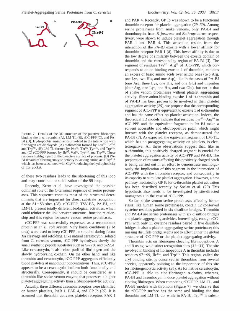
The analysis of the 3D molecular model of theL. mutathrombin-like enzyme, which has been reported recently (1),has revealed a unique hydrophobic pocket (Phe95-Trp99,Tyr172, Phe214, and Trp215) comparable to that found in thethrombin involved in the binding of fibrinopeptide A. Thestudies of Castro et al. revealed first that Arg16 of fibrin-opeptide A is making a salt bridge with Asp189 of LM-TLand keeping it proximal to the catalytic triad. Second, asexpected, Phe8, which is believed to be critical for efficientthrombin-catalyzed proteolysis of fibrinogen, and Leu9 offibrinopeptide A occupy the hydrophobic pocket of LM-TLin a manner similar to that of the thrombin (1). The arylbinding site is therefore similar in thrombin and LM-TL.The analysis of the 3D models shows that the hydrophobicamino acids involved in the interaction with fibrinogen areLeu99, Ile174, and Trp215 in R-thrombin (Figure 7A), Phe95,Thr96, Tyr172, and Trp215 in LM-TL (Figure 7B), and Ile98,Val99, Tyr172, and Trp215 in rCC-PPP (Figure 7C). The 90-loop (Phe90-Val99), Tyr172, and Trp215 residues of rCC-PPPthen form a hydrophobic pocket, and a similar structure existsin R-thrombin and LM-TL (Figure 7A,B). Therefore, therCC-PPP hydrophobic pocket should be participating in thecleavage of fibrinogen as a consequence of accommodationof the apolar residues usually found in fibrinopeptide A (Phe8
and Leu9) (1). More interesting is the Trp215 conserved inhumanR-thrombin and in venom serine proteinases rCC-PPP and LM-TL, pointing to the importance of this residuein fibrinogenolytic activity.
DISCUSSION
Several proteins, which share common properties withthrombin, have been purified to homogeneity fromC.cerastesvenom. They include cerastocytin (7) and cerastotin(26). These proteins are characterized by a marked plateletproaggregant action and a weak procoagulant activity.Nanomolar concentrations of cerastocytin induce aggregationof blood platelets (7). Studies of structure-function relation-ships of proaggregant protein and the analysis of theiractivities in ViVo require large amounts of protein. Toovercome this obstacle, we cloned and expressed, for thefirst time, a platelet proaggregant protein from snake venom.
In this report, we describe the construction of a cDNAencoding the precursor of aC. cerastesvenom protein, byRACE-RT-PCR. This protein, whose amino acid sequenceis more than 96% similar with the known polypeptidesequence of cerastocytin (Figure 2), is called rCC-PPP. Thesmall differences in the amino acid sequence between rCC-PPP and cersatocytin might result from individual and/orgeographical variations between the snake used for thecollection of venom proteins and the venom glands. Thededuced amino acid sequence of rCC-PPP contains twopotential N-glycosylation sites and is between 66 and 81%similar with sequences of other venom serine proteinases.The similarity of rCC-PPP with venom and mammalianserine proteinases was further confirmed by the presence ofthe highly conserved amino acid residues, which form theircatalytic triad of His57, Asp102, and Ser195 (chymotrypsinogennumbering) and their flanking sequences.
All the venom serine proteinases contain six well-conserved disulfide bridges, whereas rCC-PPP contains only11 half-cystine residues, which probably form only fiveintrachain disulfide bridges. The missing bridge is the Cys42-Cys58 bridge. In both PA-BJ and TSV-PA, the Cys42-Cys58
disulfide bridges form with residues 40 and 41, the S1′ site(21). This cavity is rather open and exposed to the solvent;for rCC-PPP, the lack of this cystine bridge should keep thiscavity more exposed.
The residue at position 193, which belongs to the S2′subsite, plays a key role in the determination of themacromolecular specificity of serine proteinases. In TSV-PA, Phe193 is involved in the hydrolysis of plasminogen andin the reduced sensitivity of this enzyme to Kunitz-typeinhibitors such as bovine pancreatic trypsin inhibitor (21,27). In LM-TL, this residue is an Arg193 and has beensuggested by Castro et al. (1) to have a similar role in thefibrinogen clotting activity of this enzyme. However, thisresidue is Gly193 in rCC-PPP, as in thrombin and many otherserine proteinases. Thus, the fibrinogen clotting activity doesnot seem to require an arginine residue at position 193, atleast in thrombin and rCC-PPP. Our protein, like thrombin,might be then sensitive to Kunitz-type inhibitors.
Compared to other serine proteinases, rCC-PPP presentsa two-residue deletion (Lys95 and Asp96). This region, calledthe 99-loop, is located at the north of the active site andprojects out of the molecular surface to form a highly chargedrim (Lys94-Lys95-Asp96-Asp97-Glu98-Val99). A salt bridge,between Asp97 and Arg174, stabilizes this loop and occludesthe S4 binding pocket (21). In rCC-PPP, the 99-loop presentsa deletion of Lys95 and Asp96 and the salt bridge thatstabilizes the loop is missing since Asp97 and Arg174 are
FIGURE 5: Hydrolysis of fibrinogen by rCC-PPP. Purified fibrino-gen (1%) was incubated with purified rCC-PPP (1.5µg/mL).Aliquots were removed at the indicated time and analyzed by SDS-PAGE (15%) under reducing conditions: lane 1, molecular massstandard proteins; and lanes 2-8, fibrinogen incubated with purifiedrCC-PPP for 0 min, 5 min, 30 min, 1 h, 3 h, 8 h, and 24 h,respectively.
Platelet-Aggregating Serine Proteinase fromC. cerastes Biochemistry, Vol. 42, No. 36, 200310615
substituted with Ser and Val, respectively. Therefore, in rCC-PPP, the 99-loop is held only by the disulfide bridge between
Cys91 and Cys250 from the C-terminal extension that is strictlyconserved among snake venom serine proteinases. The lack
FIGURE 6: Structure and electrostatic potentials, respectively, of thrombin (A and B), LM-TL (C and D), and rCC-PPP (E and F). Molecularmodeling was performed starting from the experimentally determined 3D structure of TSV-PA (PDB entry 1BQYA). The topology ofrCC-PPP appears to be conserved, tough the Cys42-Cys58 bond is missing. Exosite 1 involved in the proaggregant activity of thrombin (A)and the equivalent segment in LM-TL (C) and rCC-PPP (E) are showen in blue. Electrostatic potentials of residues Arg67-Ile80 obtainedwith the SWISS-PDB VIEWER were computed from the humanR-thrombin heavy chain (PDB entry 1PPB) and from theoretical structuresof residues Tyr67-Glu80 of LM-TL and residues Tyr67-Arg80 of rCC-PPP. The different pictures reveal the differences in charge betweenthe molecules, which can be observed as differences in color. Strong positive surfaces of the segment (in blue) are seen in rCC-PPP andthrombin, whereas LM-TL exhibits a mostly negative surface segment (in red).
10616 Biochemistry, Vol. 42, No. 36, 2003 Dekhil et al.
of these two residues leads to the shortening of this loopand may contribute to stabilization of the 99-loop.
Recently, Krem et al. have investigated the possibledominant role of the C-terminal sequence of serine protein-ases. This sequence contains most of the structural deter-minants that are important for direct substrate recognitionat the S1-S3 sites (28). rCC-PPP, TSV-PA, PA-BJ, andLM-TL present totally different biological activities, whichcould reinforce the link between structure-function relation-ship and this region for snake venom serine proteinases.
rCC-PPP was successfully expressed as a recombinantprotein in anE. coli system. Very harsh conditions (2 Murea) were used to keep rCC-PPP in solution during factorXa cleavage and refolding. Like natural cerastocytin isolatedfrom C. cerastesvenom, rCC-PPP hydrolyzes slowly thesmall synthetic peptide substrates such as S-2238 and S-2251.Like cerastocytin, it also clots purified fibrinogen and theslowly hydrolyzing R-chain. On the other hand, and likethrombin and cerastocytin, rCC-PPP aggregates efficientlyblood platelets at nanomolar concentrations. Thus, rCC-PPPappears to be a cerastocytin isoform both functionally andstructurally. Consequently, it should be considered as athrombin-like snake venom enzyme that possesses a higherplatelet aggregating activity than a fibrinogenolytic activity.
Actually, three different thrombin receptors were identifiedon human platelets, PAR 1, PAR 4, and GP Ib (29). It isassumed that thrombin activates platelet receptors PAR 1
and PAR 4. Recently, GP Ib was shown to be a functionalthrombin receptor for platelet aggregation (29, 30). Amongserine proteinases from snake venoms, only PA-BJ andthrombocytin, fromB. jararacaandBothrops atrox, respec-tively, were shown to induce platelet aggregation throughPAR 1 and PAR 4. This activation results from theinteraction of the PA-BJ exosite with a lower affinity forthrombin receptor PAR 1 (8). This lower affinity is due tothe low degree of similarity between the exosite domain ofthrombin and the corresponding region of PA-BJ (3). Thesegment of residues Tyr67-Arg80 of rCC-PPP, which cor-responds to anion-binding exosite 1 of thrombin, containsan excess of basic amino acids over acidic ones (two Arg,one Lys, two His, and one Asp), like in the cases of PA-BJ(one Arg, three Lys, one His, and one Glu) and thrombin(four Arg, one Lys, one His, and two Glu), but not in thatof snake venom proteinases without platelet aggregatingactivity. Since anion-binding exosite 1 ofR-thrombin andof PA-BJ has been proven to be involved in their plateletaggregation activity (25), we propose that the correspondingsegment of rCC-PPP is equivalent to exosite 1 ofR-thrombinand has the same effect on platelet activation. Indeed, thetheoretical 3D models indicate that residues Tyr67-Arg80 inrCC-PPP and the equivalent fragment in PA-BJ make asolvent accessible and electropositive patch which mightinteract with the platelet receptor, as demonstrated forPA-BJ (3). As expected, the equivalent segment of LM-TL,which has no proaggregating activity on platelets, is elec-tronegative. All these observations suggest that, like inR-thrombin, this positively charged segment is crucial forthe platelet aggregation activity of rCC-PPP and PA-BJ. Thepreparation of mutants affecting this positively charged patchis being carried out in an effort to demonstrate unambigu-ously the implication of this segment in the interaction ofrCC-PPP with the thrombin receptor, and consequently inits capacity to stimulate platelet aggregation. However, a newpathway mediated by GP Ib forR-thrombin platelet activationhas been described recently by Soslau et al. (29) Thishypothesis also needs to be investigated by site-directedmutagenesis in the case of rCC-PPP.
So far, snake venom serine proteinases affecting hemo-stasis, like human serine proteinases, contain 12 conservedcysteine residues paired in six disulfide bridges. Thrombinand PA-BJ are serine proteinases with six disulfide bridgesand platelet aggregating activities. Interestingly, enough rCC-PPP with only 11 cysteine residues paired in five disulfidebridges is also a platelet aggregating serine proteinase; thismissing disulfide bridge seems not to affect either the globalstructure of rCC-PPP or the platelet aggregating activity.
Thrombin acts on fibrinogen cleaving fibrinopeptides Aand B using two distinct recognition sites (31-33). The siteinvolved in binding of fibrinopeptide A in thrombin includesresidues 97-99, Ile174, and Trp215. This region, called thearyl binding site, is conserved in thrombins from severalspecies, apparently pointing to the importance of this sitefor fibrinogenolytic activity (34). As for native cerastocytin,rCC-PPP is able to clot fibrinogenR-chains, whereas,PA-BJ and thrombocytin induce platelet aggregation withoutclotting fibrinogen. When comparing rCC-PPP, LM-TL, andPA-BJ models with thrombin (Figure 7), we observe thatthe rCC-PPP surface has the same aryl binding site thatthrombin and LM-TL do, while in PA-BJ, Trp215 is substi-
FIGURE 7: Details of the 3D structure of the putative fibrinogenbinding site inR-thrombin (A), LM-TL (B), rCC-PPP (C), and PA-BJ (D). Hydrophobic amino acids involved in the interaction withfibrinogen are displayed: (A)R-thrombin formed by Leu99, Ile174,and Trp215, (B) LM-TL formed by Phe95, Thr96, Tyr172, and Trp215,and (C) rCC-PPP formed by Ile98, Val99, Tyr172, and Trp215. Theseresidues highlight part of the bioactive surface of proteins. (D) PA-BJ devoid of fibrinogenolytic activity is lacking amino acid Trp215,which has been substituted with Gly215, reducing the hydrophobicityof this pocket.
Platelet-Aggregating Serine Proteinase fromC. cerastes Biochemistry, Vol. 42, No. 36, 200310617
tuted with Gly. Because of its location, surrounding the S3binding pocket, this substituted residue could be involvedin the limited recognition and specific cleavage of fibrinogen.The lack of an aromatic ring of tryptophan in this environ-ment reduces the hydrophobicity of this pocket. This effectsuggests that Trp215 resides in the putative bioactive surfaceof PA-BJ upon fibrinogen. It is tempting to speculate thatthe aryl binding site of rCC-PPP is also involved infibrinogen clotting activity. The importance of these regionson fibrinogen binding should be further investigated bymutational studies.
As suggested by Stubbs et al. (33), the thrombin andfibrinogen molecules complement each other over a largesurface, with simultaneous interactions at the aryl bindingsite, the fibrinogen recognition exosite, and subsites S3-S3′. The S3′ site in thrombin formed by Glu39, Leu41, Cys42-Cys58, and Glu192 is implicated in the catalysis and releaseof fibrinopeptideR- andâ-chains. As far as we know, theCys42-Cys58 disulfide bridge is only implicated in the S3′site of thrombin; in this context, mutation of Glu39 to Lys(35) decreases the ability of thrombin to clot fibrinogen. InrCC-PPP, arginine at position 39 together with the missingCys42-Cys58 disulfide bridge may contribute to a release offibrinopeptideR- and â-chains that is slower than that forthe wild type. So far, this missing disulfide bridge does notinterfere with the two main activities of rCC-PPP (i.e.,fibrinogenolysis and platelet aggregation).
In conclusion, we propose that the effect of viper venomserine proteinase rCC-PPP on platelets and fibrinogen ismediated by the positively charged segment of residuesTyr67-Arg80 and the aryl binding site, respectively. Themissing disulfide bridge is not involved in the two mainactivities of rCC-PPP. The equivalent regions mediate thethrombin effects on platelets and fibrinogen.
ACKNOWLEDGMENT
We gratefully acknowledge Professor Koussay Dellagi forhis constant interest in this work and for his helpfulencouragement. We thank Dr. Ben Lasfar Zakaria, from theSerpentarium of Institut Pasteur de Tunis, for providingC.cerastessnakes and venoms.
REFERENCES
1. Castro, H. C., Silva, D. M., Craik, C., and Zingali, R. B. (2001)Biochim. Biophys. Acta 1547, 183-195.
2. Stocker, K. (1990) Snake venom proteins affecting hemostasis andfibrinolysis, in Medical use of snakeVenom proteins(Stocker,K., Ed.) pp 97-160, CRC Press, Boca Raton, FL.
3. Serrano, S. M., Mentele, R., Sampaio, C. A., and Fink, E. (1995)Biochemistry 34, 7186-7193.
4. Schmaier, A. H., and Colman, R. W. (1980)Blood 56, 1020-1028.
5. Serrano, S. M., Matos, M. F., Mandelbaum, F. R., and Sampaio,C. A. (1993)Toxicon 31, 471-481.
6. Kirby, E. P., Niewiarowski, S., Stocker, K., Kettner, C., Shaw,E., and Brudzynski, T. M. (1979)Biochemistry 18, 3564-3570.
7. Marrakchi, N., Zingali, R. B., Karoui, H., Bon, C., and El Ayeb,M. (1995)Biochim. Biophys. Acta 1244, 147-156.
8. Santos, A. B. F., Serrano, S. M. T., Kuliopulos, A., andNiewiarowski, S. (2000)FEBS Lett. 477, 199-202.
9. Itoh, N., Tanaka, N., Mihashi, S., and Yamashina, I. (1987)J.Biol. Chem. 262, 3132-3135.
10. Au, L. C., Lin, S. B., Chou, J. S., Teh, G. W., Chang, K. J., andShih, C. M. (1993)Biochem. J. 294, 387-390.
11. Zhang, Y., Wisner, A., Xiong, Y., and Bon, C. (1995)J. Biol.Chem. 270, 10246-10255.
12. Chomczynski, P., and Sacchi, N. (1987)Anal. Biochem. 162, 156-159.
13. Serrano, S. M., Hagiwara, Y., Murayama, N., Higuchi, S., Mentele,R., Sampaio, C. A., Camargo, A. C., and Fink, E. (1998)Eur. J.Biochem. 251, 845-853.
14. Deshimaru, M., Ogawa, T., Nakashima, K., Nobuhisa, I., Chijiwa,T., Shimohigashi, Y., Fukumaki, Y., Niwa, M., Yamashina, I.,Hattori, S., and Ohno, M. (1996)FEBS Lett. 397, 83-88.
15. Frohman, M. A. (1990)Amplifications 5, 11-15.16. Zhang, Y., Wisner, A., Maroun, R. C., Choumet, V., Xiong, Y.,
and Bon, C. (1997)J. Biol. Chem. 272, 20531-20537.17. Ardlie, N. G., Perry, D. W., Packham, M. A., and Mustard, J. F.
(1971)Proc. Soc. Exp. Biol. Med. 136, 1021-1023.18. Born, G. V. P., and Cross, M. J. (1963)J. Physiol. 168, 178-
195.19. Thompson, J. D., Gibson, T. J., Plewniak, F., Jeanmougin, F., and
Higgins, D. G. (1997)Nucleic Acids Res. 25, 4876-4882.20. Bode, W., Mayr, I., Baumann, U., Huber, R., Stone, S. R., and
Hofsteenge, J. (1989)EMBO J. 8, 3467-3475.21. Parry, M. A., Jacob, U., Huber, R., Wisner, A., Bon, C., and Bode,
W. (1998)Structure 6, 1195-1206.22. Degen, S. J., and Davie, E. W. (1987)Biochemistry 26, 6165-
6177.23. Krem, M. M., and Di Cera, E. (2001)EMBO J. 20, 3036-3045.24. Dang, Q. D., and Di Cera, E. (1996)Proc. Natl. Acad. Sci. U.S.A.
93, 10653-10656.25. Jakubowski, J. A., and Maraganone, J. M. (1990)Blood 75, 399-
406.26. Marrakchi, N., Barbouche, R., Guermazi, S., Karoui, H., Bon, C.,
and El Ayeb, M. (1997)Eur. J. Biochem. 247, 121-128.27. Braud, S., Bon, C., and Wisner, A. (2000)Biochimie 82, 851-
859.28. Krem, M. M., Rose, T., and Di Cera, E. (1999)J. Biol. Chem.
274, 28063-28066.29. Soslau, G., Class, R., Morgan, D. A., Foster, C., Lord, S. T.,
Marchese, P., and Ruggeri, Z. M. (2001)J. Biol. Chem. 276,21173-21183.
30. Ramakrishnan, V., DeGuzman, F., Bao, M., Hall, S. W., Leung,L. L., and Phillips, D. R. (2001)Proc. Natl. Acad. Sci. U.S.A. 98,1823-1828.
31. Bode, C., Baumann, H., von Hodenberg, E., Freitag, M., and Nordt,T. (1993)Z. Kardiol. 82, 125-128.
32. Di Cera, E., Dang, Q. D., and Ayala, Y. (1997)Cell. Mol. LifeSci. 53, 701-730.
33. Stubbs, M. T., Oschkinat, H., Mary, I., Huber, R., Angliker, H.,Stone, S. R., and Bode, W. (1992)Eur. J. Biochem. 206, 187-195.
34. Banfield, D. K., and MacGillivray, R. T. A. (1992)Proc. Natl.Acad. Sci. U.S.A. 89, 2779-2783.
35. LeBonniec, B. F., MacGillivray, R. T., and Esmon, C. T. (1991)J. Biol. Chem. 266, 13796-13803.
BI034790B
10618 Biochemistry, Vol. 42, No. 36, 2003 Dekhil et al.
Copyright © 2022 FDOKUMEN