Generation of Pluripotent Stem Cells from Neonatal Mouse Testis
Upload
independentCategory
view
0download
0
Introduction
Spermatogenesis is a complex, highly organized processthat originates from stem cell spermatogonia. In non-primate mammals, the As (A single) spermatogonia areconsidered to be the stem cells of spermatogenesis(Huckins, 1971; Oakberg, 1971; de Rooij, 1973). Upondivision of the As spermatogonia, the daughter cells eithermigrate away from each other and become two new stemcells, or stay together through an intercellular bridge and become A-paired (Apr) spermatogonia. The Apr sper-matogonia develop further into chains of four, eight or 16 A-aligned (Aal) spermatogonia. The Aal spermatogoniadifferentiate into A1 spermatogonia and after six mitoticdivisions result in A2, A3, A4 and, finally, B spermatogonia,which give rise to spermatocytes at the last mitotic division.Irrespective of species-specific differences, in the bovinetestis, a comparable classification with another terminologyhas been reported (Wrobel et al., 1995a). In this species,
spermatogonial precursor cells have been divided into basalstem cells (BSC), aggregated spermatogonial precursor cells(ASPC) and committed spermatogonial precursor cells(CSPC), which, according to this classification, representAs–Apr spermatogonia, Aal spermatogonia and A1–A4 differ-entiating spermatogonia, respectively. Hence, in bulls, Aprspermatogonia are also thought to have stem cell properties(Wrobel, et al., 1995a).
As there are relatively few stem cells that can be definedonly by their function, the identification and isolation of thesecells has been very difficult. To date, the most effective wayto enrich germ cell populations for stem cells is to purify allforms of type A spermatogonia. In the adult mammaliantestis, owing to the presence of multiple generations ofgerminal cells, purification of spermatogonia is more diffi-cult than it is before puberty. Bellve et al. (1977) obtained a90% pure fraction of type A spermatogonia from immaturemice. Similarly, highly purified spermatogonia have beenobtained from immature rat (Morena et al., 1996) and pig(Dirami et al., 1999) testes. In addition, vitamin A-deficientanimals can be used as a source of spermatogonia as, inthese animals, spermatogenesis stops at a spermatogonial
Isolation and purification of type A spermatogonia from the bovine testis
F. Izadyar1,2, G. T. Spierenberg3, L. B. Creemers1,2, K. den Ouden1,2 and D. G. de Rooij1,2
1Department of Endocrinology, Faculty of Biology, University Medical Center Utrecht,Utrecht, The Netherlands; and Departments of 2Cell Biology and 3Immunology, University
Medical Center Utrecht, Utrecht, The Netherlands
Reproduction (2002) 124, 85–94 Research
The aim of this study was to isolate and purify bovine typeA spermatogonia. Testes from 5–7-month-old calves wereused to isolate germ cells using a two-step enzymaticdigestion. During the isolation and purification steps, the viability of cells was determined using live/deadstaining. The identity of type A spermatogonia during isola-tion and purification was determined under a light micro-scope equipped with a Nomarski lens. Isolated cells werecharacterized further by using specific markers for type Aspermatogonia, including Dolichos biflorus agglutinin(DBA) and c-kit. The cell suspension was transplanted intoimmunodeficient recipient mouse testes and the colon-ization was assessed 1–3 months after transplantation, toassess the stem cell population among the isolated cells.After isolation, a cell suspension was obtained containingabout 25% type A spermatogonia, which was enrichedfurther by differential plating and separation on a discon-tinuous Percoll gradient. Finally, fractions containing
65–87% pure type A spermatogonia were obtained. Largeand small type A spermatogonia with different numbersand sizes of nucleoli were found. DBA stained both largeand small type A spermatogonia and its application influorescence-activated cell sorting (FACS) resulted incomparable percentages of type A spermatogonia to thosedetermined by morphological examination under a lightmicroscope equipped with a Nomarski lens. Nearly all of the large type A spermatogonia showed strong c-kitimmunoreactivity, indicating that these cells had under-gone at least an initial differentiation step. In contrast,approximately half of the small type A spermatogonia werenegative for c-kit, indicating the presence of the sper-matogonial stem cells in this population. At 3 months aftertransplantation, groups of bovine type A spermatogoniawere found in most tubule cross-sections of the recipientmouse testes, showing the presence of spermatogonialstem cells among the isolated cells.
© 2002 Society for Reproduction and Fertility1470-1626/2002
Email: [email protected]

differentiation step (van Pelt et al., 1996). At present, withthe exception of a single study in which a mixed germ cellpopulation, including type A spermatogonia, was collectedfrom bovine testes and used for transplantation (Dobrinskiet al., 2000), no information is available on the isolation ofpure bovine type A spermatogonia.
In studies on spermatogonial isolation and purification,the availability of markers that can conclusively establishthe identity of the spermatogonia is essential. One such amarker is c-kit, the receptor for stem cell factor (SCF), whichis expressed by some Aal and by A1–A4, In and B sper-matogonia, but not by As and Apr spermatogonia (Schrans-Stassen et al., 1999). Therefore, application of c-kit forisolation of spermatogonia results in the selection of moredifferentiated spermatogonia than spermatogonial stemcells (Shinohara et al., 2000). In contrast, isolation of mousespermatogonia on the basis of α-6 and β-1 integrin resultedin an enriched population of spermatogonial stem cells(Shinohara and Brinster, 2000). In bulls, the lectin Dolichosbiflorus agglutinin (DBA), which has a specific affinity for α-D-N-acetyl-galactosamine, can be used as a specificmarker for gonocytes and spermatogonia in the testis duringthe first 30 weeks after birth (Ertl and Wrobel, 1992). Wehave developed a procedure by which highly purified typeA spermatogonia can be isolated from the testes of 5-month-old bull calves. The aim of the present study was to inves-tigate the morphological and physiological characteristicsof the purified type A spermatogonia and their stem cellpotential.
Materials and Methods
Collection of testes and histology
Testes from 3–7-month-old calves were collected froman abattoir, placed on ice and transferred to the laboratorywithin 2 h, to study the development of spermatogenic cellsduring the prepubertal period. Approximately 20 g of testic-ular tissue was used for each cell isolation procedure and asample was taken for histological examination. Testicularsamples were fixed in Bouin’s solution and embedded in glycol methacrylate Technovit 7100 (Kulzerand Co.,GmbH, Wehrheim). Sections of 5 µm were cut, stained withperiodic acid–Schiff and Gill’s haematoxylin and evaluatedunder a light microscope.
Cell isolation, purification and identification
After decapsulation, the testes were minced into smallpieces and suspended in minimum essential medium(MEM; Gibco Life technology, Paisley) supplemented with14 mol NaHCO3 l–1, 4 mol L-glutamine l–1 (both fromSigma, St Louis, MO), single-strength non-essential aminoacids, 100 iu ml–1–100 µg ml–1 penicillin–streptomycin,40 µg gentamycin ml–1 and 15 mol Hepes l–1 (all fromGibco) (subsequently referred to as MEM). Seminiferousepithelial cells were dispersed with enzyme and separatedusing the method of van Pelt et al. (1996) with minor
modifications. In brief, minced testis pieces were suspendedin MEM containing 1 mg collagenase ml–1 and 1 mg trypsin ml–1 (both from Worthington, Freehold, NJ), 1 mghyaluronidase type II ml–1 (Sigma) and 5 µg DNAse I ml–1
(Boehringer, Mannheim) and incubated at 328C for 60 minin a shaking waterbath operated at 140 cycles min–1. Afterthree washes in MEM medium and removal of most of theinterstitial cells, seminiferous cord fragments were incu-bated in MEM containing collagenase, hyaluronidase andDNAse for 45 min as described above. Cells were separatedfrom the remaining tubule fragments by centrifugation at30 g for 2 min. After filtration through 77 and 55 µm nylonfilters, the cells were pelleted and subjected to differentialplating to eliminate the somatic cells (myoid and Sertolicells). The pooled cells were incubated overnight in MEMcontaining 10% FCS (Gibco) at 378C. After removal ofSertoli and myoid cells, spermatogonia, which remained insuspension, were collected and loaded onto a discontin-uous Percoll density gradient for further purification (vanPelt et al., 1996). Fractions containing > 50% type A sper-matogonia were washed, counted and used. The purity ofthe cell suspension at the various steps was determinedusing Nomarski interference microscopy.
Viability assays
The viability of the cells during isolation and purificationsteps was determined using a live–dead kit (MolecularProbes, Eugene, OR). Viability was assessed by determiningthe number of calcein-AM-positive cells (1 µmol l–1 inMEM) and ethidium homodimer-positive cells (1 µmol l–1 inMEM) after 10 min of incubation at 378C. Binding ofcalcein-AM to the enzyme esterase, which is present in thecytoplasm of living cells only, produces a green colour.Ethidium homodimer passes through the plasma membraneof dead cells only, binding to the nucleus and producing ared colour.
DBA staining and FACS analysis
Approximately 2 3 106 fixed cells were washed twice inPBS containing 5 mg BSA ml–1 (PBS–BSA) to eliminate thefixative. After centrifugation at 2000 g for 5 min, the pelletwas resuspended (approximately 1 3 106 cells ml–1) influorescein isothiocyanate (FITC)-conjugated DBA at aconcentration of 100 µg ml–1 in PBS containing 10 mg BSAml–1 for 15 min at 378C. Cells were washed in PBS–BSAthree times and finally the pellet was resuspended in 0.5 mlPBS–BSA. Cells were first analysed under a fluorescencemicroscope (Nikon TE 200, Tokyo) to check the labelling.The fluorescence emission of the stained cells was mea-sured on a FACScan flow cytometer interfaced to aMacintosh computer equipped with Cell Quest software(Becton & Dickinson, San Jose, CA). The excitation sourcesconsisted of an argon ion laser operated at 488 nm. FITCfluorescence was monitored using a 530 nm band-pass filterand at least 10 000 events were evaluated for each sample.
86 F. Izadyar et al.

Immunohistochemcal localization of DBA and c-kit
In addition to evaluation under a light microscope, type A spermatogonia were identified using DBA immuno-histochemistry as described by Ertl and Wrobel (1992).Testicular sections fixed in Bouin’s solution and embeddedin paraffin wax, and isolated cells on glass chamber slides(Nunc, Life Technologies, Roskilde), also fixed in Bouin’ssolution and preserved in 70% ethanol, were used forimmunohistochemistry. In brief, after removal of paraffinwax (for tissue sections only) and rehydration, sections were treated with 3% (v/v) H2O2 (Merck, Darmstadt) for10 min to inhibit endogenous peroxidase and were sub-sequently rinsed in PBS. Incubation in PBS containing50 mg BSA ml–1 for 15 min before lectin incubation wasadvantageous to block non-specific adhesion sites. Thesections were then incubated in DBA conjugated withhorseradish peroxidase (DBA–HRP; E. Y. Laboratories, SanMateo, CA) at 1:100 in PBS and 1% (w/v) BSA for 1 h at378C in a moist chamber. After incubation with lectin, thesections were rinsed three times in PBS. Staining of theDBA–HRP was performed by treating the sections for5–15 min with PBS containing 25 mg 3,39-diaminobenzi-dine tetrahydrochloride (DAB) (Sigma), 1 ml nickelammonium sulphate solution (10 mg ml–1), 1.25 ml cobaltchloride solution (10 mg ml–1) and 17 µl of 35% (v/v) H2O2per 50 ml. The slides were rinsed thoroughly in distilledwater and, if necessary, counterstained with haematoxylin.The sections were dehydrated in graded alcohol, cleared inxylol and mounted with Pertex (Cell Path; Compulink,Bedford). Negative control sections were incubated in 1%BSA in PBS without lectin.
The differentiation stage of the type A spermatogonia wasstudied using c-kit immunolabelling. Testicular sectionsfixed in Bouin’s solution and embedded in paraffin wax, andisolated cells also fixed in Bouin’s solution and preserved in70% ethanol, were used. The slides were washed in PBS for10 min, treated with 3% (v/v) H2O2 for 10 min to inhibitendogenous peroxidase and rinsed in PBS. The slides werethen incubated in the blocking solution containing 5%normal goat serum (Vector, Burlingame, CA) with 10 mgBSA ml–1 in PBS for 1 h. All incubations were performed in amoist chamber at room temperature. After blocking, theslides were rinsed in acetylated BSA (BSA-c) (0.5 mg ml–1) in PBS (PBS–BSA-c) and incubated overnight with poly-clonal rabbit anti-c-kit (C-19) antibody (1:100) diluted inPBS–BSA-c. After three rinses in PBS–BSA-c, the slides wereincubated for 1 h with biotinylated goat anti-rabbit antibody(1:100) diluted in PBS–BSA-c. After rinsing in PBS, the slideswere incubated in avidin–biotin complex (ABC) for 60 min.The slides were then rinsed in Tris buffer solution (TBS)(0.05 mol Tris l–1 (Merck, Amsterdam) and 0.85 mol NaCll–1) containing 0.1% (v/v) Tween-20 (Merck), and wereincubated for 15–30 min in TBS containing 25 mg DAB and15 µl of 3.5% (v/v) H2O2. After rinsing thoroughly in water,the sections were studied under a Nikon inverted lightmicroscope as described earlier.
Preparation of donor cells and recipient testes, andtransplantation
After isolation and purification, the cells were re-suspended in MEM–BSA–DNAse at a concentration of20 3 106 cells ml–1 and kept on ice until transplantation.Adult NMRI mice HsdCpb (nu/nu; Harlan, Horst) were usedas recipients. NMRI mice lack T cells and are immuno-deficient; therefore, they were kept under specific pathogen-free conditions and food, water and bedding were autoclavedbefore use. The mice were housed in a 12 h light :12 h darkcycle at constant temperature and provided with food andwater ad libitum. Recipient mice were given a fractionatedX-ray dose of 1.5 and 12.0 Gy with an interval of 24 h, todestroy endogenous spermatogenesis. This radiation dose issufficient to block virtually all endogenous spermatogenesisand does not have any apparent harmful effect on support-ing Sertoli cells (Creemers et al., in press). At 1 month after irradiation, the recipient mice were anaesthetized by i.p. administration of a mixture of fentanyl, fluanison andmidazolam (10 mg kg–1; Roche, Mijdrecht). Testes wereexposed through a mid-line abdominal incision, and donorcells (about 25 µl) were injected via a micropipette throughthe efferent ducts into the rete testis as described by Ogawaet al. (1997). The contralateral testis was used as the nega-tive control. Testes were harvested at 1–3 months aftertransplantation, fixed in Bouin’s solution and the coloniza-tion efficiency was assessed using DBA immunohisto-chemistry. The experimental protocol of this study followedthe guidelines of the care and use of laboratory animals andwas approved by the animal care and use committee ofUtrecht University.
Statistical analysis
The results are presented as means 6 SEM and statisticalanalysis was performed by Student’s t test. Differences wereconsidered significant when the P value was < 0.05.
Results
Histology
The presence of the various types of spermatogenic cellsin the testes of prepubertal bulls of various ages (3–7months) was studied (Figs 1 and 2). At 3 months of age,gonocytes were the only germ cells present. The gonocyteswere large, localized mostly in the middle of the seminif-erous tubules and occasionally at the basal membrane (Fig.2a). However, by 5 months, most tubules contained type Aspermatogonia and gonocytes, and a few contained sper-matocytes as the most advanced germ cells (Fig. 2b). At 6months, the diameter of the seminiferous tubules increasedand a lumen was observed for the first time. About 60% ofthe tubule cross-sections contained primary spermatocytesand 20% contained spermatids (Fig. 2c). At 7 months, thediameter of the seminiferous tubules had increased further
Isolation of bovine spermatogonia 87

and a lumen was clearly visible. Elongated spermatids werethe most advanced cells observed at 7 months (Fig. 2d).
Cell isolation and purity
After isolation, the cell suspension contained on average25.5 6 2.8% type A spermatogonia. Differential plating and subsequent Percoll gradient separation significantlyenhanced the purity of these cells to 45 6 3.1 (P < 0.01) and73 6 2.9% (P < 0.001), respectively. The viability of thecells after isolation was > 90%, which diminished slightlyafter overnight culture and exposure to a Percoll gradient(Fig. 3a,b). Regardless of the age of the testes, most of thespermatogonia were found in fraction three of the Percollgradient (Fig. 4), and the purity of the spermatogonialfraction was higher at 5 months than at 6 or 7 months (Fig. 5). When testes from 5-month-old calves were used,approximately 1 3 106 type A spermatogonia per gram oftestes of a purity of about 75% could be obtained routinely.
FACS analysis
The purity of type A spermatogonia in the cell suspensionwas assessed using DBA–FITC and flow cytometry. Twosubpopulations of DBA–FITC-positive cells were detectedcontaining small and large cells, presumably small or largetype A spermatogonia. The population of large cells was on average tenfold greater than the population of small cells(Fig. 6a,b). The purity of the type A spermatogonia, asassessed by morphological examination under a lightmicroscope equipped with a Nomarski lens, was similar tothat estimated by DBA flow cytometry.
Identification of spermatogonia in the testis and afterisolation
In the seminiferous epithelium of 5-month-old calves,type A spermatogonia were round cells of various sizes with
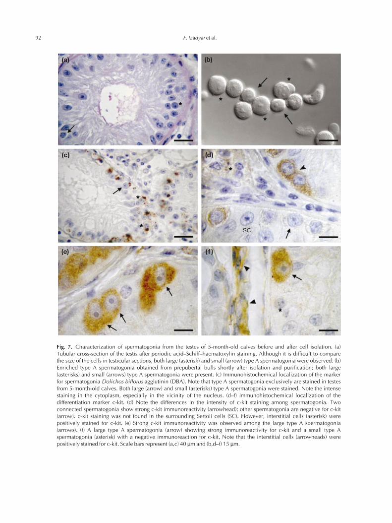
a spherical nucleus and one to three centrally located densenucleoli of about 1–3 µm in diameter (Fig. 7a). Purifiedspermatogonia contained both large and small type Aspermatogonia (Fig. 7b). Immunohistochemical staining forDBA showed that, in testes from 5-month-old calves, onlytype A spermatogonia were stained. DBA staining waslocalized mostly as a dense coloured area at the Golgicomplex in the vicinity of the nucleus and was notdetectable in the nucleus (Fig. 7c). In the testes of five-month-old calves, the intensity of staining for c-kit in type Aspermatogonia varied and, in general, large type A sper-matogonia had stronger c-kit immunoreactivity than didsmall type A spermatogonia (Fig. 7d–f): > 90% of the largetype A spermatogonia showed c-kit immunoreactivity,whereas about 50% of the small type A spermatogonia werenegative for c-kit (Table 1).
Evaluation of stem cell activity
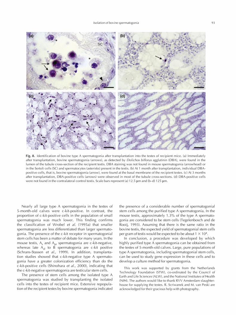
Isolated cells were transplanted into the testes ofrecipient immunodeficient mice to investigate the function-ality of the spermatogonial stem cells among the isolatedtype A spermatogonia. Immediately after transplantation,bovine type A spermatogonia, as detected with DBAstaining, were found in the seminiferous tubule lumen of the recipient mice (Fig. 8a). At 1 month after trans-plantation, a few bovine spermatogonia were found at thebasal membrane of the tubules (Fig. 8b). At 3 months aftertransplantation, groups of DBA-positive cells, that is, bovinespermatogonia, were found in the tubule cross-sections ofthe recipient testes (Fig. 8c). At this time, bovine sper-matogonia were found on average in > 60% of the tubulecross-sections. Bovine germ cells at advanced stages ofdevelopment were not found in the mouse testis. DBA-positive cells were not found in the contralateral controltestis (Fig. 8d).
Discussion
This study demonstrates that highly purified type Aspermatogonia can be obtained from the testes of 5–7-month-old calves. In terms of the most appropriate age ofcalves from which to isolate type A spermatogonia, itappeared that at 5 months most of the tubule cross-sections
88 F. Izadyar et al.
3 5 6 70
20
40
60
80
100
Approximate age (months)Sem
inife
rous
cro
ss-s
ectio
ns(%
)
Fig. 1. Percentage of seminiferous tubules containing differenttypes of germ cell in the testes of bull calves at different ages: (d)spermatogonia and gonocytes; (j) primary and secondary sper-matocytes; (m) round and elongated spermatids. The results repre-sent pooled data obtained from 11 different animals (mean 6 SEM).
Table 1. c-kit immunoreactivity of bovine type A spermatogonia inrelation to the size of the spermatogonia
c-kit immunoreactivity
Size of type A spermatogonia Positive Negative
Large 93.4 6 3.6a 6.6 6 3.7a
Small 52.2 6 5.7b 47.8 6 8.2b
Results are the pooled data from three independent experiments.abWithin columns, values with different superscripts are significantlydifferent (P < 0.05).

contained type A spermatogonia as the most advanced typeof germ cell, and these testes proved to be the best sourcefor isolation of this type of spermatogonia. Highly enrichedpopulations of type A spermatogonia to a final purity of upto 75% could be isolated routinely. Cell recovery was about1 3 106 type A spermatogonia per gram of testis and theviability of the isolated spermatogonia was always > 80%.These results are comparable to those reported for theisolation of type A spermatogonia from prepubertal mice(Bellve et al., 1977), rats (Morena et al., 1996) and pigs(Dirami et al., 1999).
In the testicular sections of 5-month-old calves, differentsubpopulations of type A spermatogonia were observed:large cells with a large central nucleolus and small cellswith one to three nucleoli. The isolated type A sper-matogonia also contained populations of small and largespermatogonia. The difference in size of the spermatogoniamay be related to the phase of the cell cycle (Lok et al.,1982). However, as the population of large spermatogonia
was tenfold greater than the population of small sper-matogonia, and as the G1 phase, during which the cells aresmall, occupies much more than 10% of the duration of the cell cycle, this explanation is unlikely. Therefore, thedifference in size of the spermatogonia is more likely to bethe result of the differentiation stage of these cells. Morpho-logical studies of spermatogonia in the prepubertal bovinetestis (Wrobel et al., 1995a,b; Wrobel, 2000), on the basis ofelectron microscopy and whole mount immunohisto-chemistry, showed that three groups of spermatogonia ofdifferent sizes can be distinguished: basal stem cells,aggregated spermatogonia and committed spermatogonia.Basal stem cells are small round cells containing one tothree irregular nucleoli and are comparable to As and Aprspermatogonia. Aggregated spermatogonia are of differentsizes from small to large, contain one to two nucleoli andresemble Aal spermatogonia. Committed spermatogonia are the largest type of spermatogonia found in the testis,have a large centrally located nucleolus and are com-
Isolation of bovine spermatogonia 89
(a) (b)
(c) (d)
PS
L
PS
RS
L
RS
Fig. 2. Histological sections of testes from prepubertal calves aged 3–7 months. (a) At 3 months of age, only gonocytes (arrows) and pre-Sertoli cells (arrowheads) were present. (b) By 5 months, spermatogonia (arrows) were the most frequent type of germ cell found at thebasement membrane of the seminiferous tubules surrounded by Sertoli cells (arrowhead). Note the different sizes of spermatogonia. Nocentral lumen was present. (c,d) At 6 and 7 months, the diameter of the seminiferous tubules had increased and a lumen (L) was present.In addition to spermatogonia, germ cells at more advanced stages of development were observed, including spermatocytes (PS), roundspermatids (RS) and elongated spermatids (arrow). Scale bars represent 20 µm.

parable to A1–A4 differentiating spermatogonia. Therefore,the large spermatogonia observed in the present studyprobably consisted of differentiating (A1–A4) subpopula-tions of spermatogonia. In addition, in the testes of 5-month-old calves, a few seminiferous tubules contained
gonocytes. Although gonocytes are large cells and afterisolation might be mistaken for large type A spermatogonia,there are very few of these cells and the size and number of nucleoli make them distinguishable from the large type A spermatogonia.
90 F. Izadyar et al.
(b) (c)
(d) (e)
(a)
100
80
60
40
20
0Isolation Differential
platingPercoll
Type
A s
perm
atog
onia
(%
)
Fig. 3 (a) Purity (j) and viability (h) of bovine type A spermatogoniaat various stages of the cell isolation and purification procedure. Theresults represent pooled data obtained from 15 different experimentsand are presented as mean 6 SEM. (b–e) Purity of type A spermatogoniaafter isolation and during purification stages, as assessed by Nomarskiinterference microscopy. (b) Note the presence of cell clumps (arrows)accompanying the isolated type A spermatogonia (asterisks). (c) Twolarge cell clumps removed from the cell suspension after purification(arrows). Note the morphology of the pre-tubular cell in one of theclumps (asterisk). (d) Morphology of the contaminating somatic cells(asterisks) and cell debris accompanying the type A spermatogonia(arrows). (e) Large (arrow) and small (arrowhead) spermatogonia and atesticular somatic cell (asterisk). Scale bars represent (b–d) 12.5 µmand (e) 5 µm.

Isolation of bovine spermatogonia 91
100
80
60
40
20
01Ty
pe A
spe
rmat
ogon
ia (
%)
2 3 4 5Fractions
Fig. 4. Relative numbers of bovine type A spermatogonia presentin different fractions of the Percoll gradient. The results representpooled data obtained from ten different experiments and arepresented as mean 6 SEM.
100
80
60
40
20
0Type
A s
perm
atog
onia
(%
)
5 6 7
Age (months)
Fig. 5. Efficiency of the purification procedure for bovine type Aspermatogonia from calves of various ages. The results representpooled data obtained from 12 different experiments and arepresented as mean 6 SEM.
0
SS
C
255
0FSC
255
(a1)
R2
R1
0
SS
C
255
0FSC
255
(a2)
R2
R1
0FSC
255
(b1)
100
101
102
103
104
FIT
C
(b2)
0FSC
255100
101
102
103
104
FIT
C
Fig. 6. Dolichos biflorus agglutinin (DBA) fluorescence distribution after staining of bovine spermatogonia withDBA–fluorescein isothiocyanate (FITC). Dot plot representation showing subpopulations of DBA–FITC-labelled cellsaccording to their size or the fluorescence intensity. (a) Cell size measured by forward scatter (FSC) against side scatter(SSC); (b) fluorescence measured by forward scatter (FSC) against FITC. (1) Pure type A spermatogonia without DBAstaining as the negative control; (2) the same cell population stained with DBA–FITC. R1: large cells; R2: small cells.This experiment consisted of three replicates. The result is a representative data set from an independent experiment.
92 F. Izadyar et al.
(a) (b)
(c) (d)
(e) (f )
SC
Fig. 7. Characterization of spermatogonia from the testes of 5-month-old calves before and after cell isolation. (a)Tubular cross-section of the testis after periodic acid–Schiff–haematoxylin staining. Although it is difficult to comparethe size of the cells in testicular sections, both large (asterisk) and small (arrow) type A spermatogonia were observed. (b)Enriched type A spermatogonia obtained from prepubertal bulls shortly after isolation and purification; both large(asterisks) and small (arrows) type A spermatogonia were present. (c) Immunohistochemical localization of the markerfor spermatogonia Dolichos biflorus agglutinin (DBA). Note that type A spermatogonia exclusively are stained in testesfrom 5-month-old calves. Both large (arrow) and small (asterisks) type A spermatogonia were stained. Note the intensestaining in the cytoplasm, especially in the vicinity of the nucleus. (d–f) Immunohistochemical localization of thedifferentiation marker c-kit. (d) Note the differences in the intensity of c-kit staining among spermatogonia. Twoconnected spermatogonia show strong c-kit immunoreactivity (arrowhead); other spermatogonia are negative for c-kit(arrow). c-kit staining was not found in the surrounding Sertoli cells (SC). However, interstitial cells (asterisk) werepositively stained for c-kit. (e) Strong c-kit immunoreactivity was observed among the large type A spermatogonia(arrows). (f) A large type A spermatogonia (arrow) showing strong immunoreactivity for c-kit and a small type Aspermatogonia (asterisk) with a negative immunoreaction for c-kit. Note that the interstitial cells (arrowheads) werepositively stained for c-kit. Scale bars represent (a,c) 40 µm and (b,d–f) 15 µm.
Nearly all large type A spermatogonia in the testes of 5-month-old calves were c-kit-positive. In contrast, theproportion of c-kit-positive cells in the population of smallspermatogonia was much lower. This finding confirms the classification of Wrobel et al. (1995a) that smaller spermatogonia are less differentiated than larger spermato-gonia. The presence of the c-kit receptor in spermatogonialstem cells has been a matter of debate for many years. In themouse testis, As and Apr spermatogonia are c-kit-negative,whereas late Aal to B spermatogonia are c-kit positive(Schrans-Stassen et al., 1999). In addition, transplanta-tion studies showed that c-kit-negative type A spermato-gonia have a greater colonization efficiency than do the c-kit-positive cells (Shinohara et al., 2000), indicating thatthe c-kit-negative spermatogonia are testicular stem cells.
The presence of stem cells among the isolated type Aspermatogonia was studied by transplanting the isolatedcells into the testes of recipient mice. Extensive repopula-tion of the recipient testes by bovine spermatogonia indicated
the presence of a considerable number of spermatogonialstem cells among the purified type A spermatogonia. In themouse testis, approximately 1.3% of the type A spermato-gonia are considered to be stem cells (Tegelenbosch and deRooij, 1993). Assuming that there is the same ratio in thebovine testis, the expected yield of spermatogonial stem cellsper gram of testis would be expected to be about 1 3 104.
In conclusion, a procedure was developed by whichhighly purified type A spermatogonia can be obtained fromthe testes of 5-month-old calves. Large, pure populations oftype A spermatogonia, including spermatogonial stem cells,can be used to study gene expression in these cells and todevelop a culture method for spermatogonia.
This work was supported by grants from the NetherlandsTechnology Foundation (STW), co-ordinated by the Council ofEarth and Life Sciences (ALW), and the National Institutes of Health(NIH). The authors would like to thank RVV Amsterdam slaughter-house for supplying the testes. R. Scriwanek and M. van Peski areacknowledged for their gracious help with photography.
Isolation of bovine spermatogonia 93
(a) (b)
(c) (d)
SC
Fig. 8. Identification of bovine type A spermatogonia after transplantation into the testes of recipient mice. (a) Immediatelyafter transplantation, bovine spermatogonia (arrows), as detected by Dolichos biflorus agglutinin (DBA), were found in thelumen of the tubule cross-section of the recipient testis. DBA staining was not found in mouse spermatogonia (arrowhead) orin the Sertoli cells (SC) and spermatocytes (asterisks) present in the testis. (b) At 1 month after transplantation, individual DBA-positive cells, that is, bovine spermatogonia (arrow), were found at the basal membrane of the recipient testes. (c) At 3 monthsafter transplantation, DBA-positive cells (arrows) were observed in most of the tubule cross-sections. (d) DBA-positive cellswere not found in the contralateral control testis. Scale bars represent (a) 12.5 µm and (b–d) 125 µm.

ReferencesBellve AR, Cavicchia JC, Millette CF, O’Brien DA, Bhatnagar YM and
Dym M (1977) Spermatogenic cells of the prepubertal mouse. Isolationand morphological characterization Journal of Cell Biology 74 68–85
Creemers LB, Meng X, den Ouden K, van Pelt AMM, Izadyar F, Santoro M,Sariola H and de Rooij D Transplantation of germ cells from GDNF-overexpressing mice to host testes depleted from endogenous sper-matogenesis by fractionated irradiation Biology of Reproduction (inpress)
de Rooij DG (1973) Spermatogonial stem cell renewal in the mouse. I.Normal situation Cell and Tissue Kinetics 6 281–287
Dirami G, Ravindranath N, Pursel V and Dym M (1999) Effects of stem cellfactor and granulocyte macrophage-colony stimulating factor onsurvival of porcine type A spermatogonia cultured in KSOM Biology ofReproduction 61 225–230
Dobrinski I, Arvarbock MR and Brinster RL (2000) Germ cell trans-plantation from large domestic animals into mouse testes MolecularReproduction and Development 57 270–279
Ertl C and Wrobel KH (1992) Distribution of sugar residues in the bovinetestis during postnatal ontogenesis demonstrated with lectin–horseradish peroxidase conjugates Histochemistry 97 161–171
Huckins C (1971) The spermatogonial stem cell population in adult rats. I.Their morphology, proliferation and maturation Anatomical Record 169533–557
Lok D, Weenk D and de Rooij DG (1982) Morphology, proliferation anddifferentiation of undifferentiated spermatogonia in the Chinese hamsterand the ram Anatomical Record 203 83–99
Morena AR, Boitani C, Pesce M, De Felici M and Stefanini M (1996)Isolation of highly purified type A spermatogonia from prepubertal rattestis Journal of Andrology 17 708–717
Oakberg EF (1971) Spermatogonial stem-cell renewal in the mouseAnatomical Record 169 515–531
Ogawa T, Arechaga JM, Avarbock MR and Brinster RL (1997)Transplantation of testis germinal cells into mouse seminiferous tubulesInternational Journal of Developmental Biology 41 111–122
Schrans-Stassen BHGJ, van de Kant HJG, de Rooij DG and van Pelt AMM(1999) Differential expression of c-kit in mouse undifferentiated anddifferentiating type A spermatogonia Endocrinology 140 5894–5900
Shinohara T and Brinster RL (2000) Enrichment and transplantation ofspermatogonial stem cells International Journal of Andrology 23 89–91
Shinohara T, Orwing KE, Avarbock MR and Brinster RL (2000)Spermatogonial stem cell enrichment by multiparameter selection of mouse testis cells Proceedings National Academy of Sciences USA 978346–8351
Tegelenbosch RA and de Rooij DG (1993) A quantitative study of sper-matogonial multiplication and stem cell renewal in the C3H/101 F1hybrid mouse Mutation Research 290 193–200
van Pelt AMM, Morena AR, van Dissel-Emiliani FMF, Boitani C, GaemersIC, de Rooij DG and Stefanini M (1996) Isolation of the synchronized Aspermatogonia from adult vitamin A-deficient rat testes Biology ofReproduction 55 439–444
Wrobel KH (2000) Prespermatogenesis and spermatogoniogenesis in thebovine testis Anatomy and Embryology 202 209–222
Wrobel KH, Bickel D, Kujat R and Schimmel M (1995a) Evolution andultrastructure of the bovine spermatogonia precursor cell line Cell andTissue Research 281 249–259
Wrobel KH, Bickel D, Kujat R and Schimmel M (1995b) Configuration anddistribution of bovine spermatogonia Cell and Tissue Research 279277–289
Received 20 November 2001.First decision 25 January 2002.Revised manuscript received 22 February 2002.Accepted 14 March 2002.
94 F. Izadyar et al.
Copyright © 2022 FDOKUMEN