Generation of Pluripotent Stem Cells from Neonatal Mouse Testis
12
Cell, Vol. 119, 1001–1012, December 29, 2004, Copyright ©2004 by Cell Press Generation of Pluripotent Stem Cells from Neonatal Mouse Testis plications for germ cell biology and opens the possibil- ity of using these cells for biotechnology and medicine. Mito Kanatsu-Shinohara, 1 Kimiko Inoue, 5 Jiyoung Lee, 6 Momoko Yoshimoto, 4 Narumi Ogonuki, 5 Hiromi Miki, 5 Shiro Baba, 4 Takeo Kato, 4 Yasuhiro Kazuki, 7 Shinya Toyokuni, 2 Introduction Megumi Toyoshima, 3 Ohtsura Niwa, 3 Germ cells are unique in that they have the capacity to Mitsuo Oshimura, 7 Toshio Heike, 4 contribute genes to offspring. Although germ cells are Tatsutoshi Nakahata, 4 Fumitoshi Ishino, 6 highly specialized cells for the generation of gametes, Atsuo Ogura, 5 and Takashi Shinohara 1,8, * several lines of evidence suggest their multipotentiality. 1 Horizontal Medical Research Organization For example, teratomas are tumors containing many 2 Department of Pathology and Biology of Diseases kinds of cells and tissues at various stages of matura- Graduate School of Medicine tion, which occur almost exclusively in the gonads (Ste- 3 Radiation Biology Center vens, 1984). Furthermore, primordial germ cells (PGCs) Kyoto University from embryos between 8.5 and 12.5 days postcoitum Kyoto 606-8501 (dpc) give rise to pluripotent cells when cultured under 4 Department of Pediatrics appropriate conditions (Resnick et al., 1992; Matsui et Graduate School of Medicine al., 1992). These EG cells have differentiation properties Kyoto University similar to ES cells isolated from inner cell mass (Martin, Kyoto 606-8507 1981; Evans and Kaufman, 1981). While these observa- 5 The Institute of Physical and Chemical Research tions suggest that the germline lineage retains the ability RIKEN to generate pluripotent cells, it has not been possible to Bioresource Center establish pluripotent cells from normal neonatal gonads Ibaraki 305-0074 (Labosky et al., 1994). 6 Medical Research Institute We recently reported the in vitro culture of mouse Tokyo Medical and Dental University spermatogonial stem cells (Kanatsu-Shinohara et al., Tokyo 101-0062 2003a), the only type of stem cell in the body that trans- 7 Department of Molecular and Cell Genetics mits genetic information to offspring (Meistrich and van School of Life Sciences Beek, 1993; de Rooij and Russell, 2000). When neonatal Faculty of Medicine testis cells were cultured in the presence of glial cell line- Tottori University, Yonago derived neurotrophic factor (GDNF), leukemia inhibitory Tottori 683-8503 factor (LIF), epidermal growth factor (EGF), and basic Japan fibroblast growth factor (bFGF), the germ cells devel- oped uniquely shaped colonies, and the stem cells pro- liferated logarithmically over a 5 month period. Upon transplantation into the seminiferous tubules of infertile Summary mice, the cultured cells produced normal sperm and offspring, and neither somatic differentiation nor tera- Although germline cells can form multipotential em- toma formation was observed, indicating that the cul- bryonic stem (ES)/embryonic germ (EG) cells, these tured cells were fully committed to the germ cell lineage cells can be derived only from embryonic tissues, and (Kanatsu-Shinohara et al., 2003a). This was in contrast such multipotent cells have not been available from to ES cells, which produced teratoma after being trans- neonatal gonads. Here we report the successful estab- ferred into seminiferous tubules (Brinster and Avarbock, lishment of ES-like cells from neonatal mouse testis. 1994). Based on these results, we named these cells These ES-like cells were phenotypically similar to ES/ germline stem (GS) cells to distinguish them from ES or EG cells. Thus, GS cells represent a third method of EG cells except in their genomic imprinting pattern. expanding germline cells, but they are clearly distinct They differentiated into various types of somatic cells from ES/EG cells in their differentiation capacity. in vitro under conditions used to induce the differentia- In this manuscript, we describe the derivation of pluri- tion of ES cells and produced teratomas after inocula- potent stem cells from the neonatal mouse testis. Neo- tion into mice. Furthermore, these ES-like cells formed natal testis cells were cultured in conditions similar to germline chimeras when injected into blastocysts. those used for GS cell culture. In addition to the GS Thus, the capacity to form multipotent cells persists cell colonies, colonies indistinguishable from ES cell in neonatal testis. The ability to derive multipotential colonies appeared. This second cell type could be ex- stem cells from the neonatal testis has important im- panded selectively under culture conditions used for ES cells. Although they produced teratomas when trans- planted subcutaneously or into the seminiferous tubules *Correspondence: [email protected] of the testis, they participated in normal embryonic de- 8 Present address: Department of Molecular Genetics, Graduate School of Medicine, Kyoto University, Kyoto 606-8507, Japan. velopment following injection into blastocysts.
Transcript of Generation of Pluripotent Stem Cells from Neonatal Mouse Testis

Cell, Vol. 119, 1001–1012, December 29, 2004, Copyright ©2004 by Cell Press
Generation of Pluripotent StemCells from Neonatal Mouse Testis
plications for germ cell biology and opens the possibil-ity of using these cells for biotechnology and medicine.
Mito Kanatsu-Shinohara,1 Kimiko Inoue,5
Jiyoung Lee,6 Momoko Yoshimoto,4
Narumi Ogonuki,5 Hiromi Miki,5 Shiro Baba,4
Takeo Kato,4 Yasuhiro Kazuki,7 Shinya Toyokuni,2 IntroductionMegumi Toyoshima,3 Ohtsura Niwa,3
Germ cells are unique in that they have the capacity toMitsuo Oshimura,7 Toshio Heike,4
contribute genes to offspring. Although germ cells areTatsutoshi Nakahata,4 Fumitoshi Ishino,6
highly specialized cells for the generation of gametes,Atsuo Ogura,5 and Takashi Shinohara1,8,*several lines of evidence suggest their multipotentiality.1Horizontal Medical Research OrganizationFor example, teratomas are tumors containing many2 Department of Pathology and Biology of Diseaseskinds of cells and tissues at various stages of matura-Graduate School of Medicinetion, which occur almost exclusively in the gonads (Ste-3 Radiation Biology Centervens, 1984). Furthermore, primordial germ cells (PGCs)Kyoto Universityfrom embryos between 8.5 and 12.5 days postcoitumKyoto 606-8501(dpc) give rise to pluripotent cells when cultured under4 Department of Pediatricsappropriate conditions (Resnick et al., 1992; Matsui etGraduate School of Medicineal., 1992). These EG cells have differentiation propertiesKyoto Universitysimilar to ES cells isolated from inner cell mass (Martin,Kyoto 606-85071981; Evans and Kaufman, 1981). While these observa-5 The Institute of Physical and Chemical Researchtions suggest that the germline lineage retains the abilityRIKENto generate pluripotent cells, it has not been possible toBioresource Centerestablish pluripotent cells from normal neonatal gonadsIbaraki 305-0074(Labosky et al., 1994).6 Medical Research Institute
We recently reported the in vitro culture of mouseTokyo Medical and Dental University
spermatogonial stem cells (Kanatsu-Shinohara et al.,Tokyo 101-0062 2003a), the only type of stem cell in the body that trans-7 Department of Molecular and Cell Genetics mits genetic information to offspring (Meistrich and vanSchool of Life Sciences Beek, 1993; de Rooij and Russell, 2000). When neonatalFaculty of Medicine testis cells were cultured in the presence of glial cell line-Tottori University, Yonago derived neurotrophic factor (GDNF), leukemia inhibitoryTottori 683-8503 factor (LIF), epidermal growth factor (EGF), and basicJapan fibroblast growth factor (bFGF), the germ cells devel-
oped uniquely shaped colonies, and the stem cells pro-liferated logarithmically over a 5 month period. Upontransplantation into the seminiferous tubules of infertile
Summary mice, the cultured cells produced normal sperm andoffspring, and neither somatic differentiation nor tera-
Although germline cells can form multipotential em- toma formation was observed, indicating that the cul-bryonic stem (ES)/embryonic germ (EG) cells, these tured cells were fully committed to the germ cell lineagecells can be derived only from embryonic tissues, and (Kanatsu-Shinohara et al., 2003a). This was in contrastsuch multipotent cells have not been available from to ES cells, which produced teratoma after being trans-neonatal gonads. Here we report the successful estab- ferred into seminiferous tubules (Brinster and Avarbock,lishment of ES-like cells from neonatal mouse testis. 1994). Based on these results, we named these cellsThese ES-like cells were phenotypically similar to ES/ germline stem (GS) cells to distinguish them from ES or
EG cells. Thus, GS cells represent a third method ofEG cells except in their genomic imprinting pattern.expanding germline cells, but they are clearly distinctThey differentiated into various types of somatic cellsfrom ES/EG cells in their differentiation capacity.in vitro under conditions used to induce the differentia-
In this manuscript, we describe the derivation of pluri-tion of ES cells and produced teratomas after inocula-potent stem cells from the neonatal mouse testis. Neo-tion into mice. Furthermore, these ES-like cells formednatal testis cells were cultured in conditions similar togermline chimeras when injected into blastocysts.those used for GS cell culture. In addition to the GSThus, the capacity to form multipotent cells persistscell colonies, colonies indistinguishable from ES cellin neonatal testis. The ability to derive multipotentialcolonies appeared. This second cell type could be ex-stem cells from the neonatal testis has important im-panded selectively under culture conditions used for EScells. Although they produced teratomas when trans-planted subcutaneously or into the seminiferous tubules*Correspondence: [email protected] the testis, they participated in normal embryonic de-8 Present address: Department of Molecular Genetics, Graduate
School of Medicine, Kyoto University, Kyoto 606-8507, Japan. velopment following injection into blastocysts.

Cell1002
Figure 1. ES-Like Colonies from NeonatalTestis Cells
(A) Morphology of ES-like cells. (Top, left) Atypical colony of GS cells. (Top, right) ES-like colony (arrow) and cultured epiblast-likecolony (arrowhead), which appeared underGS cell culture conditions, at 40 days after theinitiation of culture. (Bottom, left) Establishedculture of ES-like cells at 50 days. (Bottom,right) A typical colony of ES-like cells at 95days.(B) Distribution of metaphase spreads withdifferent chromosome numbers. At least 20cells were counted. Scale bar, 50 �m.
Results absence of GDNF, an essential growth factor for the self-renewing division of spermatogonial stem cells (Menget al., 2000). Cytogenetic analysis by quinacrine plusDevelopment of ES-Like Colonies from Neonatal
Testis Cells in the Presence of Growth Factors Hoechst 33258 staining showed that the ES-like cellshad a normal karyotype (40, XY) in 70%�85% of meta-When neonatal ddY mouse testis cells were cultured in
a primary medium containing GDNF, bFGF, EGF, and phase spreads (Figure 1B). The morphology of the ES-like cells did not change as long as the cells were main-LIF, the majority of the colonies that formed had the
typical appearance of GS cells, which are characterized tained in ES cell culture conditions, and they could bepropagated in vitro for more than 5 months with 48by intercellular bridges and a morula-like structure (Fig-
ure 1A, top left) (Kanatsu-Shinohara et al., 2003a). How- passages while maintaining an undifferentiated state(Figure 1A, bottom right). Established cultures wereever, we occasionally found colonies that were remark-
ably similar to ES cells or cultured epiblast cells (Figure passed at a dilution of 1:3 to 1:6 every 3 days.These results were reproducible; similar cells were1A, top right) (Kanatsu and Nishikawa, 1996). These col-
onies were more tightly packed and generally appeared obtained from mice with a different genotype (DBA/2),and ES-like cells were successfully established in fourwithin 4–7 weeks after initiation of the culture (�four
to seven passages). After these ES cell-like colonies of 21 experiments (19%). The overall frequency of form-ing ES-like cells was 1 in 1.5 � 107 cells (equivalent toappeared, they outgrew the GS cells and became the
dominant population within 2–3 weeks. Although these �35 newborn testes). Significantly, neither GS nor ES-like cells appeared when newborn testis cells were cul-ES-like colonies generally differentiated to lose their ES
cell-like appearance after several passages under GS tured directly in ES culture conditions in at least 20experiments. Likewise, neither GS nor ES-like cells ap-cell culture conditions, they retained ES-like appearance
and could be selectively expanded when cultured in a peared when neonatal testis cells were cultured in thepresence of membrane bound Steel factor (mSCF), LIF,secondary medium containing 15% fetal calf serum
(FCS) and LIF (standard ES cell culture conditions). and bFGF (EG cell culture condition) in at least 15 experi-ments; the addition of GDNF was a prerequisite for theAfter two to three passages, most colonies in the
culture consisted of these ES-like colonies (Figure 1A, development of both GS and ES-like colonies. In addi-tion, when CD9-selected adult spermatogonial stembottom left), which could be maintained with standard
ES cell culture conditions. In contrast, GS cells could cells from 3- to 8-week-old mice were cultured (Kanatsu-Shinohara et al., 2004), GS cells appeared in three of 15not be propagated under these conditions due to the
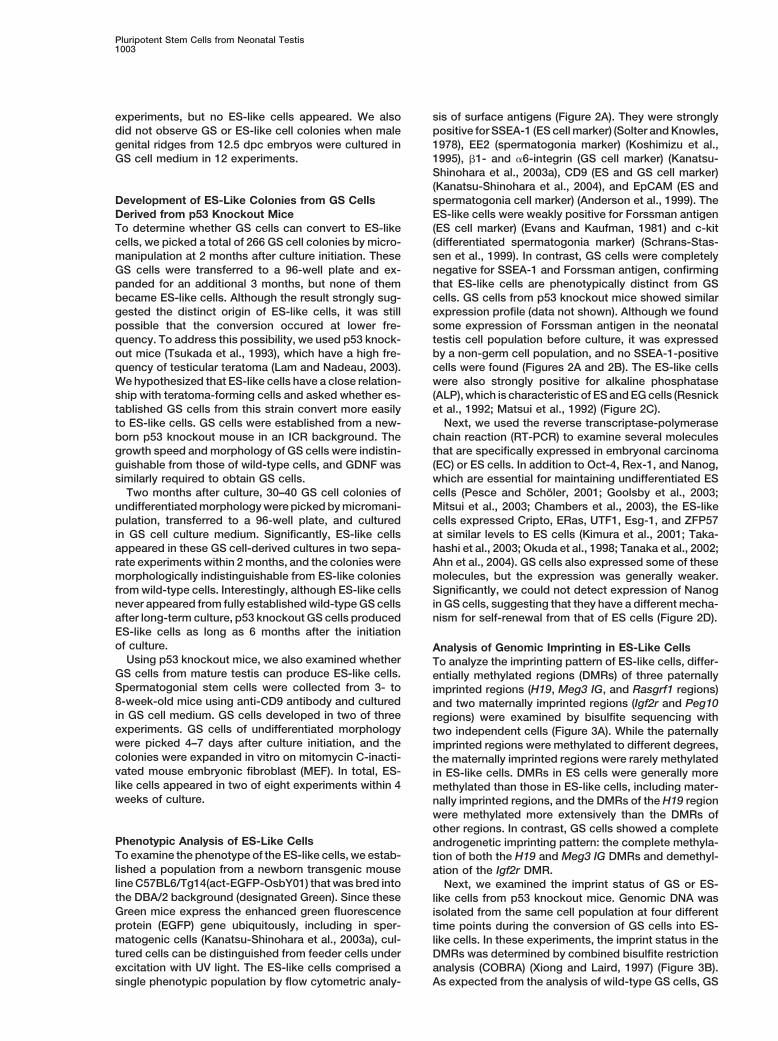
Pluripotent Stem Cells from Neonatal Testis1003
experiments, but no ES-like cells appeared. We also sis of surface antigens (Figure 2A). They were stronglypositive for SSEA-1 (ES cell marker) (Solter and Knowles,did not observe GS or ES-like cell colonies when male
genital ridges from 12.5 dpc embryos were cultured in 1978), EE2 (spermatogonia marker) (Koshimizu et al.,1995), �1- and �6-integrin (GS cell marker) (Kanatsu-GS cell medium in 12 experiments.Shinohara et al., 2003a), CD9 (ES and GS cell marker)(Kanatsu-Shinohara et al., 2004), and EpCAM (ES andspermatogonia cell marker) (Anderson et al., 1999). TheDevelopment of ES-Like Colonies from GS Cells
Derived from p53 Knockout Mice ES-like cells were weakly positive for Forssman antigen(ES cell marker) (Evans and Kaufman, 1981) and c-kitTo determine whether GS cells can convert to ES-like
cells, we picked a total of 266 GS cell colonies by micro- (differentiated spermatogonia marker) (Schrans-Stas-sen et al., 1999). In contrast, GS cells were completelymanipulation at 2 months after culture initiation. These
GS cells were transferred to a 96-well plate and ex- negative for SSEA-1 and Forssman antigen, confirmingthat ES-like cells are phenotypically distinct from GSpanded for an additional 3 months, but none of them
became ES-like cells. Although the result strongly sug- cells. GS cells from p53 knockout mice showed similarexpression profile (data not shown). Although we foundgested the distinct origin of ES-like cells, it was still
possible that the conversion occured at lower fre- some expression of Forssman antigen in the neonataltestis cell population before culture, it was expressedquency. To address this possibility, we used p53 knock-
out mice (Tsukada et al., 1993), which have a high fre- by a non-germ cell population, and no SSEA-1-positivecells were found (Figures 2A and 2B). The ES-like cellsquency of testicular teratoma (Lam and Nadeau, 2003).
We hypothesized that ES-like cells have a close relation- were also strongly positive for alkaline phosphatase(ALP), which is characteristic of ES and EG cells (Resnickship with teratoma-forming cells and asked whether es-
tablished GS cells from this strain convert more easily et al., 1992; Matsui et al., 1992) (Figure 2C).Next, we used the reverse transcriptase-polymeraseto ES-like cells. GS cells were established from a new-
born p53 knockout mouse in an ICR background. The chain reaction (RT-PCR) to examine several moleculesthat are specifically expressed in embryonal carcinomagrowth speed and morphology of GS cells were indistin-
guishable from those of wild-type cells, and GDNF was (EC) or ES cells. In addition to Oct-4, Rex-1, and Nanog,which are essential for maintaining undifferentiated ESsimilarly required to obtain GS cells.
Two months after culture, 30–40 GS cell colonies of cells (Pesce and Scholer, 2001; Goolsby et al., 2003;Mitsui et al., 2003; Chambers et al., 2003), the ES-likeundifferentiated morphology were picked by micromani-
pulation, transferred to a 96-well plate, and cultured cells expressed Cripto, ERas, UTF1, Esg-1, and ZFP57at similar levels to ES cells (Kimura et al., 2001; Taka-in GS cell culture medium. Significantly, ES-like cells
appeared in these GS cell-derived cultures in two sepa- hashi et al., 2003; Okuda et al., 1998; Tanaka et al., 2002;Ahn et al., 2004). GS cells also expressed some of theserate experiments within 2 months, and the colonies were
morphologically indistinguishable from ES-like colonies molecules, but the expression was generally weaker.Significantly, we could not detect expression of Nanogfrom wild-type cells. Interestingly, although ES-like cells
never appeared from fully established wild-type GS cells in GS cells, suggesting that they have a different mecha-nism for self-renewal from that of ES cells (Figure 2D).after long-term culture, p53 knockout GS cells produced
ES-like cells as long as 6 months after the initiationof culture. Analysis of Genomic Imprinting in ES-Like Cells
Using p53 knockout mice, we also examined whether To analyze the imprinting pattern of ES-like cells, differ-GS cells from mature testis can produce ES-like cells. entially methylated regions (DMRs) of three paternallySpermatogonial stem cells were collected from 3- to imprinted regions (H19, Meg3 IG, and Rasgrf1 regions)8-week-old mice using anti-CD9 antibody and cultured and two maternally imprinted regions (Igf2r and Peg10in GS cell medium. GS cells developed in two of three regions) were examined by bisulfite sequencing withexperiments. GS cells of undifferentiated morphology two independent cells (Figure 3A). While the paternallywere picked 4–7 days after culture initiation, and the imprinted regions were methylated to different degrees,colonies were expanded in vitro on mitomycin C-inacti- the maternally imprinted regions were rarely methylatedvated mouse embryonic fibroblast (MEF). In total, ES- in ES-like cells. DMRs in ES cells were generally morelike cells appeared in two of eight experiments within 4 methylated than those in ES-like cells, including mater-weeks of culture. nally imprinted regions, and the DMRs of the H19 region
were methylated more extensively than the DMRs ofother regions. In contrast, GS cells showed a complete
Phenotypic Analysis of ES-Like Cells androgenetic imprinting pattern: the complete methyla-To examine the phenotype of the ES-like cells, we estab- tion of both the H19 and Meg3 IG DMRs and demethyl-lished a population from a newborn transgenic mouse ation of the Igf2r DMR.line C57BL6/Tg14(act-EGFP-OsbY01) that was bred into Next, we examined the imprint status of GS or ES-the DBA/2 background (designated Green). Since these like cells from p53 knockout mice. Genomic DNA wasGreen mice express the enhanced green fluorescence isolated from the same cell population at four differentprotein (EGFP) gene ubiquitously, including in sper- time points during the conversion of GS cells into ES-matogenic cells (Kanatsu-Shinohara et al., 2003a), cul- like cells. In these experiments, the imprint status in thetured cells can be distinguished from feeder cells under DMRs was determined by combined bisulfite restrictionexcitation with UV light. The ES-like cells comprised a analysis (COBRA) (Xiong and Laird, 1997) (Figure 3B).
As expected from the analysis of wild-type GS cells, GSsingle phenotypic population by flow cytometric analy-

Cell1004
Figure 2. Phenotypic Characterization of ES-Like Cells
(A) Flow cytometric characterization of ES-like cells. Black line, control immunoglobulin; red line, specific antibody.(B) Double immunostaining of neonatal testis cells by anti-EE2 and anti-Forssman antigen antibodies.(C) ALP staining. GS cells (left) are weakly positive, whereas ES-like (middle) and ES cells (right) are strongly positive.(D) RT-PCR analysis. Three-fold serial dilutions of cDNA from GS, ES-like, and ES cells were amplified with specific primers. Scale bar, 200 �m.
cells from p53 KO mice had an androgenetic imprint of cell types were identified including hematopoieticcells, vascular cells, and spontaneously beating myo-pattern. However, a loss of methylation in the DMRs of
H19, Meg 3IG, and Rasgrf1 regions and methylation of cytes (Figures 4A–4H). Hematopoiesis could also be in-duced when ES-like cells were cultured in methylcellu-the DMRs in the Igf2r region were observed immediately
after the appearance of ES-like cells. The perturbation lose to form embryoid bodies (Figure 4I). When wetransferred ES-like cells onto gelatin-coated dishes forof imprint patterns continued even when GS cells disap-
peared, and only the DMR of the Peg10 region was the differentiation of neural-lineage cells (Ying et al.,2003), they formed neurons or glial cells (Figures 4J–4L).intact, 18 days after the appearance of ES-like cells.
DMR of Oct-4 region in ES and ES-like cells were all Dopaminergic neurons were also found, albeit at lowfrequency (Figure 4M). When we compared the differen-hypomethylated, which confirms their undifferentiated
state (Hattori et al., 2004) (Figure 3C). tiation efficiency using ES cells, ES-like cells producedmore glial cells than did ES cells, and there were signifi-cantly more vessel or heart muscle cell colonies fromDifferentiation Potential of ES-Like Cells
In Vitro and In Vivo ES-like cells. However, ES-like cells could produce allof the expected lineages using protocols for ES cellTo determine whether ES-like cells can differentiate into
somatic cell lineages, we used methods designed to differentiation (Table 1).ES-like cells were further examined for their ability toinduce differentiation of ES cells in vitro. ES-like cells
were first transferred to an OP9 stromal feeder layer, form teratomas in vivo by subcutaneous injection intonude mice. Transplanted cells gave rise to typical terato-which can support differentiation of mesodermal cells
such as hematopoietic or muscle cells (Nakano et al., mas in all recipients (eight of eight) by 4 weeks aftertransplantation (Figure 4N). The tumors contained deriv-1994; Schroeder et al., 2003). Within 10 days, a variety

Pluripotent Stem Cells from Neonatal Testis1005
Figure 3. Analysis of Imprinting in ES-Like Cells
(A) DMR methylation of H19, Meg3 IG, Rasgrf1, Igf2r, and Peg10 regions. DNA methylation was analyzed by bisulfite genomic sequencing.Black ovals indicate methylated cytosine-guanine sites (CpGs), and white ovals indicate unmethylated CpGs.(B) COBRA of GS and ES-like cells from p53 knockout mice. The day when ES-like colonies were found was designated day 0, and cells werecollected at the indicated time. In this culture, only ES-like cells were found by day 12.(C) COBRA of Oct-4 gene upstream region. Open arrowheads indicate the size of unmethylated DNA. Closed arrowheads indicate the sizeof methylated DNA. Enzymes used to cleave each locus are indicated in parentheses. U, uncleaved; C, cleaved.
atives of the three embryonic germ layers: squamous using the spermatogonial transplantation technique(Brinster and Zimmermann, 1994). This method allowscell epithelium, neuroepithelium, and muscle. Similar
results were obtained with three different clones or with spermatogonial stem cells to recolonize the empty semi-niferous tubules of infertile animals and differentiate intoES-like cells from p53 knockout mice (eight of eight),
and we did not observe a significant histological differ- mature sperm. We transplanted the cultured cells intoimmune-suppressed immature W mice (Kanatsu-Shino-ence from teratomas derived from ES cells. In contrast,
no tumors developed after transplantation of GS cells hara et al., 2003b). These mice are congenitally infertileand have no differentiating germ cells (Brinster and Zim-or fresh testis cells (data not shown).
Since the ES-like cells originated from testis, their mermann, 1994). One month after transplantation, allrecipient animals (ten of ten) developed teratomas inability to differentiate into germline cells was examined

Cell1006
Figure 4. In Vitro and In Vivo Differentiation of ES-Like Cells
(A–H) Differentiation on OP9 cells. (A) Cobblestone formation on day 8. (B) CD45-positive hematopoietic cell development on day 7 aftercoculture (left). In this cell population, Gr1-positive granulocytes, Mac1-positive macrophages, or Ter119-positive erythrocytes were found(right). (C) May-Giemsa staining of harvested cells. Myeloid progenitor (arrowhead) and erythroblast (arrow) were observed. (D and E) Vascularcell differentiation. Flk-1-positive cells were sorted on day 4 after coculture, and CD31-positive (D) or VE-cadherin-positive (E) vascular cellsappeared at 6 days after cell sorting. (F–H) Heart muscle differentiation. The Flk-1-positive cells were differentiated into MF20-positive (F) orcTn-I-positive (G) heart muscle at 6 days after sorting. (H) ANP-positive (blue) atrial muscle and MLC2v-positive (brown) ventricular muscle.(I) Erythroid cells that developed from embryoid body in methylcellulose at 8 days after culture. Note the red color of the cells.(J–M) Neuronal cell differentiation on gelatin-coated plates. Tuj-positive neurons (J) on day 5, GFAP-positive astrocytes (K) and MBP-positive

Pluripotent Stem Cells from Neonatal Testis1007
Table 1. In Vitro Differentiation of ES-Like Cells from Testis
Hematopoiesisa,b Vasculogenesisa,c Neurogenesisd
Increase in cell Granulocyte/Cell type number (fold) Macrophage (%) Erythrocytee (%) Vessele Hearte Neurone Astrocytee Oligodendrocytee
ES-like 116.7 � 15.4 7.6 � 0.2 19.9 � 0.7 111.5 � 12.0 8.0 � 4.5 126.7 � 14.4 34.6 � 4.4 4.6 � 2.5ES cell 102.3 � 11.6 7.6 � 0.4 24.7 � 0.9 49.0 � 9.2 3.8 � 2.0 162.2 � 14.5 10.5 � 3.3 0.2 � 0.1
Values are mean � SEM. Results from at least three experiments. ES cells were derived from 129 mice, whereas ES-like cells were derivedfrom DBA/2 mice.a Flk-1-positive cells (5 � 103) were sorted 4 days after coculture and replated on OP9 feeder in a 24-well plate.b Cells were recovered 7 days after sorting and analyzed by flow cytometry. Erythrocytes, macrophages, and granulocytes were identified byanti-Ter119, anti-Mac1, and anti-Gr1 antibodies, respectively.c Numbers of positive cells in each well, 8 days after sorting. Vascular cells were determined by the uptake of DiI-acetylated low-densitylipoprotein. Heart muscle colonies were identified by counting beating colonies.d Cells (2.5 � 104) were plated on gelatin in a 48-well plate, and numbers of positive cells per cm2 were determined by immunocytochemistry5 (neuron) or 7 (astrocytes or oligodendrocytes) days after plating. Neurons were identified by anti-Tuj antibody, whereas astrocytes andoligodendrocytes were identified by anti-GFAP or anti-MBP antibodies, respectively. Dopaminergic neurons were produced �10 cells per well.e Statistically significant by Student’s t test (p � 0.05).
the testis. The seminiferous tubules were disorganized, Since donor cells were also found in the testis ofa chimeric animal at 6 weeks of age (Figure 5J), weand no sign of spermatogenesis was found in histologi-
cal sections. The cell composition found in the terato- performed microinsemination to obtain offspring. Roundspermatids were collected and microinjected intomas was similar to that of tumors that developed after
subcutaneous injection (data not shown). In contrast, C57BL/6 � DBA/2 (BDF1) oocytes. Of 81 cultured em-bryos, 64 (79%) developed into 2-cells and were trans-both wild-type and p53 KO GS cells produced normal
spermatogenesis when transplanted into the seminifer- ferred into five psudopregnant females. Eighteen (22%)embryos were implanted, and one of the two offspringous tubules (Figures 4O–4Q).from a recipient mouse showed EGFP fluorescence, in-dicating the donor origin (Figure 5K). Interestingly, whileContribution of ES-Like Cells to Normalcontrol ES cells showed wide contribution to embryos,Embryonic Developmentno donor cell contribution was observed in experimentsafter Blastocyst Injectionusing GS cells (Table 2).Finally, we microinjected ES-like cells into blastocysts
To determine the full developmental potential of ES-to examine whether they can contribute to chimeraslike cells, we used tetraploid complementation tech-in vivo. Five to fifteen cells were injected into C57BL/6nique (Nagy et al., 1993). This technique allows the pro-blastocysts. The ratio of euploid cells, which signifi-duction of live animals that consist entirely of donor EScantly influences the rate of chimerism or germline trans-cells. A total of 92 tetraploid embryos were created bymission (Longo et al., 1997; Liu et al., 1997), was 70%electrofusion, aggregated with ES-like cells, and trans-at the time of injection.ferred to pseudopregnant ICR females. When some ofSome of the recipient animals were analyzed at 12.5the recipient animals were sacrificed at 10.5 dpc, wedpc to look for chimerism, and others were allowed tofound one normal-looking fetus and several resorptionsdevelop to term. Chimerism was observed in 25% (threewith normal placentas. The fetus showed some growthof 12) of the 12.5 dpc embryos (Figure 5A) and in 36%retardation but clearly expressed the EGFP gene(13 of 36) of the newborn animals (Figure 5B), as judgedthroughout its body, including the yolk sac (Figure 5L),by the expression of EGFP observable under UV illumi-indicating that it was derived from donor ES-like cells.nation. Chimerism was also confirmed by the coat colorHowever, none of the pseudopregnant mothers siredat mature stage (Figure 5C). We found six dead fetuseslive offspring from both ES-like and ES cells.that showed EGFP expression, and some embryos were
partially or completely absorbed. The pattern of contri-bution was similar at both stages analyzed; EGFP-posi- Discussiontive donor cells were found in the central nervous sys-tem, liver, heart, lung, somites, intestine, and other The results of our experiments revealed the presence of
multipotential stem cells in the neonatal testis. Althoughtissues, including the yolk sac and chorionic membraneof the placenta (Figures 5D–5I). some cases of the “stem cell plasticity” phenomenon
oligodendrocytes (L) on day 7 after induction. TH and Tuj-double positive dopaminergic neurons (arrow) appeared among Tuj-positive neurons(arrowhead) (M).(N) Section of a teratoma under the skin. The tumors contained a variety of differentiated cell types, including muscle (m), neural (n), andepithelial (e) tissues.(O–Q) Spermatogenesis from p53 knockout GS cells. (O) A macroscopic comparison of untransplanted (left) and transplanted (right) recipienttestes. Note the increased size of the transplanted testis. (P and Q) Histological appearance of the untransplanted (P) and transplanted (Q)W testes. Note the normal appearance of spermatogenesis (Q). Color staining: Cy3, red (J–M); Alexa Fluor 488, green (M). Scale bar, 50 �m(A, D–I, J, K, and M), 20 �m (C and L), 200 �m (N, P, and Q), 1 mm (O).

Cell1008
have been attributed to cell fusion (Wagers and Weiss-man, 2004), our case cannot be explained by the samemechanism because the ES-like cells formed teratomasafter subcutaneous transplantation. These ES-like cellsfrom the testis can be considered the neonatal counter-parts of ES/EG cells. The result was unexpected, sincePGCs become resistant to experimental teratocarcino-genesis or EG cell formation after 13.5 dpc (Stevens,1984; Labosky et al., 1994). To our knowledge, EG cellsare the only example of the isolation of multipotent stemcells from primary germ cells (Resnick et al., 1992; Mat-sui et al., 1992). EG cells were derived from primarygerm cells harvested from 8.5 to 12.5 dpc fetuses andcultured in vitro with a mixture of mSCF, LIF, and bFGF.However, pluripotent cells could not be isolated fromneonatal germ cells using the same culture conditions(Labosky et al., 1994), except when cells from teratomaswere used (Robertson and Bradley, 1986). ES-like cellsare unlikely to be derived from teratoma cells for tworeasons. First, the frequency of derivation of ES-likecells in our study was significantly higher than the negli-gible rate of spontaneous teratoma formation in strainsother than 129 and A/He mice (one teratoma out of11,292 males in 129 hybrid backgrounds) (Stevens andMackensen, 1961). Second, growth factor supplementa-tion was essential for the establishment of ES-like cells.In fact, few EC cell lines have been obtained from spon-taneously occurring teratocarcinomas (Robertson andBradley, 1986). These findings strongly suggest that theability to become multipotent stem cells persists in neo-natal testis. Based on the results reported here, we pro-pose to name these ES-like cells multipotent germlinestem cells, or mGS cells, to distinguish them from GScells, which can differentiate only into germline cells(Kanatsu-Shinohara et al., 2003a).
An important question that arises from this study is theorigin of mGS cells. One possibility is that they appearindependently from GS cells and originate from a popu-lation of undifferentiated pluripotent cells that persist inthe testis from the fetal stage. Although EG cells havebeen established from �12.5 dpc PGCs (Matsui et al.,1992; Labosky et al., 1994), cells with similar characteris-tics might remain in neonatal testis and produce ES-like cells. Indeed, the results of the imprinting analysisof wild-type mGS cells suggest a distinct origin for mGScells. In male germ cells, genomic imprinting is erasedduring the fetal stage, and male-specific imprinting be-gins to be acquired around birth in prospermatogoniaand is completed after birth (Davis et al., 1999, 2000;Figure 5. Production of Chimeric AnimalsKafri et al., 1992). While GS cells had a typical androgen-(A) A 12.5 dpc chimeric embryo (arrow) showing fluorescence underetic imprinting pattern, the imprinting pattern of mGSUV light. No fluorescence was observed in a control embryo (ar-
rowhead). cells clearly differed from those of androgenetic germ(B) A newborn chimeric animal (arrow) showing fluorescence. cells or somatic cells, which suggested that mGS cells(C) Mature chimeric animals. Note the donor cell-derived coat originate from partially androgenetic germ cells thatcolor (cinnamon). have undergone imprint erasure.(D–I) Parasagittal section of a 12.5 dpc chimeric embryo. Fluores-
Another possibility is that mGS cells are derived fromcence was observed in the brain (D), intestine (E), heart (F), liver (G),spermatogonial stem cells and that the ability to becomelower spinal cord (H), and placenta (I).
(J) A testis from a chimeric mouse showing fluorescence. EGFP multipotential cells may be one of the general character-expression was observed in some germ cells in the testis cell sus-pension (inset).(K) Offspring derived from a chimera. One of the offspring showedfluorescence, confirming the donor origin (arrow). No fluorescence was observed in the placenta (arrowhead). Coun-(L) A 10.5 dpc embryo (arrow) and yolk sac produced from an aggre- terstained with propidium iodide (PI) (D–I). Color staining: EGFP,gation of ES-like cells with tetraploid embryo showing fluorescence. green (A, B, and D–L); PI, red (D–I). Scale bar, 100 �m (D–I), 1 mm (J).

Pluripotent Stem Cells from Neonatal Testis1009
Table 2. Contribution of ES-Like Cells to Embryonic Development
Chimera (%)Number of Number of Number of Number of
Type of Cells Embryos Transferred Recipients Pups Borna Live Pupsb Male Female
ES-like 193 11 54 36 9/22 (41) 4/14 (29)ES 91 14 14 4 2/2 (100) 2/2 (100)GS 124 7 28 16 0/8 (0) 0/8 (0)4n rescue ES-like 92 4 0 NA NA NA4n rescue ES 30 2 0 NA NA NA
NA, not applicable.a In some experiments, fetuses were delivered by cesarean section at 19.5 dpc.b Number of live pups on the next day after birth.
istics of germline cells. Possibly, the interaction with taneous teratomas in mice occur almost exclusively inthe 129/Sv background and are considered to developSertoli cells normally directs germ cells to spermatogen-
esis and inhibits multilineage differentiation in the testis. from PGCs (Stevens, 1984). However, our resultsstrongly suggest that spermatogonial stem cells areHowever, when germline cells are continuously stimu-
lated to expand in the absence of Sertoli cells, as in our multipotential.Interestingly, the acquisition of multipotentiality inculture conditions, germ cells may be released from this
inhibition and some of the cells converted to pluripotent mGS cells was concurrent with the loss of spermatogo-nial stem cell potential. Despite their testicular origin,cells. Teratogenesis from germline cells is susceptible
to environmental influences; for example, teratoma for- mGS cells formed teratomas in the seminiferous tubules,indicating that this environment was no longer sufficientmation can be significantly enhanced (�10-fold) in vivo
by ectopic transplantation of the fetal genital ridge (Ste- for spermatogenesis after the cells became pluripotent.This contrasts with GS cells, which produce spermato-vens, 1984). As PGCs can become pluripotential only
after in vitro culture and cytokine supplementation was genesis on transfer to the seminiferous tubules (Ka-natsu-Shinohara et al., 2003a). Therefore, mGS cells arealso necessary for EG cell conversion (Matsui et al.,
1992; Resnick et al., 1992), growth stimulation and re- more closely related to ES/EG cells in terms of cell func-tion. The reason for the loss of spermatogonial stemlease from somatic cells may modify the differentiation
program of germline cells. cell potential is unknown; however, we speculate thatit may be related to the loss of responsiveness to GDNFSeveral lines of evidence in our study provide support
for the multipotential nature of spermatogonial stem during the course of the establishment of mGS cells,as GDNF is essential for the self-renewing division ofcells. First, we did not find PGC-like germ cells in the
neonatal testis, and we failed to induce mGS cells from spermatogonial stem cells (Meng et al., 2000). Anotherquestion that remains to be answered is why GS cellsneonatal testis in EG cell culture conditions (mSCF �
LIF � bFGF). Therefore, the mGS cells arose through a converted to mGS cells only at early passages. In ourexperiments, mGS cells appeared within 7 weeks ofdifferent mechanism from that of EG cells, and the re-
sults suggest that PGC-like cells in neonatal testis, if culture initiation but not at later stages. Once estab-lished, however, GS cells were stably committed to theany, are not responsible for the generation of mGS cells.
Second, results of p53 knockout mouse experiments germline, because we did not observe any mGS cellconversion when they were expanded in large-scale cul-showed that mGS cells develop from GS cells. The use of
the p53 knockout mouse was based on previous studies ture or transplanted in vivo. The loss of multipotentialitymight be ascribed to the nonoptimal culture condition;that showed an increased frequency of teratoma in this
strain; it is estimated that loss of the p53 gene results it is widely known that ES cells differentiate easily andlose germline potential in the absence of LIF (Smith,in a 100-fold increase in the susceptibility to testicular
teratoma (Lam and Nadeau, 2003). Nevertheless, GS 2001). Likewise, germline cells may tend to lose somaticcell potential in nonoptimal culture conditions. In thiscells from this strain were phenotypically similar to wild-
type spermatogonia and could produce normal-appearing sense, it is interesting that, in contrast to mGS cells fromwild-type mice, mGS cells developed in the long-termspermatogenesis when transferred into seminiferous tu-
bules. In this sense, they are indistinguishable from wild- in p53 knockout mice. Although the mechanism for themaintenance or loss of multipotentiality of germline cellstype GS cells and fulfill the criteria for spermatogonial
stem cells. Using this model, we found that the partial is currently unclear, the results suggest that this geneis involved in these processes, and GS cells from p53androgenetic imprint in mGS cells occurred with loss of
the androgenetic imprint in GS cells. Perhaps the same knockout mice may be useful for analyzing how germlinecells retain multipotentiality.is true of wild-type mGS cells; the partial androgenetic
imprint patterns may not indicate the origin of mGS cells The most striking result from our experiments is thecontribution of mGS cells to normal embryo develop-directly but rather reflect epigenetic instability in vitro,
as reported for ES/EG cells (Labosky et al., 1994; Dean ment. Donor cell makers were present in various partsof the body, including the germline cells. These resultset al., 1998; Humpherys et al., 2001). Although these
results are based on a mutant mouse model, they demonstrate that mGS cells not only produce tumors butalso can contribute to normal embryonic development.strongly suggest that GS cells are multipotential or can
acquire multipotentiality by loss of a single gene. Spon- However, the function of the cells may not be completely

Cell1010
cells were cultured on Sl4-m220 (gift from Dr. T. Nakano, Osaka Uni-normal, because we could not recover live offspring inversity).tetraploid complementation experiments, which indi-
For adult testis culture, 2 � 107 cells from 3- to 8-week-old wild-cates that mGS cells alone cannot produce a normaltype and p53 knockout mice were used to recover spermatogonial
whole embryo. The failure is most likely related to the stem cells with anti-CD9 antibody as described elsewhere (Kanatsu-imprint status of mGS cells, since altered imprinted gene Shinohara et al., 2004), and selected cells were plated on gelatin-
coated plate (3 � 105 cells/9.5 cm2). GS cell colonies were pickedmethylation causes fetal abnormalities with ES cellsby micromanipulation and transferred to MEF for expansion.(Dean et al., 1998; Surani, 2001). Nevertheless, the im-
Standard ES cell medium was used to culture D3 ES cells thatprint status of mGS cells did not influence the germlineubiquitously express the EGFP gene under the CAG promoter (pro-competence, and normal offspring were obtained fromvided by Dr. M. Okabe, Osaka University; Niwa et al., 1991).
the chimeric animal. This agrees with the previous re-ports that both ES and EG cells can produce germline
Antibodies and Stainingchimera (Robertson and Bradley, 1986; Labosky et al.,The following primary antibodies were used: rat anti-EpCAM (G8.8),1994; Stewart et al., 1994), even with androgenetic im-mouse anti-SSEA-1 (MC-480), mouse anti-sarcomeric protein (MF20;print patterns (Narasimha et al., 1997).Developmental Studies Hybridoma Bank, University of Iowa), rat anti-
The derivation of multipotent stem cells from the neo- mouse Forssman antigen (M1/87), rat anti-human �6-integrinnatal testis may have practical value for medicine and (GoH3), biotinylated hamster anti-rat �1-integrin (Ha2/5), biotinyl-biotechnology. These cells are different from other re- ated rat anti-mouse CD9 (KMC8), allophycocyanin (APC)-conju-
gated rat anti-mouse c-kit (2B8), rat anti-mouse CD31 (MEC 13.3),ported multipotent cells in terms of morphology, markerphycoerythrin (PE)-conjugated rat anti-mouse Ter119 (TER-119),expression, and capacity for differentiation (Verfaillie,biotinylated rat anti-mouse Mac1 (M1/70), biotinylated rat anti-2002; Wagers and Weissman, 2004). While it is importantmouse Gr1 (RB6-8C5), rat anti-mouse VE-cadherin (11D4.1), APC-
to study the biology of individual cell types and assess conjugated rat anti-mouse CD45 (30-F11; BD Biosciences), rat anti-their potential for clinical application, a major advantage TDA (EE2; provided by Dr. Y. Nishimune, Osaka University), APC-of mGS cells is that techniques currently used to derive conjugated rat anti-mouse Flk-1 (Avas 12�1; provided by Dr. S.
Nishikawa, RIKEN), goat anti-mouse cardiac troponin-I (cTn-I)specific lineages of cells from ES cells are applicable(Santa Cruz Biotechnology, Santa Cruz, CA), mouse anti-humandirectly. Clearly, the derivation of mGS cells has fewermyosin light chain 2v (MLC2v) (Alexis Biochemicals Inc, Montreal,ethical concerns than does the derivation of ES cells,Canada), rabbit anti-mouse atrial natriuretic peptide (ANP) (Protos
because mGS cells can be obtained without sacrificing Biotech Corporation, NY), mouse anti-human myelin basic proteinthe conceptus or embryos. Furthermore, the availability (MBP) (Pm43), rabbit anti-glial fibrillary acidic protein (GFAP), rabbitof histocompatible, multipotent tissue for autotrans- anti-mouse tyrosine hydroxylase (TH), and mouse anti-human
�-tubulin III (Tuj) (SDL.3D10) (Sigma, St. Louis, MO). APC-conjugatedplantation would circumvent immunological problemsgoat anti-rat-IgG (Cedarlane Laboratories, ON, Canada), APC-conju-associated with ES cell-based technology. Although wegated streptavidin (BD Biosciences), Alexa Fluor 488-conjugated goatfailed to obtain mGS cells from mature wild-type ani-anti-mouse IgG, Alexa Fluor 647-conjugated goat anti-rat IgM, Alexa
mals, this was likely due to the low success rate of Fluor 633-conjugated goat anti-mouse IgM (Molecular Probes, Eu-GS cell establishment. The results of the p53 knockout gene, OR), Cy3-conjugated donkey anti-mouse IgG, Cy3-conjugatedmouse experiment suggest that mGS cells can arise donkey anti-rabbit IgG, ALP or peroxidase-conjugated donkey anti-
mouse IgG, ALP-conjugated donkey anti-rabbit IgG (Jackson Immu-from mature testis. Development of more efficient sys-noresearch, West Grove, PA), ALP-conjugated rabbit anti-goat IgGtems to derive GS cells from mature testis is necessary(Vector Laboratories, Burlingame, CA), or ALP-conjugated goat anti-at this stage of research, and suppression of p53 expres-rat IgG (Chemicon) were used as secondary antibodies. The cell
sion in GS cells, such as by RNA interference, may be staining and analysis was carried out with a FACSCalibur systemuseful for enhancing the frequency of derivation. Future (BD Biosciences) (Kanatsu-Shinohara et al., 2003a). ALP or DABstudies should also be directed toward examining the staining was carried out using a VECTOR alkaline phosphatase sub-
strate kit or DAB substrate kit (Vector Laboratories), respectively,effect of imprinting on the range and efficiency of differ-according to manufacturer’s protocol.entiation. Such studies will provide important informa-
tion for potential clinical applications.
Differentiation into Specific Lineages In VitroExperimental Procedures For differentiation into mesodermal lineages, ES-like cells were cul-
tured on OP9 feeder layers, and cell differentiation was induced asCell Culture described (Nishikawa et al., 1998; Schroeder et al., 2003, HirashimaTestis cells were collected from newborn (0–2 days old) ddY or et al., 1999). Vascular cells were identified by the uptake of DiI-DBA/2 mice (Japan SLC, Shizuoka, Japan). For some experiments, acetylated low-density lipoprotein (Molecular Probes). Methylcellu-testis cells were collected from a newborn Green mouse (Kanatsu- lose culture was performed as described previously (Nishikawa etShinohara et al., 2003a) or p53 knockout mouse in ICR background al., 1998). All cytokines were provided by Kirin Brewery (Tokyo,(Tsukada et al., 1993). Testis cell culture was performed according to Japan). Neural cell differentiation was induced as previously de-the previously published protocol (Kanatsu-Shinohara et al., 2003a), scribed (Ying et al., 2003).with slight modifications. In brief, testis cells were allocated to agelatin-coated tissue culture plate (2 � 105 cells/3.8 cm2). The nextday, floating cells were recovered and passed to secondary culture Analysis of Marker Gene Expression
RT-PCR for Nanog, Rex-1, ERas, Esg-1, Cripto, and ZFP57 wereplates. After 7 days in culture, the cells were passed to a freshculture plate at a 1:2 dilution. When the cells were confluent (�7 days carried out using specific primers, as described (Mitsui et al., 2003;
Goolsby et al., 2003; Takahashi et al., 2003; Tanaka et al., 2002;after the second passage), they were passed again (1:1 dilution). Atthe third or fourth passage, the cells were maintained on mitomycin Kimura et al., 2001; Ahn et al., 2004). PCR amplifications for Oct-4,
UTF1, and HPRT were carried out by using specific primers (5-C-inactivated MEF. ES-like cells were cultured in Dulbecco’s modi-fied Eagle’s medium supplemented with 15% FCS, 5 � 10�5 M AGCTGCTGAAGCAGAAGAGG-3 and 5-GGTTCTCATTGTTGTCG
GCT-3 for Oct-4, 5-GATGTCCCGGTGACTACGTCT-3 and 5-TCG2-mercaptoethanol, and 103 units/ml ESGRO (Invitrogen, Carlsbad,CA). To induce EG cells from neonatal testis, the same medium was GGGAGGATTCGAAGGTAT-3 for UTF1, and 5-GCTGGTGAAAAG
GACCTCT -3 and 5- CACAGGACTAGAACACCTGC-3 for HPRT).also supplemented with 20 ng/ml human bFGF (Invitrogen), and

Pluripotent Stem Cells from Neonatal Testis1011
Analysis of Imprinted Genes Brinster, R.L., and Avarbock, M.R. (1994). Germline transmissionof donor haplotype following spermatogonial transplantation. Proc.Bisulfite genomic sequencing of DMRs of imprinted genes was car-
ried out as described (Lee et al., 2002). PCR amplifications of each Natl. Acad. Sci. USA 91, 11303–11307.DMR region from bisulfite-treated genomic DNAs was carried out Brinster, R.L., and Zimmermann, J.W. (1994). Spermatogenesis fol-by using specific primers (5-GGAATATTTGTGTTTTTGGAGGG-3 lowing male germ-cell transplantation. Proc. Natl. Acad. Sci. USAand 5-AATTTGGGTTGGAGATGAAAATATTG-3 for H19, 5-GGTTT 91, 11298–11302.GGTATATATGGATGTATTGTAATATAGG-3 and 5-ATAAAACACCA
Chambers, I., Colby, D., Robertson, M., Nichols, J., Lee, S., Tweedie,AATCTATACCAAAATATACC-3 for Meg3 IG, 5-GTGTAGAATATG
S., and Smith, A. (2003). Functional expression cloning of Nanog, aGGGTTGTTTTATATTG-3 and 5-ATAATACAACAACAACAATAACA
pluripotency sustaining factor in embryonic stem cells. Cell 113,ATC-3 for Rasgrf1, 5-TTAGTGGGGTATTTTTATTTGTATGG-3 and
643–655.5-AAATATCCTAAAAATACAAACTACACAA-3 for Igf2r, 5-GTAAAG
Davis, T.L., Trasler, J.M., Moss, S.B., Yang, G.J., and Bartolomei,TGATTGGTTTTGTATTTTTAAGTG-3 and 5-TTAATTACTCTCCTACM.S. (1999). Acquisition of the H19 methylation imprint occurs differ-AACTTTCCAAATT-3 for Peg10, and 5- GGTTTTTTAGAG GATGGTentially on the parental alleles during spermatogenesis. GenomicsTGAGTG-3 and 5- TCCAACCCTACTAACCCATCACC-3 for Oct-4).58, 18–28.The DNA sequences were determined in both directions. For CO-Davis, T.L., Yang, G.J., McCarrey, J.R., and Bartolomei, M.S. (2000).BRA, PCR products were digested with restriction enzymes with aThe H19 methylation imprint is erased and re-established differen-recognition sequence containing CpG in the original unconvertedtially on the parental alleles during male germ cell development.DNA (Xiong and Laird, 1997). Intensity of digested DNA bands wasHum. Mol. Genet. 9, 2885–2894.quantified with ImageGauge software (Fuji Photo Film, Tokyo,
Japan). Dean, W., Bowden, L., Aitchison, A., Klose, J., Moore, T., Menesses,J.J., Reik, W., and Feil, R. (1998). Altered imprinted gene methylation
Transplantation and expression in completely ES cell-derived mouse fetuses: asso-For subcutaneous injections, approximately 2 � 106 cells were in- ciation with aberrant phenotypes. Development 125, 2273–2282.jected into KSN nude mice (Japan SLC). For microinjections into de Rooij, D.G., and Russell, L.D. (2000). All you wanted to knowthe seminiferous tubules, approximately 3 � 105 cells were injected about spermatogonia but were afraid to ask. J. Androl. 21, 776–798.into the seminiferous tubules of an immune-suppressed W mouse
Evans, M.J., and Kaufman, M.H. (1981). Establishment in culture of(Japan SLC) recipient through the efferent duct (Kanatsu-Shinoharapluripotential cells from mouse embryos. Nature 292, 154–156.et al., 2003b).Goolsby, J., Marty, M.C., Heletz, D., Chiappelli, J., Tashko, G., Yar-nell, D., Fishman, P.S., Dhib-Jalbut, S., Bever, C.T., Jr., and Trisler,Chimera Formation and MicroinseminationD. (2003). Hematopoietic progenitors express neural genes. Proc.Cells were injected into the blastocoel of 3.5 dpc blastocysts ofNatl. Acad. Sci. USA 100, 14926–14931.C57BL/6 mice using a Piezo-driven micromanipulator (Kimura and
Yanagimachi, 1995). The blastocysts were returned to the oviducts Hattori, N., Nishino, K., Ko, Y.-G., Hattori, N., Ohgane, J., Tanaka,or uteri of 2.5 dpc pseudopregnant ICR foster mothers on the day S., and Shiota, K. (2004). Epigenetic control of mouse Oct-4 geneof microinjection. Tetraploid embryo aggregation chimeras were expression in embryonic stem cells and trophoblast stem cells. J.produced using the method developed by Nagy et al. (1993), except Biol. Chem. 279, 17063–17069.that two-cell blastomeres were electrofused by applying an electric Hirashima, M., Kataoka, H., Nishikawa, S., Matsuyoshi, N., and Nishi-pulse (2500 V/cm, 10 �sec) in 300 mM mannitol solution. Mi- kawa, S.-I. (1999). Maturation of embryonic stem cells into endothe-croinsemination was carried out as described using BDF1 oocytes lial cells in an in vitro model of vasculogenesis. Blood 93, 1253–1263.(Kimura and Yanagimachi, 1995). The embryos were transferred on
Humpherys, D., Eggan, K., Akutsu, H., Hochedlinger, K., Rideout,the next day after culture.W.M., III, Biniszkiewicz, D., Yanagimachi, R., and Jaenisch, R. (2001).Epigenetic instability in ES cells and cloned mice. Science 293,
Histology95–97.
Tissues were fixed in 10% formalin and processed for paraffin sec-Kafri, T., Ariel, M., Brandeis, M., Shemer, R., Urven, L., McCarrey,tioning. Chimeric embryos were fixed in 4% paraformaldehyde andJ.R., Ceder, H., and Razin, A. (1992). Developmental pattern of gene-frozen in Tissue-Tek OCT compound (Sakura Finetechnical, Tokyo,specific DNA methylation in the mouse embryo and germ line. GenesJapan) for cryosectioning. Slides were analyzed with an OlympusDev. 6, 705–714.confocal laser scanning microscope.Kanatsu, M., and Nishikawa, S.-I. (1996). In vitro analysis of epiblasttissue potency for hematpoietic cell differentiation. DevelopmentAcknowledgments122, 823–830.
We declare that none of the authors have a financial interest related Kanatsu-Shinohara, M., Ogonuki, N., Inoue, K., Miki, H., Ogura, A.,to this work. We thank Drs. Y. Kaziro and Y. Matsui for discussion Toyokuni, S., and Shinohara, T. (2003a). Long-term proliferation inand encouragement, and Ms. A. Wada for technical assistance. culture and germline transmission of mouse male germline stemFinancial support for this research was provided by the Inamori cells. Biol. Reprod. 69, 612–616.Foundation; the Ministry of Health and Welfare; and the Ministry of Kanatsu-Shinohara, M., Ogonuki, N., Inoue, K., Ogura, A., Toyokuni,Education, Culture, Sport, Science, and Technology of Japan. S., Honjo, T., and Shinohara, T. (2003b). Allogeneic offspring pro-
duced by male germ line stem cell transplantation into infertileReceived: April 30, 2004 mouse testis. Biol. Reprod. 68, 167–173.Revised: October 7, 2004
Kanatsu-Shinohara, M., Toyokuni, S., and Shinohara, T. (2004). CD9Accepted: November 2, 2004is a surface marker on mouse and rat male germline stem cells.Published: December 28, 2004Biol. Reprod. 70, 70–75.
Kimura, Y., and Yanagimachi, R. (1995). Mouse oocytes injectedReferenceswith testicular spermatozoa or round spermatids can develop intonormal offspring. Development 121, 2397–2405.Ahn, J.-I., Lee, K.-H., Shin, D.-M., Shim, J.-W., Lee, J.-S., Chang,Kimura, C., Shen, M.M., Takeda, N., Aizawa, S., and Matsuo, I. (2001).S.Y., Lee, Y.-S., Brownstein, M.J., Lee, S.-H., and Lee, Y.-S. (2004).Complementary functions of Otx2 and Cripto in initial patterning ofComprehensive transcriptome analysis of differentiation of embry-mouse epiblast. Dev. Biol. 235, 12–32.onic stem cells into midbrain and hindbrain neurons. Dev. Biol.
265, 491–501. Koshimizu, U., Nishioka, H., Watanabe, D., Dohmae, K., and Nishi-mune, Y. (1995). Characterization of a novel spermatogenic cellAnderson, R., Schaible, K., Heasman, J., and Wylie, C.C. (1999).
Expression of the homophilic adhesion molecule, Ep-CAM, in the antigen specific for early stages of germ cells in mouse testis. Mol.Reprod. Dev. 40, 221–227.mammalian germ line. J. Reprod. Fertil. 116, 379–384.

Cell1012
Labosky, P.A., Barlow, D.P., and Hogan, B.L.M. (1994). Mouse em- Development, J. Rossant, and R.A. Pedersen, eds. (Cambridge:Cambridge University Press), pp. 475–508.bryonic germ (EG) cell lines: transmission through the germline and
differences in the methylation imprint of insulin-like growth factor Schrans-Stassen, B.H.G.J., van de Kant, H.J.G., de Rooij, D.G., and2 receptor (Igf2r) gene compared with embryonic stem (ES) cell van Pelt, A.M.H. (1999). Differential expression of c-kit in mouselines. Development 120, 3197–3204. undifferentiated and differentiating type A spermatogonia. Endocri-
nology 140, 5894–5900.Lam, M.-Y.J., and Nadeau, J.H. (2003). Genetic control of suscepti-bility to spontaneous testicular germ cell tumors in mice. APMIS Schroeder, T., Fraser, S.T., Ogawa, M., Nishikawa, S., Oka, C., Born-111, 184–191. kamm, G.W., Nishikawa, S.-I., Honjo, T., and Just, U. (2003). Recom-
bination signal sequence-binding protein J alters mesodermal cellLee, J., Inoue, K., Ono, R., Ogonuki, N., Kohda, T., Kaneko-Ishino,fate decisions by suppressing cardiomyogenesis. Proc. Natl. Acad.T., Ogura, A., and Ishino, F. (2002). Erasing genomic imprinting mem-Sci. USA 100, 4018–4023.ory in mouse clone embryos produced from day 11.5 primordial
germ cells. Development 129, 1807–1817. Smith, A.G. (2001). Embryo-derived stem cells: of mice and men.Annu. Rev. Cell Dev. Biol. 17, 435–462.Liu, X., Wu, H., Loring, J., Hormuzdi, S., Disteche, C.M., Bornstein,
P., and Jaenisch, R. (1997). Trisomy eight in ES cells is a common Solter, D., and Knowles, B.B. (1978). Monoclonal antibody definingpotential problem in gene targeting and interferes with germ line a stage-specific mouse embryonic antigen (SSEA-1). Proc. Natl.transmission. Dev. Dyn. 209, 85–91. Acad. Sci. USA 75, 5565–5569.Longo, L., Bygrave, A., Grosveld, F.G., and Pandolfi, P.P. (1997). Stevens, L.C. (1984). Spontaneous and experimentally induced tes-The chromosome make-up of mouse embryonic stem cells is pre- ticular teratomas in mice. Cell Differ. 15, 69–74.dictive of somatic and germ cell chimaerism. Transgenic Res. 6, Stevens, L.C., and Mackensen, J.A. (1961). Genetic and environmen-321–328. tal influences on teratocarcinoma in mice. J. Natl. Cancer Inst. 27,Martin, G.R. (1981). Isolation of a pluripotent cell line from early 443–453.mouse embryos cultured in medium conditioned by teratocarcinoma Stewart, C.L., Gadi, I., and Bhatt, H. (1994). Stem cells from primor-stem cells. Proc. Natl. Acad. Sci. USA 78, 7634–7638. dial germ cells can reenter the germline. Dev. Biol. 161, 626–628.Matsui, Y., Zsebo, K., and Hogan, B.L.M. (1992). Derivation of pluri- Surani, M.A. (2001). Reprogramming of genome function throughpotential embryonic stem cells from murine primordial germ cells epigenetic inheritance. Nature 414, 122–128.in culture. Cell 70, 841–847.
Takahashi, K., Mitsui, K., and Yamanaka, S. (2003). Role of ERas inMeistrich, M.L., and van Beek, M.E.A.B. (1993). Spermatogonial promoting tumor-like properties in mouse embryonic stem cells.stem cells. In Cell and Molecular Biololgy of the Testis, C. Desjardins, Nature 423, 541–545.and L.L. Ewing, eds. (New York: Oxford University Press), pp. Tanaka, T.S., Kunath, T., Kimber, W.L., Jaradat, S.A., Stagg, C.A.,266–295. Usuda, M., Yokota, T., Niwa, H., Rossant, J., and Ko, M.S.H. (2002).Meng, X., Lindahl, M., Hyvonen, M.E., Parvinen, M., de Rooij, D.G., Gene expression profiling of embryo-derived stem cells reveals can-Hess, M.W., Raatikainen-Ahokas, A., Sainio, K., Rauvala, H., Lakso, didate genes associated with pluripotency and lineage specificity.M., et al. (2000). Regulation of cell fate decision of undifferentiated Genome Res. 12, 1921–1928.spermatogonia by GDNF. Science 287, 1489–1493. Tsukada, T., Tomooka, Y., Takai, S., Ueda, Y., Nishikawa, S.-I., Yagi,Mitsui, K., Tokuzawa, Y., Itoh, H., Segawa, K., Murakami, M., Taka- T., Tokunaga, T., Takeda, N., Suda, Y., Abe, S., et al. (1993). En-hashi, K., Maruyama, M., Maeda, M., and Yamanaka, S. (2003). The hanced proliferative potential in culture of cells from p53-deficienthomeoprotein Nanog is required for maintenance of pluripotency in mice. Oncogene 8, 3313–3322.mouse epiblast and ES cells. Cell 113, 631–642. Verfaillie, C.M. (2002). Adult stem cells: assessing the case for pluri-Nagy, A., Rossant, J., Nagy, R., Abramow-Newerly, A., and Roder, potentcy. Trends Cell Biol. 12, 502–508.J.C. (1993). Derivation of completely cell culture-derived mice from Wagers, A.J., and Weissman, I.L. (2004). Plasticity of adult stemearly-passage embryonic stem cells. Proc. Natl. Acad. Sci. USA cells. Cell 116, 639–648.90, 8424–8428.
Xiong, Z., and Laird, P.W. (1997). COBRA: a sensitive and quantita-Nakano, T., Kodama, H., and Honjo, T. (1994). Generation of lympho- tive DNA methylation assay. Nucleic Acids Res. 25, 2532–2534.hematopoietic cells from embryonic stem cells in culture. Science
Ying, Q.L., Stavridis, M., Griffiths, D., Li, M., and Smith, A. (2003).265, 1098–1101.
Conversion of embryonic stem cells into neuroectodermal precur-Narasimha, M., Barton, S.C., and Surani, M.A. (1997). The role of sors in adherent monoculture. Nat. Biotechol. 21, 183–186.the paternal genome in the development of the mouse germ line.Curr. Biol. 7, 881–884.
Nishikawa, S.-I., Nishikawa, S., Hirashima, M., Matsuyoshi, N., andKodama, H. (1998). Progressive lineage analysis by cell sorting andculture identifies FLK1�VE-cadherin� cells at a diverging point ofendothelial and hemopoietic lineages. Development 125, 1747–1757.
Niwa, H., Yamamura, K., and Miyazaki, J. (1991). Efficient selectionfor high-expression transfectants with a novel eukaryotic vector.Gene 108, 193–200.
Okuda, A., Fukushima, A., Nishimoto, M., Orimo, A., Yamagishi, T.,Nabeshima, Y., Kuro-o, M., Nabeshima, Y., Boon, K., Keaveney, M.,et al. (1998). UTF1, a novel transcriptional coactivator expressed inpluripotent embryonic stem cells and extra-embryonic cells. EMBOJ. 17, 2019–2032.
Pesce, M., and Scholer, H.R. (2001). Oct-4: gatekeeper in the begin-ning of mammalian development. Stem Cells 19, 271–278.
Resnick, J.L., Bixler, L.S., Cheng, L., and Donovan, P.J. (1992). Long-term proliferation of mouse primordial germ cells in culture. Nature359, 550–551.
Robertson, E.J., and Bradley, A. (1986). Production of permanentcell lines from early embryos and their use in studying developmentalproblems. In Experimental Approaches to Mammalian Embryonic