Salinity regulates claudin mRNA and protein expression in the teleost gill
Upload
independentCategory
view
1download
0
TWO SPLICE VARIANTS OF CLAUDIN-10 IN THE KIDNEY CREATE
PARACELLULAR PORES WITH DIFFERENT ION SELECTIVITIES
Christina M. Van Itallie1, Sarah Rogan2, Alan Yu3, Lucia Seminario Vidal2, Jennifer Holmes2
and James M. Anderson2
Departments of 1Medicine and 2Cell and Molecular Physiology, University of North Carolina at Chapel Hill, Chapel Hill, North Carolina 27599-7545 USA and 3Departments of Medicine and Physiology and Biophysics, University of Southern California Keck School of Medicine, Los
Angeles, California 90033
Running title: Cldn-10a and 10b have different TJ charge selectivity
Please address correspondence to:
Christina M. Van ItallieUniversity of North Carolina at Chapel Hill6314 MBRB CB#7545103 Mason Farm RoadChapel Hill, NC 27599-7545Phone: 919-966-6412Fax: 919-966-6413Email: [email protected]
Page 1 of 51Articles in PresS. Am J Physiol Renal Physiol (June 27, 2006). doi:10.1152/ajprenal.00138.2006
Copyright © 2006 by the American Physiological Society.
2
Members of the large claudin family of tight junction proteins create the differences in
paracellular conductance and charge selectivity observed among different epithelia. Previous
studies demonstrated that ionic charge selectivity is influenced by acidic or basic amino acids on
the first extracellular domain of claudins. We noted two alternatively spliced variants of claudin-
10 in the database, 10a and 10b, which are predicted to encode two different first extracellular
domains and asked if this might be a novel mechanism to generate two different
permselectivities from a single gene. Using quantitative PCR, we found that claudin-10b is
widely expressed among tissues including the kidney; however claudin-10a is unique to the
kidney. Using a nondiscriminating antibody we found that claudin-10 (a plus b) is expressed in
most segments of the nephron. In situ hybridization, however, showed mRNA for 10a is
concentrated in the cortex and mRNA for 10b is more highly expressed in the medulla.
Expression in MDCK II and LLC-PK1 cells reveals that both variants form low resistance pores,
and that claudin-10b is more selective for cations than claudin-10a. Charge-reversing mutations
of cationic residues on 10a reveal positions that contribute to its anion selectivity. We conclude
that alternative splicing of claudin-10 generates unique permselectivities and might contribute to
the variable paracellular transport observed along the nephron.
Key words: tight junction, claudin, kidney, permselectivity, MDCK, LLC-PK1
Page 2 of 51
3
INTRODUCTION
Tight junctions regulate the movement of solutes and electrolytes through the paracellular
pathway across epithelia (3; 11). Permeability characteristics of tight junctions vary widely
among different tissues as characterized by electrical resistance, preference for cations or anions
and the flux for noncharged solutes; these tight junction characteristics are collectively referred
to as permselectivity (21). Current evidence suggests that these variable properties are based on
differential expression of members of the claudin family of transmembrane proteins (reviewed in
(29)).
Claudins are small proteins with four transmembrane segments and two extracellular
domains. They assemble into rows of adhesive cell-cell contacts which appear as interconnected
transmembrane strands in freeze-fracture electron micrographs (12). For many years the strand
barriers have been modeled as rows of pores with size and charge selectivity (4; 10).There are at
least 23 claudin genes in mammals and each epithelial cell type has a distinct expression profile
(see for example, (18), (17),(14)), presumably resulting in its unique collective permselectivity.
When expressed in a cultured epithelial cell monolayers, different claudins cause
different permselectivity phenotypes, that is, they behave like different pores (29). For example,
claudin-2 forms electrically low resistance, cation-selective pores when expressed in high
resistance MDCK I cells (1). In contrast, expression of claudin-8 (32) and -14 (2) results in
increased transepithelial resistance and specifically decreased paracellular cation permeability.
The result of charge reversing mutagenesis on two claudins reveals the ability of some charged
residues on the first extracellular domain to influence ionic permeability, as if these residues
lined the pore (8). We noted two alternatively spliced cDNAs for claudin-10 in mice and humans
which encode two different first extracellular domains and asked if they represented a naturally
Page 3 of 51
4
occurring mechanism to generate different permselectivities. Our findings are most relevant to
the physiology of the kidney which is the only organ were we observe detectable levels of both
splice variants (this paper).
Transcellular transport as well as paracellular permselectivity varies along the nephron as
the basis of water, solute, salt and pH homeostasis. In general, paracellular electrical resistance
increases along the nephron from the leaky proximal tubule (6-7 ohms x cm2) to tight collecting
ducts and bladder (300,000 ohms x cm2) (21). Most segments are more permeable for cations
than anions, although junctions of the superficial straight proximal tubule are anion selective
(27). While we still know little about the properties of most claudins, several groups have studied
their expression profiles along the nephron with the ultimate goal of explaining transport
difference among segments. Even a limited review of the published immunolocalization reveals
strong regional specialization: glomerulus expresses claudin-1 (6; 18), proximal tubule expresses
relatively high levels of claudin 2 (6; 18), the thick ascending limb of the loop of Henle
expresses claudin-16 (Paracellin-1) (25), the distal nephron expresses claudin-7 and -8 (19),
collecting tubule contains claudin-4 (6; 18), and claudin-5 (18) is localized to endothelial cells.
However, the detailed profiles are incomplete and there exists some disagreement about the
distribution of other claudins. Human mutations in claudin-16 (Paracellin-1) result in magnesium
wasting, which was rationalized as loss of paracellular cation pores in the thick ascending limb
of the loop of Henle, the only segment where appreciable Mg++ resorption normally occurs(25).
There remains much to learn about how claudins make selective pores. Insights about the
basis for charge selectivity are inferred from a single study of experimental mutagenesis (8). To
address this limitation we investigated the consequences of expressing in cultured epithelial cells
two naturally occurring isoforms of claudin-10 which differ only in the first transmembrane and
Page 4 of 51
5
extracellular domains and share the remaining sequence. Inspection of the charged residues of
the first extracellular domain suggested to us that the claudin-10b isoform was likely to be a
cation pore, while claudin-10a was likely to create a pore with less selectivity for cations.
Claudin-10 had been previously reported to be expressed in proximal tubule and thick ascending
limb of the loop of Henle by Tsukita and colleagues (18) and in proximal and distal tubule, thick
ascending limb of Henle and vasa recta by Inai et al. (16). However, neither group addressed
the issue that claudin-10 is expressed as two different isoforms, and in each case the detecting
antibody did not discriminate between these isoforms. Because the tissue distribution of these
two isoforms in the body has not been previously described, we first characterized their
distribution by quantitative PCR and the distribution of claudin-10 by immunoblotting and
immunofluorescence microscopy. Each isoform was then expressed separately in the cation-
selective background of MDCK II cells and the anion-selective background of LLC-PK1 cells
and the physiologic properties assessed. The results presented here suggest that the two naturally
occurring isoforms of claudin-10 differ in their localization in kidney and in their physiologic
properties.
MATERIALS AND METHODS
Plasmid constructs and cell lines. Mouse claudin-10a (clone ID 5356256) and 10b (clone
ID 5040100) cDNAs were obtained from Open Biosystems (Huntsville, AL), cloned into the
pTRE vector (Clontech Laboratories, Palo Alto, CA) and verified by sequencing in both
directions. Claudin-10a as obtained from Open Biosystems contains a deletion within the
carboxyl terminal half, which we corrected by replacement with sequence encoding the shared
Page 5 of 51
6
carboxyl terminal domain from claudin-10b. Mutants of claudin-10a were created with the PCR-
mediated Quik-change mutagenesis kit (Stratagene, La Jolla, CA) using appropriate
oligonucleotide primers to generate the charge-reversing mutations m1 (R32D), m2 (R61D) and
m3 (K68D) and the resulting mutations were verified by sequencing. Clonal cell lines of both
MDCK II and LLC-PK1 cells were generated as described previously (30); 3-5 separate stable
clones were derived for each construct. Cells were maintained in the presence of 50 ng/ml
doxycycline to repress transgene expression and plated for experiments onto removable filters
(Snapwell, Corning Life Sciences, Acton, MA) in the presence (not induced) or absence
(induced) of doxycycline as described previously (30). Physiological studies, immunoblots and
immunofluorescence microscopy of the cultured cells were performed as described previously
(8; 30). All antibodies except the anti-ZO-1 rat monoclonal (26) were from Zymed Laboratories
(Invitrogen R&D, Carlsbad, CA).
Quantitative Real-Time PCR, tissue immunoblotting and immunofluorescence
microscopy. Tissue for RNA isolation and for immunofluorescence microscopy (except for
kidney) was obtained from 3 or 4 adult C57 Black6 mice and preserved by submersion in
RNALater RNA stabilization solution (Ambion, Inc., Austin, TX). Tissues for
immunofluorescence were flash frozen in liquid nitrogen in O.C.T embedding compound
(Tissue-Tek #4583, Sakura Finetek U.S.A., Inc., Torrance, CA). RNA was isolated using an
RNeasy kit (Qiagen, Inc., Valencia, CA) according to kit protocols. 5 ug of RNA for each tissue
was treated with TURBO DNase (TURBO DNA-free kit, Ambion, Inc., Austin, TX) per
instructions to remove genomic DNA contamination. cDNA was synthesized from 2.5 ug of
treated RNA using Superscript III reverse transcriptase (Invitrogen Corporation, Carlsbad, CA)
with an equal amount of RNA included in a no-RT control for each separate RNA sample.
Page 6 of 51
7
Quantitative PCR was performed with 1:50 dilutions of both the cDNA (in triplicate) and
no-RT control for each sample as well as no-template reaction controls. Reaction conditions,
threshold cycle determinations and quality control measures have been previously described
(14). The cycle at which each sample crossed a fluorescence threshold, Ct (at 0.1-0.2
fluorescence units), was determined, and the triplicate values for each cDNA were averaged.
RNA for Eef1a1, eukaryotic translation elongation factor 1 alpha 1, served as a housekeeping
gene for normalization between samples and was included in each cycling run (13); (14)). Gene
expression was normalized to Eef1a1 expression by calculating a ∆Ct (Ct of claudin-10a or 10b
minus the Ct of Eef1a1). Relative expression values were calculated as 2(-∆Ct), setting the
expression value of Eef1a1 to 1.0. Experimental error was estimated for each gene in each tissue
by the CV (%) of the average ∆Ct value of that gene between animals, error = ((2%CV)/100)*
relative expression value. If a sample signal had not risen above the threshold value within 37
cycles, it was considered not detected (nd). Primers for quantitative PCR are shown in Table 1.
Mouse kidney immunofluorescence. Mouse kidneys were perfusion fixed,
immunofluorescently stained and micrographic images acquired as described previously (19).
Secondary antibodies used were goat anti-rabbit, -mouse, or -guinea pig IgG conjugated to Alexa
Fluor 488 or 555. Identification of tubule segments was made based on histological criteria and
immunolocalization with the segment-specific marker proteins, Na-Ca exchanger/NCX1 (in the
connecting tubules and cortical collecting duct principal cells) and vacuolar H-ATPase (apical
membrane of α-intercalated cells, α-IC, and brush border of the proximal convoluted tubule)
(19).
In situ hybridization. In situ hybridization was performed on fresh frozen kidney sections.
PCR primers containing T7 and Sp6 promoter sites were used to generate DNA templates
Page 7 of 51
8
specific for mouse claudin-10a or 10b. Digoxin-labeled RNA probes were used for hybridization
and developed with HNPP (2-hydroxy-3-naphtoic acid-2’-phenylanilide phosphate) fluorescent
substrate by Dr. Yongqin Wu in the UNC Neuroscience Center Expression Localization Core.
RESULTS
Alternative splicing generates two variants of claudin-10. Our previous work showed
that the different charge selectivities of claudins are influenced by the number and position of
charged amino acid residues in the first extracellular domains (7; 8; 15). Thus, it was of interest
to find two splice variants of claudin-10 in the cDNA sequence database s, which differ in their
first exon, and encode proteins which differ from the N-terminus up to the last seven residues of
the first extracellular domain, Figure 1. Inspection of the mouse claudin-10 gene (Mouse
Genome Informatics Number 1913101, chromosome 14) reveals exon 1a precedes 1b and their
splicing to shared exons 2 through 5 generates transcript variant-1 (claudin-10a, NM_023878)
and variant-2 (claudin-10b, NM_021386), respectively. This led us to ask if the two forms of
claudin-10 have different tissue distributions and charge selectivities and thus represent a novel
mechanism to generate physiologically diversity from a single claudin gene. Claudin-18 has
already been reported to undergo an analogous splicing of the first extracellular domain (20)
although physiologic implications were not investigated.
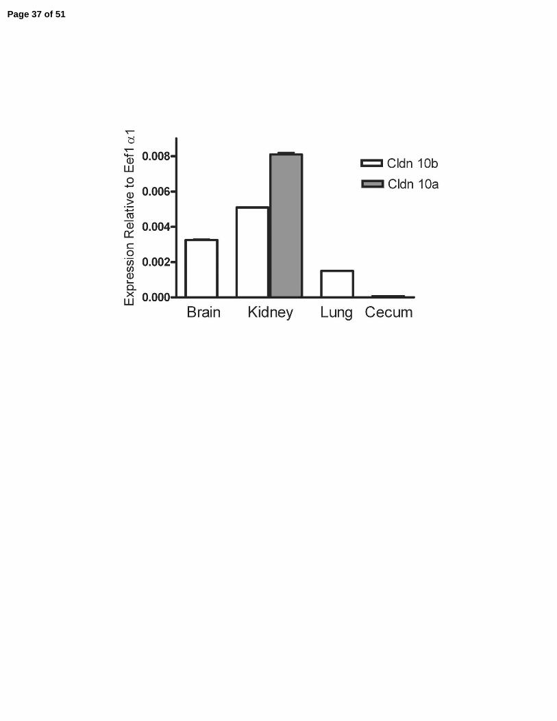
Quantitative Real-Time PCR reveals that claudin-10b is ubiquitously expressed, while
claudin-10a is unique to the kidney. Since none of the available antibodies discriminate between
claudin-10a and 10b, we used quantitative PCR to determine whether the relative expression of
the two transcripts differs among mouse tissues. Expression was measured relative to a control
housekeeping transcript Eef1a1. Claudin-10b was found to be expressed in all tissues tested,
Page 8 of 51
9
although it is most highly expressed in brain (cortex), kidney and lung, Figure 2A, with lower
levels in other tissues including esophagus, jejunum, cecum, colon, heart and spleen. Clau din-
10a, on the other hand, was only detected in kidney, where its level was similar to that of
claudin-10b, Figure 2A.
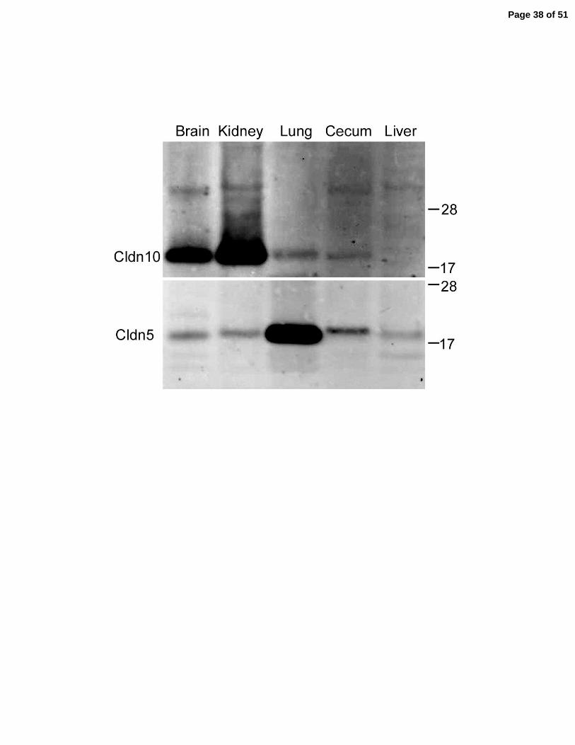
Although the available antibodies do not distinguish between the isoforms, we attempted
to verify expression of claudin-10 protein by immunoblotting in those tissues with the highest
RNA levels, Figure 2B. Consistent with the qRT-PCR results, claudin-10 protein levels were
highest in brain and kidney, but are also detectable in both lung and cecum. In liver, claudin-10
levels were undetectable by qRT-PCR or by immunoblot, although immunofluorescence
microscopy revealed weak claudin-10 immunoreactivity in the tight junctions of hepatocytes
(data not shown). For comparison, claudin-5 expression is ubiquitous and especially high in lung,
Figure 2B.
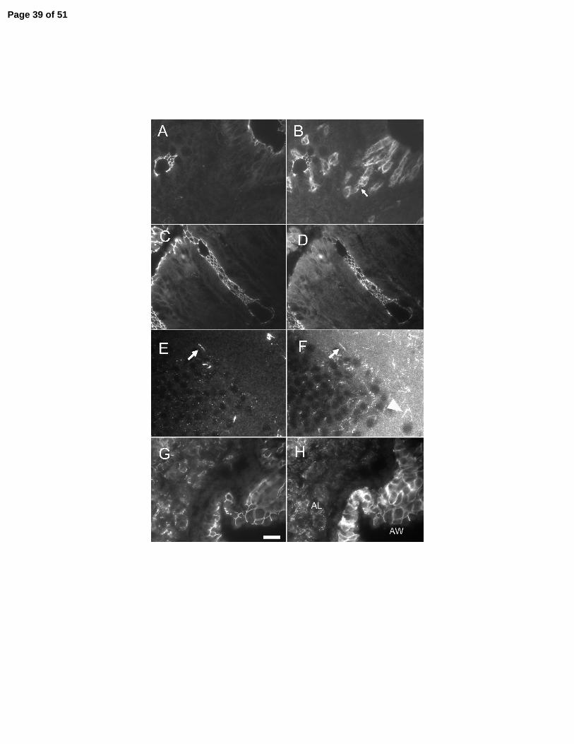
Localization of claudin-10 in kidney and other tissues. We previously described the
distribution of claudin-10 protein along the gastrointestinal tract where it localizes to both
endothelial and epithelial tight junctions (14). In retrospect, this represented only the claudin-10b
isoform. In the current study we performed additional immunolocali zation of claudin-10 starting
with tissues where the signal, based on mRNA analysis, represents exclusively the 10b isoform.
Some atypical localizations were observed. In cecum, claudin-10, Figure 3B&D, colocalizes
with its cytoplasmic binding partner ZO-1, Figure 3A&C, at epithelial tight junctions, but is also
located on the lateral membranes in a subset of cells along the crypt-surface axis, Figure 3B. It is
not obvious based on histological grounds what this unusual claudin distribution reveals and how
these cells are unique. In brain, claudin-10 is colocalized with ZO-1 in endothelial cells, Figure
3E&F (arrows), as well as in as yet unidentified small fibrillar structures. In lung, claudin-10,
Page 9 of 51
10
Figure 3H, is strongly colocalized with ZO-1, Figure 3G, in large airway epithelial cells and
alveoli. In heart, claudin-10 is found with ZO-1 in endothelial cells, but not in the ZO-1 positive
intercalated disks (data not shown).
In the kidney claudin-10 is widely expressed along the nephron; this is in contrast to
other claudins which show restricted patterns, such as claudin-2 in the proximal convoluted
tubule (PCT) (18) and claudin-16 in the TALH (Paracellin-1) (25). Claudin-10 colocalized with
ZO-1 in numerous tubule types, including weak expression in both PCT and distal convoluted
tubule, Figure 4A-C, high expression in the macula densa (Figure 4E), very high expression in
both cortical and medullary thick ascending limbs of the loop of Henle (cTAL and mTAL),
Figures 4D, 4G and both cortical (CCD) and inner medullary collecting ducts (IMCD). In the
inner medulla, claudin-10 is strongly coexpressed with ZO-1 in the thin ascending limbs (Fig. 4H
and at higher magnification in 4I) where the tight junctions appear tortuous due to the extensive
lateral interdigitations between adjacent cells (24). Only the various cell types of the glomerulus
lack detectable expression of claudin-10.
Because the claudin-10 antibody does not distinguish between 10a and 10b isoforms, we
used in situ hybridization to determine if their transcripts were expressed in different regions of
the kidney, Figure 5. The in situ probes include only sequences from either exon 1a or 1b and
have no shared sequence. As one control for their specificity we performed in situ hybridization
on sections of mouse brain (data not shown). We detected a signal for only the claudin-10b
transcript, consistent with RT-PCR results which detect only the claudin-10b transcript in brain,
Figure 2A. Relative expression for claudin-10a mRNA is higher in tubules of the cortex than the
medulla. In contrast, claudin-10b mRNA shows the opposite relative expression, higher in the
medulla than cortex. Although clau din-1 is found in endothelia in other parts of the body, we
Page 10 of 51
11
found no protein or transcript in the vasa recta. Based on the available data we conclude claudin-
10 is widely expressed along the nephron and that 10a and 10b transcripts are more highly
expressed in cortex and medulla, respectively.
Claudin-10a and 10b have different charge selectivities, which are conferred by charged
residues in the first extracellular domain. Claudin-10a and 10b differ in the charge
characteristics of their first extracellular domains. Most claudins share a set of membrane
proximal charged residues in this domain which in claudin-10a correspond to positions E26,
K28, and R75, Figure 6A. The corresponding charges on claudin-10b are at positions D29, K32
and R81, Figure 6A. When consideration of these shared positions is excluded, claudin-10a is
found to contain more positively charged residues (R32, R59 and K66, 3 positive residues) and
claudin-10b more negatively charged residues (D37, K52, D57, K65, D66 and D74, 4 negative
and 3 positive residues). This led us to predict that claudin-10a might form paracellular pores
that are more permeable to anions than those formed by claudin-10b. To test this hypothesis, we
expressed both isoforms of claudin-10 separately in cultured epithelial models which are either
cation-selective (MDCK II ) or anion selective (LLC-PK1) and looked for transgene-induced
changes from cell background selectivities. In addition, to test the idea that the positively
charged residues in claudin-10a might enhance permeability of anions relative to cations, we
mutated the three positive residues, indicated in Figure 6A, to negative aspartic acid residues and
tested the physiologic effect of their expression in MDCK II cells. These mutations correspond
to m1 (R32D), m2 (R59D) and m3 (K66D). Several (3-5) stable, independent clonal cell lines
expressing claudin-10a, 10b and the mutants of 10a were generated for each construct, using a
tetracycline-repressible promoter system and the Tet-off MDCK II cells previously described
(28). This system allows maintenance of the transfected cells in the absence of transgene
Page 11 of 51
12
expression and permits comparison of each clonal line with and without induction of the claudin.
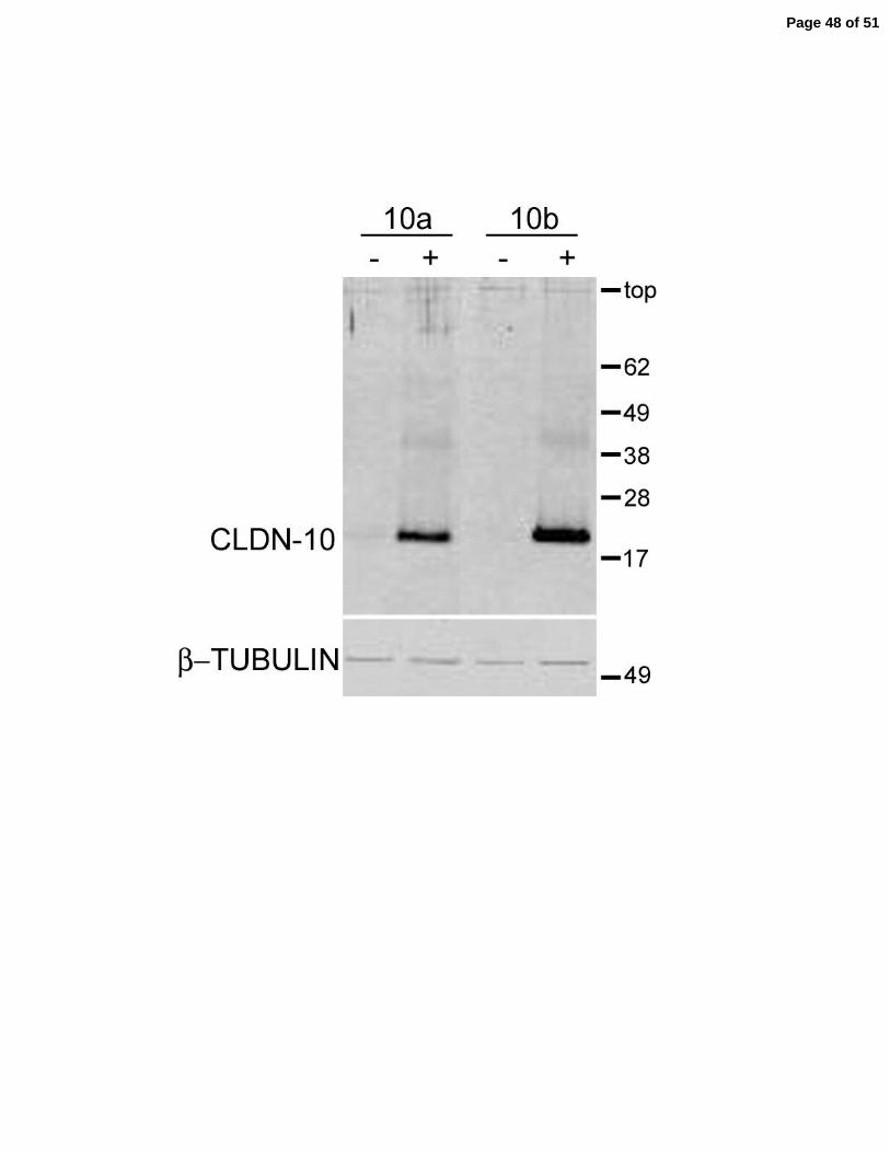
Claudin-10 was not detectable in the parental MDCK II cells nor in the transfected lines when
expression was repressed, Figure 6B top panel, “-“ lanes. Claudin-10a, 10b and the three
mutants of 10a could all be induced by removal of doxycycline, Figure 6B top panel, “+” lanes.
Induction of any form of claudin-10 had little consistent effect on the expression of claudin-2,
claudin-4 (data not shown) or occludin; β-tubulin was used as a control to verify approximately
equal loading of cell protein. Cl audin-10a, 10b, Figure 6C, and the three mutants, Figure 7,
colocalized with ZO-1 at cell contacts as well as in intracellular vesicles, similar to the pattern
seen for several other claudins (30).
MDCK II cells normally form a paracellular barrier with a strong cation preference.
Using our protocol, dilution potentials for NaCl are in the range of +8 to +12 mV, consistent
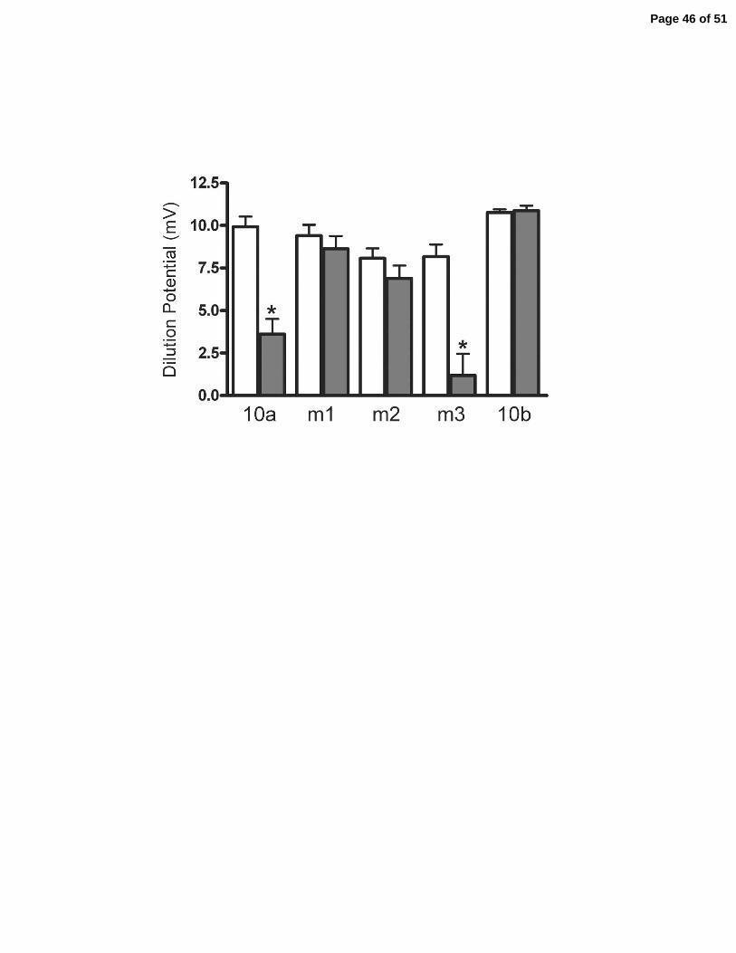
with a PNa/PCl ≈ 3.5. As predicted, expression of claudin-10a in this background resulted in a
sharp decrease in dilution potential, from +10 mV down to +3 mV, consistent with a relative
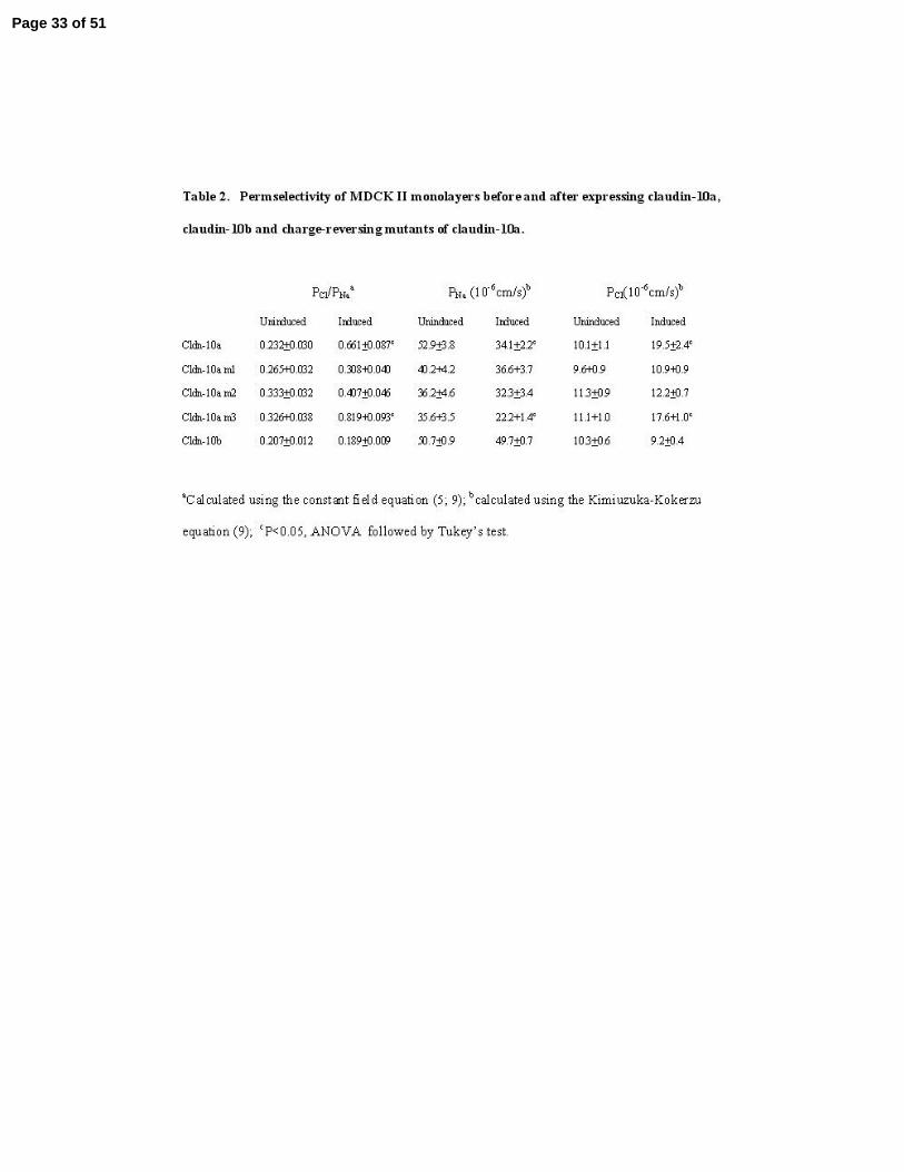
decrease in the ratio Na+ to Cl- permeability, Figure 8A and Table 2. Mutation of either of the
first two positively charged amino acid residues to aspartic acid (m1-R32D and m2-R59D)
eliminates their ability to decrease PNa/PCl, while the third mutant (m3-K66D) retain the ability of
wild-type claudin-10a to decrease PNa/PCl. We conclude the first two but not the third position
can influence permeability of ions moving through the tight junction. Expression of claudin-10b
did not significantly change the dilution potential from that of uninduced MDCK II cells. We
conclude claudin-10b was likely to confer a high PNa/PCl, similar to and thus indistinguishable
from the background properties of MDCK II cells.
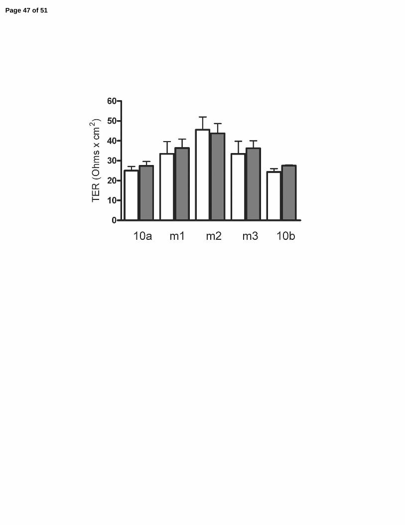
Transepithelial electrical resistance was not significantly changed by expression of
claudin-10a, 10b or the mutants, Figure 8B. The ability of claudin-10a to reduce the dilution
Page 12 of 51
13
potential without changing TER is the result of an opposing decrease in Na+ permeability and
increase in Cl- permeability, Table 2. This is the first time we have seen an increase in Cl-
permeability in MDCK II cells and the result is a leaky pore with half the normal cation
selectivity that characterizes the MDCK II background. This pattern of opposing affects on ionic
permeabilities is distinct from those observed following expression of claudin-4 (28), claudin-11
(30) or claudin-14 (2) in this same cell line. These claudins decrease Na+ permeability, with no
effect on Cl- permeability, with the result that total permeability and conductance (1/TER) are
decreased.
The inability of claudin-10b to alter the dilution potential in MDCK II cells could either
be attributed to intrinsic cation selectivity or might mean that the protein was not functional. To
determine whether expression of 10b in a different cell background could reveal a functional
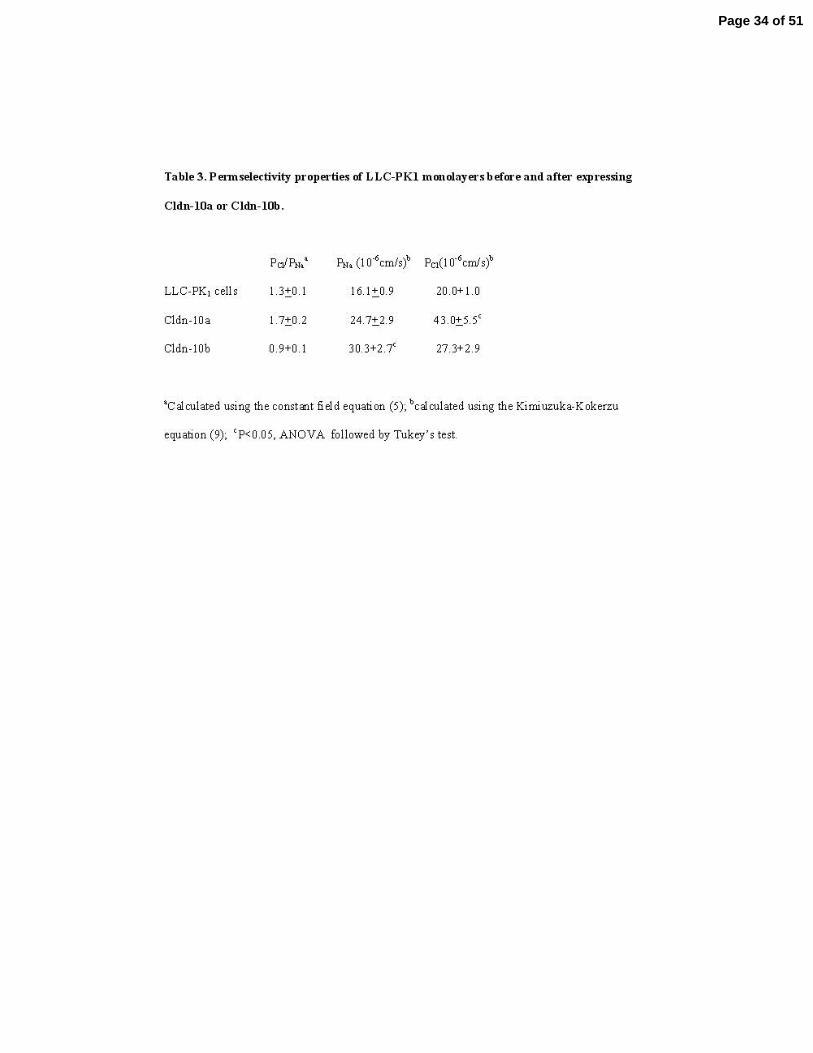
cation-selectivity, we expressed it and 10a in the anion-selective LLC-PK1 cell line. Both
claudin-10 isoforms were inducible in the tet-off LLC-PK1 cells, Figure 9A, and localize to both
cell borders and in some intracellular vesicles, Figure 9B, as was seen in the MDCK II cells. The
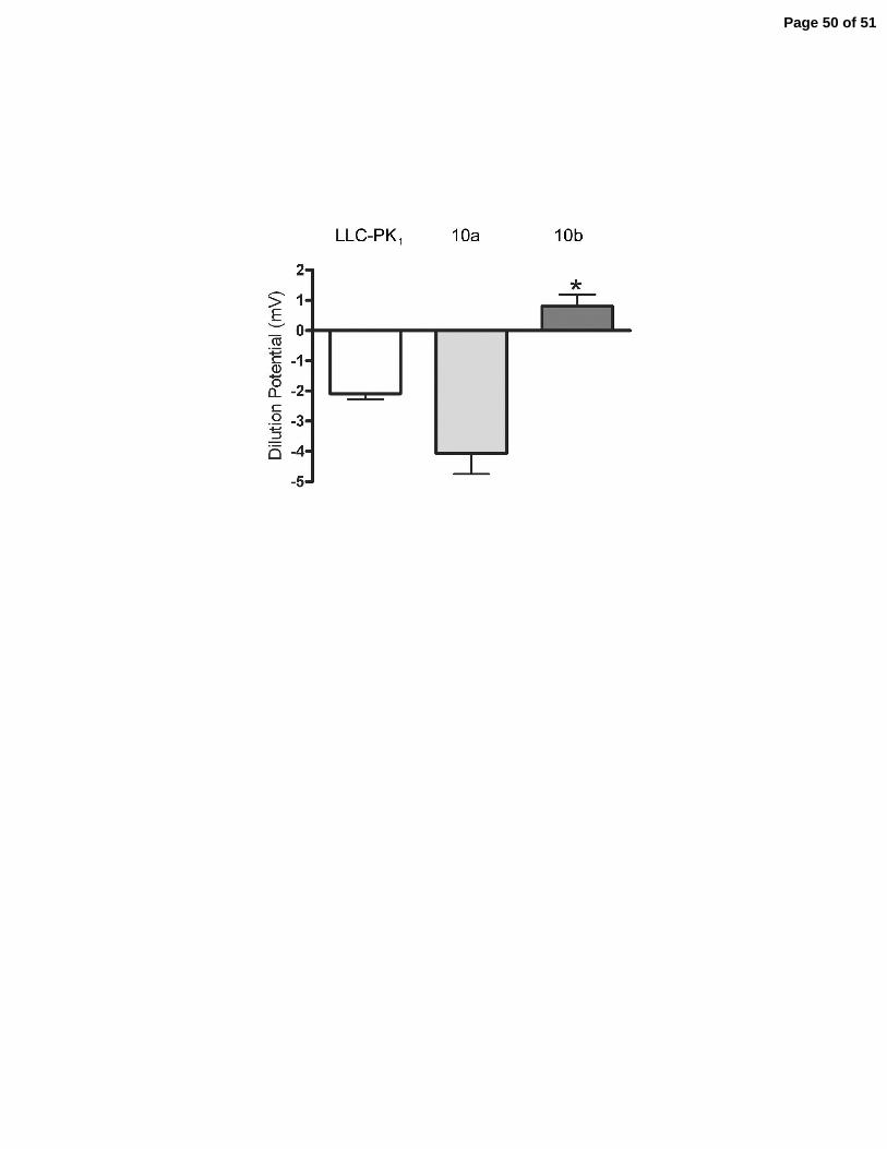
normal dilution potential for untransfected LLC-PK1 cells under conditions described here (30) is
-2 to -3 mV, Figure 9C, expression of claudin-10a in this cell background resulted in a slightly
but not significantly more negative dilution potential. In contrast, expression of claudin-10b
resulted in a significant increase in the dilution potential from approximately -2 to +1mV.
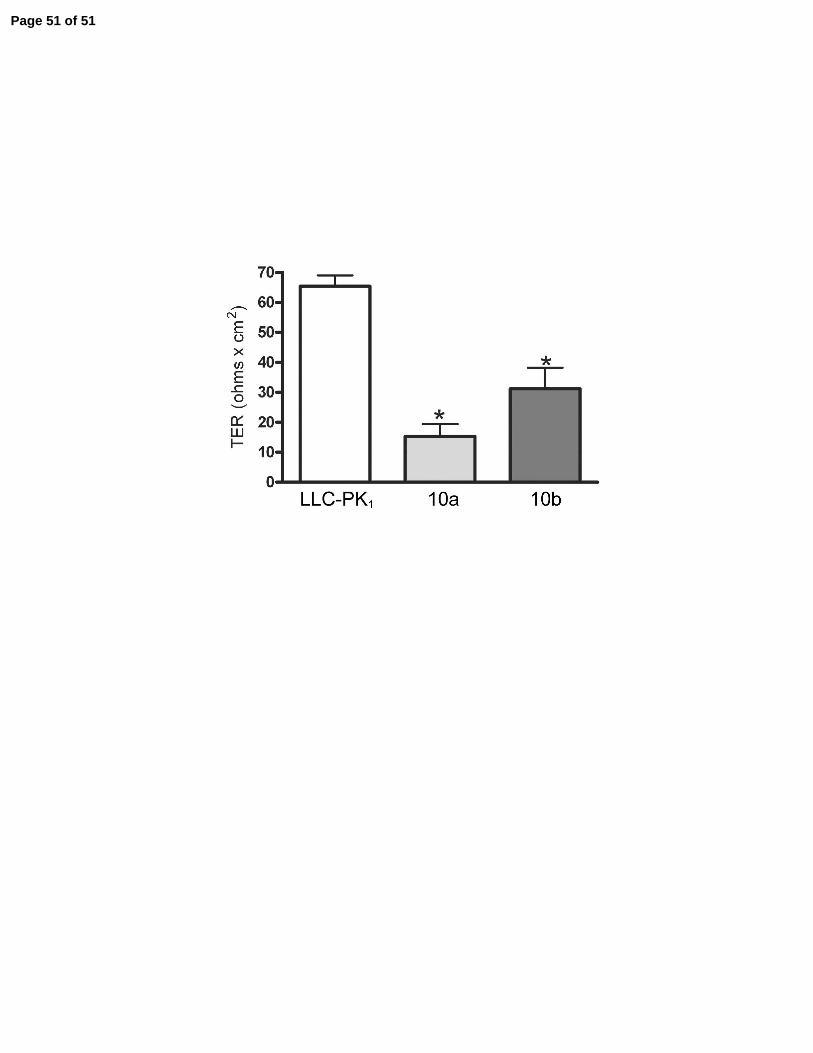
Expression of both claudin-10a and 10b decreased TER, Figure 9D, however, in the case of
claudin-10a, this was attributable to a significant increase in Cl- permeability while in the case of
claudin-10b, this was due mainly to a significant increase in Na+ permeability, Table 3. We
conclude that claudin-10a creates leaky pores relatively more Cl- permeable than Na+ and
claudin-10b creates pores more permeable to Na+ than Cl-.
Page 13 of 51
14
DISCUSSION
In this study, we investigated whether claudin-10a and 10b, two naturally occurring
splice variants confer different effects on tight junction permselectivity. The two variants use
alternative first exons resulting in different first extracellular d omains, which could be significant
because previous studies on other claudins implicate the first extracellular domains in charge
selectivity (7). We find that claudin-10b is widely expressed in many tissues, but consistent with
the possibility of distinct physiologic functions, claudin-10a is unique to the kidney. While we
are unable to assign expression of claudin-10a and 10b to specific nephron segments, their
mRNAs are preferentially expressed in either the medulla or cortex respectively. Directly
expressing the two isoforms in cultured epithelial monolayers reveals that claudin-10a creates
pores preferentially permeable to anions and 10b to cations. Site-specific mutagenesis reveals
some but not all positively charged residues in the first extracellular domain of claudin-10a are
required for its anionic selectivity. We conclude that alternative splicing of the first extracellular
domain of claudin-10 represents a novel mechanism to generate diversity in paracellular
permselectivity. This may also be true of claudin-18 (20) which shows a similar splicing pattern
although its physiology has not been studied.
The finding that claudin-10a appears unique to kidney and the fact that we lack
antibodies to distinguish it from claudin-10b raise the question of whether it is a real splice
variant and protein. We tried several different biochemical approaches to distinguish between the
isoforms. Isoelectric focusing followed by immunoblot did not resolve the two isoforms, but this
technique is generally difficult for small, basic membrane proteins. We were unable to identify
unique chemical or enzymatic cleavage sites which would generate different sized products from
Page 14 of 51
15
10a and 10b on immunoblots. Attempts at proteomic discrimination of the variants in
immunoprecitated material was also unsuccessful; both proteins are of low abundance in native
kidney. Nevertheless, we think the circumstantial evidence is overwhelming that claudin-10a is
a real protein and not, for example, a pseudogene. First, 10a and 10b splice forms can be found
in the database for humans, mice, dogs and cows. Second, the fact that highly homologous
cDNAs have been sequenced for multiple species strongly supports proper RNA transcription
and splicing from active genes. Protein encoded by human exon 1a is 97% identical to the same
regions of both the mouse and dog sequences. In addition, when protein is expressed from the
cldn-10a cDNA in either MDCK or LLC-PK1 cells it localizes properly to the junction and has
unique affects on tight junction ion selectivity. We thus conclude 10a is a real splice variant and
protein from the claudin-10 gene.
Claudin-10 has previously been reported in both endothelial (14) and epithelial (14; 16;
17) cells, raising the question of whether the splice variants segregate between these cell types.
This is not the case since in tissues like cecum which express only claudin-10b, the protein is
detected by immunofluorescence microscopy in both epithelial and endothelial cells. Also, in
kidney, transcripts for both variants are detected in tubular epithelial cells; in some segments
they appear to be expressed in the same cells. Beyond its expression in epithelial and endothelial
cells, claudin-10b in the brain is located in small fibrillar structures which could represent glial
or neural cell process. Identification of these structures awaits further study.
Ion permeability analysis of the two isoforms of claudin-10 revealed some distinct
differences from other claudins. In the cation-selective MDCK II cell background, expression of
a number of claudins were shown to decrease paracellular conduction by specifically decreasing
cation permeability and without changing anion permeability. These include claudin-4 (28),
Page 15 of 51
16
claudin-5 (31), claudin-8 (32), claudin-11 (30), and claudin-14 (2). In contrast, expressing
claudin-10a simultaneously decreased PNa+ while increasing PCl-, creating a unique type of pore
with less overall ionic discrimination . The result of these counter balancing changes is that
overall resistance does not change. This increase in PCl- was also observed when this claudin was
expressed in the LLC-PK1 cell line. In contrast to claudin-10a, claudin-10b behaved in a fashion
similar to that previously observed for claudin-2 (1; 30), in that it makes highly conductive and
cation-selective pores in both MDCK and LLC-PK1 cells. Consistent with our previous work on
other claudins, some but not all, charged residues on the first extracellular domain of claudin-10a
influence ionic permeability. In claudin-10a, R32 and R61 limit Na+ permeability and
presumably line the junctional pores. To summarize our observations on six different claudins,
charge selectivity can be influenced by residues along the entire first extracellular loop, but not
all charged residue positions exert equal influence. Further rationalization of selectivity will
require more structural information on the pore. The unique permselectivity of each tissue likely
results from the profile of claudins expressed, with each claudin having a unique influence on
anion and cation permeability and resistance.
Some claudins have very restricted expression patterns along the nephron where they are
proposed to confer segment-specific paracellular resistance and ion selectivity (6; 18; 22; 25). In
contrast, we find that claudin-10 is expressed in all segments except the glomerulus. The most
obvious finding from the in situ hybridization studies was that claudin-10b was very heavily
expressed in the TAL, where by immunofluorescence the protein is also highly expressed and
localized not only to tight junctions but also found on the lateral cell membrane. In contrast,
claudin-10a mRNA was more abundant in the kidney cortex than in medulla, but there was
considerable overlap between 10a and 10b mRNA expression. Immunofluorescent analysis
Page 16 of 51
17
revealed that claudin-10 protein was expressed in almost every tubule segment in the kidney,
usually but not always colocalized with ZO-1. This finding is different from results reported by
Kiuchi-Saishin et al. (18), who reported the presence of claudin-10 only in proximal tubule and
the thick ascending limb of the loop of Henle, but more similar to findings of Inai et al. (16) and
Chabardes-Garonne et al. (6). This latter group used microarray analysis in freshly dissected
kidney tubules to determine the overall gene expression profile and found claudin-10 expression
in all tubule segments except glomerulus and collecting duct, although the heaviest expression
was in the thick ascending limb of Henle. Our immunofluorescence results suggest that
collecting duct does express claudin-10, although at relatively low levels, while glomerulus
appears to be negative. Unlike what we have seen in other tissues, claudin-10 immunofluorescent
labeling of blood vessels was either faint or undetectable, although others have reported vasa
recta staining with the same claudin-10 antibody (16) . The relatively heavy staining for claudin-
10 in macula densa is intriguing but the physiological relevance is unclear.
It is tempting to speculate that the general distribution of the claudin-10 isoforms as
identified by in situ hybridization in the kidney could be rationalized with current knowledge
about the functions of different parts of the tubule. Claudin-10b creates leaky, cation-permeable
pores. It is more highly expressed in kidney medulla than is claudin-10a, apparently in both the
thin and thick ascending limbs of the loop of Henle. If this is true, it is consistent with the
previously described Na+ and Cl- transport in these segments, since in these tubule segments
paracellular Na+ resorption is driven by lumen positive voltage, but Cl- resorption is mediated
mostly by transcellular transporters. Claudin-10a, which forms leaky anion permeable pores, is
more highly expressed in cortex than medulla. By immunofluorescence, claudin-10 is more
prominent in proximal tubules than in distal tubules. Claudin-2, which is a leaky cation pore, is
Page 17 of 51

18
highly expressed in the proximal tubules, but if claudin-10a is also a component of this barrier, it
would help explain the high paracellular Cl- permeability that characterizes this tubule segment,
particularly the distal proximal tubule. The expression of claudin-10 (if present as 10a) in
collecting duct would be consistent with the reported paracellular Cl- permeability in this
segment, but the resolution of the in situ hybridization was insufficient to indicate the exact
localization of claudin-10a. In any case, the exact molecular constituents of the tight junctions of
these tubule segments, their organization and relative abundances are still unknown, and it is
premature to infer much about the physiologic properties of the junction based on the reported
presence of even several claudins. The role of claudin-10 in the kidney requires further study
although our observations clearly show that alternative splicing is an additional mechanism for
generating diversity in permselectivity of tight junctions.
Page 18 of 51
19
Disclosures: none
Acknowledgments
We acknowledge helpful discussions with Dr. Alan Fanning. These studies were supported by
NIH DK 045134 (to JMA), DK062283 (to ASLY), P30 DK 034987 and the State of North
Carolina. The UNC Neuroscience Center Expression Localization Core is supported by National
Institutes of Neurological Diseases and Stroke Grant NS 031768.
Page 19 of 51
20
References
1. Amasheh S, Meiri N, Gitter AH, Schoneberg T, Mankertz J, Schulzke JD and Fromm
M. Claudin-2 expression induces cation-selective channels in tight junctions of epithelial
cells. J Cell Sci 115: 4969-4976, 2002.
2. Ben Yosef T, Belyantseva IA, Saunders TL, Hughes ED, Kawamoto K, Van Itallie
CM, Beyer LA, Halsey K, Gardner DJ, Wilcox ER, Rasmussen J, Anderson JM,
Dolan DF, Forge A, Raphael Y, Camper SA and Friedman TB. Claudin 14 knockout
mice, a model for autosomal recessive deafness DFNB29, are deaf due to cochlear hair cell
degeneration. Hum Mol Genet 12: 2049-2061, 2003.
3. Berry CA and Boulpaep EL. Nonelectrolyte permeability of the paracellular pathway in
Necturus proximal tubule. Am J Physiol 228: 581-595, 1975.
4. Cereijido MG-MLCG. Tight Junction: Barrier Between Higher Organisms and
Environment. News In Physiological Sciences 4: 72-75, 1989.
5. Cereijido M, Robbins ES, Dolan WJ, Rotunno CA and Sabatini DD. Polarized
monolayers formed by epithelial cells on a permeable and translucent support. J Cell Biol
77: 853-880, 1978.
6. Chabardes-Garonne D, Mejean A, Aude JC, Cheval L, Di Stefano A, Gaillard MC,
Imbert-Teboul M, Wittner M, Balian C, Anthouard V, Robert C, Segurens B,
Page 20 of 51
21
Wincker P, Weissenbach J, Doucet A and Elalouf JM. A panoramic view of gene
expression in the human kidney. Proc Natl Acad Sci U S A 100: 13710-13715, 2003.
7. Colegio OR, Van Itallie C, Rahner C and Anderson JM. Claudin extracellular domains
determine paracellular charge selectivity and resistance but not tight junction fibril
architecture. Am J Physiol Cell Physiol 284: C1346-C1354, 2003.
8. Colegio OR, Van Itallie CM, McCrea HJ, Rahner C and Anderson JM. Claudins
create charge-selective channels in the paracellular pathway between epithelial cells. Am J
Physiol Cell Physiol 283: C142-C147, 2002.
9. Delamere NA and Duncan G. A comparison of ion concentrations, potentials and
conductances of amphibian, bovine and cephalopod lenses. J Physiol 272: 167-186, 1977.
10. Diamond JM. Twenty-first Bowditch lecture. The epithelial junction: bridge, gate, and
fence. Physiologist 20: 10-18, 1977.
11. Diamond JM. Channels in epithelial cell membranes and junctions. Fed Proc 37: 2639-
2643, 1978.
12. Furuse M, Fujita K, Hiiragi T, Fujimoto K and Tsukita S. Claudin-1 and -2: novel
integral membrane proteins localizing at tight junctions with no sequence similarity to
occludin. J Cell Biol 141: 1539-1550, 1998.
Page 21 of 51
22
13. Hamalainen HK, Tubman JC, Vikman S, Kyrola T, Ylikoski E, Warrington JA and
Lahesmaa R. Identification and validation of endogenous reference genes for expression
profiling of T helper cell differentiation by quantitative real-time RT-PCR. Anal Biochem
299: 63-70, 2001.
14. Holmes JL, Van Itallie CM, Rasmussen JE and Anderson JM. Claudin profiling in the
mouse during postnatal intestinal development and along the gastrointestinal tract reveals
complex expression patterns. Gene Expr Patterns 2006.
15. Hou J, Paul DL and Goodenough DA. Paracellin-1 and the modulation of ion selectivity
of tight junctions. J Cell Sci 118: 5109-5118, 2005.
16. Inai T, Sengoku A, Guan X, Hirose E, Iida H and Shibata Y. Heterogeneity in
expression and subcellular localization of tight junction proteins, claudin-10 and -15,
examined by RT-PCR and immunofluorescence microscopy. Arch Histol Cytol 68: 349-
360, 2005.
17. Kitajiri SI, Furuse M, Morita K, Saishin-Kiuchi Y, Kido H, Ito J and Tsukita S.
Expression patterns of claudins, tight junction adhesion molecules, in the inner ear. Hear
Res 187: 25-34, 2004.
18. Kiuchi-Saishin Y, Gotoh S, Furuse M, Takasuga A, Tano Y and Tsukita S. Differential
expression patterns of claudins, tight junction membrane proteins, in mouse nephron
segments. J Am Soc Nephrol 13: 875-886, 2002.
Page 22 of 51
23
19. Li WY, Huey CL and Yu AS. Expression of claudin-7 and -8 along the mouse nephron.
Am J Physiol Renal Physiol 286: F1063-F1071, 2004.
20. Niimi T, Nagashima K, Ward JM, Minoo P, Zimonjic DB, Popescu NC and Kimura S.
claudin-18, a novel downstream target gene for the T/EBP/NKX2.1 homeodomain
transcription factor, encodes lung- and stomach-specific isoforms through alternative
splicing. Mol Cell Biol 21: 7380-7390, 2001.
21. Powell DW. Barrier function of epithelia. Am J Physiol 241: G275-G288, 1981.
22. Reyes JL, Lamas M, Martin D, del Carmen NM, Islas S, Luna J, Tauc M and
Gonzalez-Mariscal L. The renal segmental distribution of claudins changes with
development. Kidney Int 62: 476-487, 2002.
23. Rozen S and Skaletsky H. Primer3 on the WWW for general users and for biologist
programmers. Methods Mol Biol 132: 365-386, 2000.
24. Schwartz MM, Karnovsky MJ and Venkatachalam MA. Regional membrane
specialization in the thin limbs of Henle's loops as seen by freeze-fracture electron
microscopy. Kidney Int 16: 577-589, 1979.
25. Simon DB, Lu Y, Choate KA, Velazquez H, Al Sabban E, Praga M, Casari G,
Bettinelli A, Colussi G, Rodriguez-Soriano J, McCredie D, Milford D, Sanjad S and
Page 23 of 51
24
Lifton RP. Paracellin-1, a renal tight junction protein required for paracellular Mg2+
resorption. Science 285: 103-106, 1999.
26. Stevenson BR, Siliciano JD, Mooseker MS and Goodenough DA. Identification of ZO-
1: a high molecular weight polypeptide associated with the tight junction (zonula
occludens) in a variety of epithelia. J Cell Biol 103: 755-766, 1986.
27. Takahashi M, Taniguchi J, Muto S, Tsuruoka S and Imai M. Effect of protamine on
ion selectivity of superficial and juxtamedullary proximal straight tubules. Nephron 83:
154-159, 1999.
28. Van Itallie C, Rahner C and Anderson JM. Regulated expression of claudin-4 decreases
paracellular conductance through a selective decrease in sodium permeability. J Clin Invest
107: 1319-1327, 2001.
29. Van Itallie CM and Anderson JM. CLAUDINS AND EPITHELIAL PARACELLULAR
TRANSPORT. Annu Rev Physiol 68: 403-429, 2006.
30. Van Itallie CM, Fanning AS and Anderson JM. Reversal of charge selectivity in cation
or anion-selective epithelial lines by expression of different claudins. Am J Physiol Renal
Physiol 285: F1078-F1084, 2003.
Page 24 of 51

25
31. Wen H, Watry DD, Marcondes MC and Fox HS. Selective decrease in paracellular
conductance of tight junctions: role of the first extracellular domain of claudin-5. Mol Cell
Biol 24: 8408-8417, 2004.
32. Yu AS, Enck AH, Lencer WI and Schneeberger EE. Claudin-8 expression in Madin-
Darby canine kidney cells augments the paracellular barrier to cation permeation. J Biol
Chem 278: 17350-17359, 2003.
Page 25 of 51
26
Figure Legends
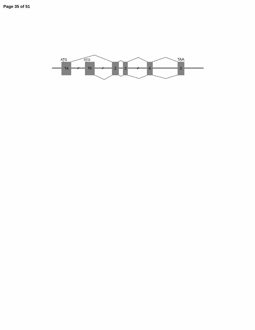
Figure 1. Alternative splicing generates two isoforms of claudin-10, which differ in their first
extracellular domains. A: Schematic organization of the exons within the mouse claudin-10 gene
(Mouse Genome Informatics Number 1913101) on chromosome 14. Alternative slicing of exons
1a and 1b to shared exons 2 through 5 generates transcript variant-1 (claudin-10a, NM_023878)
and variant-2, (claudin-10b, NM_021386). Start and stop codons are noted. B: Amino acid
sequence alignments of claudin-10a and 10b. Identical sequences encode by exons 2-5 are
underlined and shaded. The four transmembrane segments are boxed. The sequences of claudin-
10a and 10b differ up to exon 2, which begins with the last 7 residues of the first extracellular
domain, GYIQACR. Sequences encoded by exons 1a and 1b are 53% homologous and their
identical residues are shaded.
Figure 2. Relative expression of mRNA for claudin-10a and claudin-10b and for total claudin-
10 protein in various mouse tissues. A: Expression of claudin-10a and 10b transcripts
determined by qRT-PCR in brain cortex, whole kidney, lung and cecum expressed relative to
Ef1a1. Claudin-10b is widely expressed but at different levels, while 10a is detected only in the
kidney. In most cases the variation (see methods) is too small to be visible on the graph. B:
Relative protein expression of claudin-10 in brain cortex, whole kidney, lung, cecum and liver.
Antibodies used do not discriminate claudin-10a from 10b. Expression profile for claudin-5 is
shown for comparison. 5 ug of protein was loaded in each lane. Mr markers in kDa.
Page 26 of 51
27
Figure 3. Immunofluorescent protein colocalization of ZO-1 (left panels) with claudin-10 (right
panels). In cecum, claudin-10 (B and D) colocalizes ZO-1 (A and C) at epithelial tight junctions
from the crypt base to the surface. Claudin-10 is also found on the lateral membranes of a subset
of unidentified epithelial cells (arrow). In cerebellar cortex (E and F), claudin 10 colocalizes
with ZO-1 in endothelial cells (arrows) and is also found in fine fibrillar structure with lack ZO-1
staining (arrowhead). In the lung, ZO-1 (G) and claudin-10 colocalize to tight junctions in alveoli
(AL) and large airway surface cells (AW). Bar, 20µm.
Figure 4. Immunolocalization of claudin-10 in mouse kidney cryosections. Claudin-10 is stained
green in every panel. A-C. Double staining for claudin-10 (A), ZO-1 (red, B) and merged image
(C). Claudin-10 is weakly expressed in proximal convoluted tubule (PCT) and distal convoluted
tubule (DCT). Claudin-10 overlaps with ZO-1 at the tight junction; note though that ZO-1
extends further along the lateral membrane, presumably into the adherens junction, where
claudin-10 is absent (arrowheads in C). D. Claudin-10 is strongly expressed in the cortical thick
ascending limb (cTAL) at the basolateral membrane (note deep infoldings of the basal
membrane) and at the tight junction (arrowheads). E. Claudin-10 expression in a section of the
cTAL (asterisk denotes the lumen) at the level of the juxtaglomerular apparatus. Note strong
claudin-10 staining along the basolateral membrane of macula densa cells (arrow, note the
simplified basal membrane), which abut the vascular pole of the glomerulus (G), as well as in
more typical cTAL cells on the opposite side of the tubule (with typical deep infoldings of the
basal membrane). The section is counterstained with antibody to claudin-2 which happens to
highlight the glomerular basement membrane and PCT. F. Claudin-10 is expressed in tight
junctions of the cortical collecting duct (arrows, CCD) and connecting tubule (CNT). Tubules
Page 27 of 51
28
are identified by staining with antibody against the Na-Ca exchanger, NCX1 (red, CNT and CCD
principal cells) and the vacuolar H-ATPase (blue, apical membrane of α-intercalated cells, α-IC,
and brush border of the PCT). G. Claudin-10 is also strongly expressed at the basolateral
membrane and tight junction (arrowheads) of the medullary thick ascending limb (mTAL) and
the tight junction of the outer medullary collecting duct (OMCD). Here the tight junctions are
highlighted with anti-ZO-1 (red). H. In the inner medulla, claudin-10 is strongly expressed in
thin ascending limbs (arrows) and weakly expressed at the tight junctions of thin descending
limbs (asterisks) and inner medullary collecting ducts (IMCD). I. Higher magnification view of
two thin ascending limbs shows claudin-10 colocalization with ZO-1 (red) at their tight
junctions, which appear serpentine and tortuous due to the extensive lateral interdigitations
between neighboring epithelial cells. Scale bars represent 25 µm.
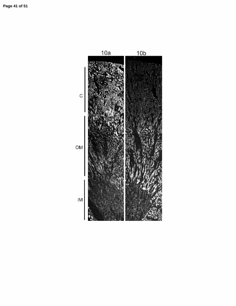
Figure 5. In situ hybridization for claudin-10a and 10b transcripts in mouse kidney. Collages
from cortex to inner medulla were assembled digitally from sequential immunofluorescence
microscopic images all taken at a single exposure setting. The relative expression of 10a is
higher in cortex and 10b in medulla. C (cortex), OM (outer medulla) and IM (inner medulla).
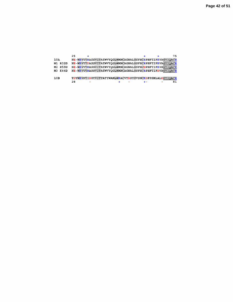
Figure 6. Expression of claudin-10a, 10b and three charge-reversing mutations of claudin-10a
in MDCK II cells monolayers. A: Sequences of the first extracellular domains of claudin-10a,
10b and 10a mutants m1(R32D), m2(R52D) and m3 (K66D). The final seven residues of the first
extracellular domains are encoded by exon 2 and are underlined. Identical resides in 10a and 10b
are shaded. Positive residues are indicated in blue, negative residues are indicated in red.
Position numbers of the first and last residues in the extracellular domains are indicated. B:
Page 28 of 51
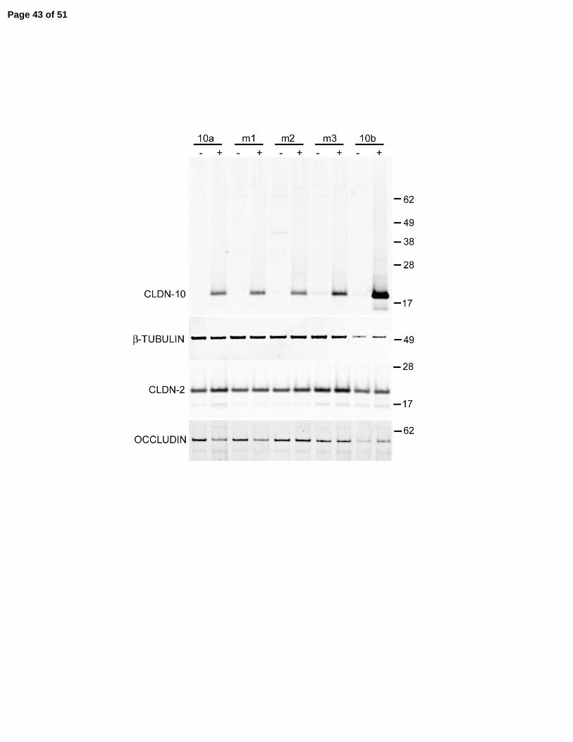
29
Immunoblot showing inducible expression of all proteins in representative cell lines. Repressed
in the presence of doxycycline (-) and induced in its absence (+). Induction has no reproducible
effect on the levels of endogenous claudin-2 and occludin. Beta-tubulin was used as a loading
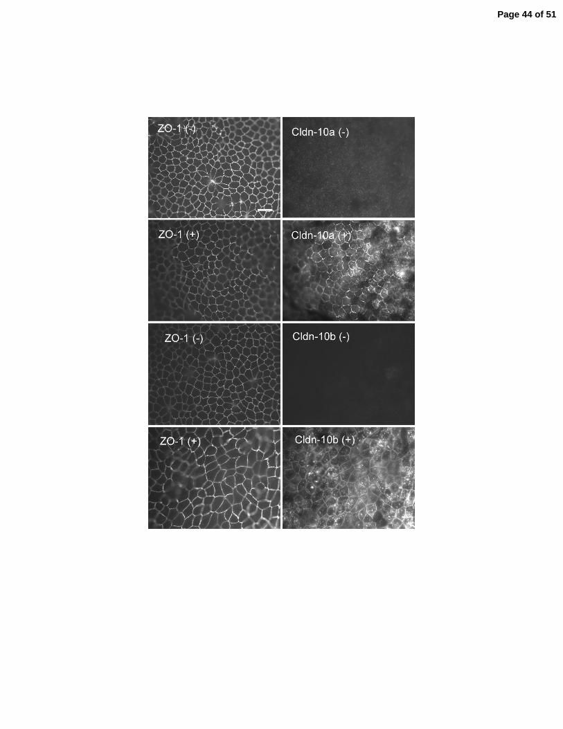
control. C: Immunofluorescent co-localization of ZO-1 (left) with claudin-10a and 10b before (-
) and after (+) induction. The induced proteins colocalize with ZO-1 at tight junctions and
additional pools are observed in the cytoplasm.
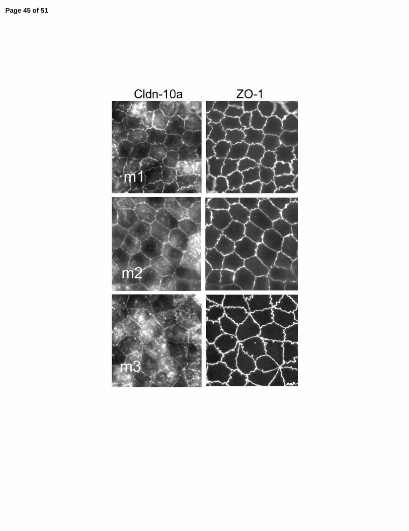
Figure 7. Immunofluorescent co-localization of claudin-10a mutants (left) with ZO-1 (right) in
MDCK cell monolyers induced to express the transgenes. All the induced mutant proteins (m1,
m2 and m3) colocalize with ZO-1 at tight junctions and additional pools are observed in the
cytoplasm.
Figure 8. Electrophysiologic consequences of expressing claudin-10a, 10b and three charge-
reversing mutations of claudin-10a in MDCK II cell monolayers. A: Dilution potential (mV)
across MDCK monolayers following a 120 to 60mM apical NaCl dilution for uninduced (open
bars) and their induced matched clones (filled bars). The positive dilution potential reveals the
cation selectivity of the parental MDCK monolayers. Expression of claudin-10a reduces cation
selectivity while expression of claudin-10b confers no change. M1 and m2 eliminate the effect
of expressing claudin-10a while m3 shows the selectivity of wild-type 10a. B. Despite different
affects on charge selectivity there was no statistically significant effect on transepithelial
electrical resistance (TER). Average of 3-5 clones and duplicate measurements on each; means +
SE. * P < 0.05. Ion permeabilities are reported in Table 1.
Page 29 of 51
30
Figure 9. Consequences of expressing claudin-10a and 10b in anion-selective LLC-PK1 cell
monolayers. A: Immunoblot showing doxycycline-regulated induction of claudin-10a and 10b in
representative LLC-PK1 clones. (-) promoter suppressed in the presence of doxycycline; (+)
activated in its absence. Five clones were studied for each claudin. Immunoblotting for β-tubulin
on the same blot confirms similar protein loading. Mr markers in kDa. B: Immunofluorescent
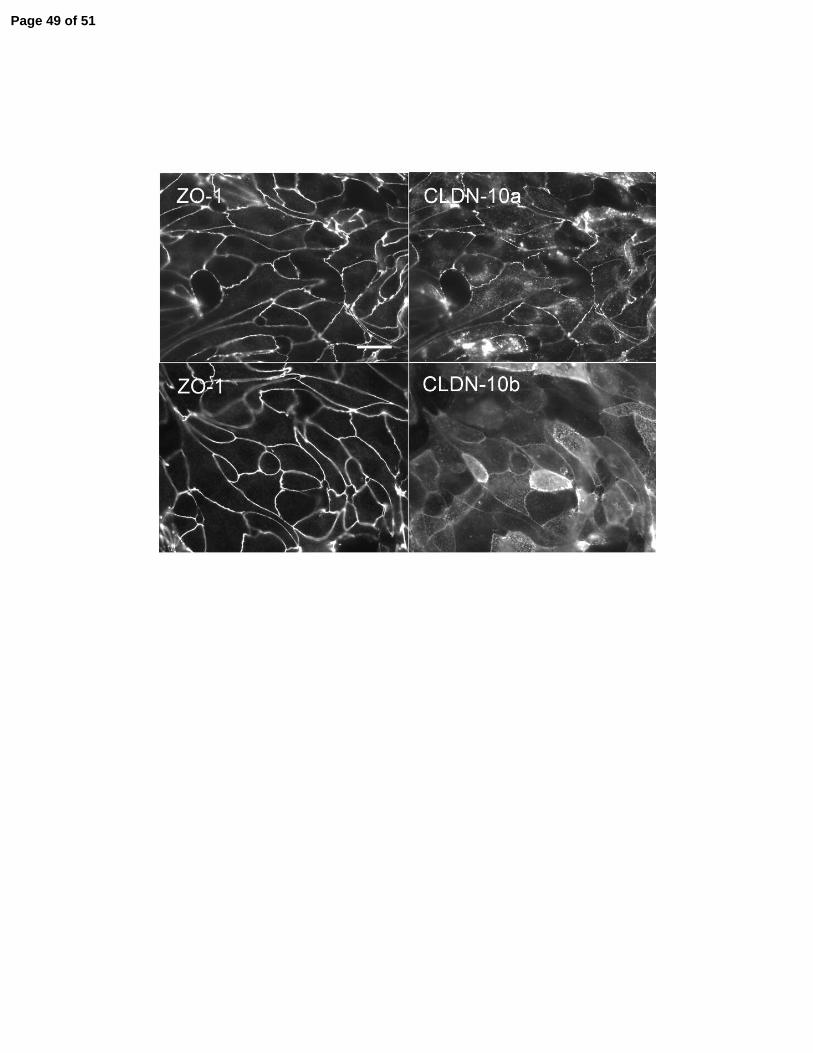
microscopic colocalization of ZO-1 with induced claudin-10a (top panels) and 10b (bottom
panels). C: Dilution potentials (mV) across LLC-PK1 monolayers following a 120mM to 60
mM NaCl dilution for untransfected cells (LLC-PK1), claudin-10a and 10b. The negative dilution
potential reveals the anion selective background of LLC-PK1 monolayers. Expression of claudin-
10a does not alter the anion selectivity while expression of claudin-10b produces a cation-
selective monolayer. D: Despite different affects on charge selectivity both 10a and 10b cause a
statistically significant reduction in transepithelial electrical resistance. Average of 3-5 clones
and duplicate measurements on each, means + SE. * P < 0.05.
Page 30 of 51
31
Tables and table legends
Table 1. Quantitative PCR Primers
All primers were designed using Primer3 software (23).
Table 2. Permselectivity of MDCK II monolayers before and after expressing claudin-10a,
claudin-10b and charge-reversing mutants of claudin-10a.
aCalculated using the constant field equation (5; 9); bcalculated using the Kimiuzuka-Kokerzu
equation (9); cP<0.05, ANOVA followed by Tukey’s test.
Table 3. Permselectivity properties of LLC-PK1 monolayers before and after expressing
Cldn-10a or Cldn-10b.
aCalculated using the constant field equation (5); bcalculated using the Kimiuzuka-Kokerzu
equation (9); cP<0.05, ANOVA followed by Tukey’s test.
Page 31 of 51
Page 32 of 51
Page 33 of 51
Page 34 of 51
Page 35 of 51

Page 36 of 51
Page 37 of 51
Page 38 of 51
Page 39 of 51

Page 40 of 51
Page 41 of 51
Page 42 of 51
Page 43 of 51
Page 44 of 51
Page 45 of 51
Page 46 of 51
Page 47 of 51
Page 48 of 51
Page 49 of 51
Page 50 of 51
Page 51 of 51
Copyright © 2022 FDOKUMEN