Histological features and histochemistry of the mucous glands ...
Lectin histochemistry in the human epididymisM. I. Arenas, M. P. de Miguel, F. R. Bethencourt, B. Fraile,
M. Royuela and R. Paniagua1Department of Cell Biology and Genetics, University of Alcalá de Henares, E-28871 Alcalá de Henares
(Madrid), Spain; and Department of Urology, Hospital Principe de Asturias, E-28871 Alcalá de Henares(Madrid), Spain
A histochemical study using lectins to identify glycoconjugates present in the efferent ductsand ductus epididymidis of men without testicular or related disease was carried out. Thelectins used and the oligosaccharide residues linked were: wheat germ agglutinin (WGA) for\g=b\-N-acetylglucosamineand sialic acid, concanavalin A (ConA) for \g=a\-mannose, Ulex europaeusagglutinin (UEA-I) for \g=a\-fucose, Dolichos biflorus agglutinin (DBA) for \g=a\-N\x=req-\acetylgalactosamine, soy bean agglutinin (SBA) for \g=b\-N-acetylgalactosamine,and peanutagglutinin (PNA) for \g=b\-galactose.The lectin-binding pattern in the human epididymispresents similarities and differences to those observed in other mammals which also showeddifferences between species. The present results revealed that regional differences along thehuman ductus epididymidis were less pronounced than those reported in other mammals.The epithelial cells in the efferent ducts reacted positively to WGA. All along the length ofthe ductus epididymidis, the principal cells and the luminal content showed staining affinityfor WGA and ConA. The epididymal principal cells and luminal content also reactedpositively to DBA for \g=a\-N-acetylgalactosaminebut only in the cauda epididymidis. Apositive reaction to UEA-I was observed only in the luminal content of the caudaepididymidis. This finding suggests that changes in the oligosaccharide chains of secretionsleading to a positive UEA-I reaction occur in the cauda epididymidis. The epididymalprincipal cells showed positive reactions to SBA and PNA over the apical microvilli but notin the cytoplasm. The reaction was observed in the caput and corpus epididymidis but notin the cauda. Positive reactions to SBA and PNA were also detected in the epididymal fluidand in the cytoplasm of mitochondria-rich cells (a minor population of epididymal epithelialcells). These cells also reacted to other lectins such as WGA, ConA and DBA, which were
also detected in the principal cells.
Introduction
Mammalian spermatozoa are not mature when they leave thetestis; their development continues during their passagethrough the epididymis (spermatozoon maturation) (Bedford,1975). A contribution from the epididymis is necessary for thismaturational process (Echeverría et al, 1984) and, most impor¬tantly, spermatozoa acquire the ability to fertilize in theepididymis. Although the precise region of the epididymiswhere this acquisition occurs varies with species, it occurs
generally in the corpus or proximal cauda epididymidis(Bedford, 1975).
The ductuli efferentes are a series of tubules that conductspermatozoa from the rete testis to the epididymis (Ilio andHess, 1994). These tubules leave the testis, initially as parallelstraight tubules containing ciliated and non-ciliated cells in an
epithelium of irregular height (Saito et al, 1990; Yeung et al,
1991). These ducts continue with the ductus epididymidis, a
convoluted duct that extends from the caput to the caudaepididymidis. The epithelium lining the ductus epididymidis iscolumnar pseudostratified and comprises mainly principal cellsand basal cells (Morita, 1966). Isolated mitochondria-rich cells(also called narrow cells, apical cells or flask cells according totheir shape) may be seen in the caput and, more rarely, in thecorpus epididymidis (Palacios et al, 1991). Intraepitheliallymphocytes are evident in increasing numbers as one proceedsfrom the caput more distally (Ritchie et al, 1984). Anotherepithelial cell type, designated as the clear cell, has beenreported in mice and rats but not in humans (Toshimoriet al, 1988). The epididymal epithelium displays regionalchanges in features such as luminal diameter and smoothmuscle investment (Vendrely, 1981). The epididymis has beenimplicated in the secretion of a variety of substances rangingfrom small molecules to proteins (Hamilton, 1975; Turner,1979). Biochemical analyses in various species have demon¬strated the presence of specific epididymal proteins, which varywith the epididymal region (Lea et al, 1978; Echeverría et al,
*Correspondence and reprint requests.Received 21 September 1995.

1982; Russell et al, 1984; Olson and Hinton, 1985). Synthesisand secretion of proteins and glycoproteins by the epididymalepithelium have also been demonstrated using radioauto-graphy (Kopecny and Pech, 1977) or immunocytochemicalprocedures (Tezon et al, 1985). Light microscope autoradio¬graphy studies have shown the greatest activity for protein or
glycoprotein synthesis in the initial segment of the epididymis,with progressively lower protein incorporation in the middleand terminal segment (Kopecny and Pech, 1977). In the mouse
epididymis, the synthesis and transport of proteins seem to bemore rapid in the middle than in the initial segment (Flickinger,1981).
Lectins have been used extensively as biochemical tools toisolate glycoproteins, glycolipids and polysaccharides and as
probes to investigate the sugar residues of the cell surface.When conjugated to a marker, such as fluorescent dye, peroxi¬dase or colloidal gold, lectins may be used as histochemicalprobes to identify and localize specific carbohydrate residues(Roth, 1978; Chang and Wong, 1992). Lectins are especiallyuseful in studies of the quality, distribution, assembly andturnover of glycoconjugates in normal and pathological tissues,as well as during embryonic differentiation (Brown and Hunt,1978; Lis and Sharon, 1986). Lectin histochemistry has beencarried out in the epididymis of the mouse (Mus musculus),guinea-pig (Cavia porcellus), gerbil (Jaculus jaculus) and nutria(Lutra lutra) (Arya and Vanha-Perttula, 1986; Burkett et al,1987). The results of these studies revealed positive reactionsfor wheat germ agglutinin (WGA), peanut agglutinin (PNA)and concanavalin A (ConA), although the staining patternvaried among species and among regions of the epididymis.The aim of the present study was to examine the location anddistribution of glycoconjugates in the human epididymis usinga number of biotinylated lectin—peroxidase complexes.
Materials and Methods
Human epididymides from 17 adult men (aged from 25 to 55
years) were obtained at autopsy (6-10 h after death). Thesemen had died either in traffic accidents (seven men) or frommyocardial (five men) or cerebral (five men) infarction. Thecondition for selection was that the testes showed a normalhistological pattern, including complete spermatogenesis.Samples were fixed in 10% (v/v) buffered formaldehyde for24 h at room temperature. Tissue specimens were dehydratedthrough a graded ethanol series, cleared in xylene and embed¬ded in paraffin wax (Paraplast: Panreac, Barcelona). Postmortemchanges in the autopsy specimens were evaluated by immedi¬ately fixing and processing three epididymides obtained fromtesticular tumour surgery in the same way. Removal of tissuesand the study of autopsy samples were approved by the EthicsCommittee of the Hospital and made with the consent of thepatients' relatives.
Thick sections, 5 µ , were cleared of paraffin with xyleneand rehydrated through a series of decreasing ethanol concen¬
trations followed by PBS. For glycoconjugate demonstration,in addition to histochemical staining with Alcian blue (pH 2.5)/periodic-acid-Schiff (AB/PAS) (Mowry, 1963), different horse¬radish peroxidase-labelled lectins were used at concentrationsof 10-20 µgmrI in 0.1 mol PBS 1" 1 (pH 7.2) for 60 min at
room temperature. Previously, the endogenous peroxidase was
blocked by incubation in 3% H202. The lectins used were thefollowing: wheat germ agglutinin (Triticum vulgäre, WGA),concanavalin A (Concanavalia ensiformis, ConA), Ulex europaeusagglutinin-I (UEA-I), Dolichos biflorus agglutinin (DBA), soybean agglutinin (Glycine max, SBA), and peanut agglutinin(Arachis hypogea, PNA). The peroxidase activity was revealedby a diaminobenzidine—hydrogen peroxide system (DAB;Sigma, Barcelona). All lectins were supplied by Sigma.
For lectin negative controls, the following saccharides were
added at a final concentration of 1 mg ml~
to the respectivelectin solutions: N-acetyl-D-glucosamine for WGA, ct-methyl-D-mannoside for ConA, L-fucose for UEA-I, N-acetyl-D-galactosamine for DBA and SBA, and ß-galactose for PNA.Exposure to peroxidase and DAB system without lectins was
performed as an additional negative control. The reactions ofcertain control sections were examined with DAB only todetect endogenous peroxidase activity in tissues. The sectionswere counterstained with Harris' haematoxylin, dehydrated,and mounted in DePex (Probus, Barcelona).
Mitochondrial-rich cells were identified in sections from thesame specimens used here by means of enzymohistochemicaland immunohistochemical techniques, using principally anti-AE1/AE3 cytokeratin antibodies, which immunoreact intenselyagainst these cells. These techniques and the results obtainedhave been reported by Palacios el al (1991).
Results
Comparison of the epididymides obtained during surgerywith the autopsy specimens showed neither histological nor
histochemical changes in the autopsy specimens.AB/PAS staining showed differential reactions in the epi¬
didymal epithelium. The luminal surfaces of the epithelial cellspresented positive reaction to Alcian blue (acidic glyco¬proteins) in the efferent ducts and along the whole length ofthe ductus epididymidis. The apical cytoplasm of principal cellsin the ductus epididymidis showed positive reaction to PAS(neutral glycoproteins). The luminal content of cauda epi¬didymidis presented a strong positive reaction to Alcian blueand PAS.
The results of lectin histochemistry are summarized(Table 1). Reaction to WGA was positive in all epididymalregions. In the efferent ducts, the reactivity was localized in thesupranuclear cytoplasm of the principal cells and ciliated cells(Fig. la, b). In the ductus epididymidis, the cytoplasm of allepithelial cell types stained diffusely with WGA (Fig. la, c). Apositive reaction was also observed in the tubular lumen fromthe caput epididymidis onwards. The interstitial connectivetissue and the smooth muscle tissue surrounding the tubulesalso stained all along the epididymis (Fig. lb, c).
An intense reaction to ConA lectin was observed in theprincipal cells of the caput, corpus and cauda epididymidis andin the mitochondria-rich cells (present only in the caput andcorpus epididymidis) (Fig. Id). The luminal content of theductus epididymidis also labelled for ConA in the corpus andcauda. No positive reaction to this lectin was found in theefferent ducts. The interstitial connective tissue and the smoothmuscle tissue surrounding the tubules also stained all along theepididymis.

Table 1. Histochemistry of carbohydrates in the human epididymis
LectinsCarbohydrate
binding
Efferent ducts
PC CC PC
Ductus epididymidisCaputMRC PC
CorpusMRC
Cauda
PC
WGA ß-GlcNAc/sialic acidConA a-MannoseUEA-1 a-FucoseDBA a-GalNAcSBA ß-GalNAcPNA ß-Galactose
++
+ "
+ a
++ '
+ '
++ '
+'
++
++
PC: principal cells; CC: ciliated cells; L: luminal fluid; MRC: mitochondria-rich cells; WGA: wheat germ agglutinin; ConA: concanavalinA; DBA: Dolichos biflorusagglutinin; SBA: soy bean agglutinin; PNA: peanut agglutinin; GlcNAc: N-acetylglucosamine; GalNAc: N-acetylgalactosamine; aStaining is visualized only in theadluminal surface. Basal cells were labelled with WGA and only occasionally with the other lectins.
UEA-I lectin labelling was only visualized in the luminal fluidof the cauda epididymidis (Fig. le, f). The epithelium of theefferent ducts, caput, corpus and cauda epididymidis showed a
negative reaction to this lectin.A strong reaction to DBA was found in the mitochondria-
rich cells of the caput and corpus, the principal cells and luminalcontent of the cauda epididymidis and in fibroblast-like cells inthe interstitial connective tissue throughout the epididymallength (Fig. 2a—c).
An intense reaction to SBA was observed in the luminalsurface of the epithelial cells of the caput and corpus epi¬didymidis as well as in their luminal content (Fig. 2d). Themitochondria-rich cells also presented a strong positivereaction to SBA (Fig. 2d). In the cauda, the epithelial cellsshowed no reaction to SBA, whereas the luminal contentreacted positively to this lectin (Fig. 2e). All along the length ofthe ductus epididymidis, small round cells that appearedisolated in the epididymal epithelium and, more rarely, in theinterstitial connective tissue (probably lymphocytes), alsoreacted to SBA (Fig. 2d, e).
Reaction to PNA was localized in the luminal surface of theprincipal cells and luminal content in all the epididymal regionsexcept for the efferent ducts (Fig. 2f, g). A strong labelwas also observed in the cytoplasm of mitochondria-rich cells(Fig· 2g).
Discussion
The lectin-binding pattern of the human epididymis obtained inthe present study presents similarities and differences withregard to that observed in other mammals, such as gerbils,guinea-pigs, nutria, and mice (Arya and Vanha-Perttula, 1986;Burkett et al, 1987) which, in turn, showed differences amongspecies. In addition, the present results reveal that regionaldifferences along the human ductus epididymidis were lesspronounced than those reported in other mammals.
In the human epididymis, a diffuse and uniformly distributedWGA labelling (ß-N-acetylglucosamine and sialic acid detec¬tion) was localized in all the epithelial cells in all the epididymal
segments. A positive staining to this lectin was also found ingerbils, guinea-pigs, nutria, and mice (Arya and Vanha-Perttula,1986; Burkett et al, 1987), although only epididymal regions Iand II were stained in mice, and only regions II and III werestained in nutria. The wide reaction to this lectin may be relatedto structural oligosaccharides. In the mouse epididymis, Burkettet al (1987) observed a strong staining for WGA in the ciliaand the apical surfaces of ciliated cells in the efferent ducts ofmice. As the ciliated cells are not known to secrete macro-
molecules, it is more probable that newly synthesizedglycoconjugates are incorporated directly into the cell plasma¬lemma as integral membrane glycoproteins than that they are
exocytosed.Nevertheless, WGA staining also appears in the epididymal
lumen. Although the presence of stained material in the lumenand epithelial cells does not necessarily imply that the proteinis secreted (it could be taken up from the lumen), it is wellknown that, in contrast to the absorptive function of theefferent duct epithelial cells (Ilio and Hess, 1994), the ductusepididymidis epithelium is principally involved in glycoproteinsecretion (Yeung et al, 1991), and thus, the present findingssuggest that some WGA-labelled glycoprotein is also secretedin the human epididymis. In the boar epididymis, Dacheauxand Dacheaux (1988) immunolabelled antagglutinin in all theprincipal cells of the caput and corpus epididymidis as well asin the lumen. These authors also found that antagglutinin is a
sialoprotein that binds WGA and ConA in the corpus andcauda epididymidis of this species. In addition, Bedford et al(1973), in humans, and Toshimori et al (1988), in mice, detectedsialic acid residues in spermatozoa and assumed that theseresidues are from the sperm maturation antigen (SMA).Toshimori et al (1988) concluded that mouse SMA is secretedby the epididymal clear cells
-
and not by the principal cells-because only the clear cells of the corpus epididymidis showed
a positive reaction to sialic acid. In the study reported here,where WGA lectin labelled the SMA, this would not have beensynthesized by the clear cells, which are absent in mostmammals including humans, but by other epithelial cells. Inmice, the clear cells are present only as isolated cells in thecorpus epididymidis. It may be that, in humans and in some

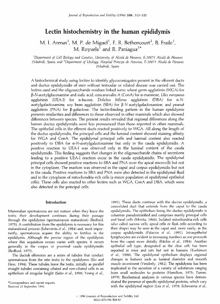
Fig. 1. (a-c) Wheat germ agglutinin staining for the demonstration of the presence of ß-N-acetylglucosamine/sialic acid in the human epididymis.(a) Control section of the caput epididymidis from a 41-year-old man. Positive staining (due to peroxidase activity) is observed only in lysosomalgranules (arrow) of the efferent duct epithelium (EF). DE: ductus epididymidis. (b) Efferent ducts from a 26-year-old man. In addition to thelysosomal reaction, the supranuclear cytoplasm of the epithelial cells (arrow) and the interstitial connective tissue (star) were weakly stained, (c)Caput epididymidis from a 30-year-old man. The epithelium of the ductus epididymidis (DE), luminal content (asterisk), and the connective tissue(star) are diffusely stained, (d) Concanavalin A staining for the demonstration of the presence of mannose in the corpus epididymidis from a
33-year-old man. The epithelial cells reacted intensely to this lectin (DE). A weaker reaction was observed in the connective tissue (star), (e, f) Ulexeuropaeus agglutinin-I staining for fucose. (e) Control section of the corpus (asterisk) and cauda (star) epididymidis from a 42-year-old man. Nopositive reaction is observed, (f) Corpus (asterisk) and cauda (star) epididymidis from a 39-year-old man. A positive reaction is observed only inthe luminal content but not in the epithelium (E) of the cauda. All sections were counterstained with Harris' haematoxylin. Scale bar: 1 µ forall micrographs.
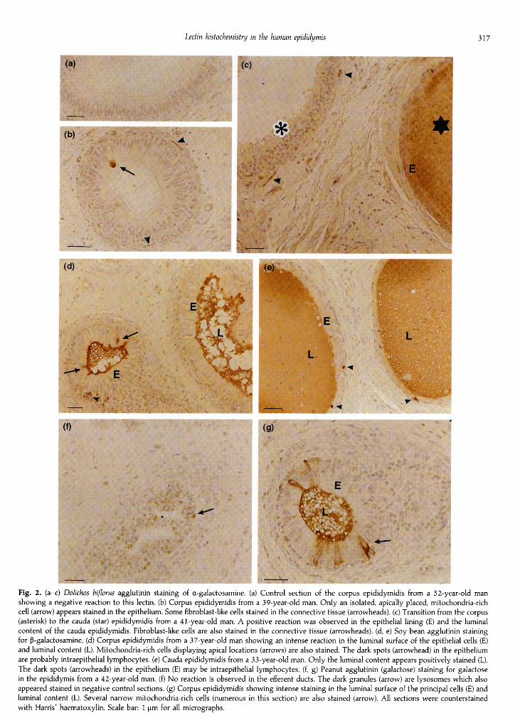
Fig. 2. (a-c) Dolichos bifloras agglutinin staining of -galactosamine. (a) Control section of the corpus epididymidis from a 52-year-old man
showing a negative reaction to this lectin. (b) Corpus epididymidis from a 39-year-old man. Only an isolated, apically placed, mitochondria-richcell (arrow) appears stained in the epithelium. Some fibroblast-like cells stained in the connective tissue (arrowheads), (c) Transition from the corpus(asterisk) to the cauda (star) epididymidis from a 41-year-old man. A positive reaction was observed in the epithelial lining (E) and the luminalcontent of the cauda epididymidis. Fibroblast-like cells are also stained in the connective tissue (arrowheads), (d, e) Soy bean agglutinin stainingfor ß-galactosamine. (d) Corpus epididymidis from a 37-year-old man showing an intense reaction in the luminal surface of the epithelial cells (E)and luminal content (L). Mitochondria-rich cells displaying apical locations (arrows) are also stained. The dark spots (arrowhead) in the epitheliumare probably intraepithelial lymphocytes, (e) Cauda epididymidis from a 33-year-old man. Only the luminal content appears positively stained (L).The dark spots (arrowheads) in the epithelium (E) may be intraepithelial lymphocytes, (f, g) Peanut agglutinin (galactose) staining for galactosein the epididymis from a 42-year-old man. (f) No reaction is observed in the efferent ducts. The dark granules (arrow) are lysosomes which alsoappeared stained in negative control sections, (g) Corpus epididymidis showing intense staining in the luminal surface of the principal cells (E) andluminal content (L). Several narrow mitochondria-rich cells (numerous in this section) are also stained (arrow). All sections were counterstainedwith Harris' haematoxylin. Scale bar: 1 µ for all micrographs.

other mammals, an ill-defined subtype of principal cells or othercell type of the epididymal epithelium present in very smallnumbers, such as the mitochondria-rich cell, may be responsiblefor this secretion.
ConA binds tightly to mannose-rich precursor oligosac¬charide chains, complex mannose-type oligosaccharides foundin membrane glycoproteins, and lysosomal enzymes (Shibuyaet al, 1988). ConA does not bind to complex chains with threeor more N-acetylglucosamine branches (Baenziger and Fiete,1979). Several researchers have reported ConA-binding sites inthe cisternae of the rough endoplasmic reticulum (Chang andWong, 1992), and within the cisternae of the Golgi apparatus(Roth, 1983; Pavelka and Ellinger, 1985). We observed an
intense staining affinity for ConA in the cytoplasm of principalcells in the caput, corpus and cauda epididymidis. A positivereaction to this lectin was also found in the luminal content butonly in the last two regions. These observations differ from thelectin histochemistry study of the epididymis of gerbils,guinea-pigs, nutria, and mice by Arya and Vanha-Perttula(1986). These researchers only observed a cytoplasmic positivereaction to ConA in gerbils and only in region II of theepididymis. If antagglutinin
—
which may be detected withWGA and ConA in boars (Dacheaux and Dacheaux, 1988)
—
or
other similar glycoproteins are also present in other mammalsincluding humans, it is possible that the epididymal principalcells that are positive to these two lectins are responsible forthe synthesis and secretion of these proteins in men andgerbils, whereas in other mammals, such as guinea-pigs, nutria,and mice, the cells involved in these secretions are only WGApositive. We can speculate that, in humans, one of theseproteins may be HE2, a secretory epididymal glycoprotein thatassociates with the equatorial region of the sperm head(Osterhoff et al, 1994).
The oligosaccharide residues detected by DBA (a-N-acetylgalactosamine) could be contained in residual proteins inthe lysosomes or in mucin-type-O-linked glycoproteins frommucous secretions (Madrid et al, 1994). Positive reactions toDBA have been reported in the cytoplasm of epididymalepithelial cells in most mammals studied. However, whereas in
guinea-pigs this reaction was positive all along the length ofthe ductus epididymidis, in mice, gerbils and nutria, thereaction was limited to the initial epididymal regions (I and II)(Arya and Vanha-Perttula, 1986). Lectin histochemical studieson the mouse epididymis by Burkett et al (1987) found a
positive reaction to DBA all along the epididymis, but only inbasal cells. In the present study, a reaction to DBA was
observed only in the epithelial cells and luminal content of thecauda epididymis. In mice, a reaction to DBA may be related tothe secretion of a glycoprotein (CP27) which is the majorglycoprotein secreted in the cauda epididymidis of this speciesthat is involved in maturation of spermatozoa (Flickinger et al,1988). However, there is no information about a similar proteinin humans.
A positive reaction to UEA-I lectin, which preferentiallylabels terminal fucose residues located in the outer region ofoligosaccharide chains (Sugii and Kabat, 1982), has beenreported in the epididymal principal cells of gerbils (regions IIand III), guinea-pigs (region III) and mice (regions I and II)but not in nutria (Arya and Vanha-Perttula, 1986). Weobserved that the luminal content of the cauda epididymidis
presents a positive reaction to this lectin. However, thereaction to this lectin was not detected in the epithelial cellsin any epididymal region. This suggests that, in men andnutria, the UEA-labelled secretion is not synthesized andsecreted in its final form and that the changes leading to a
positive UEA reaction in the oligosaccharide chains of secre¬
tions occur in the cauda epididymidis. These changes may beexplained by two mechanisms: either a fucose residue isadded to the oligosaccharide chain or this residue is elimi¬nated by enzymatic digestion. The last hypothesis is inagreement with the high content of a-glucosidase found inthe human cauda epididymidis (Yeung et al, 1990; Purvis andEgdetveit, 1993).
In the results presented here, the principal cells of theepididymis showed positive reactions only to SBA (ß-N-acetylgalactosamine) or PNA (ß-galactose) over the apicalmicrovilli. SBA staining was localized in the caput and corpusepididymidis, whereas PNA staining was observed all alongthe epididymis, except for the efferent ducts. Arya andVanha-Perttula (1986) observed that in the epididymis ofgerbils, guinea-pigs, mice and nutria, the apical surface andmicrovilli are strongly reactive to PNA in all regions, whileSBA displayed weak staining. These findings suggest that theglycoconjugates forming the glycocalyx of the epithelial cellsdiffer along the epididymal length. It has been reported that theresidues labelled by these lectins could also be part of theglycoproteins secreted by epithelial cells as cementing or
barrier substances (Meyer, 1986). In addition, other saccharideresidues that were also detected in the cytoplasm, such as sialicacid and a-mannose, also appear in the glycocalyx of theepithelial cells in some epididymal regions. Lectins have beenused to characterize the glycoconjugates in the glycocalyx ofvarious epithelial cells bearing microvilli (Roth, 1983; Gonnellaand Neutra, 1985; Tanimura et al, 1987) and, it has been shownthat, along the length of the renal tubules, the glycocalyxexhibits marked variations that have been related to theirspecialized functions (Spicer et al, 1981; Roth and Taatjes,1985).
In the present study, positive reactions to SBA and PNAwere also detected in the epididymal fluid and in the cytoplasmof mitochondria-rich cells. Although staining of the intra-luminal content might be attributed to the glycocalyx ofsloughed principal cells, this staining might also be the result ofsecretion by mitochondria-rich cells. These cells also reacted toWGA and ConA, and they were the only epididymal cellspositive to DBA (a-N-acetylgalactosamine), which was alsodetected in the intraluminal content. These cells form a minorpopulation of epididymal epithelial cells in most mammalsstudied and are usually present only in the initial epididymalsegments (Martínez-García et al, 1995). The morphologicalpattern of these cells varies, even in the same epithelium, fromslender cells (narrow cells) to round apical cells withoutapparent connection with the epithelial basal lamina (apicalcells); but they all contain short microvilli and an electron-dense cytoplasm with abundant mitochondria (mitochondria-rich cells). Histochemical and biochemical studies have alsoshown differences between these cells and the principal cells inthe epididymis with respect to enzymatic activities and inter¬mediate filament types (Palacios et al, 1991). Although thefunction of the apical mitochondria-rich cells is unknown, the

following possible functions have been suggested: holocrinesecretion (Martan and Risley, 1963), cooperation with theprincipal cells in reabsorption of testicular fluid (Sun andFlickinger, 1980), and acidification of epididymal fluid(Kierszenbaum et al, 1981). The present results suggest thathuman mitochondria-rich cells are also secretory cells respon¬sible for a secretion that reacts to SBA and PNA and, perhaps,to WGA, ConA and DBA. These secretions probably varyfrom one species to another since lectin studies in mice, gerbils,nutria and guinea-pigs indicated that the mitochondria-richcells are WGA- and ConA-positive in the four species: SBA ispositive in mice, gerbils and nutria; PNA is positive in mice,gerbils and guinea-pigs; DBA is positive in gerbils and guinea-pigs and UEA is positive in mice (Arya and Vanha-Perttula,1986).
The small intraepithelial and interstitial cells that react withSBA are probably lymphocytes. These cells, present in smallnumbers, are a normal constituent of epididymal epithelium(Ritchie et al, 1984). In the samples used in this study, we didnot observe inflammatory infliltrates into the epithelium or
interstitium.
This work was supported by a grant from the 'Fondo deInvestigaciones Sanitarias de la Seguridad Social', Madrid, Spain.
References
Arya M and Vanha-Perttula (1986) Comparison of lectin-staining pattern intestis and epididymis of gerbil, guinea pig, mouse, and nutria AmericanJournal of Anatomy 175 449-469
Baenziger JU and Fiete D (1979) Structural determinants of concanavalin. Aspecificity for oligosaccharides Journal of Biological Chemistry 254 2400-2407
Bedford JM (1975) Maturation, transport, and fate of spermatozoa in theepididymis. In Handbook of Physiology, Section 7 Endocrinology Vol. V MaleReproductive System pp 303-317 Eds DW Hamilton and EB Astwood.American Physiological Society, Washington
Bedford JM, Calvin HI and Cooper GW (1973) The maturation of spermatozoain the human epididymis Journal of Reproduction and Fertility Supplement 18199-213
Brown JC and Hunt RC (1978) Lectins International Review of Cytology 52277-349
Burkett BN, Schulte BA and Spicer SS (1987) Histochemical evaluation ofglycoconjugates in the male reproductive tract with lectin—horseradishperoxidase conjugates: II. Staining of ciliated cells, basal cells, flask cells, andclear cells in the mouse American Journal of Anatomy 178 23—29
Chang L and Wong YC (1992) Localization of prostatic glycoconjugates by thelectin—gold method Ada Anatomica 143 27—40
Dacheaux F and Dacheaux JL (1988) Immunocytochemical localization ofantagglutinin in the boar epididymis Cell and Tissue Research 252329-337
Echeverría FG, Cuasnicu PS and Blaquier JA (1982) Identification of androgen-dependent glycoproteins in the hamster epididymis and their associationwith spermatozoa Journal of Reproduction and Fertility 64 1—7
Echeverría FG, Cuasnicu PS, Piazza A, Pineiro L and Blaquier JA (1984) Additionof an androgen-free epididymal protein extract increases the ability ofimmature hamster spermatozoa to fertilize in vivo and in vitro Journal ofReproduction and Fertility 71 433-437
Flickinger CJ (1981) Regional differences in synthesis, intracellular transport,and secretion of protein in the mouse epididymis Biology of Reproduction 25871-883
Flickinger CJ, Herr JC and Kotz KL (1988) Immunocytochemical localization ofthe major glycoprotein of epididymal fluid from the cauda in the epitheliumof the mouse epididymis Cell and Tissue Research 251 603-610
Gonnella PA and Neutra MR (1985) Glycoconjugate distribution and mobilityin apical membranes of absorptive cells of suckling rat ileum in vivoAnatomical Record 213 520-528
Hamilton DW (1975) Structure and function of the epithelium lining the ductuliefferentes, ductus epididymidis, and ductus deferens in the rat. In Handbookof Physiology Section 7 Endocrinology Vol. V Male Reproductive Systempp 259—301 Eds Hamilton DW and Greep RO. American PhysiologicalSociety, Washington
Ilio KY and Hess RA (1994) Structure and function of the ductuli efferentes: a
review Microscopy Research and Technique 29 432—467Kierszenbaum Al, Lea 0, Petrusz P, French FS and Tress LL (1981) Isolation,
culture and immunohistochemical characterization of epididymal epithelialcells from pubertal and adult rats Proceedings National Academy of SciencesUSA 78 1675-1679
Kopecny V and Pech V (1977) An autoradiographic study of macromolecularsyntheses in the epithelium of the ductus epididymis in the mouse.
II. Incorporation of L-fucose-I3 H Histochemistry 50 229—238Lea OA, Petrusz and French FS (1978) Purification and localization of acidic
epididymal glycoprotein (AEG): a sperm coating protein secreted by the rat
epididymis International Journal of Andrology Supplement 2 592—607Lis H and Sharon (1986} Lectins as molecules and as tools Annual Review of
Biochemistry 55 35-67Madrid JF, Castells MT, Martínez-Menárguez JA, Aviles M, Hernández F and
Ballesta J (1994) Subcellular characterization of glucoproteins in the principalcells of human gallbladder. A lectin cytochemical study Histochemistry 101195-204
Martan JP and Risley (1963) Holocrine secretory cells of the rat epididymisAnatomical Record 146 173-189
Martínez-García , Regadera J, Cobo , Paniagua R and Nïstal M (1995) Theapical mitochondria-rich cells of the mammalian epididymis Andrologia 27195-206
Meyer W (1986) Die Haut des Schweines-Vergleichende histologische undhistochemische Untersuchungen an der Haut von Wildschweinen Haus¬schweinen und Kleinschweinen pp 1—228 Schlütersche, Hannover
Morita I (1966) Some observations on the fine structure of the human ductuliefferentes Archives of Histology Japónica 26 341—356
Mowry RW (1963) The special value of methods that colour with acidic andvicinal hydroxyl groups in the histochemical study of mucins. With reviseddirections for the colloidal iron stain, the use of Alcian blue 8GX and theircombination with the periodic acid—Schiff reaction Annals of the New YorkAcademy of Sciences 106 402-423
Olson GE and Hinton BT (1985) Regional differences in luminal fluid poly-peptides of the rat testis and epididymis revealed by two-dimensional gelelectrophoresis Journal of Andrology 6 20—34
Osterhoff C, Kirchohoff C, Krull and Ivell R (1994) Molecular cloning andcharacterization of a novel human sperm antigen (HE2) specifically expressedin the proximal epididymis Biology of Reproduction 50 516—525
Palacios J, Regadera J, Nistal M and Paniagua R (1991) Apical mitochondria-richcells in the human epididymis: an ultrastructural, enzymohistochemical, andimmunohistochemical study Anatomical Record 231 82—88
Pavelka M and Eilinger A (1985) Localization of binding sites for concanavalinA, Ricinus communis I and Helix pomatia lectin in the Golgi apparatus of ratsmall intestinal absorptive cells Journal of Histochemistry and Cytochemistry 33905-914
Purvis and Egdetveit I (1993) Segmentai distribution of a-glucosidase,ornithine decarboxylase and polyamines in the human epididymis Journal ofReproduction and Fertility 97 575-580
Ritchie AWS, Hargreave TB, James and Chisholm GD (1984) Intraepitheliallymphocytes in the normal epididymis. A mechanism for tolerance to spermauto-antigens? British Journal of Urology 56 79—83
Roth J (1978) The lectins: molecular probes in cell biology and membraneresearch Experimental Pathology Supplement 3 1-186
Roth J (1983) Application of lectin-gold complexes for electron microscopiclocalization of glycoconjugates on thin section Journal of Histochemistry andCytochemistry 31 987-999
Roth J and Taatjes DJ (1985) Glycocalyx heterogeneity of rat kidney urinarytubule: demonstration with a lectin-gold technique specific for sialic acidEuropean Journal of Cell Biology 39 449-457
Russell LD, Peterson RN, Hunt W and Strack LE (1984) Post-testicular modifi¬cations and contributions of reproductive tract fluids to the surface poly¬peptide composition of boar spermatozoa Biology of Reproduction 30959-978

Saito , Terada and Hatakeyama S (1990) A morphological study of theefferent ducts of the human epididymis International Journal of Andrology 13369-376
Shibuya N, Goldstein IJ, Van Damme EJM and Peumans WJ (1988) Bindingproperties of a mannose specific lectin from the snowdrop (Galanthus nivalis)bulb Journal of Biological Chemistry 263 728-734
Spicer SS, Baron DA, Sato A and Schutle BA (1981) Variability of cell surfaceglycoconjugates. Relation to differences in cell function Journal ofHistochemistry and Cytochemistry 29 994—1002
Sugii S and Kabat EA (1982) Further immunochemical studies of the combiningsites of Lotus teiragonolobus and Ulex europaeus I and II lectins CarbohydrateResearch 99 99-101
Sun EL and Flickinger CJ (1980) Morphological characteristics of cells withapical nuclei in the initial segment of the adult rat epididymis AnatomicalRecord 196 285-293
Tanimura F, Morioka H and Tachibana M (1987) Glycoconjugates in theglycocalyx of the guinea pig middle ear mucosa as revealed by post-embedding staining with lectin gold complexes Acta Histochemica et
Cytochemica 20 315-320
Tezon JG, Ramella E, Cameo MS, Vazquez MH and Blaquier JA (1985)Immunochemical localization of secretory antigens in the human epididymisand their association with spermatozoa Biology of Reproduction 32591-597
Toshimori K, Araki S and Öura C (1988) Masking of sperm maturation antigenby sialic acid in the epididymis of the mouse. An immunohistochemicalstudy Histochemistry 90 195-200
Turner TT (1979) On the epididymis and its function Investigations in Urology16 311-321
Vendrely E (1981) Histology of the epididymis in the human adult. In Progressin Reproductive Biology. Vol. 8 Epididymis and Fertility: Biology and Pathology 21 Eds C Bollack and A Clavert. Karger, Basel
Yeung CH, Cooper TG and Senge (1990) Histochemical localization andquantification of ü-glucosidase in the epididymis of men and laboratoryanimals Biology of Reproduction 42 669—676
Yeung CH, Cooper TG, Bergmann M and Schulze H (1991) Organization oftubules in the human caput epididymidis and the ultrastructure of theirepithelia American Journal of Anatomy 191 261—279
Copyright © 2022 FDOKUMEN