Lectin-Microorganism Interactions - Taylor & Francis eBooks
310
Lectin- Microorganism Interactions
-
Upload
khangminh22 -
Category
Documents
-
view
1 -
download
0
Transcript of Lectin-Microorganism Interactions - Taylor & Francis eBooks

Lectin-Microorganism
Interactions

Lectin-Microorganism
Interactions

Lectin-Microorganism
Interactions
c&©Jo~c&©J [b)w
[R1 o Jl [Q)©W~® University of Louisville
Louisville, Kentucky
Allegheny General Hospital and Medical College of Pennsylvania
Pittsburgh, Pennsylvania
C\ Taylor & Francis ~ Taylor&FrancisGroup
LONDON AND NEW YORK

Published by Taylor & Francis 270 Madison Ave, New York NY 10016 2 Park Square, Milton Park, Abingdon, Oxon, OX14 4RN
Transferred to Digital Printing 2010
Library of Congress Cataloging-in-Publication Data
Lectin-microorganism interactions/edited by R. J. Doyle, Malcolm Slifkin.
p. em. Includes bibliographical references and index. ISBN 0-8247-9113-4 (alk. paper) 1. Lectins. 2. Microbial polysaccharides. I. Doyle, Ronald J.
II. Slifkin, Malcolm. [DNLM: 1. Lectins--physiology. 2. Lectins--diagnostic use.
3. Microbiology.] QP552.L42L415 1994 574.19'245--dc20 93-46008
CIP
The publisher offers discounts on this book when ordered in bulk quantities. For more information, write to Special Sales/Professional Marketing at the address below.
Copyright @ 1994 by Taylor & Francis All Rights Reserved.
Neither this book nor any part may be reproduced or transmitted in any form or by any means, electronic or mechanical, including photocopying, micro-filming, and recording, or by any information storage and retrieval system, without permission in writing from the publisher.
Publisher's Note The publisher has gone to great lengths to ensure the quality ofthis reprint but points out that some imperfections in the original may be apparent.

Preface
Lectins have become irreplaceable tools for modern microbiologists, molec-ular biologists, and biochemists. These carbohydrate-binding proteins have been employed as diagnostic reagents for viruses, bacteria, fungi, and pro-tozoa. They have also been used in epidemiological investigations in in-fectious diseases. The use of lectins to isolate microbial toxins, microbial mutants, cell surface glycoconjugates, and viral coat glycoproteins is now well established. This is the first book devoted solely to lectin-microorgan-ism interactions. The text contains an introduction to lectins and their interac-tions with microorganisms as well as chapters on lectins as probes for viral, bacterial, fungal and protozoal surfaces. Two chapters were contributed by Russian scientists who have reviewed much of the lectin-microorganism liter-ature from Eastern Europe. Another chapter is concerned about advances in the use of lectins in studying blood groups. Although blood cells do not qual-ify as microorganisms, the traditional association between blood bank labo-ratories and diagnostic microbiology laboratories is strong enough to justify a chapter on lectin-red cell interactions. A proposal to provide a uniform meth-od to abbreviate lectins is also presented. The book's abundant references en-compass most of the literature on lectin interactions with microorganisms. The text provides a thorough overview of lectin-microorganism interactions including applications and fundamental aspects ofthe interactions.
A list of the most common applications of lectins in microbiology and serology would include the following:
Diagnostic microbiology Epidemiological characterization of microorganisms
iii

iv
Research on protozoa Applications to fungi and yeasts Routine grouping of erythrocytes Automated blood-grouping apparatuses Assembly of cell surface in Bacillus subtilis Study of bacteriophage receptors Use in microbial ultrastructure Purification of teichoic acids Purification of viral components Characterization of glycoproteins in tissue culture cells Purification of microbial enzymes important in biotechnology Mechanism of bacterial adhesion Solution structure of teichoic acids Mechanism of root nodulation Structural determination of microbial polysaccharides
Preface
Characterization of lipopolysaccharide structure of Neisseria gonorrhoeae
This book is intended for all scientists who employ lectins as tools. Although the book does not provide detailed methods or physicochemical descriptions of lectins, microbiologists, biochemists, bio-tech engineers, physicians, epidemiologists, serologists, and other health care workers will find this volume an invaluable resource. The book can also serve as a text for a one-semester course in lectins and their applications to microbiology.
The versatility of lectins as reagents and tools in microbiology and serology is emphasized throughout the book. The availability of new lectins with new specificities will make it possible to identify even more applica-tions for lectins in microbiology and serology.
R. J. Doyle Malcolm Slifkin

Contents
Preface Contributors
1. Introduction to Lectins and Their Interactions
iii vii
with Microorganisms 1 R. J. Doyle
2. Use of Lectins in General and Diagnostic Virology 67 Sigvard Olofsson, Stig Jeansson, and John-Erik Stig Hansen
3. Epidemiological Applications of Lectins to Agents of Sexually Transmitted Diseases 111 William 0. Schalla and Stephen A. Morse
4. Application of Lectins in Clinical Bacteriology 143 Malcolm Slifkin
5. Lectin Specificities Relevant to the Medically Important Yeast Candida albicans 173 Hans C. Korting and Markus W. 01/ert
6. Lectin-Leishmania Interaction 191 R. L. Jacobson
7. Trypanosome-Lectin Interactions 225 Justus Schotte/ius and Martins S. 0. Aisien
v

vi Contents
8. Lectin Sorbents in Microbiology 249 V. M. Lakhtin
9. Microbial Lectins for the Investigation of Glycoconjugates 299 K. L. Shakhanina, N. L. Kalinin, and V. M. Lakhtin
10. Lectin-Blood Group Interactions 327 C. Levene, Nechama Gilboa-Garber, and Nachman C. Garber
Index 393

Contributors
Martins S. 0. Aisien Department of Zoology, University of Benin, Benin City, Nigeria
R. J. Doyle Professor of Microbiology, Department of Microbiology, School of Medicine; Associate Dean for Research, School of Dentistry, University of Louisville, Louisville, Kentucky
Nachman C. Garber Professor of Microbiology, Department of Life Sci-ences, Bar-Han University, Ramat-Gan, Israel
Nechama Gilboa-Garber Professor of Biochemistry, Department of Life Sciences, Bar-Han University, Ramat-Gan, Israel
John-Erik Stig Hansen Head of Research, Laboratory of Infectious Dis-eases, Hvidovre Hospital, Hvidovre, Denmark
R. L. Jacobson Medical Parasitologist, Department of Parasitology, He-brew University-Hadassah Medical School, Jerusalem, Israel
Stig Jeansson Associate Professor, Department of Clinical Virology, Uni-versity of Gothenburg, Gothenburg, Sweden
N. L. Kalinin Department of Biological Sciences (Immunology), Gamaleya Institute of Epidemiology and Microbiology, Russian Academy of Medical Sciences, Moscow, Russia
vii

viii Contributors
Hans C. Korting Department of Dermatology, University of Munich, Mu-nich, Germany
V. M. Lakhtin Head, Laboratory' of Lectinology, Institute for Applied Sci-ence of Moscow University, and Institute of Food Substances, Russian Academy of Medical Sciences, Moscow, Russia
C. Levene Director, Reference Laboratory for Immunohematology and Blood Groups, Ministry of Health, Jerusalem, Israel
Stephen A. Morse Director, Division of Sexually Transmitted Diseases Lab-oratory Research, Centers for Disease Control and Prevention, Public Health Service, U.S. Department of Health and Human Services, Atlanta, Georgia
Markus W. Ollert Department of Biochemistry and Molecular Biology, University of Hamburg, Hamburg, Germany
Sigvard Olofsson Associate Professor, Department of Clinical Virology, University of Gothenburg, Gothenburg, Sweden
William 0. Schalla Chief, Model Performance Evaluation Program, Divi-sion of Laboratory Systems, Centers for Disease Control and Prevention, Public Health Service, U.S. Department of Health and Human Services, Atlanta, Georgia
Justus Schottelius Privatdozent, Department of Protozoology, Bernhard Nocht Institute for Tropical Medicine, Hamburg, Germany
K. L. Shakhanina Head, Department of Biological Sciences (Immunology), Gamaleya Institute of Epidemiology and Microbiology, Russian Academy of Medical Sciences, Moscow, Russia
Malcolm Slifkin Head, Section of Microbiology, Department of Labora-tory Medicine, Allegheny General Hospital; Professor of Microbiology and Immunology, and Professor of Pathology and Laboratory Medicine, Medi-cal College of Pennsylvania, Allegheny Campus, Pittsburgh, Pennsylvania

Lectin-Microorganism
Interactions

1 Introduction to Lectins and Their Interactions with Microorganisms
R. ). DOYLE University of Louisville, Louisville, Kentucky
I. INTRODUCTION AND THE DEFINITION OF A LECTIN
Lectin research is now more than 100 years old. Most lectinologists ac-knowledge the valuable contribution of Stillmark [1] as the beginning of the centennial on lectin identification, purification, characterization, biological properties, and functions. Stillmark, for his Ph.D. thesis at the University of Dorpat (now Tartu, in Estonia), recorded the hemagglutinating proper-ties of extracts of Ricinus communis seeds and of members of the family Euphorbaceae. He observed that red cells of some species were refractory to hemagglutination, giving rise to the concept of lectin specificity. Since the 1960s, lectin research has seemed to gain an exponential strength. This has necessitated a critical examination of the word "lectin," followed by new attempts to define a lectin.
The original definition of a lectin was proposed by Boyd and Shapleigh to account for the blood group specificity of plant extracts. The word lectin itself is taken from the Latin Iegere, meaning to select or choose. As a tangential comment, Boyd and Shapleigh [2] made the prophetic statement "They [the lectins] promise to have practical and theoretical importance." Some researchers simply called the extracts possessing blood group specific-ity as agglutinins, hemagglutinins, or phytohemagglutinins. As pointed out by Boyd and Shapleigh [2], some immunologists did not seem very happy by applying the word "agglutinin" to a material from a plant tissue. The word lectin is now in much more common use, no doubt because red cell-agglutinating substances are found in almost all living tissues examined.
Because of the genesis of lectin research, Goldstein et al. [3] were
1

2 Doyle
prompted to redefine a lectin. They proposed a lectin as "a sugar-binding protein or glycoprotein of non-immune origin which agglutinates cells and or precipitates glycoconjugates." This definition assumes that alllectins are multivalent. It assumes that the specificity of the lectin is largely dependent on monosaccharide terminii. The definition takes into account the fact that lectins may be soluble or tissue-bound. The definition ascribes lectinlike properties to certain enzymes, such as amylases or phosphorylases, which may precipitate polysaccharides. Kocourek and Horejsi [4] contested the definition of lectin of Goldstein et al. [3]. They proposed that "lectins are sugar-binding proteins or glycoproteins of non-immune origin which are devoid of enzymatic activity towards sugars to which they bind and do not require free glycosidic hydroxyl groups on these sugars for their binding." This definition, therefore, dispenses with enzymes as lectins, and it also dispenses with the requirement of multivalency. Kocourek and Horejsi [4] agree that lectins are nonimmune proteins. Dixon [5], on behalf of the Nomenclature Committee of the International Union of Biochemistry, ac-cepted the definition of Goldstein et al. [3] for a lectin. Dixon argued that the definition proposed by Kocourek and Horejsi [4] was" ... too broad to be useful, since it includes substances such as sugar-transport proteins, chemotaxis receptors, certain bacterial toxins, hormones and interferons." Dixon further argued that some " . . . certain proteins hitherto known as lectins possess glycosidase activity." Dixon and the committee chose to remove the word "glycoprotein" from the Goldstein et al. definition because glycoproteins are a class of proteins. It seems clear that a single, simple definition of a lectin may be impossible. Both the Goldstein et al. and the Kocourek and Horejsi definitions have merit and the criticisms of Dixon are reasonable. The now-known multiple functions of lectins may compro-mise any of the foregoing definitions. Barondes [6] has now made a con-vincing attempt to establish a new definition for lectins. Barondes has pointed out that many of the well-characterized lectins have binding sites for noncarbohydrate ligands. For example, discoidin I, a multivalent pro-tein from Dictyostelium discoideum, binds N-acetylgalactosamine (Gal-NAc) and galactose (Gal) and contains an Arg-Gly-Asp (RGD) sequence. The RGD sequence is important to cell-substratum adhesive events in ani-mal cells, but it is also required for the developmental cycle of D. dis-coideum. Small peptides containing the RGD sequence interfere with devel-opmental processes of the slime mold. Therefore, it is clear that discoidin I is bifunctional, of which one function or property is dependent on carbohy-drate and the other on a noncarbohydrate-binding amino acid sequence. Similarly, the asialoglycoprotein receptor (Gal,-GalNAc-specific lectin) also contains an amino acid sequence that tethers it to cellular membranes. Barondes [6], in an effort to take into account the known properties of

Lectin-Microorganism Complexes 3
carbohydrate-binding proteins, has defined a lectin as "a carbohydrate-binding protein other than an enzyme or an antibody." This is the most satisfying, least-restrictive definition of a lectin yet proposed. But this definition is not perfect, as it must include periplasmic (nonmembrane-anchored) carbohydrate transport proteins of bacteria, such as the arabin-ose- and galactose-binding proteins of Escherichia coli. It also may be that certain proteins, such as limulin, a sialic acid-binding protein from the horseshoe crab Limulus polyphemus is a type of "immune" protein, exhibit-ing antibodylike properties. Nevertheless, the definition of a lectin given by Barondes will be adhered to in this book. A lucid and detailed history of the development of research on lectins has been given by Kocourek [7].
This book is concerned with the interactions between lectins and micro-organisms, including bacteria, fungi and yeasts, protozoa, metazoa, and viruses. Only a brief review will be presented on the properties of lectins. Lectins of microorganisms will not be discussed in terms of their functional roles as adhesins. A comprehensive review of the chemical and biological properties of lectins and their functions and applications was published in 1986 [8]. A readable account of lectinology has also been published by Sharon and Lis [9]. No single comprehensive review on lectin-microorgan-ism interactions is available, although reviews by Pistole [10], Doyle and Keller [11], Slifkin and Doyle [12] and Doyle and Slifkin [13] outline se-lected areas of the lectin-microbe literature. Table 1 provides a brief de-scription of selected papers on lectins and lectin-microorganism complexes. The table is designed to provide an overview of how lectins have been used to study microbial surfaces and glycoconjugates. Some of the experiments cited in Table 1 will be discussed more thoroughly in this and other chapters of the book.
II. SOURCES AND FUNCTION OF LECTINS
Lectins seem ubiquitous in nature. They occur in the simplest life forms (viruses) to the most complex (mammalian tissues). In plants, more than 1000 species have been reported to possess lectins. In fact, most plants examined yield lectins or lectinlike activities. There are no rapid-screening methods for all lectins. Because most lectins tend to be multivalent, they generally have the ability to aggregate cells, such as erythrocytes. In exam-ining biological specimens or their extracts for lectins, it must be considered that frequently lectins exhibit a narrow specificity. One kind of red cell may be agglutinated by a lectin, or only the red cells of one or a few species may be susceptible to aggregation. This is because different red cells have unique glycoconjugate compositions and unique distributions of lectin re-ceptors. Nevertheless, hemagglutination is the most reliable and direct

Table 1 Selected Major Experiments in Lectin Research and Lectin-Micro-organism Interactions
Year
1888
1936
1948-52
1957
1960
1960s
1960s
1968-70
1971
1972-73
1973
1973
1977
1978
1979
1984
4
Contributor(s)
Stillmark (1)
Sumner and Howell (14)
Renkonen (15); Bird (16-18)
Makela (19) (also work of Morgan and Watkins; Boyd, Reguera, and oth-ers; further reviewed by Levene et al. (Chapter 10 of this book), Bird (20) and Crookston (21)
Nowell (22)
Goldstein et al. (23)
Kohler et al. (24-27); Wagner (28)
Doyle et al. (29); Goldstein and Staub (30)
Tkacz et al. (31)
Archibald and Coapes (32); Birdsell and Doyle (33)
Doyle et al. (34)
Martinez-Palomo et al. (35)
Ebisu et al. (36)
Stoddart et al. (37)
Schaefer et al. (38)
Graham et al (39)
Observations
Plant extracts could specifically aggluti-nate erythrocytes of various animals.
ConA aggregated members of the gen-era Mycobacterium and Actinomy-ces. Lipid extracts of M. paratubercu-losis were aggregated by ConA.
Developed use of lectins as blood group reagents.
Further studies on blood group antigen interactions with lectins.
Discovery of lectin-induced mitogenesis of lymphocytes.
Specificity of ConA for nonreducing sugar termini shown.
Demonstrated lectin specificity for mi-croorganisms.
ConA was shown to specifically bind li-popolysaccharides of certain gram-negative bacteria.
Identification of budding sites in Sac-charomyces.
ConA blocked binding of bacteriophage to Bacillus subtilis.
First affinity purification of a teichoic acid employing ConA-agarose col-umns.
Lectins employed as probes for patho-genic protozoa.
Lectins were used as structural probes for a streptococcal group-specific polysaccharide.
Identification of fungi in paraffin sec-tions of tissues.
First use of lectins in diagnostic microbi-ology.
Enzyme-linked lectinosorbent assay (ELLA) developed for bacteria and bacterial spores.

Lectin-Microorganism Complexes
Table 1 (Continued)
Year
1984
1985
1988
1989
Contributor(s)
Mobley et al. (40)
Schalla et al. ( 41)
Karayannopoulou et al. (42)
Slifkin and Cumbie (43)
5
Observations
ConA was used to monitor the insertion of and subsequent fate of teichoic acids of Bacillus subtilis.
Lectins were first employed as reagents in the epidemiology of bacterial infec-tious agents.
In situ identification of fungi in tissue sections.
Use of lectins in diagnostic virology with infected tissue cultures.
means of screening for lectins. Furthermore, once a lectin has been shown to clump a particular cell (red cells, fungi, bacteria, or other), the specificity of the lectin can be determined by hapten-inhibition experiments. Knowl-edge of the specificity then frequently leads to affinity purification methods for the isolation of the glycoconjugate-binding proteins. One reason mono-valent lectins have not been discovered may be that there are no rapid means for their detection. It may be possible for monovalent lectins to compete with polyvalent lectins and, thereby, render the latter incapable of causing cellular aggregation, but as far as is known, no systematic search for monovalent lectins has been undertaken. Monovalent lectins would also be expected to be retarded, but not retained, by aff~nity columns.
Table 2 outlines the major sources of lectins. In plants, lectins have been found in the roots, sap, fruit, seeds, flowers, barks, stem, and leaves. Some plants have more than one lectin, and some lectins are synthesized as allelic variants or as isolectins. Some lectins are glycoproteins, but in many instances, the carbohydrate is not required for lectin activity. In bacteria, lectins may occur on the cell surface or may be found in the periplasm (transport proteins) or cytoplasm. The bacterium Pseudomonas aerugi-nosa is the only known prokaryote to express internal lectins [44]. The spectrum of lectin sources is impressive.
For some years, it was a theme of some lectin researchers to find a universal function for the proteins. Now, it seems that the function is related more to the origin of the lectin (Table 3). For example, surface lectins of bacteria are thought to be important in adhesive events. Mutants lacking surface lectins tend to be avirulent [45]. Furthermore, inhibitors of bacteriallectins have been reported to reduce the incidence of experimental infections [46]. Similarly, virallectins (the spikes of influenza viruses are

6
Table 2 Sources of Lectins in Nature
Avian Eggs, serums, tissues
Bacteria
Invertebrates Crustaceans, insects, slugs, snails
Mammals
Doyle
Cell wall Cytoplasm Cytoplasmic membrane Fimbriae (pili)
Eggs, lymphocytes, serum, sperm, various tissues
Outer membrane Peri plasm
Fish, eels, snakes Serums Venoms
Fungi, yeasts, protozoa Surface structures
Plants Flowers Fruit Leaves Roots Saps Seeds Stems
Viruses Bacteriophages Spikes of some animal viruses
the best example) and fungallectins may have roles in adhesion to glycocon-jugates [47]. The influenza virus binds to receptors containing terminal sialic acids. Sialidase treatment of receptor-containing cells renders the cells resistant to the influenza virus. In bacteria, many bacteriophage particles require a-glycosylated teichoic acids as receptors [48].
Etzler [49] has reviewed many of the proposed functional roles for lectins in plants. In one interesting experiment, Marsh [50] grew Dolichos
Table 3 Some Proposed Functional Roles for Lectins
Source(s)
Bacteria, viruses Fungi, molds Nematodes, protozoa Eel and fish serum,
crustacean· tissues Mammalian tissues
Plants
Insects Eggs
Function(s)
Adhesion to glycoconjugates Adhesion; mating factors; differentiation Adhesion; trapping of potential nutrients Primitive antibodies; agglutinins for bacteria
Lectinophagocytosis; removal of desialyated gly-coproteins
Anti-insect; anti-fungal; primitive immune pro-teins; symbiosis; storage protein
Immune factors against protozoa Recognition of sperm glycoconjugates

lectin-Microorganism Complexes 7
biflorus in the presence and absence of blood group A antigen and then analyzed the seeds for the anti-A lectin. Both groups of seeds gave rise to the anti-A lectin, suggesting that the lectin was not synthesized by virtue of antigenic stimulation. It seems likely that lectins of plants are not analogous to antibodies in animals. Plants, however, may not have a need to respond to potential antigens. The lectin may play a more direct role against imme-diate challenges such as from fungi or viruses. Wheat germ agglutinin can inhibit the growth of the plant pathogen Trichoderma viride [51]. Further-more, the lectin can bind to hyphal tips and septa of the fungus. Antifungal properties of the lectin of Solanum tuberosum have been reported. The lectin, which binds oligomers of N-acetylglucosamine, inhibited hyphal ex-tension of Botrytis cinera [52] and caused the release of cytoplasmic constit-uents of Phytophthora injestans [53]. Other studies have appeared that describe antifungal properties of lectins. Lectin from barley is known to reduce the infectivity of barley stripe mosaic virus [54]. In work in the author's laboratory, severallectins capable of binding to bacteria were inca-pable of inhibiting cell division, so it appears unlikely that lectins possess general antibacterial properties. Specific lectins, however, may inhibit se-lected bacteria. If, indeed, lectins do play a general role in reducing the infectiveness of plant pathogens, it must be through an as yet to be deter-mined mechanism.
Lectins of some plants may be toxic to insect predators. The larvae of bruchid beetles are killed by the lectin of Phaseolus vulgaris [55]. For some years, there has been a controversy surrounding the role of lectins in symbi-osis between nitrogen-fixing bacteria and legumes. It is clear that the root lectins of some sprouts of plants can specifically bind nitrogen-fixing bacte-ria. But it is also clear that these same lectins can bind other kinds of microorganisms as well. Furthermore, occasionally, the absence of lectin in mutants of the plants has not led to loss of the ability of the bacteria to adhere to root tips [reviewed in 49].
In animal tissues, some macrophages possess cell surface lectins capa-ble of recognizing bacterial (and possibly other microbial) glycoconjugates. These lectins may be required to achieve nonopsonic phagocytosis. This process has been termed lectinophagocytosis by Ofek and Sharon [56]. Liver cells possess lectins capable of binding asialoglycoproteins. These lectins presumably function in the removal of the glycoproteins from circu-lation. The hepatocyte lectins are sometimes called C-lectins as they are Ca2+ -dependent. The C-lectins bind galactose residues in mammals and are involved in endocytosis of asialoglycoproteins. Another class of animal lectins is called S-lectins. These lectins are soluble in the absence of deter-gents and are usually specific for fl-galactosides. Neither C- nor S-type lectins have been employed in microbiology, as far as is known. Animal

8 Doyle
celllectins [reviewed in 57] are also considered to be involved in egg-sperm recognition, cellular differentiation, metastasis, lymphocyte migration, and hormonal function [a summary of these proposed functions is given in 9]. Lectins have now been firmly established in numerous biological processes. The increasing availability of lectins from many sources provides more probes for the study of microbial and viral glycoconjugates.
Ill. SPECIFICITIES OF LECTINS Traditionally, the specificities of lectins have been defined based on the simplest monosaccharides to inhibit hemagglutination or to bind directly to the protein. Some lectins, however, cannot be inhibited by monosaccha-rides (or disaccharides). For example, the a-1,6-glucan-binding lectin of Streptococcus cricetus cannot be inhibited by high concentrations of iso-maltotriose, but can be inhibited by relatively low concentrations of isomal-tooctaose or higher oligomers [58]. Moreover, peptides contribute to the specificity of some lectins capable of complexing with complex saccharides. Figure 1 shows the structures of most of the sugars and monosaccharides that have been reported to bind with lectins. The figure shows the Haworth structures and, for some, the stable chair conformations. Symbols are in-cluded for the monosaccharides commonly found in polysaccharides or glycoconjugates of bacteria, plants, and animals (all possible monosaccha-rides with symbols cannot be described in this brief overview of lectin specificities, but the figure contains the best-represented ones in the litera-ture). Microorganisms, including viruses for purposes of this book, are known to contain glycoconjugates possessing all the structures shown in Figure 1. In addition, lectins have been reported that interact with microbial and viral glycoconjugates containing the structures shown in the figure. Figure 2 shows ways of presenting some of the oligosaccharide structures known to interact with lectins. Some of the structures are animal cell-derived, but many of the structures are found on viral coats or on cells transformed by viruses (see Chapter 2 for the complete description of viral glycoconjugates capable of interacting with lectins).
Concanavalin A (ConA), the first lectin for which the specificity was studied in detail, binds to unsubstituted nonreducing a-n-glucose (Glc) or a-o-mannose (Man) residues [23]. The basic requirement is that hydroxyls at C-3, C-4, and C-6 must be available. Microbial polymers, such as dex-trans (a-1,6-glucan), which have only one nonreducing o-glucose per chain, bind to ConA, but the protein cannot precipitate with them. Introduction of branches into the linear dextran may result in increased availability of nonreducing termini, leading to precipitation with the lectin. Linear tei-choic acids may precipitate with ConA because of the substitution (in ef-

D·GLUCOSE
D-GALACTOSE
D·MANNOSE
L-FUCOSE
N-ACETYL-0-GLUCOSAMINE
N-ACETYL-0-GALACTOSAMINE
N-ACETYLNEURAMINIC ACID (SIALIC ACID)
Q-NHz
0-GLUCOSAMINE ~OH
4ft~ N-£-CH,
0 N·ACETYLMURAMIC ACID
l\ml m)_(
OH
b.---OH
~~\ HO~
OH
Q--
0
•
-• D
•
METHYL-a-D-GLUCOSIDE OH
METHJ_!.-JI·D-GLUCOSIDE
HO.k\,011 ~
OH OH
a·L·RHAMNOSE
Q~ OH
D·GALACTURONIC ACID
Figure 1 Lectin-reactive carbohydrate structures. The figure shows the most com-mon structure known to complex with lectins. Symbols for some of the structures are shown to the very right. The figure also shows Haworth (pyranose) and chair conformations for many of the carbohydrates. (For carbohydrate structures fre-quently found on glycoproteins; see Fig. 2. Modified from Ref. 9.)
9

10
Mana 3 (Mana6) Man
Mana 6 '-Man Mana 3/
ManC< 2 Mana 3 [Mana 3 (Mana 6) ManC< 6]Man~4GicNAci}4GicNAc
ManC< 6-.........._ /ManaS-........._
Mana 3 __,., Man~4GicNAcj}4GicNAc Mana2Mana3.......-
GaiJl4GicNAcJl2Mana3(GaiJl4GicNAcJl2Mana6)ManJl4GicNAcJl4(L-Fuca6)GicNAc
Doyle
L-FUCC<6 GaiJl4GicNAc~2ManC<6 -- I
o-e-•, .,. o-•-•/•-•-• -- Man~4GicNAcJl4GicNAc
Ga1Jl4GicNAc~2Mana3
GlcNAcJl2Mana3[GicNAc~2(GicNAcJl6)Mana6]ManJl4GicNAc~GicNAc
GlcNAc~6 --.... GlcNAc~2ManC<6 > Man~4GicNAcJl4GicNAc GlcNAcJl2Mana3
GlcNAc~2Mana3(GicNAcJl4)(GicNAc~2Mana6)Man~4GicNAc~4GicNAc ·-· '\ GlcNAc~2Mana6 ·-·-·-· GlcNAc~ -- Man~4GicNAc~4GicNAc / GlcNAc~2Mana3 :__..;;;;-"' ·-·
GlcNAcJl2Mana3[Mana3(Mana6)Mana6]Man~4GicNAc~4GicNAc
Mana6 -- Mana6 ManC<3--------- ... -~----. ManJl4GicNAcJl4GicNAc GlcNAcJl2Mana3---
Figure 2 Representation of lectin-reactive sites commonly found on glycoproteins. The saccharides may be derived from animal cells, viral-transformed cells, or micro-organisms, such as fungi. Lectins frequently complex with oligosaccharides in glyco-proteins. Usually, the most important residues are the nonreducing termini and their accompanying penultimate residues. (Modified from Ref. 9.)
feet, a type of branching) of a-D-glucose residues on the glycerol or ribitol moiety (a later section describes the microbial structures that may interact with lectins). Mannans and glycoproteins may contain a-1,2-mannose link-ages that readily interact with ConA.
The Appendix lists the most common lectins studied to date. Specifici-ties are given in terms of monosaccharides or oligosaccharides that best complex with the lectins. For many of the lectins, only monosaccharides have been studied as inhibitors, whereas for others, inhibitors have yet to

Lectin-Microorganism Complexes 11
be discovered. The table also lists the common name of the lectin source and, when known, the blood group specificity. A new means for the abbre-viation of lectins is proposed as well. Some common abbreviations, such as WGA, for wheat germ agglutinin, are too well entrenched in the literature to propose a change. When possible, abbreviations should begin with the first two letters of the genus, followed by the first letter of species. When abbreviations overlap, multiple letters of both the genus and species should be employed. Throughout this book, the abbreviations shown in the Ap-pendix will be employed.
An inspection of the specificities of the lectins listed in the Appendix reveals that numerous lectins bind Gal or GalNAc residues. A few of the lectins are specific for anomeric linkages, although most Gal or GalNAc-binding lectins can complex with either a- or ~-linked saccharides. Impor-tantly, although a particular lectin may bind a particular saccharide, there is no certainty that the lectin will bind those residues on a microbial surface. Frequently, hydrophobic residues enhance the interaction between a sac-charide and a lectin. Probably the most unimportant hydroxyl group recog-nized by lectins is C-2. For example, ConA can bind mannose or glucose, monosaccharides that differ only in the spatial orientation of the C-2 hy-droxyl. In general, galactose-specific lectins have no affinity for glucose, and vice versa, showing the role of C-4 in lectin recognition. For many lectins, the penultimate saccharide has a large influence on binding. For example, the lectin from Eranthis hyemalis (ERH) can bind Gal~-1,4GlcNAc somewhat better than Gal~-1,4Glc. Similarly, the Japanese pa-goda tree lectin (SOJ, from Sophora japonica) binds Gal~-1, 3GalNAc somewhat better than Gal~-1,3GlcNAc. Most GlcNAc-binding lectins com-bine best with oligomers of GlcNAc, although as far as is known, all of these lectins can complex with GlcNAc alone. In the Appendix, it should be noted that there is a paucity of ~-glucose-binding lectins. A lectin from the fungus Sclerotium roljsii (SCR) has been reported to bind Glc~-1,3Glc. In addition, Cytisus sessifolius (CSS) has a weak affinity for cellobiose (Glc~-1,4Glc). Lectins capable of binding ~-glucosides would be welcome in diagnostic microbiology, as many bacteria and fungi produce ~-linked
glucosidic polymers. The so-called ~-lectins [69] are known to interact with hydrophobic ~-glucosides, but these have not yet proved to be of value in microbiology. There are also only a few lectins specific for uronic acids. The Ap/ysia depilans (APD) lectin is inhibited by galacturonic acid, but this lectin also binds galactose. The lectin from Abramis brama (ABD) has been reported to complex with rhamnose (Rha) residues, but this lectin also binds galactose. The very few lectins reported to be specific for ~-o-glucose, uronic acids, or rhamnose may reflect that researchers have not looked for such specificities. The ubiquitous occurrence of ~-o-glucose, a-L-rhamnose, and uronic acids suggests that lectins are available that can bind these

12 Doyle
monosaccharides. Lectins specific for sialic acids frequently complex with N-acetylneuraminic acid (NeuAc), whereas others may complex with 9-0-acetylneuraminic acid or N-glycolylneuraminic acid. Only a few bacteria make sialic acid-containing polymers, but many viral protein coats contain sialoglycoproteins that can interact with lectins. Some lectins interact pri-marily with mucin-type or 0-linked glycoconjugates, including the lectins from Agaricus bisporus and Bauhinia purpurea. Other lectins seem to have high affinities for N-linked glycoconjugates, such as the lectin from Ricinus communis. The now widespread availability of numerous lectins increases the probability that lectins will be applied more extensively to the study of microbial surfaces.
IV. THE HYDROPHOBIC EFFECT AND LECTINS
Severallectins are known to possess multiple-combining sites. In addition to saccharide-specific sites, lectins may bind metal ions and hydropho-bic ligands. A common occurrence in legume lectins is a hydrophobic-combining site spatially separated from the saccharide-combining site. In addition, there are hydrophobic sites very near saccharide-specific sites. Concanavalin A can bind p-nitrophenyl-a-o-mannoside better than it can bind methyl-a-o-mannoside which, in turn, is complexed better than a-D-mannose. Also, ConA has a site specific for hydrophobic groups, such as inositol, -adenine, and various fluorescent dyes. Frequently, the affinity constant for the binding of a hydrophobic group is greater than the affinity constant between the lectin and its specific monosaccharide. Microorgan-isms have numerous hydrophobins [70] on their surfaces, including pro-teins, glycolipids, lipoteichoic acids, and others. These hydrophobins, which may or may not be associated with glycoconjugates, no doubt con-tribute to the ability of a lectin to bind to a microbial surface. It is interest-ing that, among the legumes, the hydrophobic cleft has been largely con-served throughout evolution, suggesting a functional role for the site. Hydrophobic sites adjacent to carbohydrate-specific sites also extend to unrelated species. Pseudomonas aeruginosa lectins bind hydrophobic sac-charides better than unsubstituted saccharides. Similarly, the mannose-specific lectin of Escherichia coli has a much higher affinity for hydropho-bic mannosides than for hydroxylated mannosides [71].
V. ISOLATION AND PURIFICATION OF LECTINS
There are no general strategies for the isolation of lectins. The isolation procedure(s) are usually dictated by the source (seed, serum, bacteria, or other) and may involve classic protein purification schema. Some extracts

lectin-Microorganism Complexes 13
may be initially fractionated by ammonium sulfate and then by ion-exchange chromatography. If the specificity of a lectin is known, the lectin may be purified to homogeneity on affinity sorbents. Elution of the lectin can then be realized by use of solutions of saccharides or chaotropes, or by lowering the pH. It is essential that eluting agents be removed from the purified lectin(s) and that the agents do not irreversibly alter the saccharide-binding site(s). Affinity methods rarely separate isolectins, although a com-bination of affinity and ion-exchange techniques may afford reasonable separations. For studies on microbial surfaces, it is best that pure lectins be employed when possible. This is because many sources of lectins yield two or more carbohydrate-binding proteins with distinct specificities. The book by Liener et al. [8] provides extensive discussions on the methods involved in lectin isolation and purifications.
VI. LECTIN DERIVATIVES IN MICROBIOLOGY
The microbiological applications of lectins frequently require that the lectin possess a sensitive tag or reporter. Lectins, as proteins, can be derivatized with any of the same reagents that have been employed for antibodies or enzymes [72]. Fluorescein isothiocyanate (FITC) derivatives of lectins have been used in microbiology to detect microorganisms and spores [73,74] and to study the distribution of wall polymers [75]. Figure 3 shows an FITC derivative of soybean agglutinin binding to spores of Bacillus anthracis, a gram-positive organism possessing a cell wall polysaccharide with terminal D-galactose residues. The obvious goal of introducing a marker onto the lectin is to be able to monitor glycoconjugates on microbial surfaces or in solution. It must be remembered that the chemical modification of a lectin may led to a partial loss of its activity. Consequently, chemical modifica-tions are normally performed in the presence of specific saccharides in an effort to prevent inactivation of the combining sites.
Many lectins bind directly onto latex particles, resulting in passively sensitized spheres that can be used in aggregation reactions (Table 4). La-tex-sensitized particles also have been used in establishing specificities of lectins. Such sensitized particles may be susceptible to aggregation by mi-crobial glycoconjugates. Lectins may be coupled with enzymes, thereby permitting enzyme-linked lectinsorbent assays (ELLA). The ELLA tech-niques have been used successfully to detect low densities of bacteria and bacterial spores [39]. Salt-enhanced ELLA assays (SELLA) take advantage of the fact that many substrata for lectins can be salted-out onto plastics. The SELLA techniques make it possible to detect very low concentrations of microbial glycoconjugates. The acronym PELLA is reserved for the use of fluorescent derivatives of lectins, whereas GELLA is proposed to refer

14 Doyle
Figure 3 Binding of fluorescein-labeled soybean agglutinin (SBA) with Bacillus anthracis spores. Vegetative cells also bind SBA. The SBA-reactive material on the spore surfaces may or may not be similar in structure to that on vegetative cell walls. (From Ref. 76.)
to lectin-gold mixtures as probes for glycoconjugates. A main advantage for using dot-blot-like assays with lectin-colloidal gold is that glycoconju-gates can be detected on membrane filters and very low concentrations of gold can be detected visually. The lectin-colloidal gold (GELLA) assays are convenient for the screening of large numbers of samples for glycocon-jugates.
VII. MICROBIAL SUBSTRATA FOR LECTINS
There are numerous microbial structures that serve as receptors for lectins. Most of the lectin-reactive glycoconjugates are listed in Table 5. In bacteria, cell wall- or outer membrane-associated components are the most common lectin receptors. Teichoic acids, covalently linked to peptidoglycans, occur in many gram-positive bacteria. Polymers of glycerol phosphate or ribitol phosphate may contain a- or 13-linked carbohydrate substitutions on the carbons not attached to phosphates. In B. subtilis W23, a /3-D-glucosyl unit is attached to the C-2, 3, or 4 groups of the ribitol, whereas for B. subtilis

Lectin-Microorganism Complexes 15
Table 4 Lectins, Lectin Derivatives, and Procedures Involving Lectins in Micro-biology
Methods
Agglutination (direct) Agglutination (indirect)
BELLA
Enzyme-linked lectinsor-bent assay (ELLA)
FELLA
GEL LA
RELLA
SELLA
WELL A
Enzyme assays
Lectin in combination with flu-orogenic or chromogenic substrate
Lectinophoresis
Description
Soluble lectins bind to microbial surface. Latex spheres are passively sensitized with lee-
tins. Biotin-conjugated lectin may be detected by an
avidin-enzyme conjugate. Lectin may bind to plastic, or lectin may be used
to complex with plastic-bound microbe or an-tigen.
Fluorescent lectin is used to detect glycoconju-gates or microbes.
Lectin-colloidal gold mixture detects low densi-ties of microbes or low concentrations of glyco-conjugates.
Lectin may be derivatized to contain radioactive 3H, 14C, 1311, or 1251.
Salt-enhanced enzyme-linked lectin-sorbent assay; ammonium sulfate promotes binding of lectin or protein antigen to polysty-rene.
Western blot modified so a lectin can bind to a macromolecule in a gel.
Lectin is coupled to enzyme, such as ,8-galactosidase or a peroxidase.
Lectin may detect one kind of bacterium, but con-ventional fluorogenic or chromogenic sub-strate may detect closely related bacteria.
Lectin is substituted for antibody in rocket elec-trophoresis.
168, the C-2 of the glycerol contains a-o-glucosyl substitutions. As indi-cated in the foregoing, ConA and other a-o-Gle-specific lectins can com-bine with the teichoic acid. Figure 4 shows an example of a teichoic acid structure, along with some other microbial polymers known to be reactive with lectins. Some strains of Staphylococcus aureus possess ribitol phos-phate teichoic acids that are substituted with both a- and {3-GlcNAc. These teichoic acids interact with ConA and WGA, respectively.
Muramic acid (usually in the N-acetylated form) will react weakly with some GlcNAc- and sialic acid-binding proteins. Most muramic acid residues possess amino acid substitutions on the lactyl groups on C-3, so in pepti-

16
Table 5 Lectin-Reactive Sites on Microorganisms and Viruses
Bacteria Capsules Glycolipids Glycoproteins (infrequent) Group-specific polysaccharides Levans (polyfructans) Lipomannans Lipooligosaccharides Lipopolysaccharides Lipoteichoic acids Peptidoglycans Surface array layers Teichoic acids Teichuronic acids Type-specific polysaccharides•
Fungi Arabinans Capsules Chitin Galactans Glucans Glycoproteins Mannans
Protozoa Galactomannans Glycolipids Glycoproteins Lipophosphoglycans Phosphoglycans
Viruses Envelope glycoproteins
•Types of polysaccharides that occur in oral streptococci are not to be confused with type-specific M-protein of pyogenic cocci.
Doyle
doglycans, muramic acids generally form poor receptors. Peptidoglycans, however, are able to complex with WGA, presumably through interaction with nonreducing GlcNAc termini [77].
The lipopolysaccharide shown in Figure 4 would be expected to inter-act with WGA and galactose-binding lectins. Connelly and Allen [78] and Allen et al. [79] showed that a battery of lectins could be used in structural determinations of lipolysaccharides from Neisseria gonorrhoeae. Doyle et al. [29] found that Shigella f/exneri lipopolysaccharides could precipitate with ConA. Several other lipopolysaccharides formed weak complexes with the lectin. Goldstein and Staub [30] observed that although some lipopoly-saccharides possessed the requisite terminal a-n-glucose residues, they would not precipitate with ConA. These findings suggested that penulti-mate or nearest-neighbor residues may influence the interaction with the lectin. Mutants in lipopolysaccharide oligosaccharide structures may result in exposed or lost lectin reactive sites. Hammarstrom et al. [80] have studied the reactivity of lipopolysaccharides from a Salmonella sp. and found that the extent of mutation may lead to various lectin reactivities. For example, the wild-type salmonellar lipopolysaccharide was unreactive with Helix po-matia agglutinin (HPA; see Appendix), but mutations leading to "rough" colonies gave rise to a product capable of interaction with the lectin, and a final "deep rough" mutant was again unreactive. The lipopolysaccharides

Lectin-Microorganism Complexes 17
0 II H H H
- 0 - p - 0 - c -c -c - 0 -
A teichoic acid I H I H 0- n a-D-Gic
EthN-P
I KDO
A lipopolysaccharide I
I Lipid AI- Glc-Gai-Gic Hep-Hep-KDO-KDO ( Man-Rhm-Gal )0
I I I GlcNAc Gal P-P-EthN
A dextran (a-1 ,6 Glc)n
A peptidoglycan -(MurNAcP-1, 4 GlcNAcp)-I n peptide(s)
Chitin ( GlcNAc P - 1, 4 ln
A capsular polysaccharide (GicU p- 1 ,4 Glc)n
Figure 4 Potential lectin receptors derived from microorganisms. Teichoic acids are covalently bound to peptidoglycan of many gram-positive bacteria. Lipoteichoic acids (L T A) are anchored in cell membranes and not associated with the walls of most (group A streptococci excepted) gram-positive cells. The L TAs, consisting of a chain of poly(glycerol phosphate) may be glycosylated, similar to wall teichoic acids. Lipopolysaccharide (LPS) structures are dependent on the genus or species of the organism producing them. Capsular polysaccharides also exhibit diversities of structures, either from gram-positive or gram-negative microorganisms. The sim-plified structures shown in the figure do not represent all potential lectin-reactive materials produced by microorganisms. A more complete list is given in Table 5.

18 Doyle
of many gram-negative bacteria possess limulin-reactive 2-keto-3-deoxy-octonate (KDO; see Fig. 4) [81]. Gilbride and Pistole [82] have suggested that limulin may serve as a type of immune factor in the horseshoe crab, because of the ability of the lectin to bind lipopolysaccharides.
Branched dextrans (a-glucans) precipitate with ConA and other a-glucose-binding lectins. Increased branching, leading to a greater propor-tion of nonreducing glucose termini, enhances reactivity with ConA [23]. Therefore, ConA is a tool for studying a-glucan structures. Mannans, lev-ans, galactans, arabinogalactans, galactomannans, various group-specific polysaccharides of streptococci, and teichuronic acids have been reported to bind with one or more lectins. An acidic lipomannan from Micrococcus luteus has been detected by ConA in lectinophoresis [83] (see Table 4), a type of rocket electrophoresis substituting lectin for antibody.
Several reports describe the interaction between bacterial teichoic acids and lectins [84-86]. Precipitin reactions in gels have been employed to detect complex formation between ConA and teichoic acids from members of the genus Bacillus [87]. In addition, soluble teichoic acids are precipi-tated by ConA. Cell wall-bound teichoic acids can be detected on the sur-faces of the bacilli by FITC-ConA. Lectins from Triticum vulgaris (WGA) and Helix pomatia (HP A) have been used to study cell wall teichoic acids from staphylococci. Reeder and Ekstedt [85] found that an a-o-gluco-sylated teichoic acid from a strain of Staph. epidermidis was precipitated by ConA. This was unusual because most teichoic acids from Staph. epider-midis strains are not a-o-glucosylated. Also, ConA can be used as a probe for the surface teichoic acid of Streptococcus jaecalis (Enterococcus hirae) [87]. When the teichoic acid is removed by extraction with acids, the reac-tivity of Strep. jaecalis with ConA is abolished.
Individual chapters in this book will provide details on interactions between lectins and various specific surface glycoconjugates of microorgan-isms and fungi. The reactivity of a lectin with a particular glycoconjugate depends on several factors (Table 6). For example, lectin molecular weights vary from a few thousand to over one-half million. Therefore, some lectins can penetrate to sites inaccessible to antibodies or other higher molecular weight lectins. Furthermore, some glycoconjugates may be masked, render-ing them inaccessible to any lectin. An example is the peptidoglycan of certain bacteria that is not available to lectins because of outer membranes or capsules. Dextran-covered streptococci could be made ConA-reactive by incubation of the cells with dextranase [88; see also 89]. In Mycoplasma, lectin-reactive sites are exposed by treating the bacteria with proteases [90]. In addition, salts may decrease the interaction between lectins and glyco-conjugates by causing an unfavorable conformation in the glycoconjugate [91]. Work in my laboratory has shown that some bacteria become lectin-

Lectin-Microorganism Complexes
Table 6 Some Factors Governing the Reactivity of Microbial Glycoconju-gates with Lectins
Factors
Lectin molecular weight (MW)
Adjacent hydrophobic resi-dues
Presence of salts
Cell wall turnover
Time of interaction
Location of lectin- reactive site(s)
Proteases
Comment
High MW lectins may be excluded from carbohydrate receptors.
Some lectins preferentially bind to sites near hydrophobic groups.
Salts induce a rigid-rod to random coil conformation in teichoic acids, thereby diminishing the rate of bind-ing with lectins.
In some bacteria, lectin-reactive sites are shed into medium ("turned over").
In microorganisms with a low density of lectin receptor, .considerable time may be required to detect an interac-tion.
Some glycoconjugates may be masked by capsule, outer membrane, peptid-oglycan, or other factor, thereby pre-venting their accessibility to lectins.
Proteases may expose (or abolish) lec-tin-reactive sites on microorganisms.
19
reactive only after prolonged incubation times with the protein. Hydropho-bic interactions may play a role in complex formation between lectins and glycoconjugates. For example, a-glucosylated lipoteichoic acids of B. subti-lis cannot be readily eluted from ConA-Sepharose columns unless mild chaotropes are present along with methyl-a-o-mannoside. Finally, some microorganisms shed their cell surface components during growth. These shed or turned over materials may not be replaced, resulting in loss of lectin-reactive sites [92].
VIII. APPLICATIONS OF LECTINS IN MICROBIOLOGY
Lectins have now become essential tools for the study of microbial glyco-conjugates. A few of the uses of lectins in microbiology were given in Table 1, where some of the historical aspects of lectin-microorganism interactions were listed. The purpose of this section is to provide an overview of the applications of lectins in various practical and research problems related to

20 Doyle
microbiology. Table 7 lists some of the most common applications of lee-tins in microbiology reported to date.
In diagnostic microbiology, lectins have now become standard re-agents. In a typical application, a suspension of a microorganism may be mixed with a solution of lectin on a glass slide. An agglutination can often be taken as a confirmation of an organism, providing that some knowledge of the history of the organism is available. For example, N. gonorrhoeae can be confirmed by agglutinating with WGA, assuming the isolate came from a urethral exudate and was a gram-negative diplococcus. If the isolate originated from a spinal tap, a throat swab, or the skin, WGA may be expected to aggregate a few strains of N. menigitidis or N. lactamica. The agglutination of N. gonorrhoeae by WGA requires a microgram or less of the lectin, whereas the other bacteria are usually agglutinated by higher concentrations. In fact, one of the advantages of employing lectins as diag-nostic reagents is that they are generally active at very low concentrations. Moreover, agglutination reactions are usually rapid. Th~ use of lectin deriv-atives (see Table 4) potentiates the possibilities for lectin applications in diagnostic procedures. The detection of herpesviruses in tissue cultures [43] and the detection of infectious agents in tissue sections [42] are good exam-ples of how derivatized lectins can be used in diagnostic microbiology pro-cedures. Table 8 offers an outline of some of the major uses of lectins in diagnostic procedures. Slifkin, Chapter 4 in this book, provides a compre-hensive review of the uses of lectins in clinical diagnostic microbiology.
Table 7 General Applications of Lectins in Microbiology
Affinity sorbents for microbial polymers and microbial products Detection of intracellular viruses and microorganisms Detection of microorganisms in situ Detection of mutants in surface glycoconjugates Probes for monitoring insertion and fate of cell surface glycoconjugates Probes for solution properties of polyelectrolytes Purification of immunoglobulins Reagents for diagnostic microbiology Reagents to be employed in establishing structures of glycoconjugates Reagents to establish epidemiological patterns of infectious agents Study of symbiosis between bacteria and sponges Reagents that can be employed to determine receptor identities for bacteriophages Selective inhibitors of enzymes Studies on adhesion mechanisms of microorganisms Use in identification of antigens, including blood group antigens
Source: Details may be found in Refs. 93-111.

Table 8 Summary of Some Specific Applications of Lectins in Diagnostic Microbi-ology and Epidemiology
The pathogen Bacillus anthracis can be distinguished from other bacilli by its growth at 37°C and aggregation with the Glycine max lectin.
Neisseria gonorrhoeae and nonencapsulated N. meningtidis are selectively aggluti-nated by low concentrations of wheat germ agglutinin.
Serogroup A Campylobacter fetus could be correctly identified with lectins. Group B streptococci can be specifically agglutinated by lectins bound to polysty-
rene particles. Cross-reactivity with streptococcal groups A,C,D,F, and G was not observed. A lectin from Cepaea hortensis is specific for group B streptococci.
Group C streptoccal antigen is selectively agglutinated with a lectin from Dolichos biflorus bound to polystyrene particles.
Enzyme-linked lectinsorbent (ELLA) assays can be used to detect low numbers of Bacillus anthracis.
Surface antigen of Streptococcus faecal is isolates from endocarditis patients could be identified by blotting techniques using lectins.
Most common isolates of Listeria monocytogenes could be grouped with a battery of lectins.
Fluorescein-conjugated lectins selectively bound to microbial isolates from the hu-man cornea.
Fungi could be directly observed in tissue sections by use of lectins. Rhabdoviruses of plants could be distinguished from plant tissue by use of three lee-
tins. Subtypes of Marek disease virus could be discriminated by a battery of lectins. Lectins could discriminate between pathogenic and nonpathogenic South American
trypanosomes Smooth and rough strains of Brucella sp. displayed unique agglutination patterns
with lectins. Fluorescein-labeled Helix pomatia lectin could rapidly distinguish herpes simplex
types I and 2 in cell culture. Thermophilic Campylobacter sp. demonstrated unique reactivities with a battery of
lectins and plant agglutinins. A battery of lectins was used to distinguish between Neisseria gonorrhoeae isolates
from various geographic locations. Campylobacter jejuni and C. coli were grouped according to their reactivities with
severallectins. Isolates of Hemophilus ducreyi from different geographic areas gave rise to unique
agglutination patterns with lectins Wheat germ agglutinin has been proposed as a reagent to discriminate between
gram-positive and gram-negative bacteria.
Source: Refs. 11-13, 110-128, 131, 132. Chapter 10 describes the use of individuallectins in blood banking.

22 Doyle
Levy [133] showed that 2-10 ~g/ml of WGA blocked attachment of Chlamydia psittaci to mouse fibroblasts. The blockage could be inhibited by GlcNAc, but not other saccharides or sugars. Moreover, ConA and RCA-1,11 were unable to block the chlamydial attachment. A strain of C. trachoma/is, isolated from a lymphogranuloma venereum lesion, was also prevented from adhering to the fibroblasts by WGA. Identification of car-bohydrates involved in bacterium-animal cell interaction may lead to new means of preventing certain infections. An agglutinin (lectinlike substance of unknown composition) from Persea americana prevents the adhesion of Strep. mutans to saliva-coated hydroxylapatite [98]. The saliva-coated hydroxylapatite (S-HA) serves as a model for tooth surfaces, so consider-able efforts have been made to prevent attachment of cariogenic strepto-cocci to the surfaces of the S-HA beads. Streptococcus downei (formerly Strep. mutans serotype h) adhesion to the S-HA was not inhibited by vari-ous carbohydrates, but the binding was abolished by a protease. The bacte-ria had to be pretreated with P. americana agglutinin to achieve significant inhibition of adhesion. Halverson and Stacey [95] have employed various lectins as mediators of adhesion between Rhizobium (Bradyrhizobium) ja-ponicum and soybean root. A mutant of R. japonicum, incapable of caus-ing rood modulation, could be made phenotypically wild-type (modulation-positive) by very low concentrations of soybean agglutinin (SBA). Concentrations of SBA as low as ten molecules per bacterium were effective in restoring the wild-type characteristics. The results suggested that modula-tion was dependent on adhesion of the bacterium to the root surface. Ef-forts are underway in several laboratories to study the role of lectins in mediating attachment of bacteria to plant tissues. Electron microscopic techniques, employing SBA-ferritin, were used to localize the lectin binding site on Rhizobium japonicum. The SBA-ferritin binds to a capsular poly-saccharide at one end of the cell [134]. The capsular polysaccharide was not contaminated with lipopolysaccharide, nor did outer membrane bind with the lectin. The polysaccharide is a good candidate for bridging between nitrogen-fixing bacteria and legume root tip lectins. Furthermore, the local-ization of capsule at one end of the bacterium raises an important question in bacteria physiology about sites of secretion of exopolymers.
The capsular polysaccharides of pneumococci vary considerably in composition and linkages. The pneumococcal S-14 polysaccharide studied by Lindberg et al. [135] was proposed to have the following structure
- GlcNAc{3-1 ,3Gal{3-1 ,3Glc{3-1 ,6-Gal/3-1 ,3
Ebisu et al. [36] found that WGA and RCA-I, II could precipitate the S-14 polysaccharide, but the a-D-Gal-specific lectin from Griffonia (Bandeiraea) simplicifolia could not. The lack of reactivity with the griffonial lectin

Lectin-Microorganism Complexes 23
confirmed the existence of the ~-o-galactose terminal linkage. A linear polymer of S-14 could be derived by periodate oxidation, followed by Smith degradation, which removes the ~-o-galactosyl residues. The linear polymer retained its ability to bind with WGA, but its reaction with RCA was lost, as expected. These results show the versatility of lectins in structural work on bacterial glycoconjugates.
Wheat germ agglutinin has an affinity for sialic acid residues, as well as ~-linked GlcNAc residues. Gray et al. [136] took advantage of the reac-tivity of WGA for sialic acids to aid in the purification of streptococcal group B, type-specific polysaccharides. The type-specific, but not the group-specific, polysaccharide, could be eluted from WGA-Sepharose col-umns in reasonably high yield and free of contaminants.
Lectin bound to magnetic microspheres has also been employed in detecting and concentrating bacteria in dilute suspensions. Patchett et al. [109] coated spheres with the H. pomatia lectin and observed that strains of Listeria monocytogenes would bind avidly to the lectin-sphere conju-gate. They further showed that L. monocytogenes would adhere to HPA-agarose columns, only to be eluted by GalNAc. The column and sphere techniques made it possible to concentrate L. monocytogenes from low densities of cells. Such techniques may have value in the food and dairy industries for which L. monocytogenes is a frequent contaminant. Interest-ingly, of several GalNAc-specific lectins, only HPA interacted with most of the strains of L. monocytogenes. Most other food-borne bacteria, such as members of the genera Salmonella, Bacillus, and Staphylococcus, did not complex with HPA.
It has been suggested that lectins can be employed to enumerate yeasts in a suspension [137]. Lectin-conjugates were loaded into plastic syringes, then suspensions of yeasts were poured over the columns. The yeasts were assayed by their ability to produce metabolites, the concentrations of which were proportional to the densities of yeasts bound onto the columns. The method is clever, but there are several items of concern. Some yeasts simply do not interact with any known lectins. Some yeasts, although capable of interacting with a lectin, may not respire or ferment. The method has prom-ise for distinguishing between limited numbers of metabolically active yeasts, but for a pure culture, direct-counting methods would seem supe-rior.
The detection of human immunodeficiency virus (HIV) envelope anti-gens has become important in clinical and hospital laboratories. The anti-gens are generally glycosylated and, therefore, are potentially able to inter-act with lectins. Robinson et al. [138] coated microtiter plates with solutions of ConA, then used the coated wells to trap soluble HIV envelope antigens. The antigens were obtained from detergent-solubilized glycoproteins re-

24 Doyle
leased into the culture medium of HIV-1-infected cells grown in serum-free medium. The HIV antigens, trapped on the solid surface, could then be quantitated by use of enzyme-linked immunosorbent assay (ELISA) tech-niques. No false-positive results occurred among 16 HIV-negative sera, and no false-negative results occurred among 14 HIV-positive sera.
IX. BACTERIAL CELL WALLS, BACTERIOPHAGES, AND LECTINS
Bacterial viruses frequently bind to carbohydrates as a first step in the infective process. In B. subtilis, glucosylated teichoic acids are required for the binding of many bacteriophage particles [48]. The teichoic acid, how-ever, must be covalently bound to peptidoglycan to serve as phage receptor sites. Concanavalin A and bacteriophage t/>25 competed for the same site(s) on the cell wall of B. subtilis 168 [33], a bacterium containing a glucosylated poly(glycerol phosphate) teichoic acid. Table 9 shows that phage t/>25 ab-sorbs to glucosylated walls, but, when ConA is present, no adsorption occurs. Removal of the teichoic acid with 100 mM sodium hydroxide re-sulted in a wall incapable of binding phage t/>25. Soluble glucosylated tei-choic acid did not compete with cell walls, but neutralized the effect of the lectin. Figure 5 shows that ConA, when added to a mixture of walls and phage t/>25, interrupted the adsorption of the virus. When the ConA iiihibi-tor, methyl-a-o-glucopyranoside, was added, adsorption commenced. The ConA may be directly competing with the virus for receptors, or the lectin may cover a composite site consisting of teichoic acid and peptidoglycan. A soluble autolysate or lysozyme digest of the cell wall will not bind to the virus, showing that an intact cell wall containing glucosylated teichoic acid is the actual phage receptor [33]. Concanavalin A not only blocked phage receptor sites on B. subtilis, but also blocked sites on Staph. aureus, provid-ing the staphylococcus possessed nonreducing a-o-N-acetylglucosaminyl res-idues in its teichoic acid [32]. As a control, it was shown that ConA would not alter phage binding to B. subti/is W23, a bacillus possessing ,S-linked o-glucose residues in its teichoic acids.
Lactobacillus casei, another gram-positive rod, possesses a cell surface polysaccharide outside the peptidoglycan that serves as a receptor for bacte-riophage PL-1. L-Rhamnose, a component of the wall-associated polysac-charide, inhibits phage adsorption to the cells [139]. Slight inhibition was observed for o-mannose and L-fucose. A streptomyces hemagglutinin (crude lectin) of anti-B activity (specific for L-rhamnose and o-galactose; see Appendix) inhibits phage binding to cell walls of L. casei [140]. In addition, small reductions in phage binding were noted for o-glucose (or

Table 9 Effects of ConA on Phage Adsorption to Bacillus subtilis Strains
Adsorption (OJo)
Walls Walls +
Hexose/ Walls + teichoic Carbon phosphorus Walls + teichoic acid+
Strain Phenotype source ratio alone ConA acid ConA
168 Trpt/>25S Glucose 0.8:1.0 99.9 0 100 67 Galactose 0.8:1.0 100 0 100 39
gtaB290 Trpt/>25R Glucose 0.06:1.0 0 0 NO NO Galactose 0.04:1.0 0 0 NO NO
gtaC10 Trpt/>25R Glucose 0.8:1.0 0 0 NO NO Galactose 0.30:1.0 99.1 0 NO NO
Strain 168 is wild-type; strain gtaB290 is a mutant unable to glucosylate its wall teichoic acid; strain gtaClO glucosylates its teichoic acid at permissive conditions (growth on galactose). Reaction mixtures contained 100 1-lg cell walls, 2.0 mg ConA, 2.0 mg teichoic acid, 3 x 10⁷ plaque-forming units (pfu) of phage t/125 in a total volume of 1.0 mi. Cell walls were suspended in a minimal medium that contained the concentration of ConA or teichoic acid, or both, desired in 0.9 rnl volume. After 10 min at 30°C, 3.1 x 10⁷ pfu of phage t/125 (0.1 ml) were added, and incubation continued for 15 min. The adsorption was terminated by a 1:100 dilution into cold medium, following by further dilution and plating by the agar-overlay technique. Teichoic acid was from B. subtilis 168. R, resistant; S, sensitive. Source: Ref. 33. ·
[ ~· ~ ;::;· g
~ = ;;· 3 ("l Q 3
"CI
[
N 1.11

26
2
•
4 6 TIME (MIN)
Doyle
• 8 10
Figure 5 Effect of ConA on the kinetics of bacteriophage ¢J25 adsorption to cell walls of B. subtilis 168. The reaction mixtures contained (in a total volume of 1.0 ml) 0.2 mg of cell walls, 2.0 mg of ConA, 1.5 x 10⁷ plaque-forming units of ¢J25, and 0.1 M methyl-a-o-glucopyranoside. At the times indicated, samples were removed, diluted 1 : 100 in cold medium, further diluted, and plated by the agar-overlay procedure. Symbols: 0, untreated cell walls; •, cell walls plus methyl-o:-D-glucopyranoside (a-MG); _., ConA added at zero time; •. ConA added at first arrow and the glucoside added at second arrow. (From Ref. 33.)
D-GlcNAc), but not for lectins that bound only GlcNAc. It was suggested that L-rhamnose was the primary receptor for the phage [140].
X. INTERACTION OF TEICHOIC ACIDS WITH LECTINS
Teichoic acids are frequently substituted with lectin-reactive carbohydrates. In B. subtilis 168, the teichoic acid is a:-n-glucosylated at the glycerol C-2, whereas in B. subtilis W23, 13-n-glucosyl substitutions occur on carbon positions 2, 3, or 4, but these glucose residues are not receptors for readily available lectins. In Staph. aureus, the presence of teichoic acid a:-D-glucosaminyl residues (mostly N-acetylated) renders the cells agglutinable by ConA. Doyle and Birdsell [86] found that double diffusion in agar gels

Lectin-Microorganism Complexes 27
was a good way to monitor lectin-teichoic acid interactions. They observed that teichoic acids of B. subtilis 168 would form precipitin bands with ConA in agar gels. When the gels were soaked in ConA inhibitors (o-mannose or methyl-a-o-mannopyranoside), the precipitin lines dissolved. Reeder and Ekstedt [85] used a similar method to study ConA-teichoic acid complexes in staphylococci. When soluble B. subtilis 168 teichoic acid was allowed to interact with ConA, typical precipitinlike profiles were ob-tained, characterized by a zone of teichoic acid excess, an equivalence zone, and a zone of ConA excess [91]. Inhibition of precipitation was brought about by the same inhibitors of ConA-neutral polysaccharide complexes. Results suggest that teichoic acids may exist in two conformations. One conformation is random-coil, found in reasonably high salt solutions. The other is rigid-rod, found in dilute salts and buffers. The teichoic acid in the rigid-rod conformation is readily precipitated by ConA, whereas the random-cell teichoic acid is less readily able to interact with the lectin (Fig. 6) [91]. Interaction of teichoic acids with lectins, therefore, is salt-dependent, whereas neutral polysaccharide structure is largely unaffected by salts [91] (Table 10). The rigid-rod conformation of teichoic acids in low-ionic-strength medium is probably due to electrostatic repulsion groups in the teichoic acid backbone structure. Ions would tend to neutral-ize the phosphate groups, resulting in a random-coil conformation. Tei-choic acid conformation may be important in interactions with specific antibodies or autolysin binding to walls. Concanavalin A has proved to be a good probe to distinguish between random-coil and rigid-rod conforma-tions of teichoic acids.
Peptidoglycans of Staph. aureus are receptors of WGA, if the pepti-doglycans possess terminal nonreducing GlcNAc residues [77]. Similarly, staphylococci possessing teichoic acids substituted with {j-GlcNAc residues were good receptors for WGA. As far as is known, there have been no attempts to purify soluble peptidoglycans using WGA affinity sorbents (see also Chapter 8).
Classic preparative schema for cell wall teichoic acids involve extrac-tion of walls with acids or bases. These methods yield polydisperse and impure preparations. Doyle et al. [34] were able to isolate an undegraded (based on physical properties) teichoic acid of B. subtilis 168 by use of ConA-agarose column (Fig. 7). They showed that when autolysates were poured over the ConA column, most of the peptidoglycan and protein emerged near the void volume. The teichoic acid was eluted only by ConA inhibitors. The teichoic acid isolated by ConA affinity chromatography gave a narrow, single band in the analytical ultracentrifuge. In contrast, teichoic acids prepared by conventional extraction procedures were polydis-perse, as revealed by analytical ultracentrifugation. Interestingly, the ConA

28 Doyle
140 2.8 • I T II 120 II 2.4 I
II I II • II
100 II I 2.0
I I E' I I c
80 I 1.6 0 I (\J I (\J I -
E I w ....... 60 I 1.2 0 a. I z I <( Cl I :::L 40 • aJ
I I~ 0.8 a: e I 0 I I C/) I I 20 I I 0.4 aJ
I I <( I I
0 0 20 60 100 140 180 220240
EFFLUENT VOLUME (ml)
Figure 6 Affinity chromatography of a bacterial teichoic acid on ConA-agarose. An autolysate of cell walls of B. subtilis 168 was poured over a ConA-agarose column. The glucosylated teichoic acid was eluted with the addition of methyl-a-D-glucopyranoside to the column (elution with the glucoside began at the arrow). (From Ref. 34.)
column has proved useful in the isolation of mutants deficient in cell wall glucosylation. Teichoic acid, prepared by the affinity method, is a good antigen when mixed with a polymer of opposite charge.
Several other reports document the use of lectin affinity chromatogra-phy for the isolation of teichoic acids. Ndule et al. [141] observed that a small fraction of GlcNAc-containing teichoic acid from Staph. aureus is retained on WGA-Ultrogel. The teichoic acid fraction seemed to partially bind to the column by ionic phenomena. Later, Ndule and Flandrois [142] showed that the wall fraction contained ribitol phosphate, GlcNAc, and alanine. A GlcNAc-containing ribitol-teichoic acid of B. subtilis W23 can be resolved on WGA-Sepharose [143]. Lectin chromatography was useful in the separation of glycerol teichoic acids and mannitol teichoic acids in members of the genus Brevibacterium [144]. The streptococcal group N antigen could be purified as a galactosyllipoteichoic acid [145]. Recently, Leopold and Fischer [146] were able to purify lipoteichoic acids from En-

lectin-Microorganism Complexes
Table 10 Solubilities of Concanavalin A-Teichoic Acid and Conca-navalin A-Glycogen Complexes
Complex
Teichoic acid" Tris (50 mM, pH 7 .5) NaCl (100 mM) NaCl (1.0 M) KCI (1.0 M) Galactose (100 mM) Methyl-a-o-mannopyranoside (100 mM)
Glycogen Tris (50 mM, pH 7.5) NaCl (l.OM) Galactose ( 100 mM) Methyl-a-D-mannopyranoside (100 mM)
Soluble ConA (p.g/4 ml)
49 390 645 580
55 827
33 50 53
761
Complexes were from reaction mixtures containing 1.0 mg ConA, 1.0 mg B. subtilis 168 teichoic acid, or 2.0 mg rabbit liver glycogen, in 50 mM Tris (pH 7 .5) in 2.0 ml volumes. Following incubation for 2 hr at 3 °C, the precipitates were collected by centrigugation, washed twice with Tris, and finally, sus-pended in 4 ml of the indicated solvents. Soluble protein was measured following an additional2 hr incubation at room temperature. •For teichoic acid-ConA complexes, radioactive ConA was employed, the soluble contents of which were determined by scintillation counting. For ConA-glycogen complexes the soluble ConA was assayed by a colorimetric method. Source: Ref. 91.
29
terococcus jaeca/is, E. hirae, and Leuconostoc mesenteroides on columns containing ConA. Interestingly, the LTAs were shown to be heterogeneous in the extent of their glucosylation and chain length. The amounts of ala-nine ester in the L T A were uniform. Furthermore, the ester-linked alanine and the glucose moieties were found on the same poly(glycerol phosphate) chains.
Various papers in the 1960s and early 1970s suggested that cell wall teichoic acids were arranged on the outer surface of the gram-positive cell wall. The appearance of electron-dense outer regions of the walls gave rise to the notion that the teichoic acids were distributed asymmetrically. Doyle et al. [75] found that when walls were partially autolyzed, the walls bound more ConA (Fig. 8). The amount of ConA bound to a partially autolyzed wall was greater than the lectin bound to native walls. However, the relative amount of hexose remaining in the wall was virtually constant, a finding

30
1.0
0.8
"0 -~0.6 0> E
~ 0.4 0 u
0.2
0.125 0.25 0.5 TEICHOIC ACID (mg)
c 0.75
/ I I
1.5
Doyle
3.0
Figure 7 Precipitin profile between ConA and the teichoic acid of B. subtilis 168. ConA (1.0 mg), with the indicated concentration of teichoic acid (2.0 ml final vol-ume), was incubated for 2 hr at 25°C. The precipitates were removed by centrifuga-tion, washed once with 5 ml of the indicated solvent, and analyzed for protein content. (A), 50 mM tris(hydroxylmethylamino)methane (Tris), plus 1 mM Mg2+,
pH 7.5; (B), 50 mM Tris; (C), 50 mM Tris plus 1M sodium chloride. (From Ref. 91.)
that suggested that walls were "loosened" by autolysis, making masked receptors available for interaction with the lectin. Calculations suggested that at least one-half of the teichoic acid of B. subtilis 168 was intercalated within the wall matrix and available for interaction with the lectin only after autolysis (or lysozyme digestion).
Anderson et al. [147], Mobley et al. [40] Kirchner et al. [148], and Kemper et al. [149] applied Fl-ConA to the study of surface expansion in bacterial gram-positive rods, particularly B. subtilis. For several years, it was a mystery how B. subtilis expanded its surface during division. Several authors assumed that surface expansion in bacilli was analogous to that of streptococci, where a single growth zone defined the boundary of expan-sion. Mobley et al. [40] were able to make use of several known obser-vations on the cell walls and teichoic acids of B. subtilis. First, ConA specifically and reversibly binds to a-D-glucosylated teichoic acids. Second, teichoic acids and peptidoglycan are coordinately assembled in B. subtilis.

Lectin-Microorganism Complexes 31
Finally, phosphoglucomutase mutants (gtfC) cannot glucosylate their tei-choic acids. Mobley et al. used fluorescein-labeled ConA to study the inser-tion and fate of cell wall in temperature-sensitive gtfC mutants of B. subti-lis. They found (Fig. 9) that when ConA-reactive cells, grown at the permissive temperature, were shifted to the nonpermissive temperature, the lectin-reactive sites disappeared randomly over the cell cylinder surfaces, but were retained in the polar areas. In contrast, when cells were shifted from nonpermissive to permissive conditions, ConA-reactive sites (new wall) were found very early in cell septa, but appeared diffusely in cell cylinders (Fig. 10). Old poles did not bind the lectin at all for several generations. The results were taken as evidence !or the diffuse intercalation of wall in the cell side walls during division process. Poles (matured septa) were considered to be assembled in a manner analogous to that for strepto-cocci. Side walls seemed to elongate because of the random (or diffuse) addition of new wall polymers on the face of the wall near the plasma
s:: (/) • 0.9 0 0 .. -·----.---.-------- r r 0.8 ~ c
m 0.24 17 0.7 ~ r m
'1 '--l -u 0.6 6 I 0.20 J: 100 ~
E _,... m -u
c ? 15 "' ~ X I
0 0 / 0 0 0 0.16 / (f) 80 IJ <D / [!1 c w / , (f)
u /. I ~ / 0 z 0.12 ~ 13 /
c{ II/ (f) 60 2. -u ID u / I s:: a: -l..-.; / 0 N 0 Iii (f) 0.08 ~ / ' IJ 3' ID /{ --. c 40
c{ ' ' !!J c 11 / ' 2 / ...... /
f 0.04 / /I ...... -- 4.. 20
)'' ,..--* ~--
0.00 9 ... 0 0 30 60 90 120 150
AUTOLYSIS TIME (MIN)
Figure 8 Release of ConA-reactive teichoic acid from cell walls of B. subtitis 168. Autolysis was carried out at room temperature in 40 mM Tris-HCl (pH 7 .4). As autolysis proceeded, samples were withdrawn, heat inactivated at 100°C for 15 min, and mixed with radioactive ConA (final volumes were 2.0 ml containing 2.0 mg ConA of 1940 cpm/mg). After an incubation, ConA-teichoic acid or autolysate complexes were removed by centrifugation, washed, and radioactivity determined on supernatant fractions. Wall concentration was 0.5 mg/ml. (From Ref. 75.)

32 Doyle
Figure 9 Binding of fluorescein-ConA to B. subtilis gta strain C33 after a shift from permissive (35°C) to nonpermissive (45°C) conditions. Intervals (generations) after the shift: 0 (A), 1.6 (B), 2.8 (C), 3.2 (D) and 5.0 (E). Growth rates were 33 min per generation at the nonpermissive temperature. (From Ref. 40.)

lectin-Microorganism Complexes 33
Figure 10 Interaction between tluorescein-ConA and B. subtilis gtaC33 after a shift from nonpermissive to permissive conditions. Cells were prepared for photog-raphy 0.8 generations after the temperature shift. Note that the cell poles are not as fluorescent as the cell cylinders or the septa. Growth rate in the minimal medium was 40 min per generation at the permissive temperature. Cells grown at the nonper-missive temperature were unable to bind the lectin. Bar, 15 1-1m. (From Ref. 40.)
membrane, followed by even more addition of wall. New wall pushed old wall away from the cytoplasmic side to the cell surface. More recent refine-ments of the mechanisms of surface expansion of bacilli have been pre-sented by Kirchner et al. [148] and by Kemper et al. [149]. The results provide a good description of how a lectin (ConA) has been used to study a major problem in bacterial physiology. Figure 11 shows that as the temper-ature-sensitive bacilli are shifted from permissive to nonpermissive condi-tions, nearly three generations are required before most ConA-reactive sites are turned over or diluted. Presumably, these ConA-binding sites are resid-ual side wall materials and old poles. In contrast, when the cells are shifted from nonpermissive conditions, only one generation is needed for ConA-reactive sites to appear. The newly inserted sites are from new poles (septa) and side wall material "pushed" to the outer surface.

34 Doyle
a: 260 0 u.. o_ 35° ~ 45° 45° ~ 35° w-a:.€ -o ::J:t 180 o-wz a:o <-
~ z(!) 120 -'w
~a: <((!) Z(!) << {) z 40 0 {)
2 3 4 5 2 3 4
GENERATIONS
Figure 11 Agglutination of B. subtilis gtaC33 by ConA. The panel on the left shows the amount of ConA required to aggregate the cells when the growth tempera-ture was shifted from a permissive to a nonpermissive condition. Cells were re-moved, mixed with ConA and examined for aggregation under a dissecting micro-scope. The panel on the right shows the aggregation results when the cells were shifted from a nonpermissive to permissive temperature. (Some of the results cour-tesy ofHLT Mobley.)
XI. MICROBIAL ULTRASTRUCTURE AS PROBED BY LECTINS
Concanavalin A has also been employed to study the organization of tei-choic acid in the wall of B. subtilis 168 [150]. Thin sections of the walls of the bacterium were relatively uniform, but ConA-bound walls exhibited an asymmetry (Figs. 12 and 13). The ConA-treated walls exhibited irregular fuzzy external surfaces, whereas untreated walls were relatively smooth. It was speculated that the lectin condensed the peripheral teichoic acids, re-sulting in the observed asymmetry of the walls (Fig. 14). The current view is that the outer surface of the walls are serrated because of cellular turgor and autolysins [149].
In Staph. aureus, walls also seem to be serrated or rough on the outer surface. Morioka et al. [148] observed that colloidal gold-WGA labeled partially autolyzed cells very densely (Fig. 15). Wall material could be ob-served sloughing off from the cell surface. The sloughed off wall material

Lectin-Microorganism Complexes 35
Figure 12 Thin sections of B. subtilis 168. (a) Untreated; (b) treated with ConA. (From Ref. 150.)
is, no doubt, due to turnover of wall during normal cell surface expansion [92]. Figure 15 shows the labeling pattern of WGA-gold on S. aureus. Morioka et al. [151] considered that the WGA was binding to cell wall teichoic acids. They did not show micrographs of isolated walls, but be-cause of staining of thin sections postembedding, were able to show the binding pattern of the lectin on the inner- and outer-wall faces. Interest-ingly, the WGA seemed to bind to three distinct layers in the septum or

36 Doyle
Figure 13 Thin sections of cell walls of B. subtilis 168. (a,b) untreated controls; (c,d) treated with ConA. (From Ref. 150.)
cross-wall. When staining with WGA-gold was done in the presence of GlcNAc there was no evidence of labeling.
Electron microscopic techniques with lectin probes have been used to study the surface localization of lipoteichoic acids in group A streptococci [152]. Concanavalin A labeled with ferritin was employed to show that the lipoteichoic acid (L T A) could be found on the outer surface of the cell. Pepsin digestion of the cells resulted in increased binding of ConA-ferritin. Furthermore, isolated walls were also capable of binding the lectin, but

Lectin-Microorganism Complexes 37
only on the periphery of the walls. Strains exhibiting high hydrophobicities bound more lectin than the hydrophilic strains. The results are consistent with the view that LT A molecules are surface-exposed in group A strepto-cocci and may serve in interacting with mucosal cells. The specificity of ConA binding to cellular (and wall) LT A was confirmed by first mixing the lectin with purified L T A. The purified L T A caused a reduction in the binding of ConA to cell surfaces. The results are interesting from the view-point that a membrane-anchored L T A molecule can become exteriorized and bind strongly to cell walls. The walls must have complementary recep-tors for the L T A molecule. The addition of L T A to L T A-depleted walls was not attempted, although such experiments may be useful in establishing the identity of LT A receptors on cell walls. The ConA-ferritin (and gold) probes employed [152] demonstrate the value of lectins in characterizing microbial surface adhesins.
Maruyama et al. [101] isolated two mutants of E. coli that were highly sensitive to sodium dodecyl sulfate (SDS). Both of these mutants were of
Untreated Cell Walls
I 30 ••
Cell Walls fallowing Fixation and Dehydration
41 nm
25 nm
Surface Teichoic Acid Stainable Cell Wall
Cell Walls plus Concanavalin A
Concanavalin A
• • • • • • • • • • • • • • • • • • Figure 14 Schematic representation of the organization of the cell surface of B. subtilis 168. (From Ref. 150.)

38 Doyle
Figure 15 Binding of wheat germ agglutinin to cell walls of Staphylococcus aureus. (A) Thin section of Staph. aureus incubated with WGA-gold. Label is found on both the inner and outer wall faces. (B) Same as A, but the label is shown to bind strongly to a septum. (C) Interaction of WGA-gold with a partially autolyzed Staph. aureus. (From Ref. 151.)
readily aggregated by ConA, but the parent isogenic strain was relatively refractory to the lectin. Presumably, the mutants contained defective outer membrane components, leading to exposure of glycoconjugates on the plasma membrane. Maruyama [153-155] employed the agglutinability by ConA to assay for spheroplast formation in Escherichia coli. The identity

Lectin-Microorganism Complexes 39
the glycoconjugate(s) on the spheroplast surface capable of interacting with ConA has not been defined, although lipopolysaccharide structures may be involved.
Lectins have been employed to study the surface carbohydrates in vari-ous morphological forms of rumen fungi [156]. For example, the fucose-specific Laccaria amesthystina lectin bound spores, flagella, sporangia, and rhizoids of a Neocallimastix strain, but in another strain, the flagella were unable to bind the lectin. Similarly, the same lectin bound to the rhizoids of some Piromonas strains, but not to others. Lectins not only distinguished between various morphological structures, but also distinguished the genera Neocallimastix, Piromonas, and Sphaeromonas [156]. The lectin-binding sites were located on the fungal structures by fluorescence. Lectins thus serve as powerful probes to study the surface differentiation of life cycles of fungi.
Bonfante-Fasolo et al. [157] were able to use WGA-gold to localize chitin in the spore walls of the fungus Glomus versiforme. Chitin was localized in the fibrillar wall components. Labeling was not observed in the cytoplasm or the areas separating primary and secondary wall. The ultrastructural results are in agreement with the notion that chitin synthesis occurs at the plasmalemma level. Later studies by these authors [158], employing lectin-gold markers, were directed to determining the surface location of carbohydrates in the fungal symbiont, Hymenoscyphus ericae. They showed that WGA-gold bound exclusively to septa and to an inner electron transparent layer on longitudinal walls. The ConA-reactive mate-rial was seen to be radiating from the wall of infective strains, suggesting that this material was involved in adhesion to host cells. In fact, when the infective strain was in contact with the host, there was evidence for an abundance of ConA-gold sites. The results may be useful in establishing the molecular basis for fungi-plant interactions.
Miragall et al. [159] and Rico et al. [160] have studied the surface expansion in Candida albicans, an opportunistic fungal pathogen. They observed that protoplasts of C. albicans lacked WGA-gold-reactive sites (or WGA-peroxidase sites), but when the protoplasts were allowed to re-generate their wall materials, the WGA sites were synthesized slowly. The initial WGA-reactive sites were found on or near the plasmalemma, but later sites were marked rather uniformly over the entire surface. The ConA-ferritin sites appeared on the very outermost wall layer as the protoplasts were converted into vegetative cells. Although the ConA sites were consid-ered to be synthesized only during later stages of division, there is no obvious explanation of how the sites are assembled on the surface. Rico et al. [160] established that a lytic process in localized areas of the wall was required for surface expansion. This process may result in cell wall turnover [92], but this is not known with certainty. It is thought that yeasts add their

40 Doyle
newly synthesized materials at sites of septation and at diffuse sites covering the growing bud.
Concanavalin A is a marker for the mannoproteins of yeasts, including C. albicans [31,37 ,94]. From the results of Miragall et al. [159] and Rico et al. [160], it seems that the assembly of yeast cell wall occurs in distinct steps. The initial steps involve the synthesis of chitin (WGA-reactive), whereas the latter steps incorporate mannoproteins (ConA-reactive). This is one exam-ple of the application of lectins in the study of microbial growth processes.
XII. INFLUENCE OF LECTINS ON MICROBIAL PHYSIOLOGY In an interesting application of lectins in microbial physiology, it was ob-served that ConA prevented the uptake of DNA by B. subtilis [161]. Compe-tent cultures of B. subtilis were mixed with the lectin and transforming DNA added. The ConA prevented the expression of new genetic markers, but the effect of ConA was negated by methyl-a-n-glucoside. In control experi-ments, it was established that the lectin had no effect on cell viability. It ap-pears that ConA sequestered potential DNA receptors (probably teichoic acids) on the cell surface, rendering the cells incapable of binding the DNA.
Although ConA is unable to aggregate B. cereus 14579, the lectin can induce several physiological responses in the organism. For example, ConA stimulated growth rates and culture yields of the bacterium [162-164]. In addition, it increased oxygen uptake, and RNA, DNA, and protein synthe-sis in cultures of B. cereus. The enhanced physiological responses were unrelated to the lectin serving as a source of amino acids, because methyl-a-o-mannoside reversed the effects of ConA and, in control experiments, it was shown that the lectin was not internalized. Cell fractionations revealed that ConA was capable of binding the cytoplasmic membrane, but not the cell wall. These experiments are analogous to the now familiar studies on ConA-induced mitogenesis of lymphocytes. There is no obvious explana-tion for the effects of ConA on B. cereus. The authors speculate the cyclic guanosine 3 ',5 '-monophosphate (cGMP), also increased in the presence of the lectin, may regulate macromolecular synthesis. As far as is known, these studies have not been followed up. Additional studies are needed to define the mechanism of how ConA triggers physiological responses in bacilli. Lectin-enhanced metabolism could be of significant value in indus-trial microbiology.
XIII. LECTIN-BINDING PROTEINS OF BACTERIA Only a few bacteria are able to glycosylate their proteins [165]. The most widely studied glycoproteins of bacteria belong to the Archaeobacteria, although a few eubacteria have been reported to synthesize carbohydrate-

Lectin-Microorganism Complexes 41
conjugated proteins. Bose et al. [166] reported that purified elementary bodies of the pathogen, Chlamydia trachomatis, were unable to bind to HeLa cultures in the presence of WGA. Later, Swanson and Kuo [167,168] were able to show that C. trachomatis proteins could bind to ConA, DBA (Dolichos biflorus), Ulex europaeus (Ulex-1), SBA (soybean agglutinin), and PNA (peanut agglutinin). The results were consistent with the bac-terium having lectin receptors consisting of mannose, fucose, and N-acetylgalactosamine (or galactose). Two proteins of 18 and 32 kDa were isolated, and when treated with periodate, were unable to bind the lectins. Furthermore, the two polypeptides were able to bind to the surfaces of HeLa cells, suggesting that they were adhesins. These proteins may be candidates for a chlamydial vaccine.
Mycoplasma pneumoniae, a wall-free bacterium, has been reported to contain glycoprotein in its plasma membrane [169]. These glycoproteins may be the receptors for lectins known to selectively interact with mycoplas-mal membranes [90]. Kahane and Tully [90] found that WGA, RCA, and ConA would bind to cells of Mycoplasma and to plasma membranes, al-though the binding of WGA was low. Proteolysis of the membranes led to an increase in lectin-reactive sites, suggesting that the carbohydrates may be partially masked in vegetative cells. In addition, the lectins tended to complex only the outer surface, showing that carbohydrate distribution in the membranes was asymmetric. Later, Kahane and Brunner [169] isolated a glycoprotein with a relative molecular mass (Mr) of 60,000 from M. pneumoniae membranes. The glycoprotein contained about 70Jo of weight of carbohydrate (Ole, Gal, GleN). No studies on the linkage of carbohy-drate to peptide or carbohydrate to carbohydrate were performed, so it is not proved that the lectin receptors were indeed the 60,000 Mr protein. Carbohydrates on bacteria may be recognized by macrophage surface lee-tins, leading to lectinophagocytosis [56]. Whether the binding of carbohy-drates by macrophage lectins contributes to the pathogenesis of the organ-ism is as yet unknown.
A WELLA technique was used to detect a glycoprotein in the plasma membrane of the mollicute, Spiroplasma citra [170]. Membranes were ex-tracted with chloroform/methanol to remove lipids and then fractionat-ed on sodium dodecyl sulfate-polyacrylamide gel electrophoresis (SDS-PAGE). The proteins were blotted onto nitrocellulose membrane filters, which were then overlaid with [3H]ConA. Autoradiography revealed the presence of a ConA-binding protein of about 84,000 Mr. Affinity chroma-tography on ConA-agarose was then used to isolate the glycoprotein. Cur-rently, no function has been ascribed to the glycoprotein. Furthermore, the nature of the linkage(s) between the polypeptide and its glycan substitu-ent(s) remains to be defined.

42 Doyle
A larvacidal toxin from spores of Bacillus sphaericus contains 120Jo carbohydrate, but cannot be purified by ConA chromatography [171]. Lec-tin probes have been employed to rule out the presence of carbohydrate on a surface array protein of Campylobacter fetus [172]. Many surface array proteins studied to date have been glycoproteins. The C. fetus surface array protein, although highly hydrophobic, would not bind to ConA [172]. An interesting glycoprotein autolysin was isolated by Kawamura and Shock-man [173] with the aid of ConA-Sepharose chromatography. The autolysin was glucosylated, a rare finding for glycoproteins. It seems unlikely that glucosylation was by a nonenzymatic reaction, because subfractions of the autolysin contained oligomeric glucose residues.
Further studies on possible glycoproteins in prokaryotes have been provided by Aitchison et al. [93]. They studied surface antigens of Strep. faecalis by electrophoresing whole-cell extracts, cell walls, and other cell fractions. Following SDS-PAGE, the gels were blotted onto nitrocellulose papers and mixed with lectin-peroxidase conjugates (WELLA). It was ob-served that lectins specific for L-fucose (UEA), D-glucose to D-mannose (ConA) and N-acetylglucosamine (WGA) bound to two prominent protein bands. Proper controls were run to eliminate the contributions of growth medium constituents. The results suggest that Strep. faecalis elaborates glycoprotein antigens. The attachment of the carbohydrates to polypeptide has not been studied, however. From the reports of Kawamura and Shock-man [173] and of Aitchison et al. [93], it seems that glycoproteins may be more common in prokaryotes than heretofore recognized.
XIV. THE GRAM STAIN AND LECTINS
Sizemore et al. [132] proposed that WGA could be used to selectively bind gram-positive cells, whereas the gram-negative bacterium would fail to in-teract with the lectin. They found that all gram-positive cells tested bound fluorescein-labeled WGA. Only some Pseudomonas sp. of the gram-negative bacteria yielded binding reactions with WGA. This study, although detailed in its testing of a variety of gram-positive and gram-negative bacteria, must account for some important exceptions. For example, many members of the genus Bacillus do not bind the WGA [76]. Furthermore, the gram-negative N. gonorrhoeae readily complexes with WGA [104]. Members of the genera Brucella and Yersinia have also been reported to interact with WGA [174,175]. Gram-negative bacteria with defective outer membrane, or with lipopolysaccharides containing !3-1,4-linked GlcNAc may be ex-pected to interact with WGA. It seems unlikely that the proposed test will be adopted; however, it is possible that newly discovered lectins will be able to discriminate between gram-positive and gram-negative bacteria.

Lectin-Microorganism Complexes 43
APPENDIX
Sources and Specificities of Common Lectins
Systematic name Common name or source Specificities
A aptos papillata Sponge (AAP) (lsolectins) (GleNAc/3-1,4)n Abramis brama Fish (ABD) L-Rha >a-D-Gal
(Anti-B) Abrus precatorius Jequirty bean (ABP) /3-o-Gal > GleNAc Achatina fulica Snail (ACF) o-Gal Achatina granulata Snail (ACG) Sialic acid Adenia digitata Shrub (ADD) o-Gal Adenia volkensii Shrub (ADV) /3-D-Gal Aegopodium podagraria Ground elder (AEP) o-GalNAc Agaricus bisporus Common mushroom (AGP) Gal/3-1 ,3GalNAc;
Gal Agardhiella tenera Red alga (AGT) Complex Agaricus campestris Fungus (AGC) Complex Agrocybe aegerita Mushroom (AGEE) Complex Agropyrum repens Couch or quack grass (AGR) Gal/3-1 ,3GalNAc;
GalNAc (Anti-A) Albizzia julibrissin Mimosa tree seed (ALJ) Complex Alcyonium digitatum Marine cnidarium (ALD) a- or /3-o-Gal Aleuria aurantia Orange peel fungus (ALA) a-L-Fuc; a-L-Fuc a-
1,2Gal Allium cepa Onion (ALCE) o-Man; o-Gle Allium porrum Leek (ALPO) o-Man; o-Gle Allium sativum Garlic (ALS) Complex Allomyrina dichotoma Japanese beetle (AlloA) o-Gal/3-1 ,4GicNAc Aloe arborescens Aloe plant (Aloe) Complex Amaranthus caudatus Inca wheat (AMA) o-Gal Amaranthus leucocarpus Tropical herb (AML) Gal/3-1 ,3GalNAc
[Anti-M(T)] Amaranthus retroflexus Pigweed (AMR) Ole, Man Amaroucium stellatum Tunicate (AMS) Complex Amphicarpaea bracteata Hog peanut (AMB) GalNAca-
1,3GalNAc (Anti-A1)
Amphicarpeae edgeworthy Hog Peanut (AME) Complex (Anti A>H)
Androctonus australis Scorpion (ANA) Sialic acid Angelica archangelica Medicinal plant (ANA) Glycoprotein Anguilla anguilla Eel (AAA) L-Fuca-1 ,3Gal [Anti-
O(H)] Anguilla rostrata Eel (ANR) L-Fuc

44 Doyle
Systematic name Common name or source Specificities
Anopheles albopictus Mosquito (ANAL) Complex Antheraea pernyi Chinese oak silk moth (ANP) a({j)o-Gal Anthocidaris crassispina Sea urchin (eichinoidin) Gal{j-1 ,3GalNAc Antirrhinum majus Snapdragon (ANM) Glycoprotein Aplysia californica Sea hare (APC) Complex Aplysia depilans Sea mollusc (APD) o-Galacturonic acid;
D-Gal Arachis hypogaea Peanut (PNA) Gal{j-1 ,3GalNAc;
Gal (Anti-T1Tt, Th, TJ
Arion empiricorum Snail (ARE) Complex Aristolachia galeata Medicinal herb (ARG) D-Gal (Anti-B>A) Artocarpus altilis Breadfruit (ARA) Gal[j-1 ,3GalNAca
(Anti-T) Artocarpus heterophyllus Jackfruit Gacalin) (JCA) Gal{j-1 ,3GalNAc
(integrifolia) (Anti-T) Artocarpus lakoocha Jackfruit (ATL) Gal[j-1,3GalNAc;
a-D-Gal Ascidia malaca Sea squirt (ASM) Gala-1,6Glc>Gal Asterias forbesi Sea star (ASF) Complex Astragalus onobrychis Milk vetch (ASO) D-GalNAc (Anti-A)
(distortus) A vena sativa Oat(OAT) [j-o-Glc Axinella polypoides Sponge (AXP-1, II) (lsolec- Gal[j-1 ,6Gal
tins) Azolla caroliniana Water fern (AZC) D-Gal Balea perversa Snail (BAPE) D-Gal (Anti-B,A) Bauhinia candicans Shrub (BAC) D-Gal; GalNAc Bauhinia carronii Shrub (BACA) [j-D-Gal Bauhinia purpurea Camel's foot tree (BAP) Gal[j-1,3GalNAc;
Gal{j-1,3Gal (Anti-T)
Bauhinia tomentosa Shrub (BAT) Complex Beta vulgaris Beet root (BEV) Complex Biomphalaria glabrata Water snail (BIG) (Isolectins) Fru > L-Gal >o-Man
(Anti-A1 > A2 >B) Birgus latro Coconut crab (BIL) Sialic acid Boltenia ovipera Tunicate (BOO) Sialic acid Boodlea coacta Green alga (BOC) (lsolectins) Glycoprotein Botylloides leachii Colonial asidian (BOL) D-Gal
(Isolectins) Bowringia milbraedii Shrub (BOM) o-Man Brachypodium sylvaticum Brome grass (BRS) (GlcNAc[j-1 ,4)n
> >GlcNAc

Lectin-Microorganism Complexes 45
Systematic name Common name or source Specificities
Brussica oleracea Red cabbage (BRO) Complex Bryonica dioica White bryony (BRD) GalNAc;Gala-1 ,6Gal Bryopsis hypnoides Algae (BRH) o-Gal [Anti-
B>A(H)] Buteo frondosa Bastard teak (BUF) Fuca1 ,2Gal;
Gal{j-1 ,3GlcNAc Calendula officina/is Artichoke or pot marigold o-Gle; Man
(CAO) Callinectes sapidus Blue crab (CAS) Complex Calliphora erythrocepha/a Blowfly (CAE) o-Man; o-Gle Cu/purina aurea (CAA) o-Gal;GalNAc (Anti-
A,B) Canavalia ensiformis Jack bean (Con A) Mana-1;Glca-1;
GlcNAca-1 Canavalia gladiata Japanese jack bean (Con G) Mana;Glca Cancer antennarius California or blue crab (CAA) Sialic acid Canna indica Indian herb (CAl) Hydrophobic 13-
glucosides Capsicum annuum Hot herb (CAA) ( GlcNAc{j-1 ,4)0
Caragana arborescens Siberian pea tree (CAAR) o-GalNAc (lsolectins)
Caragna frutex Siberian pea shrub (CAFR) o-Gal [Anti-B>A(H)]
Carcinoscorpius rotunda Horseshoe crab (Carcino- Sialic acid cauda scorpin) (NeuAca-2,6Gal)
Carpopeltis flabellata Marine alga (CAF) Glycoproteins Caucasotachea astro- Snail (CAAS) o-GalNAc (Anti
/abiata A>H) Caulerpa paspa/oides Marine alga (CAP A) Complex Cepaea hortensis Snail (CEH) Sialic acid Cepaea nemora/is Snail (CEN) o-GalNAc (Anti-
A>H) Cerastium tomentosum Snow in summer (CET) Complex Cercis siliquastrum Red bud or Judas tree (CES) Complex Chamaespartium sagittale Winged broom (CHS) Complex Channa leucopunctatus Fish (CHL) (Isolectins I-III) GalNAca-
1,3GalNAc; GalNAc
Charybdis japonica Crab (CHJ) o-Gal (Anti-B >A) Chelidonium majus Greater celandine (CHM) o-GlcNAc Cicer arietinum Chick pea (CIA) Complex Citrullus vulgaris Watermelon (CIV) Man;Glc;GlcNAc Cladonia pyxidata Marine lichen (CLP) Glc;Man (Anti
A>AB)

46 Doyle
Systematic name Common name or source Specificities
Clave/ina picta Tunicate (CLPI) Complex Clerodendron inerme Asian shrub (CLI) Complex Clerodendron trichotomum Asian shrub (CL T) o-GalNAc;Gal Clitocybe geotropa Fleshy mushroom (CLG) a-L-Fuc Clitocybe nebularis Nebelkappe (CLN) o-GalNAc;Gal Clupea harengus Herring egg (CLHO) o-Rha,o-Gal (Anti-
B>A1)
Codium fragile Sponge seaweed (COF) o-GalNAc (Anti-A) Colchicum autumnale Meadow saffron (COA) a,{l-o-Gal;GalNAc Colinus coggygria Cashew like shrub (COC) Glycoprotein Colocasia esculenta Taro (COE) (lsolectins) Complex Coronilla varia Crown vetch (COY) o-Gal;GalNAc (Anti-
A>B) Cotinus ceggyaria Smoketree (COCE) Complex. Crassostrea virginica Oyster (CRY) Complex Crenomytilus grayanus Mussel (CRG) o-Gal Crotolaria aegypteaca Middle East shrub (CRA) o-GalNac (Anti
A>H) Crotolaria juncea Sunhemp (CRJ) o-Gal > GalNAc Crotolaria striata Shrub (crotalarin) o-GalNAc > o-Gal
(Anti-A) Croton tiglium (Euphorbiaceae) (CRT) Complex Cucumis sativa Cucumber (CUS) Glycoprotein Cucurbita maxima Winter squash (CUM) (GlcNAc{l-1,4). Cucurbita pepo Squash (CUP) (GlcNAc{l-1 ,4).;
GlcNAc Cuscuta europea (gronovii) Dodder (CUE) Complex Cycad siamensis Primitive gymnosperm (CYS) Complex Cynara scolymus Globe artichoke (CYSC) Glycoprotein Cystoclonium purpureum Marine alga (CYP) Complex Cytisus (Sarothamnus) Scotch broom (CYSCO) o-Gal;o-GalNAc
scoparius Cytisus sessilifolius Shrub (CYSE) (lsolectins) (GlcNAc{l-1,4).;
cellobiose; L-Fuca-1,2Gal [Anti O(H), A2]
Datura innoxia Harmless jimsonweed (DAI) (GlcNAc{l-1 ,4). Datura stramonium Thorn apple (DAS) Gal{l-1,3(4)GlcNAc
(or Jimsonweed) Daucus carota Carrot (DAC) (GlcNAc{l-1,4). Dicentrarchus, labrax Sea bass (DIL) o-Fuc (Anti-H) Dictyostelium Slime mold (discoidin) (lso- o-GalNAc;o-Fuc
discoideum lectins)

Lectin-Microorganism Complexes 47
Systematic name Common name or source Specificities
Dictyota dichotoma Brown alga (DID) Complex Didemnum candidum Ascidian (DIC-1,11) Gal(p-1,4)Fru;
(lsolectins) Galj3-1,4Glc;o-Gal Dioclea grandijlora (similar to jack bean) o-Man >o-Gle
(DIG) Dolichos bijlorus Horse gram (DBA) GalNAca-
1,3GalNAc (Anti-A1 > A 2 >Cad)
Drosera rotundifolia Sun dew (DRR) Complex Echinocereus engelmanii Cactus (ECE) Complex Electrophorus electricus Electric eel (EEL) o-Gal Elettaria cardamomun Cardomon (ELC) Complex Eobania vermicu/ata Snail (EOV) o-GalNAc (Anti-
A>H) Eranthis hyemalis Winter aconite root (ERH) Galj3-1,4GlcNAc;
Galj3-1,4Glc (Anti-O(H)>A,B)
Erythrina corrallodendron Coral tree (ECor) Galj3-1,4GlcNAc; GlcNAc > o-Gal
Erythrina cristagalli Coral tree (ECA) Galj3-1,4GlcNAc; Gala 1,6Gal;Gal
Erythrina variegata Coral tree (EVA) Galj3-1,4GlcNAc Euhadra cal/izona Snail (EUC) Complex
(amaliae) Euhadra periomphala Snail (EUP) o-GalNAc (Anti-
A>H) Euphorbia antiquorum Evergreen (EUA) o-Gal Euphorbia cyparissias Cypress spurge (EUC) a-D-Gal Euphorbia heterophylla Evergreen (EUH) o-GalNAc >Lac Evonymus europaeus Spindle tree (EUE) Gala-1,3Gal (Anti-B) Fagopyrum esculentum Buckwheat (FAE) Complex Falcata japonica Shrub (FAJ) o-GalNAc;Gal (Anti-
AI>A2) Fornes fomentarius Sapwood rot (FOF) a-o-Gal;GalNAc
(Anti-B>O) Fucus vesiculosus Brown alga (FUV) Complex Galanthus niva/is Snow drop (GAN) Mana-1,3Glc Galega officina/is Goat's rue (GAO) o-Glc;Man Ganoderma /ucidum Fairies teaspoon (GAL) !3-o-Gal
(applanatum) Geodia cydonium Sponge (GEC) Galj3-1,4GlcNAc;
Galj3-1,3GlcNAc; Gal;GalNAc
Genista tinctoria Greenweed (GET) Complex

48
Systematic name
Geum urbanum Glossina fuscipes
Glycine max (soja)
Gracilaria tikvahiae (confervoides)
Griffithsia flocculosa
Griffonia simplicifolia
Grifola frondosa
Gypsophila elegans Halichondric panicea
Halocynthia roretzi Hardenbergia comptoniana Helianthus annus Helix aspersa
Helix hortensis Helix lucorum Helix pomatia
Common name or source
Town herb (GEU) Tsetse fly (TSF) (numerous
other Glossina spp also pro-duce lectins)
Soybean (SBA) (lsolectins) I
Doyle
Specificities
Glycoprotein Complex
GalNAca(orml,3 Gal;o-Gal
Ceylon moss (GRT)
II 4-0-Methyl-D-glucuronic acid
Sialic Acid (Anti-A,B)
Red alga (GRF)
African legume (lsolectins)
GS-1
GS-IB4
GS-IAB3
GS-11
GS-IV
Mushroom (GRFR) (lsolectins) Maiden's breath (GYE) Marine sponge (AAP)
Solitary ascidian (HAR) Shrub (HAC) Sunflower (HEA) Garden snail (HEAS)
Snail (HEH) Snail (HEL) Garden snail (HEP)
(I) (II)
Complex (activity en-hanced by GalNAc or GlcNAc)
a-o-Gal;o-GalNAc (Anti-A,B)
a-D-Gal (Anti-B) a-o-Gal; o-GalNAc
(Anti-A,B) a-o-Gal;o-GalNAc
(Anti-A,B) a-o-Gal;o-GalNAc
(Anti-A,B) a-o-GalNAc
(Anti-A) o-GlcNAc (Anti-
B,Tk,T) a-L-Fuc-1,2Gal
(Anti-Leb andY) o-Gal > GalNAc o-GalNAc Complex L-Fuc; o-GlcU;
o-GalU oGal; Mel Raf Complex a-o-GalNAc
(Anti-A) Sialic acid a,(j-GalNAc GalNAca-
1,3GalNAc (Anti-A)
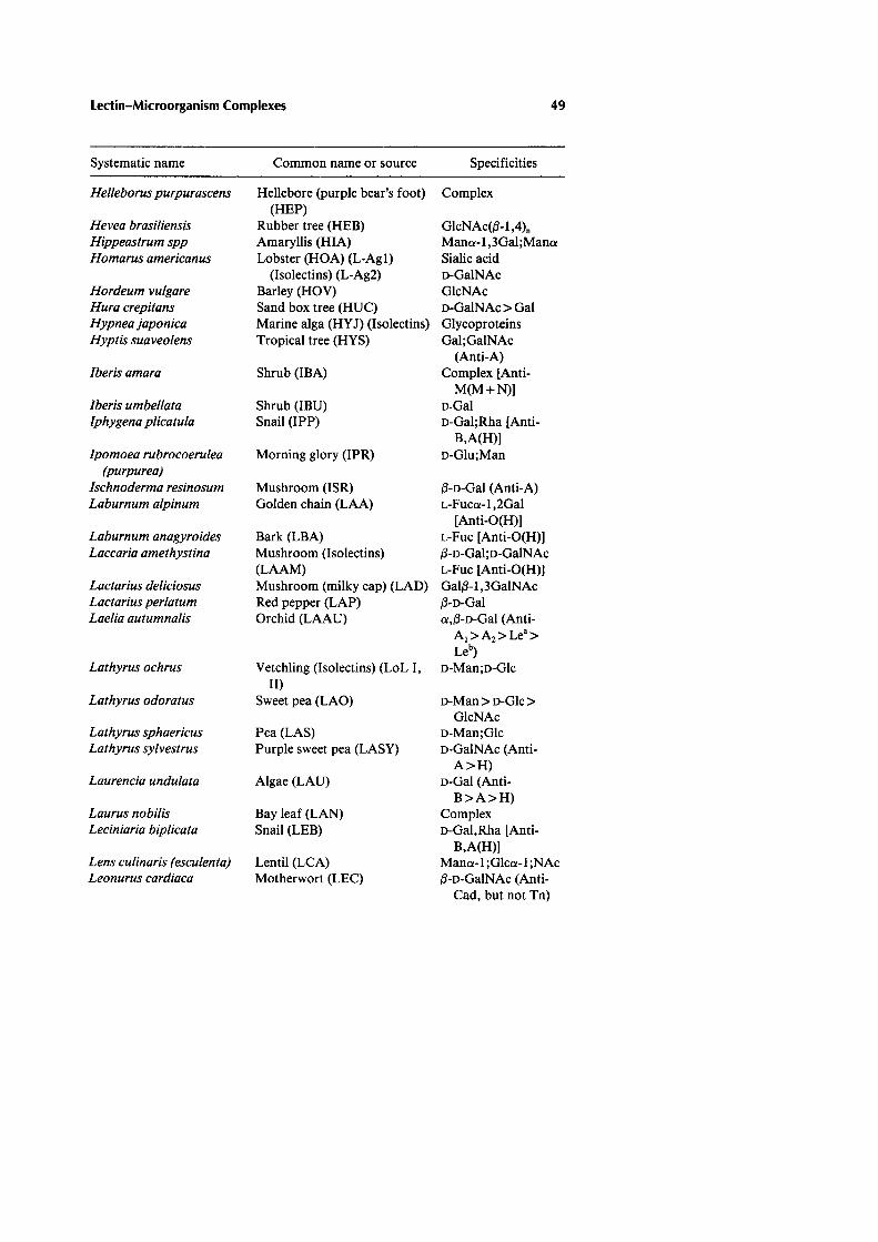
Lectin-Microorganism Complexes 49
Systematic name Common name or source Specificities
Helleborus purpurascens Hellebore (purple bear's foot) Complex (HEP)
Hevea brasiliensis Rubber tree (HEB) GlcNAc(l3-1 ,4). Hippeastrum spp Amaryllis (HIA) Mano:-1 ,3Gal;Mano: Homarus americanus Lobster (HOA) (L-Ag1) Sialic acid
(lsolectins) (L-Ag2) o-GalNAc Hordeum vulgare Barley (HOV) GlcNAc Hura crepitans Sand box tree (HUC) o-GalNAc >Gal Hypnea japonica Marine alga (HYJ) (lsolectins) Glycoproteins Hyptis suaveolens Tropical tree (HYS) Gal;GalNAc
(Anti-A) lberis amara Shrub (IBA) Complex [Anti-
M(M+N)) lberis umbellata Shrub (IBU) 0-Gal lphygena plicatu/a Snail (IPP) o-Gal;Rha [Anti-
B,A(H)] Ipomoea rubrocoerulea Morning glory (IPR) o-Glu;Man
(purpurea) Ischnoderma resinosum Mushroom (ISR) 13-o-Gal (Anti-A) Laburnum a/pinum Golden chain (LAA) L-Fuco:-1 ,2Gal
[Anti-O(H)] Laburnum anagyroides Bark (LBA) L-Fuc [Anti-O(H)] Laccaria amethystina Mushroom (Isolectins) 13-o-Gal;o-GalNAc
(LAAM) L-Fuc [Anti-O(H)] Lactarius deliciosus Mushroom (milky cap) (LAD) Gall3-1 ,3GalNAc Lactarius perlatum Red pepper (LAP) 13-o-Gal Laelia autumnalis Orchid (LAAU) o:,l3-o-Gal (Anti-
A1>A2 >Le•> Leb)
Lathyrus ochrus Vetchling (lsolectins) (LoL I, o-Man;o-Glc II)
Lathyrus odoratus Sweet pea (LAO) o-Man> o-Gle> GlcNAc
Lathyrus sphaericus Pea (LAS) o-Man;Glc Lathyrus sylvestrus Purple sweet pea (LASY) o-GalNAc (Anti-
A>H) Laurencia undulata Algae (LAU) o-Gal (Anti-
B>A>H) Laurus nobilis Bay leaf (LAN) Complex Leciniaria biplicata Snail (LEB) o-Gal,Rha [Anti-
B,A(H)] Lens culinaris (esculenta) Lentil (LCA) Mano:-1 ;Glco:-1 ;NAc Leonurus cardiaca Motherwort (LEC) 13-o-GalNAc (Anti-
Cad, but not Tn)

50 Doyle
Systematic name Common name or source Specificities
Lepidium sativum Garden cress (LES) Complex Leucojum vernum White snowflower (LEV) o-Man Levisticum officinale Aromatic herb (LEO) Complex Limax flavus Slug (LFA) Sialic acid Limulus polyphemus Horseshoe crab (limulin) (LIP) Sialic acid Listera ovata Twayblade (LIO) o-Man Litchi chinensis Lychee nut (LIC) Complex Lonchocarpus capassa Apple leaf (LOC) Complex Lophocereus shotti Cactus (LOS) Complex Lotononis bainesii Perennial pasture legume Complex
(LOB) Lotus tetragonolobus Asparagus (LOTUS) (winged L-Fuca-1 ,2Gal [Anti-
pea) O(H)] Luff a actangula Gourd (LUA) (GlcNAc{l-1 ,4). Lumbricus terrestris Earthworm (LUT) Complex Lycoperdon perlatum Puff ball mushroom (L YP) Complex Lycopersicon esculentum Tomato (LYE) (GlcNAc{l-1 ,4). Lygos monosperma Willow-like reed tree (L YM) Complex (Anti-Cad,
but not T, Tk, Th, T.)
Lygos sphaerocarpa Reed tree (L YS) Complex (Anti-T, T "' but not Cad)
Lyngbya majusarla Algae (L YMA) Complex Machaerocerus eruca Baja cactus (MAE) (lsolectins) a-L-Fuc;o-GalNAc Madura pomijera Osage orange (MAP) Gal{l-1 ,3GalNAc
(Anti-T) Macrotyloma axil/are Shrub (MAAX) o-GalNAc (Anti-
At>A2) Mangifera indica Mango (MAl) Complex Marasmius oreades Fairy-ring mushroom (MAO) o-Gal (Anti-B>O) Medicago lupulina Black medic (MEL) Glycoproteins Medicago sativa (truncu- Alfalfa (MES) o-Man;Glc
lata) Megabalanus rosa Barnacle (MER) (lsolectins) o-Gal Megapetaria squa/ida Clam (MES) o-GalNAc Microciona porifera Marine sponge (MIP) Complex Moluccella laevis Irish bell (MOL) Complex (Anti-A,N) Momordica charantia Bitter pear melon (MOC) o-Gal > GalNAc Moringia olifera Moringin Complex Myrica gale Sweet gale (MYG) Complex Narcissus pseudonarcissus Daffodil (NAP) Mana-1,3;Mana-1,6 Ocimum basilicum Basil (OCB) Complex Octopus vulgaris (bairdi) Octopus (OCV) {1-o-Gal

Lectin-Microorganism Complexes 51
Systematic name Common name or source Specificities
Oncorhynchus spp Salmon eggs a,{j-o-Gal Onobrychis viciifolia Sanfoin (ONV) a-Man> a-Ole Ononis hircinia Restharrow (ONH) a,{j-o-Gal;o-GalNAc Ononis spinosa Spiny restharrow (ONS) a-n-Gal Origanum vulgare Oregano (ORV) Complex Oryza sativa Rice (ORS) o-GlcNAc Otala lactea Snail (OTL) o-GalNAc (Anti-
A>H) Pachycereus pringlei Cactus (PAP) Complex (Anti-
A>B) Palmaria palmata Marine alga (PAP) Complex Papaver dubium Doubtful poppy (PAD) (GlcNAc{j-1,4)0
Papaver somnijerum Sleep-bringing or opium (GlcNAc(j-1 ,4)0
poppy (PAS) Pastinaca sativa Parsnip (P ASA) Complex Peltigera canina Lichen (PEC) Complex Pelvetia canaliculata Brown alga (PEC) Unknown Perea flaviatilis Perch (PEF) o-Glc;o-Man;L-Fuc Periplaneta americana Cockroach (PEAM) Complex Persea americana Avocado fruit seed (P AA) Complex Petromyzon marinus Lamprey (PEM) Complex Petroselinum crispum Parsley (PEC) Complex Phallus impudicus Stinkhorn mushroom (PHI) Complex Phanerochaete chryso- Fungus (PHC) (Glc(j-1 ,4)0
sporium Phaseolus acutiofolius Terpary bean (PHA) (lsolec- Complex
tins) Phaseolus coccineus Scarlet runner bean (PH C) Unknown Phaseolus lunatus (I, II) Lima bean (LBA) (lsolectins) GalNAca-1 ,3Gal
(Anti-A) Phaseolus vulgaris Red kidney bean (PHA) (lso- Complex
lectins) Gal(j-1,4 GlcNAc-(j-1,2Mana (Anti-A)
Phlebotomus papatasi Sandfly (PHP) (numerous Complex other Phlebotomus spp also produce lectins)
Phlomis fruticosa Jerusalem sage (PHF) Mel;GlcNAc; 2-deoxy-o-Gal
Phlox drumondii Ornamental thread (PHD) o-Glc;Man Phoradendrom califor- Desert mistletoe (PHCA) o-Gal
nicum Photo/iota squarrosa Broad-leaf tree (PHS) a-L-Fuc

52 Doyle
Systematic name Common name or source Specificities
Phragmites australis Common reed (PHAU) (lso- o-GalNAc lectins)
Phytolacca americana Pokeweed (PWM) (lsolectins) Gal/3-1,4(3)GlcNAc; (GlcNAc,j3-1,4)0 ;
Mana-1,2Man Pichia anomala Yeast (PIA) (Glcj3-1,4)0
Pi/a globosa Snail (PIG) Sialic acid Pisum sativum Pea(PEA) Mana-1 ;Glca-1 Plecoglossus altivelis Fish egg (PLA) L-Rha (Anti-B) Pleurotus ostreatus Mushroom (PLO) Fucosyllactose [Anti-
O(H)] Pleurotus spodo/eucus Mushroom (PLS) Fucosyllactose [Anti-
O(H)] P/umaria elegans Marine alga (PLE) Complex Polyandrocarpa misa- Tunicate (POM) o-Gal
kiensis Polygonatum multiflorum Perennial flower, USSR Complex
(POMU) Po/ygonum persicaria Buckwheat or lady's thumb o-Gal
smartweed (POP) Psathyrel/a velutina Mushroom (PSV) o-GlcNAc Pseudomonas aeruginosa Bacterium (PA-l, PA-Il) Thiogalactosides
>o-Gal; L-Fuc;L-Gal; o-Man
Psophocarpus tetragono- Winged bean (PST) o-Gal > o-GalNAc lobus (Anti-A,B)
Pterocarpus ango/ensis South Afr. kiatt tree (PTA) o-Gle, o-Man Ptilota plumosa Red marine algae (PTP) o-Gal (Anti-B) Quercus rubra English or red oak (QUR) (GlcNAcj3-1,4)0
Rana catesbaiena Frog eggs (RAC) Complex, Sialic acid Rana japonica Frog (RAJ) Complex Raphanus sativus Radish (RAS) Glycoproteins Rheum rhabarbarum Garden rhubard (RHR) Glycoprotein Rhodnius prolixus Reduviid bug (RHP) o-ManNAc;
o-GalNAc; o-Gal
Ricinus communis Castor bean (lsolectins) Galj3-1,3(4)GlcNAc (RCA-1,11) Galj3-1,3GalNAc;
GalNAc Robinia pseudoacacia Black locust (ROP) Complex Rumex obtrisfolia Bitterdock (RUD) Hydrophobic 13-
glycosides

Lectin-Microorganism Complexes 53
Systematic name Common name or source Specificities
Rumex patientia Shrub (RUP) Glycoprotein Rutilis rutilus Fish egg a-L-Rha > {3-o-Gal Saccharomyces cerevisiae Yeast (SAC) o-Man Salix alba White willow (SAA) (GlcNAc{3-l ,4). Salmo solar Salmon (SAS) o-Gal (Anti-B>A) Salvia horminum Bluebeard shrub (SAH) Complex (Anti-
T.+Cad) Salvia sclarea Clary shrub (SAC) GalNAca-1 ,ser
(or thr) (Anti-T.) Sambucus edulus Daneworth (SAE) o-Gal (Anti-A> B,O) Sambucus nigra Elderberry (SNA) (lsolectins o-Gai;GalNAc;
1-11) Sialic acid Sambucus racemosa Shrub (SAR) Complex Sarcophaga peregrina Flesh fly (SAP) Complex Satureja hortensia Savory herb (SAH) Complex Saxidomus giganteus Butter clam (SAG) o-GalNAc (Anti-A,) Saxidomus purpuratus Shellfish (SAPU) (lsolectins) a-o-GlcNAc Sclerotium rolfsii Fungus (SCR) (lsolectins) Glc{3-1 ,3Glc Secale cereale Rye (SCL) o-GlcNAc Sesamum indicum Sesame (SEI) o-GlcNAc Simulium ornatum Blackfly (SIO) Complex Soja hipspida Bean (SOH) o-GlcNAc;Gal Solanum alatum Winged nightshade (SOA) (GlcNAc{3-1 ,4). Solanum dulcamara Woody nightshade (SOD) Complex Solanum melongena Eggplant (SOM) (GlcNAc{3-l ,4). Solanum tuberosum Potato (ST A) (GlcNAc{3-1 ,4)z_5
Solieria chorda/is Marine alga (SOC) Glycoprotein Sophora japonica Japanese pagoda tree (SOJ) Gal{3-1 ,3GalNAc;
Gal{3-l ,3GlcNAc [Anti-B>A> >
O(H)] Sorbus aucuparia European mountain ash Complex
(SDA) Spondyliosoma cantharus Sea bream (SPC) Complex (Anti
B>H) Streptomyces spp Bacterium (STR) a-L-Fuc;o-Man Stye/a plicata Tunicate (STP) Sialic acid Synadenium grantii Community seed tree (SYG) a-D-Gal Tachypleus tridentatus Asian horseshoe crab (TAT) Sialic acid Taxus bactata Gymnosperm or Yew (TAB) Hydrophobic
{3-glucosides Tetracarpidium cono- Nigerian walnut (TEC) Gal{3-1 ,4GlcNAc
phorum

54 Doyle
Systematic name Common name or source Specificities
Tetragonolobus maritimus Winged pea (TEM) Glycoprotein Thymus vulgaris Thyme(THV) Complex Tichocarpus crinitus Red alga (TIC) Complex Tilia cordata Basswood (TICD) (GlcNAc/3-1 ,4)0
Trichosanthes anguina Gourd (hair flower) (TRA) Complex Trichosanthes kiri/owii Chinese gourd (TRK) D-Gal Tridacna crocea Mollusc (TRC) D-Gal Tridacna maxima Clam(TRM) Gal/3-1 ,4GlcNAc Trifolium repens White clover (Trifoliin) 2-Deoxyglucose Trigonella procumbens Fenugreek (TRP) Complex Trimeresurus mucrosqua- Poison scaly fungus (TRMU) Complex
matusvenum Triticum vulgaris Wheat(WGA) (GlcNAc/3-1,4)0 , Sia-
(aestivum) lie acid Tropaeolum majus Nasturtium (TRMM) D-GalNAc (Anti-A) Tu/ipa gesneriana Tulip (TUG) o-Man Ulex europaeus Gorse (or furze) (lsolectins) L-Fuca-1 ,2Gal[Anti-
(UEA-1, UEA-11) O(H)]; (GlcNAc/3-1 ,4)2 [Anti-O(H)]
Ulmus glabria Smooth elm (ULG) (GlcNAc/3-1 ,4)0
Ulva arasakei Algae(ULA) Complex [Anti-A,H(O)]
Ulva arasakii Algae (UAR) o-GalNAc (Anti-A+H)
Ulva lactuca Green marine algae (ULL) a-L-Fuc [Anti-O(H)] Urtica dioica Stinging nettle (URD) (GlcNAc/3-1 ,4)0
Vaejovis spinigerus Scorpion (VAS) Complex; sialic acids Velesunio ambigus Murray mussel (VV A) Complex Verbascum blattaria Smooth moth plant (VEB) Complex Viburnum lantana Wayfaring tree (VIL) Glycoprotein Viburnum opulus Guelder rose (VIO) Glycoprotein Vicia cracca Common vetch (VIC) o-GalNAc (Anti-A) Vicia cretica Vetch (VICRE) Complex (Anti-
TITh,A) Viciafaba Fava (broad) bean (favin) Mana-1;Glca-1 Vicia graminea Herb (VI G) Gal/3-1 ,3GalNAc
(Anti-N>O) Vicia hyrcania Shrub (VIH) (lsolectins) Gal/3-1 ;3GalNAc
(Anti-T) GlcNAc,Glc
(Anti-Tk) Vicia vi/losa Hairy vetch (VVA) (Isolectins) GalNAca-1,ser
(Anti-T0 ) (or thr)

Lectin-Microorganism Complexes 55
Systematic name Common name or source Specificities
Vigna radiata Mung bean (VIR) D-Gal Vigna ungiucu/ata Cowpea (VIU) D-Gal Viscum album Mistletoe (Isolectins) (ML-I, o-Gal; o-GalNH2
II, III) Wisteria jloribunda Japanese wisteria (WIF) GalNAca-1 ,6Gal;
GaiNAc;Gal Wisteria sinensis Flower (WIS) D-Gal Xenopus laevis Frog (XEL) a,/3-D-Gal Xylaria polymorpha Mushroom (XYP) Complex Zeamays Maize (ZEM) o-Man;Gal;GalNAc Zingiber officinale Chinese ginger (ZIO) Glycoprotein
Specificities determined by hemagglutination inhibition, equilibrium dialysis, fluorescence quenching, other. Specificities may reflect contributions of penultimate residues, or peptides. In some cases, specificities reported in the literature do not reveal anomeric preferences or blood group reactivities. In other cases, reports may disagree. When di- or tri-saccharide specificities are shown, usually the lectin has an affinity for the nonreducing carbohydrate residue. The table does not list hydrophobic derivatives of saccharides, although frequently the hydrophobic derivatives are capable of binding with higher affinities than unmodified saccharides. Many lectins have a higher affinity for di-, tri-, or multi-, antennary complex carbohydrates than for linear carbohydrate sequences, but the table has been simplifed to include only the latter. Vertebrate lectins, although now recognized to be numerous, are not thoroughly reviewed in the table. Finally, bacteriallectins, with the exception of Pseudomonas aeruginosa and Streptomyces spp, have not been purified in significant quantities and are not described in the table. Fru = fructose; Fuc = fucose; Gal = galactose; GalNAc = N-acetyl-galactosamine; Ga!NH2 = galactosamine; Ole = glucose, GlcNAc = N-acetylglucosamine; Lac = lactose; Man = mannose, Mel = melibiose; NeuAc = neuraminic acid, Raf = raffi-nose. Major commercial sources of lectins are E-Y Laboratories, San Mateo, CA (USA); Lectinola, Charles University, Prague, Czech Republic; Lectinotest, Lvov Medical Institute, Lvov, Ukraine and Sigma Chemical Company, St. Louis, MO (USA). Abbreviations are based on common usage and on first two letters of the genus and first letter of the species. In some cases, it is necessary to employ the first two letters of both the genus and species. Table was derived from the reviews of Mogos eta/. (61), Antonjuk eta/. (62), Sharon and Lis (9), Liener et a/. (8), Bird (20), Wu et al. (60), Etzler (40), Strosberg et a/. (63), Goldstein and Poretz (64), Goldstein and Hayes (59), Garber eta/. (66,67), Gilboa-Garber eta/. (65), and Mandai and Mandai (68), commercial pamphlets and original papers of numerous investigators.
REFERENCES
1. Stillmark H. Uber Ricin, ein giftiges Ferment aus den Samen von Ricinus communis und einigen anderen Euphorbiaceen. (Ph.D. Thesis), University of Dorpat (Tartu), Estonia, 1888.
2. Boyd WC, Shapleigh E. Specific precipitating activity of plant agglutinins (lectins). Science 1954; 119:419.

56 Doyle
3. Goldstein IJ, Hughes RC, Monsigny M, Osawa T, Sharon N. What should be called a lectin? Nature 1980; 285:66.
4. Kocourek J, Horejsi V. Defining a lectin. Nature 1981; 290:188. 5. Dixon HBF. Defining a lectin. Nature 1981; 292:192. 6. Barondes S. Bifunctional properties of lectins: lectins redefined. Trends Bio-
chem Sci 1988; 13:480-482. 7. Kocourek J. Historical background. In: Liener IE, Sharon N, Goldstein IJ,
eds. The lectins: properties, functions, and applications in biology and medi-cine. Orlando: Academic Press, 1986:1-32.
8. Liener IE, Sharon N, Goldstein IJ, eds. The lectins: properties, functions and applications in biology and medicine. Orlando: Academic Press, 1986.
9. Sharon N, Lis H. Lectins. London: Chapman & Hall, 1990:1-27. 10. Pistole TO. Interaction of bacteria and fungi with lectins and lectin-like
substances. Annu Rev Microbiol1981; 35:85-112. 11. Doyle R, Keller K. Lectins in diagnostic microbiology. Eur J Clin Microbiol
1983; 3:4-9. 12. Slifkin M, Doyle RJ. Lectins and their application to clinical microbiology.
Clin Microbiol Rev 1990; 3:197-218. 13. Doyle RJ, Slifkin M. Applications of lectins in microbiology. ASM News
1989; 55:655-658. 14. Sumner JB, Howell SF. The identification of the hemagglutinin of the jack
bean with concanavalin A. J Bacteriol1936; 32:227-237. 15. Renkonen KD. Studies on haemagglutinins present in seeds of some represen-
tatives of the family Leguminosae. Ann Med Exp Fenn 1948; 26:66-72. 16. Bird GWG. Specific agglutinating activity for human red blood corpuscles in
extracts of Dolichos biflorus. Curr Sci 1951; 20:298-299. 17. Bird GWG. Relationship of the blood subgroups A1, A2, and A1B, A2B to
haemagglutinins present in the seed of Dolichos biflorus. Nature 1952; 170: 674.
18. Bird GWG. George Bird's reminisces. In: B0g-Hanson TC, Freed DLJ, eds. Lectins, biology, biochemistry, clinical biochemistry, vol. 6. St. Louis: Sigma Chemical Co, 1988.
19. Makela D. Studies in hemagglutinins of leguminosae seeds. Ann Med Exp Fenn 1957; 35(suppl11):1-130.
20. Bird GWG. Lectins in haematology and blood banking. Adv Haematol1988; 17:125-148.
21. Crookston MC. Blood typing and cross-matching procedures. In: Rose NR, Friedman H, eds. Manual of clinical immunology, 2nd ed. Washington, DC: American Society for Microbiology, 1980:716-725.
22. Nowell PC. Phytohemagglutinin: an initiator of mitosis in cultures of normal human leukocytes. Cancer Res 1960; 20:462-466.
23. Goldstein IJ, Hollerman CE, Smith EE. Protein-carbohydrate interaction. II. Inhibition studies on the interaction of concanavalin A with polysaccha-rides. Biochemistry 1965; 4:876-883.
24. Kohler W, Prokop 0. Agglutination von streptokokken ver gruppe C durch ein agglutinin aus Helix pomatia. Z Immunitaetsforsch Allerg Klin Immunol 1967; 133:30-53.

Lectin-Microorganism Complexes 57
25. Kohler WD, Prokop 0. Agglutinationsversuche am streptokokken mit dem phytogslutinin aus Dolichos bif/orus. Z Immunitaetsforsch Allerg Klin Im-munol1967; 133:171-175.
26. Prokop 0, Kohler W. Agglutinations reactions en von mikroorganiumen mit Helix pomatia eiweiss drusnextract. Z Immunitaetsforsch Allerg Klin Immunol1967; 133:176-179.
27. Kohler W, Prokop 0, Kuhnemund 0. Routine identification of group C streptococci by means an agglutinin (protectin) from the albumen gland of the edible snail, Helixpomatia. J Med Microbiol1973; 6:127-130.
28. Wagner M. Interaction of wheat-germ agglutinin with streptococci and strep-tococcal cell wall polymers. Immunobiology 1979; 156:57-64.
29. Doyle RJ, Woodside EE, Fishel CW. Protein-polyelectrolyte interactions. The concanavalin A precipitin reaction with polyelectrolytes and polysaccha-ride derivatives. Biochem J 1968; 106:35-40.
30. Goldstein IJ, Staub AM. Interaction of concanavalin A with polysaccharides of salmonellae. Immunochemistry 1970; 7:315-319.
31. Tkacz JS, Cybolska EB, Lampen JO. Specific staining of wall mannan in yeast cells with fluorescein-conjugated concanavalin A. J Bacteriol1971; 105: 1-5.
32. Archibald AR, Coapes HE. Blocking of bacteriophage receptor sites by con-canavalin A. J Gen Microbiol1972; 73:581-585.
33. Birdsell DC, Doyle RJ. Modification of bacteriophage cp25 adsorption to Bacillus subtilis by concanavalin A. J Bacterial 1973; 113:198-202.
34. Doyle RJ, Birdsell DC, Young FE. Isolation of teichoic acid of Bacillus subtilis 168 by affinity chromatography. Prep Biochem 1973; 3:13-17.
35. Martinez-Palomo A, Gonzales-Robles A, de Ia Torre M. Selective agglutina-tion of pathogenic strains of Entamoeba histolytica induced by concanavalin A. Nature 1973; 245:186-187.
36. Ebisu S, Lonngren J, Goldstein IJ. Interaction of pneumococcal S-14 poly-saccharide with lectins from Ricinus communis, Triticum vulgaris, and Ban-deiraea simplicifolia. Carbohydr Res 1977; 58:187-191.
37. Stoddart RW, Herbertson BM. The use of fluorescein-labelled lectins in the detection and identification of fungi pathogenic for man: a preliminary study. J Med Microbiol1978; 11:315-324.
38. Schaefer RL, Keller KF, Doyle RJ. Lectins in diagnostic microbiology: use of wheat germ agglutinin for laboratory identification of Neisseria gonorr-hoeae. J Clin Microbiol1979; 10:669-672.
39. Graham K, Keller K, Ezzell J, Doyle R. Enzyme-linked lectinosorbent assay (ELLA) for detecting Bacillus anthracis. Eur J Clin Microbial 1984; 3:210-212.
40. Mobley HL T, Koch AL, Doyle RJ, Streips UN. Insertion and fate of cell wall in Bacillus subtilis. J Bacteriol1984; 158:169-179.
41. Schalla WD, Whittington WL, Rice RJ, Larsen SA. Epidemiological charac-terization of Neisseria gonorrhoeae by lectins. J Clin Microbial 1985; 22: 379-382.
42. Karayannopoulou G, Weiss J, Damjanov I. Detection of fungi in tissue sections by lectin histochemistry. Arch Pathol Lab Med 1988; 112:746-748.

58 Doyle
43. Slifkin M, Cumbie R. Rapid detection of herpes simplex virus with fluoresce-in-labeled Helix pomatia lectin. J Clin Microbiol1989; 27:1036-1039.
44. Gilboa-Garber N. Lectins of Pseudomonas aeruginosa: Properties, biological effects and applications. In: Mirelman D, ed. Microbiallectins and aggluti-nins. New York: John Wiley & Sons, 1986:255-269.
45. Sharon N. Bacteriallectins, cell-cell recognition and infectious disease. FEBS Lett 1987; 217:145-157.
46. Aronson M, Medalia 0, Schori L, Mirelman D, Sharon N, Ofek I. Preven-tion of colonization of the urinary tract of mice with Escherichia coli by blocking of bacterial adherence with methyl-cx-o-mannopyranoside. J Infect Dis 1979; 139:329-332.
47. Wiley DC, Skehel JJ. The structure and function of the hemagglutinin mem-brane glycoprotein of influenza virus. Annu Rev Biochem 1987; 56:365-394.
48. Young FE. Requirement of glucosylaied teichoic acid for adsorption of phage in Bacillus subtilis 168. Proc Nat! Acad Sci USA 1967; 58:2377-2383.
49. Etzler ME. Distribution and function of plant lectins. In: Liener IE, Sharon N, Goldstein IJ, eds. The lectins: properties, functions, and applications in biology and medicine. Orlando: Academic Press, 1986:371-435.
50. Marsh WL. The effect of plant root environment on the activity of Dolichos biflorus lectin. Vox Sang 1967; 12:68-70.
51. Mirelman D, Galan E, Sharon N, Lotan R. Inhibition of fungal growth by wheat germ agglutinin. Nature 1975; 256:414-416.
52. Callow JA. Recognition, resistance and role of plant lectins in host-parasite interactions. Adv Bot Res 1977; 4:1-49.
53. Garas NE, Kuc J. Potato lectin lyses zoospores of Phytophthora infestans and precipitates elicitors of terpenoid accumulation produced by the fungus. Physiol Plant Pathol1981; 18:227-237.
54. Partridge J, Shannon L, Gumpf D. A barley lectin that binds free amino sugars. I. Purification and characterization. Biochim Biophys Acta 1976; 451:470-483.
55. Janzen DH, Juster HB, Liener IE. Insecticidal activity of the phytohemagglu-tinin in black beans on a bruchid beetle. Science 1976; 192:795-796.
56. Ofek I, Sharon N. Lectinophagocytosis: a molecular mechanism of recogni-tion between cell surface sugars and lectins in the phagocytosis of bacteria. Infect Immun 1988; 56:539-547.
57. Drickamer K. Two distinct classes of carbohydrate-recognition domains in animallectins. J Bioi Chern 1988; 263:9557-9560.
58. Drake D, Taylor KG, Bleiweis AS, Doyle RJ. Specificity of the glucan-binding lectin of Streptococcus cricetus. Infect Immun 1988; 56:1864-1872.
59. Goldstein IJ, Hayes CE. The lectins: carbohydrate-binding proteins of plants and animals. Adv Carbohydr Chern Biochem 1978; 35:127-340.
60. Wu AM, Sugii S, Herp A. A guide for carbohydrate specificities of lectins. Adv Exp Med Biol1988; 288:819-847.
61. Mogos SM, Stefan E, Hulea SA, Andras M, Trif M, Pop A, Buzila L, Dihoru G, Roman N, Motas C. Screening for new lectins in the Romania flora. Rev Rom Biochim 1989; 26:139-144.

Lectin-Microorganism Complexes 59
62. Antonyuk VA, Formagyuk V, Levashou V. Use of lectins in microbiology. Z Mikrobiol Epidemiol Immunobiol1987; 6:97-104.
63. Strosberg A, Buffard D, Lauwereys M, Foriers A. Legume lectins: a large family of homologous proteins. In: Liener IE, Sharon N, Goldstein 11, eds. The lectins: properties, functions, and applications in biology and medicine. Orlando: Academic Press, 1986:249-264.
64. Goldstein J, Poretz RD. Isolation, physicochemical characterization, and carbohydrate-binding specificity of lectins. In: Liener E, Sharon N, Goldstein J, eds. The lectins: properties, functions, and applications in biology and medicine. Orlando: Academic Press, 1986:33-247.
65. Gilboa-Garber N, Susswein A, Mizrahi L, Avichezer D. Purification and characterization of the gonad lectin of Ap/ysia depilans. FEBS Lett 1985; 181:267-270.
66. Garber N, Guempel U, Gilboa-Garber N, Doyle RJ. Specificity of the fucose-binding lectin of Pseudomonas aeruginosa. FEMS Microbiol Lett 1987; 48: 331-334.
67. Garber N, Guempel U, Belz A, Gilboa-Garber N, Doyle RJ. On the specific-ity of the o-galactose-binding lectin (PA-l) of Pseudomonas aeruginosa and its strong binding to hydrophobic derivatives of o-galactose and thiogalac-tose. Biochim Biophys Acta 1992; 1116:331-333.
68. Mandai C, Mandai C. Sialic acid binding lectins. Experientia 1990; 46:433-441.
69. Jermyn MA. Flavonoid glycosides, beta-lectins and plant cell membranes. Proc Aust Biochem Soc 1977; 10:89-104.
70. Rosenberg M, Doyle RJ. Microbial cell surface hydrophobicity: history, mea-surement and significance. In: Doyle RJ, Rosenberg M, eds. Microbial cell surface hydrophobicity. Washington, DC: American Society for Microbiol-ogy, 1990:1-37.
71. Firon N, Ashkenazi S, Mirelman D, Ofek I, Sharon N. Aromatic alpha-glycosides of mannose are powerful inhibitors of the adherence of type 1 fimbriated Escherichia coli to yeast and intestinal epithelial cells. Infect Im-mun 1987; 55:472-476.
72. Bayer EA, Skutelsky E, Wilchek M The ultrastructural visualization of cell surface glycoconjugates. Methods Enzymol1982; 83:195-215.
73. Damjanov I. Biology of disease. Lectin cytochemistry and histochemistry. Lab Invest 1987; 57:5-20.
74. Jackson M, Chan R, Matoba AY, Robin JB. The use of fluorescein-conjugated lectins for visualizing atypical mycobacteria. Arch Ophthalmol 1989; 107:1206-1209.
75. Doyle RJ, McDannel ML, Helman JR, Streips UN. Distribution of teichoic acid in the cell wall of Bacillussubtilis. J Bacteriol1975; 122:152-158.
76. Cole HB, Ezzell JW Jr, Keller KF, Doyle RJ. Differentiation of Bacillus anthracis and other Bacillus species by lectins. J Clin Microbiol 1984; 19:48-53.
77. Lotan R, Sharon N, Mirelman D. Interaction of wheat-germ agglutinin with bacterial cells and cell-wall polymers. Eur J Biochem 1975; 55:257-262.

60 Doyle
78. Connelly MC, Allen PZ. Chemical and immunochemical studies lipopolysac-charides from pyocin 103-sensitive and resistant Neisseria gonorrhoeae. Car-bohydr Res 1983; 120:171-186.
79. Allen PZ, Connelly MC, Apicella MA. Interactions of lectins with Neisseria gonorrhoeae. Can J Microbiol1980; 26:468-474.
80. Hammarstrom S, Lindberg AA, Robertsson ES. Precipitation of lipopolysac-charides from rough mutants of Salmonella typhimurium by an A-hemag-glutinin fromHelixpomatia. Bur J Biochem 1972; 25:274-282.
81. Pistole TO. Bacterial agglutinins from Limulus polyphemus-an overview. Prog Clin Bioi Res 1979; 29:547-553.
82. Gilbride KJ, Pistole TO. Isolation and characterization of a bacterial aggluti-nin in the serum of Limulus polyphemus. Prog Clin Bioi Res 1979; 29:525-535.
83. Owen P, Salton MRJ. Membrane asymmetry and expression of cell surface antigens of Micrococcus lysodeikticus established by crossed immunoelectro-phoresis. J Bacteriol1977; 132:974-985.
84. Archibald AR, Coapes HE. The interaction of concanavalin A with teichoic acids and bacterial walls. Biochem J 1971; 123:665-667.
85. Reeder WJ, Ekstedt RD. Study of the interaction of concanavalin A with staphylococcal teichoic acids. J lmmunol1971; 106:334-340.
86. Doyle RJ, Birdsell DC. Interaction of concanavalin A with the cell wall of Bacillus subtilis. J Bacteriol1972; 109:652-658.
87. Garland JM, Archibald AR, Baddiley J. An electron microscopic study of the location of teichoic acid and its contribution to staining reactions in walls of Streptococcusfaecalis 8191. J Gen Microbiol1975; 89:73-86.
88. Hamada S, Gill K, Slade HD. Binding of lectins to Streptococcus mutans cells and type-specific polysaccharides, and effect on adherence. Infect Im-mun 1977; 18:708-716.
89. Kashket S, Guilmette KM. Aggregation of oral streptococci in the presence of concanavalin A. Arch Oral Biol1975; 20:375-379.
90. Kahane I, Tully JG. Binding of plant lectins to Mycoplasma cells and mem-branes. J Bacteriol1970; 128:1-7.
91. Doyle RJ, McDannel ML, Streips UN, Birdsell DC, Young FE. Polyelectro-lyte nature of bacterial teichoic acids. J Bacteriol1974; 118:606-615.
92. Doyle RJ, Chaloupka J, Vinter V. Turnover of cell walls in microorganisms. Microbiol Rev 1988; 52:554-567.
93. Aitchison EJ, Lambert PA, Farrell ID. Antigenic composition of an endocar-ditis associated isolate of Streptococcus faecalis and identification of its gly-coprotein antigens by ligand blotting with lectins. J Med Microbiol1986; 21: 161-167.
94. Bauer H, Horisberger M, Bush D, Sigarlakie E. Mannan as a major compo-nent of bud scars of Saccharomyces cerevisiae. Arch Microbiol1972; 85:202-272.
95. Halverson LJ, Stacey G. Effect of lectin on nodulation by wild-type Bradyhi-zobium japonicum and nodulation-defective mutant. Appl Environ Micro-biol1986; 51:753-760.

Lectin-Microorganism Complexes 61
96. Fogg GC, Yang L, Wang E, Blaser MJ. Surface array proteins of Campylo-bacter fetus block lectin-mediated binding to type A lipopolysaccharide. In-fect Immun 1990; 58:2738-2744.
97. Holm SE, Bersholm A, Wagner B, Wagner M. A sialic-acid-specific lectin from Cepaea hortensis that promotes phagocytosis of a group-B, type Ia, streptococcal strain. J Med Microbiol1985; 19:317-323.
98. Staat RH, Langley SD, Doyle RJ. Streptococcus mutans adherence: pre-sumptive evidence for protein-mediated attachment followed by glucan-dependent cellular accumulation. Infect Immun 1980; 27:675-681.
99. Meade NA, Staat RH, Langley SD, Doyle RJ. Lectin-like activity from Per-sea americana. Carbohydr Res 1980; 78:349-363.
100. Goldstein IJ, Misaki A. Interaction of concanavalin A with an arabinogalac-tan from the cell wall of Mycobacterium bovis. J Bacterial 1970; 103:422-425.
101. Maruyama HB, Arisawa M, Ono-Onitsuka M. Simplified assay of concanav-alin A-dependent bacterial agglutination by using cell surface mutants. Infect Immun 1975; 11:1320-1324.
102. Doyle RJ, Keller KF, Ezzell JW. Bacillus. In: Lennette EH, Balows A, Hausler HJ Jr, Shadomy HD, eds. Manual of clinical microbiology, 4th ed. Washington, DC: American Society Microbiology, 1985:211-215.
103. Delucca AJ 2nd. Lectin grouping of Bacillus thuringiensis serovars. Can J Microbiol1984;30:1100-1104.
104. Doyle RJ, Nedjat-Haiem F, Keller KF, Frasch CE. Diagnostic value of inter-actions between members of the family Neisseriaceae and lectins. · J Clin Microbiol1984; 19:383-387.
105. Senne JE. Lectin agglutination of Neisseria gonorrhoeae. Clin Microbial Newslett 1981; 3:10.
106. Frasch EC. Role of lipopolysaccharide in wheat germ agglutinin mediated agglutination of Neisseria meningitidis and Neisseria gonorrhoeae. J Clin Microbiol1980; 12:498-501.
107. Yajko DM, ChuA, Hadley WK. Rapid confirmatory identification of Neisse-ria gonorrhoeae with lectins and chromogenic substrates. J Clin Microbial 1984; 19:380-382.
108. Vazquez JA, Berron S. Lectin agglutination test as an epidemiological marker for Neisseria gonorrhoeae. Genitourin Med 1990; 66:302.
109. Patchett RA, Kelley AF, Kroll RG. The adsorption of bacteria to immobi-lized lectins. J Appl Bacteriol1991; 71:277-284.
110. Flemming C. The kinetics of lectin-mediated cell agglutination. Adv Lectin Res 1991; 4:97-121.
111. Wong KH, Skelton SK, Feeley JC. Interaction of Campylobacter jejuni and Campylobacter coli with lectins and blood group antibodies. J Clin Microbial 1985; 22:134-135.
112. Corbel JJ, Gill KPW. Lectin agglutination of thermophilic Campylobacter species. Vet Microbiol1987; 15:163-173.
113. Gill KPW, Corbel MJ. Lectin agglutination for the differentiation of Campy-lobacter fetus serotypes. Vet Rec 1986; 118:158.

62 Doyle
114. Slifkin M, Gil OM. Rapid biochemical tests for the identification of groups A,B,C,F, and G streptococci from throat cultures. J Clin Microbiol 1983; 18:29-32.
115. Slifkin M, Gil OM. Identification of group C streptococcal antigen extracts with lectin-bound polystyrene particles. J Clin Microbiol1984; 19:83-84.
116. Pistole, T. Naturally occurring bacterial agglutinin in the serum of the horse-shoe crab Limulus polyphemus. J Invert Path 1976; 28:153-154.
117. Wagner M. Agglutination of bacteria by a sialic acid-specific lectin of the snail Cepaea hortensis. Acta Histochem 1982; 71:35-39.
118. Slifkin M, Cumbie R. Identification of group-B streptococcal antigen with lectin-bound polystyrene particles. J Clin Microbiol1987; 25:1172-1175.
119. Davidson SK, Keller KF, Doyle RJ. Differentiation of coagulase-positive and coagulase-negative staphylococci by lectins and plant agglutinins. J Clin Microbiol1982; 15:547-553.
120. Adam G, Heegard P, Bog-Hansen TC, Mundry KW. Lectins as probes for the assay of rhabdovirus infections in plants. J Virol Methods 1987; 17:263-275.
121. Svennerholm B, Olofsson S, Jeansson S, Vahlne A, Lycke E. Herpes simplex virus type selective enzyme-linked immunosorbent assay with Helix pomatia lectin-purified antigens. J Clin Microbiol1984; 19:235-239.
122. Corbel MJ, Cockren DS, Brewer RA. Differentiation of smooth and rough Brucella strains by lectins. Vet Rec 1983; 113:261-262.
123. Chatterjee BP, Guha AK, Pal P, Bhattachayya M. Lectin typing of Pseudom-onas aeruginosa strains of different serotypes, Habs and Fisher types. Zen-tralbl Bakteriol Hyg 1989; A271:362-371.
124. Eaves DJ, Doyle RJ. Surface characteristics of Pseudomonas cepacia. Mi-crobios 1988; 53:119-128.
125. Avni I, Affra RC, Robin JB, Rao NA. Lectins for the identification of ocular bacterial pathogens. Metab Pediatr Syst Ophthalmol1987; 19:45-47.
126. Malkinson M, Orgad U, Becker Y. Use of lectins to detect and differentiate subtypes of Marek's disease virus and turkey herpesvirus glycoproteins in tissue culture. J Virol Methods 1986; 13:129-133.
127. deMiranda Santos IKF, Pereira MEA. Lectins discriminate between patho-genic and non-pathogenic South American trypanosomes. Am J Trop Med Hyg 1984; 33:839-844.
128. Korting HC, Abeck D, Johnson AP, Ballard RC, Taylor-Robinson D, Braun-Falco 0. Lectin typing of Haemophilus ducreyi. Eur J Clin Microbiol Infect Dis 1988; 7:678-680.
129. LeMinor L, Tournier P, Chalon AM. Agglutinabilite par Ia concanavaline a de divers bacilles a gram negatif. Etude chez les Salmonella de Ia correlation avec Ia structure antigenique 0. Ann Microbiol (Paris) 1973; 124A:467-476.
130. Doyle RJ, Nedjat-Haiem F, Miller RD, Keller KF. Interaction between plant agglutinins and Legionella species. J Clin Microbiol1982; 15:973-975.
131. Nutsubidze NN, Lakhtin VM, Demina VA, Obraztsova IN, Marinova DN. Possibility of identifying microorganisms by the rate of their agglutina,tion by lectins. Drikladnaya Bioklim Mikrobiol1991; 27:274-278.

lectin-Microorganism Complexes 63
132. Sizemore RK, Caldwell JJ, Kendrick AS. Alternate Gram staining technique using a fluorescent lectin. Appl Environ Microbiol1990; 56:2245-2247.
133. Levy NJ. Wheat germ agglutinin blockage of chlamydia! attachment sites: antagonism by N-acetyl-o-glucosamine. Infect Immun 1979; 25:946-953.
134. Tsien HC, Schmidt EL. Localization and partial characterization of soybean lectin-binding polysaccharide of Rhizobium japonicum. J Bacteriol 1981; 145:1063-1074.
135. Lindberg B, Lonngren J, Powell DA. Structural studies on the specific type-14 pneumococcal polysaccharide. Carbohydr Res 1977; 58:177-186.
136. Gray BM, Dillon HC Jr, Pritchard DG. Interaction of group B streptococcal type-specific polysaccharides with wheat germ agglutinin and other lectins. J Immunol Methods 1984; 72:269-277.
137. Mattiasson B, Johansson P-A. A simple rapid method for quantifying micro-organisms by their metabolic activity when bound to a specific adsorbent. J Immunol Methods 1982; 52:233-240.
138. Robinson JE, Holton D, Liu J, McMurdo H, Murciano A, Gohd D. A novel enzyme-linked immunosorbent assay (ELISA) for the detection of antibodies to HIV-1 envelope glycoproteins based on immobilization of viral glycopro-teins in microtiter wells coated with concanavalin A. J Immunol Methods 1990; 132:63-71.
139. Watanabe K, Takesue S, Ishibashi K. Reversibility of the adsorption of bacte-riophage PL-1 to the cell walls isolated from Lactobacillus casei. J Gen Virol 1977; 34:189-194.
140. Ishibashi K, Takesue S, Watanabe K, Oishi K. Use of lectins to characterize the receptor sites for bacteriophage PL-1 of Lactobacillus casei. J Gen Micro-biol1982; 128:2251-2259.
141. Ndulue A, Flandrois JP, Marmet D. Abnormal affinity of Staphylococcus aureus N-acetylglucosamine ribitol teichoic acid for wheat-germ agglutinin. J Chromatogr 1981; 209:323-328.
142. Ndulue A, Flandrois J-P. Immunochemical studies of Staphylococcus aureus Oeding-Haukenes antigen as: a phosphorus-containing polysaccharide. J Gen Microbiol1983; 129:3603-3610.
143. Hancock IC. The biosynthesis of wall teichoic acid by toluenised cells of BacillussubtilisW23. Bur J Biochem 1981; 119:85-90.
144. Fiedler F, Bude A. Occurrence and chemistry of cell wall teichoic acids in the genus Brevibacterium. J Gen Microbiol198; 135:2837-2846.
145. Wicken AJ, Knox KW. Characterization of group N Streptococcus lipotei-choic acid. Infect Immun 1975; 11:973-981.
146. Leopold K, Fischer W. Separation of the poly(glycerophosphate) lipoteichoic acids of Enterococcus faecalis Kiel 27738, Enterococcus hirae ATCC 9790 and Leuconostoc mesenteroides DSM 20343 into molecular species by affinity chromatography on concanavalin A. Bur J Biochem 1991; 196:475-482.
147. Anderson AJ, Green RS, Sturman AJ, Archibald AR. Cell wall assembly in Bacillus subtilis: location of wall material incorporated during pulsed release of phosphate limitation, its accessibility to bacteriophages and concanavalin A, and its susceptibility to turnover. J Bacteriol1978; 136:886-899.

64 Doyle
148. Kirchner G, Kemper MA, Koch AL, Doyle RJ. Zonal turnover of cell poles of Bacillus subtilis. Ann Inst Pasteur (Microbiology) 1988; 139:645-654.
149. Kemper MA, Mobley HLT, Doyle RJ. How do bacilli elongate? In: Actor P, Daneo-Moore L, Higgins ML, Salton MRJ, Shockman GD, eds. Antibiotic inhibition of bacterial cell surface assembly and function, Washington, DC: American Society for Microbiology, 1988:98-108.
150. Birdsell DC, Doyle RJ, Morgenstern M. Organization of teichoic acid in the cell wall of Bacillus subtilis. J Bacteriol1975; 121:726-734.
151. Morioka H, Tachibana M, Suganuma A. Ultrastructural localization of car-bohydrates on thin sections of Staphylococcus aureus with silver methena-mine and wheat germ agglutinin-gold complex. J Bacteriol1987; 169:1358-1362.
152. Rye M, Wagner B, Wagner M, Bicova R. Electron microscopic localization of lipoteichoic acid on group A streptococci. Zentralbl Bakteriol Hyg 1988; A269:168-178.
153. Maruyama HB. Agglutination of bacterial spheroplast. I. Effect of conca-navalin A. Biochim Biophys Acta 1972; 274:499-504.
154. Maruyama HB. Agglutination of bacterial spheroplast: II. Agglutination-dependent degradation of Escherichia coli ribosomal ribonucleic acid. J Bac-teriol1973; 115:47-51.
155. Maruyama HB. Agglutination of bacterial spheroplast. III. Relationship of labelled concanavalin A to the agglutinability. J Biochem 1974; 75:165-170.
156. Guillot J, Breton A, Damez M, Dusser M, Gaillard-Martinie B, Millet. Use of lectins for a comparative study of cell wall composition of different anaer-obic rumen fungal strains. FEMS Microbiol Lett 1990; 67:151-156.
157. Bonfante-Fasolo P, Vian B, Testa B. Ultrastructural localization of chitin in the cell wall of a fungal spore. Bioi Cell1987; 57:265-270.
158. Bonfante-Fasolo P, Perotto S, Testa B, Faccio A. Ultrastructural localization of cell surface sugar residues in ericoid mycorrhizal fungi by gold-labeled lectins. Protoplasma 1987; 139:25-35.
159. Miragall F, Rico H, Sentandreu R. Regeneration of the cell wall in proto-plasts of Candida albicans. A cytochemical study using wheat germ agglutinin and concanavalin A. Arch Microbiol1988; 149:286-290.
160. Rico H, Herrero E, Miragall F, Sentandreu R. An electron microscopy study of wall expansion during Candida albicans yeast and mycelial growth using concanavalin A-ferritin labelling of mannoproteins. Arch Microbiol 1991; 156:111-114.
161. Chu C-Y, Chen K-C. Inhibition of DNA-induced transformation by conca-navalin A in Bacillus subtilis. Biochem Biophys Res Commun 1979; 91:170-176.
162. Chan K-Y, Lau T-M. Physiological responses of Bacillus species to concanav-alin A. 1. The binding of concanavalin A to B. cereus ATCC 14579 and B. licheniforms IFO 12107. Microbios 1984; 39:121-128.
163. Chan K-Y, Lau T-M. Physiological responses of Bacillus species to concanav-alin A. 3. Effect on RNA, DNA and protein synthesis, and galactose uptake of B. cereus ATCC 14579. Microbios 1984; 40:195-204.

Lectin-Microorganism Complexes 65
164. Lau T -M, Chan K-Y. Physiological responses of Bacillus species to concanav-alin A. 2. Effect on growth, oxygen uptake, enzyme activities and intracellu-lar cyclic guanosine 3 ',5 '-monophosphate level of B. cereus ATCC 14579. Microbios 1984; 39:137-150.
165. Lechner J, Wieland F. Structure and biosynthesis of prokaryotic glycopro-teins. Annu Rev Biochem 1989; 58:173-194.
166. Bose SK, Smith GB, Paul RG. Influence of lectins, hexoses, and neuramini-dase on the association of purified elementary bodies of Chlamydia tracho-matis UW-31 with HeLa cells. Infect lmmun 1983; 40:1060-1067.
167. Swanson AF, Kuo C-C. Identification of lectin-binding proteins in Chla-mydia species. Infect Immun 1990; 58:502-507.
168. Swanson AF, Kuo C-C. The characterization of lectin-binding proteins of Chlamydia trachomatis as glycoproteins. Microb Pathogen 1991; 10:465-473.
169. Kahane I, Brunner H. Isolation of a glycoprotein from Mycoplasma pneu-moniae membranes. Infect Immun 1977; 18:273-277.
170. Simoneau P, Labarere J. Detection of a concanavalin A binding protein in the mollicute Spiroplasma citri and purification from the plasma membrane. Arch Microbiol1989; 152:488-491.
171. Narasu ML, Gopinathan KP. Purification of larvicidal protein from Bacillus sphaericus 1593. Biochem Biophys Res Commun 1986; 141:756-761.
172. Durbreuil JD, Logan SM, Cubbage S, Eidhin DN, McCubbin WD, Kay CM, Beveridge TJ, Ferris FG, Trust TJ. Structural and biochemical analyses of a surface array protein of Campylobacter fetus. J Bacteriol 1988; 170:4165-4173.
173. Kawamura T, Shockman GD. Purification and some properties of the endog-enous, autolytic N-acetylmuramoylhydrolase of Streptococcus faecium, a bacterial glycoenzyme. J Bioi Chern 1983; 258:9514-9521.
174. Corbel MJ, Cockrem DS, Brewer RA. The interaction of Brucella cell surface components with plant agglutinins. Dev Bioi Stand 1984; 56:169-175.
175. Cavalcanti MSM, Almeida AMP, Coelho LCBB. Interaction of lectins with Yersiniapestis strains. Appl Biochem Biotechnol1990; 26:125-131.

References
246 2 4 6
1. Stillmark H. Uber Ricin, ein giftiges Ferment aus denSamen von Ricinus communis und einigen anderenEuphorbiaceen. (Ph.D. Thesis), University of Dorpat(Tartu), Estonia, 1888.
2. Boyd WC, Shapleigh E. Specific precipitating activity ofplant agglutinins (lectins). Science 1954; 119:419. 3.Goldstein IJ, Hughes RC, Monsigny M, Osawa T, Sharon N.What should be called a lectin? Nature 1980; 285:66. 4.Kocourek J, Horejsi V. Defining a lectin. Nature 1981;290:188. 5. Dixon HBF. Defining a lectin. Nature 1981;292:192. 6. Barondes S. Bifunctional properties oflectins: lectins redefined. Trends Biochem Sci 1988;13:480-482. 7. Kocourek J. Historical background. In:Liener IE, Sharon N, Goldstein IJ, eds. The lectins:properties, functions, and applications in biology andmedicine. Orlando: Academic Press, 1986:1-32. 8. LienerIE, Sharon N, Goldstein IJ, eds. The lectins: properties,functions and applications in biology and medicine.Orlando: Academic Press, 1986. 9. Sharon N, Lis H.Lectins. London: Chapman & Hall, 1990:1-27. 10. PistoleTO. Interaction of bacteria and fungi with lectins andlectin-like substances. Annu Rev Microbiol1981; 35:85-112.11. Doyle R, Keller K. Lectins in diagnostic microbiology.Eur J Clin Microbiol 1983; 3:4-9. 12. Slifkin M, DoyleRJ. Lectins and their application to clinical microbiology.Clin Microbiol Rev 1990; 3:197-218. 13. Doyle RJ, SlifkinM. Applications of lectins in microbiology. ASM News 1989;55:655-658. 14. Sumner JB, Howell SF. The identificationof the hemagglutinin of the jack bean with concanavalin A.J Bacteriol1936; 32:227-237. 15. Renkonen KD. Studies onhaemagglutinins present in seeds of some representativesof the family Leguminosae. Ann Med Exp Fenn 1948; 26:66-72.16. Bird GWG. Specific agglutinating activity for human redblood corpuscles in extracts of Dolichos biflorus. CurrSci 1951; 20:298-299. 17. Bird GWG. Relationship of theblood subgroups A 1 , A 2 , and A 1 B, A 2 B tohaemagglutinins present in the seed of Dolichos biflorus.Nature 1952; 170: 674. 18. Bird GWG. George Bird'sreminisces. In: B0g-Hanson TC, Freed DLJ, eds. Lectins,biology, biochemistry, clinical biochemistry, vol. 6. St.Louis: Sigma Chemical Co, 1988. 19. Makela D. Studies inhemagglutinins of leguminosae seeds. Ann Med Exp Fenn1957; 35(suppl11):1-130. 20. Bird GWG. Lectins inhaematology and blood banking. Adv Haematol1988;17:125-148. 21. Crookston MC. Blood typing and

cross-matching procedures. In: Rose NR, Friedman H, eds.Manual of clinical immunology, 2nd ed. Washington, DC:American Society for Microbiology, 1980:716-725. 22.Nowell PC. Phytohemagglutinin: an initiator of mitosis incultures of normal human leukocytes. Cancer Res 1960;20:462-466. 23. Goldstein IJ, Hollerman CE, Smith EE.Protein-carbohydrate interaction. II. Inhibition studieson the interaction of concanavalin A with polysaccharides.Biochemistry 1965; 4:876-883. 24. Kohler W, Prokop 0.Agglutination von streptokokken ver gruppe C durch einagglutinin aus Helix pomatia. Z Immunitaetsforsch AllergKlin Immunol 1967; 133:30-53. 57
25. Kohler WD, Prokop 0. Agglutinationsversuche amstreptokokken mit dem phytogslutinin aus Dolichosbif/orus. Z Immunitaetsforsch Allerg Klin Immunol1967;133:171-175.
26. Prokop 0, Kohler W. Agglutinations reactions en vonmikroorganiumen mit Helix pomatia eiweiss drusnextract. ZImmunitaetsforsch Allerg Klin Immunol1967; 133:176-179.
27. Kohler W, Prokop 0, Kuhnemund 0. Routineidentification of group C streptococci by means anagglutinin (protectin) from the albumen gland of theedible snail, Helixpomatia. J Med Microbiol1973; 6:127-130.
28. Wagner M. Interaction of wheat-germ agglutinin withstreptococci and streptococcal cell wall polymers.Immunobiology 1979; 156:57-64.
29. Doyle RJ, Woodside EE, Fishel CW.Protein-polyelectrolyte interactions. The concanavalin Aprecipitin reaction with polyelectrolytes andpolysaccharide derivatives. Biochem J 1968; 106:35-40.
30. Goldstein IJ, Staub AM. Interaction of concanavalin Awith polysaccharides of salmonellae. Immunochemistry 1970;7:315-319.
31. Tkacz JS, Cybolska EB, Lampen JO. Specific staining ofwall mannan in yeast cells with fluorescein-conjugatedconcanavalin A. J Bacteriol1971; 105: 1-5.
32. Archibald AR, Coapes HE. Blocking of bacteriophagereceptor sites by concanavalin A. J Gen Microbiol1972;73:581-585.
33. Birdsell DC, Doyle RJ. Modification of bacteriophagecp25 adsorption to Bacillus subtilis by concanavalin A. J

Bacterial 1973; 113:198-202.
34. Doyle RJ, Birdsell DC, Young FE. Isolation of teichoicacid of Bacillus subtilis 168 by affinity chromatography.Prep Biochem 1973; 3:13-17.
35. Martinez-Palomo A, Gonzales-Robles A, de Ia Torre M.Selective agglutination of pathogenic strains of Entamoebahistolytica induced by concanavalin A. Nature 1973;245:186-187.
36. Ebisu S, Lonngren J, Goldstein IJ. Interaction ofpneumococcal S-14 polysaccharide with lectins from Ricinuscommunis, Triticum vulgaris, and Bandeiraea simplicifolia.Carbohydr Res 1977; 58:187-191.
37. Stoddart RW, Herbertson BM. The use offluorescein-labelled lectins in the detection andidentification of fungi pathogenic for man: a preliminarystudy. J Med Microbiol1978; 11:315-324.
38. Schaefer RL, Keller KF, Doyle RJ. Lectins in diagnosticmicrobiology: use of wheat germ agglutinin for laboratoryidentification of Neisseria gonorrhoeae. J ClinMicrobiol1979; 10:669-672.
39. Graham K, Keller K, Ezzell J, Doyle R. Enzyme-linkedlectinosorbent assay (ELLA) for detecting Bacillusanthracis. Eur J Clin Microbial 1984; 3:210212.
40. Mobley HL T, Koch AL, Doyle RJ, Streips UN. Insertionand fate of cell wall in Bacillus subtilis. JBacteriol1984; 158:169-179.
41. Schalla WD, Whittington WL, Rice RJ, Larsen SA.Epidemiological characterization of Neisseria gonorrhoeaeby lectins. J Clin Microbial 1985; 22: 379-382.
42. Karayannopoulou G, Weiss J, Damjanov I. Detection offungi in tissue sections by lectin histochemistry. ArchPathol Lab Med 1988; 112:746-748. 43. Slifkin M, Cumbie R.Rapid detection of herpes simplex virus withfluorescein-labeled Helix pomatia lectin. J ClinMicrobiol1989; 27:1036-1039. 44. Gilboa-Garber N. Lectinsof Pseudomonas aeruginosa: Properties, biological effectsand applications. In: Mirelman D, ed. Microbiallectins andagglutinins. New York: John Wiley & Sons, 1986:255-269.45. Sharon N. Bacteriallectins, cell-cell recognition andinfectious disease. FEBS Lett 1987; 217:145-157. 46.Aronson M, Medalia 0, Schori L, Mirelman D, Sharon N, Ofek

I. Prevention of colonization of the urinary tract of micewith Escherichia coli by blocking of bacterial adherencewith methyl-cx-o-mannopyranoside. J Infect Dis 1979;139:329-332. 47. Wiley DC, Skehel JJ. The structure andfunction of the hemagglutinin membrane glycoprotein ofinfluenza virus. Annu Rev Biochem 1987; 56:365-394. 48.Young FE. Requirement of glucosylaied teichoic acid foradsorption of phage in Bacillus subtilis 168. Proc Nat!Acad Sci USA 1967; 58:2377-2383. 49. Etzler ME.Distribution and function of plant lectins. In: Liener IE,Sharon N, Goldstein IJ, eds. The lectins: properties,functions, and applications in biology and medicine.Orlando: Academic Press, 1986:371-435. 50. Marsh WL. Theeffect of plant root environment on the activity ofDolichos biflorus lectin. Vox Sang 1967; 12:68-70. 51.Mirelman D, Galan E, Sharon N, Lotan R. Inhibition offungal growth by wheat germ agglutinin. Nature 1975;256:414-416. 52. Callow JA. Recognition, resistance androle of plant lectins in host-parasite interactions. AdvBot Res 1977; 4:1-49. 53. Garas NE, Kuc J. Potato lectinlyses zoospores of Phytophthora infestans and precipitateselicitors of terpenoid accumulation produced by the fungus.Physiol Plant Pathol1981; 18:227-237. 54. Partridge J,Shannon L, Gumpf D. A barley lectin that binds free aminosugars. I. Purification and characterization. BiochimBiophys Acta 1976; 451:470-483. 55. Janzen DH, Juster HB,Liener IE. Insecticidal activity of the phytohemagglutininin black beans on a bruchid beetle. Science 1976;192:795-796. 56. Ofek I, Sharon N. Lectinophagocytosis: amolecular mechanism of recognition between cell surfacesugars and lectins in the phagocytosis of bacteria.Infect Immun 1988; 56:539-547. 57. Drickamer K. Twodistinct classes of carbohydrate-recognition domains inanimallectins. J Bioi Chern 1988; 263:9557-9560. 58. DrakeD, Taylor KG, Bleiweis AS, Doyle RJ. Specificity of theglucanbinding lectin of Streptococcus cricetus. InfectImmun 1988; 56:1864-1872. 59. Goldstein IJ, Hayes CE. Thelectins: carbohydrate-binding proteins of plants andanimals. Adv Carbohydr Chern Biochem 1978; 35:127-340.60. Wu AM, Sugii S, Herp A. A guide for carbohydratespecificities of lectins. Adv Exp Med Biol1988;288:819-847. 61. Mogos SM, Stefan E, Hulea SA, Andras M,Trif M, Pop A, Buzila L, Dihoru G, Roman N, Motas C.Screening for new lectins in the Romania flora. Rev RomBiochim 1989; 26:139-144.
62. Antonyuk VA, Formagyuk V, Levashou V. Use of lectins inmicrobiology. Z Mikrobiol Epidemiol Immunobiol1987;6:97-104.

63. Strosberg A, Buffard D, Lauwereys M, Foriers A. Legumelectins: a large family of homologous proteins. In:Liener IE, Sharon N, Goldstein 11, eds. The lectins:properties, functions, and applications in biology andmedicine. Orlando: Academic Press, 1986:249-264.
64. Goldstein J, Poretz RD. Isolation, physicochemicalcharacterization, and carbohydrate-binding specificity oflectins. In: Liener E, Sharon N, Goldstein J, eds. Thelectins: properties, functions, and applications in biologyand medicine. Orlando: Academic Press, 1986:33-247.
65. Gilboa-Garber N, Susswein A, Mizrahi L, Avichezer D.Purification and characterization of the gonad lectin ofAp/ysia depilans. FEBS Lett 1985; 181:267-270.
66. Garber N, Guempel U, Gilboa-Garber N, Doyle RJ.Specificity of the fucosebinding lectin of Pseudomonasaeruginosa. FEMS Microbiol Lett 1987; 48: 331-334.
67. Garber N, Guempel U, Belz A, Gilboa-Garber N, Doyle RJ.On the specificity of the o-galactose-binding lectin(PA-l) of Pseudomonas aeruginosa and its strong binding tohydrophobic derivatives of o-galactose and thiogalactose.Biochim Biophys Acta 1992; 1116:331-333.
68. Mandai C, Mandai C. Sialic acid binding lectins.Experientia 1990; 46:433441.
69. Jermyn MA. Flavonoid glycosides, beta-lectins and plantcell membranes. Proc Aust Biochem Soc 1977; 10:89-104.
70. Rosenberg M, Doyle RJ. Microbial cell surfacehydrophobicity: history, measurement and significance. In:Doyle RJ, Rosenberg M, eds. Microbial cell surfacehydrophobicity. Washington, DC: American Society forMicrobiology, 1990:1-37.
71. Firon N, Ashkenazi S, Mirelman D, Ofek I, Sharon N.Aromatic alphaglycosides of mannose are powerful inhibitorsof the adherence of type 1 fimbriated Escherichia coli toyeast and intestinal epithelial cells. Infect Immun 1987;55:472-476.
72. Bayer EA, Skutelsky E, Wilchek M The ultrastructuralvisualization of cell surface glycoconjugates. MethodsEnzymol1982; 83:195-215.
73. Damjanov I. Biology of disease. Lectin cytochemistryand histochemistry. Lab Invest 1987; 57:5-20.

74. Jackson M, Chan R, Matoba AY, Robin JB. The use offluoresceinconjugated lectins for visualizing atypicalmycobacteria. Arch Ophthalmol 1989; 107:1206-1209.
75. Doyle RJ, McDannel ML, Helman JR, Streips UN.Distribution of teichoic acid in the cell wall ofBacillussubtilis. J Bacteriol1975; 122:152-158.
76. Cole HB, Ezzell JW Jr, Keller KF, Doyle RJ.Differentiation of Bacillus anthracis and other Bacillusspecies by lectins. J Clin Microbiol 1984; 19:4853.
77. Lotan R, Sharon N, Mirelman D. Interaction ofwheat-germ agglutinin with bacterial cells and cell-wallpolymers. Eur J Biochem 1975; 55:257-262. 78. ConnellyMC, Allen PZ. Chemical and immunochemical studieslipopolysaccharides from pyocin 103-sensitive andresistant Neisseria gonorrhoeae. Carbohydr Res 1983;120:171-186. 79. Allen PZ, Connelly MC, Apicella MA.Interactions of lectins with Neisseria gonorrhoeae. CanJ Microbiol1980; 26:468-474. 80. Hammarstrom S, LindbergAA, Robertsson ES. Precipitation of lipopolysaccharidesfrom rough mutants of Salmonella typhimurium by anA-hemagglutinin fromHelixpomatia. Bur J Biochem 1972;25:274-282. 81. Pistole TO. Bacterial agglutinins fromLimulus polyphemus-an overview. Prog Clin Bioi Res 1979;29:547-553. 82. Gilbride KJ, Pistole TO. Isolation andcharacterization of a bacterial agglutinin in the serum ofLimulus polyphemus. Prog Clin Bioi Res 1979; 29:525535.83. Owen P, Salton MRJ. Membrane asymmetry and expressionof cell surface antigens of Micrococcus lysodeikticusestablished by crossed immunoelectrophoresis. JBacteriol1977; 132:974-985. 84. Archibald AR, Coapes HE.The interaction of concanavalin A with teichoic acids andbacterial walls. Biochem J 1971; 123:665-667. 85. ReederWJ, Ekstedt RD. Study of the interaction of concanavalin Awith staphylococcal teichoic acids. J lmmunol1971;106:334-340. 86. Doyle RJ, Birdsell DC. Interaction ofconcanavalin A with the cell wall of Bacillus subtilis. JBacteriol1972; 109:652-658. 87. Garland JM, Archibald AR,Baddiley J. An electron microscopic study of the locationof teichoic acid and its contribution to staining reactionsin walls of Streptococcusfaecalis 8191. J GenMicrobiol1975; 89:73-86. 88. Hamada S, Gill K, Slade HD.Binding of lectins to Streptococcus mutans cells andtype-specific polysaccharides, and effect on adherence.Infect Immun 1977; 18:708-716. 89. Kashket S, GuilmetteKM. Aggregation of oral streptococci in the presence ofconcanavalin A. Arch Oral Biol1975; 20:375-379. 90. Kahane

I, Tully JG. Binding of plant lectins to Mycoplasma cellsand membranes. J Bacteriol1970; 128:1-7. 91. Doyle RJ,McDannel ML, Streips UN, Birdsell DC, Young FE.Polyelectrolyte nature of bacterial teichoic acids. JBacteriol1974; 118:606-615. 92. Doyle RJ, Chaloupka J,Vinter V. Turnover of cell walls in microorganisms.Microbiol Rev 1988; 52:554-567. 93. Aitchison EJ,Lambert PA, Farrell ID. Antigenic composition of anendocarditis associated isolate of Streptococcus faecalisand identification of its glycoprotein antigens by ligandblotting with lectins. J Med Microbiol1986; 21: 161-167.94. Bauer H, Horisberger M, Bush D, Sigarlakie E. Mannan asa major component of bud scars of Saccharomyces cerevisiae.Arch Microbiol1972; 85:202272. 95. Halverson LJ, Stacey G.Effect of lectin on nodulation by wild-type Bradyhizobiumjaponicum and nodulation-defective mutant. Appl EnvironMicrobiol1986; 51:753-760. 61
96. Fogg GC, Yang L, Wang E, Blaser MJ. Surface arrayproteins of Campylobacter fetus block lectin-mediatedbinding to type A lipopolysaccharide. Infect Immun 1990;58:2738-2744.
97. Holm SE, Bersholm A, Wagner B, Wagner M. Asialic-acid-specific lectin from Cepaea hortensis thatpromotes phagocytosis of a group-B, type Ia, streptococcalstrain. J Med Microbiol1985; 19:317-323.
98. Staat RH, Langley SD, Doyle RJ. Streptococcus mutansadherence: presumptive evidence for protein-mediatedattachment followed by glucandependent cellularaccumulation. Infect Immun 1980; 27:675-681.
99. Meade NA, Staat RH, Langley SD, Doyle RJ. Lectin-likeactivity from Persea americana. Carbohydr Res 1980;78:349-363.
100. Goldstein IJ, Misaki A. Interaction of concanavalin Awith an arabinogalactan from the cell wall ofMycobacterium bovis. J Bacterial 1970; 103:422425.
101. Maruyama HB, Arisawa M, Ono-Onitsuka M. Simplifiedassay of concanavalin A-dependent bacterial agglutinationby using cell surface mutants. Infect Immun 1975;11:1320-1324.
102. Doyle RJ, Keller KF, Ezzell JW. Bacillus. In: LennetteEH, Balows A, Hausler HJ Jr, Shadomy HD, eds. Manual ofclinical microbiology, 4th ed. Washington, DC: AmericanSociety Microbiology, 1985:211-215.

103. Delucca AJ 2nd. Lectin grouping of Bacillusthuringiensis serovars. Can J Microbiol1984;30:1100-1104.
104. Doyle RJ, Nedjat-Haiem F, Keller KF, Frasch CE.Diagnostic value of interactions between members of thefamily Neisseriaceae and lectins. · J Clin Microbiol1984;19:383-387.
105. Senne JE. Lectin agglutination of Neisseriagonorrhoeae. Clin Microbial Newslett 1981; 3:10.
106. Frasch EC. Role of lipopolysaccharide in wheat germagglutinin mediated agglutination of Neisseriameningitidis and Neisseria gonorrhoeae. J ClinMicrobiol1980; 12:498-501.
107. Yajko DM, ChuA, Hadley WK. Rapid confirmatoryidentification of Neisseria gonorrhoeae with lectins andchromogenic substrates. J Clin Microbial 1984; 19:380-382.
108. Vazquez JA, Berron S. Lectin agglutination test as anepidemiological marker for Neisseria gonorrhoeae.Genitourin Med 1990; 66:302.
109. Patchett RA, Kelley AF, Kroll RG. The adsorption ofbacteria to immobilized lectins. J Appl Bacteriol1991;71:277-284.
110. Flemming C. The kinetics of lectin-mediated cellagglutination. Adv Lectin Res 1991; 4:97-121.
111. Wong KH, Skelton SK, Feeley JC. Interaction ofCampylobacter jejuni and Campylobacter coli with lectinsand blood group antibodies. J Clin Microbial 1985;22:134-135.
112. Corbel JJ, Gill KPW. Lectin agglutination ofthermophilic Campylobacter species. Vet Microbiol1987;15:163-173.
113. Gill KPW, Corbel MJ. Lectin agglutination for thedifferentiation of Campylobacter fetus serotypes. Vet Rec1986; 118:158. 114. Slifkin M, Gil OM. Rapid biochemicaltests for the identification of groups A,B,C,F, and Gstreptococci from throat cultures. J Clin Microbiol 1983;18:29-32. 115. Slifkin M, Gil OM. Identification of groupC streptococcal antigen extracts with lectin-boundpolystyrene particles. J Clin Microbiol1984; 19:83-84.116. Pistole, T. Naturally occurring bacterial agglutinin

in the serum of the horseshoe crab Limulus polyphemus. JInvert Path 1976; 28:153-154. 117. Wagner M. Agglutinationof bacteria by a sialic acid-specific lectin of the snailCepaea hortensis. Acta Histochem 1982; 71:35-39. 118.Slifkin M, Cumbie R. Identification of group-Bstreptococcal antigen with lectin-bound polystyreneparticles. J Clin Microbiol1987; 25:1172-1175. 119.Davidson SK, Keller KF, Doyle RJ. Differentiation ofcoagulase-positive and coagulase-negative staphylococci bylectins and plant agglutinins. J Clin Microbiol1982;15:547-553. 120. Adam G, Heegard P, Bog-Hansen TC, MundryKW. Lectins as probes for the assay of rhabdovirusinfections in plants. J Virol Methods 1987; 17:263275.121. Svennerholm B, Olofsson S, Jeansson S, Vahlne A, LyckeE. Herpes simplex virus type selective enzyme-linkedimmunosorbent assay with Helix pomatia lectin-purifiedantigens. J Clin Microbiol1984; 19:235-239. 122. CorbelMJ, Cockren DS, Brewer RA. Differentiation of smooth andrough Brucella strains by lectins. Vet Rec 1983;113:261-262. 123. Chatterjee BP, Guha AK, Pal P,Bhattachayya M. Lectin typing of Pseudomonas aeruginosastrains of different serotypes, Habs and Fisher types.Zentralbl Bakteriol Hyg 1989; A271:362-371. 124. Eaves DJ,Doyle RJ. Surface characteristics of Pseudomonas cepacia.Microbios 1988; 53:119-128. 125. Avni I, Affra RC, RobinJB, Rao NA. Lectins for the identification of ocularbacterial pathogens. Metab Pediatr Syst Ophthalmol1987;19:45-47. 126. Malkinson M, Orgad U, Becker Y. Use oflectins to detect and differentiate subtypes of Marek'sdisease virus and turkey herpesvirus glycoproteins intissue culture. J Virol Methods 1986; 13:129-133. 127.deMiranda Santos IKF, Pereira MEA. Lectins discriminatebetween pathogenic and non-pathogenic South Americantrypanosomes. Am J Trop Med Hyg 1984; 33:839-844. 128.Korting HC, Abeck D, Johnson AP, Ballard RC,Taylor-Robinson D, Braun-Falco 0. Lectin typing ofHaemophilus ducreyi. Eur J Clin Microbiol Infect Dis 1988;7:678-680. 129. LeMinor L, Tournier P, Chalon AM.Agglutinabilite par Ia concanavaline a de divers bacillesa gram negatif. Etude chez les Salmonella de Iacorrelation avec Ia structure antigenique 0. AnnMicrobiol (Paris) 1973; 124A:467-476. 130. Doyle RJ,Nedjat-Haiem F, Miller RD, Keller KF. Interaction betweenplant agglutinins and Legionella species. J ClinMicrobiol1982; 15:973-975. 131. Nutsubidze NN, Lakhtin VM,Demina VA, Obraztsova IN, Marinova DN. Possibility ofidentifying microorganisms by the rate of theiragglutina,tion by lectins. Drikladnaya BioklimMikrobiol1991; 27:274-278.

132. Sizemore RK, Caldwell JJ, Kendrick AS. Alternate Gramstaining technique using a fluorescent lectin. ApplEnviron Microbiol1990; 56:2245-2247.
133. Levy NJ. Wheat germ agglutinin blockage of chlamydia!attachment sites: antagonism by N-acetyl-o-glucosamine.Infect Immun 1979; 25:946-953.
134. Tsien HC, Schmidt EL. Localization and partialcharacterization of soybean lectin-binding polysaccharideof Rhizobium japonicum. J Bacteriol 1981; 145:1063-1074.
135. Lindberg B, Lonngren J, Powell DA. Structural studieson the specific type14 pneumococcal polysaccharide.Carbohydr Res 1977; 58:177-186.
136. Gray BM, Dillon HC Jr, Pritchard DG. Interaction ofgroup B streptococcal type-specific polysaccharides withwheat germ agglutinin and other lectins. J Immunol Methods1984; 72:269-277.
137. Mattiasson B, Johansson P-A. A simple rapid method forquantifying microorganisms by their metabolic activity whenbound to a specific adsorbent. J Immunol Methods 1982;52:233-240.
138. Robinson JE, Holton D, Liu J, McMurdo H, Murciano A,Gohd D. A novel enzyme-linked immunosorbent assay (ELISA)for the detection of antibodies to HIV-1 envelopeglycoproteins based on immobilization of viralglycoproteins in microtiter wells coated with concanavalinA. J Immunol Methods 1990; 132:63-71.
139. Watanabe K, Takesue S, Ishibashi K. Reversibility ofthe adsorption of bacteriophage PL-1 to the cell wallsisolated from Lactobacillus casei. J Gen Virol 1977;34:189-194.
140. Ishibashi K, Takesue S, Watanabe K, Oishi K. Use oflectins to characterize the receptor sites forbacteriophage PL-1 of Lactobacillus casei. J GenMicrobiol1982; 128:2251-2259.
141. Ndulue A, Flandrois JP, Marmet D. Abnormal affinity ofStaphylococcus aureus N-acetylglucosamine ribitolteichoic acid for wheat-germ agglutinin. J Chromatogr1981; 209:323-328.
142. Ndulue A, Flandrois J-P. Immunochemical studies ofStaphylococcus aureus Oeding-Haukenes antigen as: a

phosphorus-containing polysaccharide. J Gen Microbiol1983;129:3603-3610.
143. Hancock IC. The biosynthesis of wall teichoic acid bytoluenised cells of BacillussubtilisW23. Bur J Biochem1981; 119:85-90.
144. Fiedler F, Bude A. Occurrence and chemistry of cellwall teichoic acids in the genus Brevibacterium. J GenMicrobiol198; 135:2837-2846.
145. Wicken AJ, Knox KW. Characterization of group NStreptococcus lipoteichoic acid. Infect Immun 1975;11:973-981.
146. Leopold K, Fischer W. Separation of thepoly(glycerophosphate) lipoteichoic acids of Enterococcusfaecalis Kiel 27738, Enterococcus hirae ATCC 9790 andLeuconostoc mesenteroides DSM 20343 into molecular speciesby affinity chromatography on concanavalin A. Bur JBiochem 1991; 196:475-482.
147. Anderson AJ, Green RS, Sturman AJ, Archibald AR. Cellwall assembly in Bacillus subtilis: location of wallmaterial incorporated during pulsed release of phosphatelimitation, its accessibility to bacteriophages andconcanavalin A, and its susceptibility to turnover. JBacteriol1978; 136:886-899. 148. Kirchner G, Kemper MA,Koch AL, Doyle RJ. Zonal turnover of cell poles ofBacillus subtilis. Ann Inst Pasteur (Microbiology) 1988;139:645-654. 149. Kemper MA, Mobley HLT, Doyle RJ. How dobacilli elongate? In: Actor P, Daneo-Moore L, Higgins ML,Salton MRJ, Shockman GD, eds. Antibiotic inhibition ofbacterial cell surface assembly and function, Washington,DC: American Society for Microbiology, 1988:98-108. 150.Birdsell DC, Doyle RJ, Morgenstern M. Organization ofteichoic acid in the cell wall of Bacillus subtilis. JBacteriol1975; 121:726-734. 151. Morioka H, Tachibana M,Suganuma A. Ultrastructural localization of carbohydrateson thin sections of Staphylococcus aureus with silvermethenamine and wheat germ agglutinin-gold complex. JBacteriol1987; 169:13581362. 152. Rye M, Wagner B, WagnerM, Bicova R. Electron microscopic localization oflipoteichoic acid on group A streptococci. ZentralblBakteriol Hyg 1988; A269:168-178. 153. Maruyama HB.Agglutination of bacterial spheroplast. I. Effect ofconcanavalin A. Biochim Biophys Acta 1972; 274:499-504.154. Maruyama HB. Agglutination of bacterial spheroplast:II. Agglutinationdependent degradation of Escherichia coliribosomal ribonucleic acid. J Bacteriol1973; 115:47-51.

155. Maruyama HB. Agglutination of bacterial spheroplast.III. Relationship of labelled concanavalin A to theagglutinability. J Biochem 1974; 75:165-170. 156. GuillotJ, Breton A, Damez M, Dusser M, Gaillard-Martinie B,Millet. Use of lectins for a comparative study of cellwall composition of different anaerobic rumen fungalstrains. FEMS Microbiol Lett 1990; 67:151-156. 157.Bonfante-Fasolo P, Vian B, Testa B. Ultrastructurallocalization of chitin in the cell wall of a fungalspore. Bioi Cell1987; 57:265-270. 158. Bonfante-Fasolo P,Perotto S, Testa B, Faccio A. Ultrastructural localizationof cell surface sugar residues in ericoid mycorrhizal fungiby gold-labeled lectins. Protoplasma 1987; 139:25-35.159. Miragall F, Rico H, Sentandreu R. Regeneration of thecell wall in protoplasts of Candida albicans. Acytochemical study using wheat germ agglutinin andconcanavalin A. Arch Microbiol1988; 149:286-290. 160. RicoH, Herrero E, Miragall F, Sentandreu R. An electronmicroscopy study of wall expansion during Candida albicansyeast and mycelial growth using concanavalin A-ferritinlabelling of mannoproteins. Arch Microbiol 1991;156:111-114. 161. Chu C-Y, Chen K-C. Inhibition ofDNA-induced transformation by concanavalin A in Bacillussubtilis. Biochem Biophys Res Commun 1979; 91:170176. 162.Chan K-Y, Lau T-M. Physiological responses of Bacillusspecies to concanavalin A. 1. The binding of concanavalinA to B. cereus ATCC 14579 and B. licheniforms IFO 12107.Microbios 1984; 39:121-128. 163. Chan K-Y, Lau T-M.Physiological responses of Bacillus species to concanavalinA. 3. Effect on RNA, DNA and protein synthesis, andgalactose uptake of B. cereus ATCC 14579. Microbios 1984;40:195-204.
164. Lau T -M, Chan K-Y. Physiological responses ofBacillus species to concanavalin A. 2. Effect on growth,oxygen uptake, enzyme activities and intracellular cyclicguanosine 3 ',5 '-monophosphate level of B. cereus ATCC14579. Microbios 1984; 39:137-150.
165. Lechner J, Wieland F. Structure and biosynthesis ofprokaryotic glycoproteins. Annu Rev Biochem 1989;58:173-194.
166. Bose SK, Smith GB, Paul RG. Influence of lectins,hexoses, and neuraminidase on the association of purifiedelementary bodies of Chlamydia trachomatis UW-31 with HeLacells. Infect lmmun 1983; 40:1060-1067.
167. Swanson AF, Kuo C-C. Identification of lectin-bindingproteins in Chlamydia species. Infect Immun 1990;

58:502-507.
168. Swanson AF, Kuo C-C. The characterization oflectin-binding proteins of Chlamydia trachomatis asglycoproteins. Microb Pathogen 1991; 10:465473.
169. Kahane I, Brunner H. Isolation of a glycoprotein fromMycoplasma pneumoniae membranes. Infect Immun 1977;18:273-277.
170. Simoneau P, Labarere J. Detection of a concanavalin Abinding protein in the mollicute Spiroplasma citri andpurification from the plasma membrane. Arch Microbiol1989;152:488-491.
171. Narasu ML, Gopinathan KP. Purification of larvicidalprotein from Bacillus sphaericus 1593. Biochem BiophysRes Commun 1986; 141:756-761.
172. Durbreuil JD, Logan SM, Cubbage S, Eidhin DN, McCubbinWD, Kay CM, Beveridge TJ, Ferris FG, Trust TJ. Structuraland biochemical analyses of a surface array protein ofCampylobacter fetus. J Bacteriol 1988; 170:41654173.
173. Kawamura T, Shockman GD. Purification and someproperties of the endogenous, autolyticN-acetylmuramoylhydrolase of Streptococcus faecium, abacterial glycoenzyme. J Bioi Chern 1983; 258:9514-9521.
174. Corbel MJ, Cockrem DS, Brewer RA. The interaction ofBrucella cell surface components with plant agglutinins.Dev Bioi Stand 1984; 56:169-175.
175. Cavalcanti MSM, Almeida AMP, Coelho LCBB. Interactionof lectins with Yersiniapestis strains. Appl BiochemBiotechnol1990; 26:125-131.

2 Use
19. Campadelli-Fiume G, Poletti L, Dall'Olio F,Serafini-Cessi F. Infectivity and glycoprotein processingof herpes simplex virus type 1 grown in a ricinresistantcell line deficient in N-acetylglucosaminyl transferase I.J Virol1982; 43:1061-1071.
20. Campadelli-Fiume G, Serafini-Cessi F. Processing of theoligosaccharide chains of herpes simplex virus type 1glycoproteins. In: Roizman B, ed. The herpeseviruses. NewYork: Plenum Press, 1985:357-382.
21. Serafini-Cessi F, Dall'Olio F, Scannavini M,Campadelli-Fiume G. Processing of herpes simplex virusglycans in cells defective in glycosyl transferases of theGolgi system: relation to cell fusion and virion egress.Virology 1983; 131:59-70.
22. Lundstrom M, Jeansson S, Olofsson S. Host cell-induceddifferences in the 0-glycosylation of herpes simplexvirus gC-1. II. Demonstration of cellspecificgalactosyltransferase essential for formation of 0-linkedoligosaccharides. Virology 1987; 161:395-402.
23. Onodera K, Yamaguchi N, Kuchino T, Aoi Y. Alterationsin surface glycoproteins and levels of sialyl transferaseof cells transformed by a temperaturesensitive mutant ofsimian virus 40. Proc Natl Acad Sci USA 1976; 73:40904094.
24. Adachi M, Hayami M, Kashiwagi N, Mizuta T, Ohta Y, GillMJ, Matheson DS, Tamaoki T, Shiozawa C, Hakomori S.Expression of Ley antigen in human immunodeficiencyvirus-infected human T cell lines and in peripherallymphocytes of patients with acquired immune deficiencysyndrome (AIDS) and AIDS-related complex (ARC). J Exp Med1988; 167:323-331.
25. Hansen JE, Clausen H, Nielsen C, Teglbjaerg LS, HansenLL, Nielsen CM, Dabelsteen E, Mathiesen L, Hakomori Sl,Nielsen JO. Inhibition of human immunodeficiency virus(HIV) infection in vitro by anticarbohydrate monoclonalantibodies: peripheral glycosylation of HIV envelopeglycoprotein gp120 may be a target for virusneutralization. J Virol1990; 64:2833-2840.
26. Fossum C, Burny A, Portetelle D, Mammerickx M, MoreinB. Detection of B and T cells with lectins or antibodiesin healthy and bovine leukemia virus-infected cattle. VetImmunol Immunopathol1988; 18:269-278.

27. Clausen H, Hakomori S. ABH and related histo-bloodgroup antigens; immunochemical differences in carrierisotypes and their distribution. Vox Sang 1989; 56:1-20.
28. Bolscher JG, van der Bijl MM, Neefjes JJ, Hall A, SmetsLA, Ploegh HL. ras (proto)oncogene induces N-linkedcarbohydrate modification: temporal relationship withinduction of invasive potential. EMBO J 1988; 7:3361-3368.
29. Bruyneel EA, Debray H, De Mets M, Mareel MM, MontreuilJ. Altered glycosylation in Madin-Darby canine kidney(MDCK) cells after transformation by murine sarcoma virus.Clin Exp Metastasis 1990; 8:241-253.
30. Hurley JB, Simon Ml, Teplow DB, Robishaw JD, Gilman AG.Homologies between signal transducing G proteins and rasgene products. Science 1984; 226:860-862. 31. Beadling L,Rothfield L. Modulation of the conformation of a membraneglycosyl transferase by specific lipids. Proc Natl AcadSci USA 1978; 75: 3669-3672. 32. Mookerjea S, Yung JWM.Studies on uridine-galactose pyrophosphatase and uridinediphosphate-galactose : glycoprotein galactosyl transferaseactivities in microsomal membranes. Arch Biochem Biophys1975; 166:223-236. 33. Olofsson S, Khanna B, Lycke E.Altered kinetic properties of sialyl and galactosyltransferases associated with herpes simplex virus infectionof GMK and BHK cells. J Gen Virol1980; 47:1-9. 34.Kumarasamy R, Blough HA. Characterization ofoligosaccharides of highly purified glycoprotein gC ofherpes simplex virus type 1. Biochem Biophys Res Commun1982; 109:1108-1115. 35. Tollefsen SE, Kornfeld R. The B 4lectin from Vicia vil/osa seeds interacts withN-acetylgalactosamine residues a-linked to serine orthreonine residues in cell surface glycoproteins. J BioiChern 1983; 258:5172-5176. 36. Goldstein IJ, Poretz RD.Isolation, physicochemical characterization, andcarbohydrate-binding specificity of lectins. In: LienerIE, Sharon N, Goldstein IJ, eds. The lectins: properties,functions, and applications in biology and medicine.Orlando: Academic Press, 1986:233-245. 37. Montreuil J.Structure and conformation of glycoprotein glycans In:Olden K, Parent JB, eds. Vertebrate lectins. New York: VanNostrand, 1987:1-26. 38. Ozanne B, Sambrook J. Binding ofradioactively labelled concanavalin A and wheat germagglutinin to normal and virus-transformed cells. Nature1971; 232:156-160. 39. Zarling JM, Tevethia SS. Expressionof concanavalin A binding sites in rabbit kidney cellsinfected with vaccinia virus. Virology 1971; 45:313-316.40. Ito M, Barron AL. Inactivation of herpes simplex virus

by concanavalin A. J Virol1974; 13:1312-1318. 41.Kornfeld K, Reitman ML, Kornfeld R. Thecarbohydrate-binding specificity of pea and lentillectins. Fucose is an important determinant. J Bioi Chern1981;256:6633-6640. 42. Cummings RD, Kornfeld S.Fractionation of asparagine-linked oligosaccharides byserial lectin-agarose affinity chromatography. A rapid,sensitive technique. J Bioi Chern 1982; 257:11235-11240.43. Cummings RD, Kornfeld S. Characterization of thestructural determinants required for the high affinityinteraction of asparagine-linked oligosaccharides withimmobilized Phaseo/us vulgaris leukoagglutinating anderythroagglutinating lectins. J Bioi Chern 1982;257:11230-11234. 44. Green ED, Baenziger JU.Oligosaccharide specificities of Phaseolus vulgarisleukoagglutinating and erythroagglutinatingphytohemagglutinins. Interactions with N-glycanase-releasedoligosaccharides. J Bioi Chern 1987; 262: 12018-12029.45. Olofsson S, Datema R. New virus-selective inhibitor ofterminal glycosylation increasing immunological reactivityof a viral glycoprotein. Antiviral Chern Chemother 1990;1:17-24. 46. Deom CM, Schulze IT. Oligosaccharidecomposition of an influenza virus
Use of Lectins in Virology 103 hemagglutinin withhost-determined binding properties. J Bioi Chern 1985;260:14771-14774.
47. Hansen JE, Nielsen CM, Nielsen C, Heegaard P, MathiesenLR, Nielsen JO. Correlation between carbohydratestructures on the envelope glycoprotein gp120 of HIV-1 andHIV-2 and syncytium inhibition with lectins. AIDS 1989;3:635-641.
48. Olofsson S, Lundstrom M, Datema R. The antiherpes drug(E)-5-(2bromovinyl)2 '-deoxyuridine (BV dU) interfereswith the formation of Nlinked and 0-linked oligosaccharidesof the herpes simplex virus type 1 glycoprotein C. Virology1985; 147:201-205.
49. Olofsson S, Sjoblom I, Jeansson S. Activity of herpessimplex virus type !-specified glycoprotein C antigen siteII epitopes modulated by peripheral fucose or galactoseunits of glycoprotein oligosaccharides. J Gen Virol1990;71:889-895.
50. Ohlson C, Karlsson JO. Glycoproteins of axonaltransport: polypeptides interacting with the lectin fromAleuria aurantia. Brain Res 1983; 264:99104.

51. Gustavsson S, Ohlson C, Karlsson JL. Glycoproteins ofaxonal transport: affinity on fucose-specific lectins. JNeurochem 1982; 38:852-855.
52. Kornfeld R, Kornfeld S. Structure of glycoproteins andtheir oligosaccharide units. In: Lennarz WJ, ed. Thebiochemistry of glycoproteins and proteoglycans. New York:Plenum Press, 1981:1-34.
53. Green ED, Brodbeck RM, Baenziger JU. Lectin affinityhigh-performance chromatography liquid chromatography.Interactions with Ricinus communis agglutinin 1 andRicinus communis II. J Bioi Chern 1987; 262:1203012039.
54. Pierce-Cretiel A, Izhar Y, Nuchmanowitz Y, Strecker J,Montreuil J, Spik G, Mirelman D. Oliogosaccharidestructure of the soluble agglutinin released from guineapig colonic epithelial cells. FEMS Lett 1983; 20:237-242.
55. Sjoblom I, Lundstrom M, Sjogren-Jansson E, Glorioso JC,Jeansson S, Olofsson S. Demonstration and mapping ofhighly carbohydrate-dependent epitopes in the herpessimplex virus type !-specified glycoprotein C. J GenVirol1987; 68:545-554.
56. Olofsson S. Studies on herpes simplex virusglycoproteins. Dissertations of the Medical Faculty,University of Goteborg, 1980.
57. Olofsson S, Blomberg J, Lycke E. 0-Glycosidiccarbohydrate-peptide linkages of herpes simplex virusglycoproteins. Arch Virol1981; 70:321-329.
58. Olofsson S, Jeansson S, Lycke E. Unusual lectin-bindingproperties of a herpes simplex virus type !-specificglycoprotein. J Virol1981; 38:564-570.
59. Hammarstrom S, Murphy LA, Goldstein IJ, Etzler M.Carbohydrate binding specificity of fourN-acetyl-o-galactosamine-"specific"lectins: Helix pomatiaA hemagglutinin, soy bean agglutinin, lima bean lectin andDolichos biflorus lectin. Biochemistry 1977; 16:2750-2755.
60. Mansson JE, Olofsson S. Binding specificities of thelectins from Helix pomatia, soybean and peanut againstdifferent glycosphingolipids in liposome membranes. FEBSLett 1983; 156:249-252. 61. Olofsson S, Sjoblom I,Lundstrom M, Jeansson S, Lycke E. Glycoprotein C of herpessimplex virus: characterization of 0-linkedoligosaccharides. J Gen Virol1983; 64:2735-2747. 62.

Lundstrom M, Olofsson S, Jeansson S, Lycke E, Datema R,Mlmsson J-E. Host cell induced differences in0-glycosylation of the herpes simplex virus gC-1. I.Structures of nonsialylated HPAand PNA-bindingcarbohydrates. Virology 1987; 161:385-394. 63. Osawa T,Tsuji T. Fractionation and structural assessment ofoligosaccharides and glycopeptides by use of immobilizedlectins. Annu Rev Biochem 1987; 56:21-42. 64. Elia G,Ferrantini M, Belardelli F, Proietti E, Gresser I, Amici C,Bendetto A. Wheat germ agglutinin-binding protein changesin highly malignant Friend leukemia cells metastasizing tothe liver. Clin Expl Metastatis 1988; 6:347-362. 65.Jeansson S, Elwing H, Nilsson L-A. Thin-layer immunoassayfor determination of antibodies to herpes simplex virus. JClin Microbiol1979; 9:317-322. 66. Allen AK, Neuberger A,Sharon N. The purification, composition, and specificityof wheat germ agglutinin. Biochem J 1973; 131:155-162. 67.Peters BP, Ebisu S, Goldstein lJ, Flashner M. Interactionof wheat germ lectin with sialic acid. Biochemistry 1979;27:5505-5511. 68. Kronis KA, Carver JP. Wheat germagglutinin dimers bind siayloligosaccharides at four sitesin solution: proton magnetic resonance temperature studiesat 360 MHz. Biochemistry 1985; 24:826-833. 69. Wright CS.Localization of the N-acetyl-o-neuraminic acid binding sitein wheat germ agglutinin: a crystallographic study at 2-8A resolution. J Mol Biol1980; 141:267-291. 70. Badia M,Querol E. Detection and purification of bovine herpesvirus1 glycoproteins by lectin affinity. J Virol Methods 1988;22:23-29. 71. Adam G, Heegardy P, B0g-Hansen T. Lectins asprobes for the assay of rhabdovirus infections in plants.J Virol Methods 1987; 17:263-275. 72. Hammarstrom S, KabatEA. Purification and characterization of a blood groupA-reactive hemagglutinin from the snail Helix pomatia anda study of its combining site. Biochemistry 1969;8:2696-2705. 73. Lis H, Sharon N. Lectins: their chemistryand application to immunology In: Sela M, ed. Theantigens, vol4. New York: Academic Press, 1977:429529.74. Laemmli UK. Cleavage of structural proteins during theassembly of the head of bacteriophage T4. Nature 1970;227:680-685. 75. Fargeau~ D, Jeannin CB, Kato F, ChappuisG. Biochemical study of the feline herpesvirus 1.Identification of glycoproteins by affinity. Arch Virol1984; 80:69-82. 76. Tow bin H, Staehelin T, Gordon J.Electrophoretic transfer of proteins from polyacrylamidegels to nitrocellulose sheets: procedure and someapplications. Proc Natl Acad Sci USA 1979; 76:4350-4354.77. Russell DL, Consigli RA. Glycosylation of purifiedenveloped nucleocapsids of the granulosis virus infectingPlodia interpunctel/a as determined by lectin binding.Virus Res 1985; 4:83-91. 105

78. Benko DM, Gibson WA. Primate cytomegalovirusglycoproteins: lectinbinding properties and sensitivitiesto glycosidases. J Virol1986; 59:703-715.
79. Hammar L, Eriksson S, Morein B. Human immunodeficiencyvirus glycoproteins: lectin binding properties. AIDS ResHum Retroviruses 1989; 5:495-506.
80. Kuroda K, Geyer H, Geyer R, Doerfler W, Klenk HD. Theoligosaccharides of influenza virus hemagglutininexpressed in insect cells by a baculovirus vector.Virology 1990; 174:418-429.
81. Butters TD, Hughes RC, Vischer P. Steps in thebiosynthesis of mosquito cell membrane glycoproteins andthe effects of tunicamycin. Biochim Biophys Acta 1981;640:672-686.
82. Slifkin M, Cumbie R. Rapid detection of herpes simplexvirus with fluorescein-labeled Helix pomatia lectin. JClin Microbiol1989; 27:1036-1039.
83. Poste G, Reeve P. Increased mobility and redistributionof concanavalin A receptors on cells infected withNewcastle disease virus. Nature 1984; 247: 469-471.
84. Kaiiriainen L, Virtanen I, Saraste J, Keranen S.Transport of virus membrane glycoproteins, use oftemperature-sensitive mutants and organelle-specificlectins. Methods Enzymol1983; 96:453-456.
85. Malkinson M, Orgad U, Becker Y. Use of lectins todetect and differentiate subtypes of Marek's disease virusand turkey herpesvirus glycoproteins in tissue culture. JVirol Methods 1986; 13:129-133.
86. Moreno-Lopez J, Kristiansen T, Karsnas P. Bovine viraldiarrhea virus: affinity chromatography onCrotalariajuncea lectin. J Virol Methods 1981; 2: 293-300.
87. Neukirch M, Moennig V, Liess B. A simple procedure forthe concentration and purification of hog cholera virus(HCV) using the lectin of Ricinus communis. Arch Virol1981;69:287-290.
88. Stewart ML, Summers DF, Soeiro R, Fields BN, Maizel JV.Purification of oncornaviruses by agglutination withconcanavalin A. Proc Natl Acad Sci USA 1973; 79:1308-1312.

89. Montelaro RC, West M, Issei CJ. Isolation of equineinfectious anemia virus glycoproteins. Lectin affinitychromatography procedures for high avidity glycoproteins.J Virol Methods 1983; 6:337-346.
90. Hayman MJ, Skehel JJ, Crumpton MJ. Purification ofvirus glycoproteins by affinity chromatography using Lensculinaris phytohemagglutinin. FEBS Lett 1973; 29:185-188.
91. Kristiansen T, Sparrman M, Heller L. Towards a subunitinfluenza vaccine prepared by affinity chromatography onimmobilized lectin. J Biosci 1983; 5:149-155.
92. Gething MJ, White JM, Waterfield MD. Purification ofthe fusion protein of Sendai virus: analysis of the NH 2-terminal sequence generated during precursor activation.Proc Natl Acad Sci USA 1978; 75:2737-2740.
93. Lund GA, Salmi AA. Purification and characterization ofmeasles virus haemagglutinin protein G. J Gen Virol1981;56:185-193.
94. Christie M, Endresen C, Haukenes G. Purification ofmeasles virus H polypeptide and ofF polypeptide. ArchVirol1981; 69:177-187. 95. Devare SG, Stephenson JP.Biochemical and immunological characterization of themajor envelope glycoprotein of bovine leukemia virus. JVirol 1977; 23:443-437. 96. Westenbrink F, Koornstra W,Bentvelzen P. The major polypeptides of themurine-mammary-tumor virus isolated by plant-lectinaffinity chromatography. Eur J Biochem 1977; 76:85-90.97. Zwaagstra JC, Armstrong GD, Leung WC. The use of lectinaffinity columns for selection of precursor or fullyglycosylated forms of glycoprotein gD1 of herpes simplexvirus type 1. J Virol Methods 1988; 20:21-32. 98. RespessRA, Pancake BA, Edson CM, Schaffer PA. A rapid procedurefor the enrichment of undenaturated, antigenically activeherpes simplex virus glycoproteins. J Virol Methods 1984;8:27-45. 99. Olofsson S, Lundstrom M, Marsden H, JeanssonS, Vahlne A. Characterization of a herpes simplex virustype 2-specified glycoprotein with affinity forN-acetylgalactosamine-specific lectins and itsidentification as g92K or gG. J Gen Virol1986;67:737-744. 100. Friedrichs WE, Grose C. Glycoproteingp118 of varicella-zoster virus: purification by serialaffinity chromatography. J Virol1984; 49:992-996. 101.Thorley-Lawson DA, Poodry CA. Identification and isolationof the main component (gp350-gp220) of Epstein-Barr virusresponsible for generating neutralizing antibodies invivo. J Virol1982; 43:730-736. 102. Qualtiere LF, ChaseR,

Pearson GR. Purification and biologic characterization of amajor Epstein Barr virus-induced membrane glycoprotein. JImmunol1982; 129:814-818. 103. Pachl C, Burke RL, StuveLL, Sanchez-Pescador L, VanNest G, Masiary F, Dina D.Expression of cell-associated and secreted forms of herpessimplex virus type 1 glycoprotein Bin mammalian cells. JVirol1987; 61:315-325. 104. Persson H, Katze MG, PhilipsonL. Purification of a native membraneassociated adenovirustumor antigen. J Virol1982; 42:905-917. 105. Neurath AR,Prince AM, Lippin A. Affinity chromatography of hepatitis Bantigen on concanavalin A linked to Sepharose. J Gen Virol1973; 19:391395. 106. Ponce de Leon M, Hessle H, Cohen GH.Separation of herpes simplex virus antigens byconcanavalin A affinity chromatography. J Virol 1973;12:766774. 107. Keller PM, Lonergan K, Neff BJ, Morton DA,Ellis RW. Purification of individual varicella-zostervirus (VZV) glycoproteins gpl, gpll, and gplll and theiruse in ELISA for detection of VZV glycoprotein-specificantibodies. J Virol Methods 1986; 14:177-188. 108.Helenius A, Simons K. Solubilization of membranes bydetergents. Biochim Biophys Acta 1975; 415:29-79. 109.Lotan R, Beattie G, Hubbell W, Nicolson GL. Activities oflectins and their immobilized derivatives in detergentsolutions. Implications on the use of lectin affinitychromatography for the purification of membraneglycoproteins. Biochemistry 1977; 16:1787-1794.
110. Krah DL. Characterization of octylglucoside-solubilized cell membrane receptors for bindingmeasles virus. Virology 1989; 172:386-390.
111. Spear PG, Roizman B. Proteins specified by herpessimplex virus. V. Purification and structural proteins bythe herpesvirion. J Virol1972; 9:143-159.
112. Svennerholm B, Vahlne A, Jeansson S, Lunden R,Olofsson S, Svantesson G, Lycke E. Separation of herpessimplex virus and nucleocapsids on Percoll gradients. JVirol Methods 1980; 1:303-309.
113. Whittaker GR, Wheldon LA, Giles LE, Stocks J-M,Halliburton IW, Killington RA, Meredith DM.Characterization of the high MW glycoprotein (gp300) ofequine herpesvirus type 1 as a novel glycoprotein withextensive 0-linked carbohydrate. J Gen Virol1990;71:2407-2416.
114. Kari B, Gehrz R. Isolation and characterization of ahuman cytomegalovirus glycoprotein containing a highcontent of 0-linked oligosaccharides. Arch Virol1988;

98:171-188.
115. Pinter A, Honnen WJ. 0-Glycosylation of retroviralenvelope gene products. J Virol1988; 62:1016-1021.
116. Wertz GW, Collins PL, Huang Y, Gruber C, Levine S,Ball LA. Nucleotide sequence of the G protein gene ofhuman respiratory syncytial virus reveals an unusual typeof viral membrane protein. Proc Natl Acad Sci USA 1985;82:4075-4079.
117. Kaplan A, Erickson JS, Ben-Porat T. Synthesis ofproteins in cells infected with herpesvirus. X. Proteinsexcreted by cells infected with herpes simplex virus type1 and 2. Virology 1975; 64:132-143.
118. Alexander S, Elder JH. Carbohydrate dramaticallyinfluences immune reactivity of antisera to viralglycoprotein antigens. Science 1984; 226:1328-1330.
119. Skehel JJ, Stevens DJ, Daniels RS, Douglas HR, KnossowM, Wilson lA, Wiley DC. A carbohydrate side chain onhemagglutinin of Hong Kong influenza viruses inhibitsrecognition by a monoclonal antibody. Proc Natl Acad SciUSA 1984; 81:1779-1783.
120. van der Schaal lAM, Logman TJJ, Diaz CL, Kijne JW. Anenzyme-linked lectin binding assay for quantitativedetermination of lectin receptors. Anal Biochem 1984;140:48-55.
121. Robinson WE Jr, Montefiori DC, Mitchell WM. Evidencethat mannosyl residues are involved in humanimmunodeficiency virus type 1 (HIV-1) pathogenesis. AIDSRes Hum Retroviruses 1987; 3:265-282.
122. Lifson J, Coutre S, Huang E, Engleman E. Role ofenvelope glycoprotein carbohydrate in humanimmunodeficiency virus (HIV) infectivity and virusinducedfusion. J Exp Med 1986; 164:2101-2106.
123. Larkin M, Childs RA, Matthews TJ, Thiel S, Miziochi T,Lawson AM, Savill JS, Haslett C, Diaz R, Feizi T.Oligosaccharide-mediated interactions of the envelopeglycoprotein gp120 of HIV-1 that are independent of CD4recognition. AIDS 1989; 3:793-798.
124. Mastromarino P, Conti C, Orsi N. Effect ofconcanavalin A on early interactions of Sindbis virus withgoose erythrocytes and BHK 21 cells. Microbiologica 1986;

9:295-303. 125. Bowen DL, Isaak DD, Cerny J. Inhibition ofin vitro Friend murine leukemia virus infection oflipopolysaccharide-activated B-cells with concanavalin A.JNCI 1979; 62:1497-1502. 126. Ito M, Girvin L, Barron AL.Inactivation of human cytomegalovirus byphytohemagglutinin. Arch Virol1978; 57:97-105. 127.Cartwright B. Effect on concanavalin A on vesicularstomatitis virus maturation. J Gen Virol1977; 34:249-256.128. Dent PB. Inhibition of mortality and induction ofimmunity to Friend disease by lectin-treated virus. JNCI1973; 50:511-513. 129. Ezekowitz RAB, Kuhlman M, GroopmanJE, Byrn RA. A human serum mannose-binding proteininhibits in vitro infection by the human immunodeficiencyvirus. J Exp Med 1989; 169:185-196. 130. Griffiths G,Quinn P, Warren G. Dissection of the Golgi complex. I.Monensin inhibits the transport of viral membrane proteinsfrom medial to trans Golgi cisternae in baby hamsterkidney cells infected with Semliki Forest virus. J CellBiol1983; 96:835-850. 131. Griffiths G, Brands R, Burke B,Louvard D, Warren G. Viral membrane proteins acquiregalactose in trans Golgi cisternae during intracellulartransport. J Cell Biol1982; 95:781-792. 132. Nilheden E,Jeansson S, Vahlne A. Typing of herpes simplex virus by anenzyme linked immunosorbent assay with monoclonalantibodies. J Clin Microbiol1983; 17:677-680. 133. FrameB, Mahony JB, Balachandran N, Rawls WE, Chernensky MA.Identification and typing of herpes simplex virus byenzyme immunoassay with monoconal antibodies. J ClinMicrobiol1984; 20:162-166. 134. Murphy BR, Webster RG.Orthomyxoviruses. In: Fields BN, Knipe DM, eds. Fieldsvirology, 2nd ed. New York: Raven Press, 1990:1091-1144.135. Luther P, Adamczyk B, Bergman KC. Simple test fordetection of virus neuraminidase and antineuraminidaseusing lectins. Zentralbl Bakteriol [A] 1980;248:281-285.136. Luther P, Bergman KC, Oxford JS. An investigation ofantigenic drift of neuraminidase of influenza A (H1N1)viruses. J Hyg (Lond) 1984; 92:223229. 137. Bird GWG.Anti-Tin peanuts. Vox Sang 1964; 9:748-752. 138.Svennerholm B, Olofsson S, Jeansson S, Vahlne A, Lycke E.Herpes simplex virus type-selective enzyme-linkedimmunosorbent assay with Helix pomatia lectin-purifiedantigens. J Clin Microbiol1984; 19:235-239. 139.Suchankova A, Hirsch I, Kremar M, Vonka V. Determination ofherpes simplex virus type-specific antibodies bysolid-phase RIA on Helix pomatia lectin-purified antigens.J Infect Dis 1984; 149:964-972. 140. Ades AE, Peckham CS,Dale GE, Best JM, Jeansson S. Prevalence of antibodies toherpes simplex virus types 1 and 2 in pregnant women, andestimated rates of infection. J Epidemiol Community Health1989; 43:53-60. 141. Lowhagen GB, Jansen E, Nordenfelt E,

Lycke E. Epidemiology of genital herpes infection inSweden. Acta Derm Venereol (Stockh) 1990; 70:330-334. 142.Wasmuth EH, Miller WJ. Sensitive enzyme-linkedimmunosorbent assay for
Use of Lectins in Virology 109 antibody tovaricella-zoster virus using purified VZV glycoproteinantigen. J Med Virol1990; 32:189-193.
143. Provost PJ, Krah DL, Kuter BJ, Morton DH, SchofieldTL, Wasmuth EH, White CJ, Miller WJ, Ellis RW. Antibodyassays suitable for assessing immune responses to livevaricella vaccine. Vaccine 1991; 9:111-116.
144. Kitamura K, Namazue J, Campo-Vera H, Ogino T,Yamanishi K. Induction of neutralizing antibody againstvaricella-zoster virus (VZV) by VZV gp3 and crossreactivity between VZV gp3 and herpes simplex gB. Virology1986; 149:74-82.
145. Robinson JE, Holton D, Liu J, McMurdo H, Murciano A,Gohd R. A novel enzyme-linked immunosorbent assay (ELISA)for the detection of antibodies to HIV-1 envelopeglycoproteins based on immobilization of viralglycoproteins in microtiter wells coated with concanavalinA. J lmmunol Methods 1990; 132:63-71.
146. Midoux P, Negre E, Roche AC, Mayer R, Monsigny M,Balzarini J, De Clercq E, Mayer E, Ghaffar A, Gangemi JD.Drug targeting: anti-HIV-1 activity of mannosylatedpolymer-bound 9-(2-phosphonylmethoxyethyl)adenine. BiochemBiophys Res Commun 1990; 167:1044-1049.
147. Roche AC, Midoux P, Pimpaneau V, Negre E, Mayer R,Monsigny M. Endocytosis mediated by monocyte andmacrophage membrane lectinsapplications to antiviral drugtargeting. Res Virol1990; 141:243-249.
148. Schlesinger S, Schlesinger MJ. Replication ofTogaviridae and Flaviviridae. In: Fields BN, Knipe DM,eds. Fields virology, 2nd ed. New York: Raven Press, 1990:697-712.
149. Niemann H, Geyer R, Klenk H-D, Stirm S, Wirth M. Thecarbohydrates of mouse hepatitis virus (MHV) A59:structures of the 0-glycosidically linked oligosaccharidesof glycoprotein El. EMBO J 1984; 3:665-670.
150. Schachter H. Biosynthetic controls that determine thebranching and microheterogeneity of protein-bound

oligosaccharides. Biochem Cell Bioi 1986; 64:163-181.
151. Frink RJ, Eisenberg R, Cohen G, Wagner EK. Detailedanalysis of the portion of herpes simplex virus genomeencoding glycoprotein C. J Virol 1983; 45:634-647.
152. Swain MA, Peet RW, Gallaway DA. Characterization ofthe gene encoding herpes simplex virus type 2 glycoproteinand comparison with the type 1 counterpart. J Virol1985;53:561-569.
153. Dowbenko DJ, Laskey LA. Extensive homology between theherpes simplex virus type 2 glycoprotein F gene and theherpes simplex virus type 1 glycoprotein C gene. JVirol1984; 52:154-163.
3
Epidemiological Applications of
Lectins to Agents of
Sexually Transmitted Diseases
WILLIAM 0. SCHALLA and STEPHEN A. MORSE Centers for Disease
Control and Prevention, Public Health Service, U.S.Department of Health and
Human Services, Atlanta, Georgia
I. INTRODUCTION
The first reported use of lectins to agglutinate bacteriawas published in
1936 and involved mycobacteria [1]; however, it was notuntil the 1960s
that lectins were used to study the cell surfacecarbohydrate moieties of
bacteria and fungi [2-5]. Since that time, the amount ofpublished informa
tion on lectin characterization of pathogenic andnonpathogenic microor
ganisms has dramatically increased. A review of theinteraction of bacteria

and fungi with lectins and lectinlike substances has beenpublished [6]. The
interested reader is referred to this as well as otherreviews [7-11] for further
information. Many studies on lectin-microorganisminteractions have focused on
lectin binding to whole organisms. Virtually anycarbohydrate-containing
cell surface structure has the potential of binding one ormore lectins.
Some of these components have been identified and includefungal cell
wall constituents, such as mannans [6,7] and chitin [6,7],and bacterial
components, such as capsular polysaccharides [6,7,12-14],lipopolysaccha
rides [6,7,12,13,15], teichoic acids [6,7,16,17], andnoncapsular polysaccha
rides [18,19] (see Chapter 1). Although it has beenreported [20] that lectin-binding may differentiate
between microbial genera, it is evident thatlectin-binding can be used as a
typing method to study intraspecies differences [8,21-32].Because lectins
Use of trade names is for identification only and does notconstitute endorsement by the
Public Health Service or by the U.S. Department of Healthand Human Services. 111 have the ability to bind to awide variety of microbial components that contain eithersimple or complex carbohydrates, they can be used todetect changes in the bacterial cell envelope, identifycell surface carbohydrates, or differentiate betweenstrains of bacteria on the basis of variations in cellsurface carbohydrates. The latter use has epidemiologicalimplications in that it can provide information that canbe used either directly or in conjunction with other

typing systems to study the population dynamics ofpathogenic microorganisms. This chapter will focus on theuse of lectins to study sexually transmittedmicroorganisms such as Neisseria gonorrhoeae, Haemophilusducreyi, Treponema pallidum subsp pallidum, and Treponemapallidum subsp pertenue. II. NEISSERIA CONORRHOEAE A. TheOrganism Neisseria gonorrhoeae is a gram-negativediplococcus that colonizes human mucosal surfaces.Gonococci cause symptomatic or asymptomatic localizedinfections including urethritis, cervicitis, proctitis,pharyngitis, and conjunctivitis. Disseminated infectionsoccur either by direct extension to adjacent organs[pelvic inflammatory disease (PID}, epididymitis], or bybacteremic spread (skin lesions, tenosynovitis, septicarthritis, endocarditis, and meningitis). B. CellEnvelope The outer membrane is composed of proteins,phospholipids, and lipopolysaccharide. Neisseriagonorrhoeae and other mucosal pathogens, such as N.meningitidis and Haemophilus inf/uenzae, produce highlybranched and relatively short lipopolysaccharides [33].These lipopolysaccharides lack the repeating and variable0-antigens that are characteristic of thelipopolysaccharide of the Enterobacteriaceae. To reflectthese structural differences, gonococcallipopolysaccharide and other bacteriallipopolysaccharidesthat share similar features are referred to aslipooligosaccharides (LOS). 1. Properties of GonococcalLipoo/igosaccharide Gonococcal LOS consists of severalcomponents with molecular weights ranging from 3200 to7100 [34]. Immunochemical analysis of gonococcal LOS usingmonoclonal antibodies has revealed the presence of discreteindividual LOS components [35,36]. Gonococcal LOS sharestructures and epitopes with human glycosphingolipids thatare precursors to blood group antigens [37]. These LOSare sialylated in vivo by host cytidinemonophospho-N-acetylneuraminic acid in a manner similar tohost glycosphingolipids
[38,39]. The sialic acid is readily lost during in vitrocultivation [40]. The
structures of the oligosaccharide component of the twomajor LOS compo
nents of N. gonorrhoeae strain F62 have recently beendetermined [41] and
are illustrated in Figure 1. These are the only LOSstructures that have

been completely determined. The terminal Gal,8-1,4GlcNAcm(see Fig. 1B)
is the structure that is sialylated in vivo [38]. Gonococciare apparently able
to vary the expression of LOS determinants [42]. The basisfor this varia
tion is incompletely understood.
C. Interaction with Lectins
The LOS is primarily responsible for the interaction of N.gonorrhoeae
with lectins, such as wheat germ agglutinin (WGA) [13].Schaefer et al. [26]
observed that WGA reacted with 165 of 165 strains of N.gonorrhoeae and
proposed that agglutination by WGA could be used as an aidin identifying
this organism. Yajko et al. [27] observed that rarestrains (4 of 126) were
not agglutinated by WGA and proposed that a combination ofWGA,
soybean agglutinin (SBA), and chromogenic substrates couldbe used for
the confirmatory identification of N. gonorrhoeae.Lectin-binding studies have been useful in determining thestructure of
the gonococcal LOS. Allen et al. [15] observed that somelectins [WGA,
SBA, Phaseolus vulgaris (PHA), and ricin] agglutinated allstrains, suggest
ing that ,B-linked o-N-acetylglucose-(GlcNAc) and,8-o-galactose (Gal) units
were common structural features of the LOS. Other lectins[Dolichos

bif/oris (DBA), limulin, and Sophora japonica] agglutinatedonly some
strains, suggesting that a-o-GalNAc units and ,B-o-Gallinked to GalNAc
GalNAc~l ~3Gal~l--+1G!cNAc~l ~4-Glc~l ~4Hepa~KDO 3 (A) iGlcNAcal ~2Hepal Gal~l~4GlcNAc~l~3Gal~l~4-Glc~l~4Hepa~KDO3 (B) i GlcNAcal ~ 2Hepal
Figure 1 Structure of the two major dioligosaccharidesderived from the lipooligo
saccharides of N. gonorrhoeae strain F62. (Data from Ref.41.) occurred as structural features of the LOS of somestrains. In retrospect, these results are consistent withthe recently determined structures shown in Figure 1.The differential binding of lectins toN. gonorrhoeae thatwas observed in previous studies [15,24] suggested thepresence or exposure of specific carbohydrates may varybetween strains, and these differences may be used toprovide useful epidemiological information. 1. MethodologyThe following protocol has been used successfully forlectin-typing of N. gonorrhoeae. Fourteen lectins withspecificities for the carbohydrates found in gonococcalLOS, including many of those used by Allen et al. [15] toobtain structural information about the gonococcal cellenvelope and by Doyle et al. [24] to distinguish betweenmembers of the family Neisseriaceae, were obtained ascommercially prepared lectin agglutination panels from EYLaboratories (San Mateo, California). The 14 lectins andtheir carbohydrate specificities are shown in Table 1.Gonococcal (GC) agar base (Difco Laboratories, Detroit,Michigan) containing 1 OJo (vollvol) IsoVitaTable 1Lectins Used in the Commercially Prepared Panels and theirCarbohydrate Specificities• Lectin Symbol SpecificitybConcanavalin A Con A a-D-Man > a-D-Glc > a-D-GlcNAc Lensculinaris LCA a-D-Man > a-D-Glc Trichosanthes kirilowiiTRK D-Gal Griffonia simplicifolia I GS-I a-D-Gal >a-D-GalNAc Arachis hypogeae PNA D-Gal-(3-(1-+3) >/3-D-GalNH 2 = a-D-Gal Glycine max SBA a-D-GalNAc >/3-D-GalNAc > a-D-Gal Dolichos bijlorus DBA a-D-GalNAc >a-D-Gal Griffoniil simplicifolia II GS-11 a-D-GlcNAc =/3-D-GlcNAc Solanum tuberosum STA (/3-D-GlcNAc)2-5 >(13-D-GlcNAc) Triticum vulgaris WGA (/3-D-GlcNAc) 3 >(13-D-GlcNAc) 2 > (13-D-GlcNAc) Limaxflavus LFAN-Acetylneuraminic acid Phaseolus vulgaris PHA D-GalNAcUlex europaeus I UEA-I a-L-Fuc Lotus tetragonolobus LOTUSa-L-Fuc "Lectin specificities according to Goldstein andHayes [7] and E-Y Laboratories. bMan, mannose; Ole,

glucose; Fuc, fucose; Gal, galactose. Source: Ref. 47.
Applications of Lectins to Agents of STD 115
leX enrichment (BBL, Cockeysville, Maryland) and 20Jo(vol!vol) fetal bo
vine serum (Hyclone Laboratories, Ogden, Utah), pH 7 .2,is the medium
of choice for cultivating gonococci for lectin typing. Theinclusion of
Hyclone fetal bovine serum had no effect on theagglutination patterns of
control strains of N. gonorrhoeae (unpublished data).Likewise, varying
the initial pH of the medium from 6.5 to 7.8 did notaffect the agglutination
patterns of control strains. Cultures of N. gonorrhoeaegrown on chocolate
agar for more than 18 hr often exhibited cell lysis andautoagglutination,
causing difficulty in interpreting lectin agglutinationpatterns [15]. This
can be prevented by using a commercially availableantiautoagglutination
reagent [27] or by incorporating deoxyribonuclease (DNase)in the suspend
ing buffer [15]. To reduce the effects of cell lysis andautoagglutination,
cultures of N. gonorrhoeae are incubated for 16-18 hr at35°C under aero
bic conditions with 5% C0 2 • For agglutination studies,cotton-tipped wood
applicator sticks are used to transfer cell growth totubes containing Tris
buffered saline (TBS), pH 7.5. For standardization, the

cell suspensions
are adjusted spectrophotometrically (535 nm) to theoptical density of a
McFarland No. 4 standard. This results in a suspensioncontaining about
10 9 colony-forming units (cfu)/ml, determined to beoptimum for providing
agglutination reactions that could be visually read withoutrequiring the
use of a magnifying device. A 50to 100-J.Ll volume ofcell suspension is added to each well in the
panel of dried lectins. The panels are placed on agyratory platform, rotated
at room temperature for 5-10 min, read for agglutinationactivity, and the
reactions recorded. Although each panel contains a cover toprevent drying
of the lectin-cell suspension, it is still very importantto use only the number
of panels that can be easily read and interpreted within a10to 15-min
period. Additionally, gonococcal strains with knownagglutination patterns
should be included as controls with each set of panel runsto determine any
lot-to-lot variation in manufactured lectin panels. Toensure the specificity
of the reactions, samples of carbohydrate solutionsspecific for each lectin
are added to the wells to demonstrate inhibition of theagglutination reac
tions. Also, gonococcal strains are blindly repeated ondifferent days to

ensure that agglutination patterns do not change over time.
2. Epidemiologic Applications
Vasquez and Berron [43] suggested that auxotyping [44] andserotyping
[45,46] were not an efficient means of discriminating amongmany gono
coccal isolates and proposed that adding the lectin-bindingpattern would
markedly increase the discriminating power. We have usedlectin agglutina
tion to study the characteristics of 150 strains of N.gonorrhoeae that were
isolated during epidemiological investigations inCalifornia, Hawaii, Georgia, and Pennsylvania [47].Twenty-four different agglutination patterns were observed(Table 2). All of the strains were agglutinated by lectins(TRK, SBA, and GS-I) that exhibit specificities for a-D-Galor a-DGalNAc. Conversely, none of the strains wasagglutinated by LFA, which has a specificity forN-acetylneuraminic acid (sialic acid). Interestingly, notall of the strains were agglutinated by lectins sharing aspecificity for D-galactose. For example, PNAagglutinated 149/150 isolates; whereas, DBA agglutinatedonly 57/150, and PHA agglutinated 7/150 isolates. It ispossible that the specific carbohydrate is either in thewrong conformation, is inaccessible to the lectin, or thatit is not present on every strain. Six isolates were notagglutinated with WGA; five of these strains were isolatedfrom patients with gonococcal meningitis and will bediscussed in detail later. The agglutination patternsobserved with the 150 isolates were designated as lectingroups. Table 3 shows that most strains (670Jo) belonged tolectin groups 6 and 7. These lectin groups accounted for70.6% of the isolates from California and Hawaii, 56.8%of the isolates from Georgia, and 72.7% of the isolatesfrom Pennsylvania. In this limited study, we were unableto observe any correlation between a specific lectin group,the sex of the patient, or geographic area. Fortunately,paired isolates were available for five couples fromGeorgia (Table 4). The auxotype and serovar agreed for allof the sexual partner isolates; however, the lectin groupagreed in only three of five isolate pairs. Among thedisparate pairs, the difference in lectin group

represented the loss and gain of reactivity to just asingle lectin. Interestingly, one male sexual partner(patient A) had four female partners, and the lectin groupwas the same in three of four female partners. In lightof the variability of gonococcal LOS, the role of hostfactors (e.g., antibodies) in LOS variation (and lectingroup variability) should be examined. Twelve pairs ofisolates from sexual partners were also available amongthe isolates from California and Hawaii (Table 5). Threeof four paired isolates belonging to different lectingroups also differed in either serovar or auxotype,suggesting that these may represent infections withmultiple strains. Approximately 15% of patients have beeninfected with more than one strain of N. gonorrhoeae[48]. The lectin group, serovar, and auxotype agreed for 5of 12 paired isolates; in only one pair were the auxotypeand serovar the same and the lectin group different.Previous studies have shown that the auxotype [49] andserovar [46] are relatively stable characteristics of aparticular strain of N. gonorrhoeae; therefore, it islikely that the recently described heterogeneity ofgonococcal LOS [42] is responsible for the variability oflectin agglutination patterns among partner isolates withidentical auxotypes and serovars. When the two prepon>Lectin 'E.. group Con A LCA TRK GS-1 PNA SBA DBA GS-IISTA WGA LFA PHA UEA-1 LOTUS ;::;~ s· 1 + + + + + ++ + :II "' 2 + + + + + + + Q 3 + + + + + ++ + + I'"" ID l"l 4 + + + + + + :r 5 + + + ++ + + "' 6 + + + + + + + Q > 7 + + + + + + ~ 8+ + + + + + + :II "' 9 + + + + + + + + Q 10 + + + ++ + + + Ill -1 11 + + + + + + + + + 0 12 + + + ++ + + 13 + + + + + + + + 14 + + + + + + + + + 15+ + + + + + + + 16 + + + + + + + + 17 + + + + + +18 + + + + + + + 19 + + + + + + + + + 20 + + ++ + 21 + + + + + + 22 + + + + + 23 + + + + 24 + ++ + + •con A, concanavalin A; LCA, Lens culinaris; TRK,Trichosanthes kirilowii; GS-I, Griffonia simplicifolia I;PNA, Arachis hypogeae; ... ... SBA, Glycine max; DBA,Dolichos biflorus; GS-11, Griffonia simplicifolia II; STA,Solanum tuberosum; WGA, Triticum vulgaris; LFA, ......Lim ax flavus; PHA, Phaseolus vulgaris; UEA-I, Ulexeuropaeus I; LOTUS, Lotus tetragonolobus. Source: Ref. 47.Table 3 Geographic Distribution of Lectin AgglutinationGroups No. of isolates from California Lectin group andHawaii Georgia Pennsylvania 1 0 1 0 2 0 2 0 3 0 0 42 0 5 0 1 0 6 13 12 9 7 23 13 31 8 1 0 9 0 010 0 0 11 0 0 12 1 1 9 13 3 4 0 14 0 1 0 15 1 1 016 0 1 0 17 1 0 0 18 0 1 0 19 1 0 0 20 1 0 0 21 00 4 22 1 0 1 23 2 0 0 24 0 derant lectin groupsrecorded for this study (Table 6) were examined, it was

observed that lectin group 6 and lectin group 7 could notbe differentiated based on lA or IB servovars. Although lAand IB serovars were found in both groups, IB serovarswere more frequently present in both groups. Auxotropicrequirements showed that most of these isolates requiredproline or were prototrophic. Plasmid analysis revealedthat all of the isolates contained the 2.6-MDa crypticplasmid, and most contained either no {3lactamase plasmid(3.2and 4.4-MDa plasmids), either a combination of the2.6-MDa cryptic plasmid, and the 4.4or 3.2-MDa {3-lactamaseplasmid, or the combination of the 4.4 {3-lactamaseplasmid and the 24.5-MDa
conjugative plasmid. No differentiation of these lectingroups could be
based on plasmid content. Lectins have also been used toretrospectively examine chromosomally
mediated penicillin-resistant (Pen') strains isolatedduring an epidemiologi
cal investigation in New Mexico [50]. Nineteen Pen'isolates of N. gonor
rhoeae were obtained from 8 heterosexual and 11 homosexualmen; 21
penicillin-susceptible (Pen') isolates were obtained from15 heterosexual
and 6 homosexual men. All of the patients resided in thesame area of
Albuquerque, New Mexico. Strains were characterized byserotype [45,46],
auxotype [44], plasmid content [51,52], and lectin group.The results are
presented in Table 7. All of the heterosexual isolatesbelonged to lectin
groups 7 and 12, with the exception of one isolate thatbelonged to lectin
group 25. Both the Pen' and Pen' heterosexual isolateswere distributed
between lectin groups 7 and 12. All of the Pen' and Pen'

isolates obtained
from homosexual men belonged to lectin group 7, however,indicating that
isolates infecting these homosexual men shared a commoncharacteristic.
Among these gonococcal isolates, eight were from homosexualpartners
and two from heterosexual partners (Table 8). All of thesexual partner
isolates belonged to lectin group 7, and all had 2.6-MDacryptic plasmids;
two isolates contained 24.5-MDa conjugative plasmids. Eightof the ten
sexual partners were the same serovar, whereas only fourof ten had similar
auxotrophic requirements. The two heterosexual partnersisolates that be
longed to lectin group 7 had the same serovar andauxotrophic require
ments. When all of the isolates from this study werecompared, the hetero
Table 4 Relationships Among Sexual Pairs, Serovars, Lectin
Groups, and Auxotypes for Georgia Isolates
Patient Sex• Serovar Lectin group Auxotype
A M IB-5 6 Prototrophic
B F IB-5 8 Prototrophic
A M IB-5 6 Prototrophic
c F IB-5 6 Prototrophic
D M IB-5 5 Prototrophic
E F IB-5 6 Prototrophic

A M IB-5 6 Prototrophic
E F IB-5 6 Prototrophic
A M IB-5 6 Prototrophic
F F IB-5 6 Prototrophic
"M, male; F, female.
Source: Ref. 47. TableS Relationships Among Sexual Pairs,Serovars, Lectin Groups, and Auxotypes for California andHawaii Isolates Patient No. Sex• Serovar Lectin groupAuxotype 1 M IB-5 6 Prototrophic 2 F IB-5 6Prototrophic 3 M IB-13 8 Prototrophic 4 F IB-5 7Prototrophic 5 F IB-1 6 Prob 6 M IB-1 6 Prob 7 FIA-3 22 Prototrophic 8 M IA-3 6 Prob 9 F IBC 6Prototrophic 10 M IBC 6 Prototrophic 11 F IB-5 18Prototrophic 12 M IB-5 6 Prototrophic 13 F IB-1 7Prototrophic 14 M IB-1 7 Prob 15 F IB-5 7 Prototrophic16 M IB-13 7 Prototrophic 17 M IB-5 7 Prototrophic 18 FIA-6 7 Prob 19 M IA-3 6 Prob 20 F IA-3 22Prototrophic 21 F IB-5 6 Prototrophic 22 M IB-5 6Prototrophic 23 F IA-4 7 Prototrophic 24 M IA-4 7Prototrophic •M, male; F, female. bPro, prolinerequiring. <unclassified in the present nomenclature forserological classsification. Source: Ref. 47. sexual Pen•isolates had proline growth requirements, whereas thehomosexual Pen• and Pen• isolates and the heterosexual Pen•isolates required either proline or were prototrophic.Homosexual and heterosexual Pen• isolates belonged toprotein IB serovars, whereas the homosexual andheteroxexual Pen• isolates belonged to protein lA and IBserovars. This was the first reported study in whichgonococcal isolates from the same geographic area showedthe same lectin agglutination group for the sexual partnersand that isolates obtained from different patientanatomical sites agreed with lectin group and serovar.
Table 6 Serovars, Auxotypes, and Plasmid ContentsAssociated with the Two Pre
ponderant Lectin Groups Plasmid content
Lectin group Serovar Auxotype• (MOab)
6(34t IA(l4) Arg/Pro (5), 2.6 (18) Pro (14) 2.6, 3.2 (1)IB(20) Arg (1) 2.6, 4.4 (5) Proto (14) 2.6, 4.4, 24.5(10)
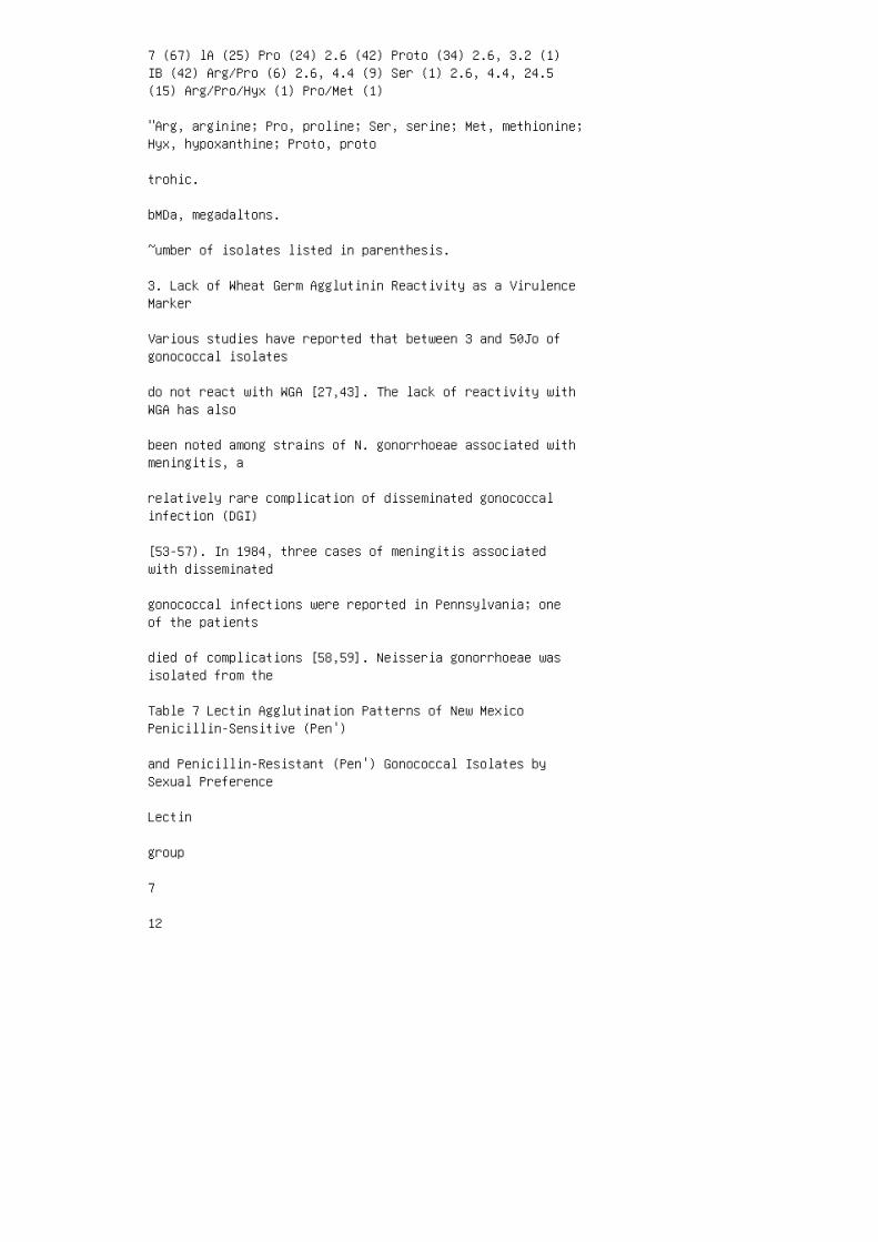
7 (67) lA (25) Pro (24) 2.6 (42) Proto (34) 2.6, 3.2 (1)IB (42) Arg/Pro (6) 2.6, 4.4 (9) Ser (1) 2.6, 4.4, 24.5(15) Arg/Pro/Hyx (1) Pro/Met (1)
"Arg, arginine; Pro, proline; Ser, serine; Met, methionine;Hyx, hypoxanthine; Proto, proto
trohic.
bMDa, megadaltons.
~umber of isolates listed in parenthesis.
3. Lack of Wheat Germ Agglutinin Reactivity as a VirulenceMarker
Various studies have reported that between 3 and 50Jo ofgonococcal isolates
do not react with WGA [27,43]. The lack of reactivity withWGA has also
been noted among strains of N. gonorrhoeae associated withmeningitis, a
relatively rare complication of disseminated gonococcalinfection (DGI)
[53-57). In 1984, three cases of meningitis associatedwith disseminated
gonococcal infections were reported in Pennsylvania; oneof the patients
died of complications [58,59]. Neisseria gonorrhoeae wasisolated from the
Table 7 Lectin Agglutination Patterns of New MexicoPenicillin-Sensitive (Pen')
and Penicillin-Resistant (Pen') Gonococcal Isolates bySexual Preference
Lectin
group
7
12

25 Sexual preference Heterosexual Pen' 6 9 0 Pen' 43 1 Homosexual Pen' Pen' 6 0 0 11 0 0 Table 8Relationship of Lectin Group, Serovar, Plasmid Content, andAuxotype of Isolates for the New Mexico Sexual Partners•Sexual Culture partners Sexb site< Serovar Auxotype AM R IB-1 Prototrophic B M u IB-1 Proline c M u IB-1Proline D M R IB-1 Prototrophic E M u IB-4 PrototrophicF M R IB-4 Proline G M u IB-4 Prototrophic H M u IB-1Prototrophic I F c IB-1 Proline J M u IB-1 Proline"All isolates were in lectin group 7, and all had 2.6 MDaplasmids. bM, male; F, female. <R, rectum; U, urethra; C,cervix. Source: Data from Ref. 50. blood and cervix ofthe deceased patient, from the cerebrospinal fluid andcervix of the second patient, and from the blood of thethird patient. When examined with lectin panels, all fiveisolates failed to react with WGA. Fifty urogenital andtwo DGI arthritis-associated isolates of N. gonorrhoeaewere then obtained from the same Pennsylvania community andtested for agglutination by WGA; 49 of SO urogenital andthe 2 arthritisassociated isolates were agglutinated byWGA. Results of the lectin agglutination of the Smeningitis-associated, 50 urogenital, and 2 DOlarthritisassociated isolates obtained from this geographicarea, and 13 PID isolates obtained from the Centers forDisease Control and Prevention (CDC) culture collection areshown in Table 9. One isolate obtained from a case ofuncomplicated or urogenital gonorrhoea did not agglutinateWGA. The failure of WGA to agglutinate the strainsisolated from patients with gonococcal meningitis may be amarker for a virulence factor. A comparison of serovar andlectin reactivity with WGA between the S Pennsylvaniameningitis-associated isolates, 10 DOl isolates notassociated with meningitis and obtained from the CDCculture collection, and the SO Pennsylvania urogenitalisolates causing uncomplicated gonorrhoea is summarized inTable 10. The meningitis-associated isolates did notagglutinate WGA and were preponderantly lA serovars. TheDOl isolates not associated with meningitis were alsopreponderantly lA serovars; however, they were agglu
Applications of Lectins to Agents of STD 123
Table 9 Various Lectin Reactions with the PennsylvaniaN.gonorrhoeae Isolates
from Different Anatomical Sites No. of reactiveisolates/total DGI," DGI,
Lectin Symbol Urogenital PID" arthritis meningitis

Concanavalin A ConA 0/50 0/13 0/2 0/5
Arachis hypogeae PNA 50/50 13/13 2/2 515
Dolichos biflorus DBA 13/50 13/13 2/2 515
Solanum tuberosum STA 46/50 12/13 2/2 515
Triticum vulgaris WGA 49/50 12/13 2/2 0/5
"PID, pelvic inflammatory disease; DGI, disseminatedgonococcal infection.
Source: Data from Ref. 58.
tinated by WGA. The isolates from uncomplicated infectionswere prepon
derantly IB serovars, and only one isolate did notagglutinate WGA. Frasch
[13] observed that encapsulated strains of N. meningitidiswere not aggluti
nated by WGA, whereas nonencapsulated strains wereagglutinated by
WGA. No capsule could be demonstrated when the fivegonococcal isolates
from meningitis patients were examined further. Asubsequent study re
vealed that a gonococcal isolate associated withmeningitis, obtained from
a different geographic area of the United States, wasagglutinated by WGA
[60]. These findings suggested that the strains isolated inPennsylvania may
represent the spread of a single clone; however, this isunlikely, since sero
type analysis indicated that these strains belonged toboth lA and IB sero
vars.

Table 10 Comparison of Serogroup and Wheat GermAgglutination Among
Patient Isolates of N. gonorrhoeae
Diagnosis
DOl" /meningitis
DOl/without meningitis
Uncomplicated gonorrhoea No. of patients 5 10 50
"DOl, disseminated gonococcal infection.
Source: Data from Ref. 59. Protein I serogroup lA IB 41 8 2 6 44 Wheat germ agglutination yes no 5 1049 4. Use of Lectins to Assess Treatment Failure Anotherapplication of lectin typing has been in drug treatmenttrials to characterize preand posttreatment isolates ofN. gonorrhoeae. Two studies have been reported thatcharacterized preand posttreatment isolates followingenoxacin [61] or cefodizime versus cefotaxime [62] therapyfor gonorrhea. Lectin typing has been used in conjunctionwith auxotyping, serovar analysis, and minimum inhibitoryconcentration (MIC) to the investigational drug toascertain whether the preand posttreatment isolates wereidentical, thus representing either treatment failure,reinfection from an untreated partner, or emergence of anantibiotic-resistant strain. Nonidentical preandposttreatment isolates suggest reinfection with a differentstrain. Twelve pairs of preand posttreatment isolates wereavailable from the enoxacin trial. The results of thisstudy are shown in Table 11. Eleven of twelve pairs ofpreand posttreatment isolates had identical auxotypes andserovars; two isolate pairs differed in lectin group. Inone pair, the difference represented the loss ofreactivity with GS-11 and the gain of reactivity with DBA.In the other pair, the posttreatment isolate lostreactivity with GS-11. In addition, 11 of 12 pairs ofpreand posttreatment strains had the same MIC to enoxacin.One of the posttreatment strains exhibited a 67-foldincrease in the MIC value and probably represents theselection of a resistant strain. In the other studyinvolving treatment of uncomplicated gonorrhea in men andwomen with either cefodizime or cefotaxime [62], all ofthe preand posttreatment isolates were identical forauxotype, serovar, and lectin group. The results of thesestudies suggest that, with one exception, the

posttreatment isolates represent either treatment failureor reinfection from an untreated partner. Ill. HAEMOPHILUSDUCREYI A. The Organism Haemophi/us ducreyi is agram-negative, nonmotile rod that is the etiological agentof chancroid. Chancroid is one of the five classic venerealdiseases (gonorrhea, syphilis, lymphogranuloma venereum,donovanosis, and chancroid) and is characterized bypainful genital ulcers and frequent lymphadenopathy.Chancroid was considered to be a relatively uncommonsexually transmitted disease in the United States;however, the number of reported cases have increased morethan sixfold since 1984 [63]. Haemophilus ducreyi is amajor cause of genital ulcers in Asia and Africa, where theprevalence of chancroid often exceeds that of syphilis.This bacterium is difficult to isolate directly fromgenital lesions [63]. Sensitivity of culture is dependenton the medium, the specimen, and the Table 11 TypingPatterns for N. gonorrhoeae Strains Isolated Before andAfter Therapy with Enoxacin Pretreatment strainsPosttreatment strains Strain MIC Lectin MIC number(/Lg/ml) Auxotype" Serovar group (/Lg/ml) Auxotype"Serovar 1 0.03 NR IB-3 6 0.03 NR IB-3 2 0.06 ProlineIB-5 7 0.06 Proline IB-5 3 0.03 Proline IB-4 7 0.03Proline IB-4 4 0.06 Proline IB-4 7 0.06 Proline IB-45 0.06 NrPhe IB-6 7 0.06 NRPhe IB-6 6 0.03 ProlineIB-4 6 0.06 Proline IB-4 7 0.03 Prototrophic IB-3 60.03 Prototrophic IB-3 8 0.03 Prototrophic IB-2 72.00 Prototrophic IB-2 9 0.03 NRPhe IB-6 7 0.03NrPhe IB-6 10 0.06 Prototrophic NTb 12 0.06Prototrophic NT 11 0.03 NR IB-6 6 0.06 NR IB-6 12O.o3 NRPhe IB-6 12 0.03 NRPhe IB-6 "NR Phe, notrequiring and inhibited by phenylalanine; NR, norequirement. bNT, not typable. Source: Data from Ref. 60.Lectin group 6 7 7 7 7 6 6 7 7 6 6 7 > ,'2.. ;:r !!:. =· = "' Q .... tD !l :r "' Q >~ = "' Q Ill -1 c ... N 1;,11 expertise of thelaboratorian. Relatively little is known about theepidemiology of chancroid because there are relatively fewphenotypic characteristics that can be used todifferentiate among isolates of H. ducreyi. Outer membraneprotein profiles [64,65] and amino peptidase profiles[66,67] have been used, but do not provide suitablediscrimination. Recently, the taxonomy of H. ducreyi hasbeen called into question [68,69]. Sequence analysis ofribosomal RNA genes indicates that H. ducreyi belongs inthe family Pasteurel/aceae, but is not a member of thegenus Haemophilus [70]. It has been suggested that H.ducreyi may comprise a single-species genus and, thus, maybe of clonal origin. Therefore, it is not surprising thatit has been difficult to differentiate among strains of

H. ducreyi. The identification of restriction fragmentlength polymorphisms (RFLPs) among ribosomal RNA genes(ribotyping) has recently been useful in typing isolatesof H. ducreyi [71]. B. Cell Envelope In electronmicrographs, the outer membrane of H. ducreyi appearsmorphologically similar to those of other gram-negativebacteria [63,72]. The outer membrane profile is alsotypical of gram-negative bacteria, with one to severalproteins predominating [64,73,74]. Changes in growth mediumcomposition, atmospheric conditions, and temperature do notappear to affect the outer membrane protein profile;however, the electrophoretic pattern of the LPS isapparently affected by conditions of growth [75]. Thepresence of a capsule has not been adequatelydemonstrated. Bertram [76] observed the presence ofantibody-stabilized extracellular capsular material byelectron microscopy. In contrast, Johnson et al. [77]failed to observe extracellular acidic polysaccharide onthin sections of H. ducreyi stained with ruthenium red.1. Properties of Haemophilus ducreyi Lipoo/igosaccharidesHaemophilus sp. have been reported to produce both smoothand rough LPS [78]. Several investigators have recentlyexamined the type of LPS produced by H. ducreyi.Haemophilus ducreyi possesses a low molecular weight LPSthat contains heptose, ketodeoxyoctonate, glucose,galactose, glucosamine, and galactosamine [79]. It issimilar in size to that of other mucosal pathogens andtherefore, is referred to as LOS. The H. ducreyi LOS ismore toxic than the LOS from H. inf/uenzae or the LPS fromEscherichia coli, and has produced skin abscesses in animalmodels [80]. Two patterns of reactivity with monoclonalantibodies have been observed among isolates of H.ducreyi [81]. The LOS from 24 of 25 strains examined boundmonoclonal antibody 3Fll, which recognizes a terminalGal(j
1 ,4GlcNAc epitope that has a structure similar to humanblood group anti
gens [37,81]. This epitope is also present on somegonococcal LOSs and is
apparently sialylated in vivo [39]. Whether H. ducreyi LOSis sialylated in
vivo remains to be determined. One strain has beenidentified that does not
react with monoclonal 3Fll. This strain produces a smallerLOS than the

other strains that have been examined [81].
C. Interaction of Haemophilus ducreyi with Lectins
1. Methodology
Laboratory cultivation of H. ducreyi can be accomplishedwith heart infu
sion agar (Difco Laboratories) supplemented with 50Jorabbit erythrocytes,
10% fetal bovine sera (Hyclone Laboratories), and 1%IsoVitaleX (BBL).
Various media have been described for cultivating H.ducreyi [63,82-91].
After growth for 18 hr at 35°C in 5% C0 2 , cells weresuspended in TBS,
pH 7 .5, as described for N. gonorrhoeae. Unlike gonococci,H. ducreyi
cells form clumps or chains when in suspension thatinterfere in interpreting
lectin agglutination reactions. By allowing suspensions ofH. ducreyi to
stand for 1-2 min, cell clumps and chains of cells settleto the bottom of
the tube. The liquid at the top of the tube, containingsingle or pairs of
cells, is removed and transferred to another tube. Thissuspension can now
be adjusted spectrophotometrically (535 nm) to an opticaldensity of a
McFarland No. 4 standard. Samples (50J,tl) of the cellsuspension are added
to the lectin panel wells, panels are placed on a gyratoryplatform, rotated
for 5-10 min, and agglutination read. Additionally,

plastic panels contain
ing no lectins were used to place a suspension of eachisolate. With the aid
of a stereomicroscope (Bausch and Lomb) at 5 x or lOxmagnification,
the granulation between wells containing organisms andlectins could be
compared with the wells containing only organisms. In thoseinstances
when there was difficulty determining if agglutination wasoccurring, or
granulation was causing a false-positive reaction,specific carbohydrates
were used to show reversibility of agglutination.
2. Epidemiological Applications
A total of 63 isolates of H. ducreyi obtained fromoutbreaks in California,
Florida, Georgia, New York, and Massachusetts wereexamined. The re
sults of the H. ducreyi lectin agglutination activity areshown in Table 12.0f
the 63 isolates examined, two preponderant lectin groupswere observed.
Differences in lectin agglutination focused on thereactivity of isolates with
GS-11. Among these isolates, 27 (43%) did not agglutinatewith GS-11,
whereas 36 (57%) isolates did agglutinate with this lectin.What appeared
to be agglutination activity with LCA and PHA could not bereversed Table 12 Lectin Agglutination Patterns of 63Haemophilus ducreyi Isolates Geographic Number Lectin"location and of year of isolation Isolates ConA LCA TRKGS-I PNA SBA DBA GS-11 STA WGA LFA PHA UEA-I LOTUS

California Orange County (1982) 5 + + + + + + + +8 + + + + + + + + + Long Beach (1987) 5 + + + + ++ + + + Georgia Atlanta (1958) 1 + + + + + + + + + 1+ + + + + + + + Atlanta (1981) 3 + + + + + + + + + 3+ + + + + + + + New York New York City (1985) 6 + ++ + + + + + 7 + + + + + + + + + ... 1-.J = Cll n::r !.. iii Ill :I Cl. ~ Q ... ; Boston (1985) 4+ + + + Florida West Palm Beach (1983) 7 + + + + ++ + + Orlando (1985) 1 + + + + + + + + Jacksonville(1986) 4 + + + + + + + + Ohio Cleveland (1985) 2 ++ + + Texas Dallas (1987) 3 + + + + •Lectinabbreviations shown in Table 1. + + + + + + + + + + + ++ + + + + + + + + + + + + + + + + + + + + + + ++ + + + + + > "5!.. ;::;~ o· :I II> Q I""'I'D 1"1 :r II> Q > (JQ I'D :I II> Q VI ~ c.... 1-.1 loCI with specific carbohydrates. It wasconcluded that this activity was due to nonspecificclumping that resulted in false-positive agglutination. Theresults presented in Table 12 are different from thosepresented by Korting et al. [92]. In that study, all ofthe isolates were agglutinated by LCA and PHA, with someagglutination activity reported for DBA, UEA-1, and Lotus.In contrast, we were unable to show agglutination activityby LCA, DBA, PHA, UEA-1 or Lotus. However, all of theisolates were agglutinated by TRK, GS-1, PNA, SBA, andSTA. Similar to the results reported by Korting et al.[92], some isolates were agglutinated by GS-11. Nodifference was found in agglutination activity of isolatesrelative to geographic location in the United States. Thesimilarity in lectin agglutination patterns for theseisolates may suggest that these outbreaks were caused by afew strains; however, the inability of lectins todifferentiate many agglutination types among the H.ducreyi strains examined is consistent with data suggestingthat few phenotypic properties exist that can be used forstrain typing [63]. IV. TREPONEMA PALL/DUM SUBSPECIESPALL/DUM AND TREPONEMA PALL/DUM SUBSPECIES PERTENUE A.The Organism Treponema pallidum subsp. pallidum (T.pallidum) and T. pallidum subsp. pertenue (T. pertenue)are nonculturable (pathogenic) spirochetes that are thecausal agents of syphilis and yaws, respectively. Theybelong to the family Spirochaetaceae, genus Treponema.Syphilis is a chronic disease that occurs throughout theworld. Infection within the host is characterized by aprimary lesion or soft chancre that usually develops withina few weeks. Treponemes can often be observed by directexamination of lesion fluid by darkfield microscopy.Treponema pertenue is the causative agent of yaws,transmission occurs through skin contact, and the diseaseoccurs primarily in the tropics. Both T. pallidum and T.

pertenue are gram-negative organism and are considered tobe microaerophilic [93,94]. Treponema pallidum takes upoxygen and possesses an electron transport system [95,96].Both microorganisms exhibit corkscrew motility that isaccompanied by rotation around the longitudinal axis and abending or flexing. The ends of these spirochetes aresomewhat pointed or tapered [97 ,98]. Electron microscopyhas shown that these treponemes possess an outer membrane,also referred to as an outer envelope, which covers theaxial filaments located on the surface [97,98].Antigenically, these treponemes are considered identical,and a species-specific antigen has not yet been identified.Pathogenic and nonpathogenic treponemes can bedifferentiated by DNA homology, but
Applications of Lectins to Agents of STD 131
this method does not differentiate between T. pallidum andT. pertenue
[99,100].
B. The Outer Envelope and Cell Wall
The pathogenic treponemes are nonculturable, althoughlimited success has
been obtained with tissue cell cultures [101-103]. Becauseit is difficult to
grow pathogenic treponemes in vitro, very littleinformation is available
concerning their biochemical reactions, metabolism, andchemical composi
tion, although the lipid composition of T. pallidum wasreported by Ma
thews et al. [104]. Some reports have indicated that thepathogenic trepo
nemes possess a surface acidic polysaccharide [105-107],but other reports
question whether this polysaccharide is synthesized by thetreponeme, or is
derived from host tissue [1 08-111]. The polysaccharidepossesses glucose

and galactose residues [ 104, 107, 11 0].
C. Interaction with Lectins
Very little information is available about the interactionof T. pallidum or
T. pertenue with lectins. Baseman et al. [112] used lectinsnoncovalently
bound to Leighton tube coverslips to bind freshly extractedtreponemes
and facilitate the removal of cellular debris and fluids.They observed that
T. pallidum bound to lectin film coverslips containingConA, WGA, PHA,
and pokeweed mitogen (PWM). Binding ofT. pallidum to ConAoccurred
with greater affinity than to WGA and PHA. Fitzgerald andJohnson [109]
reported that T. pallidum interacted with WGA and SBA, andthey postu
lated that the ligand was the acidic polysaccharide. Thesurface polysaccha
ride of T. pallidum is reported to be composed ofhyaluronic acid and
chondroitin sulfate or related acidic glycosaminoglycans[107]. Hyaluronic
acid, consisting of N-acetyl-o-glucosamine-o-glucuronicacid, will interact
with WGA. Similarly, chondroitin sulfate consists ofN-acetyl-o-galac
tosamine-o-glucuronic acid, which will interact with SBA.These acidic
polysaccharides are present in syphilitic lesions[108-111], and it is unclear

whether the polysaccharide material is part of thetreponemal cell surface,
or results from host tissue damage during the infectionprocess [108-111].
1. Methodology
Since T. pallidum and T. pertenue cannot be easilysubcultured in vitro,
growth of the organisms is accomplished by intratesticularpassage in
healthy rabbits. Rabbit testes are excised 11-14 dayspostinfection. The
treponemes are harvested by slicing the testes, placingthem in a solution of
0.02 M phosphate-buffered saline (pH 7 .2), containing0.075 M sodium
citrate, and teasing the treponemes out of the testiculartissue with gentle agitation. The pathogenic treponemesare separated from other cellular debris by densitygradient centrifugation in 200Jo Percoll (Pharmacia FineChemicals, Piscataway, New Jersey). Treponemes purified bydensity gradient centrifugation are washed three times withTBS, pH 7.5, to remove contaminating Percoll. Treponemaphagedenis biotype Reiter was cultivated in NIHthioglycolate broth (Difco Laboratories) containing 10%inactivated normal rabbit sera. Growth of thisnonpathogenic treponeme was accomplished at 35°C for 4-7days. Purified pathogenic treponemes and the nonpathogenicT. phagedenis were suspended in TBS, pH 7.5, adjustedspectrophotometrically to yield a suspension equivalent toa McFarland No. 4 standard, and examined for agglutinationactivity with the panel of 14 lectins. Normal rabbittesticular extract (NRTE) was prepared by pulverizingnitrogen-frozen testicular tissue in a stainless steelmill. The powdered testicular tissue was extracted threetimes with TBS, pH 7.5, and layered onto 20% Percoll fordensity gradient centrifugation as described earlier. TheNRTE and 20% Percoll were also examined for agglutinationactivity with the 14 panellectins. 2. EpidemiologicalApplications Three strains of T. pallidum and two strainsof T. pertenue, all isolated from different geographicareas, and one nonpathogenic strain were examined forlectin reactivity. The results of lectin agglutination

with the treponemal strains are shown in Table 13. Thesame lectin agglutination pattern was observed for all T.pallidum and T. pertenue strains. The pathogenictreponemes reacted with ConA, GS-1, SBA, and WGA. The NRTEalso showed reactivity with these same lectins. It isuncertain whether the treponemal reactivity with theselectins is due to treponemal cell surface carbohydrate,or to contaminating testicular tissue present on thesurface of the treponemes. The pathogenic strains couldnot be differentiated from the T. phagedenis biotypeReiter owing to the uncertainty of testicular tissuecontamination. Agglutination of lectins with thepathogenic treponemal suspensions was reversed withspecific carbohydrates, indicating specific reactivity;however, with the uncertainty of testicular tissuecontamination, the specificity of these reactions mayreside in contaminating testicular material. The 200JoPercoll preparation did not show lectin agglutination. V.CONCLUSIONS The information reported in this chapterrepresents only preliminary information concerning theapplications of lectins to some organisms associated withsexually transmitted diseases. All agglutinationcharacteristics of these microorganisms were determinedusing prepared panels of 14lectins. Fur.... l.ol l.olTable 13 Lectin" Agglutination Activity of PathogenicTreponema! Strains, Compared with One NonpathogenicTreponema! Strain Con A LCA TRK OS-I PNA SBA DBA GS-11STA WGA LFA PHA UEA-1 LOTUS T. phagedenis + + + + + + +biotype Reiter T. pa/lidum subsp. pa/lidum Strain 1 + ++ + + + + + + ± + Strain 2 + + + + + + + + + ± +Strain 3 + + + + + + + + + ± + T.pallidum subsp.pertenue Strain 1 + + + + + + + + + ± + Strain 2 + ++ + + + + + + ± + 200Jo Percoll NRTEb + + + + + + ++ "See Table 1 for identification of lectins. ~RTE,normal rabbit testicular extract . ther characterizationof cell surface carbohydrates and their role in cellsurface structure may require the use of more-specificlectin reagents than those contained in panels used forstudies reported in this chapter. The agglutinationreactions of N. gonorrhoeae, based on structuralcomponents of the LOS, were carbohydrate-specific andcould be inhibited. The agglutination of N. gonorrhoeaeappeared to be due to the availability of exposed terminalstructures of the carbohydrate residues. Masking of somecarbohydrate structures or influence of a neighboringstructural charge phenomenon may offer an explanation whylectins of similar specificity do not agglutinate the samegonococcal strains. Twenty-four different lectin groupswere observed in these studies. It has been suggested thatgonococcal LOS epitopes may undergo phase variation, which

may, in turn, affect lectin agglutinability [42].However, subculture did not appear to affect lectinagglutination of N. gonorrhoeae strains used in thesestudies. Strains of N. gonorrhoeae were repeatedlysubcultured and used as blind controls. The originalagglutination pattern of each strain did not change aftersubculture. The observation that differences in lectinagglutination with GS-11 and DBA may be suggestive ofstructural differences in a-o-GalNAc or a-o-GlcNAcresidues, lectin accessibility to residues, absence ofthese residues in some strains, or the population ofresidues presented to lectin reagents. In the studiespresented in this chapter, two strains of N. gonorrhoeaeagglutinated with ConA, which represented an uncommonlectin agglutination pattern among the 150 strainsexamined. In contrast, Vazquez and Berron [43] observed amore frequent agglutination of N. gonorrhoeae strains byConA. Perhaps these strains, which were isolated indifferent geographic areas of the world, exhibited moreLOS structural differences than the 150 strains examinedfrom the United States. The strains isolated in theUnited States showed infrequent agglutination with UEA-1,LCA, and PHA, when compared with the agglutination patternsreported by Vazquez and Berron [43]. The data presentedin this chapter concerning lectin agglutination differencesamong strains of N. gonorrhoeae represent a differenttyping system that can be used in conjunction withserotyping and auxotyping. The use of lectinagglutination, serotyping, and auxotyping can be useful inclinical studies to determine antibiotic treatmentfailures, emergence of antibiotic-resistant strains, andreinfection from an untreated sexual partner. Strains ofH. ducreyi reported in this chapter did not show a highdegree of variability in lectin agglutination patterns.Repeated subculture of these H. ducreyi strains did notaffect the reproducibility of lectin agglutinationpatterns, suggesting that subculturing did not affect LOSepitopes present on the cell surface. That only twolectin agglutination patterns for
the isolates of H. ducreyi were available for examinationin this study is
consistent with results of studies indicating fewphenotypic characteristics
are available for differentiating strains of thisorganism. Studies of lectin
agglutination with H. ducreyi reported by Korting et al.

[92] with strains
isolated from other world geographic areas did showdifferences in lectin
agglutination. At least 20 different lectin agglutinationpatterns were re
corded, suggesting that there may be methodologicaldifferences or differ
ences in phenotypic characteristics among these strains.The lectin agglutination patterns of the pathogenictreponemes were
identical. No differences in lectin agglutination wereobserved between the
strains examined. A previous report indicated that lectinagglutination with
T. pallidum was due to acidic polysaccharides [107].However, whether this
polysaccharide is of treponema! origin or host tissueorigin is still unclear
[1 08-111]. Difficulty in obtaining high yields oftreponemes for lectin ag
glutination or in preparing pathogenic treponemes to ensureabsence of
host tissue fluid or debris will be a continual problem toproduce reliable
and accurate lectin agglutination results. Otherpreliminary studies have been initiated to examine otheragents
of sexually transmitted diseases. With the same panel of14lectins, strains
of Chlamydia trachomatis have been examined foragglutination by lectins.
Strains of C. trachomatis were agglutinated by lectinsspecific for a-o-Gle,
a-D-Gal, and a-o-GalNAc. McCoy tissue culture cells used

for passage of
C. trachomatis were also agglutinated by the same lectins,however, raising
concern that the agglutination may be due to contaminationby McCoy
tissue culture cell epitopes. Additionally, the panel of14 lectins has been
used to determine lectin agglutination patterns ofMycoplasma hominis.
Only after proteolytic treatment were M. hominis strainsagglutinated by
lectins specific for a-o-Gle, a-n-Gal, and a-o-GalNAc, inaccord with re
sults reported by Kahane and Tully [113]. Schiefer et al.[114], however,
did not observe agglutination of M. hominis strains bylectins, and treating
of these strains with pronase failed to enhanceagglutination reactivity.
Further examination of these microorganisms is necessaryto determine the
suitability of lectin applications and to determine thestability and repro
ducibility of lectin agglutination activity. Theapplication of lectin agglutination to agents of sexuallytransmit
ted diseases may facilitate the detection of interandintrastrain variations.
ACKNOWLEDGMENTS
The authors thank R. E. George, E. F. Hunter, and S. A.Larsen for their
technical assistance and guidance in growth of pathogenictreponemes, and

21. Cole HB, Ezzell JW Jr, Keller KF, Doyle RJ.Differentiation of Bacillus anthracis and other Bacillusspecies by lectins. J Clin Microbiol 1984; 19:4853.
22. Curtis GDW, Slack MPE. Wheat-germ agglutination inNeisseria gonorrhoeae. Br J Vener Dis 1981; 57:253-255.
23. · Davidson SK, Keller KF, Doyle RJ. Differentiation ofcoagulase-positive and coagulase-negative staphylococci bylectins and plant agglutinins. J Clin Microbiol1982;15:547-553.
24. Doyle RJ, Nedjat-Haiem F, Keller KF, Frach CE.Diagnostic value of interactions between members of thefamily Neisseriaceae and lectins. J Clin Microbiol1984;19:383-387.
25. Senne JE. Lectin agglutination of Neisseriagonorrhoeae. Clin Microbiol Newsl1981; 3:10.
26. Schafer RL, Keller KF, Doyle RJ. Lectins in diagnosticmicrobiology: use of wheat germ agglutinin for laboratoryidentification of Neisseria gonorrhoeae. J ClinMicrobiol1979; 10:669-672.
27. Yajko DM, ChuA, Hadley KW. Rapid confirmatoryidentification of Neisseria gonorrhoeae with lectins andchromogenic substrates. J Clin Microbiol 1984; 19:380-382.
28. Doyle RJ, Nedjat-Haiem F, Miller RD, Keller KF.Interaction between plant agglutinins and Legionellaspecies. J Clin Microbiol1982; 15:973-975.
29. Ottensooser F, Nakamizo Y, Sato M, Miyamoto Y, TakizawaK. Lectins detecting group C streptococci. Infect Immun1974; 9:971-973.
30. Slifkin M, Cumbie R. Identification of group Bstreptococcal antigen with lectin-bound polystyreneparticles. J Clin Microbiol1987; 25:1172-1175.
31. Wong KH, Skelton SK, Feeley JC. Interaction ofCampylobacter jejuni and Campylobacter coli with lectinsand blood group antibodies. J Clin Microbiol 1985;22:134-135.
32. Meade NA, Staat RH, Langley SD, Doyle RJ. Lectin-likeactivity from Persea americana. Carbohydr Res 1980;78:349-363.

33. Mintz CS, Apicella MA, Morse SA. Electrophoretic andserological characterization of the lipopolysaccharideproduced by Neisseria gonorrhoeae. J Infect Dis 1984;149:544-552.
34. Griffiss JM, O'Brien JP, Yamasaki R, Williams GD, RicePA, Schneider H. Physical heterogeneity ofneisseriallipooligosaccharides reflects oligosaccharidesthat differ in apparent molecular weight, chemicalcomposition, and antigenic expression. Infect Immun 1987;55:1792-1800.
35. Dudas KC, Apicella MA. Selection and immunochemicalanalysis of lipooligosaccharide mutants of Neisseriagonorrhoeae. Infect Immun 1988; 56:499504.
36. Griffiss JM, Schneider H, Mandrell RE, Yamasaki R,Jarvis GA, Kim JJ, Gibson BW, Hamadeh R, Apicella MA.Lipooligosaccharides: the principal glycolipids of theneisserial outer membrane. Rev Infect Dis 1988; 10(suppl2):S287-S295. 37. Mandrell RE, Griffiss JM, Macher BA.Lipooligosaccharides (LOS) of Neisseria gonorrhoeae andNeisseria meningitidis have components that areimmunochemically similar to precursors of human bloodgroup antigens. J Exp Med 1988; 168:107-126. 38.Parsons NJ, Patel PV, Tan EL, Andrade JRC, Nairn CA,Goldner M, Cole JA, Smith H. Cytidine 5'-monophospho-N-acetylneuraminic acid or a related compoundis the low molecular weight factor from human red bloodcells which induce gonococcal resistance to killing byhuman serum. J Gen Microbiol1988; 134:3295-3306. 39.Mandrell RE, Lesse AJ, Sugai JV, Shero M, Griffiss JM,Cole JA, Parsons NJ, Smith H, Morse SA, Apicella MA. Invitro and in vivo modification of Neisseria gonorrhoeaelipooligosaccharide epitope structure by sialylation. JExp Med 1990; 171:1649-1664. 40. Ward ME, Watt PJ, GlynnAA. Gonococci in urethral exudate possess a virulencefactor lost on subculture. Nature 1970; 227:382-384. 41.Yamasaki R, Bacon B, Nasholds W, Schneider H, Griffiss JM.Structural determination of the oligosaccharides derivedfrom lipooligosaccharide (LOS) of Neisseria gonorrhoeaeF62 by chemical and 2D NMR (500 MHz) methods: MAbs 1-1Mand3Fll-defined LOS epitopes have aN-acetylgalactosaminylneolactotetraose and neolactotetraoseat their non-reducing terminus, respectively. Biochemistry1991; 30:10566-10575. 42. Schneider H, Hammack CA,Apicella MA, Griffiss JM. Instability of expression oflipooligosaccharides and their epitopes in Neisseriagonorrhoeae. Infect Immun 1988; 56:942-946. 43. VasquezJA, Herron S. Lectins agglutination test as an

epidemiological marker for Neiserria gonorrhoeae.Genitourin Med 1990; 66:302-305. 44. Catlin BW.Nutritional profiles of Neisseria gonorrhoeae, Neisseriameningitidis, and Neisseria lactamica in chemicallydefined media and the use of growth requirements forgonococcal typing. J Infect Dis 1975; 128:178-194. 45. TamMR, Buchanan TM, Sandstrom EG, Holmes KK, Knapp JS, SiadakA W, Nowinski RC. Serological classification of Neisseriagonorrhoeae with monoclonal antibodies. Infect Immun 1982;36:1042-1053. 46. Knapp JS, Tam MR, Nowinski RC, HolmesKK, Sandstrom EG. Serological classification of Neisseriagonorrhoeae with use of monoclonal antibodies togonococcal outer membrane protein I. J Infect Dis 1984;150:44-48. 47. Schalla WO, Whittington WL, Rice RJ,Larsen SA. Epidemiological characterization of Neisseriagonorrhoeae by lectins. J Clin Microbiol 1985; 22:379-382. 48. Short HB, Ploscoe VB, Weiss JA, Young FE.Rapid method for auxotyping multiple strains of Neisseriagonorrhoeae. J Clin Microbiol1977; 6:244-248. 49. CatlinBW. Characteristics and auxotyping of Neisseriagonorrhoeae. In: Bergan T, Norris JR, eds. MethodsMicrobiol1978; 10:345-380. 50. Schalla WO, Rice RJ, BiddleJW, JeanLouis Y, Larsen SA, Whittington WL. Lectincharacterization of gonococci from an outbreak caused bypenicillin-resistant Neisseria gonorrhoeae. J ClinMicrobiol1985; 22:481-483. 51. Meyers JA, Sanchez D,Elwell LP, Falkow SL. Simple agarose gel electro
Applications of Lectins to Agents of STD 139 phoresismethod for the identification and characterization ofplasmid DNA. J Bacteriol1976; 127:1629-1633.
52. Perine PL, Thornsberry C, Schalla WO, Biddle J, SeigelMS, Wong KH, Thompson SE. Evidence for two distinct typesof penicillinase-producing Neisseria gonorrhoeae. Lancet1977; 2:993-995.
53. Holmes KK, Counts GW, Beaty HN. Disseminatedgonocococal infection. Ann Intern Med 1971; 74:979-993.
54. Sayeed ZA, Bharudi U, Howell E, Meyers HL Jr.Gonococcal meningitis: a review. JAmMed Assoc 1972;1730-1731.
55. Swierczewski JA, Mason EJ, Cabrera PB, Liber M.Fulminating meningitis with Waterhouse-Friderichesensyndrome due to Neisseria gonorrhoeae. Am J ClinPathol1970; 54:202-204.
56. Pasquariello CA, Plotkin SA, Rice RJ, Hackney JR. Fatal

gonococcal septicemia. Pediatr Infect Dis 1985; 4:204-206.
57. Granato P A, Howard R, Wilkinson B, Laser J. Meningitiscaused by maltose-negative variant of Neisseriameningitidis. J Clin Microbiol 1980; 11: 270-273.
58. Rice RJ, Schalla WO, Whittington WL, Biddle JW,JeanLouis Y, DeWitt WE, Thompson SE. Investigation ofNeisseria gonorrhoeae causing disseminated infection,endotoxemia, and meningitis and identification of apossible virulence marker. In: Schoolnik GK, Brooks GF,Falkow S, Frasch CE, Knapp JS, McCutchan JA, Morse SA,eds. Pathogenic neisseriae. Washington, DC: AmericanSociety for Microbiology, 1985:61-65.
59. Rice RJ, Schalla WO, Whittington WL, JeanLouis Y,Biddle JW, Goldberg M, Dewitt W, Pasquariello A, AbrutynE, Swenson R. Phenotypic characterization of Neisseriagonorrhoeae isolated from three cases of meningitis. JInfect Dis 1986; 153:362-365.
60. Del Rio C, Stephens DS, Knapp JS, Rice RJ, Schalla WO.Comparison of isolates of Neisseria gonorrhoeae causingmeningitis and report of gonococcal meningitis in apatient with CS deficiency: J Clin Microbiol 1989;27:10451049.
61. Van Der Willigen AH, Van Der Hoek JCS, Wagenvoort JHT,Van Vliet HJA, Van Klingeren B, Schalla WO, Knapp JS, VanJost T, Michael MF, Stolz E. Comparative double-blindstudy of 200and 400-mg enoxacin given orally in thetreatment of acute uncomplicated urethral gonorrhea inmales. Antimicrob Agents Chemother 1987; 31:535-538.
62. VanDer Willigen AH, Wagenvoort JHT, Schalla WO, KnappJS, Boot JM, Heeres-Weststrate PL, Michel MF, VanKlingeren B, Stolz E. Randomized comparative study of 0.5and 1 g of cefodizime (HR 221) versus 1 g of cefotaxime foracute uncomplicated urogenital gonorrhea. Antimicrob AgentsChemother 1988; 32:426-429.
63. Morse SA. Chancroid and Haemophilus ducreyi. ClinMicrobiol Rev 1989; 2:137-157.
64. Odumeru JA, Ronald AR, Albritton WL. Characterizationof cell proteins of Haemophilus ducreyi by polyacrylamidegel electrophoresis. J Infect Dis 1983; 148:710-714. 65.Taylor DN, Echeverria P, Hanchalay S, Pitarangsi C,Slootmans L, Piot P. Antimicrobial susceptibility andcharacterization of outer membrane proteins of Haemophilus

ducreyi in Thailand. J Clin Microbiol1985; 21:442-444. 66.Casin IM, Sanson-Le Pors MJ, Gorce MF, Ortenberg M, PerolY. The enzymatic profile of Haemophilus ducreyi. AnnMicrobiol (Paris) 1982; 133B:379-383. 67. Van Dyck E,Piot P. Enzyme profile of Haemophilus ducreyi strainsisolated on different continents. Eur J ClinMicrobiol1987; 6:40-43. 68. Casin IM, Grimont F, GrimontPAD, Sanson-Le Pors MJ. Lack of deoxyribonucleic acidrelatedness between Haemophilus ducreyi and otherHaemophilus species. Int J Syst Bacteriol1985; 35:23-25.69. Carlone GM, Schalla WO, Moss CW, Ashley DL, Fast DM,Holler JS, Plikaytis BD. Haemophilus ducreyi isoprenoidquinone content and structure determination. Int J SystBacteriol1988; 38:249-253. 70. Rossau R, Duhamel M, JannesG, Decourt JL, Van Heuverswyn H. The development ofspecific rRNA-derived oligonucleotide probes forHaemophi/us ducreyi, the causative agent of chancroid. JGen Microbiol 1991; 137: 277-285. 71. Sarafian SK, WoodsTC, Knapp JS, Swaminathan B, Morse SA. Molecularcharacterization of Haemophilus ducreyi by rRNAfingerprinting. J Clin Microbiol1991; 29:1949-1954. 72.Kilian M, Theilade J. Cell wall ultrastructure of strainsof Haemophilus ducreyi and Haemophilus pisicum. Int JSyst Bacteriol1975; 25:351-356. 73. Abeck D, Johnson AP.Identification of surface-exposed proteins in Haemophilusducreyi. FEMS Microbiol Lett 1987; 44:49-51. 74. SaundersJM, Folds JD. Immunoblot analysis of antigens associatedwith Haemophilus ducreyi using serum from immunizedrabbits. Genitourin Med 1986; 62:321-328. 75. Abeck D,Johnson AP, Ballard RC, Dangor Y, Fontaine EA,TaylorRobinson D. Effect of cultural conditions on theprotein and lipopolysaccharide profiles of Haemophilusducreyi analyzed by SDS-P AGE. FEMS Microbioi Lett 1987;48:397-399. 76. Bertram PD. Studies on Haemophilusducreyi. M.Sc. thesis, University of Manitoba, Winnipeg,May 1980. 77. Johnson AP, Abeck D, Davies HA. Thestructure, pathogenicity and genetics of Haemophilusducreyi. J Infect Dis 1988; 17:99-106. 78. Roberts MC,Mintz CS, Morse SA. Characterization of Haemophilusparainfluenzae strains with low-M. or ladder-likelipopolysaccharides. J Gen Microbiol1986; 132:611-616. 79.Odumeru JA, Wiseman GM, Ronald AR. Relationship betweenlipopolysaccharide composition and virulence of Haemophilusducreyi. J Med Microbiol 1987; 23:155-162. 80. CampagnariAA, Wild LM, Griffiths GE, Karalus RJ, Wirth MA, SpinolaSM. Role of lipooligosaccharide in experimental dermallesions caused by Haemophilus ducreyi. Infect Immun 1991;59:2601-2608. 141
81. Campagnari AA, Spinola SM, Lese AJ, Kwaik YA, Mandrell

RE, Apicella MA. Lipooligosaccharide epitopes shared amonggram-negative non-enteric mucosal pathogens. MicrobPathogen 1990; 8:353-362.
82. Hammond GW, Slutchuk M, Scatliff J, Sherman E, Wilt JC,Ronald AR. Epidemiologic, clinical, laboratory andtherapeutic features of an urban outbreak of chancroid inNorth America. Rev. Infect Dis 1980; 2:867-879.
83. Plummer FA, Nsanze H, Karasira P, D'Costa LJ, DylewskiJ, Ronald AR. Epidemiology of chancroid and Haemophilusducreyi in Nairobi, Kenya. Lancet 1983: 2:1293-1295.
84. Rajan VS, Sng EH, Lim AL. The isolation of Haemophilusducreyi in Singapore. Ann Acad Med (Singapore) 1983;12:57-60.
85. Taylor DN, Duangmani C, Suvonge C, O'Connor R,Pitarangsi C, Panikabutra K, Echeverria P. The role ofHaemophilus ducreyi in penile ulcers in Bangkok, Thailand.Sex Transm Dis 1984; 11:148-151.
86. Sng EH, Lim AL, Raj an VS, Gob AJ. Characteristics ofHaemophilus ducreyi. Br J Vener Dis 1982; 58:239-242.
87. Sottnek FO, Biddle JW, Kraus SJ, Weaver RE, Stewart JA.Isolation and identification of Haemophilus ducreyi in aclinical study. J Clin Microbiol 1980; 12:170-174.
88. Hannah P, Greenwood JR. Isolation and rapididentification of Haemophilus ducreyi. J ClinMicrobiol1982; 16:861-864.
89. Dylewski J, Nsanze H, Maitha G, Ronald A. Laboratorydiagnosis of Haemophilus ducreyi: sensitivity of culturemedia. Diagn Microbiol Infect Dis 1986; 4:241-245.
90. Lubwama SW, Plummer FA, Ndinya-Achola H, Nsanze W,Namaara LJ. Isolation and identification of Haemophilusducreyi in a clinical laboratory. J Med Microbiol1986;22:175-178.
91. Schalla WO, Sanders LL, Schmid GP, Tam MR, Morse SA.Use of dotimmunobinding and immunofluorescence assays toinvestigate clinically suspected cases of chancroid. JInfect Dis 1986; 153:879-887.
92. Korting HC, Abeck D, Johnson AP, Ballard RC,Taylor-Robinson D, Braun-Falco 0. Lectin typing ofHaemophilus ducreyi. Eur J Clin Microbiol Infect Dis

1988; 7:678-680.
93. Norris SJ, Miller JN, Sykes JA, Fitzgerald TJ.Influence of oxygen tension, sulfhydryl compounds, andserum on the motility and virulence of Treponema pallidum(Nichols strain) in a cell-free system. Infect Immun 1978;22: 689-697.
94. Fieldsteel AH, Stout JG, Becker FA. Comparativebehavior of viru1ent strains of Treponema pallidum andTreponema pertenue in gradient cultures of variousmammalian cells. Infect Immun 1979; 24:337-345.
95. Cox CD, Barber MK. Oxygen uptake by Treponemapallidum.Infect Immun 1974; 10:123-127.
96. Lysko PG, Cox DC. Terminal electron transport inTreponema pallidum. Infect lmmun 1977; 16:885-890.
97. Hovind-Hougen K. Determination by means of electronmicroscopy of morphological criteria of value forclassification of some spirochetes, in particulartreponemes. Acta Pathol Microbiol Scand Suppl1976;255:1-41. 98. Fitzgerald TJ, Cleveland P, Johnson RC,Miller JN, Sykes JA. Scanning electron microscopy ofTreponema pallidum (Nichols strain) attached to culturedmammalian cells. J Bacteriol1977; 130:1333-1344. 99. MiaoRM, FieldsteelAH. Genetics of Treponema: relationshipbetween Treponema pa/lidum and five cultivable treponemes.J Bacteriol1978; 133: 101-107. 100. Miao RM, FieldsteelAH. Genetic relationship between Treponema pallidum andTreponema pertenue, two noncultivable human pathogens. JBacteriol 1980; 141:427-429. 101. Fieldsteel AH, Cox DL,Moeckli RA. Cultivation of virulent Treponema pallidum intissue culture. Infect lmmun 1981; 32:908-915. 102.Fieldsteel AH, Cox DL, Moeckli RA. Further studies onreplication of virulent Treponema pa/lidum in tissuecultures of SflEp cells. Infect Immun 1982; 35:449-455.103. Norris SJ. In vitro cultivation of Treponema pa/lidum:independent confirmation. Infect Immun 1982; 36:437-439.104. Mathews HM, Yang TK, Jenkin HM. Unique lipidcomposition of Treponemapallidum (Nichols virulent strain).Infect Immun 1979; 24:713-719. 105. Fitzgerald TJ, JohnsonRC, Wolfe ET. Mucopolysaccharide material resulting fromthe interaction of Treponema pa/lidum (Nichols strain) withcultured mammalian cells. Infect Immun 1978; 22:575-584.106. VanDerSluis JJ, Ten Kate FJ, Vuzevski VD, Stolz E.Light and electron microscopy of rabbit testes infectedwith Treponema pallidum (Nichols strain): nature ofdeposited mucopolysaccharides and localisation of

treponemes. Genitourin Med 1987; 63:297-304. 107.Fitzgerald TJ, Johnson RC. Surface mucopolysaccharides ofTreponemapallidum. Infect Immun 1979; 24:244-251. 108.Fitzgerald TJ, Johnson RC, Ritzi DM. Relationship ofTreponema pallidum to acidic mucopolysaccharides. InfectImmun 1979; 24:252-260. 109. Fitzgerald TJ, Johnson RC.Mucopolysaccharidase of Treponema pallidum. Infect Immun1979; 24:261-268. 110. Wos SM, Wicher K. Antigenicevidence for host origin of exudative fluids in lesions ofTreponema pa/lidum-infected rabbits. Infect lmmun 1985;47:228233. 111. Fitzgerald T J, Repesh LA. Thehyaluronidase associated with Treponema pallidumfacilitates treponema! dissemination. Infect Immun 1987;55:10231028. 112. Baseman JB, Zachar Z, Hayes NS.Concanavalin A-mediated affinity film for Treponemapallidum. Infect lmmun 1980; 27:260-263. 113. Kahane I,Tully JG. Binding of plant lectins to mycoplasma cells andmembranes. J Bacteriol1976; 128:1-7. 114. Schiefer H-G,Gerhardt U, Brunner H, Krupe M. Studies with lectins on thesurface carbohydrate structures of mycoplasma membranes. JBacteriol1974; 120:81-88.
4
Application of Lectins in
Clinical Bacteriology
MALCOLM SLIFKIN Allegheny General Hospital and MedicalCollege of
Pennsylvania, Pittsburgh, Pennsylvania
I. INTRODUCTION
The application of lectins for the identification ofvarious microorganisms
was first brought to the attention of the microbiologistthrough the investi
gations of Sumner and Howell, in 1936 [1]. Theseinvestigators observed
that various lectins capable of agglutinating agglutinatederythrocytes could
be inhibited by simple sugars [2]. These observations werethe first to pres

ent evidence that the lectin from the jackbean,concanavalin A (ConA),
agglutinated certain species of Mycobacterium andActinomyces. The lipids
of Mycobacterium paratuberculosis, when extracted withacetone and ether
and incorporated in a salt suspension, were reported toagglutinate in the
presence of ConA. This latter report of Sumner and Howell,therefore,
may be viewed as one that brought forth the initialmomentum of lectin
application into the realm of clinical microbiology aswell as into many
other facets of microbiology. Bacterial lectins have beendemonstrated to be associated with a role
in infectious disease [3]. The bacterial surface lectinsserve as molecules of
recognition in cell-cell interactions [2,4]. The reader isthus referred to
interactions [5]. There are many reviews on lectins andapplications of these unique
proteins [6-17]. At present, there are several reviews onthe application of
lectins as diagnostic reagents for use in clinicalmicrobiology [18-23]. This
chapter will (1) review the pertinent applications ofnonbacteriallectins for
the identification or classification of variousgram-positive and gram143 negative bacteria and (2)present examples of the use of lectins as diagnosticprobes. Special attention is devoted to the presentationof methods applicable to the detection of variousbacteria, by lectin interaction, for the clinicalbacteriology laboratory. II. USE OF LECTINS FORGRAM-POSITIVE BACTERIA A. Bacillus Concanavalin A reacts

with a wall polymer of B. subtilis, represented bypolyglucosylglycerol phosphate teichoic acid [24]. Variouslectins have made it possible to eliminate the relativelyexpensive and time-consuming culturing or serologicaltesting. For example, the laboratory identification of B.anthracis has traditionally been a time-consuming andinexact exercise for several reasons. These problemsinclude overlap of common antigens, an overlap for 'Yphage receptor sites, and the need for special media andreagents. Some lectins bind to plastic surfaces allowing aprocedure to be developed known as an enzyme-linkedlectinosorbent assay (ELLA) (see Chapter 1). This assaywas demonstrated to detect as few as 26,000 cells of B.anthracis employing horseradish peroxidase-conjugatedsoybean lectin [25]. Soybean, Abrus precatorius andGriffonia simplicifolia (GSA-1) agglutinins were reportedto agglutinate B. anthracis and B. mycoides [25].Bacillus anthracis will bind with fluorescein-conjugatedsoybean agglutinin [27]; however, B. mycoides does notdivide at 37°C, so a Bacillus culture incubated at 37°Cand agglutinated by soybean agglutinin may be considered tobe B. anthracis [18]. Confirmation of B. anthracis dependson the determination of exotoxins or on the cellwall-associated polysaccharide [27]. A procedure using asnail lectin, Helix pomatia, and the soybean lectin forthe differentiation of B. anthracis can be found in thefourth edition of the Manual of Clinical Microbiology,published in 1985 [27]. This agglutination procedure,reported by Cole et al. [26], can be performed withminimal reagents and can be completed in only minutes. ABacillus isolate mixed in a solution of soybean lectinwill agglutinate within 1-2 min if the bacterium is B.anthracis or B. mycoides. A second lectin, Helix pomatia,is positive for only B. mycoides. Lectins have also beenused to classify many strains of the insect pathogen B.thuringiensis [28]. B. Staphylococcus and Related BacteriaA relatively wide spectrum of gram-positive bacteria havebeen examined for their respective affinity for plantoranimal-derived lectins. Various lectins have proved usefulreagents for probing structural features of poly145
saccharides. Lectins can recognize N-acetyl-o-galactosamine(GalNAc)
structures. These lectins appear to respond to differencesof terminal aor
/3-galactose or aor /3-GalNAc [29]. Some a-GalNAc-specificlectins are

Dolichos biflorus, Griffonia simplicifolia, and Salviasclarea. The lectins
derived from Helix pomatia, Vicia villosa, Wistariajloribunda, and Gly
cine max react with aas well as /3-linked GalNAc residues.Concanavalin A was reported to precipitate witha-glucosylated, but
not with {3-glucosylated teichoic acids, from Staph.epidermidis by agar
diffusion [30]. This lectin reacted with only a-linkedN-acetylglucosamine
teichoic acids from strains of Staph. aureus. ConcanavalinA is reported to
precipitate the teichoic acid associated with a strain ofStaph. aureus that
contains a-N-acetyl-o-glucosaminyl residues, but does notprecipitate with
teichoic acids of other strains containing/3-N-acetyl-o-glucosaminyl groups
[31]. Staphylococcus aureus and the related bacteriumMicrococcus lyso
deikticus were reported to react with concanavalin A [32).Staphylococcus
aureus was reported to be agglutinated by a fraction ofhorseshoe crab
distinct from limulin [33]. Purified lectin from thehorseshoe crab Limulus
polyphemus agglutinates Staph. aureus cells [33].Generally, most laboratories employ the catalase andcoagulase tests,
along with Gram stain reaction, for the differentiation ofStaph. aureus
from the coagulase-negative staphylococci [34]. Coagulaseproduction is

reported to be associated with false-negative [35) andfalse-positive re
sponses [35,36]. A battery of lectins have beendemonstrated to be useful
for the differentiation of coagulase-negative andcoagulase-positive staphy
lococci [37]. This method provides a means todifferentiate these two
groups of staphylococci in only 5 mins. Staphylococcusaureus will not
agglutinate in the presence of the lectin from cells ofthe horseshoe crab
L. polyphemus nor the combination of lectins derived fromthe mango,
Mangifera indica, and from wheat germ, Triticum vulgaris.Agglutination
with these lectins occurs with the coagulase-negativestaphylococcal strains
tested. This slide agglutination test correctlydifferentiated 29 strains of
Staph. aureus and 30 strains of coagulase-negativestaphylococci.
C. Streptococcus
Most strains of Strep. mutans tested were reported to beagglutinated by
concanavalin A [38-40]. Enhancement of this agglutinationresponse was
achieved by addition of sucrose or addition of dextranaseto the bacterial
cells grown in the presence of sucrose. Inhibition studiesindicated that
different binding sites on the bacterial cells fromvarious serogroups were

responsible for the binding of this lectin. Otherinvestigations have reported
similar conclusions [39). In another investigation, ConAwas reported to bind to isolated cell walls of variousstrains of Strep. mutans [41-42]. Streptococcus mutansgrown in sucrose medium bound more ConA than those grown inglucose medium. After treatment with dextranase, thesucrose-grown cells bound twoto fourfold more of thelectin [42]. The albumen gland of the edible snail, H.pomatia (HPA), contains an agglutinin that specificallybinds to group A human erythrocytes, with a specificitydirected to terminal nonreducing N-acetyl-o-galactosamineresidues [43-46]. The lectins derived from H. pomatia andthe plants, Dolichos bif/orus (DBA) and Wisteriafloribunda (WF A), will specifically agglutinate group Cstreptococci owing to their high affinity forN-acetylgalactosamine, the major group-specificpolysaccharide of group C streptococci. There are manyother lectins that are effective in distinguishing group Cstreptococci from the other clinically significantiS-hemolytic streptococci (Table 1). In earlyinvestigations, crude extracts containing H. pomatialectins were shown to identify group C streptococcispecifically, without agglutination responses from eithergroups A, B, C, F, or G streptococci. The lectins from D.bif/orus and W. floribunda also agglutinate group CiS-hemolytic streptococci, without reactivity with otherclinically important serogroups [47]. Prokop et al. [48]determined that group C streptococci were specificallyagglutinated by the lectin from the H. pomatia. Theactivity of this lectin is directed toward terminal aand!3-N-acetyl-o-galactosamine residues. Group C streptococciwere reported to be agglutinated by the lectin from thealbumen gland of the snail Cepaea hortensis [49] and fromthe seeds of the tropical leguminous plant D. bif/orus[44,46]. One report has demonstrated, on the basis of morethan 4000 strains of iS-hemolytic streptococci, that H.pomatia was highly specific in agglutination of group Cstreptococci [48]. More recently, an investigation thatcomprised 1045 strains of group C streptococci, includingStrep. equisimilis, Strep. zooepidemicus, and Strep.dysgalactiae, were agglutinated by the Helix lectin [48].None of 12,264 strains of group A, 1346 strains of groupG, 330 strains of group B, as well as other streptococcalgroups were agglutinated. A few strains of Strep.anginosus (S. millen) associated with group G-specificcarbohydrate, agglutinated in the presence of the Helixlectin. Streptococcus sanguis strains with group Hantigens were also agglutinated with this lectin [ 48].
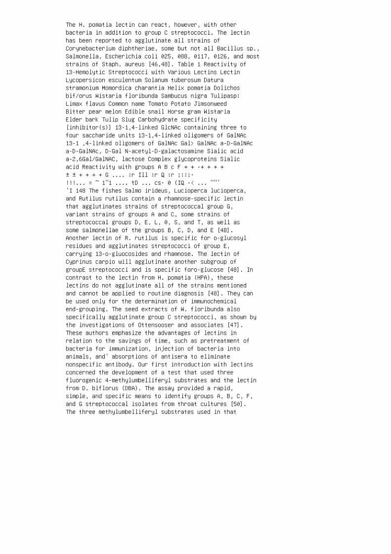
The H. pomatia lectin can react, however, with otherbacteria in addition to group C streptococci. The lectinhas been reported to agglutinate all strains ofCorynebacterium diphtheriae, some but not all Bacillus sp.,Salmonella, Escherichia coli 025, 088, 0117, 0126, and moststrains of Staph. aureus [46,48]. Table 1 Reactivity of13-Hemolytic Streptococci with Various Lectins LectinLycopersicon esculentum Solanum tuberosum Daturastramonium Momordica charantia Helix pomatia Dolichosbif/orus Wistaria floribunda Sambucus nigra Tulipasp:Limax flavus Common name Tomato Potato JimsonweedBitter pear melon Edible snail Horse gram WistariaElder bark Tulip Slug Carbohydrate specificity[inhibitor(s)] 13-1,4-linked GlcNAc containing three tofour saccharide units 13-1,4-linked oligomers of GalNAc13-1 ,4-linked oligomers of GalNAc Gal> GalNAc a-D-GalNAca-D-GalNAc, D-Gal N-acetyl-D-galactosamine Sialic acida-2,6Gal/GalNAC, lactose Complex glycoproteins Sialicacid Reactivity with groups A B c F + + -+ + + +± ± + + + + G .... :r Ill :r Q :r ;::;·!!!... = ~ 1"1 .... tD ... cs· 0 (IQ -< ... ""''I 148 The fishes Salmo irideus, Lucioperca lucioperca,and Rutilus rutilus contain a rhamnose-specific lectinthat agglutinates strains of streptococcal group G,variant strains of groups A and C, some strains ofstreptococcal groups D, E, L, 0, S, and T, as well assome salmonellae of the groups B, C, D, and E [48].Another lectin of R. rutilus is specific for o-glucosylresidues and agglutinates streptococci of group E,carrying 13-o-gluocosides and rhamnose. The lectin ofCyprinus carpio will agglutinate another subgroup ofgroupE streptococci and is specific foro-glucose [48]. Incontrast to the lectin from H. pomatia (HPA), theselectins do not agglutinate all of the strains mentionedand cannot be applied to routine diagnosis [48]. They canbe used only for the determination of immunochemicalend-grouping. The seed extracts of W. floribunda alsospecifically agglutinate group C streptococci, as shown bythe investigations of Ottensooser and associates [47].These authors emphasize the advantages of lectins inrelation to the savings of time, such as pretreatment ofbacteria for immunization, injection of bacteria intoanimals, and' absorptions of antisera to eliminatenonspecific antibody. Our first introduction with lectinsconcerned the development of a test that used threefluorogenic 4-methylumbelliferyl substrates and the lectinfrom D. biflorus (DBA). The assay provided a rapid,simple, and specific means to identify groups A, B, C, F,and G streptococcal isolates from throat cultures [50].The three methylumbelliferyl substrates used in that

investigation provided a means to accurately identifygroups A, B, and F 13-hemolytic streptococci isolates fromthroat cultures. Some strains of group C and Gstreptococci, however, could not be differentiated becauseof overlapping 4-methylumbelliferyl substrate patterns. Theuse of soluble preparation of the lectin DBA as anagglutination reagent provided a rapid and specificagglutination response when mixed on a microscope slidewith five or more colonies of group C organisms. Resultswere obtained within 30 sec of a hand rotation on amicroscope slide. No agglutination was detected with groupG or with groups A, B, or F streptococcal cells. The useof the combination of the methylumbelliferyl substrates andthe lectin reagent provided a nonserological method thatpermitted accurate identification of isolated colonies of13-hemolytic streptococci obtained from throat cultures.The methylumbelliferyl-lectin protocol was later applied byother investigators for the identification of streptococciisolated from cows with mastitis [51]. As with theprevious investigations [50], the use of the lectin D.biflorus was required to identify and differentiate Strep.dysgalactiae (group C) from the other groupablestreptococci. The demonstration of species-specific enzymeactivities of the streptococci with various4-methylumbelliferyl-conjugated substrates in combinationwith the lectin agglutination
Lectins in Clinical Bacteriology 149
permitted differentiation within 1-2 hr at 37°C. This is incontrast with the
standard identification techniques of these serogroupswith biochemical
tests that require an incubation period of 4-24 hr. Theseinvestigations led to the development of tests employinglectins
as diagnostic tools for the identification of variousserogroups of the (3
hemolytic streptococci. Thus, it was later reported thatcrude extracts of
DBA, when coupled to polystyrene particles with a spacearm, yielded
an effective lectin-latex microsphere reagent thatspecifically agglutinated

group C streptococcal antigens prepared as nitrous acid,autoclaved, or
enzyme extracts. The coated beads could also agglutinatesuspensions of
isolated colonies [52]. This latex-lectin preparation couldbe used to replace
latex microspheres conjugated to antibody to group Cstreptococci for use
in test kits for the identification of (3-hemolyticstreptococci from isolated
colonies. Many lectins are extremely stable at ambienttemperatures. The lectin
of W. floribunda was absorbed onto polystyrene latexmicrospheres that
were stained blue. Fifty microliters of these lectinconjugated microspheres
were placed on cardboard slides and dried overnight atambient tempera
ture. The dried lectin reagent cards were mixed with 50ILl of phosphate
buffer (pH 7 .2) at various times and tested with nitrousacid extracts of
group C streptococci. Strong and highly specificagglutination responses
were observed with the polystyrene lectin reagent dried oncards 7 months
previously. No reactions were observed with group Astreptococci or with
other clinically significant (3-hemolytic streptococci(Fig. 1). The use of HP A was used as a membraneparticle-capture assay to
detect nitrous acid-extracted group C streptococci. TheHPA lectin-latex

microsphere reagent was spotted onto a porous membrane anddried.
Group C streptococcal nitrous acid extract was added ontoa lectin-capture
substrate and incubated for 5 min at room temperature.Peroxidase labeled
HP A was then added to sandwich the analyte, and colordetection proceeded,
as with an enzyme-linked immunosorbent assay (ELISA) format(Fig. 2). Holm and associates have shown that blocking thesialic acid of the
type-specific polysaccharide of a type III strain of groupB streptococcal
cells by the sialic acid-specific lectin from the snailCepaea hortensis, leads
to the promotion of phagocytosis of a group Bstreptococcus of type Ia
strain [53]. The effect of the lectin was dose-dependentand required the
presence of complement. The sialic acid-specific lectinfrom C. hortensis
and C. nemoralis agglutinates all group B streptococcalstrains that contain
a type-specific polysaccharide [49,54]. However, group Bstreptococci that
lack a type-specific polysaccharide, as well as strains ofother streptococcal
groups and many other bacteria, including speciescontaining sialic acid,
are not agglutinated [53]. 150 Slifkin Figure 1Reactivity of dried polystyrene latex microspheresconjugated to Wisteria floribunda for a nitrous acidextract of group C streptococci. The microspheres havebeen dried on a cardboard card. The microspheres on thebottom left of the card have been mixed with a nitrousacid extract of group A streptococci and show no

agglutination, whereas the microspheres on the bottomright have agglutinated in the presence of group Cstreptococcal extract. A relatively wide spectrum oflectins were demonstrated to agglutinate various serotypesof group B streptococci [55] . In some instances treatmentof the bacterial cells with sialidase removed thereactivity of various lectins with those cells. In otherinstances, agglutination with lectins occurred only afterenzyme treatment of the bacterial cells. Group Bstreptococci can be specifically agglutinated or labeledwith fluorescein-conjugated lectins (Fig. 3) derived fromcertain plants of the Solanaceae family [56]. The pulplectin from the tomato, Lycopersicon esculentum (LYE),as well as the related plant, the potato, Solanum tuberosum(ST A), can be passively coupled to amide-modifiedpolystyrene microspheres [56]. These reagents could be usedfor the specific identification of group B streptococcigrown in selective Todd-Hewitt broth, such as Lim's broth,as well as nonselective broth [57]. These lectins do notagglutinate the other clinically significant serogroupablestreptococci. The lectins could 151
possibly be of value for their ability to assist in theidentification of group
B streptococcal colonization in women and infants. Thesetwo lectins have
an affinity, in part, for N-acetylglucosamine, the majorgroup-specific
polysaccharide for group B streptococci. Either the lectinof the tomato (LYE) or potato (ST A) conjugated to
latex particles will agglutinate group B streptococci.Particulate organisms,
as well as nitrous acid, autoclaved, or enzyme extractswere agglutinated or
precipitated, respectively, by STA. These streptococci,like those of group
A and group C streptococci, contain relatively high amountsof N-acetyl-o
glucosamine, compared with members of groups D, F, or G[58]. The
unique presence of 1 ,4-anhydroglucitol in the cells of

group B streptococci
[59] may, in part, be related to the affinity of the tomatoand the potato
lectins for group B streptococci. It has been shown thattomato lectin binds
oligosaccharides containing the repeating disaccharide({j-1 ,4-GlcNAc-{j),
or poly-N-acetyllactosamine sequence, requiring at leastthree repeating
disaccharide units for interaction [60]. Although notprecisely defined, the relevant antigenic determinants of
group B polysaccharide appear to be associated with therhamnose-glucitol
Figure 2 Microsphere-enzymatic procedure using H. pomatialectin as a capture
reagent for a nitrous acid extract of group Cstreptococci. The microparticle-capture
membrane on the left did not indicate a positive reactionwhen group A nitrous
acid extract was added to the membrane. A positiveenzymatic response is seen on
the membrane on the right when group C nitrous acid extractwas added. figure 3 Fluorescent response of group Bstreptococci labeled with fluoresceinconjugated L.escu/entum. units or terminal rhamnose on side chainsassociated with N-acetylglucosamine [59]. It is possiblethat the tomato and potato lectin can interact with othersubstrates associated with group B streptococci. The lectinfrom the garden snail Cepaca hortensis has a specificityfor sialic acid-containing polysaccharides and can reactwith group B streptococci [53]. Various typeable andnontypeable strains of group B streptococci were examinedto determine their respective reactivity to LYE coupled topolystyrene spheres (Table 2). All the strains grown ineither Todd-Hewitt or Lim's broth reacted with the lectinreagent. Nitrous acid, Streptomyces a/bus lysozymeenzyme, and autoclaved extracts of group B streptococcalcells yielded results similar to those with particulategroup B streptococcal cells grown in these culture broths.

None of the type 1b strains grown on Columbia sheep bloodagar, however, reacted with the LYE-conjugatedmicrospheres. Observation of medium-dependentagglutination reactions have been previously reported withLegionella-lectin interactions [61]. The latterinvestigators [61] emphasized that when lectins areemployed as diagnostic reagents, growth conditions must becarefully standardized. Fluorescein-conjugated LYE labeledall the group B streptococcal cells grown from the bloodagar medium. The serotype 1 b strains, however, did notfluoresce as brightly as did the other group B serotypes.No fluorescence or agglutination response were observedwhen the other serogroups of 153
streptococci were examined. The lectin from the solanaceousplant Datura
stramonium did not agglutinate group B streptococci northe other sera
groups tested. Datura stramonium, similar to LYE and STA,is a member of the
Solanaceae family of plants. This lectin, like that of thetomato and potato
lectins, is specific for A, B, and 0 erythrocytes andbinds to chitooligosac
charides [60]. The lack of group B streptococcal activitywith D. stramo
nium may be related to the inability of relatively smallconcentrations of
N-acetyl-o-glucosamine to block the agglutination of humanerythrocytes
with this lectin, or to its lack of reactivity with dimersor trimers of N
acetyl-o-glucosamine [60]. It was previously reported thatvarious commercially available serogro
uping reagents cross-reacted with the Todd-Hewitt brothmanufactured by
Difco Laboratories, but not with that manufactured byBaltimore Biologi

cal Laboratories [62]. The tomato or potato lectins do notprecipitate in
inoculated Todd-Hewitt broth of either manufacturer.Three elderberry species contain two identicalcarbohydrate-binding
sites per molecule and exhibit a very high specificity forthe Neu5Aca-2,6
Gal/GalNAc sequence [63-65]. Relative affinities forvarious oligosaccha
rides were significantly different among them, suggestingdifferences in the
detailed structure of the carbohydrate-binding sites ofthese related lectins.
Previous to these reports, no plant lectin had beenreported to bind specifi
Table 2 Agglutination of Group B Streptococci withLycopersicon esculen
tum or Solanum tuberosum Lectin Coupled to PolystyreneSpheres Agglutination-response (no. agglutinated/no.tested) No. Columbia sheep Todd-Hewitt Lim's
Organisms tested blood agar broth broth
Nontypeable 2 0/2 2/2 2/2
Ia 4 3/4 4/4 4/4
lb 4 0/4 4/4 4/4
Ic (la/c) 4 4/4 4/4 4/4
II 4 4/4 4/4 4/4
III 4 4/4 4/4 4/4
Group A 28 0/28 0/28 0/28
Group C 20 0/20 0/20 0/20
Group F 20 0/20 0/20 0/20
Group G 20 0/20 0/20 0/20 cally to sialic acid or to

oligosaccharide units that included this carbohydrateresidue [66]. Sialic acids represent about 30 derivativesof N-acetylor N-glycolylneuraminic acids [67]. Varioussialic acid derivatives exhibit interesting speciesandtissue-specific distribution. Sialic acids play animportant role as receptors for viruses [68] as well asin the social behavior of cells. They also act as maskingagents on antigens, receptors, and other recognition sitesof the cell surface [16]. There is a relatively smallselection of lectins that specifically bind to sialicacids [67]. The lectin isolated from elderberry, Sambucusnigra (SNA), binds the sequence NeuNAca-2,6-o-Gal/o-GalGalNAc with high specificity [63]. Thislectin has a relatively weak affinity for o-galactose oro-GalNAc, but does not bind to either NeuNAc or NeuNGuc.It has a high affinity for terminal sialic acid that islinked a~2,6to o-galactose, compared with the a-2-3-linkedisomer [63]. On the basis of inhibitions with simplesugars, the elderberry bark lectin was demonstrated to bespecific foro-galactose [63]. The lectin from the tulip(Tulipa; TUG) is derived from the bulb of the plant, amember of the family Liliaceae [69]. This lectin wasreported to be the first lectin to be isolated from thisplant family and is the first example of aphytohemagglutinin obtained from a monocotyledonous speciesother than the Graminaeae family associated with wheat,rye, barley, and rice [69]. Two lectins with differentagglutinating affinities are associated with T. gesnerianabulbs [70]. One TUG lectin component will agglutinatetrypsinized human or rabbit erythrocytes [69], but willnot agglutinate nontreated human red blood cells [69]. Asecond TUG lectin was previously shown to agglutinate thecells of the Saccharomyces genus at a concentration of15-30 ~-tllml, but did not agglutinate various species ofCandida, E. coli, Staph. aureus or B. subtilis [71]. Thelack of agglutination response of group A streptococcalextracts with TUG-bound latex particles appears toindicate that the lectin substrates may be either denaturedor not extractable. The group A streptococcal extracts, incontrast, agglutinated latex particles bound with antibodythat is directed to the specific group antigen or"C-substance" reported to be N-acetylglucosamine [58]. Inthe literature, the tulip lectin has been reported not tohave an affinity for this glycoprotein [69,71]. Accordingto one investigation, o-mannose, o-mannose-6-phosphate,L-fucose, and L-fucosylamine were demonstrated to be potentinhibitors of this lectin in the binding to Saccharomycescerevisiae cells [70]. In a hapten-inhibitioninvestigation, agglutination of human erythrocytes withTUG lectin was inhibited by N-acetylgalactosamine, lactose,

fucose, and galactose [69]. 155 It has been determinedthat the TUG lectin derived from the bulb, or
the lectin from the bark of Sambucus nigra, the elderberry,will agglutinate
both group A and C streptococci (see Table 1).Agglutination of group A
streptococcal organisms was affected with tulip lectin at aconcentration of
0.3 mg/ml on latex spheres. All of the 40 stains of group Astreptococci
grown on Columbia sheep blood agar agglutinated with thislectin reagent
in 15-30 sec by manually rocking the reactants on a glassmicroscope slide.
Within this period, the reactions appeared at anagglutination reactivity
strength of 2-3 + (Fig. 4). The tulip lectin reagentagglutinated 12 of the 40 (300Jo) test strains of
the group C streptococci. These relatively weakeragglutination responses,
however, occurred after 30-60 sec of manual rocking of thereactants. The
reactions of these group C strains produced only 1 +agglutination re
sponses within this observation period. All 40 strains ofgroup A strepto
cocci were also agglutinated in the presence ofmicrospheres prepared with
0.01 mg of lectin per milliliter. These 2-3 + agglutinationreactions were
observed within 30-60 sec of manual rocking of thereactants on micro
scope slides. Furthermore, at this lower concentration oftulip lectin, none

Figure 4 Agglutination of group A streptococci with Tulipalectin conjugated on
microspheres. The lectin-conjugated particles on the leftwere mixed with group C
streptococci. The right side shows the lectin-microphaseand group A streptococci. 156 of the group Cstreptococcal strains agglutinated within 60 sec ofobservation. Conjugation of the TUG lectin onto 0.2-J.'mpolystrene microspheres also yields agglutination withgroup A streptococci and none with the other clinicallysignificant serogroups. In contrast, the use of largermicrospheres effected no differentiation between group Aor C streptococci (Table 3). None of the strains of groupB, F, or G streptococci agglutinated in the presence ofthe TUG-latex reagent. The use of different lots ofColumbia sheep blood agar yielded the same agglutinationpatterns with these streptococci. Nitrous acid, enzyme,autoclave, or sonic-derived extracts of the various13-hemolytic streptococcal serogroups did not produceresponses when tested with the TUG-latex reagent. Theseextracts, however, did produce a specific agglutinationresponse when mixed with their respective commerciallyprepared serodiagnostic-latex reagent. Inhibition studieswere performed to determine the effects of various sugarsand glycoprotein inhibitors on the reactivity of TUGlectin, with either group A or group C streptococci. Thisinhibition protocol was performed by preparing twofoldserial dilutions of various sugars and glycoproteins inphosphate buffer, as shown in Table 4. These preparationswere respectively added to 0.1 ml of TUG-latex reagent.The latter contained 30 1-'g of lectin per milliliter. Thelectin and inhibitors were mixed on a glass microscopeslide with a wooden applicator stick for 10 sec. Four tosix colonies of group A or group C streptococci wereremoved from a sheep blood agar plate with a woodenapplicator stick and mixed into the lectinlatex inhibitormixture for 30 sec. Four to six colonies of group Astreptococci were also mixed into 0.1 ml of TUG-latexreagent that did not contain any potential inhibitorsolution to serve as a control. A determination of Table3 Effect of Microsphere Size on Specificity and ReactionTime with Interaction of Tulip Lectin and Group A andGroup C Streptococci Reaction time (sec) Latex particleSerogroupA Serogroup C size {J£m) 30 60 90 30 60 90 0.73+" 3+ 3+ 1+ 1+ 2+ 0.5 2+ 3+ 3+ 1+ 1+ 2+ 0.3 2+ 2+ 3+ 1+0.2 1+ 2+ 3+ "Strength of reaction: -, no agglutination;1 + , many fine clumps; 2 + , a few moderate-sized clumps;

3 +, many moderate-sized clumps. 157
Table 4 Inhibitory Effect of VariousCarbohydrate-Glycoproteins on the Aggluti
nation of Tulip Lectin to Groups A and C Streptococci
Carbohydrate/ glycoprotein
Asialofetuin
Fetuin
N-Acetylmuramic acid
N-Acetylneuraminic acid
N-Acetyl-o-galactosamine
Methyl-{j-o-galactopyranoside
Lactose
Mannose
Fucose
Group C streptococcal nitrous acid extract Minimalconcentration required for inhibition of tulip-mediatedagglutination of group A streptococci (mM)" 0.2 0.53.0 1.5 NI NI NI NI NI NI
"NI, no inhibition at concentrations less than 200 mM.
agglutination was made after manually rocking the slide for1 min. The
inhibition studies demonstrated the various sialoproteinsare effective as
inhibitors of agglutination by TUG-microsphere reagent,with either group
A or group C streptococci (see Table 4). In contrast,various simple sugars
or oligosaccharides do not inhibit this agglutinationresponse. Groups A
and C streptococci have been reported to contain slightly
higher concentra
tions of the sialoprotein and N-acetylmuramic acid thangroups B, D, F, or
G streptococci [58]. Furthermore, the lectin ofLimaxflavus, which is very
specific for sialic acid [72], will also agglutinate onlygroups A and C
streptococci (see Table 1). Accordingly, there appears tobe no interaction
of TUG lectin for either N-acetylglucosamine orN-acetylsialolactosamine
(see Table 4). Thus, group A streptococcal binding sitesassociated with tulip lectin
appear not to be entirely related to the polysaccharidesestablished as the
respective group-specific antigens [58]. Therefore, onemay consider that
these binding sites may be associated, in part, with thesegroup-specific
polysaccharides as well as other substrates. All lectinmolecules are multiva
lent and have two or more carbohydrate binding sites[8,17]. However,
binding of lectins cannot always be fully correlated withtheir reported
biochemical specificities [6,13,74]. An extensivedetermination of all poten
tial inhibitors may not always be possible. Lectin affinitymay also be partly
related to the method used to purify the lectin [7]. 158A similar association of binding of groups A and Cstreptococci was reported with wheat germ agglutinin (WGA)[75]. This lectin has an affinity forN-acetyl-o-glucosamine and its derivatives [76]. The wheatgerm lectin was demonstrated to agglutinate all strains of

groups A and C streptococci tested [75]. Some strains ofgroups B, E, H, L, 0, and S streptococci also agglutinatein the presence of this lectin. As with the tulip andSambucus nigra lectins, the binding of groups A and Cstreptococci may not depend on a terminal position ofGlcNAc [75]. Primary colonies of group A streptococci canbe accurately identified with the use of tulip lectin asa lectin-bound latex reagent. The tulip lectinreagent thuscan be applied as an alternative method for the rapididentification of group A streptococcal colonies. Thelectin provides a relatively cost-effective means toidentify colonies of group A streptococcal isolates. Theuse of the tulip lectin, similar to the previouslyreported D. bif/orus lectin [52], affords the availabilityof a diagnostic reagent that can be prepared withrelatively less complexity than the production of anantibody for serogrouping procedures. Table 1 shows asummary of some of investigations on the interactions ofvarious lectins with serogroups A, B, C, F, and G{j-hemolytic streptococci. Kohler and Nagai [48] havereported that some {j-hemolytic strains of Strep.anginosus (Strep. millen) can react with HPA. They observedthat not all strains reacting with group-specific anti-Cantiserum were agglutinated with HPA. It was suggestedthat N-acetyl-o-galactosamine is not in the terminalposition. The agglutination by the HP A by nongroup CStrep. milleri strains also indicated that other antigensmay be involved. All eight strains of group C-associatedStrep. anginosus strains examined reacted with H. pomatiabound to polystyrene spheres (Table 5). This lectinreagent did not agglutinate group A, group F, ornongroupable Strep. anginosus strains. These reactionswere similar when either nitrous acid extracts ornonextracted cells were used. The TUG lectin reacted withall eight strains of group A Strep. anginosus and 250Joof the group C streptoTable 5 Interaction of VariousLectins with Streptococcus anginosus Lectin TulipaSambucus nigra Helix pomatia A 8/8 6/8 0/8 Serogroupof S. anginosus (no. tested/no. reactive) c FNongroupable 2/8 7/8 8/8 0/6 0/6 0/6 0/4 0/4 0/4
coccal strains. No reactivity was observed with the group For nongroup
able strains. The SNA-latex reagent reacted with most ofthe group A and
C strains of Strep. anginosus tested. This reactivity fornon-Strep. angino

sus group C streptococci on the SNA lectin was notpreviously observed
[48], which suggests the presence of other SNA-bindingsubstrates on the
Strep. anginosus strains. The lectin from Misgurnusanguillicaudatus has a relatively strong af
finity for rhamnose [77]. This lectin, therefore, may be ofvalue in the
identification of streptococci [22].
D. Listeria
Recently, Sonnenfeld and Doyle [see 18] reported that therelatively com
mon serotypes of L. monocytogenes could be differentiatedwith the use of
a panel of lectins. Four lectins, including the applicationof a lectinlike seed
extract (Persea americana), were demonstrated to be usefulin distinguish
ing the major serotypes of L. monocytogenes. The lectinsincluded in the
panel were Griffonia simplicifolia, Vicia villosa, HPA,and ConA. The
agglutination procedure required that the L. monocytogenesisolates be
heated to expose the lecting-binding sites of theorganisms.
Ill. USE OF LECTINS FOR GRAM-NEGATIVE BACTERIA
A. Enterobacteriaceae
Concanavalin A agglutinates a variety of gram-negativebacteria. The bind
ing site of this lectin appears to be related to thecarbohydrate moieties on

the lipopolysaccharide molecule [74]. Polysaccharides ofvarious serotypes
of Salmonella precipitate with ConA [78], and it can beapplied in micro
scopic slide agglutination tests [79]. This lectin has beenreported to react
with Salmonella strains containing the 0 : 1 factor, andit will also bind to
E. coli [80]. This lectin, therefore, could be employed toclassify lipopoly
saccharides derived from Salmonella, although notalllipopolysaccharides
presumed to contain a-o-glucosyl side chains form aprecipitate with ConA.
No precipitate was observed when ConA was reacted with thelipopolysac
charide from Sal. typhi [81]. The polysaccharide isreported to contain side
chains similar to Sal. typhimurium [78] with which faintprecipitation was
detected [78]. Concanavalin A was reported to precipitatelipopolysaccharide ex
tracts obtained from Shigella flexneri and Sal.abortivoequina, but not Sal.
enteritidis [81]. Yersinia entercolitica strains,possessing the antigenic factor
0: 12 are agglutinated with ConA [79]. In the genusErwinia, the strains of 160 Slifkin E. amylovora willagglutinate in the presence of ConA in contrast with E.caratovora and E. chrysanthemi [79]. Concanavalin Aconjugated with fluorescein did not react with Pseudomonasaeruginosa, Haemophilus influenzae, or Proteus mirabilisstrains [82]. The application of lectins for theidentification of bacteria commonly associated with ocularinfections has been demonstrated. Some lectinbindingpatterns were reported to be specific for the bacterialpathogens examined [82]. Litchi chinensis lectin was

demonstrated to be applicable for the identification ofmany 13-hemolytic strains of E. coli and of Proteus sp.isolates [83]. The H. pomatia lectin failed toprecipitate the lipopolysaccharide derived from Sal.typhimurium rough mutants of type Ra and Rb, but wassomewhat reactive with an SR mutant [84]. The H. pomatialectin [48] and the lectin from the horseshoe crab Limuluspolyphemus [85] also have been reported to agglutinateE. coli and other members of the Enterobacteriaceae.Salmonella minnesota cells were agglutinated with wholeLimulus serum, but did not agglutinate in the presence inL. polyphemus serum purified by affinity column procedure[86]. The sialic acid-binding lectin from the horseshoecrab Carcinoscorpius rotunda cauda, also known ascarcinoscorpin, will agglutinate E. coli K12 and Sal.minnesota R595 cells [86]. Vibrio cholerae does notagglutinate in the presence of this lectin. B.Pseudomonas A lectin obtained from the potato was reportedto distinguish strains of P. solanacearum [87]. Strainsof this bacterial species that are pathogenic for thepotato do not react with this lectin, whereas avirulentstrains of this species are agglutinated. Theinvestigators determined that an extracellularpolysaccharide, associated with virulent strains, appearsto inhibit the reactivity of the lectin with its ligandsite on a lipopolysaccharide. Sixteen serotypes of P.aeruginosa were typed with a panel of lectins [88]. Allserogroups were highly agglutinated by the lectin ofMoringa olifera seeds, except serotype H7. Differentiationof the various serotypes was accomplished withagglutination (and precipitation) tests when a panel oflectins derived from M. olifera, Artocarpus integrifolia,and Artocarpus lakoocha and lipopolysaccharide extractsof various serotypes of P. aeruginosa were used. Previousinvestigations on 9 strains of P. cepacia with arelatively large panel of lectins indicated aheterogeneity of lectin-binding sites [89]. All 161
strains did not react with the lima bean lectin. The latterresearch demon
strated that this lectin interacted only with P. cepacia,suggesting that this
lectin may be species-specific. The investigatorsrecommended that more
pseudomonad isolates be examined to confirm thisobservation.

C. Legione/la
A panel of plant lectins gave differential reactivitieswith many of six sera
groups of L. pneumophila tested [61]. The agglutinationpatterns appeared
not to be related to the serogroup of these bacteria, as itwas determined
that five of the strains of serotype 1 had uniqueagglutination patterns.
These investigations suggested that the cell surfaces ofLegione/laceae do
not contain carbohydrate groups that are accessible forbinding by lectins.
Some of these bacteria were agglutinated by lectinlikesubstances from
Persea americana, Mangijera indica, Aloe arborescens, andAlbizzia juli
brissin. These plant extracts are associated withpolyphenol derivatives,
tannins, and may be capable of binding cell surfaces [90].
D. Neisseria and Related Bacteria
Among the gram-negative bacteria, the application oflectins as diagnostic
reagents is especially associated with the members of thegenus Neisseria.
The use of wheat germ lectin to agglutinate strains of N.gonorrhoeae has
been reported [91-92]. Nonencapsulated N. meningitidis andN. gonor
rhoeae will also agglutinate in the presence of thislectin, whereas encapsu
lated N. meningitidis will not [93]. This observationsuggested that gono

coccal agglutination with wheat germ lectin may be relatedto the lack of
capsular material on the gonococci. Other investigationsalso have shown
that wheat germ lectin is of use for the identification ofN. gonorrhoeae
[94]. From an panel of 14 lectins, all strains of thegonococcal isolates
examined were agglutinated by wheat germ, soybean, andricin lectins [95].
None of the differential patterns of gonococciagglutination by lectins was
related to GC serogroups. The authors concluded that GCserogroup anti
genic determinants involve portions of sugar structurethat are larger than,
or separate from, those recognized by lectin-combiningsites. It was sug
gested that agglutination by these lectins is mediated bycell envelope lipo
polysaccharides, and this interaction involved elements ofcommon lipo
polysaccharide core structures. A panel of 22 lectins wasused to demon
strate the diagnostic effect of lectin on theidentification of the family
Neisseriaceae [96]. DeHormaeche et al. [97] determinedthat the lipopolysaccharide of
N. gonorrhoeae, but not nonpathogenic neisseriae, containedan epitope
consisting of N-acetylglucosamine andN-acetylgalactosamine, and was re162 active with wheatgerm agglutinin. Other investigators, using a battery oflectins, have suggested that the lipopolysaccharides from

pyocin-sensitive and pyocin-resistant strains of N.gonorrhoeae possesses terminal GlcNAc or Gal(or GalNAc)residues [95,98]. The use of wheat germ agglutinin forthe identification of N. gonorrhoeae was associated withnonspecific agglutination [94] and autoagglutination ofgonococcal suspensions [96]. Doyle et al. [96] employedwheat germ and soybean agglutinins in conjunction with aa-glutamylaminopeptidase assay. All of the gonococcalstrains and lectin-reactive meningococci weredistinguishable by the hydrolysis ofa-glutamyl-13-naphythylamide by the former. This assay isapplicable for distinguishing Moraxella cat arrha/is, N.lactamica, and N. meningitidis. The autoagglutinationphenomenon was overcome by DNase treatment of thebacterial cells. Other investigators used both wheat germand soybean agglutinins in conjunction with variouschromogenic substrates [99]. They identified N.gonorrhoeae and differentiated it from other Neisseria sp.examined. Recently, an investigation of 140 strains of N.gonorrhoeae has shown that 4.90Jo of the strains are notagglutinated with WGA [100] as previously reported [99].The use of ten lectins, however, yielded discriminatinggroups of serotypes lA and lB and prototroph andproline-dependent auxotypes [100]. This method of typingwas reproducible and may be considered for epidemiologicalapplications. E. Campy/obacter (Helicobacter) In a recentinvestigation, it was determined that C. fetus strainslacking surface array proteins with type Alipopolysaccharide were agglutinated with Griffoniasimplicifolia II, H. pomatia, and wheat germ lectins. Noagglutination was observed with C. fetus mutants thatlacked surface array proteins with type B or type Clipopolysaccharide or strains containing a surface arrayprotein layer [101]. The authors concluded that knowledgeof the inhibition of lectin-binding to lipopolysaccharideby the surface array proteins will aid in thecharacterization of the lipopolysaccharide interaction. Inanother investigation, various serotypes of C. jejuni andC. coli displayed differential reactivities withgalactose-binding lectins, the reactions of which appearedto be strain-specific [102]. Other investigators have usedlectins for the differentiation of various Campylobactersp. Strains of C. jejuni and C. coli interact specificallywith certain lectins and blood group antibodies [103].Agglutination by lectins was also reported effective forthe differentiation of C. jejuni and C. coli [104]. Asimple and effective agglutination procedure using a panelof lectins was developed for differentiating strains ofvarious species of 163

Campylobacter [104]. All strains tested were agglutinatedby the protein
reactive agglutinins of Mangifera indica (mango) andPersea americana
(avocado). A large number of the Campylobacter strains alsoagglutinated
in the presence of ConA and Triticum vulgaris. Thereactions of the other
lectins used in the investigation varied. The data impliedthat lectins could
be used in a supplementary procedure for fingerprintingindividual isolates.
IV. USE OF LECTINS FOR MYCOBACTERIA
Fluorescein-conjugated ConA was effective in thevisualization of Myco
bacterium fortuitum and M. chelonei in both pure cultureand experimental
keratitis samples from corneal scrapings [105]. Thislectin yields only mini
mal background staining of corneal tissue [106].Concanavalin A binds to
nonreducing terminal o-arabinofuranosyl residues associatedwith the end
chain of arabinogalactan of the cell wall of M. bovis [107].
V. EPIDEMIOLOGICAL APPLICATIONS
Applications of lectins for epidemiological investigationsand strain charac
terization of Haemophilus ducreyi [108], Campy/obacterjejuni [103], and
N. gonorrhoeae [109,110] have been reported. Theseinvestigations have
been applied in certain instances for clinical studies to

differentiate between
treatment failure and reinfection; to study transmission,and to investigate
the presence of subtypes of various pathogens. In-depthcoverage of this
topic is found in Chapter 3.
VI. CONCLUSIONS
The selection of a lectin as a potential diagnostic reagentfor use in bacteri
ology first requires that a reasonable protocol befollowed. Thus, the selec
tion of a lectin or a panel of lectins may be based on theaffinity of these
lectins for a particular marker for a given bacterialspecies. The perfect
example is clearly seen with group C streptococci and thereactivity of
those lectins for N-acetylgalactosamine. This relation oflectin with group C
streptococci yields a very effective means to discriminateone serogroup
from other clinically significant serogroups of~-hemolytic streptococci.
This group of streptococci contains relatively largeramounts of GalNAc in
comparison with that of other serogroups. The reactivityof the lectins that
bind with this carbohydrate moiety is not specific,however, for only this
one serogroup. These lectins can also bind to othergram-positive and gram
negative bacteria. Thus, the observation of characteristicgram-positive

morphology, catalase reactivity, and hemolytic responseprovides the 164 primary guidelines that establishes theuse of these lectins for ?-hemo? lytic streptococci.Without these first-stage guidelines, a lectin, such asthat of Helix pomatia, that differentiates group Cstreptococci from other ~-hemolytic streptococci can alsointeract, for example, with Staph. aureus [31]. On theother hand, the determination that LFA and TUG arerelatively effective for their respective binding withgroup B and group A streptococci is not based on thereactivity of these lectins for the antigenic determinantsgenerally associated with the serogroups. Instead,reactivity of these lectins for the serogroups does notappear to be for the N-acetylglucosamine determinants,but possibly for sialic acid residues as well as othersubstrates. Furthermore, chemical modification of thelectin, the lectin concentration, as well as the testassay system are some of the factors that will influencethe potential for a lectin to be used as a diagnosticprobe. Relative to microsphere size, it was recentlyreported that the use of relatively small-diameterpolystyrene particles will decrease the sensitivity of alatex agglutination test [111]. For tulip lectin, however,small microspheres yield a highly specific response togroup A streptococcal organisms. This, in part, may be dueto possible differences in the sialopolysaccharideconcentration associated with groups A and C streptococci.Given the reported observations of lectin interactionswith various bacteria, the most convenient means oftesting lectins as a diagnostic tool is generallyassociated with the application of isolated bacterialcolonies. The direct detection of a particular bacterialagent in a clinical specimen with the use of a lectinassay may be difficult to attain. This is due to thepotential for a relatively wide variety of carbohydratesubstrates to be available for lectin interaction in agiven clinical specimen. Inoculation of clinical specimensonto tissue culture cells has permitted a specific andsensitive means to detect herpes simplex virus with useof fluorescein-conjugated H. pomatia lectin [112]. Theusefulness of lectins in clinical microbiology isassociated with an extremely broad spectrum of advantagesthat render them of value in routine diagnostic procedures.Many of these diagnostic procedures associated withlectins are relatively inexpensive and can be rapidlycompleted. Lectins can also be used for light and electronmicroscopic investigations [113]. Some of the significantadvantages are (1) their stability, (2) their activity invery small concentrations, (3) the commercial availability

of many leetins, and (4) their ability to probe subtlesurface structural differences between various isolates[22]. The cost-effectiveness of these unique naturallyoccurring cytochemical-histochemical tools furtheremphasizes their value as diagnostic reagents for theclinical microbiologist. It was previously suggested thatbecause of the binding specificity of 165
lectins, they may be able to replace immune sera indetecting various carbo
hydrate residues on microbial cells and, by extrapolation,particular micro
organisms [19]. This chapter has emphasized that lectinsare versatile re
agents that can be applied in certain instances to (1)provide definitive
identification, (2) characterize a strain of a particularorganism, and (3)
function as epidemiological markers. The effect ofstandard methodology and contemporary emphasis on
molecular biology technology associated with monoclonalantibodies and
nucleic acid probes has, in part, diverted attention fromthe potential of
lectins in applied and clinical microbiology. Furthermore,the unique carbo
hydrate specificities of lectins in comparison with thespecificity attained
with the use of monoclonal antibodies or nucleic acidprobes may also be a
factor considered by various investigators. In contrast, anexamination of
an increase in patented processes on diagnostic uses oflectins for the last 5
years, clearly indicate a significant increase in the useof lectins in scientific

research and in clinical microbiology. The emphasis ofthis chapter is on the application of lectins derived
from plant seeds, pulp, bark, root, bulb, and invertebrateextracts for the
identification and characterization of bacteria. The useof lectins obtained
from certain bacteria may also be of value as diagnostictools for clinical
microbiology. The interaction of lectins from Pseudomonasaeruginosa,
for example, may be of value for the identification ofvarious E. coli strains
[114,115]. The mannosephilic hemagglutinin of P.aeruginosa is reported
to agglutinate the cells of the enteropathogenic E. coli0128: B12 and
086 : B7. The binding of the lectin was shown by specificagglutination of
the E. coli strains by hemagglutination tests [ 114]. Theuse of this bacterial
lectin for the typing of these potential enteropathogenswas considered to
have certain distinct advantages [115]. Although lessspecific than antibod
ies, the bacterial lectin probe can be conjugated with aperoxidase signal
[115]. Furthermore, in the realm of clinical microbiology,certain lectins
may also have antibacterial activity [116], and they havebeen considered as
carriers of chemotherapeutic agents [12]. There are now awide variety of lectins available in unconjugated as
well as conjugated forms from several commercial sources.Furthermore,

panels of lectins under the trade name of Taxonolectinsare commercially
available for the identification and characterization ofbacteria from E-Y
Laboratories, San Mateo, California. The published reviewson the diagnostic applications of lectins in mi
crobiology as well as the commercial availability of arelatively wide source
of lectins should provide ample opportunities to furtherassess the applica
23. Doyle R, Keller K. Lectins in diagnostic microbiology.Eur J Clin Microbial 1984; 3:4-9.
24. Doyle RJ, Birdsell DC. Interaction of concanavalin Awith the cell wall of Bacillus subtilis. J Bacteriol1972;109:652-658.
25. Graham K, Keller K, Ezzell J, Doyle R. Enzyme-linkedlectinosorbent assay (ELLA) for Bacillus anthracis. Eur JClin Microbiol1984; 3:210-212.
26. Cole HB, Ezzell JW, Keller KF, Doyle RJ.Differentiation of Bacillus anthracis and other Bacillusspecies by the use of lectins. J Clin Microbiol1984;19:48-53.
27. Doyle RJ, Keller KF, Ezzell JW. Bacillus In: LennetteEH, Balows A, Hausler JR, Shadomy HD, eds. Manual ofclinical microbiology, 4th ed. Washington, DC: AmericanSociety for Microbiology, 1985:211-215.
28. DeLucca AJ 2nd. Lectin grouping of Bacillusthuringiensis serovars. Can J Microbiol1984;30:1100-1104.
29. Piller V, Piller F, Cartron JP. Comparison of thecarbohydrate-binding specificities of sevenN-acetyl-o-galactosamine-recognizing lectins. Eur J Biochem1990; 191:461-466.
30. Reeder NJ, Ekstedt RD. Study of the interaction ofconcanavalin A with staphylococcal teichoic acids. JImmunol1971; 196:334-340.

31. Hammerstrom S, Kabat EA. Studies on specificity andbinding properties of the blood group A reactivehemagglutinin from Helix pomatia. Biochemistry 1971;10:1684-1692.
32. Owen P, Salton MRJ. Membrane asymmetry and expressionof cell surface antigens of Micrococcus lysodeikticusestablished by crossed immunoelectrophoresis. JBacteriol1977; 132:974-985.
33. Gilbride KJ, Pistole TO. Isolation and characterizationof a bacterial agglutinin in the serum ofLimuluspolyphemus. Prog Clin Bioi Res 1979; 29:525-535.
34. Kloos WE, Jorgensen JH. Staphylococci. In: Balows A,Hausler JR, Shadomy NJ, eds. Manual of clinicalmicrobiology, 4th ed. Washington, DC: American SocietyMicrobiology, 1985:143-153.
35. Wegrzynowicz PB, Hcezko J, Jeljaszewicz J, NeugebauerM, Pulverer G. Pseudocoagulase activity of staphylococci.J Clin Microbiol1979; 9:15-19.
36. Sperber WH, Tatime SR. Interpretation of the tubecoagulase test for identification of Staphylococcus aureus.Appl Microbiol1975; 29:502-505.
37. Davidson SK, Keller KF, Doyle RJ. Differentiation ofcoagulase-positive and coagulase-negative staphylococci bylectins and plant agglutinins. J Clin Microbiol1982;15:547-553.
38. Ellwood DC, Hardie TM, Browning PM, Bowden GH.Carbohydrate composition of cell walls of Streptococcusmutans and Streptococcus sanguis. J Dent Res 1973;52:955.
39. Kashket S, Guilmette KM. Aggregation of oralstreptococci in the presence of concanavalin A. Arch OralBiol1975; 20:375-379.
40. Hamada S, Gill K, Slade HD. Binding of lectins toStreptococcus mutans cells and type-specificpolysaccharides, and effect on adherence. Infect Immun1977; 18:708-716.
41. Staat RH, Langley SD, Doyle RJ. Streptococcus mutansadherence: presumptive evidence for protein-mediatedattachment followed by glucandependent cellularaccumulation. Infect Immun 1980; 27:675-681. 42. Staat RH,

Doyle RJ, Langley SD, Suddick RP. Modification of in vitroadherence of Streptococcus mutans by plant lectins. AdvExp Med Bioi 1978; 107:639-647. 43. Kohler W, Prokop 0.Agglutinatin von streptokokken ver gruppe C durch einagglutinin aus Helix pomatia. Z Immunitaetsforch AllergKlin Immunol 1967; 133:30-53. 44. Kohler W, Prokop 0.Agglutinationsversuche am streptokokken mit demphytogslutinin aus Dolichos bif/orus. Z ImmunitaetsforchAllerg Klin Immunol1967; 133:171-175. 45. Kohler WD,Prokop 0, Kuhnemund 0. Routine identification of group Cstreptococci by means of an agglutinin (protectin) fromthe .albumen gland of the edible snail, Helixpomatia. JMed Microbiol1973; 6:127-130. 46. Prokop D, Kohler W.Agglutinations reactions en von mikroorganiumen mit Helixpomatia eiweiss drusnextract. Z Immunitaetsforsch AllergKlin Immunol1967; 133:176-179. 47. Ottensooser F,Nakamizo Y, Sato M, Miyamito Y, Takizawa K. Lectinsdetecting group C streptococci. Infect Immun 1974;9:971-973. 48. Kohler W, Nagai T. Reactions of the lectinanti-Ahp from the edible snail Helix pomatia withN-acetyl-o-galactosarnine of streptococci. Kitasato ArchExp Med 1989; 62:107-113. 49. Wagner M. Agglutination ofbacteria by a sialic acid-specific lectin of the snailCep_aea hortensis. Acta Histochem 1982; 71:35-39. 50.Slifkin M, Gil GM. Rapid biochemical tests for theidentification of groups A, B, C, F, and G streptococcifrom throat cultures. J Clin Microbiol1983; 18:29-32. 51.Schanufuss P, Lammler C, Lobel B. A rapid differentiationof streptococci isolated from cows with mastitis. J ClinMicrobiol1986; 24:1098-1099. 52. Slifkin M, Gil GM.Identification of group C streptococci antigen extractswith lectin-bound polystyrene particles. J ClinMicrobiol1984; 19:83-84. 53. Holm SE, Bersholm A, WagnerM. A sialic-acid~specific lectin from Cepaea hortensisthat promotes phagocytosis of a group-B, type Ia,streptococcal strain. J Med Microbiol1985; 19:317-323.54. Wagner M. Interactions of wheat-germ agglutinins withstreptococci and streptococcal cell wall polymers.Immunobiology 1979; 156:57-64. 55. DietzE, Bohmer G,Solter J, Uhlenbruck G. Adhesion of bacteria and tumors:inhibition by lectin blocking. In: B0g-Hansen TC, FreedDLJ, eds. Lectinsbiology, biochemistry, clinicalbiochemistry, vol 6. St Louis: Sigma Chemical Co,1988:27-33. 56. Slifkin M, Cumbie R. Identification ofgroup B streptococcal antigen with lectin-bound polystreneparticles. J Clin Microbiol1987; 25:1172-1175. 57. Lim DV,Morales WJ, Walsh AF, Kazanis D. Reduction of morbidity andmortality rates for neonatal group B streptococcal diseasethrough early diagnosis and chemoprophlaxis. J ClinMicrobiol1986; 23:489-492.

58. Pritchard DG, Coligan JE, Speed SE, Gray BM.Carbohydrate fingerprints of streptococcal cells. J ClinMicrobiol1981; 13:89-92.
59. Pritchard DG, Gray BM, Dillon HC. Characterization ofthe group-specific polysaccharide of group Bstreptococcus. Arch Biochem Biophys 1984; 235: 385-372.
60. Nachbar MS, Oppenheim DJ, Thomas JD. Lectins in the USdiet. Isolation and characterization of a lectin from thetomato (Lycopersicon escu/etum). J Bioi Chern 1980;225:2056-2066.
61. Doyle RJ, Nedjat-Haiem F, Miller RD, Keller KF.Interaction between plant agglutinins and Legionellaspecies. J Clin Microbiol1982; 15:973-975.
62. Slifkin M, Pouchet-Melvin GR. Evaluation of threecommercially available test products for serogroupingbeta-hemolytic streptococci. J Clin Microbiol 1980;11:249-255.
63. Kaku H, Peumans WJ, Goldstein 11. Isolation andcharacterization of a second lectin (SNA-Il) present inelderberry (Sambucus nigra L) bark. Arch Biochem Biophys1990; 277:255-262.
64. Broekaert WF, Nsimba-Lubaki M, Peters B, Peumans WJ. Alectin from elder (Sambucus nigra L) bark. Biochem J1984; 221:163-169.
65. Shibuya N, Goldstein 11, Broekaert WF, Nsimba-Lubaki M,Peeters B, Peumans WJ. The elderberry (Sambucus nigra L)bark lectin recognizes Neu5Ac(a2-6)Gal/GalNAc sequence. JBioi Chern 1987; 262:1596-1601.
66. Shibuya N, Goldstein 11, Broekaert WF, Nsimba-Lubaki M,Peeters B, Peumans W J. Fractionation of sialylatedoligosaccharides, glycopeptides, and glycoproteins onimmobilized elderberry (Sambucus nigra L) bark lectin.Arch Biochem Biophys 1987; 254:1-8.
67. Mandai C, Mandai C. Sialic acid binding lectins.Experientia 1990; 46:433441.
68. Weiss W, Brown JH, Cusack S, Paulson JC, Skehel JJ,Wiley DC. Structure of the influenza virus hemagglutinincomplexed with its receptor sialic acid. Nature 1988;333:426-431.

69. Cammue BPA, Peeters B, Peumans WJ. A new lectin fromtulip (Tulipa) bulbs. Planta 1986; 169:583-588.
70. Oda Y, Ichida S, Aonuma S, Shibahara T. Studies onchemical modification of Tulipa gesneriana lectin. ChernPharm Bull1989; 37:2170-2173.
71. Oda, Y, Minami K. Isolation and characterization of alectin from tulip bulbs, Tulipa gesneriana. Bur J Biochem1986; 159:239-245.
72. Miller RL, Colla wan JF, Fish WW. Purification andmacromolecular properties of a sialic acid-specific lectinfrom the slug Limax flavus. Bioi Chern 1982;257:7574-7580.
73. Hsu SM, Ree HJ. Histochemical studies on lectin bindingin reactive lymphoid tissues. J Histochem Cytochem 1983;31:538-546.
74. Karayannopoulou G, Weiss J, Damjanov I. Detection offungi in tissue sections by lectin histochemistry. ArchPathol Lab Med 1988; 112:746-748.
75. Wagner M. Interaction of wheat-germ agglutinin withstreptococci and streptococcal cell wall polymers.Immunobiology 1979; 156:57-64. 76. Allen AK, Neuberger A,Sharon N. The purification, composition and specificity ofwheat-germ agglutinin. Biochem J 1973; 131:155-162. 77.Sakakibara F, Takayanagi G, Kawauchi H. AnL-rhamnose-binding lectin in the eggs of Misgurnusanguillicaudatus. Yakugaku-Zasshi 1981; 101:918-925 (InJapanese). 78. Goldstein IJ, Staub AM. Interaction ofconcanavalin A with polysaccharides of salmonellae.Immunochemistry 1970; 7:315-319. 79. LeMinor L, TournierP, Chalon AM. Agglutinabilite pour Ia concanavaline a dedivers bacilles a gram negatif. Etude chez les Salmonellade Ia correlation avec Ia structure antigenique 0. AnnMicrobiol (Paris) 1973; 124A:467-476. 80. Maruyuma HB,Arisawa M, Ono-Onitsuka M. Simplified assay forconcanavalin A-dependent bacterial agglutination by usingcell surface mutants. Infect Immun 1975; 11:1320-1324.81. Doyle RJ, Woodside EE, Fishel CW.Protein-polyelectrolyte interactions. The concanavalin Aprecipitin reaction with polyelectrolytes andpolysaccharide derivatives. Biochem J 1968; 106:35-40.82. Avni I, Arffa RC, Robin JB, Rao NA. Lectins for theidentification of ocular bacterial pathogens. MetabPediatr Syst Ophthalmol1987; 10:45-47. 83. Uppal JB. Useof Litchi chinesis lectins (agglutinins) in diagnostic

microbiology. J Pak Med Assoc 1985; 35:100-111. 84.Hammarstrom S, Lindberg AA, Robertsson ES. Precipitation oflipolysaccharides from rough mutants of Salmonellatyphimurium by an Ahemagglutinin from Helixpomatia. Eur JBiochem 1972; 25:274-282. 85. Pistole TG. Bacterialagglutinins from Limulus polyphemus-an overview. Prog ClinBioi Res 1979; 29:547-553. 86. Dorai DT, Srimal S, MahanS, Bachhanat BL, Bolgenesh TS. Recognition of2-keto-3-deoxyoctonate in bacterial cells andlipopolysaccharides by the sialic acid binding lectin fromthe horseshoe crab Carcinoscorpius rotunda caudia. BiochemBiophys Res Commun 1982; 104:141-147. 87. Sequeira L,Graham TL. Agglutination of avirulent strains ofPseudomonas solanacearum by potato lectin. Physiol PlantPathol1977; 11:43-57. 88. Chatterjee BP, Guha AK, Pal P,Bhattacharya M. Lectin typing of Pseudomonas aeruginosastrains of different serotypes, Habs and Fisher types.Zentralblat Bakteriol1989; 271:364-371. 89. Eaves DJ,Doyle RJ. Surface characteristics of Pseudomonas cepacia.Microbios 1988; 53:119-128. 90. Meade NA, Staat RH,Langley SD, Doyle RJ. Lectin-like activity from Perseaamericana. Carbohydr Res 1980; 78:349-363. 91. Senne JE.Lectin agglutination in Neisseria gonorrhoeae. ClinMicrobiol Newsl1981; 3:10. 92. Schaefer RL, Keller KF,Doyle RJ. Lectins in diagnostic microbiology: use of wheatgerm agglutinin for the laboratory identification ofNeisseria gonorrhoeae. J Clin Microbiol1979; 10:669-672.93. Frasch EC. Role of lipopolysaccharide in wheat germagglutinin-mediated agglutination of Neisseria meningitisand Neisseria gonorrhoeae. J Clin Microbiol1980;12:498-501.
94. Curtis GDW, Slack MPE. Wheat-germ agglutination ofNeisseria gonorrhoeae. A laboratory investigation. Br JVener Dis 1981; 57:252-255.
95. Allen PZ, Connelly MC, Apicella MA. Interactions oflectins with Neisseria gonorrhoeae. Can J Microbiol1980;26:468-474.
96. Doyle RJ, Nedjat-Haiem F, Keller KF, Frasch CF.Diagnostic value of interactions between members of thefamily Neisseriaceae and lectins. J Clin Microbiol1984;19:383-387.
97. deHormaeche RD, Burdell C, Chong H, Taylor DW, Wildy P.Definition of a virulence-related antigen of Neisseriagonorrhoeae with monoclonal antibodies and lectins. JInfect Dis 1986; 153:535-546.

98. Connelly MD, Allen PZ. Chemical and immunochemicalstudies on lipopolysaccharides from pyocin 103-sensitiveand resistant Neisseria gonorrhoeae. Carbohydr Res 1983;120:171-186.
99. Yajko DM, ChuA, Hadley WK. Rapid confirmatoryidentification of Neisseria gonorrhoeae with lectins andchromogenic substrates. J Clin Microbiol 1984; 19:380-382.
100. Vazquez JA, Herron S. Lectin agglutination test as onepidemiological marker for Neisseria gonorrhoeae.Genitourin Med 1990; 66:302.
101. Fogg GC, Yang L, Wang E, Blaser MJ. Surface arrayproteins of Campylobacter fetus block lectin-mediatedbinding to type A lipopolysaccharide. Infect Immun 1990;58:2738-2744.
102. Alaba A, Coker AD, Okotore RD. Interaction of lectinswith the surface coats of Campylobacter species inNigeria. In: B0g Hanson TC, Freed DLJ, eds. Lectins:biology, biochemistry, clinical biochemistry, vol 6. StLouis: Sigma Chemical Co, 1988:565-570.
103. Wong KH, Skelton SK, Feeley JC. Interaction ofCampy/obacter jejuni and Campylobacter coli with lectinsand blood group antibodies. J Clin Microbiol 1985;22:134-135.
104. Corbel JJ, Gill KPW. Lectin agglutination ofthermophilic Campylobacter species. Vet Microbiol1987;15:163-173.
105. Jackson M, Chan R, Matoba AY, Robin JB. The use offluoresceinconjugated lectins for visualizing atypicalmycobacteria. Arch Ophthalmol 1989; 197:1206-1209.
106. Robin JB, Schmiodt L, Haimov T, Nielsen SA, Salazar J.Fluoresceinconjugated lectin visualization of infectiousmicrorganisms. In: Caranaga H, ed. Proceedings of thethird world congress on cornea. New York: Raven Press,198:485-489.
107. Goldstein IJ, Misaki A. Interaction of concanavalin Awith an arabinogalactan from the cell wall ofMycobacterium bovis. J Bacteriol 1970; 103:422425.
108. Korting HC, Abeck D, Johnson AP, Ballard RC,Taylor-Robinson, BraunPalco 0. Lectin typing of Haemophilusducreyi. Bur J Clin Microbiol Infect Dis 1988; 7:678-680.

109. Schalla WD, Rice RJ, Biddle JW, Jean Louis Y, LarsenSA, Whittington WL. Lectin characterization of gonococcifrom an outbreak caused by penicillin-resistant Neisseriagonorrhoeae. J Clin Microbiol1979; 10:669-672. 110.Schalla WD, Whittington WL, Rice RJ, Larsen SA.Epidemiological characterization of Neisseria gonorrhoeaeby lectins. J Clin Microbiol 1985; 22: 481-483. 111.Bangs LB. New developments in particle-based tests andimmunoassays. J Int Fed Clin Chern 1990; 2:1-6. 112.Slifkin M, Cumbie R. Rapid detection of herpes simplexvirus with fluorescein-labeled Helix pomatia lectin. JClin Microbiol 1989; 27:1036-1039. 113. Taatjes DJ, RothJ, Peumans W, Goldstein IJ. Elderberry bark-lectin-goldtechniques for the detection of Neu5Ac (a-2-6) Gal!GalNAcsequences: applications and limitations. Histochem J 1988;20:478-490. 114. Gilboa-Garber N, Nir-Mizrahi I, MizrahiL. Specific agglutination of Escherichia coli 0 128 B12 bythe mannose-binding proteins of Pseudomonas aeruginosa.Microbios 1977; 18:99-109. 115. Garber N, Glick J,Gilboa-Garber N, Heller A. Interactions of Pseudomonasaeruginosa lectins with Escherichia coli strains bearingblood group determinants. J Gen Microbiol1981; 123:359-363.116. Friis-Christiansen P, Thiel S, Svehag SE, Dessau R,Svendsen P, Andersen 0, Laursen SB, Jensenius JC. In vitroand vitro antibacterial activity of conglutinin, amammalian plasma lectin. Scand J Immunol 1990; 31:453460.

5 Lectin
16. Geisow M. Shifting gear in carbohydrate analysis.Biotechnology 1992; 10: 277-280.
17. Marquis G, Garzon S, Montplaisir S, Strykowski H,Benhamou N. Histochemical and immunochemical study of thefate of Candida a/bicans inside human neutrophilphagolysosomes. J Leukocyte Biol1991; 50:587-599.
18. Richardson MD, Kearns MJ, Smith H. Differentiation ofextracellular from ingested Candida albicans blastosporesin phagocytosis tests by staining withfluorescein-labelled concanavalin A. J Immunol Methods1982; 52:241-244.
19. Broom MF, Shepherd MG, Sullivan PA. Changes in cellenvelope glycoproteins during germ-tube formation ofCandida albicans. Microbios 1991; 67:721.
20. Fukayama M, Calderone RA. Adherence of cell surfacemutants of Candida albicans to buccal epithelial cells andanalyses of the cell surface proteins of the mutants.Infect Immun 1991; 59:1341-1345.
21. Tronchin G, Bouchara JP, Robert R, Senet J-M. Adherenceof Candida a/hicans germ tubes to plastic: ultrastructuraland molecular studies of fibrillar adhesins. Infect Immun1988; 56:1987-1993.
22. Casanova M, Chaffin WL. Phosphate-containing proteinsand glycoproteins of the cell wall of Candida albicans.Infect Immun 1991; 59:808-813.
23. Tojo M, Shibata N, Osanai T, Mikami T, Suzuki M, SuzukiS. Sandwich enzyme-linked immunosorbent assay ofD-mannans of Candida albicans NIH A-207 and NIH B-792strains using concanavalin A and polyclonal rabbit anti-C.albicans antisera. Carbohydr Res 1991; 213:325-330.
24. Montreuil J, Bouquelet S, Bebray H, Fournet B, Spit G,Strecker G. Glycoproteins. In: Chaplin MF, Kennedy JF, eds.Carbohydrate analysis: a practical approach. Oxford: IRLPress, 1986:143-204.
25. Calderone RA, Linehan L, Wadsworth E, Sandberg AL.Identification of C3d receptors on Candida albicans.Infect Immun 1988; 56:252-258.
26. Linehan L, Wadsworth E, Calderone RA. Candida albicans

C3d receptor, isolated by using a monoclonal antibody.Infect Immun 1988; 56: 1981-1986.
27. Bramley TA, Menzies OS, Williams RJ, Kinsman OS, AdamsDJ. Binding sites for LH in Candida albicans: comparisonwith the mammalian corpus luteum LH receptor. JEndocrinol1991; 130:177-190.
28. Loose DS, Feldman D. Characterization of a uniquecorticosterone-binding protein in Candida a/bicans. JBioi Chern 1982; 257:4925-4930.
29. Critchley lA, Douglas LJ. Role of glycosides asepithelial cell receptors for C. albicans. J GenMicrobiol1987; 133:637-643.
30. Sandin RL, Rogers AL, Patterson RJ, Beneke ES. Evidencefor mannosemediated adherence of Candida albicans to humanbuccal cells in vitro. Infect Immun 1982; 35:79-85.
31. Brassart D, Woltz A, Golliard M, Neeser J-R. In vitroinhibition of adhesion of Candida albicans clinicalisolates to human buccal epithelial cells byFuca12Gal/3-bearing complex carbohydrates. Infect Immun1991; 59:1605-1613.
32. Jimenez-Lucho V, Ginsburg V, Krivan HC. Cryptococcusneojormans, Candida albicans, and other fungi bindspecifically to the glycosphingolipid lactosylceramide(GAI{j1-4Glq31-1Cer), a possible adhesion receptor foryeasts. Infect Immun 1990; 58:2085-2090. 33. BlackwellCC, Thorn SM, Weir DM, Kinane DF, Johnstone DF.Host-parasite interactions underlying nonsecretion ofblood-group antigens and susceptibility to infections byC. albicans. In: Lark DL, ed. Protein-carbohydrateinteractions in biological systems. London: Academic Press,1986:231-233. 34. Maisch PA, Calderone RA. Role of surfacemannan in the adherence of Candida albicans to fibrinplatelet clots formed in vitro. Infect Immun 1981;32:92-97. 35. Ray TL, Digre KB, Payne CD. Adherence ofCandida species to human epidermal corneocytes and buccalmucosal cells: correlation with cutaneous pathogenicity. JInvest Dermatol1984; 83:37-41. 36. Rotrosen D, Edwards JEJr, Gibson TR, Moore JC, Cohen AH, Green I. Adherence ofCandida to cultured vascular endothelial cells: mechanismsof attachment and endothelial cell penetration. J InfectDis 1985; 152:1264-1274. 37. Klotz SA Penn RL. Multiplemechanisms may contribute to the adherence of Candidayeasts to living cells. Curr Microbiol1987; 16:119-122.38. Segal E, Lehrer N, Ofek I. Adherence of Candida

albicans to human vaginal epithelial cells: inhibition byamino sugars. Exp Cell Bioi 1982; 50:13-17. 39. MiyakawaY, Kuribayashi T, Kagaya K, Suzuki M, Nakase T, Fukazawa Y.Role of specific determinants in mannan of Candida albicansserotype A in adherence to human buccal epithelial cells.Infect Immun 1992; 60:2493-2499. 40. Hasenclever HR,Mitchell WO. Antigenic studies of Candida. I. Observationof two antigenic groups in Candida albicans. JBacteriol1961; 82:570-573. 41. Kagaya K, Miyakawa Y,Fujihara H, Suzuki M, Soe G, Fukazawa Y. Immunologicsignificance of diverse specificity of monoclonalantibodies against mannans of Candida albicans. Jlmmunol1989; 143:3353-3358. 42. Kobayashi H, Shibata N,Suzuki S. Evidence for oligomannosyl residues containingboth {j-1,2 and a-1,2 linkages as a serotype A-specificepitope(s) in mannans of Candida albicans. Infect lmmun1992; 60:2106-2109. 43. Shibata N, Arai M, Haga E, KikuchiT, Najima M, Satoh T, Kobayashi H, Suzuki S. Structuralidentification of an epitope of antigenic factor 5 inmannans of Candida albicans NIH B-792 (serotype B) andJ-1012 (serotype A) as (j-1,2-linked oligomannosylresidues. Infect Immun 1992; 60:4100-4110. 44. Warnock DW,Speller DCE, Day JK, Farell AJ. Resistogram method fordifferentiation of strains of Candida albicans. J ApplBacteriol1979; 46:571578. 45. Odds FC, Abbott AB. A simplesystem for the presumptive identification of Candidaalbicans and differentiation of strains within the species.Sabouraudia 1980; 18:301-317. 46. Kohler G, Milstein C.Continuous cultures of fused cells secreting antibody ofpredefined specificity. Nature 1975; 256:495. 47. Tojo M,Shibata N, Kobayashi M, Mikami T, Suzuki M, Suzuki S.Preparation of monoclonal antibodies reactive with(j-1,2-linked oligomannosyl residues in thephosphomannan-protein complex of Candida albicans NIHB-792 strain. Clin Chern 1988; 34:539-543.
48. Schalla WO, Whittington WL, Rice JC, Larsen SA.Epidemiological characterization of Neisseria gonorrhoeaeby lectins. J Clin Microbiol1985; 22:379382.
49. Korting HC, Abeck D. Lektin Typisierung alsleistungsfii.higes epidemiologisches markersystem fiirNeisseria gonorrhoeae-lnfektionen. Zentralbl Bakteriol Hyg1988; A269:506-512.
50. Korting, HC, Abeck D, Johnson AP, Ballard RC,Taylor-Robinson D, BraunPalco 0. Lectin typing ofHaemophilus ducreyi. Eur J Clin Microbiol Infect Dis 1988;7:678-680.

51. Korting HC, Abeck D. The lectin type of Candidaalbicans-an epidemiological marker relevant topathogenesis. Mycoses 1992; 35:89-94.
52. Dean JW III, Chandrasekaran S, Tanzer ML. A biologicalrole of the carbohydrate moieties oflaminin. J Bioi Chern1990; 265:12553-12562.
6
Lectin-Leishmania Interaction
R. L. JACOBSON The Hebrew University-Hadassah MedicalSchool, Jerusalem,
Israel
I. INTRODUCTION
Leishmania are digenetic (heteroxenous) parasitic protozoaof humans and
animals that are found alternatively as flagellated, motilepromastigotes
and paramastigotes in the alimentary tract of phlebotominesandflies, or as
obligate intracellular aflagellate amastigotes in thephagolysosomes of host
macrophages. The genus Leishmania is of the orderKinetoplastida, family
Tryposomatidae. There are over 20 designated species andseveral unnamed
species grouped into two subgenera [1] (Table 1). Thesubgenus Leishmania
has been further divided into three complexes, the L. (L.)donovani com
plex, for which development in the sandfly vector isrestricted to the midgut
and foregut of the alimentary canal (suprapylaria), andthose Old World
species outside this complex (e.g., L.(L.) major) and the

New World com
plexes L.(L.) mexicana and L.(L.) hertigi. The othersubgenus is L.(Vian
nia) braziliensis and is characterized by prolific andprolonged phases of
development in the hindgut of the sandfly vector, withlater migration of
the flagellates to the midgut and foregut (peripylaria).This genus is re
stricted to the American tropics and subtropics. Othermethods, such as
isoenzyme profiles, serotyping, and kinetoplast DNA buoyantdensities,
have been used in attempts to characterize the differentspecies groups, and
each method has its own criteria. Lectins, when used astaxonomic tools,
follow more closely the broad pathological conditionscaused by the para
sites than do some other methods. The leishmaniases are agroup of pathological conditions ranging from
a simple cutaneous lesion caused, for example, by L.major, through to the 191 Table 1 Taxonomy of the genusLeishmania Subgenus Leishmania Viannia Source: Updatedfrom Ref. 61. Complex L. donovani L. tropica L. majorL. aethiopica L. mexicana Not pathogenic to humans L.hertigi L. braziliensis L. guyanensis UnassignedSpecies L. archibaldi L. chagasi L. donovani L.infantum L. killicki L. tropica L. major L. aethiopicaL. amazonensis L. garnhami L. mexicana L. pifanoi L.venezuelensis L. arabica L. gerbilli L. turanica L.aristidesi L. enriettii L. hertigi L. deanei L.braziliensis L. peruviana L. guyanensis L. panamensisL. /aisoni disfiguring diffuse cutaneous leishmaniasis (L.aethiopica) and mucocutaneous leishmaniasis (L.braziliensis) and, finally, to the fatal visceralleishmaniasis (L. donovani). There is variation withinspecies, as L. tropica is both cutaneous and occasionallyvisceralizing and L. donovani is found in a condition

known as post-kala-azar dermal leishmaniasis. Both formsof the parasite invade macrophages and, as primary contactis at the cell surface level, it is this host-parasiteinterface that has been extensively studied. Cleardifferences in the surfaces of the parasite have beenobserved among strains that are otherwise closely related.Soluble and membrane-bound proteins linked tooligosaccharides and termed glycoconjugates, constitutesome of the major components of surface material. Specificprobes, such as lectins, have added extensively toknowledge of the cell surface carbohydrates and are beingused to study their function. Released and cellmembrane-bound carbohydrate determinants have
both been cited as being important in the biology ofleishmania! parasites.
Surface carbohydrates of leishmania! promastigotes areimportant for at
tachment to the vertebrate host macrophage [2-4] and thesandfly digestive
tract [5]. It has also been proposed that they play a rolein the protection
and survival of the parasite at the onset and during theinfection of the
host macrophage [6, 7] and against the digestive tractenzymes of the sandfly
vector [8]. The antigenic determinants on the surface ofthe parasite are oligosac
charides, but the nature of their structural associationwith glycoproteins,
glycolipids, or other molecules is still beinginvestigated. Lectin probes
have been used to determine the presence of carbohydratereceptors on
Leishmania. Dwyer [9] was the first to report the bindingof lectins to
saccharides on the surface of promastigotes. Agglutinationtechniques and
fluorescein-labeled lectins have revealed that othercarbohydrate residues
are on the surface of the parasites as well as in theexcreted glycoconjugate.
Differences have been found in the configuration andquantity of the carbo
hydrate moieties among species and strains [10-12]. Thesurface membrane carbohydrates of different strains ofLeish
mania have been examined with lectins [13-16]. Theseauthors showed vari
ation in the carbohydrate determinants of the differentspecies and have
revealed interand intraspecific differences in promastigotesurface carbo
hydrate moieties. Glucose or mannoselike residues are foundon the sur
faces of all Leishmania, but in greater abundance in theL. tropica strains
[13]. Five L. tropica strains were tested forlectin-mediated agglutination
with seven lectins and only ConA, RCA, and SBA, (seeChapter 1 for ab
breviations) were positive [17]. Galactose residues asreflected by lectin agglu
tination, are common to all species, but to a smallerextent in L. donovani
and L. mexicana strains. Eleven strains of L. major wereinvestigated, and
it was reported that the surface components and strains ofthis species
display marked heterogeneity [18]. In retrospect, theseearlier studies of surface and released carbohy
drates used material harvested at a given point during the

parasite's growth
cycle, but did not investigate the infective potential ofthe parasites. It
has since been shown that stationary-phase (S-phase)promastigotes, which
agglutinate with lectins differently from exponential phaseparasites, are
more infective to mice [19]. The lectin RCA-I (RCA 60 ),which is specific for
galactose ({jGal) and, but less so, forN-acetyl-o-galactosamine (GalNAc),
agglutinated fewer cells from the S-phase of L. donovani.Promastigotes
from this phase gave a greater parasite burden in the liverof the infected
mice [19]. The lectin of Arachis hypogaea (peanut; PNA),which is specific
for Gal{j-1,3GalNAc > {jGal, was used as a marker to showthat S-phase promastigotes that did not agglutinate withthis lectin caused larger lesions in infected mice [20].These promastigotes are the infective forms and the finalstage of metacyclogenisis. More recently, alipophosphoglycan (LPG) has been defined, consisting of atripartite molecular structure [21]. This LPG consists of apolymer of repeating phosphorylated saccharide unitsattached by a carbohydrate core to a novel lipid anchor.The prominent feature of LPG is the polymer of 16phosphorylated disaccharides of [P0 4 -+ 6Gal~1 -+ 4)Mana1]units. In L. major, the analogous portion is composed of aseries of small oligosaccharides, consisting of severalcommon hexoses and the pentose arabinose [22]. Thestructurally related molecule, phosphoglycan, is shed bypromastigotes in vitro and is present in spent culturemedium [23]. It has been suggested that LPG is amultifunctional molecule [21]. There is evidence that inthe natural habitat of the promastigote, the sandfly gut,the shed carbohydrates are found as a gellike matrix inboth the midgut and the cardia in histological sections[24] and by immunocytochemical analysis [25]. The releasedglycoconjugate also enhances the survival of foreignpromastigotes in the indigenous host, indicating a

vector-specific function of the shed material [26]. II.LECTIN-MEDIATED AGGLUTINATION A. Single Strains The factthat lectins could agglutinate promastigotes and be used todetermine the presence of carbohydrate receptors onLeishmania was first described in 1974 [9]. In this firstreport, the binding of lectins to saccharides on thesurface of promastigotes was described in a single species,L. donovani, with three lectins. This author was able tofind evidence of glucose or mannose andN-acetylgalactosamine randomly distributed on the surfaceof the parasite using the lectins ConA andphytohemagglutinins M and P. There have been additionaldescriptions of single isolates and their reactions tovarious lectins, using a variety of techniques. Theseincluded agglutination with five lectins (ConA, RCA, WGA,SBA, and PHA-P) of different stages of growth of L.braziliensis [11]; an agglutination and electronmicroscopic study of horseradishperoxidase (HRPO)-labeledConA on the surface of L. braziliensis guyanensis [27].Another study reported agglutination tests with 31 lectinsof which 17 reacted with the promastigotes, andfluorescent microscopic examination of 6 fluoresceinisothiocyanate (FITC)-labeled lectins on the surface ofL. tropica [15]. The original work on L. donovani clone1-S, Cl 2 , was expanded with agglutination tests withfive lectins (ConA, PHA-P, SBA, WGA, and LOTUS) and use of[ 3 H]ConA
Lectin-Leishmania Interactions 195
and HRPO-ConA [10]. The results of these studies showedthat, whereas
mannose and glucose and galactose were common to allpromastigotes,
GalNAc, GlcNAc, and fucose were not found on the New Worldstrains. It
was also noted that three types of agglutination occurred,flagellar-flagel
lar, flagellar-body, and body-body, indicating that theoligosaccharides
are widely distributed on the surface of leishmania! cells[9,27]. It has also
been reported that there are indications that sugarssimilar to a-o-mannose

and a-D-glucose, o-galactose, N-acetylgalactosamine,N-acetylglucosam
ine, and a-L-fucose are present on the surface of variousLeishmania species
[28]. Mannose or glucose and galactose appear to be commonmembrane
lectin receptors for most species of Leishmania; however,the literature is
somewhat equivocal concerning the other sugars. Theforegoing studies
were based on single isolates of four species.
B. Comparison of Strains and Species
A comparison of five isolates of L. tropica from Iran andL. d. chagasi, L.
m. mexicana, and L. m. amazonensis with seven lectins(ConA, RCA,
SBA, WGA, PHA, PWM, and LOTUS) indicated some speciesspecificity
[17]. All strains tested were positive with ConA. All fivestrains of L.
tropica were positive with RCA and SBA, but were negativewith the other
lectins. Leishmania was positive with RCA and WGA; L. d.chagasi was
positive for RCA; and L. m. amazonensis was negative exceptfor the
mannose-glucose-specific lectin. It was then suggestedthat lectins could be
used for taxonomic determination of species. Lectinagglutination of promastigotes was linked to an existingsero
type system [13] and was also suggested as an additionaltaxonomic tool

[13,14,16]. Table 2 is an attempt to tabulate all thelectin agglutination
data currently available for which the strains arereasonably well docu
mented, and the more popular lectins are available to otherresearchers. In
Table 2, the many variations of scoring results byindividuallectinologists
have been reduced to the simple formula of "+" for strongagglutination,
"::1:" for weak results, and "-" for nonagglutination. Themain conclusions
that can be drawn from this body of work is that theMiddle-Eastern strains
of cutaneous leishmaniasis, caused by either L. major or L.tropica, were
nearly always strongly agglutinated with SBA (GalNAc) andUEA-I (Fuc)
or UEA-II(GlcNAc} 2 • In the New World, the one speciesthat has a clearly
defined lectin profile is L. m. pifanoi [14]. The onestrain of this species,
LRC-L90, that was tested, was agglutinated by ConA, RCA,PNA, SOJ,
SOH, UEA, but not with PHA or AAP [14]. No other New Worldspecies
reflected such a diverse and rich pattern ofoligosaccharides on their sur
faces. The Old World species L. aethiopica would appear tobe at the Table 2 Lectin-Mediated Agglutination ofLeishmania Species ConA PNA o-Man RCA120 {3-o-GalSpecies Designation Ref. o-Gle {3-o-Gal 1-3GalNAc OldWorld L. dono1-S 10 + nd nd vani l-S,3S,L-51 ,L V-13914 + + + L. do 16 + nd L. doT 16 + + ndLRCL-52,L-133,L-210 13,34 + ± ±I+ L-133 16 + + nd

Sudanese 43 + + + L. d. LV-140 14 + + infantum K26316 + + nd LV9 54 + + + L. major L V252,L V -253,L-3814 + + nd (USSR) LRC-L38 18 + + + Neal-P 18 ± ++ L. major Ko,Ha,Schwe,Ne,Ro,Ve 14 + + + (M. East)LRCLl37 ,L287 ,L288,L31 13,18,34 ± + ±/+ L306,L464,L505,L23 18 + + + L223 16 + + nd L251,L137 54 + + +L. major L448,L461 18 + + -I+ (Kenya) Ll19 18 ± +± L. tropica LV-249,LRC-L39 14 + + + LRCL-32,L-289,L-3913,34 + + + LV556,L-32 16 + + nd L-36 54 + + LV-116 + + nd L. aethioLV-1,LV-15,LV-24,LV-26 14 + + picaLRC-L134,Ll47 13,34 ±I+ + ± New World L. b. brazili11+ + nd ens is M2903 54 + + ± M2903,LRC-L 77 14 + +L.b. panaWR120 54 + + mens is L.m. LRC-L94 13 + +mexicana LRC-L94,M379 14 + + Ll1 16 + + nd Lll 54± + L.m. LRC-L259 16 + + nd amazoLTB0016 54 + ±nensis H21,M1696 14 + + L.m. LRC-L90 14 + + + pifanoiKey: -, no reaction; ±, weak reaction; +, strong reaction;nd, not done. 197 Lectin SJH PHA WGA SBA (jo-GalNAcULEX-1 ULEX-11 o-GalNAc (o-GlcNAch AAP
o-GalNAc (jo-G a! L-Fucose (o-GlcNAc) 2 L-Fucose NeuNAco-GlcNAc + nd nd nd + + nd nd nd nd ± ± nd nd nd+ ± nd nd nd nd nd nd + + + nd nd nd nd nd nd nd+ + + nd nd + + ± nd nd + nd nd nd nd nd nd + nd+ nd nd nd nd + nd nd nd nd nd + + + nd + nd ±I++ nd + nd + + nd nd nd nd + ± + nd nd + nd ndnd nd nd + nd + nd nd nd + nd + + nd + + + ++ nd nd + nd nd + + + + nd nd + nd nd nd nd nd nd+ nd nd nd nd -I± nd nd nd nd nd nd nd ± nd ndnd nd nd nd nd ± nd nd nd nd nd ± nd nd nd ndnd nd nd ± nd nd nd nd nd nd nd nd nd nd ± ndnd nd nd nd nd nd nd + + + nd opposite end of thespectrum, as promastigotes of some strains of this speciesare strongly agglutinated only by RCA-II and very weakly soby ConA, PNA, SOJ, and SBA [13,14,16,28]. The questionmust arise whether enough lectins have been used to form apanel of lectins that could separate the main complexes,if not individual species. Thirty-one lectins were used toexplore the surface membranes of L. tropica and Crithidia/ucillae (an insect protozoan) [15], and 23lectins wereused by the same group of researchers in an attempt toidentify and classify different species [16].Notwithstanding that some of the strains used have beenreclassified as different species [i.e., L. major (simplecutaneous leishmaniasis) is now a species distinct from L.tropica and L. infantum (visceral leishmaniasis with ananimal reservoir) is distinct from L. donovani (man theonly reservoir)], these authors concluded that lectinagglutinations added to our general knowledge of the"calling card" of the leishmaniae, but could not be used

definitively for taxonomic classification [29]. In my ownlaboratory, six lectins (ConA, RCA, PNA, SBA, UEA-1, andUEA-11) have been found to be the most useful in membraneagglutination analysis. We have also used LAA todifferentiate L. arabica, a rodent parasite found inPsammomys obesus, from human isolates of L. major fromTurkestan, where the reservoir host is Rhombomys opinus.This lectin gave identical agglutination results for L.arabica from Saudi Arabia and for L. major strains inIsrael, whether isolated from humans or the reservoir hostP. obesus. As there is such variation among the L. majorstrains and isolates, we decided to investigate 11 suchstrains with a standard panel oflectins [18]. 1. LectinSpecificity of 11 Strains of Leishmania major The resultsof the lectin-mediated agglutination tests are presenteddiagrammatically in Figure 1. Agglutination; shown on thevertical axis, is graded from 0.0 (no agglutination) to4.0 (all parasites agglutinated). Little uniformity wasseen in the lectin agglutination profiles of the strains,even those of the same serotype. A lectin specific for{3-1-o-galactose, RCA-II, agglutinated all the strainsstrongly. Concanavalin A, specific for o-mannose ando-glucose, caused different degrees of agglutinationamong the strains. Strain LRCL119 and the P strain wereweakly agglutinated, whereas strains LRC-L23 and LRC-L38reacted strongly. The PNA lectin, with an affinity for thegalactose-N-acetylgalactosamine dimer, caused moderateagglutination in most strains, but only feeblyagglutinated strain LRC-L119 and had no effect on strainLRC-L461. The SBA lectin, specific forN-acetylgalactosamine, also agglutinated most strainsmoderately, but did not agglutinate strain LRC-L38. LectinUEA-1, specific for L-fucose, agglutinated G) '0 c c 0; (11 c .. :I en en <1: >< G) '0 c c 0 ..(11 c .. :I en en <1: ,.... CD 'It tO C") 0 CD 0..M 'It &tl RCA co Q. a) Q) 'It .... C") U) ... ~ ConA I 4 1 Ulex-1 co Q. co Q) ..... .... ,.... U) "" IJ) (")co Q. co (J) OCDONM V.,...(")U) MOCDONM ""'~"'"MID C")'It &.n 'It ,... 'It .M o::t IJ) 'It .,... 'It LeishmaniaReference Centre (LRC) strain number SBA ,.... CD 'It IJ)C") CO Q. CO (J) .,... .,... C") 0 CD 0 N C") 'It .,...(") CD .... (") 'It IJ) 'It .... 'It ,.... CD 'It IJ) C")CO Q. CO Q) M 0 CD 0 N C") 'It .,... C") CD 'It IJ) 'It.,... 'It Ulex-11 o::t ..n M co c.. co en M 0 <0 0 N C")... ., Leishmania Reference Centre (LRC) strain number "'... Figure 1 Lectin-mediated agglutination of 11 strainsof L. major. Agglutination index; scale range: 0, noagglutination; 4, all cells agglutinated. (Modified fromRef. 18.) s· I ,.... ~ ;;;· :r 3 AI :I 6'' ::1~ ... AI !l i5' ::1 <I> .... '-'1 '-'1 strains

LRC-L137 and LRC-L23, but reacted only weakly, or not atall, with the other strains, and UEA-11, specific fordi-N-acetyl-o-chitobiose, failed to agglutinate strainLRC-L38 and the P strain, but agglutinated all otherstrains moderately. When promastigotes were incubated withfluorescein-labeled lectins, we were unable to detect anylabeling, as monitored by microscopy, with those lectinsthat had also failed to agglutinate them. Inhibition ofthe lectins with the appropriate specific sugars revealedsome unexpected findings. Of the 11 strains agglutinatedby ConA, only 2, strains LRC-L461 and LRC-L119, were notinhibited by a-1-o-mannopyranoside, but were with glucose.Lactose failed to inhibit UEA-11 agglutinin in four outof eight strains checked, LRC-L306, LRC-L464, LRC-L505,and LRC-L31. Two of these strains, LRC-L464 and LRC-L505,were inhibited from agglutinating by chitobiose, whereasthe other two were not. All 11 strains of L. major testedhave {3-1-o-galactose moieties on their surface membranes,as evidenced by Ricinus 120 binding. These results were inagreement with previous studies [13,14]. Concanavalin A hasalso been reported to agglutinate all species ofLeishmania [13,14,16]. This usually indicates the presenceof mannose and glucose residues on the surface membrane.The saccharide, a-1-o-mannopyranoside, failed to inhibitConA agglutination of strains LRC-L119 and L461, but wasinhibited by glucose. These two Kenyan strains areradically different from other strains of L. major.Strain LRC-L119 does not release glycoconjugates detectableby our methods [4;30] and LRC-L461 is one of severalstrains of L. major that produces serotype B EF [31-33].The lectin UEA-1, which is specific only for L-fucoseresidues, reacted diversely with the 11 strains. StrainsLRC-L38 and P, two lines derived from an original isolate,from a Rhombomys opinus caught in Turkestan, and both EFsubserotype ~. failed to agglutinate and, therefore,appear to lack L-fucose as terminal residues. StrainsLRC-L448 and LRC-L461, both from Kenya and subserotype ~B2 and B 2 , respectively, also lacked L-fucose. Very weakagglutination occurred in the other Kenyan strain,LRC-L119, and the Israeli strain LRCL31, a strainrepresenting the EF subserotype A 1 B 2 • Conversely,strain LRC-Ll19, as reported [34], was not agglutinatedby an UEA lectin that could be inhibited by lactose, butnot fucose. This anomaly may have been due to the potencyof the reagents used; as both UEA-11, specific for GalNAc,and UEA-1 did agglutinate cells from this strain(LRC-L119). The other Israeli strains showed variousreactions to UEA-1, from weak in strain LRC-L306subserotype A 1 , to very strong in strain LRC-L23,subserotype A 4 • Strain LRC-L38 and other Turkestani

strains of L. major also lack L-fucose residues [14].This might explain some of their differences from strainsfound in the Middle East, as was suggested on clinicalgrounds [35]. 201
Strains LRC-L119 and LRC-L461 also appeared to lackgalactose (as shown
by PNA agglutination reactions) or have considerablyreduced amounts of
it when it is coupled to part of the N-acetylgalactosaminedimer. However,
both have sufficient {3-1-D-galactose andN-acetylgalactosamine to react
with RCA-II and SBA agglutinins. The promastigotes ofthese two strains,
therefore, seem to have lectin profiles that differconsiderably from the
other L. major strains (see Fig. 1). Strain LRC-L38 andthe P strain, both derived from the same isolate,
were maintained in Jerusalem and London, respectively, formany years.
Although these two lines have identical enzyme and serotypeprofiles (zy
modeme LON 1) and EF subserotype (A 4 ), their reactionswith lectins are
different. Promastigotes of LRC-L38 appeared to lackGalNAc on their
surface membrane, whereas the promastigotes of the Pstrain bound well
with SBA. Conversely, strain LRC-L38 bound strongly withConA, show
ing an abundance of mannose and glucose residues, and Pstrain showed
very weak agglutination with this lectin. There is alwaysthe possibility of a

human error, but it is unlikely here, because the enzymeprofiles are identi
cal and, more importantly, both had A 4 subserotypes.Therefore, this shows
clearly that the strains have become different in theirreactivity to some
lectins, even though they are now grown under the sameconditions. The strains LRC-L306 and LRC-L31 differ inboth enzyme profile and
serotype, yet their lectin profile is very similar.Although both are readily
agglutinated by UEA-11 lectin, neither reaction wasinhibited by the addi
tion of lactose or chitobiose, indicating that both havean unusual carbo
hydrate configuration on their surface membrane. If acontaminant was
present in the UEA-11 lectin it would be very difficult toascertain which
saccharide residue was responsible for the agglutinationreaction. Some
UEA-11 commercial reagents may agglutinate cells with asaccharide chain
of L-Fuca1,2Gal{31,4GlcNAc, but considering the weakerreactions these
two strains gave with other lectins, this configurationseems unlikely. This study showed that L. major strainshave a diversity in their lectin
specificities, as they do in their serotypes. There is nodirect evidence that a
universal increase in one carbohydrate denotes the decreasein another.
Nor does there appear to be a direct correlation betweenthe carbohydrate

topography expressed on the surface membranes and releasedcarbohydrate
moieties as determined by lectins. The one lectin thatseems to indicate a
species specificity is SBA, which is specific for GalNAc,and the only excep
tion was the strain LRC-L38, which was agglutinated byneither UEA-1 nor
UEA-11. This diversity of lectin-mediated agglutinationindicates that no
single strain typifies this species with respect to itssurface and released
carbohydrates. These diversities were so pronounced forthese 11 strains that it was decided to investigatewhether different-growing conditions could modulate thesurface carbohydrates expressed by the promastigotes [36].2. Surface Carbohydrate Expression of Leishmania major inTwo Media The two media chosen were the biphasic (Novy,MacNeal, and Nicolle's) (NNN) medium, rich in bloodcomponents and Schneider's medium (SDM), a semidefined allliquid medium, in which inactivated fetal calf serum servesas a protein source. A cloned line of L. major, strainLRC-L137, was initiated and a duplicate series of culturetubes were inoculated with a standard dose ofpromastigotes from the S-phase of growth, washed free ofmedium components,. in phosphate-buffered saline (PBS), pH7 .2. Promastigotes were tested daily for their ability toreact with the panel of lectins [36]. In the NNN mediumbetween day 3 and 4, there was an increase in the numberof promastigotes that could be agglutinated by the lectinsRCA, PNA, SBA, and UEA-I (Fig. 2), followed by a decreaseuntil the end of the growth cycle. In SDM, thepromastigotes manifested trends similar to those in NNNfor the lectin UEA-I, but with PNA, RCA, and SBA there waseither an increase or agglutination remained the sameduring the stationary phase (see Fig. 2). In both media,there was some increase in the number of cellsagglutinated during the growth cycle when they wereincubated with ConA and UEA-11. As a population ofcells, the promastigotes grown in NNN medium to stationaryphase, showed a clear tendency to lose their ability tobind lectins that were specific for galactose,N-acetylgalactosamine, and fucose. Although cells grown ina semidefined medium showed a similar trend for the

fucose-specific lectin (UEA-1), they were bound more tothose lectins specific for either {3-1-galactose or GalNAc(PNA, RCA, and SBA) during the stationary phase than theywere during the exponential phase. This clearly indicatesthat the cell surface receptors can be affected by thenutrient contents of their surrounding medium, and thatthe promastigote population adapts either by selection, orchanges metabolically with the digestive tract of thesandfly [37] or with the extracellular milieu of themammalian host. It has been suggested that the uptake ofthe natural carbohydrate diet of the sandfly in the wildmay profoundly affect the ability of the parasite tosurvive and be transmitted to the mammalian host [38]. Inanother study, the presence of lectinlike receptors wasreported in Phlebotomus papatasi homogenates, the mainvector of L. major [39]. This lectin activity was
Lectin-Leishmania Interactions 203 2
)(
Q)
-o 0 c 4
c 3 0
c 2
c I
:::1 0
C>
C>
<t
Figure 2 Daily changes of lectin-mediated agglutination ofpromastigotes of L.
major (LRC-L137) grown in two media: filled circle, NNN;open circle, SDM.
Agglutination index; scale range: 0, no agglutination; 4,all promastigotes aggluti
nated. (From Ref. 36.)

inhibited by only two oligosaccharides, trehalose andturanose, for which
there is no specific plant lectin analogue. Trehalose isthe main disaccharide
found in the hemolymph of many insects as well as beingwidely distributed
in bacteria, yeasts, and fungi, whereas turanose can befound in the disac
charide fraction of honey [40]. This would suggest thatthese inhibiting
saccharides are either of insect origin or are taken up aspart of the sandfly
carbohydrate diet [41]. The sequentially changingconfigurations of carbo
hydrate moieties that were found in L. major may well bein response
to the presence of these lectinlike receptors in the headsand midguts of
sandflies. Although agglutination of promastigotes by thelectins has increased
our knowledge of the carbohydrate residues of the cells,the use of labeled
lectins has permitted a more intensive investigation ofthe surface oligosac
charides. Ill. LABELED LECTINS AND SURFACE TOPOGRAPHY Theuse of labeled lectins, whether conjugated with fluorescentdyes, ferritin, horseradish peroxidase, or radioactivelabels, has enabled various workers to map the surfacemembranes of leishmania! promastigotes. A. FluorescentLabels There have been two main tools used to detect thepresence of fluorescent labels on the surface ofpromastigotes: the fluorescence microscope and thefluorescence-activated cell sorter (F ACS). The microscopehas been useful in detecting the region of the lectinreceptors and visualizing some receptors that gavenegative reactions in agglutination tests. The FACS, withits argon laser, has been used to show kinetic labeling indifferent populations of cells, as well as revealing

insufficient lectin receptors to cause agglutination [42].1. Fluorescence Microscopy In an early study [15], astrain of L. tropica, (unfortunately unidentified), wascompared with Crithidia /ucillae, a nonpathogenic insectflagellate. Rather surprisingly, only fluorescein-RCAandfluorescein-UEA labeled the parasites, whereas labeledPEA, LCA, and LAO were negative. This is rather anunusual finding, as these latter lectins are specific formannose and glucose, which are common to almost everystrain of Leishmania ever tested. A Sudanese strain ofL. donovani was comprehensively studied at both theamastigote (intracellular) and promastigote (extracellular)stages [43]. The six FITC-labeled lectins used were ConA,PNA, WGA, RCA, SBA, and LOTUS. In this elegant study,there was a clear distinction between the stages of theparasite according to their lectin binding. Whereas LOTUSand SBA were negative, indicating the absence of GalNAc andfucose, PNA was positive only for promastigotes, and WGAwas positive only for amastigotes. Another importantdifference indicated in this work was that, whereasFITC-ConA binding on promastigotes was inhibited by 0.8 mMo-mannose, only 10 mM a-methylmannopyranose would inhibitthe FITCConA binding to amastigotes. The authors alsoshowed that the WGA binding on amastigotes was decreasedby GlcNAc and not sialic acid, even after sialidasetreatment. In a more recent study [44], three Indianstrains have been investigated with eight FITC-lectins,including ConA, DBA, PHA-P, PNA, RCA-II, SBA, UEA-I, andWGA. The three strains investigated were a visceral L.donovani, a post-kala-azar dermal leishmaniasis L.donovani, and a cutaneous L. tropica. The FITC-labeledSBA, PNA, and WGA failed to
label the visceral leishmanial strain, whereas the othertwo strains were
labeled by all the lectins. The authors point out thatthese results are not in
agreement with their previously published results usinglectin agglutination
tests [45].
2. Flow Cytometry
The other use of FITC-lectins has been with studies ofpopulations of
parasites using flow cytometry with fluorescence-activated

cell sorters
(FACS). Both the kinetics of changing surface carbohydratesduring the
sequential growth of a parasite population and comparisonsof different
species have been actively pursued in my laboratory. Wehave made a comparison of the FITC-lectin receptors on sixL.
major strains, three isolated in Israel (LRC-L306, -L464,and -L23), two
from Turkestan (LRC-L38 and P strain), and one from theSinai Desert
(LRC-L505). The FITC-lectins used were RCA, ConA, PNA,WGA, and
SBA. All the parasites were grown in Schneider's definedmedium, with
lOOJo fetal calf serum, until the stationary phase.Promastigotes were har
vested, washed three times in PBS, pH 7 .2, and fixed in1% formaldehyde
in PBS, with 2% glucose and 0.1% sodium azide. TheFITC-lectins have
been standardized in our department, and are used asfollows: FITC-PNA
and FITC-WGA, 20 p.g/ml; FITC-SBA, 10 p.g/ml; FITC-RCA, 5p.g/ml;
and FITC-ConA, 0.6 p.g/ml. The results showed markedheterogeneity
among the strains and little correlation withlectin-mediated agglutination
results [36,42] (Table 3 and see Fig. 1). Strains thatwere not agglutinated,
such as LRC-38 by SBA, bound well with the FITC-labeledlectin. Further

more, FITC-WGA labeled between 20 and 40% of all the cellstested in this
series and, although there are almost no reports of thislectin agglutinating
promastigotes, the presence of GlcNAc has been confirmedby other meth
ods [22]. The use of flow cytometric techniques allowedus to measure the changes
of the carbohydrates on the surface of the parasites duringthe various
phases of growth [36,42]. Although very little variationwas found in FITC
ConA and FITC-RCA binding during the growth of strainLRC-L544 (a
freshly isolated Israeli L. major), indicating theconstancy of mannose and
galactose, other lectins showed definite changes. TheFITC-SBA labeled
twice as many cells at the early exponential and latestationary phases (days
3, 9, and 12) as at the late exponential/early stationaryphase (Fig. 3). This
dramatic loss of GalNAc and surprising increase in GlcNAc(as seen with
FITC-WGA) may indicate the metacyclogenic stage of theparasite, as seen
in in vitro cultures. As the FITC-WGA and FITC-SBA gavesuch interesting results in the
foregoing study, I have been using these lectins with otherstrains and Table 3 Percentage of Cells Labeled byFITC-Lectins for All Strains Tested Under the Same GrowthConditions in Semidefined Media and Harvested at theStationary Phase Species and FITC-Lectin LRC number PNAConA SBA WGA RCA L. major L306 70.9 79.7 19.4 39.7 85.3L464 39.0 84.2 46.2 39.8 95.0 L23 69.7 90.5 40.7 21.7

90.9 L38 45.6 86.6 41.6 39.6 81.2 "P" 60.0 56.7 52.830.0 82.4 L505 27.6 75.3 36.4 38.0 84.7 L. donovaniL133 0.11 99.0 1.02 60.5 91.2 L. aethiopica L1470.64 91.3 0.38 75.4 96.4 L. amazonensis L259 1.2 94.3 0.987.0 49.9 L. enriettii L327 0.0 56.5 0.1 40.0 67.5Source: From Refs. 42 and 58. 100 ~WGA 90 80l.lilll!JJ!ilB SBA ~ 70 C=:J PNA G) 60 > 50 ;:'iii 40 0 30 a.. 20 10 0 3 6 9 12 Day Figure 3Fluorescent lectin-labeling of L. major (LRC-L544)promastigotes during the growth cycle. (From Ref. 36.)
species of Leishmania grown in different media. Thepercentage of L. don
ovani (LRC-Ll33) promastigotes, grown in biphasic medium(NNN), that
are labeled with FITC-WGA increased with time until thelate stationary
phase of growth (about day 9), when there was a rapiddecline in the
number of cells labeled. When the same cells were culturedin a semidefined
medium (SDM), only a steady increase of binding, duringgrowth, was
observed. The FITC-SBA did not label any L. donovani cells.When L.
tropica (LRC-L36) promastigotes were similarly labeled, thedramatic re
duction (that was also seen in L. major [36]) of thepercentage of cells that
lose the ability to bind to FITC-SBA during the stationaryphase was also
observed, but only in the NNN medium. In the SDM medium,the promas
tigotes of this strain were poorly labeled with FITC-SBAand seemed to
lose any GalNAc-binding sites after the fifth day inculture. These results

indicate that the parasites adapt rapidly to thesurrounding medium and
changes occur in the surface carbohydrate topography. Thesechanges may
reflect the various sandfly diets that the parasites areexposed to in nature. Table 3 summarizes the results fromthe F ACS studies and shows the
percentage of binding to each FITC-lectin that was used.All cells were
grown in semidefined medium and harvested at the stationaryphase of
growth. Only those strains that were examined with thepanel of five lectins
are tabulated.
B. Horseradish Peroxidase and Ferritin
Several authors have used horseradish peroxidase (HRPO) anddiamino
benzidine (DAB) or ferritin and radioactive 3 H-labeledlectins to map the
surface membranes of Leishmania [10,27,43]. These studiesused electron
microscopy to visualize the actual location of thelectin-binding sites on
the surface of the parasites. When using ConA, anasymmetric coat of
a-o-glucopyranosyl, a-o-mannopyranosyl, or{j-o-fructopyranosyl was dis
tributed over the surface of L. braziliensis guyanensis toa mean thickness
of 50 nm [27]. In a thorough investigation of the surfaceof L. donovani
(clone 1-S, Cl 2 _ 0 ), HRPO-ConA, HRPO-SBA, and HRPO-WGAwere

used (as well as 3 H-labeled lectins) to map surfacecarbohydrates and follow
the kinetics of binding [10]. Dense ConA-HRPO-DAB reactionproduct
was restricted to the pellicular and flagellar outerlamina. Cells treated with
SBA-HRPO or DAB had no dense-staining products, but whenused in
combination (SBA-HRPO-DAB}, large amounts of the lectinreaction
product were noted on portions of the surface membranecovering the
subpellicular microtubules. Similar results were obtainedwith WGA
HRPO-DAB, with the additional observation that the lectindeposits were
also found on the flagellar membrane, within the flagellarreservoir, and 208 the membrane lining this cavity. Theamount of cell-bound [ 3 H] ConA was determined from meansaturation-binding kinetics, which was calculated to be2.78-4.86 x 10⁴ /Lmol/10 8 which probably represents theminimal number of a-o-mannose ligands stericallyavailable for binding with the lectin and does not reflectthe total number of moieties present on the surface ofthe promastigote [10]. In another study of L. donovani,WGA-HRPO and PNA-HRPO were used to study stage-specificconversion of amastigote to promastigote and vice versa[43]. In this study, in which agglutination, FITC-lectins,and HRPO-lectins, all were applied to the two stages, theauthors showed that HRPO-PNA bound to the promastigote,but not to the amastigote. Conversely, HRPO-WGA boundhomogeneously only to the amastigote and not to the maturepromastigote. During stage conversion, from amastigote topromastigote, the HRPO-PNA was first found inside theflagellar pocket of the promastigote and, later, over theentire surface of the promastigote. This presumed loss ofPNA-binding sites in the infectious metacyclic form hasbeen the subject of much debate in several papers[20,46,47], but the significance of WGA-binding sites hasbeen mostly ignored [36]. C. Enzyme-Linked Lectin AssayThe lectin receptors of formalin-fixed promastigotes wereprobed using enzyme-linked lectins. The parasites of L.

d. donovani, L. d. chagasi and L. m. amazonensis werefixed in formalin and attached with poly-L-lysine tomicrotiter plates. The parasites were then incubated withenzyme-linked ConA, RCA, WGA, PNA, and SBA. Only theparasites of the L. d. chagasi strain reacted with SBA,but all three strains reacted with the other lectins.Trypsinization of the cells did not remove the lectinreceptors [48] as had been previously shown [49]. IV.INFECTIVITY OF LEISHMANIA AND LECTINS The fact thatlectins agglutinated promastigotes differently, dependingon the phase of growth, was described over 10 years ago,and these differences were correlated with the infectivityof L. donovani promastigotes for golden hamsters [19]. A.Leishmania donovani and Ricinus communis Lectin The threelectins used for this study were ConA, RCA-I, and RCA-II,and the strain of L. donovani was the Sudanese 3S strain.The results showed that 10-day cultured promastigoteswere less agglutinated than 3-day cultured cells whenincubated with low concentrations (25 /Lg/ml) of RCA-I
and ConA. Of these 10-day promastigotes, about 150Jo couldestablish
themselves in macrophages, whereas fewer than 2% of 3-daycultures would
infect the mammalian cells. The authors concluded that, asRCA-11
mediated agglutination did not change over time,a-D-galactose was con
stant, but as RCA-I binding did, GalNAc was the variantcarbohydrate on
the surface.
B. Leishmania major and Peanut Lectin
Another group, using L. major (Friedlin strain NIH, Clone1A), and the
lectins ConA, RCA-II, SBA, WGA, UEA-1, and PNA atconcentrations
from 0.37 to 100 p.g/ml, showed with this strain thatexponentialand
stationary-phase promastigotes varied only with PNA, andless so with

RCA-II [20,46]. Here, about 10% of the 3-day promastigoteswere infective
to mouse peritoneal cells and to 50% of the 5-day-culturedcells.
C. Leishmania donovani and Peanut Agglutinin
As PNA agglutination was thought to be a suitable markerfor the infectiv
ity (metacyclogenic) of cells, other groups tried torepeat the work [46] with
different strains. An L. donovani strain (MHOM/ET /67/HU3) was grown
in a specialized medium (modified Gracie's medium) fromamastigotes de
rived from hamster spleen [47]. Three lectins (ConA, WGA,and PNA)
were used to test the changes in agglutination betweenexponentialand
stationary-phase promastigotes. The WGA failed toagglutinate any cells,
ConA binding was the same for both stages, but only about20% of S-phase
cells agglutinated with PNA (125 p.g/ml) against almost100% of theE
phase cells. The authors concluded that the change insurface galactose, as
promastigotes matured toward metacyclogenesis, wasprobably a common
phenomenon of the Leishmania. A method for purifyingpopulations of
PNApromastigotes has been published in which sucrosegradients were
used [50].

D. Leishmania enriettii and Other Lectins
A different approach to the problem of variation on thesurface of the
promastigote during maturation was that of comparing aninfective and a
noninfective strain of the same species. Two stocks ofL. enriettii, with differing pathogenicity to guinea pigs,
were studied according to their lectin-mediatedagglutination with the fol
lowing lectins: ConA, RCA-II, EUE, SOH, PNA, UEA-1,UEA-11, LO
TUS, and LAA [51]. The FITC-UEA-1, FITC-PNA, and FITC-LOTUS
complex, were also used to ascertain differences betweenthe two stocks. The concentrations of lectins in thisstudy were high compared with other studies (seeforegoing), as 1000 J.Lglml was an average for most tests.The EVE and UEA-1 were negative for both stocks. At thesame concentration ConA, PNA, LAA, and UEA-11 werenegative for the pathogenic stock and only LOTUS wasnegative for the nonpathogenic stock. The FITCLOTUS complexlabeled only the pathogenic stock, whereas FITC-UEA-1labeled neither stock. The main conclusion that can bederived from this study is that the two stocks differquite radically in their surface carbohydrates. Theapparent lack of ConA receptors (indicating loss ofmannose residues) is quite different from other studies,as the parasites of both stocks were harvested at S-phaseand, for most Leishmania strains, no difference has beenreported. Although LOTUS lectin strongly agglutinated thepathogenic stock (fucose receptors), UEA-1 failed toagglutinate either stock. Similarly, UEA-11 and LAA[(GlcNAc) 2 and Fuc,Gal] agglutinated only thenonpathogenic stock, indicating receptors for the LOTUSlectin and N,N-diacetylchitobiose as membrane componentsof the New World nonpathogenic L. enriettii stock. E.Leishmania braziliensis panamensis and Concanavalin A andLentil Lectins Another New World species of human originthat has been studied for differences in lectin binding,during in vitro growth, was L. braziliensis panamensis(strains MHOM/PA/82/WR470 and MHOM/PA/83/WR539) [52]. Thepanel of FITC-lectins tested included PNA, WGA, SBA, UEA-1,DBA, ConA, and LCA. Only the two mannose-glucose-specificlectins (ConA and LCA) bound the promastigotes, and LCA
bound to only the stationary phase parasites. Otherlectins were used to study the glycopeptides of thisparasite and this will be discussed later. F. Leishmaniamexicana amazonensis and Other Lectins One of the morecomprehensive studies of the interaction between lectinsand a New World strain of Leishmania entailed the use of28 highly purified lectins, and a binding assay with 1251-labeled lectins [49]. The strain used was L. mexicanaamazonensis (Josefa strain), originally isolated from ahuman case of cutaneous leishmaniasis, and bothamastigotes (with and without membranes) and promastigotes(infective and noninfective) were tested. NineGalNAc-binding [JCA, BS-1( =GS-1), DBA, SBA, HPA, MPA,LBA, PHA, and WIF], four Gal-binding (AXP, PNA, RCA-I, andRCA-II), three GlcNAc-binding [AAP, BS-11 (=GS-11), WGA],two Man/ Ole-binding (ConA and LCA), and a sialic acid(LIP or limulin)-binding lectin, all were positive for thepromastigotes. Except for LBA, RCA-II,
Lectin-Leishmania Interactions 211
and LPA (which were not tested) they would also agglutinatewith the
amastigotes. The specific results indicate that LBA, WIF,and WGA react
only with infective forms, whereas PNA and AXP were moreselective for
infective forms (ten times more lectin required toagglutinate the noninfec
tive forms). Hence, for this parasite, GalNAc, GlcNAc, andGal residues
are all increased in the infective promastigote form. Thediscrimination of the binding capacity of amastigotes frompro
mastigotes was most noticeable where BS-1 ( = GS-1), DBA,PHA, SBA,
and WIF are highly specific for amastigotes and MPA forpromastigotes.
Lectins that did not react to either stage included LOTUS,UEA-1, SOJ,
BPA, VVA, STA, PWM, UEA-11, andGEC. The binding and

kinetics of 125 1-labeled lectins (WIF, WGA, and PNA)
were also studied by the same group [49]. Infective andnoninfective pro
mastigotes were easily separated by this method. With thesethree lectins
the number of binding sites per infective promastigotecould also be calcu
lated. The ratio of binding sites for WGA/WFA/PNA was30:7.9:3.1,
indicating that for this strain of L. m. amazonensis,GlcNAc was more
abundant than GalNAc and Gal.
G. Leishmania major and Fluorescein lsothiocyanateLabeled-Lectins
The problem of what constitutes an infective(metacyclogenic) promastigote
has also been studied by following the growth of theparasites in culture
and sequentially labeling with FITC-lectins. The populationof labeled par
asites was then analyzed by flow cytometry (F ACS) andsamples of the
populations injected into hamsters at each phase of thegrowth cycle [36].
The FITC-lectins used were PNA, WGA, and SBA. The parasitewas a
new human isolate of L. major (strain MHOM/IL/87/YD;LRC-L544). Promastigotes of the strain of L. majorLRC-L544, grown in SDM,
showed a diverse pattern of labeling when incubated withFITC-lectins
during the growth cycle (see Fig. 3). Most ofthe cells(82-90%) were readily

labeled with FITC-PNA throughout the entire growth period.When the
cells were incubated with FITC-WGA there was an increase inthe percent
age of cells labeled, from 27o/o on day 3 to 61 OJo on day12. When the
promastigotes were labeled with FITC-SBA, a reduction ofbinding was
reported, from 75% on day 3 to 40% on day 6. This trend wasreversed by
day 9 with 80% of the cells being labeled (Fig. 4). Whenthe parasites
with the different carbohydrate configurations, wereinjected into golden
hamsters, there was no appreciable differences in thelesions caused, regard
less of the age of the culture (Table 4). The constancy ofFITC-PNA
binding in a virulent parasite population was surprisingwhen compared 212 Jacobson Q) PNA c WGA SBA c 0..c u .......... 3 days (/) Q) u 0 loQ) ..c E:;) c Q) > +0 9days Q) a:: Relative intensity offluorescence Figure 4 Flow-cell cytometric histograms offluorescent lectin labeling during the growth of L. majorLRC-L544 promastigotes. (From Ref. 36.) with theappearance of PNApromastigotes in other strains of the samespecies [38]. The presence ofWGA receptors (GlcNAc), thatincreased over time and the decrease and reappearance ofSBA receptors (GalNAc) during a long growth cycle,indicated a change in the surface carbohydrateconfiguration that overlapped the amastigote surfacecarbohydrates described for virulent L. donovani [43].Table 4 Infectivity to Hamsters of L. major (StrainLRC-L544) Promastigotes from Different Days of CultureDays after Days of growth infection 3 6 9 21 1/3 1130/3 42 3/3 3/3 2/3 63 3/3 3/3 3/3 Source: Ref. 18.
V. LEISHMANIAL GL YCOCONJ UGATES AND LECTINS
One of the most interesting developments in the study ofleishmaniasis

has been the exploration of the leishmania!glycoconjugates. The contact
between the parasite and the host macrophage or the sandflygut microvilli
is the cell surface. Within this surface membrane isanchored a unique
lipophosphoglycan, which is released into the surroundingmedium. This
material, it has been suggested, is multifunctional [7]and has been used as
the basis for serotyping (taxonomy) [13], cell biology,and vaccination stud
ies. Lectins have been particularly useful in defining theoligosaccharides
that are the distal portion of the molecule.
A. Glycoconjugates and Surface Membranes
The presence of carbohydrate residues in the pellicularmembrane (PM)
was demonstrated by isolating the membrane from L. donovani(strain
1-S, clone 2-D}, exponential-phase promastigotes [53]. Themembranes
were solubilized in SDS, run on sodium dodecylsulfate-polyacrylamide gel
electrophoresis, (SDS-PAGE), and probed with FITC-lectins.The FITC
lectins used were ConA, DBA, RCA-I and II, SBA, UEA-1, andWGA. All
the lectins "stained" the PM bands, and it was suggestedthat PM carbohy
drate ligands were side chains on membrane glycopeptides orglycoproteins.

When lectin-ferritin conjugates were used, all the lectinsbound only to
the external lamina of the PM and, therefore, thecarbohydrates' spacial
orientation was external to the membrane plane, and therewas chemical
asymmetry of the PM relative to glycosylation [53].Another method of comparing species and strains for theirmembrane
glycoconjugate components is that of lectin blotting. Thismethod has been
used both on purified membranes [54] and extracted wholepromastigotes
[52,55]. Purified membranes of several species were probedwith either
125 1-ConA or by triple sandwich Western blots for LCA,SBA, PNA, and
RCA-II [54]. The strains tested were L. major; L. b.braziliensis, and L. b.
panamensis,· L. m. mexicana and L. m. amazonensis,· L.tropica,· L. dono
vani infantum and L. enriettii. The results showed thatthe triple-sandwich
technique for ConA gave nonspecific binding; therefore, thedirect method
was used for this lectin. Only the two species from the L.mexicana complex
were essentially similar in the overall patterns.Concanavalin A and LCA
bound to similar glycoproteins, with multiple componentswith relative
molecular masses (Mr) ranging from 27,000 to 200,000. OnlyL. b. brazil
iensis and L. enriettii were exceptional with single

components of Mr 52,000
and 200,000, respectively. High Mr doublet or triplet(160,000, 175,000, and
185,000) polypeptides are present in the other stocks.Leishmania tropica
(LRC-L36) was the only strain that did not react with thegalactose family of lectins (RCA, PNA, or SBA). The PNAbound to only three strains: L. b. panamensis, L.enriettii, and L. major (LRC-L137). The other strain of L.major (LRC-L251), which had been in continuous culture for13 months (and, therefore, nonpathogenic), was weaklypositive for PNA, but negative when freshly isolatedpromastigotes were cultured from mouse lesions.Lectin-hlotting with PNA selectively binds to 28,000,37,000, and 48,000M. membrane components of both thenon-pathogenic and the weakly pathogenic strains of L.major [54]. B. Glycoconjugates of Extracted PromastigotesA different approach to the study of the constituentcarbohydrates of the leishmanial glycoconjugates was usedby extracting late exponential-phase parasites byfreeze-thawing, separating on SDS-PAGE, and running thecomponents in Laemmli gels [55]. The lectins wereradioactively labeled, using the lodogen® method, and usedto stain the gels. Fourteen different strains ofLeishmania were probed with 14 lectins. The SBA, SNA, andNarcissus pseudonarcissus (daffodil; specific for terminala-mannose) did not bind to any component. The LYE, GAN,Sus scrofa (porcine lung lectin; PLL; 13-Gal), andColchicum autumnale (autumn crocus; Gal/3,4Glc > GalNAc >Gal) bound to the 11-kDa band in all strains, as did mostof the lectins. The PNA bound to only the 11-kDa band inL. b. panamensis and L. m. amazonensis. The GlcNAc-specificlectins DAS (the same four bands for all species 11, 19,50, and 86 kDa) and WGA (multiple binding to differentbands according to species) showed a wide range ofglycoconjugate specificities. A 62-kDa band wasconsistently labeled (8 of 14 species) with theMan-Gle-specific lectin ConA, but not by PEA lectin. Otherlectins used included ST A, which bound to only the twostrains tested from the L. donovani complex, and GS-1,which bound to the 24-kDa band of L. b. braziliensis, a53-kDa band of L. m. mexicana, and a 63-kDa band of L. d.donovani. The conclusion drawn from this study, whichsometimes agreed [14] and sometimes contrasted [13,36]with previous studies, was that it is difficult toextrapolate from lectin-mediated agglutination results theidentity of the carbohydrate moieties of individual

glycoproteins [55]. In another study, extracted L. b.panamensis exponential-phase and stationary-phaseparasites were blotted and probed with theperoxidaselabeled lectins DBA, GS-1, GS-11, ConA, PNA,WGA, MPA, RCA-I, SBA, and UEA-1 [52]. Only the lectinsPNA, WGA, DBA, and MPA bound to the Western blots; PNAbound to two glycopeptides (M. 59 and 61 kDa) ofstationary-phase promastigotes, but not to blots ofexponential (E)-phase cells. These unique glycopeptidebands of the S-phase were also 215
intensely stained by the other lectins, but so were otherbands from E-phase
promastigotes. Especially noticeable were three high M,bands (152, 165,
170 kDa) that were stained with WGA and were unique toE-phase promas
tigotes. There were some differences between these results[52] and the
other reports [54,55], but the methods and individualisolates were not
identical, so it is difficult to make a direct comparison.
C. Released Glycoconjugates
We have already seen how some leishmanial investigatorsfelt that the ab
sence of receptors for PNA (i.e., PNAcells), wasindicative of infectivity
[46], whereas others showed that if macrophages had surfacereceptors for
galactose, then this would be the most likely candidate foran immunodomi
nant carbohydrate [56]. In this latter study, only RCA-Iand RCA-II could
precipitate the released glycoconjugate (EF), whereas ConA,WGA, DBA,
LOTUS, and SBA, all failed to precipitate EF from the L.major strain

LRC-L137. The theory suggested that theleishmanial-released glycoconju
gate was, therefore, a macrophage-conditioning agent,facilitating the rapid
uptake of cells, and a lymphocyte inhibitor. RCA-II waslater used to
purify, by affinity chromatography, the releasedglycoconjugate in an at
tempt to ascertain the saccharide and amino sugar contentof the material
from L. major and L. donovani [57]. The absence of GlcNAcand the
presence of xylose as reported [57] differ somewhat fromthe currently
accepted chemical structure of the glycoconjugate of thesetwo parasites
[21,22]. In cooperation with Dr. L. F. Schnur, I havebeen studying the under
lying fundamental differences in released glycoconjugatesof Leishmania
of the B serotype complex. This group of organisms includesL. donovani
(LRC-L133), L. aethiopica (LRC-L147), L. amazonensis(LRC-L259), and
the nonpathogenic L. enriettii (LRC-L327). Each of thesefour species have
different subserotypes, so it was of interest to determineif their differences
were detectable using FITC-lectins [58].{3-D-Galactose(1-3)DGalNAc is almost undetectable on thesurface of
stationary-phase promastigotes of serotype B strains whenlabeled with

FITC-PNA and FITC-SBA, so these carbohydrates could notaccount for
the differences. a-D-Mannose and a-D-glucose (FITC-ConA),(D-GlcNAc) 2
(FITC-WGA), and {3-D-galactose (FITC-RCA-II), are allpresent, but only
50"7o of L. enriettii cells could be labeled with ConA, andonly 50"7o of L.
amazonensis were labeled with RCA. Thus, there was adifference in the
mannose/galactose ratio for these two strains. The FITC-WGAlabeling
showed the greatest diversity among the strains. Over85"7o of L. amazonen
sis cells were labeled [with 50"7o inhibition by (GlcNAc) 2], whereas only 216 Jacobson 350Jo of L. enriettii cellswere labeled [only 10% were partially inhibited by(GlcNAc) 2 ] with the same FITC-WGA; 60% of L. donovanicells were labeled [with only 25% inhibited by (GlcNAc) 2], whereas 75% L. aethiopica promastigotes bound to thislectin and there was virtually no inhibition by the(GlcNAc) 2 (Fig. 5). An examination of the histograms fromthe flow cytometry showed that the numbers of cells andthe relative intensities of fluorescence were verydifferent (Fig. 6). We believe these differences are thereason for the serotypical differences found among thesestrains [58]. VI. CYTOTOXIC LECTINS AND LEISHMANIA Thecytotoxic effect of lectins on leishmanial parasites wasfirst reported in taxonomic studies, in which it wasreported that cells agglutinated by RCA-I were notreleased by the addition of 0.5 M lactose nor was thepronounced toxic effect reversed [13]. Parasites thathave been exposed to mutagens and then grown selectively inculture media with different concentrations of RCA-I,resulted in the production of ricin agglutinin-resistantclones [59]. The mutagen used was N-methylnitroso-N'-nitroguanide, at a concentration of 3.5 ILg/ml for 3.5hr on L. donovani promastigotes. The parasites weresubsequently exposed to 100 ILg/ml of ricin on 2%agar-supplemented Dulbecco's modified Eagle medium (dOME)plates. Clones were selected on the basis of resistance toRCA-I agglutination, and one was subsequently furthercharacterized (the R2D2 clones). The clones retained their
resistance to the ricin lectin, even when grown in normalcondition, but became much more sensitive toagglutination with ConA. The R2D2 clones and wild-type L.donovani promastigotes were exposed to ricin-gold labelingand only the wildtype cells were labeled. These clones werealso incorporated in greater numbers by macrophages thanwere the wild-type promastigotes, as shown by [ 3H]uracillabeling. The conclusions drawn from these studiesindicate the R2D2 clones did not possess theglycoconjugate lipophosphoglycan, and that they would beuseful in future investigations in dissecting themolecular structure of the parasite. VII. LECTINS AND THESAUROLEISHMANIA In view of the new taxonomic status of theLeishmania, such as parasites of reptiles (they areactually a different genus, Sauroleishmania), it was feltnecessary to keep them separated from the mammalianLeishmania [1]. Sauroleishmania tarentolae senagalensis(G.10) and S. adleri (LRCL123) promastigotes were exposedto 23 lectins of diverse specificities [16].
lectin-leishmania Interactions
Q) 80
>
VI
0
0.
~ 0 60 40 20
Q) 80
>
VI
0
0.
~ 0 60 40 20
Q) 80
>

!::
VI
0
0.
~ 0 60 40 20 0 L.donovani L.aetlliopica LamazonensisL.enriellii Ldonovani L.aethiopica l.amazonensisL.enriettii l.donovani Laethiopica l.amazonensisL.enriettii 217 EJ ConA • Con +man E'JRCA • RCA +gal('JOCA • WGA+ GluNac
Figure 5 Fluorescent-lectin labeling of four B serotypeLeishmania and the effect
of inhibiting carbohydrates. Cross-hatched, labeled byFITC-lectin; black, labeled
in presence of carbohydrate. Cl> c c 0 ..c u ' C/)Cl> u 0 .... Cl> .0 E ::J c Cl> > .B Cl> 0::L. donovani L.aelhiopica L.amazonensis L.enrie11i Con ARCA WGA Relative intensity of fluorescence Figure 6Flow-cell cytometric histograms of FITC-lectin labeling offour different species of Leishmania. The four speciesare all B serotype. There were no reactions to fucoseorN-acetylchitobiose-binding lectins and little reactivityto glucose or mannose-specific lectins, except for ConA.Four lectins, SRA (Gal > > Fuc), CLN (GalNAc), and RCA-Iand RCAII, gave strong agglutination reactions with thesetwo stocks. The authors concluded that the lectin profileof the Sauroleishmania could not be superimposed on anymammalian strain [16]. A strain of S. agamae (LRC-L121)was tested for the ability of its promastigotes toagglutinate with UEA-11; a weak reaction was reportedthat was inhibited by 80 mM of lactose [34]. Thecarbohydrate moieties of S. adleri glycoconjugates havebeen partially characterized by ConA affinitychromatography [60]. The results of this studydemonstrated that the a-o-manno-pyranosyl andgalactopyranosyl units were associated with the samepolymer, and a galactomannan remained specifically attachedto the ConA. 219
VIII. CONCLUSION
Lectins have been used widely in the study of theleishmaniases both for
their surface carbohydrate moieties and theirglycoconjugates. The lectins
have helped dissect the molecular structures of both thepromastigote and
the amastigote. They have been used as taxonomic tools,infectivity and
virulence markers, and for the purification of leishmanialproducts. The
knowledge gained from the labeled lectins has greatlyincreased the under
standing of carbohydrate configurations on the surfacemembranes of these
ubiquitous parasites. The oligosaccharides on and withinthe surface mem
brane of the parasites are the key to their survival inthe hostile environment
of the macrophage phagolysosome or the alimentary tract ofthe sandfly
vector. If the disease leishmaniasis and the epidemics itcauses are ever to be
controlled, it may be through the basic understanding ofthe carbohydrate
profiles of the glycoproteins and other glycoconjugatesthat have been char
acterized with the aid of lectins.
1. Lainson R, Shaw JJ. Evolution, classification andgeographical distribution. In: Peters W, Killick-KendrickR, eds. The leishmaniasis in biology and medicine, vol. ILondon: Academic Press, 1989:1-120.
2. Bray RS. Leishmania mexicana mexicana: attachment anduptake of promastigotes to and by macrophages in vitro. J.Protozool1983; 30:314-322.
3. Blackwell J. Receptors and recognition mechanisms ofLeishmania species. Trans Roy Soc Trop Med Hyg 1985;79:606-612.

4. Handman E, Schnur LF, Spithill TW, Mitchell OF. Passivetransfer of Leishmania lipopolysaccharide confers parasitesurvival in macrophages. J Immunol 1986; 137:3608-3613.
5. Molyneux DH, Ryan L, Lainson R, Shaw JJ. TheLeishmania-sandfly interface. Leishmania. In: Rioux J-A,ed. Taxonomie et phylogenie. Applicationeco-epidemiologiques. (Coli. Int. CNRS/INNSERM 1984)Montpellier: IMEEE 1986:311-324.
6. El-On J, Bradley DJ, Freeman JC. Leishmania donovani:action of excreted factor on hydrolytic enzymes activityof macrophages of mice with genetically differentresistance to infection. Exp Parasitol1980; 49:167-174.
7. Turco SJ. The lipophosphoglycan of Leishmania. ParasitolToday 1988; 4: 255-257.
8. Schlein Y, Romano H. Leishmania major and L. donovani:effects on proteolytic enzymes of Phlebotomus papatasi(Diptera, Psychodidae). Exp Parasitol 1986; 62:376-380.
9. Dwyer DM. Lectin binding saccharides on a parasiticprotozoa. Science 1974; 184:471-473. 10. Dwyer DM.Leishmania donovani: surface carbohydrates ofpromastigotes. Exp Parasitol1977; 41:341-358. 11.Davidowicz K, Hernandez AG, Infante RB, Convit J. Thesurface membrane of Leishmania I. The effect of lectin ondifferent stages of L. braziliensis. J Parasitol1975;61:950-953. 12. Hernandez AG. Lectins as tools in parasiteresearch. In: Chance ML, Walton BC, eds. Biochemicalcharacterization of Leishmania. Geneva:UNDP/ WORLDBANK/WHO, 1982:181-196. 13. Jacobson RL, Slutzky OM,Greenblatt CL, Schnur LF. Surface reactions of LeishmaniaI. Lectin mediated agglutination. Ann Trop Med Parasitol1982; 76:45-52. 14. Schottelius J. Lectin typing ofLeishmania-strains from the New and Old World. In:B0g-Hansen TC, ed. Lectins: biology, biochemistry, clinicalbiochemistry, vol. 2. New York: Walter de Gruyter & Co,1982:531-541. 15. Petavy A-F, Guegnot J, Guillot J, DamezM. Coulet M. Fixation des lectines sur Leishmania tropicaet Crithidia lucillae. Protistologica 1978; 14:103-108.16. Gueugnot J, Guillot J, Damez M, Coulet M.Identification and taxonomy of human and animalleishmaniasis. Acta Trop 1984; 41:135-143. 17.Ebrahimzadeh A, Jones TC. A comparative study of differentLeishmania tropica isolates from Iran: correlationbetween infectivity and cytochemical properties. Am J TropMed Hyg 1983; 32:694-702. 18. Schnur LF, Jacobson RL.

Surface reaction of Leishmania IV. Variation in thesurface membrane carbohydrates of different strains ofLeishmania major. Ann Trop Med Parasitol1989; 83:455-463.19. Doran TI, Herman R. Characterization of populations ofpromastigotes of Leishmania donovani. J Protozool1981;28:345-350. 20. Sacks D, Perkins PV. Development ofinfective stage Leishmania within phlebotomine sandflies.Am J Trop Med Hyg 1985; 34:456-459. 21. Turco SJ. Theleishmania! lipophosphoglycan: A multifunctional molecule.Exp Parasitol1990; 70:241-245. 22. McConville MJ, HomansSW, Thomas-Oates JE, Dell A, Bacic A. Structure of theglycoinositol-phospholipids from Leishmania major. J BioiChern 1990; 265:7385-7394. 23. Jaffe CL, Leonor Perez M,Schnur LF. Lipophosphoglycan and secreted acidphosphatases of Leishmania tropica share species-specificepitopes. Mol Biochem Parasitol1990; 41:233-240. 24.Walters LL, Modi GB, Tesh RB, Burrage T. Host parasiterelationship of Leishmania mexicana mexicana and Lutzomyiaabonnenci (Diptera: Psychodidae). Am J Trop Med Hyg 1987;36:294-314. 25. Davies CR, Cooper AM, Peacock C, Lane RP,Blackwell JM. Expression of LPG and GP63 by differentdevelopmental stages of Leishmania major in the sandflyPhlebotomus papatasi. Parasitology 1990; 101:337-343. 26.Schlein Y, Schnur LF, Jacobson RL. Released glycoconjugateof indigenous Leishmania major enhances survival of aforeign L. major in Phlebotomus papatasi. Trans R SocTrop Med Hyg 1990; 84:353-355. 221
27. De Souza W, Brasil RP. An electron microscopic andcytochemical detection of concanavalin A receptors on thecell membrane of Leishmania braziliensis guyanensis. ZParasitenk 1976; 50:1-9.
28. Dwyer DM, Gottlieb M. The surface membrane chemistry ofLeishmania. Its possible role in parasite sequestrationand survival. J Cell Bioi 1983; 23:3545.
29. Guegnot J, Coquillard P, Guillot J. Utilisation deslectines pour !'etude des Trypanosomatidae. In: Rioux J-A,ed. Taxonomie et phylogenie applicationeco-epidemiologiques. (Coli. Int. CNRS/INNSERM 1984Montpelier: IMEEE 1986:77-84.
30. Schnur LF. The influence of host type on theinfectivity and attenuation of a leishmania! strain. In:Proceedings of the eleventh international congress ontropical medicine and malaria, Calgary. 1984:136.
31. Schnur LF, Zuckerman A. Leishmania! excreted factor(EF) serotypes in Sudan, Kenya and Ethiopia. Ann Trop Med

Parasitol1977; 71:273-294.
32. Githure J, Schnur LF, Le Blancq SM, Hendricks LD.Characterization of Kenyan Leishmania spp. andidentification of Mastomys natalensis, Taterillus eminiand Aethomys kaiseri as new hosts of Leishmania major. AnnTrop Med Parasitol1986; 80:501-507.
33. Morsy TA, Schnur LF, Feinsod FM, Salem AM, Wahba MA, ElSaid SM. Natural infections of Leishmania major indomestic dogs from Alexandria, Egypt. Am J Trop Med Hyg1987; 37:49-52.
34. Greenblatt CL, Meline D, Slutzky GM, Schnur LF, LeveneC. Surface reactions of Leishmania III. Ulex europaeus IIlectin affinity for excreted factor (EF) serotype Astrains. Ann Trop Med Parasitol1984; 78:99-107.
35. Adler S, Katzenellenbogen I. The problems of theassociation between particular strains of Leishmaniatropica and the clinical manifestations produced by them.Ann Trop Med Parasitol1952; 46:25-32.
36. Jacobson RL, Schnur LF. Changing surface carbohydrateconfigurations during the growth of Leishmania major. JParasitol1990; 76:218-224.
37. Schlein Y. Sandfly diet and Leishmania. Parasitol Today1987; 2:175-177.
38. Schlein Y, Borut S, Greenblatt CL. Development ofsandfly forms in Leishmania major in sucrose solution. JParasitol1987; 73:797-805.
39. Wallbanks KR, Ingram GA, Molyneux DH. The agglutinationof erythrocytes and Leishmania parasites by sandfly gutextracts: evidence for lectin activity. Trop MedParasitol1986; 37:409-413.
40. Noller CR. Chemistry of organic compounds.Philadelphia: WB Saunders, 1965:419.
41. Killick-Kendrick R. Biology of Leishmania ir::.phlebotomine sandflies In: Lumsden WHR, Evand DA, eds.Biology of the kinetoplastida, vol. 2. New York: AcademicPress, 1979:395-460.
42. Jacobson RL, Schnur LF, Greenblatt CL. Variation inLeishmania species expressed by antigenic glycoconjugatesand excreted factor. In: Hart DT, ed. Leishmaniasis: the

current status and new strategies of control. NATO-ASIseries A, vol. 163, New York: Plenum Press, 1987:401-408.43. Wilson ME, Pearson RD. Stage-specific variations inlectin binding to Leishmania donovani. Infect lmmun 1984;46:128-134. 44. Ghosh DK, Ghosh AK, Ghosh KN, De A,Bhattacharya A. Kinetoplastic flagellates:surface-reactive carbohydrates detected byfluorescein-conjugated lectins. J Parasitol1990;76:130-133. 45. Ghosh DK, Ghosh AK, De A, Bhattacharya A.Differentiation of pathogenic and non-pathogenickinetoplastic flagellates by lectins. In: B0g-Hansen TC,ed. Lectins: biology, biochemistry, clinical biochemistry,vol. 4. St Louis: Sigma Chemical Co, 1988:559-564. 46.Sacks DL, Hieny S, Sher A. Identification of cell surfacecarbohydrate and antigenic changes between noninfectiveand infective developmental stages of Leishmania majorpromastigotes. J Immunol1985; 135:564-569. 47. Howard KM,Sayers G, Miles MA. Leishmania donovani metacyclicpromastigotes: transformation in vitro, lectinagglutination, complement resistance and infectivity. ExpParasitol1987; 64:147-156. 48. Andrade PP, Schottelius J,Andrade CR. An enzyme-linked lectin assay for the studyof lectin receptors of Leishmania. Braz J Med Biol Res1988; 21: 517-521. 49. Saraiva EMB, Andrade AFB, PereiraMEA. Cell surface carbohydrate of Leishmania mexicanaamazonensis: differences between infective and noninfectiveforms. Eur J Cell Biol1986; 40:219-225. 50. Ready PD,Smith DF. Peanut agglutination and isolation of infectiveforms of Leishmania major. Trans R Soc Trop Med Hyg 1988;82:418. 51. Schottelius J. Selective lectin reactions oftwo stocks of Leishmania enriettii with differingpathogenicity. Parasitol Res 1987; 73:1-8. 52. Grogl M,Franke ED, McGreevy PB, Kuhn RE. Leishmania brazi/iensis:protein, carbohydrate, and antigen differences between logphase and stationary phase promastigotes in vitro. ExpParasitol1987; 63:352-359. 53. Dwyer DM. Structural,chemical and antigenic properties of surface membranesisolated from Leishmania donovani. In: Slutzky GM, ed. Thebiochemistry of parasites. Oxford: Pergamon Press,1981:10-28. 54. Jaffe CL, McMahon-Pratt D. Theidentification of membrane glycoconjugates in Leishmaniaspecies. J Parasitol1988; 74:548-561. 55. Rossell RJ,Stevens AF, Miles MA, Allen AK. A comparison of thelectinbinding properties of glycoconjugates from a rangeof Leishmania species. Parasitol Res 1990; 76:294-300.56. Slutzky GM, Greenblatt CL. Identification of galactoseas the immunodominant sugar of leishmania! excreted factorand subsequent labeling with galactose oxidase and sodiumboro[ 3 H]hydride. Infect Immun 1982; 37:10-14. 57. ZehaviU, Abrahams JC, Granoth R, Greenblatt CL, Slutzky GM, El-On

J. Leishmania! excreted factors (EFS): purification byaffinity chromatography. Z. Parasitenkd 1983; 69:695-701.58. Jacobson RL, Schnur LF. Surface carbohydrates and shedantigenic glycoconjugate expression of serotype BLeishmania species. In: Sharon N, Lis H, Duskin D, KahaneI, eds. Proceedings of lOth international symposium ofglycoconjugates. Jerusalem, 1989:182-183. 223
59. King DL, Turco SJ. A ricin agglutinin-resistant cloneof Leishmania donovani deficient in lipophosphoglycan. MolBiochem Parasitol1988; 28:285-294.
60. Palatnik CB, Previato JO, Gorin PAJ, Mendonca-PreviatoL. Partial characterization of the carbohydrate moietiesin Leishmania adleri glycoconjugates. Mol BiochemParasitol1985; 14:41-54.
61. Control of the leishmaniases. Technical Report Series793, Geneva: World Health Organization, 1990.

7 Trypanosome-Lectin Interactions
1. Lent H, Wygodzinsky P. Revision of the Triatominae(Hemiptera, reduviidae) and their significance as vectorsof Chagas' disease. Bull Am Mus Nat Hist 1979;193:125-520.
2. World Health Organization. Control of Chagas'disease-report of a WHO expert committee. WHO TechnicalReport Series 811. Geneva: World Health Organization,1991.
3. Barretto MP. Epidemiologia. In: Brener Z, Andrade Z,eds. Trypanosoma cruzi e doenca de Chagas. Rio deJaneiro: Guanabara Koogan, 1979:89-151.
4. Hoare CA. The trypanosomes of mammals. A zoologicalmonograph. Oxford: Blackwell Scientific, 1972.
5. Sherlock IA. Vectores. In: Brener Z, Andrade Z, eds.Trypanosoma cruzi e doenca de Chagas. Rio de Janeiro:Guanabara Koogan, 1979:42-88.
6. World Health Organization. WHO special programme forresearch and training in tropical diseases. Chagas' disease(1979-1981). 1983:139.
7. Carvallo RU, de Celis MR. La enfermedad de Chagas en Iaprovincia de Buenos Aires. Ministr Bien Soc La Plata1972; 141.
8. Tafuri WL. Pathogenesis of Trypanosoma cruzi infections.In: Lumsden WHR, Evans DA, eds. Biology of theKinetoplastida, vol2 1979:54~-618.
9. Maekelt A. El Cultivo "in vitro" de Trypanosoma cruzi.Caracas: Inst Med Trop Fac Med, Univ Central deVenezuela, 1981 (monograph.)
10. Rezende de JM. Chagasic mega syndromes and regionaldifferences. In: American trypanosomiasis research. PAHOSci Publ1976; 318:195-205.
11. D'Allesandro A. Biology of Trypanosoma (Herpetosoma)rangeli Tejera, 1920. In: Lumsden WHR, Evans DA, eds.Biology of Kinetoplastida. vol I. London: Academic Press,1976:32-403.
12. Dias E, Campos-Seabra CA. Sobre o Trypanosoma conorhinihemoparasito do rato transmitido pelo Triatoma

rubrofasciata. Mem Inst. Oswaldo Cruz 1943; 39:301-329.
13. Dias E, Neves 0. Determinacao do infeccao natural por 5Schizotrypanum em Triatoma rubrofasciata no estado dePernambuco. Mem Inst Oswaldo Cruz 1943; 39:331.,.335.
14. Lucena D. lnfeccao natural do Triatoma rubrofasciata(de Geer) pelo Trypanosoma.cruzi Chagas, 1909. Hospital(Rio de J) 1940; 18:91-93. 15. Markinkelle CJ.Epidemiology of Chagas' disease in Colombia. In: Americantrypanosomiasis research. PAHO Sci Publ1976; 318:340-346.16. Alves MJ, Colli W. Agglutination of Trypanosoma cruziby concanavalin A. J Protozool1974; 21:575-578. 17.Katzin AM, Del Pino EJ, Cunio RM, Raisman JS, Olmos J,Lajmanovich S, Gonzalez-Cappa SM. Receptores para lectinasen la superficie de epimastigotes de Trypanosoma cruzi.Medicine (Buenos Aires) 1979; 39:76-84. 18. Chiari E, deSouza W, Romanha AJ, Chiari CA, Brener Z. Concanavalin Areceptors on the cell membrane of Trypanosoma cruzi. ActaTrop 1978; 35: 113-121. 19. Snary D, Hudson L.Trypanosoma cruzi cell surface proteins: identification ofone major glycoprotein. FEBS Lett 1979; 100:166-170. 20.Villalta F, Katzin AM, Leon W, Gonzales-Cappa ST.Concanavalin A binding receptors on Trypanosoma cruziamastigotes. J Parasitol1980; 66:1053-1055. 21. SzarfmanA, Queiroz T. Mobility of concanavalin A receptors inTrypanosoma cruzi. J Parasitol1980; 66:1055-1057. 22.Araujo FG, Handman E, Remington JS. Binding of lectins tothe cell surface of Trypanosoma cruzi. J Protozool1980;27:397-400. 23. Pereira MEA, Loures MA, Villalta F,Andrade AFB. Lectin receptors as markers for Trypanosomacruzi. J Exp Med 1980; 152:1375-1392. 24. Katzin AM, ColliW. Lectin receptors in Trypanosoma cruzi: anN-acetyl-oglucosamine-containing surface glycoproteinspecific for the trypomastigote stage. Biochim BiophysActa 1983; 127:403-411. 25. Pereira MEA, Andrade AFB,Ribeiro JMC. Lectins of distinct specificity in Rhodniusprolixus interact selectively with Trypanosoma cruzi.Science 1981; 211:597-600. 26. Maudlin I, Welbum SC. Therole of lectins and Trypanosoma genotype in the maturationof midgut infections in Glossina morsitans. Trop MedParasitol 1988; 39:56-58. 27. Ferguson MAJ, Snary D,Allen AK. Comparative compositions of cell surfaceglycoconjugates isolated from Trypanosoma cruziepimastigotes. Biochim Biophys Acta 1985; 842:39-44. 28.Snary D, Ferguson MAJ, Allen A, Sher A. Structure andfunction of a cell surface glycoprotein from Trypanosomacruzi. Mol Biol Host Parasite Interact 1984; 239-247. 29.Schottelius J. NANA specific lectins and the Aminoff testas a tool for the differentiation between T. cruzi and T.

rangeli. Bur J Cell Biol Med 1983; 4(supp): 18. 30.Schottelius J. Differentiation between Trypanosoma cruziand Trypanosoma rangeli on the basis of their sialic acidcontent. Tropenmed Parasitol1984; 35: 160-162. 31.Schottelius J. Contribution to the characterization ofSouth American Trypanosomatidae: I. The importance oflectins, neuraminic acid and neuraminidase for thedifferentiation of trypanosomes and Leishmania from theNew World. Zool Anz 1989; 223:67-81. 32. Schottelius J.Contribution to the characterization of South AmericanTry245 panosomatidae: II. The geographical distribution ofthe lectin types of the Trypanosoma cruzi complex andtheir relation to the clinical manifestation of Chagas'disease. Zoo! Anz 1989; 223:198-210.
33. Schottelius J, Uhlenbruck G. Comparative studies ofTrypanosoma cruzi, and T. cruzi-Iike stocks from differentSouth American countries using lectins. Z Parasitenkd1983; 69:727-736.
34. Schauer R, Reuter G, Miihlpfordt M, Andrade AFB,Pereira MEA. The occurrence of N-acetylandN-glycolylneuraminic acid in Trypanosoma cruzi. HoppeSeylers Z Physiol Chern 1983; 364:1053-1057.
35. Confalonieri AN, Martin NF, Zingalis B, Colli W,Lederkremer M. Sialoglycolipids in Trypanosoma cruzi.Biochem Int 1983; 2:215-222.
36. Previato JO, Andrade AFB, Pessolani MCW,Mendonca-Previato L. Incorporation of sialic acid intoTrypanosoma cruzi macromolecules. A proposal for a newmetabolic route. Mol Biochem Parasitol1985; 16:85-96.
37. Schottelius J, Marinkelle CJ, Gomez-Leiva MA.Comparative investigations of Latin American trypanosomeswith lectins and complement lysis test. Trop MedParasitol1986; 37:54-58.
38. Piras MM, Henriques D, Piras R. The effect of fetuinand other sialoglycoproteins on the in vitro penetrationof Trypanosoma cruzi trypomastigotes into fibroblastcells. Mol Biochem Parasitol1987; 22:135-143.
39. Scharfstein J, Luquetti A, Murta ACM, Senna M, deRezende JM, Rassi A, Mendonca-Previato L. Chagas'disease: serodiagnosis with purified gp 25 antigen. Am JTrop Med Hyg 1985; 34:1153-1160.
40. Stevens AF, Miles MA, Allen AK. Trypanosoma cruzi:

studies on the interactions of lectins with glycoconjugatesof different zymodemes. Exp Parasitol 1988; 67:324-333.
41. Miles MA, Toye P J, Oswald SC, Godfrey DG. Theidentification of isoenzyme patterns of two distinctstrain-groups of Trypanosoma cruzi circulatingindependently in a rural area of Brazil. Trans R Soc TropMed Hyg 1977; 71:217225.
42. Miles MA, Povoa MM, Prata A, Cedillors RA, de Souza AA,Macedo V. Do radically dissimilar Trypanosoma cruzistrains (zymodemes) cause Venezuelan and Brazilian formsof Chagas' disease. Lancet 1981; 1:1338-1340.
43. Miles MA, Souza A, Povoa M, Shaw J, Lainson R, ToyePJ. Isoenzymatic heterogeneity of Trypanosoma cruzi inthe first autochthonous patients with Chagas' disease inAmazonian Brazil. Nature 1978; 272:819-821.
44. Schottelius J. The identification of lectins of twostrain groups of Trypanosoma cruzi. Z Parasitenkd 1982;68:147-154.
45. Harth G, Hidaris G, So M. Purification andcharacterization of stage specific glycoproteins fromTrypanosoma cruzi. Mol Biochem Parasitol1989; 33:143150.
46. Couto AS, Goncalves MF, Colli W, Lederkremer RM.TheN-linked carbohydrate chain of the 85-kilodaltonglycoprotein from Trypanosoma cruzi trypomastigotescontains sialyl, fucosyl and galactosyl(a-1,3)galactoseunits. Mol Biochem Parasitol1990; 39:101-108.
47. Yoshida N, Blanco SA, Araguth MF, RussoM, Gonzalez J.The stage-specific 90 kilodalton surface antigen ofmetacyclic trypomastigotes of Trypanosoma cruzi. MolBiochem Parasitol1990; 39:39-46. 48. Rudin W,Schwarzenbach M, Hecker H. Binding of lectins to cultureand vector forms of Trypanosoma rangeli (Tejero, 1920Protozoa, Kinetoplastida) and to structures of the vectorgut. J Protozool1989; 36:532-538. 49. Schottelius J.Limax flavus agglutinin: a new toxonolectin for theidentification of Trypanosoma cruzi, the agent of Chagas'disease. In: B0g Hansen TC, van Driessche E, eds. Lectins:biology, biochemistry, clinical biochemistry, vol5.Berlin: Walter de Gruyter, 1986:579-587. 50. MarinkelleCJ, Vallejo GA, Schottelius J, Guhl F, de Sanchez N. Theaffinity of the lectins Ricinus communis and Glycinemaxima to carbohydrates on the cell surface of variousforms of Trypanosoma cruzi and Trypanosoma rangeli and the

application of these lectins for the identification of T.cruzi in the feces of Rhodnius prolixus. Acta Trop 1986;43:215-223. 51. Schottelius J, Miihlpfordt H.Carbohydrates as markers for Trypanosoma cruzi andTrypanosoma rangeli. Hoppe Seylers Z Physiol Chern 1984;365: 1061. 52. Miranda-Santos IKF, Pereira MEA. Lectinsdiscriminate between pathogenic and nonpathogenic SouthAmerican tr}rpanosomes. Am J Trop Med Hyg 1984;33:839-844. 53. Miihlpfordt H. Vergleichendekinetoplastmorphologie verschiedener trypanosomenartenunter besonderer Beriicksichtigung von Trypanosoma cruzi.Tropenmed Parasitol1975; 26:239-246. 54. Kreutzer RD, deSouza 0. Biochemical characterization of Trypanosoma sspby isoenzyme electrophoresis. Am J Trop Med 1981;30:308-317. 55. Ebert F. Isoenzynme studies on Leishmaniastocks from Peru by ultrathinlayer isoelectrofocusing. TropMed Parasitol1987; 38:37-40. 56. Frash ACC, Goijman SG,Cazzula JJ, Stoppani AOM. Constant and variable regions inDNA minicircle from Trypanosoma cruzi and Trypanosomarangeli: applications to species and stockdifferentiation. Mol Biochem Parasitol1981; 4:163-197.57. Anthony RL, Cody TS, Constantine NT. Antigenicdifferentiation of Trypanosoma cruzi and Trypanosomarangeli by means of monoclonal-hybridoma antibodies. Am JTrop Med Hyg 1981; 30:1192-1197. 58. Miihlpfordt H,Schottelius J. Agglutinationsverhalten von T. cruzi, T.cruzilike stii.mmen, T. rangeli und T. conorhini mit demlektin von Soja hispida und demAaptospapillata protektin.Tropenmed Parasitol1977; 28:1-7. 59. Bretting H,Schottelius J. Immunfluoreszenzmikroskopischeunterscheidung zwischen T. cruzi, T. cruzi-likestii.mmen, T. conorhini und T. rangeli mit dem protektindes schwammes A aptos papil/ata. Z Parasitenkd 1978;57:213-219. 60. Schottelius J. Thiobarbituricacid/menthylumbelliferyl test for the differentiation ofTrypanosoma cruzi and Trypanosoma rangeli. ZentralblBakteriol Hyg 1987;A265:522-523. 61. Schottelius J.Neuraminidase fluorescence test for the interspecificdifferentiation of Trypanosoma cruzi Chagas, 1909 andTrypanosoma rangeli Tejero, 1920. Trop Med Parasitol1987;38:323-327. 247
62. Schottelius J, Muller V. Interspecific differentiationof Trypanosoma cruzi, Trypanosoma rangeli and Trypanosomaconorhini by lectins in combination with complement lysis.Acta Trop 1984; 41:29-38.
63. Ziegenhagen S. Anwendung von lektinen und sialidasefiir die interund intraspezifische differenzierung vonarten der gattung Schizotrypanum, megatrypanum, und

herpetosoma (Trypanosomatidae, Kinetoplastidae).Universitiit Hamburg, Fachbereich Zoologie, Diplomarbeit,1989.
64. Schottelius J, Koch 0, Uhlenbruck G. Differentiation ofTrypanosoma cruzi Chagas, 1909 and Trypanosomavespertilionis Battaglia, 1904 by various leetins.Tropenmed Parasitol1983; 34:89-92.
65. Warren L, Glick MC. Membranes of animal cells. II. Themetabolism and turnover of the surface membrane. J CellBiol1968; 37:729-746.
66. Hughes RC, Sanford BH, Jeanloz RW. Regeneration of thesurface glycoproteins of a transplantable mouse tumor cellafter treatment with neuraminidase. Proc Natl Acad SciUSA 1972; 69:942-945.
67. Prokop 0, Uhlenbruck G. Lehrbuch der menschlichenblutund serumgruppen. Leipzig: Georg Thieme, 1966.
68. Schauer R, Sialic acids: chemistry, metabolism andfunction. Cell Biol Monogr, vollO. Wien: Springer Verlag,1982.
69. Miles MA, Lanham SM, de Souza AA, Povoa M. Furtherenzymatic characters of Trypanosoma cruzi and theirevaluation for strain identification. Trans R Soc TropMed Hyg 1980; 74:221-237.
70. Barrett TV, Hoff RM, Mott KE, Miles MA, Godfrey DO,Teixeira R, Souza AA, Sherlock lA. Epidemiological aspectsof three Trypanosoma cruzi zymodemes in Bahia State,Brazil. Trans R Soc Trop Med Hyg 1980; 74:84-90.
71. Ebert F. The identification of two main groups ofTrypanosoma cruzi stocks from Brazil by their isoenzymepatterns of isoelectrofocusing. Tropenmed Parasitol1982;33:140-146.
72. Ebert F, Schaub G. The characterization of Chilean andBolivian Trypanosoma cruzi stocks from Triatoma infestansby isoelectrofocusing. Z Parasitenkd 1983; 69:283-290.
73. Petry K, Baltz T, Schottelius J. Differentiation ofTrypanosoma cruzi, Trypanosoma marinkellei, T. dionisii,and T. vespertilionis by monoclonal antibodies. Acta Trop1986; 43:5-13.
74. Petry K, Schottelius J, Baltz T. Purification of

metacyclic trypomastigotes of Trypanosoma cruzi andTrypanosoma dionisii from culture using anepimastigote-specific monoclonal antibody. Parasitol Res1987; 73:224-227.
75. Flint JE, Schechter M, Chapman MD, Miles MA. Zymodemeand speciesspecificities of monoclonal antibodies raisedagainst Trypanosoma cruzi. Trans R Soc Trop Med Hyg 1984;78:193-202.
76. Schottelius J. Trypanosoma cruzi, Trypanosoma rangeliund Trypanosoma conorhini: interund intraspezifischedifferenzierung ihrer kulturflagellaten mit hilfe vonlektinen. Habilitationsarbeit, Universitiit Hamburg,Fachbereich Zoologie, 1988.
77. Cappa SMG, Kagan IG. Agar gel and immunoelectrophoreticanalysis of several strains of Trypanosoma cruzi. ExpParasitol1969; 25:50-57. 248 Schottelius and Aisien 78.Nussenzweig V, Deane LM, Klotzel J. Differences inantigenic constitution of strains of Trypanosoma cruzi.Exp Parasitol1963; 14:221-232. 79. Nussenzweig V, GobleFC. Further studies on the antigenic constitutions ofstrains of Trypanosoma (Schizotrypanum) cruzi. ExpParasitol1966; 18:224230. 80. Ebert F. Isoenzymes ofTrypanosoma rangeli stocks and their relation to othertrypanosomes transmitted by triatomine bugs. Trop MedParasitol 1986; 37: 251-254. 81. Pifano F. Laepidemiologia de Ia enfermedad de Chagas en Venezuela. In:Sociedad Argentina de Parasitologia-simposio internacionalsobre enfermedad de Chagas, Buenos Aires, 1972:217-223.82. Mizrahi CIH. Megaesofago. Arch Hosp Vargas 1962;4:299-319. 83. Amorin OS, Manco JC, Galbo L, Neto JM.Clinica: forma cronicac cardiaca. In: Brener Z, Andrade Z,eds. Trypanosoma cruzi e doenca de Chagas. Rio deJaneiro; Guanabara Koogan, 1979:265-311. 84. Zimmermann D,Peters W, Schaub G. Differences in binding of lectin-goldconjugates by Trypanosoma cruzi and Blastocrithidiatriatomae (Trypanosomatidae) in the intestine of Triatomainfestans. Parasitol Res 1987; 74:5-10. 85. Tachibana H,Nagakura K, Kaneda Y. Species-specific monoclonalantibodies from membrane antigens in all developmentalstages of Trypanosoma cruzi. Z Parasitenkd 1986;72:433-441. 86. Tachibana H, Montenegro L T, Kurihara K,Nagakura K, Kaneda Y, Komatsu N. Localisation of theTrypanosoma cruzi specific M, 25,000 antigen by immuneelectron microscopy using monoclonal antibodies. ZParasitenkd 1986; 72:701-707.

8 Lectin Sorbents in
1. Kocourek J, Horejsi V. A note on the recent discussionon definition of the term "lectin." In: B0g-Hansen TC,Spengler GA, eds. Lectins: biology, biochemistry, clinicalbiochemistry, vol 3. Berlin: Walter de Gruyter, 1983:3-6.
2. Varbanets L. Lectins and aspects of their studies.Microbiol J (Kiev) 1990; N1, 87-90 [in English].
3. Lakhtin VM. Enzyrp.es of carbohydrate metabolism, whichcontain lectin domains of sorption on polysaccharides.In: Proceedings of the first republic conference oninvestigation and application oflectins, voll. May 31-June2, 1988, Tartu, Tallinn. Tartu: Tartu University,1989:128-131 [in Russian].
4. Lakhtin VM. Lectins and aspects of their study.Microbiol J (Kiev) 1989; 51(3):69-74 [in Russian].
5. Parchment RE, Shaper JH. Glycosyltransferases as probesfor nonreducing terminal monosaccharide residues onnitrocellulose immobilized glycoproteins: the{3-1,4-galactosyltransferase model. Electrophoresis 1987;8:421427.
6. Hanover JA, Cohen CK, Willingham MC, Park MK. 0-LinkedN-acetylglucosamine is attached to proteins of the nuclearpore. Evidence for cytoplasmic and nucleoplasmicglycoproteins. J Bioi Chern 1987; 262:9887-994.
7. Russell RRB, Donald AC, Douglas CW. Fructosyltransferaseactivity of glucan-binding protein from Streptococcusmutans. J Gen Microbiol1983; 129: 3243-3250.
8. Zakharova IY, Kovalenko EA, Buglova TT. Lectinproperties of galactosoxidase from Fusarium graminearumIMV-F-1060. Biochemistry (Mosc) 1986; 51:1249-1255 [inRussian].
9. Kovalenko EA, Pavlova IN. Biological properties ofc:x-mannanase from Rhadococcus erythropolis. Microbiology(Mosc) 1986; 55:81-85 [in Russian].
10. Lakhtin VM, Zaprometova OM. a-Galactosidase ofCephalosporium acremonium 237 and its ability to act as alectin. Biochemistry (Mosc) 1988; 53: 1270-1277 [inRussian].

11. Lakhtin VM. Lectins as metabolism regulators.Biotechnology (Mosc) 1986; N6:66-79 [in Russian].
12. Lakhtin VM. Lectins for investigation of proteins andcarbohydrates. In: Klyosov AA, ed. Achievements in scienceand technique, seria biotechnology, vol2. Moscow: Viniti,1987:1-290 [in Russian].
13. Pistole GT. Interaction of bacteria and fungi withlectins and lectinlike substances. Annu Rev Microbiol1981;35:85-112.
14. Slifkin M, Doyle RJ. Lectins and their application toclinical microbiology. Clin Microbiol Rev 1990;3:197-218. 15. Kalinin NL, Lakhtin VM, Shakhanina KL.Perspective using of lectins for identification of theinfection diseases agents. Proc Inter-Lee lith. 1989; 30.16. Goldstein IJ, Poretz RD. Isolation, physicochemicalcharacterization, and carbohydrate-binding specificity oflectins. In: Liener IE, Sharon N, Goldstein IJ, eds. Thelectins: properties, functions, applications in biology andmedicine. Orlando: Academic Press, 1986:35-247. 17.Rudiger H. Preparation of plant lectins. Adv Lectin Res1988; I :26-72. 18. Wu AM, Sugii B, Herp A. A guide forcarbohydrate specificities of lectins. In: Wu AM, AdamsLG, eds. Molecular immunology of complex carbohydrates.New York: Plenum Press, 1988:819-847. 19. Lakhtin VM.Lectins for investigation of glycoconjugates. ProcInter-Lee lith, 1989; 44. 20. Lakhtin VM.Biotechnological aspects of lectins. In: Kocourek J, FreedD, eds. Lectins: biology, biochemistry, clinicalbiochemistry, vol 7. St. Louis: Sigma Chemical Co,1990:417-426. 21. Loyenko YN, Artyukov AA, Lyamkin GP,Glazkova VE, Rutskova TA. Lectins and agglutinins frommarine algae. Plant Resources (Leningrad) 1990; 26:263-274[in Russian]. 22. Kovalenko EA. Extracellularbacteriallectins. Microbiol J (Kiev) 1990; N3: 92-99 [inRussian). 23. Gilboa-Garber N, Garber N. Microbiallectins.In: Allen HJ, Kisalius EC, eds. Glycoconjugates:composition, structure and function. New York: MarcelDekker, 1991:540-590. 24. Sharon N. Bacteriallectins,cell-cell recognition and infectious disease. FEBS Lett1987; 217:145-157. 25. Lakhtin VM. Biotechnology oflectins. Biotechnology (Mosc) 1989; 5:676691 [in Russian].26. Lis H, Sharon N. Applications oflectins. In: Liener IE,Sharon N, Goldstein IJ, eds. The lectins: properties,functions, applications in biology and medicine. Orlando:Academic Press, 1986:293-369. 27. Lakhtin VM, Yamskov lA.Lectins for investigation of receptors. Uspekhi Khim(Mosc) 1991; 60:1777-1816 [in Russian]. 28. Antonjuk VA,

Formaziuk VE, Levashev VS. Use of lectins in microbiology.J Microbiol Epidemiol Immunobiol (Mosc) 1987; N6:97-104[in Russian]. 29. Doyle RJ, Slifkin M. Applications oflectins in microbiology. ASM News 1989; 55:655-658. 30.Lakhtin VM. Purification of enzymes with lectins.Biotechnology (Mosc) 1985; N5:li-27 [in Russian]. 31.Fredrick JP. Affinity chromatography studies of the de novoglucan synthesizing phosphorylase isozyme of blue-greenalgae. Plant Sci Lett 1975; 5:131-135. 32. Terpstra W.Identification of chlorophyllase as a glycoprotein. FEBSLett 1981; 126:231-235. 33. Simonenko lA, Kovalenko EA,Lakhtin VM. Characterization of the Bacillus mesentericusextracellular lectin. Proc Inter-Lee lith, 1989; 66. 34.Doyle RJ, Birdsell DC, Young FE. Isolation of the teichoicacid of Bacillus subtilis 168 by affinity chromatography.Prep Biochem 1973; 3:13-18. 281
35. Hancock IC. The biosynthesis of wall teichoic acid bytoluenised cells of Bacil/ussubtilisW23. Eur J Biochem1981; 119:85-90.
36. Narasu ML, Gopinathan KP. Purification of larvicidalprotein from Bacillus sphaericus 1593. Biochem Biophys ResCommun 1986; 141:756-761.
37. Suzuki T, Shinji M, Eto N. Assignment of ap-nitrophenyl a-Dglucopyranosidase of Bacillusstearothermophilus ATCC 12016 to a novelexo-a-1,4-glucosidase active for oligomaltosaccharides anda-glucans. Biochim Biophys Acta 1984; 787:281-289.
38. Donovan MG, Masure HR, Storm DH. Isolation of aprotein fraction from Bordetella pertussis thatfacilitates entry of the calmodulin-sensitive adenylatecyclase into animal cells. Biochemistry 1989; 28:8124-8129.
39. Stompe H, Drescher M, Mebel S, Rustenbach S. Methodfor isolation of bacterial antigens. DDR PatentApplication N 3095875. Nov 27, 1987; published May 24, 1989[in German].
40. Fiedler F, Bude A. Occurrence and chemistry of cellwall teichoic acids in the genus Brevibacterium. J GenMicrobiol1989; 135:2837-2846.
41. Dubreuil JE, Logan SM, Cubbage S, Eidhin DN, McCubbinWD, Kay CM, Beveridge TJ, Ferris, FG, Trust TJ. Structuraland biochemical analysis of a surface array protein ofCampylobacter fetus. J Bacteriol 1988; 170:41654173.

42. Dyer JK, Bolton RW. Purification and chemicalcharacterization of an exopolysaccharide isolated fromCapnocytophaga ochracea. Can J Microbiol 1985; 31:1-5.
43. Gilkes NR, Langsford ML, Kilburn DG, Miller RC, WarrenRAJ. Mode of action and substrate specificities ofcellulases from cloned bacterial genes. J Bioi Chern1984; 259:10455-10459.
44. Poulsen OM, Petersen LW. Purification of twoimmunologically distinct endoglucanases without affinityfor microcrystalline cellulose from Cellulomonas sp. ATCC21399. Biotechnol Bioengr 1989; 34:65-71.
45. Leopold K, Fischer W. Separation of thepoly(glycerophosphate) lipoteichoic acids of Enterococcusfaecalis Kiel 27738, Enterococcus hirae ATCC 9790 andLeuconostoc mesenteroides DSM 20343 into molecular speciesby affinity chromatography on concanavalin A. Eur JBiochem 1991; 196:475-482.
46. Grasser-Regallet F, Scheftel JM, Monteil H. Isolationof heat-labile enterotoxin produced by a human strain ofEscherichia coli by wheat germ agglutinin affinitychromatography. FEMS Microbiol Lett 1986; 35:239-243.
47. Evans RM, Manner DJ, Stark JR. Partial purificationand properties of bacterial isoamylase. Carbohydr Res1979; 76:203-212.
48. Conway PL, Kjelleberg S. Protein-mediated adhesion ofLactobacillus fermentum strain 737 to mouse stomachsquamous epithelium. J Gen Microbiol 1989; 135:1175-1186.
49. Lim SH, Salton MRJ. F1-ATPase of Micrococcuslysodeikticus is not a glycoprotein. Biochim Biophys Acta1981; 638:275-281.
50. Wheeler PR, Bharadwas VP, Gregory D. Affinitychromatographic purification of{j-o-acetamido-2-deoxyglucosidase, !3-D-glucuronidase andacid phosphatase in Mycobacterium leprae. J Gen Micro bioi1982; 128:1063-1071. 51. Espitia C, Manacilla R.Identification, isolation and partial characterization ofMycobacterium tuberculosis glycoprotein antigens. Clin ExpImmunol 1989; 77:378-383. 52. Gigliotti F, Ballou LR,Hughes WT, Mosley BD. Purification and initialcharacterization of a ferret Pneumocystis carinii surfaceantigen. J Infect Dis 1988; 158:848-854. 53. Simoneau P,Labarere J. Detection of concanavalin A binding protein in

the mollicute Spiroplasma citri and purification from theplasma membrane. Arch Microbiol1989; 152:488-491. 54.Ndulue A, Flandrois JP, Marmet D. Abnormal affinity ofStaphylococcus aureus N-acetylglucosamine ribitol teichoicacid for wheat germ agglutinin. J Chromatogr 1981;209:323-328. 55. Ndulue AN, Flandrois JP. Immunochemicalstudies of Staphylococcus aureus Oeding-Haukenes antigen a5 : a phosphorus-containing polysaccharide. J GenMicrobiol1983; 129:3603-3610. 56. Kawamura T, Shockman GD.Purification and some properties of the endogenous,autolytic N-acetylmuramoyl hydrolase of Streptococcusfaecium, a bacterial glycoenzyme. J Bioi Chern 1983;258:9514-9521. 57. Wicken AJ, Knox KW. Characterization ofgroup N Streptococcus lipoteichoic acid. Infect Immun1975; 11:973-981. 58. Tsumori H, Shimamura A, Mukasa H.Purification and properties of extracellularglucosyltransferases from Streptococcus mutans serotype"a." J Gen Microbiol1983; 129:3251-3259. 59. Gray BM,Dillon HC, Pritchard DO. Interaction of group Bstreptococcal type-specific polysaccharides with wheatgerm agglutinin and other lectins. J Immunol Methods 1984;72:269-277. 60. Holm SE, Hakansson S. A simple andsensitive enzyme immunoassay for determination of solubletype-specific polysaccharide from group B streptococci. JImmunol Methods 1988; 106:89-94. 61. Slifkin M, Gil G.Identification of group C streptococcal antigen extractswith lectin-bound polystyrene particles. J ClinMicrobiol1984; 19:83-84. 62. Millikin BE, Weiss RL.Localization of concanavalin A binding carbohydrate inChlamydomonas flagella. Cell Sci 1984; 68:211-226. 63.Nebinger P. Separation and characterization of fourdifferent amylases of Entamoeba histolytica. Bioi ChernHoppe-Seyler 1986; 367: 161-167. 64. Ortega-Barria E, WardHD, Evans JE, Pereira MEA. N-Acetyl-n-glucosamine ispresent in cysts and trophozoites of Giardia Iamblia andserves as receptor for wheat germ agglutinin. Mol BiochemParasitol1990; 43:151-166. 65. Dauvrin T, Thines-SempouxD. Purification and characterization of a heterogenousglycosylated invertase from the rumen holotrich ciliateIsotricha prostoma. Biochem J 1989; 264:721-727. 66.Palatnik CB, Previato JO, Gorin PAJ, Previato LM. Partialchemical characterization of the carbohydrate moieties inLeishmania adleri glycoconjugates. Mol BiochemParasitol1985; 14:41-54. 67. Avila JL. Hermmdez-Morales D,Polegre MA, Convit J. On the acid phos283 phataseisoenyzmes existing in American Leishmania promastigotes.Comp Biochem Physiol1989; B-94:335-342.
68. Bates PA, Gottlieb M, Dwyer DM. Leishmania donovani:identification of glycoproteins released by promastigotes

during growth in vitro. Exp Parasitol 1988; 67:199-209.
68a. Campbell TA, Zlotnick GW, Neubert TA, Sacci JB,Gottlieb M. Purification and characterization of the 3'-nucleotidase/nuclease from promastigotes of Lesihmaniadonovani. Mol Biochem Parasitol1991; 47:109-118.
69. Lovelace JK, Dwyer DM, Gottlieb M. Purification andcharacterization of the extracellular acid phosphatasefrom Leishmania donovani. Mol Biochem Parasitol1986;20:243-251.
70. Lovelace JK, Gottlieb M. Evidence for phosphorylationof the extracellular acid phosphatase of Leishmaniadonovani. J Protozool1987; 34:78-79.
71. King DL, Chang YD, Turco SJ. Cell surfacelipophosphoglycan of Leishmania donovani. Mol BiochemParasitol1987; 24:47-53.
71a. Menz B, Winter G, Ilg T, Lottspeich F, Overath P.Purification and characterization of a membrane-bound acidphosphatase of Leishmania mexicana. Mol BiochemParasitol1991; 47:101-108.
72. Robertson CD, Coombs GH. Characterization of threegroups of cysteine proteinases in the amastigotes ofLeishmania mexicana mexicana. Mol Biochem Parasitol1990;42:269-276.
73. Xavier MT, Previato JO, Gorin PAJ, Mendonca-Previato L.Chemical structures of a galactose-rich glycoprotein ofLeishmania tarentolae. Comp Biochem Physiol [B]1987;88:101-104.
74. Jaffe CL, Perez LM, Schnur LF. Lipophosphoglycan andsecreted acid phosphatase of Leishmania tropica sharespecies-specific epitopes. Mol Biochem Parasitol1990;41:233-240.
75. Wright EP, Amin ERM. Leishmania infection: surfaces andimmunity. Biochem Cell Biol1989; 67:525-536.
76. lp HS, Orn A, Russell DG, Cross GAM. Leishmaniamexicana mexicana gp 63 is a site-specific neutralendopeptidase. Mol Biochem Parasitol 1990; 40: 163-172.
77. Levin AE, Travis SM, De Vito LD, Park KA, Nelson DL.Purification and characterization of a calcium-dependentA TPase from Paramecium tetraurelia. J Bioi Chern 1989;

264:4544-4551.
78. Vick GW, Blum JJ. Secretion of hexosaminidase isozymesby Tetrahymena. J Protozool1979; 26:510-518.
79. Ferguson MAJ, Cross GAM. Myristylation of the membraneform of a Trypanosoma brucei variant surface glycoprotein.J Bioi Chern 1984; 259:30113015.
80. Boutignon F, Hublart M, Gomes V, Mendonca-Previato L,Tetaert D, HuetDuvillier G, Degand P. Molecularheterogeneity of the isolated surface glycoprotein fromvariant AnTat 1.1 of Trypanosoma brucei brucei. Bioi Cell1988; 64:131-135.
81. Strickler JE, Mancini PE, Patton CL. Trypanosoma bruceibrucei: isolation of the major surface coat glycoproteinby lectin affinity chromatography. Exp Parasitol1978;46:262-276. 82. Reinwald E. Role of carbohydrates withinvariant surface glycoprotein of Trypanosoma congo/ense.Protection against proteolytic attack. Eur J Biochem 1985;151:385-391. 83. Castanys S, Gamarro F, Ruiz-Perez LM,Osuna A. Purification of a glycoprotein excreted byTrypanosoma cruzi to increase the permeability of thehost-cell membrane. Biochem Biophys Res Commun 1990;166:736-742. 84. Couto AS, Goncalves MF, Colli W, DeLoderkremer RM. The N-linked carbohydrate chain of the85-kilodalton glycoprotein from Trypanosoma cruzitrypomastigotes contains sialyl, fucosyl, and galactosyla-1 ,3-galactose units. Mol Biochem Parasitol1990;39:101-108. 85. Harth G, Haidaris CG, So M. Purificationand characterization of stagespecific glycoproteins fromTrypanosoma cruzi. Mol Biochem Parasitol1989; 33:143-150.86. Cazzulo JJ, Hellman U, Couso R, Parodi AJA. Amino acidand carbohydrate composition of a lysosomal cysteineproteinase from Trypanosoma cruzi. Absence ofphosphorylated mannose residues. Mol Biochem Parasitol1990; 38:41-48. 87. Previato JO, Andrade AFB, PessolaniMCV, Mendonca-Previato L. Incorporation of sialic acidinto Trypanosoma cruzi macromolecules. A proposal for anew metabolic route. Mol Biochem Parasitol1985; 16:85-96.88. Greenfield RA, Jones JM. Purification andcharacterization of a major cytoplasma antigen of Candidaalbicans. Infect Immun 1981; 34:469-477. 89. Linehan L,Wadsworth E, Calderone RA. Candida albicans C3d receptor,isolated by using a monoclonal antibody. Infect Immun 1988;56:1981-1986. 90. Cirigliano MC, Carman GM. Purificationand characterization of liposan, a bioemulsifer fromCandida lipolytica. Appl Environ Microbiol1985; 50:846850.91. Notario V, Villa TG, Villanueva JR. Purification of an

exo-/3-D-glucanase from cell-free extracts of Candidauti/is. Biochem J 1976; 159:555-562. 92. Freer SN.Purification and characterization of the extracellular/3-glucosidase produced by Candida wickerhamii. ArchBiochem Biophys 1985; 243:515522. 93. Peciarova A, BielyP. Affinity chromatographic purification of an endo-/31,4-n-xylanase from Cryptococcus albidus. Biochim BiophysActa 1982; 716: 391-399. 94. Workman WE, Day DF.Purification and properties of the /3-fructofuranosidasefrom Kluyveromycesfragilis. FEBS Lett 1983; 160:16-20. 95.Koenig D, Day D. The purification and characterization ofa dextranase from Lipomyces starkeyi. Eur J Biochem 1989;183:161-167. 96. Puccia RS, Schenkman S, Gorin PAJ,Travassos LR. Exocellular components of Paracoccidioidesbrasiliensis: identification of a specific antigen. InfectImmun 1986; 53:199-206. 97. Miyakawa T, Kaji M, Jeong YK,Tsuchiya E, Fukui S. Purification and characterization ofa ca+ 2 -dependent membrane peptidase involved in the 285signaling of mating pheromone in Rhodosporidium toruloides.J Bacteriol1987; 169:1626-1631.
98. Trimble RB, Maley F, Watorek W. Subunit structure andcarbohydrate composition of the extracellular acidphosphatase of Rhodotorula g/utinis. J Biol Chern 1981;256:10037-10043.
99. Watorek W, Morawiecka B. Function of carbohydratemoiety in acid phosphatase of Rhodotorula glutinis. ActaBiochim Pol1984; 31:217-221.
100. Hussain Q, Saleemuddin M. An inexpensive procedure forthe immobilization of glycoenzymes on Sephadex G-50 usingconcanavalin A. Biotechnol Appl Biochem 1989; 11:508-512.
101. Yamashita I, Hatano T, Fukui S. Subunit structure ofglucoamylase of Saccharomyces diastaticus. Agr Biol Chern1984; 48:1611-1616.
102. Penninckx MJ, Jaspers CJ. Molecular and kineticproperties of purified 'Y-glutamyltranspeptidase fromyeasts (Saccharomyces cerevisiae). Phytochemistry 1985;24:1913-1918.
103. Witt W, Mertsching A, Konig E. Secretion ofphospholipase B from Saccharomyces cerevisiae. BiochimBiophys Acta 1984; 795:117-224.
104. Onishi H, Tkacz J, Lampen J. Glycoprotein nature ofyeast alkaline phosphatase. Formation of active enzyme inthe presence of tunicamycin. J Bioi Chern 1979;

254:11943-11952.
105. Barbaric S, Kozulic B, Ries B, Mildner P. Multipleforms of acid phosphatase from Saccharomyces cerevisiae.Adv Clin Enzymol1982; 2:143-149.
106. Pugh TA, Shah JO, Magee PT, Clancy MJ.Characterization and localization of the sporulationglucoamylase of Saccharomyces cerevisiae. Biochim BiophysActa 1989; 994:200-209.
107. Correa JU, Elango N, Polacheck I, Cabib E.Endochitinase, a mannanassociated enzyme fromSaccharomyces cerevisiae. J Bioi Chern 1982; 257:1392-1397.
108. Hause E, Erkens R, Schweden J, Jaenicke L.Purification and characterization of trimming glucosidaseI from Saccharomyces cerevisiae. FEBS Lett 1986:206-212.
109. Londesborough J, Varimo K. Characterization of twotrehalases in baker's yeasts. Biochem J 1984;219:511-518.
110. Gilboa-Garber N. Lectins of Pseudomonas aeruginosa:properties, biological effects and applications. In:Mirelman D, ed. Microbiallectins and agglutinins. NewYork: John Wiley & Sons, 1986:255-269.
111. Del Rey F, Villa TO, Santos T, Garcia-Acha I, NombelaC. Purification and partial characterization of a newsporulation specific, exo-(j-glucanase from Saccharomycescerevisiae. Biochem Biophys Res Commun 1982; 105:13471353.
112. Sanchez A, Nebreda AR, Villanueva JR, Villa TO.Post-secretional modification of exo-(j-1,3-o-glucanasesfrom Saccharomyces cerevisiae. Biochem J 1983;215:471-474.
113. Sanchez A, Villanueva JR, Villa TO. Effect oftunicamycin on exo-(j-1,3-Dglucanase synthesis andsecretion of cells and protoplasts of Saccharomycescerevisiae. J Gen Microbiol1982; 128:3051-3060. 114. ChuFK, Maley F. Stabilization of the structure and activity ofyeast carboxypeptidase Y by its high-mannoseoligosaccharide chains. Arch Biochem Biophys 1982;214:134-139. 115. Turkova J, Fusek M, Maksimov JJ, AlakhovYB. Reversible and irreversible immobilization ofcarboxypeptidase Y using biospecific adsorbtion. JChromatogr 1986; 376:315-321. 116. Mizuno K, Nakamura T,Takada K, Sakakibara S, Matsuo H. A membranebound,

calcium-dependent protease of yeast a-cell cleaving on thecarboxyl side of paired basic residues. Biochem BiophysRes Commun 1987; 144:807814. 117. Dunlop PC, Meyer OM,BanD, Roon RL. Characterization of two forms ofasparaginase in Saccharomyces cerevisiae. J Bioi Chern1978; 253:1297-1304. 118. Pastor FIJ, ValentinE, HerreroE, Sentandreu R. Structure of the Saccharomyces cerevisiaecell wall. Mannoproteins released by zymolyase and theircontribution to wall architecture. Biochim Biophys Acta1984; 802:292-300. 119. Sanz P, Herrero E, Sentandreu R.Autolytic release of mannoproteins from cell walls ofSaccharomyces cerevisiae. J Gen Microbiol1985;131:2925-2932. 120. Saulnier L, Mercereau T, Vezinhet F.Mannoproteins from flocculating and non-flocculatingSaccharomyces cerevisiae yeasts. J Sci Food Agric 1991; 54:275-286. 121. Chappell TO, Warren G. Agalactosyltransferase from the fission yeastSchizosaccharomyces pombe. J Cell Biol1989; 109:2693-2702.122. Moreno S, Ruiz T, Sanchez Y, Villanueva JR, RodriguezL. Subcellular localization and glycoprotein nature ofthe invertase from the fission yeastSchizosaccharomycespombe. Arch Microbiol1985; 142:370-374.123. De Mora JF, ValentinE, Herrero E, Sentandreu R.Glycoprotein molecules in the walls ofSchizosaccharomyces pombe wild-type cells and amorphologically altered mutant resistant to paplulacandinB. J Gen Microbiol1990; 136: 2251-2259. 124. Moranelli F,Yaguchi M, Calleja GB, Nasim A. Purification andcharacterization of the extracellular a-amylase activity ofthe yeast Schizosaccharomyces al/uvius. Biochem CellBiol1987; 65:899-908. 125. Ojha M, Wallace CJA. Novel ca+2 -activated neutral protease from an aquatic fungus,Allomyces arbuscula. J Bacteriol1988; 170:1254-1260. 126.Vazquez C, Martinez MJ, Guillen F, Reyes F. (3-Glucosidaseof Alternaria alternata autolyzed cultures. In:Proceedings of the 14th congress on biochemistry. July9-15, 1988, Prague, vol4:148. 127. Kolhekar SR, MahajanPB, Ambedkar SS, Borkar PS. Purification andcharacterization of glucoamylase from a higher yieldingmutant of Aspergillus candidus Link var. aureus. ApplMicrobiol Biotechnol1985; 22:181-186. 128. Hearn WM,Mackenzie DWR. The preparation and partial purification offractions from mycelial fungi with antigenic activity. MolImmunol1980; 17: 1097-1103. 129. Fratamico PM, BuckleyHR. Identification and characterization of animmunodominant 58-kDa antigen of Aspergillus fumigatusrecognized by sera of patients with invasiveaspergillosis. Infect Immun 1991; 59:309-315. 287
130. Borchehert A, Larsson PO, Mosback K. Matrix for high

performance liquid affinity chromatography. J. Chromatogr1982; 244:49-56.
131. Bruchmann EE, Schach H, Graf H. Role and properties oflactonase in a cellulase system. Biotechnol Appl Biochem1987; 9:146-159.
132. Svensson B, Larsen K, Svendsen I. Amino acid sequenceof tryptic fragments of glucoamylase Gl from Aspergillusniger. Carlsberg Res Commun 1983; 48:517-527.
133. Woodward J, Marquess HJ, Picker CS. Affinitychromatography of glucosidase and endo-~-glucanase fromAspergillus niger on concanavalin A-Sepharose: implicationsfor cellulase component purification and immobilization.Prep Biochem 1986; 16:337-352.
134. Serban M, Roseanu A. Separation of invertase byaffinity chromatography on ConA-Sepharose. In: Proceedingsof the 14th congress on biochemistry. July 9-15, 1988,Prague, vol4:150.
135. Gunata Z, Brillouet JM, Viorin S, Baumes R, CordonnierR. Purification and some properties of ana-L-arabinofuranosidase from Aspergillus niger. Action ongrape monoterpenyl arabinofuranosylglucosides. J Agr FoodChern 1990; 38:772-776.
136. Shishido K, Habuka N. Purification of Sl-nuclease tohomogeneity and its chemical, physical and catalyticproperties. Biochim Biophys Acta 1986; 884: 215-218.
137. Reddy LG, Shankar V. Influence of lectin concentrationon the catalytic properties of S1-nuclease bound toconcanavalin A-Sepharose. Appl Biochem Biotechnol1989;22:79-94.
138. Reddy G, Shankar V. Preparation and properties ofRNAse T2 immobilized on concanavalin A-Sepharose. ApplBiochem Biotechnol 1989; 22:237246.
139. Kanaya S, Uchida T. An affinity adsorbent, 5'-adenylate-aminohexyl-Sepharose. II. Purification andcharacterization of multi-forms of RNAse T2. J Biochem1981; 90:473-481.
140. Hitomi J, Murakami Y, Saitoh F, Shigemitsu N,Yamaguchi H. Purification and characterization ofendo-~-N-acetylglucosaminidase of Aspergillus oryzae. JBiochem 1985; 98:527-533.

141. Civas A, Eberhard R, Le Dizet P, Petek F. Glycosidasesinduced in Aspergillus tamarii. Secreted a-D-galactosidaseand ~-o-mannanase. Biochem J 1984; 219:857-863.
142. Ali S, Hossain Z, Mahmood S, Alam R. Purification ofglucoamylase from Aspergillus terreus. World J MicrobiolBiotechnol1990; 6:16-18.
143. Workman WE, Day DF. Purification and properties of~-glucosidase from Aspergillus terreus. Appl EnvironMicrobiol1982; 44:1289-1295.
144. Grassin M, Dubourdieu MD. Acquisitions recentes surles enzymes extracellulaires de Botrytis cinerea. Rev FrOunol1987; 27, (Suppl)N 108:34-36.
145. Zaprometova OM, Lakhtin VM, Ulezlo IV. A study of theinteraction of a-galactosidase and~-N-acetylglucosaminidase from Cephalosporium acremonium237 with lectins. Biotechnology (Mosc) 1986; N3:57-61 [inRussian].
146. Zaprometova OM, Ulezlo IV, Lakhtin VM. Structure andproperties of a Cephalosporium acremonium a-galactosidase.Glycoconjugate J 1990; 7: 287-300. 147. Strobel G, VanAlfen N, Hapner KD, McNeil M, Albersheim P. Some phytotoxicglycopeptide from Ceratocystis ulmi, the Dutch elm diseasepathogen. Biochim Biophys Acta 1978; 538:60-75. 148.Fahnrich P, Irrgank K. Affinity chromatography ofextracellular cellulase from Chaetomium cellulolyticum.Biotechnol Lett 1984; 6:251-256. 149. Ishigami T, YamadaY. Purification and properties of polyphenol oxidase fromChaetomium thermophile, a thermophilic fungus. J Gen ApplMicrobiol1986; 32:293-301. 150. Kruse D, Cole GT.Isolation of tube precipitin antibody-reactive fractions ofCoccidioides immitis. Infect Immun 1990; 58:169-178. 151.Bonnen AM, Hammerschmidt R. Cutinolytic enzymes fromColletotrichum lagenarium. Physiol Mol Pathol1989;35:463-474. 152. Kawaguchi N, Ohmori T, Takeshita Y,Kawanishi G, Katayama S, Yamada H. Occurrence ofGal-{1-1,3-GalNAc-a-Ser/Thr in the linkage region ofpolylactosamine containing fungal glycoprotein fromCordyceps ophioglossoides. Biochem Biophys Res Commun1986; 140:350-356. 153. Brillouet JM, Moulin JC, Agosin E.Production, purification, and properties of ana-L-arabinofuranosidase from Dichomitus squalens. CarbohydrRes 1985; 144:113-126. 154. Das DVM, Weeks G. Studies onthe unmasking of membrane-bound alkaline phosphataseduring the differentiation of Dictyostelium discoideum.

Can J Biochem Cell Biol1984; 62:970-974. 155. Bulgakov R,Van Haastert PJM. Isolation and partial characterizationof a cyclic GMP-dependent GMP-specific phosphodiesterasefrom Dictyostelium discoideum. Biochim Biophys Acta1983; 756:56-66. 156. Orlow SJ, Shapiro RI, Franke J,Kessin RM. The extracellular cyclic nucleotidephosphodiesterase of Dictyostelium discoideum.Purification and characterization. J Bioi Chern 1981;256:7620-7627. 157. Franke J, Kessin RH. The cyclicnucleotide phosphodiesterase inhibitory protein ofDictyostelium discoideum. Purification andcharacterization. J Bioi Chern 1981; 256:7628-7637. 158.Killick KA. Trehalase from the cellular slime moldDictyostelium discoideum: purification andcharacterization of the homogeneous enzyme from myxamoebae.Arch Biochem Biophys 1983; 222:561-573. 159. KilpatrickDC, Stirling JL. Properties and developmental regulationof an a-D-galactosidase from Dictyostelium discoideum.Biochem J 1976; 158:409417. 160. Bernstein RL, Tabler M,Westweber D, Van Driel R. Extracellular folate deaminaseof Dictyostelium discoideum. Biochim Biophys Acta 1981;677: 295-302. 161. Saxe CL, Sussman M. Induction ofstage-specific cell cohesion in Dictyostelium discoideumby a plasma-membrane-associated moiety reactive with wheatgerm agglutinin. Cell1982; 29:755-759. 289
162. Ray J, Lerner RA. A biologically active receptor forthe carbohydratebinding protein(s) of Dictyosteliumdiscoideum. Cell1982; 28:91-98.
163. Breuer W, Siu CH. Identification of endogenous bindingproteins for the lectin discoidin-I in Dictyosteliumdiscoideum. Proc Natl Acad Sci USA 1981; 78:2115-2119.
164. Kubohara Y, Okamoto K. A stalk-specific wheat germagglutinin binding protein, wst 34, in Dictyosteliumdiscoideum can be detected with antiserum raised againstDictyosteium mucoroides stalk. Biochem Cell Bioi 1990; 68:699-704.
165. Kanaya S, Yoshida H.Phosphodiesterase-phosphomonoesterases from Fusariummoniliforme. Separation and properties of four isoenzymes.J Biochem 1979; 85:791-797.
166. Roy U, Vora VC. Purification and properties of acarboxymethylcellulase from phytophathogenic fungusMacrophomina phaseolina. Indian J Biochem Biophys 1989;26:243-248.

167. Berry RK, Dekker RFH. Fractionation of thecellulolytic enzymes produced by a species of Monilia.Purification and properties of an extracellular{3-Dglucosidase. Carbohydr Res 1986; 157:1-12.
168. Moriguchi M, Yamada M, Suenaga S, Tanaka H, WakasugiA, Hatanaka SI. Partial purification and properties of-y-glutamyltranspeptidase from mycelia of Morchel/aesculenta. Arch Microbiol1986; 144:15-19.
169. Yamamoto K, Tsuji Y, Matsushita S, Kumagai H,Tochikura T. Purification and properties of{3-N-acetylhexosaminidase from Mucor jragilis grown inbovine blood. Appl Environ Microbiol1986; 51:1019-1023.
170. Kadowaki S, Yamamoto K, Fujisaki M, Izumi K, TochikuraT, Yokoyama T. Purification and characterization of anovel fungal endo-{3-Nacetylglucosaminidase acting oncomplex oligosaccharides of glycoproteins. Agr Bioi Chern1990; 54:97-106.
171. Huge-Jensen B, Galluzzo DR, Jensen RG. Studies on freeand immobilized lipases from Mucor mihei. JAm Oil ChernSoc 1988; 65:905-910.
172. Yamada H, Hiraiwa M, Miyazaki T. Characterization ofmannoproteins from yeast and mycelial forms of Mucorrouxii. Carbohydr Res 1983; 119: 129-140.
173. Grotewold E, Taccioli GE, Aisemberg GO, Judewicz ND. Asingle-step purification of an extracellularfungallaccase. J Appl Microbiol Biotechnol1988; 4:357-363.
174. Hiura N, Nakajima T, Matsuda K. Purification and someproperties of an endo-{3-1,6-glucanase from Neurosporacrassa. Agr Bioi Chern 1987; 51: 3315-3321.
175. Ishikawa F, Oishi K. Production, purification, andcharacterization of Neurospora sitophila lectin. Agr BioiChern 1989; 53:1769-1776.
176. Voiland A, Michel G. Characterization of6-deoxy-o-altriol in the cell wall polysaccharide ofNocardia asteroides R 399. Carbohydr Res 1985; 141:283288.
177. Krysteva MA, Papukchieva SP, Zlateva TP. Affinitychromatography purification of apoglucose oxidase fromPenicillium chrysogenum. Rep Acad Sci Bulg 1990;43(9):69-72. 178. Sahasrabudhe NA, Laohke AH, Ranjekar PK.Characterization of the purified multi-functional cellulase

component of Penicillium funiculosum. Biotechnol Lett1987; 9:881-886. 179. Copa-Patino JL, Rodriquez J,Perez-Leblic MI. Purification and properties of a{j-glucosidase form Penicillium oxalicum autolysates. FEMSMicrobiol Lett 1990; 67:191-196. 180. Yamamoto K, LeeKM, Kumagai H, Tochikura T. Purification andcharacterization of (j-N-acetylhexosaminidase fromPenicillium oxalicum. Agr Bioi Chern 1985; 49:611-619.181. Kamagata Y, Yachi M, Sasaki H, Takao S. Purificationand properties of two endocellulases from Penicilliumpurpurogenum. Agr Bioi Chern 1986; 50:2989-2995. 182.Renganathan V, Miki K, Gold MH. Multiple molecular forms ofdiarylpropane oxygenase, an H 2 0 2 -requiring,lignin-degrading enzyme from Phanerochaete chrysosporium.Arch Biochem Biophys 1985; 241:304-314. 183. Tien M.Properties of ligninase from Phanerochaete chrysosporiumand their possible applications. CRC Crit RevMicrobiol1987; 15:141-168. 184. Murakami-Murofushi K,Takahashi T, Minowa Y, Iino S, Takeuchi T, Kitagaki-OgawaH, Murofushi H, Takahashi K. Purification andcharacterization of a novel intracellular acid proteinasefrom the plasmodia of a true slime mold Physariumpolycephalum. J Bioi Chern 1990; 265: 19898-19903. 185.Forster H. Pectinesterases from Phytophthora infestans.Methods Enzymol 1988; 161:355-361. 186. Parker JE,Schulte W, Hahlbrock K, Scheel D. An extracellularglycoprotein from Phytophthora megasperma f sp glycineaelicits phytoalexin synthesis in cultured parsley cellsand protoplasts. Mol Plant-Microbe Interact 1991; 4:19-27. 187. Fabre I, Bruneteau M, Ricci P, Michel G.Isolement et etude structurale de glucanes dePhytophthora parasitica. Eur J Biochem 1984; 142:99-103.188. Beissmann B, Reisner HJ. Isolation and puritydetermination of a glycoprotein elicitor from wheat stemrust by medium-pressure liquid chromatography. J Chromatogr1990; 521:187-197. 189. Mazars C, Hapner KD, Strobel CA.Isolation and partial characterization of a phytotoxicglycoprotein from culture filtrates of Rhynchosporiumsecalis (Oud). Experientia 1984; 40:1244-1247. 190.Clarke AJ, Yaguchi M. The role of carboxyl groups in thefunction of endo(j-1,4-glucanase. Eur J Biochem 1985;149:233-238. 191. Willick GE, Seligy VL. Multiplicity incellulases of Schizophyllum commune. Derivation partlyfrom heterogeneity in transcription and glycosylation. EurJ Biochem 1985; 151:89-96. 192. Patil RV, Sadana JC. Thepurification and properties of (j-1,4-o-glucancellobiohydrolase from Sclerotium roljsii: substratespecificity and mode of action. Can J Biochem CellBiol1984; 62:920-926. 193. Morpeth FF, Jones GD.Resolution, purification, and some properties of the

multiple forms of cellobiose quinone dehydrogenase from thewhite-rot fungus Sporotrichum pulverulentum. Biochem J1986; 236:221-226.
194. Mackenzie CR, Bilous D, Johnson KG. Purification andcharacterization of an exoglucanase from Streptomycesflavogriseus. Can J Microbiol 1984; 30: 1171-1178.
195. Greber UF, Kozulic B, Mosbach K. Purification ofendo-(3-N-acetyl-oglucosaminidase H by substrate-affinitychromatography. Carbohydr Res 1989; 189:289-299.
196. Wood TM, McCrae SI. Purification and some propertiesof the extracellular {3-D-glucosidase of the cellulolyticfungus Trichoderma koningii. J Gen Microbiol1982;128:2973-2982.
197. Niku-Paavola ML, Lappalsinen A, Enari TM, Nummi M. Anew appraisal of the endoglucanase of the fungusTrichoderma reesei. Biochem J 1985; 231:75-81.
198. Tikhomirov DF, Niltsubidze NN, Lakhtin VM, Klyosov AA.Isolation of multiple forms of Trichoderma reeseiendoglucanase possessing high hydrophobicity. Biochemistry(Mosc) 1987; 52:1097-1106 [in Russian].
199. Chirico WJ, Brown D. Purification and characterizationof a (3-glucosidase from Trichoderma reesei. Eur J Biochem1987; 165:333-341.
200. Salovuori I, Makarow M, Rauvala H, Knowles J,Kaiiriiiinen L. Low molecular weight high-mannose typeglycans in a secreted protein of the filamentous fungusTrichoderma reesei. Biotechnology 1987; 5:152-156.
201. Niku-Paavola ML, Lappalainen A, Enari TM, Nummi M.Trichoderma reesi cellobiohydrolase II. Purification byimmunoadsorption and hydrolytic properties. Biotechnol ApplBiochem 1986; 8:449-458.
202. Kminkova M, Kucera J. Separation of cellobiase (EC3.2.1.21) from the crude cellulase system (EC 3.2.1.4) ofTrichoderma viride using affinity chromatography on Con Abound to agarose. J Chromatogr 1982; 244:166168.
203. Wilhelm M, Sahm H. Purification and characterizationof two extracellular (3-o-glucosidases from Trichodermaviride ITCC 1433. Acta Biotechnol1986; 6:115-121.
204. Apodaca G, McKerrow JH. Purification and

characterization of a 27,000-M, extracellular proteinasefrom Trichophyton rubrum. Infect Immun 1989; 57:3072-3080.
205. Riethman HC, Mawhinney TP, Sherman LA.Characterization of phycobilisome glycoproteins in thecyanobacterium Anacystis nidulans R2. J Bacteriol 1988;170:2433-2440.
206. Holo H, Broch-Due M, Ormerod JG. Glycolipids and thestructure of chlorosomes in green bacteria. ArchMicrobiol1985; 143:94-99.
207. Torres BY, Smith DF. Purification of Forssman andhuman blood group A glycolipids by affinity chromatographyon immobilized Helix pomatia lectin. Anal Biochem 1988;170:209-219.
208. Lotan R, Nicolson GL. Purification of cell membraneglycoproteins by lectin affinity chromatography. BiochimBiophys Acta 1979; 559:329-376.
209. Muthukumar G, Nickerson KW. The glycoprotein toxin ofBacillus thuringiensis subsp israelensis indicates alectinlike receptor in the larval mosquito gut. ApplEnviron Microbiol1987; 53:2650-2655. 210. Pfannenstiel MA,Muthukumar G, Couche GA, Nickerson KW. Amino sugars in theglycoprotein toxin from Bacillus thuringiensis subspisraelensis. J Bacteriol1987; 169:796-801. 211. DavidsonEW. Variation in binding of Bacillus sphaericus toxin andwheat germ agglutinin to larval midgut cells of sixspecies of mosquitoes. J Invest Pathol1989; 53:251-259.212. Yoshimatsu T, Ozaki K, Shikata S, Ohta YI, Koike K,Kawai S, Ito S. Purification and characterization ofalkaline endo-13-1,4-glucanases from alkalophilic Bacillussp KSM-635. J Gen Microbiol1990; 136:1973-1979. 213.Nakatsuka T, Suzuki K, Nakano Y, Kitaoka S.Physicochemical properties of intracellular thiaminase IIof Bacillus aneurinolyticus. Vitamins 1988; 62: 15-22.214. Yabuki M, Uchiyama A, Suzuki K, Ando A, Fujii T.Purification and properties of chitosanase from Bacilluscirculans MH-Kl. J Gen Appl Microbiol 1988; 34:255-270.215. Cowan DA, Daniel RM. Purification and some propertiesof an extracellular protease (caldolysin) from an extremethermophile. Biochim Biophys Acta 1982; 705:293-305. 216.Kim DJ, Byun SM. Purification and properties of ampicillinacylase from Pseudomonas melanogenum. Biochim Biophys Acta1990; 1040:12-18. 217. Messner P, Sleytr UB.Asparaginyl-rhamnose: a novel type of proteincarbohydratelinkage in a eubacterial surface-layer glycoprotein. FEBS
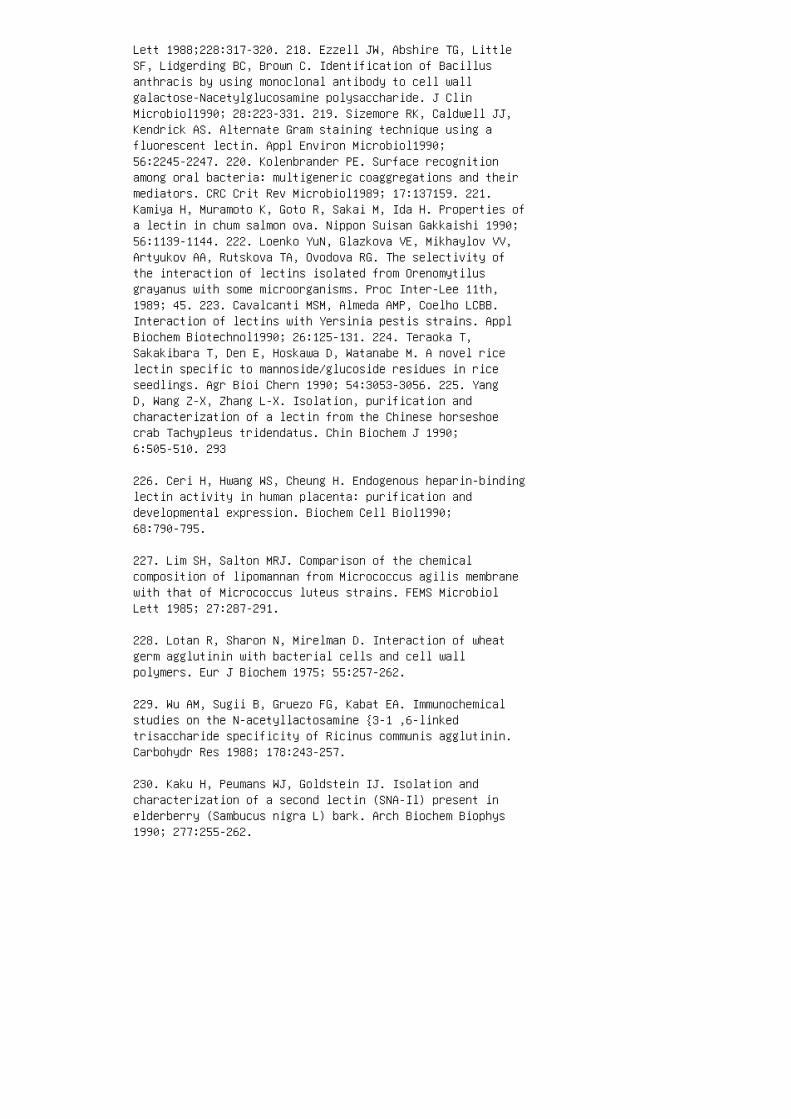
Lett 1988;228:317-320. 218. Ezzell JW, Abshire TG, LittleSF, Lidgerding BC, Brown C. Identification of Bacillusanthracis by using monoclonal antibody to cell wallgalactose-Nacetylglucosamine polysaccharide. J ClinMicrobiol1990; 28:223-331. 219. Sizemore RK, Caldwell JJ,Kendrick AS. Alternate Gram staining technique using afluorescent lectin. Appl Environ Microbiol1990;56:2245-2247. 220. Kolenbrander PE. Surface recognitionamong oral bacteria: multigeneric coaggregations and theirmediators. CRC Crit Rev Microbiol1989; 17:137159. 221.Kamiya H, Muramoto K, Goto R, Sakai M, Ida H. Properties ofa lectin in chum salmon ova. Nippon Suisan Gakkaishi 1990;56:1139-1144. 222. Loenko YuN, Glazkova VE, Mikhaylov VV,Artyukov AA, Rutskova TA, Ovodova RG. The selectivity ofthe interaction of lectins isolated from Orenomytilusgrayanus with some microorganisms. Proc Inter-Lee 11th,1989; 45. 223. Cavalcanti MSM, Almeda AMP, Coelho LCBB.Interaction of lectins with Yersinia pestis strains. ApplBiochem Biotechnol1990; 26:125-131. 224. Teraoka T,Sakakibara T, Den E, Hoskawa D, Watanabe M. A novel ricelectin specific to mannoside/glucoside residues in riceseedlings. Agr Bioi Chern 1990; 54:3053-3056. 225. YangD, Wang Z-X, Zhang L-X. Isolation, purification andcharacterization of a lectin from the Chinese horseshoecrab Tachypleus tridendatus. Chin Biochem J 1990;6:505-510. 293
226. Ceri H, Hwang WS, Cheung H. Endogenous heparin-bindinglectin activity in human placenta: purification anddevelopmental expression. Biochem Cell Biol1990;68:790-795.
227. Lim SH, Salton MRJ. Comparison of the chemicalcomposition of lipomannan from Micrococcus agilis membranewith that of Micrococcus luteus strains. FEMS MicrobiolLett 1985; 27:287-291.
228. Lotan R, Sharon N, Mirelman D. Interaction of wheatgerm agglutinin with bacterial cells and cell wallpolymers. Eur J Biochem 1975; 55:257-262.
229. Wu AM, Sugii B, Gruezo FG, Kabat EA. Immunochemicalstudies on the N-acetyllactosamine {3-1 ,6-linkedtrisaccharide specificity of Ricinus communis agglutinin.Carbohydr Res 1988; 178:243-257.
230. Kaku H, Peumans WJ, Goldstein IJ. Isolation andcharacterization of a second lectin (SNA-Il) present inelderberry (Sambucus nigra L) bark. Arch Biochem Biophys1990; 277:255-262.

231. Wessels MR, Rubens CE, Benedi VJ, Kasper DL.Definition of a bacterial virulence factor: sialylationof the group B streptococcal capsule. Proc Natl Acad SciUSA 1989; 86:8983-8987.
232. Mandai C, Mandai C. Sialic acid binding lectins.Experientia 1990; 46:433441.
233. Miller KJ, Kennedy EP, Reinhold VN. Osmotic adaptationby gram-negative bacteria: possible role for periplasmicoligosaccharides. Science 1986; 231: 48-51.
234. Ahamed NM, Kuhn HM, Widemann C, Radziejewska-LebrechtJ, Mayer H. Specific interaction of R-typelipopolysaccharides with lectins versus their nonspecificreactivity with basic and hydrophobic proteins. In:B0g-Hansen TC, ed. Lectins: biology, biochemistry,clinical biochemistry, vol 2. Berlin: Walter de Gruyter,1982:341-350.
235. Chatterjee BP, Guha AK, Pal R, Bhattacharyya M. Lectintyping of Pseudomonas aeruginosa strains of differentserogroups, Habs and Fisher types. Zentralbl Bakteriol Hyg[A] 1989; 271:364-371.
236. Parent JB. Membrane receptors on rat hepatocytes forthe inner core region of bacteriallipopolysaccharides. JBiol Chern 1990; 265:3455-3461.
237. Wright SD, Ramos RA, Tobia~ PS, Ulevitch RJ, MathisonJC. CD14, a receptor for complexes of LPS and LPS bindingprotein. Nature 1990; 249: 1431-1433.
238. Apicella MA, Mandrell RE, Shero M, Wilson ME, GriffissJM, Brooks OF, Lammel C, Breen JF, Rice PA. Modificationby sialic acid of Neisseria gonorrhoeae lipopolysaccharideepitope expression in human urethral exudates: animmunoelectron microscopic analysis. J Infect Dis 1990;162:506512.
239. Krauss JH, Reuter G, Schauer R, Weckesser J, Mayer H.Sialic acid-containing lipopolysaccharides in purplenonsulfur bacteria. Arch Microbiol 1988; 150:584-589.
240. Sacci JB, Campbell TA, Gottlieb M. Leishmaniadonovani: regulated changes in the level of expression ofthe surface 3 '-nucleotidase/nuclease. Exp Parasitol1990;71:158-168. 241. Rossell RJ, Stevens AF, Miles MA, AllenAK. A comparison of the lectinbinding properties of

glycoconjugates from a range of Leishmania species.Parasitol Res 1990; 76:294-300. 242. Grogl M, Franke ED,McGreevy PB, Kuhn RE. Leishmania braziliensis: protein,carbohydrate, and antigen differences between log phase andstationary phase promastigotes in vitro. Exp Parasitol1987;63:352-359. 243. Merkle RK, Cummings RD. Lectin affinitychromatography of glycoproteins. Methods Enzymol1987;138:232-259. 244. Osawa T. Recent progress in theapplication of plant lectins to glycoprotein chemistry.Pure Appl Chern 1989; 61:1283-1292. 245. Gallagher JT.Affinity chromatography of complex carbohydrates usingleetins. In: Oliver RWA, ed. HPLC of macromolecules: apractical approach. Oxford: IRL Press, 1989:209-227.246. Iqbal J, Saleemuddin M. Activity and stability ofglucose oxidase and invertase immobilized on concanavalinA-Sepharose: influence of lectin concentrations.Biotechnol Bioeng 1983; 25:3191-3195. 247. D'Souza SF,Nadkarni GB. Continuous conversion of sucrose to fructoseand gluconic acid by immobilized yeast cell multienzynmecomplex. Biotechnol Bioengin 1980; 22:2179-2189. 248.Hussain Q, Iqbal J, Saleemuddin M. Entrapment ofconcanavalin Aglycoenzyme complexes in calcium alginategels. Biotechnol Bioengin 1985; 27:1102-1107. 249.Hussain Q, Saleemuddin M. Immobilization of glycoenzymesusing crude concanavalin A and glutaraldehyde. EnzymeMicrobial Technol1986; 8:686690. 250. Fadda MB, Dessi MR,Maurici R, Rinaldi A, Satta G. Highly efficientsolubilization of natural lignocellulosic materials by acommercial cellulase immobilized on various solid supports.Appl Microbial Biotechnol 1984; 19:306311. 251. WoodwardJ, Zachry GS. Immobilization of cellulase through itscarbohydrate side chains: a rationale for its recovery andreuse. Enzyme Microbial Technol1982; 4:245-248. 252. ChuFK, Maley F, Tarentino AL. The use of iodinated lectinsfor determination the degree of deglycosylation ofhigh-mannose glycoproteins byendo-(3N-acetylglucosaminidase H. Anal Biochem 1981;116:152-160. 253. Iqbal J, Saleemuddin M. Sucrosehydrolysis using invertase immobilized on concanavalinA-Sepharose. Enzyme Microbial Technol1985; 7:175-178. 254.Day DF, Workman WE. A simple inulin assay for renalclearance determination using immobilized(3-fructofuranosidase. Ann NY Acad Sci 1986; 434: 504-507.255. Abbasi A, Voelter W, Zaidi ZH. Isolation,purification, and properties of a site-specificproteolytic enzyme valyl-proteinase from Candidatropicalis. Bioi Chern Hoppe-Seyler 1986; 367:441-445.256. Colas B, Boulanger Y. Glycosylation of yeastaspartyl-tRNA synthetase. Affinity labelling by glucoseand glucose-6-phosphate. FEBS Lett 1983; 163: 175-180.

257. Herrero E, Sanz P, Sentandreu R. Cell wall proteinsliberated by zymolyase from several ascomycetous andimperfect yeasts. J Gen Microbiol1987; 133: 2895-2903.
258. Mizuno T, Ohsawa K, Hagiwara N, Kuboyama R.Fractionation and characterization of antitumorpolysaccharides from "Maitake," Grifo/a frondosa. Agr BioiChern 1986; 50:1679-1688.
259. Kaku H, Van Damme EJM, Peumans WJ, Goldstein 11.Carbohydratebinding specificity of the daffodil (Narcissuspseudonarcissus) and amaryllis (Hippeastrum hybr) bulblectins. Arch Biochem Biophys 1990; 279:298-304.
260. Guillot-Breton A, Damez M, Dusser M, Gaillard-MartinieB, Millet L. Use of lectins for a comparative study ofcell wall composition of different anaerobic rumen fungalstrains. FEMS Microbiol Lett 1990; 67:151-156.
260a. Amatayakul-Chantler S, Ferguson MAJ, Dwek RA,Rademacher TW, Parekh RB, Crandall IE, Newell PC. Cellsurface oligosaccharides on Dictyostlium duringdevelopment. J Cell Sci 1991; 99:485-495.
261. Morgan AC, Woodhouse CS, Knost JA, Abrams PG, ClarkeGC, Arthur LO, Mcintyre R, Ochs JJ, Foon KA, Oldham RK.Monoclonal antibodies to human colorectal tumor-associatedantigens: improved elicitation and subclass restriction.Hybridoma 1984; 3:233-245.
262. Mattiasson B, Johansson PA. A simple rapid method forquantifying microorganisms by their metabolic activity whenbound to a specific adsorbent. J Immunol Methods 1982;52:233-240.
263. Mattiasson B. Analytical applications of immobilizedcells. In: Mattiasson B, ed. Immobilized cells andorganelles, vol 2. Boca Raton: CRC Press, 1983:95-123.
264. Warren GS, Fallon R. Reversible, lectin-mediatedimmobilization of plant protoplasts on agarose beads.Planta 1984; 161:201-206.
265. Mandenius CF, Welin S, Danielsson B, Lundstrom I,Mosbach K. The interaction of proteins and cells withaffinity ligands covalently coupled to silicon surfaces asmonitored by ellipsometry. Anal Biochem 1984; 137:106-114.
266. Mandenius CF, Mosbach K. Detection of biospecific

interactions using amplified ellipsometry. Anal Biochem1988; 170:68-72.
267. Mattiasson B. Reversible immobilization of enzymeswith special reference to analytical applications. J ApplBiochem 1981; 3:183-194.
268. Ohlson S, Hansson L, Glad M, Mosbach K, Larsson PO.High performance liquid affinity chromatography: a newtool in biotechnology. Trends Biotechno! 1989;7:179-186.
269. Welply JK. Sequencing methods for carbohydrates andtheir biological applications. Trends Biotechnol1989;7:5-10.
270. Lee KB, Loganathan D, Merchant ZM, Linhardt RJ.Carbohydrate analysis of glycoproteins. A review. ApplBiochem Biotechnol1990; 23:53-80.
271. Berezin BB, Gevorkyan RG, Lakhtin VM, Yamskov lA.Affinity sorbent containing Con A immobilized via cobalt(3 +)complex. Proc Inter-Lee 11th, 1989; 7.
272. McCoy JP. Contemporary laboratory applications oflectins. Biotechniques 1986; 4:252-262. 273. Tojo M,Shibata N, Osanai T, Mikami T, Suzuki M, Suzuki S. Sandwichenzyme-linked immunosorbent assay of o-mannans of Candidaalbicans NIH A-207 and NIH B-792 strains usingconcanavalin A and polyclonal rabbit anti-C. albicansantisera. Carbohydr Res 1991; 213:325-330. 274. RobinsonJE, Holton D, Liu J, McMurdo H, Murciano A, Gohd R. A novelenzyme-linked immunosorbent assay (ELISA) for the detectionof antibodies to HIV -1 envelope glycoproteins based onimmobilization of viral glycoproteins in microtiter wellscoated with concanavalin A. J Immunol Methods 1990;132:63-71. 275. Sjoblom I, Lundstrom M, Sjogren-JanssonE, Gloriso JC, Jeansson S, Olofsson B. Demonstration andmapping of highly carbohydrate-dependent epitopes in theherpes simplex virus type 1-specified glycoprotein C. J GenVirol 1987; 68:545-554. 276. Graham K, Keller K, EzzellJ, Doyle R. Enzyme-linked lectinosorbent assay (ELLA) fordetecting Bacillus anthracis. Eur J Clin Microbiol 1984;3:210212. 277. Shakhanina K, Kalinin N, Ruanet V,Kuljakina M, Pavlova I, Lakhtin V. ELISHA-like methods ofmicrassay of anthrax agent based on soybean agglutinin.Proc Inter-Lee 11th, 1989; 65. 278. Kalinin NL, GoncharovDB, Shakhanina KL, Safjanova VM. Comparative studies onthe interaction between lectins and Leishmania inagglutination test and enzyme-linked lectin-biotin assay(ELLBA). Proc Inter-Lee 12th, 1990; 62. 279. Lakhtin VM,

Guseva Nl, Fedurkina NV, Chibisova VA, Shakhanina KL.Differential affinity of pathogenic microbial species to aset of lectins, detected by the sandwich method with theuse of fluorescein isothiocyanate. J Microbiol EpidemiolImmunobiol (Mosc) 1985; N1:30-33. 280. Adam G, Heegard P,B0g-Hansen TC, Mundry KW. Lectins as probes for the assayof rhabdovirus infections in plants. J Virol Methods 1987;17:263275. 281. Riethman HC, Mawhinney TP, Sherman LA.Phycobilisome-associated glycoproteins in thecyanobacterium Anacystis nidulans R2. FEBS Lett 1987;215:209-214. 282. Gubler F, Hardham AR. Secretion ofadhesive material during encystment of Phytophthoracinnamomi zoospores, characterized by immunogold labellingwith monoclonal antibodies to components of peripheralvesicles. J Cell Sci 1988; 90:225-235. 283. Holden DW,Rohringer R. Proteins in intracellular washing fluid fromnoninoculated and rust-affected leaves of wheat andbarley. Plant Physiol1985; 78:715-723. 284. Liang R,Emerich DW. Analysis of lectin binding by Bradyrhizobiumjaponicum strains grown on nitrocellulose filters usingperoxidase-labeled lectin. Anal Biochem 1987;164:488-493. 285. Mirelman D, ed. Microbiallectins andagglutinins: properties and biological activity. NewYork: John Wiley & Sons, 1986. 286. Schrinner E, RichmondMH, Seibert G, Schwarz U, eds. Surface structures 297 ofmicroorganisms and their interactions with the mammalianhost. Weinheim: Verlagsgesellschaft, 1988.
287. Markwell MAK. Viruses as hemagglutinins and lectins.In: Mirelman D, ed. Microbial lectins and agglutinins:properties and biological activity. New York: John Wiley& Sons, 1986:21-53.
288. Weir DM. Carbohydrates as recognition molecules ininfection and immunity. FEMS Microbiol Immunol1989;47:331-340.
289. Keusch GT, Donohue-Rolfe A, Jacewicz M. Sugar bindingbacterial toxins. In: Mirelman D, ed. Microbiallectins andagglutinins: properties and biological activity. New York:John Wiley & Sons, 1986:271-296.
290. Uhlenbruck G. Bacteriallectins: mediators of adhesion.Zentralbl Bakteriol Hyg [A] 1987; 263:497-508.
291. Ofek I. Lectinophagocytosis mediated by bacterialsurface lectins. Zentralbl Bakteriol Mikrob Hyg [A] 1989;270:449-455.
292. Old DC. Bacterial cell envelopes in adhesion. In:

Hancock I, Poxton I, eds. Bacterial cell surfacetechniques. Chichester: John Wiley & Sons, 1988:227240.
293. Sharon N, Lis H. Lectins as cell recognitionmolecules. Science 1989; 246: 227-234.
294. Pereira MEA. Lectins and agglutinins in protozoa. In:Mirelman D, ed. Microbial lectins and agglutinins:properties and biological activity. New York: John Wiley& Sons, 1986:297-317.
295. Ishikawa F, Oishi K. Chitin-binding lectins fromfungi. In: Zikaris JP, ed. Chitin, chitosan, and relatedenzymes. Orlando: Academic Press, 1984:369382.
296. Kocourek J, Freed D, eds. Lectins: biology,biochemistry, clinical biochemistry, vol7. St. Louis:Sigma Chemical Co, 1990.
297. Norberg T, Ritzen H. Synthesis ofmethyl-a-o-Glcp-a-1,2-D-Galp-a1 ,3-o-Glcp and an acyclicanalogue thereof for probing the carbohydratebindingspecificity of bacteriophage c/>X174. Glycoconjugate J1986; 3:135142.
298. Wollin R, Bruse GW, Jansson PE, Lindberg AA.Definition of the phage G 13 receptor as structuraldomains of trisaccharides in Salmonella and Escherichiacoli core oligosaccharides. J Mol Recogn 1989; 2:37-43.
299. Drake D, Taylor KG, Bleiweis AS, Doyle RJ. Specificityof the glucanbinding lectin of Streptococcus cricetus.Infect Immun 1988; 56:1864-1872.
300. Cowan MM, Parrish K, Kessler RE, Pyle CJ, Taylor KG,Ciardi JE, Doyle RJ. Glucan-binding factor in saliva.Infect Immun 1988; 56:2912-2917.
301. Kundu M, Basu J, Chakrabarti P. Purification andcharacterization of an extracellular lectin fromMycobacterium smegmatis. FEBS Lett 1989; 256: 207-210.
302. Al-Mahmood S, Giummelly P, Bonaly R, Delmotte F,Monsigny M. Kluyveromyces bulgaticus yeast lectins.Isolation of N-acetylglucosamine and galactose-specificlectins: their relation with flocculation. J Bioi Chern1988; 263: 3930-3934.
303. Giummelly P, Separi F, Suptijah P, Bonaly R. Cell wallreceptors of the lectins produced by Kluyveromyces

bu/garicus. Yeast 1989; 5(special issue): 8309-311. 304.Barak R, Chet I. Lectin of Sclerotium ro/jsii: itspurification and possible function in fungal-fungalinteraction. J Appl Bacteriol1990; 69:101-112. 305. GuinetRMF, Rogemond V, Tessier F, Capdepuy M. Characterizationand possible role of lectinlike adhesion in theBacteroidesfragilis group. In: Proceedings ofinternational seminar on biology of anaerobic bacteria.June 1718, 1986, Lille. Amsterdam. 1986:60-68. 306.Szecowka J, Hallden G, Goldfine ID, Williams JA.Purification of the pancreatic cholecystokinin receptor.Regul Peptides 1989; 24:215-224. 307. Duong LT, Hadac EM,Miller LJ, Vlasuk GP. Purification and characterization ofthe rat pancreatic cholecystokinin receptor. J Bioi Chern1989; 264: 17990-17996. 308. Hill R, Levin J. Partialpurification of thrombopoietin using lectin chromatography.Exp Hematol1986; 14:752-759. 309. Ushikubi F, Nakajima M,Hirata M, Okuma M, Fujiwara M, Narumiya S. Purification ofthe thromboxane A2/prostaglandin H2 receptor from humanblood platelets. J Bioi Chern 1989; 264:16496-16501. 310.Giampapa CS, Abraham SN, Chiang TM, Beachey EH. Isolationand characterization of a receptor for type 1 fimbriae ofEscherichia coli from guinea pig erythrocytes. J BioiChern 1988; 263:5362-5367. 311. Geary SJ, Gabridge MG.Characterization of a human lung fibroblast receptor sitefor Mycoplasmapneumoniae. Isr J Med Sci 1987; 23:462-468.312. Hart GW, Haltiwanger RS, Holt GD, Kelly WG.Glycosylation in the nucleus and cytoplasm. Annu RevBiochem 1989; 58:841-874. 313. Belzunces LP, Theveniau M,Masson P, Bounias M. Membrane acetylcholine esterase fromApis mellifera head solubilized byphosphatidylinositol-specific phospholipase C interactswith an anti-CRD antibody. Comp Biochem Physiol B 1990;95:609-612.
9
Microbial Lectins for the Investigation
of Glycoconjugates
K. L. SHAKHANINA and N. L. KALIN IN Gamaleya Institute of
Epidemiology and Microbiology, Russian Academy of MedicalSciences,
Moscow, Russia
V. M. LAKHTIN Institute for Applied Science of MoscowUniversity
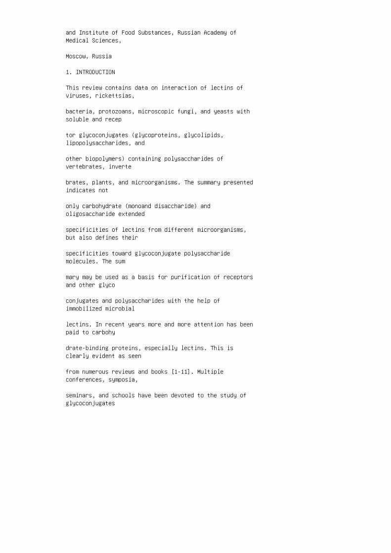
and Institute of Food Substances, Russian Academy ofMedical Sciences,
Moscow, Russia
1. INTRODUCTION
This review contains data on interaction of lectins ofviruses, rickettsias,
bacteria, protozoans, microscopic fungi, and yeasts withsoluble and recep
tor glycoconjugates (glycoproteins, glycolipids,lipopolysaccharides, and
other biopolymers) containing polysaccharides ofvertebrates, inverte
brates, plants, and microorganisms. The summary presentedindicates not
only carbohydrate (monoand disaccharide) andoligosaccharide extended
specificities of lectins from different microorganisms,but also defines their
specificities toward glycoconjugate polysaccharidemolecules. The sum
mary may be used as a basis for purification of receptorsand other glyco
conjugates and polysaccharides with the help ofimmobilized microbial
lectins. In recent years more and more attention has beenpaid to carbohy
drate-binding proteins, especially lectins. This isclearly evident as seen
from numerous reviews and books [1-11]. Multipleconferences, symposia,
seminars, and schools have been devoted to the study ofglycoconjugates

and lectins. Rapid growth in the numbers of firmsproducing complex
glycans, neoglycoconjugates, lectins, glycosidases,carbohydrate-binding
toxins, monoclonal antibodies to carbohydrate-containingtargets, and
publication of new scientific journals (e.g., Trends inGlycoscience and
Glycotechnology) are additional evidence for theimportance of studying
glycoconjugates and their interactions with lectins. Infact, glycoconjugate
recognition is the basis of several key me~hanisms ofbiological recognition
of living organisms. 299 The least studied lectins arestill those of microbial origin that interact selectivelywith glycoconjugates of host organisms (initial infectionprocesses in humans, farm animals, plants, and marine foodorganisms). Aspects of bacteria-mediated fixation ofnitrogen in tubercles of plants, the obtaining ofmicrobial insecticides, the phenomena of microparasitism,and selective coaggregation of microorganisms, allinvolve protein-glycoconjugate interactions [4, 12-17]. Inmany interactions between microbiallectins and cellsurfaces, there is insufficient information available aboutthe structures of receptor glycoconjugates and the spectrumof possible glycoconjugate targets. Study of theinteraction of purified microbiallectins with a set ofmodel glycoconjugates (glycoproteins, glycolipids,polysaccharides) with the known structures of glycanshelps define the specificities of microbial lectins.Therefore, the main task of the present review is tooutline the data on new preparations of microbial lectinsthat have not been considered in other reviews [12,18-22].II. INTERACTION OF MICROBIAL LECTINS WITHCARBOHYDRATE-CONTAINING TARGETS Agglutinins (mainlylectins) have been investigated from about 150 microbialsources and isolated from only a few species. However,lectins from only 100 microorganism species have beencharacterized for monosaccharide specificities. There islittle information about oligosaccharide specificities ofmicrobial lectins or of their specificities for wholemolecules of glycoconjugates and polysaccharides. The main

sources of microbiallectins are from viruses (defined asa microorganism for purposes of this book), bacteria,protozoans, yeasts, and fungi, including mushrooms. Thelectins that belong to each of these groups ofmicroorganisms will be considered. Ill. VIRAL LECTINSTable 1 shows some results on the interaction ofvirallectins with receptors and isolated glycoconjugates(natural or synthetic). Many viral proteins showhemagglutinating activities [23], although it is onlyrecently that hemagglutinins of viral origin have begun tobe considered as lectins [23]. Carbohydrate-containingtargets for viral lectins, as a rule, includelipopolysaccharides, gangliosides, and glycoproteins (seeTable 1). The most thorough investigation was carried outon the specificities of the hemagglutinins of influenzaviruses for mammalian glycoproteins and gangliosides[34-39]. All hemagglutinins of influenza virus types A, B,and C exhibit the properties of sialospecific lectins,interacting with terminal residues of N-acetylneuraminicor N-glycolylneuraminic acid in glycans. 301
Table 1 Interaction of Viral Lectins with Glycoconjugatesand Polysaccharides Targets containing
Source of lectins carbohydrates Ref.
Bacterial viruses Bacteriophage 013 hemRa, Rb 1 , Rb 2-type LPS from mutant 24 agglutinin strains ofSalmonella sp., Escherichia coli. Bacteriophage <!>X174Glc-a-1 ,2-Gal-a-1 ,3-Glc-containing 25 hemagglutininLPS of Salmonella
Coronaviridae Sheep encephalomyelitis9-0-Ac-Neu5Ac-containing protein 26 virus and bovinecorona of surface structure of chicken virus hemagglutininerythrocytes and human sialidasetreated erythrocytes
Hepadnaviridae Hepatitis B virus S proNeuAc,NeuAc-Lac-containing sur27 tein face structures of Verocells
Herpesviridae Herpes simplex virus type Sensitive toheparin surface structure 28 1 and pseudorabies virus ofrabbit kidney cell lines RK-13 GPll-B, GP111-C, GPV
Lentiviridae Human immunodeficienAntigen CD4 andtransmembrane 14,29 cy virus type 1 gp120 gp41 ofTlymphocytes
Reoviridae Reovirus type 3 0 proGlycoprotein A MN of

human erythro30 tein 1 cytes Orbivirus of blue linguaeNeu-Ac-a2,6, NeuG-a2,6-containing 31 of sheephemagglutinin (GP); glycophorin, mucin of bovinesubmandibular gland
Rhabdoviridae Rabies virus hemaggluGangliosides GTlb,GQ1b, GDlb 32 tinin Vesicular stomatitis viralGangliosides GM 3 of goose erythro33 hemagglutinin cytes
Orthomyxoviridae Influenza virus type A NeuAca2,3(6)NeuG-a2,3(6)34,36,37 containing gangliosides and GPNeu-Ac-containing a 2 -macroglobulins Influenza virus typeC Neu5Ac-a-containing neopolysac38 hemagglutinin charide9-0-Ac-NeuAc-containing protein of 39 surface structureof chicken erythrocytes However, the hemagglutinin ofinfluenza virus type A predominantly recognizes4-0-acetylated groups of N-acetylneuraminic acid.Influenza virus type C hemagglutinin shows a high affinityfor 9-0-acetylated derivatives of sialic acid [37-39].Moreover, influenza virus lectins are capable ofrecognizing microdomains and clusters from the sialic acidresidues within the elongated glycans, including sets ofsialic residues. Some viral lectins are specific forpolysialogangliosides (e.g., rabies virus hemagglutinin),and other virallectins for mannosialogangliosides (such asthe vesicular stomatitis virus hemagglutinin) [32,33].Specificities of virallectins for lipopolysaccharide andneopolysaccharides with known structures have also beenstudied [24,25]. Because of the density of glycanclusters, their lengths and glycoconjugate structures, andcomposition variations, it is possible to detect glycanswith maximal affinity for lectins, as has beendemonstrated for hemagglutinins of bacteriophage G13 andinfluenza viruses [24]. Chapter 2 of this book provides adetailed examination of the interaction between lectins,viruses, and viral-infected cells. Ill. BACTERIAL LECTINSSignificantly more papers have been published onspecificities of bacterial lectins [15-22,24,41-44]. Thismay be because the researchers began to considercarbohydrate-binding toxins of bacterial origin as lectins[41 ,53 ,54]. Among the sources of bacterial lectins,several investigators report nearly 100 strains from morethan 70 species of organisms. However, most of the lectinshave not been purified, or the purified lectins have notbeen exhaustively investigated from the viewpoint oftheir specificities for glycoconjugates [18]. Table 2 givesdata on the known specificities of bacterial lectins. Ascan be seen from Table 2, bacterial lectins representpreparations with different biological activities andcellular distribution. Adhesins may be found on microbial

surfaces containing fimbria! and nonfimbrial appendages.Some are extracellular toxins, and some are enzymes (seeChapter 1 for a discussion of the definition of a lectin).All the microbiallectins, as a rule, show hemagglutinatingproperties. Toxic hemagglutinins usually includeRNA-N-glycosidase or ADP-ribosyltransferase activities[41], although frequently proteolytic activity is alsodetectable in microbial hemagglutinins [18,139]. Someglycosidases and glucose oxidases have, apart from theircatalytic centers, lectinlike domains [44, 117, 179].From the examples of coliform bacteria, pseudomonads, andcholera vibrios one can observe that each of themicroorganisms synthesize a set of lectins with variousspecificities for glycoconjugate targets (see Table 2).Lectin sources Actinomyces naeslundii WVU45 fimbriae A.viscosus T14V fimbriae Aeromonas caviae, A. hydrophila, A.veroni, A. sobria hemagglutinins pilins/ adhesinsAgrobacterium tumefaciens lectins Azospirillum brasiliensehemagglutinin Bacillus mesentericus 316M extracellularhemagglutinin Bacteroides jragilis adhesin (70 kDa) B.(Prevotella) loescheii adhesin Bordetella bronchisepticaadhesin B. pertussis pertussis toxin (B subunit oligomer)(11-22 kDa) adhesin (filamentous hemagglutinin)Bradyrhizobium japonicum Campylobacter jejuniextracellular enterotoxin Carbohydrate-containing targetsGal-~1 ,3-GalNAc/GalNAc-~1 ,3-Gal-containing glycolipidsGal-~1 ,3-GalNAc/GalNAc-~1; 3-Gal-containingpolysaccharides Sensitive to mannose of animalerythrocytes and lines of mammalian cells a-L-Fuc-BSA;fucan; chondroitin-sulfate; pectin of citrusa-L-Fuc-containing substances of erythrocytes NeuG 1;NeuAc-containing substances of rabbit erythrocytes a-GleN;a-GalN-Sepharose; polymers of the bacterium Enterococcushirae; epithelial intestinal cell line of human Proandeukaryotic cells NeuAc-Lac-Gp-containing sialogangliosidesof mycelia; mucin of bovine submandibular glandNeu5Ac-a2,6-containing GP; some asialo-GP; thermoprocessedfetuin; proteins of goose erythrocytes Substances ofcomplement-3/integrin aM ~ 2 ; CD 11 b + CD 18 ofmacrophages Lac-Sepharose; ~Gal; Lac-containing polymersof soybean cell line Gangliosides SRL ... Q =Ref. et42 ID 0. :r "' 42 45 46 47 48 49,50 51 5253-57 58,59 60,61 62,62a '-"'~ (continued) = '-"'~~ Table 2 Continued ~ Lectin sources Clostridiumbotulinum botulinum toxin type E C. botulinum botulinumtoxin C 2 C. difficile extracellular enterotoxin A C.spiroforme C. tetani tetanus toxin Eikenella corrodensEscherichia coli adhesin from type 1 (hemagglutinin 28kDa) fimbriae (or pili) Fimbrial type 1 of strain RDEC-1P-fimbriae (G adhesins) hemagglutinins of uropathogenic

strains S-fimbriae CS 1, CS 2, CS 3 of uropathogenic andother enterotoxigenic strains Fimbriae K88ab fromenterotoxigenic strains for piglet intestinal mucosaFimbriae K99 of enterotoxigenic strains Fimbriae F41 ofenterotoxigenic strain Nonfimbrial adhesin NFA-3 Membranetransporter of glucose Carbohydrate-containing targetsGangliosides and phospholipids Pig thyroglobulin; mucinof bovine submandibular gland; human erythrocytes treatedby sialidase Gal-ad, 3-Gal-,81,4-GlcNAc-containingsubstances of rabbit erythrocytes; bovine thyroglobulin;antigens Lex, LeY 1 of intestinal epithelium of humansHuman erythocytes Sialoganglioside-containing materialsGal-containing receptors Erythrocytes of guinea pig;Sensitive to mannose-containing substances of erythrocytesof guinea pig Man 5 GlcNAc 2 -peptide Gal-a1, 4Gal,8-containing globosides (globo-A and other) of humanepithelial cells NeuAca2, 3-Gal-containing substances ofmammalian cells gp 40-42 kDa of mucous epithelial cellsurface of piglet intestine Glycophorin A; glycopeptidesof pig intestine mucins; NeuG1-paragloboside andNeuG1-GM3 Glycophorins of human erythrocytes GlycophorinA NN erythrocytes of human with blood group N-antigenGlucose-containing ligands Ref. 63 64 65 70 71 9922,43,72,73 74-77 77-79 80 81-83 84-85 86-88 89 9091 w = \11 for human and pig Serotype 1 toxinSerotype 2 toxin 1 Serotype 2 toxin 2 Thermolabileenterotoxin of strains pathogenic for chickens Toxinfrom pig edemic disease strain TB-1 (pCG 5) Erwiniacarotovora subsp carotovora hemagglutinin E. rhapoviticihemagglutinin Eikenella corrodens surface hemagglutininLegionella pneumophila hemagglutinin Listeriamonocytogenes surface lectins Mycobacterium smegma tisextracellular lectin Mycoplasma salivarum surfacehemagglutinins M. hypopneumoniae hemagglutinins M.pneumoniae adhesins Neisseria gonorrhoeae nonfimbrialadhesin N. meningitidis S-fimbriae of strains causingmeningitis in newborns N. subflava nonfimbrial adhesin-1(27 kDa) GMt> GD 1 y, GD 1 b, GD2, GM2, GM3, GDI•• GD 1• and GT 1 b gangliosides Substances of pig blood groupA + H asialo-GP: mucin of bovine and thyroglobulin Gb 4,Gb 3 gangliosides Sensitive to mannose; rabbiterythrocytes Asialofetuin, Gal-{j1 ,4-GlcNAc-containingsubstances of human erythrocytes Binds to galactosamineof mammalian erythrocytes Adheres to carbohydrates ofmammalian erythrocytes treated with sialidase Ole, L-FucN-containing neoGPs Yeast mannan and mycobacterialarabinogalactan Sialic acid receptors of sheeperythrocytes; human B group blood erythrocyte Sialoreceptors of turkey erythrocytesNeuAc-a2,3-Gal-containing GP, laminin, fetuin,

choriogonadotropin of humans; Gal(3-S0 4 )-{jlcontainingsulfated glycolipids of adenocarcinoma! cell line WiDrAsialo GMt> GM 2 , containing glycolipidsNeuAc-a2,3-Gal-{j1 ,4-containing substances of bloodvessel endothelial cells NeuAc-a2,3-Gal-{j1,4-containinggangliosides of human erythocytes 95 96 97 98 99 100101 102 103 104 105,106 107 108 109 (continued) ~Q a'\ Table 2 Continued Lectin sources Porphyromonasgingiva/is (Bacteroides gingiva/is) adhesin(s) Pseudomonasaeruginosa lectins PA-l PA-Il Adhesins of strain ATCC27853 Exotoxin A P. fluorescens subsp cellulosaendoglucanase domain Pseudomonas syringae phaseolicolahemagglutinin P. solanacearum hemagglutinin Renibacteriumsalmoninarum hemagglutinins (57, 58 kDa) Salmonella dublintype 1 fimbria! hemagglutinins Shigella dysenteriae type 1dysenteric toxin Staphylococcus aureus Wood 46 adhesinsS. haemolyticus S. epidermidis, S. warneri hemagglutininsS. saprophyticus surface hemagglutininCarbohydrate-containing targets Peptide (12 amino acids)rich in histidine and GP from human salivaGalactose-containing receptors of erythrocytes; substancesof blood group A + B Mannoseand fucose-containing GP ofyeasts, plants, mammals NeuAc, GlcNAc-containingsubstances of human kidney and lung cellsp-Aminophenyl-{3-n-thioGalp; fetuin {3-1 , 4-glucan(cellulose) Sensitive to N-acetylgalactosamineFucose-containing substances of rabbit erythrocytesRabbit erythrocytes and salmon spermatozoaMannose-sensitive substances of erythrocytes Gal-ad,4-Gal-{3-BSA; galabiose-containing globosides (Gb 3 andothers) of HeLa cells Fucose-containing GP: fibronectinN-Acetylgalactosamine-sensitive substances of horseerythrocytes N-Acetylgalactosamine andN-Acetylglucosamine sensitive substances of human urinarytract epithelium and horse erythrocytes Ref. 111,112113 114 115 116 117 96 118 119 120,121 122 123123,124 IN Q "' Streptomyces murinus extracellularaggregation factor of cells S. sanguis surface adhesinS. cricetus and S. sobrinus (extracellular or surfacelectins) Vibrio cholerae thermolabile enterotoxin of its"B"subunits (11.6 kDa) Fimbriae (monomer 16 kDa) ofstrain 0 1 biotype El Tor Hemagglutinin (62kDa) ofnon-OL V2 strain cells (nonfimbrial origin)Hemagglutinin/metalloprotease from culture fluid ofstrain CA401 V. furnissii adhesins/ chemoreceptors ofmarine bacteria Ureaplasma urealyticum adhesinsXanthomonas campestris hemagglutinins uin glycopeptidesand bovine substances of glycophorin AMN 131,3(6)-Glucans of sarcoma cell line, Hela cells, E. coli,Bacillus subtilis, Micrococcus /uteus, Staphylococcus au

reus NeuAc-a2, 3-Gal-131, 3-GalNAc-sensitiveglycoconjugates of human saliva Glc-a 1 ,6-containinglinear glucans GM 1 , GM 2 , asialo-GM 1 ,neoglycolipids; (GM 1 ) oligopolylysine; substances ofmouse intestine epithelial cell nuclei (probably GP),glycoproteins with activity of A blood group from mucousmembrane of pig stomach Animal erythrocytes Fetuinandasialofetuin-sensitive substances of rabbit erythrocytesErythrocytes of the chicken breed "white leghorn" GlcNAc,Man, Glc-Sepharose; (GlcNAc) Glycophorin and dextransulfate-sensitive substances of human erythrocytesGlucosamine-containing substances of rabbit erythrocytes;xylose-, galactose-, N-acetylgalactosaminesensitivesubstances of rabbit erythrocytes GP, glycoprotein;NeuAc, N-acetylneuraminic acid; NeuG, N-glycolylneuraminicacid; PS, polysaccharide. 112,126,127 128,130 92,131-136137 138 139 140,141 142 The ability of a microorganismto produce surface lectins signifies the role of lectinsin the ecology of microbes, especially for biologicalrecognition of host cells [4,17,18,22]. One can also seefrom Table 2 that bacterial lectins are capable ofrecognizing not only several types of monoanddisaccharides (specificities of lectins for simple sugars)or oligosaccharides (fine specificities of lectins), butalso macromolecular glycoconjugates and polysaccharides.From the example of monosaccharides coupled with suitablecarriers it can be observed that the lectin-inhibitingeffectiveness of the neoglycoconjugates shows 10 3 or 106 -fold increase when compared with free carbohydrates[185,186]. The effectiveness of such conjugates dependsnot only on the monosaccharide type, but also on itsdensity and distribution over the carrier surface. Lectinsof Actinomyces are capable of recognizing the samestructures of disaccharide fragments within differenttargets, such as glycolipids and polysaccharides [166-168].Pertussis toxin and elder lectins react with suchglycoproteins as fibrinogen, transferrin, and phosvitin,containing Neu-5-Ac-a2,6-residues of sialic acid (but notNeu-5-Ac-a2,3-residues). Fetuin, containing equal numbersof both types of these sialic acid residues, exhibits ahigher affinity for the toxin than for the elder lectins.Human fibrinogen was characterized by maximal affil).ityfor the toxin. Fibrinogen glycoprotein contains fouridentical biantennary asparagine-bound glycans, withterminal sialic acid residues. More weakly reacting withthe toxin were human transferrin and chicken phosvitin,which possess two biantennary or one triantennarysialylated glycans, respectively. In its ability to reactwith sialoglycoproteins, pertussis toxin could bedistinguished from a sialospecific plant lectin from wheat

embryos [53,54]. Bacteriallectins also frequently show ahigh selectivity for glycolipids (globosides andgangliosides). With these receptors, lectins ofmicroorganisms are often distinguishable from those ofplant or animal origin [10,17]. Whereas cholera toxin hasa maximal affinity for monosialoganglioside GM~> thetetanus, botulinum, and gas gangrene toxins show highaffinities for gangliosides containing three to foursialic acid residues. Here, the increased selectivity ofthe microbial lectin for glycolipids may be enhancedbecause of variations in the structure of the lipidcomponent. Thermolabile enterotoxins of various serotypesof Escherichia coli differ in their levels of affinity fora wide range of gangliosides [92]. Oral streptococcallectins [12] may be considered an example of thehigh-level selectivity of microbial lectins towardpolysaccharides. Of a set of glucans, with varyingcontents of a-1,6; a-1,4; a-3; and a-1,2linkages, thelectin reacted only with those glucans containing morethan 800Jo of the a-1,6 linkages; the maximal affinity forlectins was a glucan containing
Microbial Lectins 309
950Jo of a-1,6 linkages. Only linear glucans of theforegoing type with a
relative molecular mass (M,) of over 5 x 10 5 kDa wereable to induce rapid
aggregation of the streptococci. From Table 2, one can seethat a single
microbial lectin can show not only selective reaction witha definite type
target (glycoprotein, glycolipid, polysaccharide) but canalso recognize gly
coconjugates of various other types. Cholera toxin, forinstance, may ex
hibit a high specificity toward some glycolipids andpolysaccharides [166
168]. The ability of a microbial lectin to reactselectively with glycoconju
gates is determined by the domain and epitope structuresof the lectin

molecule. Epitope organization of the cholera toxincarbohydrate-binding
subunit [187], and the size and structure ofcarbohydrate-binding fragments
or subunits of diphtheria, pertussis, and other microbialprotein toxins
also vary. Filamentous Bordetella hemagglutinin containsbinding sites for
heparin or glycoconjugates containing terminal residues ofgalactose that
are not adjacent to each other [59]. Interaction ofClostridium toxin A with
antibodies does not interfere with hemagglutinatingactivity of this toxin
lectin, which is characterized by a h,igh selectivitytoward complex glycans
[68,69]. Therefore, variations in selectivity ofmicrobiallectins toward gly
coconjugates may occur because of the existence of avariety of sites with
different specificities for glycoconjugates and because ofstructural changes
within these sites. Lectins from actinomycetes,streptomycetes, and Sclero
tium rolfsii interact differently with variousgram-positive and gram-nega
tive bacteria [166,182,183].
IV. PROTOZOAL LECTINS
Currently, protozoallectins have been studied in limiteddetail relative to
interactions with other microorganisms. Lectins fromprotozoa are also

characterized by the aforementioned general regularities:production of a
system of lectins with different carbohydratespecificities by a microorgan
ism of one species or strain and the ability of the samelectin to interact
with various types of carbohydrate-containing targets(Table 3). In addition
to the glycoproteins and neoglycoproteins listed in Table 3that interact
with protozoallectins, there are reports on the interactionbetween Giardia
Iamblia lectin and Salmonella lipopolysaccharides [143].
V. YEAST LECTINS
As with the protozoal lectins, little information isavailable concerning
carbohydrate complexes with yeast lectins [154,155]. Themost thorough
study has been given to the preparations of lectins fromthe pathogenic yeast species Candida a/bicans, the causeof candidiasis in humans, as well as lectins of yeastswidely used in the food industry, produced by strains ofSaccharomyces cerevisiae (Table 4). It is worth mentioningthat the spectrum of biological activities of yeastlectins also includes killer activity toward other yeastspecies. Thus, extracellular lectin from Pichia anomalashows anticandidal activity owing to selective interactionwith {:l-1,6-glucan of C. albicans [160]. Anothersignificant process of biological recognition betweenyeast gametes of different sexes is controlled byparticipation of yeast a-mannan-sensitive lectins [162].Table 3 Interaction of Lectins of Protozoan Origin withGlycoconjugates and PolysaccharidesCarbohydrate-containing Lectin sources targets Ref.Entamoeba histolytica lectin Asialoorosomucoid; Gal,GalNac144 containing polymers of CHO hamster cell lineSurface adhesin N-, 0-glycan of cell surface of CHO145-147 hamster cell line, mucin of rat intestineGiardia Iamblia Portland 1 strain surfaceGlucose/mannose-containing poly148,149 lectin (57-78kDa)

mers of intestinal epidermal cells of mammalsTagerin/lectin activated Mannose-6-phosphate-containingby trypsin (28-30kDa) materials of rabbit erythrocytesLeishmania braziliensis (NR) GlcNAc-BSA,N-acetylglucosamine 150 surface lectin glucose, mannose,galactosecontaining materials of macrophage J774G8 L.donovani transporter of Glucose-containing ligands 91glucose Paramecium tetraurelia lectins Gal-BSA, Man-BSA,dextran-BSA 151 of apex of trichocysts Plasmodiumfalciparum Glycophorins, GlcNAc-Sepharose 152 (140,70,35kDa) Shizont soluble antigen Sialoreceptors of humanerythro153 (175 kDa) of camp strain cytes Pneumocystiscarinii adhesins (j-Gal-BSA, Man-BSA a-Fuc-BSA 110 oramino sugar-BSA
Microbial Lectins 311
Table 4 Interaction of Lectins of Yeast Origin withGlycoconjugates and Polysac
charides Carbohydrate-containing
Lectin sources targets Ref.
Candida albicans extracellular L-Fucose-containingsubstances of 156
lectin of GDH 2346 strain buccal and vaginal epithelialcells of mammals Extracellular lectin ofN-Acetylglucosamine-containing rna157 GDH 2023 strain C.terials of epithelial cells of mamalbicans and Cryptomals.Gal-131, 4-Glc-!31 containing coccus neojormansglycolipids adhesins
K/uyveromyces bulgaricus ex13-GlcNAc,aGal-containingmaterials 158,159
tracellular lectins of sheep and rabbit erythrocytes,yeasts
Pichia anomala extracellular 131 ,6-glucan-containingsubstances of 160
anti candidai toxin cell wall of Candida albicans
Saccharomyces cerevisiae exGalactoseand lactose-sensitivepoly161
tracellular hemagglutinin of mers of animal erythrocytes

CD115 strain Sex agglutinin (22 kDa) Mannose-containingpolymers of 162 yeasts
VI. FUNGAL LECTINS
Lectins of fungi have been less well investigated thanthose of higher plants
[10]. Many of the fungal lectins studied belong to thegroup of chitin
binding proteins [163,164]. Data on the interaction offungallectins with
other carbohydrate-containing targets are presented inTable 5. Many fun
gallectins are characterized by a high affinity forsialomucins of the bovine
submaxillary gland [164,169,170,178], whereas lectins fromAthelia ro/jsii
and Rhizoctonia so/ani mycelia show a high affinity forsialomucins of the
pig intestinal mucous membrane [172]. Some fungallectinsalso interact
well with human erythrocyte glycoproteins, bovine fetuin[164,172], and
glycosaminoglycans of the chondroitin sulfate type [170].Of considerable interest are data on specific interactionof fungal lee
tins with polysaccharides. Lectins from Rhizoctonia so/anireact well with
galactose and N-acetylgalactosamine-containingpolysaccharides of bacte
rial and plant origin [166,181]. Lectins from Phanerochaetechrysosporium
and the bacterium Streptomyces murinus [179, 183] showaffinity for {3Table 5 Interactions of Lectins of FungalOrigin with Glycoconjugates and Polysaccharides ~ ;jLectin sources Chrysosporium keratinophilum (Frey)conidial hemagglutinin Arthrobotrys ellipsospore

extracellular hemagglutinin Arthrobotrys oligosporasurface adhesins Athelia roljsii micellar hemagglutinin(17 kDa) Beauveria bassiana micellar hemagglutininsCephalosporium acremonium extracellularagalactosidase/hemagglutinin Clitocybe geotropa, Laccariaemethystina, and Photo/iota squarrosa lectinsConidiobolus obsurus lectins/adhesins of spore surfaceGanoderma lucidum micellar hemagglutinin (17 kDa)Epidermophyton floccosum, Microsporum canis, M. cookei,and M. fulvum extracellular hemagglutinins Neurosporasitophila extracellular hemagglutinin (22 kDa)Phanerochaete chrysosporium extracellular cellobioseoxidase with domain for sorption on glucan (flavin domainof enzyme) Pythium aphanidermatum lectin Rhizoctoniacrocorum micellar hemagglutinin (11 kDa) R. so/animicellar hemagglutinin (13 kDa) Sclerotium roljsiiextracellular lectins (55+ 60 kDa) Carbohydrate-containingtargets N-Acetylneuraminic acid and Ca 2 + -sensitivesubstances of rabbit erythrocytes, mucin of submandibularbovine gland Mucin of submandibular bovine gland andchondroitin sulfate containing substances of chickenerythrocytes 2-Deoxy-n-glucose substances of cell surfaceof Trichostrongylus colubriformis Mucin of pig intestine;asialofetuin-sensitive substances of trypsin-treatederythrocytes of pigs Monosaccharide-insensitivesubstances of animal erythrocytes Branched {31,4-glucanpotato starch L-Fucose-containing substances of spores,flagella, sporangia and rhizoids of fungal strains fromintestine of sheep Glc-BSA, GlcNAc-BSA Sheep erythrocytesN-Acetylneuraminic acid-containing substances of rabbiterythrocytes; mucin of bovine submandibular gland; GP ofhuman erythrocytes Mu~ of bovine submandibular gland; GPof human erythrocytes {31 ,4-Glucan (cellulose)Fucose-containing substances of Lepidum sativum Mucin ofpig stomach; fetuin-sensitive substances of trypsintreatedrabbit erythrocytes Gum arabic;N-acetylgalactosamine-sensitive substances of rabbiterythrocytes Surface polymers of E. coli Ref. 169 170171 172 173 174 175 176 177 178 163 178 171,179172 181 182
1,4-glucans or ~-1,3(6)-glucans. The lectin ofCephalosporium acremo
nium binds specifically with a-1 ,4-glucan [174]. Amongfungallectins, simi
lar to some lectins from bacteria (see Table 2) are someenzymes, such

as cellobiose oxidase and a-galactosidase, with aspecificity for glucans
[174,179]. Both of these enzymes of carbohydratemetabolism are charac
terized by the presence of carbohydrate-binding sites inaddition to a cata
lytic center [179]. As can be seen from Table 5, lectinsof fungi may be applied to the
investigation of glycoconjugates, polysaccharides, andreceptors of micro
organisms. With the help of fungal fucose-specificlectins, one can detect
considerable structural subtleties between eight strainsof five other fungal
species belonging to the genera: Neocallimastix,Piromonas, and Sphaero
monas [175].
VII. CONCLUSIONS
The foregoing summary was devoted to interaction of lectinsand aggluti
nins from more than 100 microbial sources, withglycoconjugates, polysac
charides, and cellular receptors. Most microorganisms seemcapable of
synthesizing a group (or a system) of lectins characterizedby various carbo
hydrate specificities, various cellular distributions, anda variety of biologi
cal activities. The set of lectins can vary in mutants andrecombinant micro
organisms, and they also depend on the life cycles ofmicroorganisms. The
ability of a microbial lectin to recognize carbohydrates

generally increases
within the range: free monoand disaccharides (simplecarbohydrates <
isolated oligosaccharides < mono-, di-, andoligosaccharides within glyco
conjugates and polysaccharides). Microbial lectins areable not only to rec
ognize various types of natural carbohydrate-containingtargets (glycopro
teins, gangliosides, globosides, lipopolysaccharides,glycosaminoglycans,
and polysaccharides), but they can also recognizedifferences in carbohy
drate-containing targets within each type. Factors that canpotentially af
fect the affinity of a microbial lectin for complexglycans as constituents of
carbohydrate-containing targets may be the following: (1)types of terminal
and internal carbohydrate residues; (2) presence ofterminal residues of
carbohydrates of the same type in clusters, accessible tolectin (nearest
neighbors within one glycan in a glycoconjugate, such as apolysialoganglio
side or sialylated antenna(e) in polyantennaryasparagine-bound glycans of
glycoconjugates. Shark glycans bound with serine orthreonine in glycopro
teins represent another example; (3) the degree ofpolysaccharide branch
ing. Further refinement of lectin specificity to glycanswithin carbohydrate

containing targets may be residues bearing hydrophobicgroups (methyl,
11. Kalinin N, Doyle RJ. Distribution of glucan-bindinglectin on the surface of Streptococcus. Proceedings of13th international lectin meeting. Berlin: Abstract book1991:36.
12. Sharon N. Bacteriallectins, cell-cell recognition andinfectious disease. FEBS Lett 1987; 217:1-13.
13. Sharon N. Biomedical aspects of glycoconjugaterecognition. Biomedical aspects of lectins. Biochem SocTrans 1989; 17:11-12.
14. Weir OM. Carbohydrates as recognition molecules ininfection and immunity. FEMS Microbiol Lett 1989;47:331-340.
15. Old DC. Bacterial cell envelopes in adhesion. In:Hancock I, Poxton I, eds. Bacterial cell surfacetechniques. Chichester: John Wiley & Sons, 1988:227240.
16. Ofek I. Lectinophagocytosis mediated by bacterialsurface lectins. Zentralbl Bakteriol Hyg [A1] 1989;270:449-455.
17. Karlsson KA. Animal glycosphingolipids as membraneattachment sites for bacteria. Annu Rev Biochem 1989;58:309-350.
18. Mirelman D, ed. Microbiallectins and agglutinins:properties and biological activity. New York: John Wiley &Sons, 1986.
19. Sharon N. The lectins. In: Liener IE, Sharon N,Goldstein IJ, eds. Lectins: properties, functions andapplications in biology and medicine. Orlando: AcademicPress, 1986.
20. Uhlenbruck G. Bacteriallectins: mediators of adhesion.Zentralbl Bakteriol Hyg [A] 1987; 263:497-508.
21. Gilboa-Garber N, Garber N. Microbiallectins. In: AllenHJ, Kisailus EC, eds. Handbook of glycoprotein&. NewYork: Marcel Dekker, 1989.
22. Nimich W. Adhesins of Escherichia coli. ActaBiotechnol1990; 10:151-161.

23. Markwell MAK. Viruses as hemagglutinin& and lectins.In: Mirelman D, ed. Microbiallectins and agglutinins. NewYork: John Wiley & Sons, 1986:2153.
24. Wollin R, Bruse OW, Jansson P, Lindberg S. Definitionof the phage 013 receptor as structural domains oftrisaccharide& in Salmonella and Escherichia coli coreoligosaccharide&. J Mol Recogn 1989; 2:37-43.
25. Norberg T, Ritzen H. Synthesis of methyla-o-glycopyranosyl-(1-2)-a-Dgalactopyranosyl acyclicanalogue thereof for probing the carbohydratebindingspecificity of bacteriophage t/>X174. Glycoconjugate J1986; 3:135142.
26. Schultze B, Gross HJ, Brossmer R, Klenk HD, Herrber G.Hemagglutinating encephalomyelitis virus attaches toN-acetyl-9-0-acetylneuraminic acidcontaining receptors onerythrocytes: comparison with bovine coronavirus andinfluenza C virus. Virus Res 1990; 16:185-194.
27. Komai K, Kaplan M, Peeples ME. The vero cell receptorfor the hepatitis B virus smallS protein is asialoglycoprotein. Virology 1988; 163:629-634.
28. Sawitzky D, Hampl H, Habermehl KO. Comparison ofheparin-sensitive attachment of pseudorabies virus (PRV)and herpes simplex virus type 1 and identification ofheparin-binding PRY glycoproteins. 1 Gen Virol 1990; 71:1221-1225. 29. Berman PW, Gregory TJ, Riddle L, NakamuraG, Change M. Protection of chimpanzees from infection byHIV -1 after vaccination with recombinant glycoproteingp120 but not gp160. Nature 1990; 345:622-625. 30. PaulRW, Lee PW. Glycophorin is the reovirus receptor on humanerythrocytes. Virology 1987; 159:94-101. 31. Eaton BT,Crameri OS. The site of blue tongue virus attachment toglycophorins from a member of animal erythrocytes. 1 GenVirol 1989; 70:33473353. 32. Superti F, Hauttecoeur B,Morelec MJ, Goldoni P, Bizzini B, Tsiang H. Involvementof gangliosides in rabies virus infection. 1 Gen Virol1986; 67: 47-56. 33. Mastromarino P, Conti C, Goldoni P,Hauttecoeur B, Orsi N. Characterization of membranecomponents of the erythrocytes involved in vesicularstomatitis virus attachment and fusion at acidic pH. 1 GenVirol1987; 68:23592369. 34. Suzuki Y, Nagao Y, Kato H,Matsumota M, Nerome K, Nakajimo K, Nobusawa E. Humaninfluenza A virus hemagglutinin distinguishessialyloligosaccharides in membrane-associated gangliosidesas its receptor which mediates the adsorption and fusionprocesses of virus infection. Specificity for

oligosaccharides and sialic acids and the sequence to whichsialic acid is attached. 1 Bioi Chern 1986;261:17057-17061. 35. Sauter NK, Bednarski MD, Wurzburg BA,Hanson J, Whitesides G, Skehel J, Wiley D. Hemagglutininsfrom two influenza A virus variants bind to sialic acidderivatives with millimolar dissociation constants: a500-MHz proton nuclear magnetic resonance study.Biochemistry 1989; 28:8388-8396. 36. Higa HH, Rogers ON,Paulson JC. Influenza A virus hemagglutinins differentiatebetween receptor determinants bearingN-acetyl-N-glycollyland N,Odiacetylneuraminic acids.Virology 1985; 144:279-282. 37. Pritchett T 1, Paulson JC.Basis for the potent inhibition of influenza virusinfection by equine and guinea pig a 2 -macroglobulin. JBioi Chern 1989; 264: 9850-9858. 38. Rogers ON, HerrlerG, Paulson JC, Klenk HD. Influenza C virus uses9-0acetyl-N-acetylneuramic acid as a high affinity receptordeterminant for attachment to cells. 1 Bioi Chern 1986;261:5947-5951. 39. Herrler G, Reuter G, Rott R, Klenk HD,Schauer R. N-Acetyl-9-0acetylneuramic acid the receptordeterminant for influenza C virus is a differentiationmarker on chicken erythrocytes. Bioi Chern Hoppe-Seyler1987; 368:451-454. 40. Ysen-Caldentey M, Adams-Burton CR,Hart LT, Laine RA. Cell-surface Anaplasma marginateproteins and interaction with bovine erythrocytes.Glycoconjugate 1 1988; 5:351. 41. Keusch GT,Donohue-Rolfe A, Jacewicz M. Sugar binding bacterialtoxins. In: Mirelman D, ed. Microbial lectins andagglutinins. New York: John Wiley & Sons, 1986:271-296.
42. Kolenbrander PE. Surface recognition among oralbacteria: multigenetic coaggregation and their mediators.CRC Crit Rev Microbial 1989; 17:137159.
43. Nimmich W, Zingler G, Falkenhagen U, Naumann G.Hemagglutination and fimbria! antigens of urinaryEscherichia coli. Zentralbl Bakteriol Hyg 1990;272:411-418.
44. Knowles J, Lehtovaara P, Teeri T. Seculase family andtheir genes. Trends Biotechnol1987; 5:255-261.
45. Clark RB, Knoop FC, Padgitt PJ, Hu PH, Wong JD, JandaJ. Attachment to mesophilicAeromonasto cultured mammaliancells. Curr Microbiol1989; 19:97-102.
46. Depierreux C, Michel MF, Monsigny M, Delmotte F.Evidence of Agrobacterium tumejaciens surface lectins.Proceedings of the 19th FEBS meeting, July 2-7, 1989,Rome. 1989:TH85.

47. Nikitina VE, Italianskaya JV. Fucose-specific lectin ofAzospirillum brasilense. Abstracts of proceedings of thelOth international lectin meeting, July 3-8, 1988,Prague. 1989:59.
48. Simonenko lA, Kovalenko EA, Lakhtin VM.Characterization of the Bacillus mesentericusextracellular lectin. Proc Inter-Lee llth, 1989; 96.
49. Guinet RMF, Rogemond V, Tessier F, Capdepuy A.Characterization and possible role of lectin-like adhesinin the Bacteroides jragilis group. Proceedings ofinternational seminar on biology of anaerobic bacteria,June 17-18, 1986, Lille. 1986:60-68.
50. Pruzzo C, Guzman CA, Dainelli B. Incidence ofhemagglutination activity among pathogenic andnon-pathogenic Bacteroides jragilis strains and role ofcapsule and pili in HA and adherence. FEMS Microbial Lett1989; 59: 113-118.
51. London I, Allen I. Purification and characterization ofa Bacteroids loescheii adhesin that interacts withprocaryotic and eucaryotic cells. J Bacteriol1990;172:2527-2534.
52. Ishikawa H, Isayama Y. Evidence forsialylglycoconjugates as receptors for Bordetellabronchiseptica on swine nasal mucosa. Infect Immun 1987;55: 1607-1609.
53. Heereze LD, Armstrong GD. Comparison of the lectin likeactivity of pertussis toxin with two plant lectins thathave differential specificities for a(2-6) anda(2-3)-linked sialic acid. Biochem Biophys Res Commun 1990;172: 1224-1229.
54. Tyrrell OJ, Reppler MS, Bonnah RA, Clark C, Chong P,Armstrong C. Lectin like properties of pertussis toxin.Infect Immun 1989; 57:1854-1857.
55. Bernadette MJ, Quentin-Millet J, Arminjon F. Patent N2597605. 16.04.1986, France.
56. Chong P, Klein M. Single step purification of pertussistoxin and its subunits by heat-treated fetuin-Sepharoseaffinity chromatography. Can J Biochem Cell Biol1989;67:387-391.

57. Clark CO, Armstrong GD. Lymphocyte receptor forpertussis toxin. Infect Immun 1990; 58:3840-3846. 58.Reiman D, Tuomanen E, Falkow S, Golenbock D, Saukkonen K,Wright S. Recognition of a bacterial adhesin by anintegrin: macrophage CR3 (aM {j 2 CD11b/CD18) bindsfilamentous hemagglutinin of Bordetella pertussis. Cell1990; 61:1375-1382. 59. Menozzi FD, Ganties C, Locht C.Interaction of the Bordetella pertussis filamentoushemagglutinin with heparin. FEMS Microbiol Lett 1991;78:5964. 60. Ho SC, Schindler M, Wang JL. Carbohydratebinding activities of Bradyrhizobium japonicum. 2.Isolation and characterization of a galactose-specificlectin. J Cell Biol1990; 111:1639-1643. 61. Ho SC, WangJL, Schindler M. Carbohydrate binding activities ofBradyrhizobium japonicum. 1. Saccharide-specificinhibition of homotypic and heterotypic adhesion. J CellBiol1990; 111:1631-1638. 62. Daikoku T, Kawaguchi M,Takama K, Suzuki S. Partial purification andcharacterization of the enterotoxin produced byCampylobacter jejuni. Infect Immun 1990; 58:2414-2419.63. Kamata Y, Kozaki S, Sakaguchi G. Effects of pH on thebinding of Clostridium botulinum type E derivative toxin togangliosides and phospholipids. FEMS Microbiol Lett 1988;55:71-76. 64. Sugii S, Kozaki S. Hemagglutining andbinding properties of botulinum C 2 toxin. Biochim BiophysActa 1990; 1034:176-179. 65. Krivan HC, Clark GF, SmithDF, Wilkins TD. Cell surface binding site for Clostridiumdifficile enterotoxin: evidence for a glycoconjugatecontaining the sequence Gala1,3 Gal{j1,4GlcNAc. InfectImmun 1986; 53:573-581. 66. Krivan HC, Wilkins TD.Clostridium difficile enterotoxins carbohydrate sequencestructure and functions. Infect Immun 1987; 55:1873-1877.67. Kamiya S, Reed P J, Borriello SP. Analysis of purityof Clostridium difficile toxin A derived by affinitychromathography on immobilized bovine thyroglobulin. FEMSMicrobiol Lett 1988; 56:331-336. 68. Rothman SW, GentryMK, Brown JE, Foret DA, Stone MJ, Strickler MP.lmmunochemical and structural homology of toxins A and B ofClostridium dif/icile. Toxicon 1988; 26:583-597. 69.Tucker KD, Wilkins TD. Toxin A of Clostridium difficilebind to the human carbohydrate antigens. I, X andY.Infect lmmun 1991; 59:73-78. 70. Baldassarri L, PantostiA, Caprioli A, Mastrantonio P, Donelli G. Hemagglutinationand surface structures in strains of Clostridiumspiroforme. FEMS Microbiol Lett 1989; 60:1-4. 71.Johnstone SR, Morrice LM, Van Heynigen S. The heavy chainof tetanus toxin can mediate the entry of cytotoxicgelonin into intact cells. FEBS Lett 1990;265:101-103.72. McConnell MM, Chart H, Rowe B. Antigenic homologywithin human enterotoxigenic Escherichia coli fimbrial

colonization factor antigens: CFA/1, coli-surfaceassociated antigens (CS)1, CS2, CS4 and CS17. FEMSMicrobiol Lett 1989; 61:105-108. 73. Yamamoto T.Bacterial adhesins and pathogenicity. Jpn J Bacteriol1987; 42:627-651.
74. Hanson MS, Brinton CC. Identification andcharacterization of E. coli type I pilus tip adhesionprotein. Nature 1988; 332:265-268.
75. Abraham SN, Sun D, Dale JB, Beachey E. Conservation ofthe o-mannoseadhesion protein among type I fimbriatedmembers of the family Enterobacteriaceae. Nature 1988;336:682-684.
76. Beachey EH, Giampapa CS, Abraham SN. Specificity ofbacterial lectins. Am Rev Respir Dis 1988; 138:S45-S48.
77. Wold AE, Thorsen M, HullS, Svanberg-Eden C. Attachmentof Escherichia coli via mannose or Gala1-+4Gal{j-containing receptors to human colonic epithelialcells. Infect Immun 1988; 56:2531-2537.
78. Sherman P, Soni R, Del Vedoro S, Veeser J. Effect ofoligomannoside type glycopeptides in diarrheal disease ofrabbit induced by Escherichia coli strain RDEC-1. FEMSMicrobiol Lett 1989; 61:121-126.
79. Stromberg N, Marklund BI, Lund B, liver D, Hamers A,Gaastro W, Karlsson KA, Normark S. Host-specificity ofuropathogenic Escherichia coli depends on differences inbinding specificity to Gala1,4 Gal-containingisoreceptors. EMBO J 1990; 9:2001-2010.
80. Senior D, Baker N, Cedergren B, Falk P, Larson G,Lindstedt R, Eden C. Globo-A-a new receptor specificityfor attaching Escherichia coli. FEBS Lett 1988;237:123-127.
81. Schmoll T, Hacker J, Goebel W. Nucleotide sequence ofthe sfaA gene coding for the S-fimbrial protein subunitof E. coli. FEMS Microbiol Lett 1987; 41:229-235.
82. Sjoberg PO, Lindahl M, Porath J, Wadstrom T.Purification and characterization of CS2 a sialicacid-specific haemagglutinin of enterotoxigenic Escherichiacoli. Biochem J 1988; 255:105-111.
83. Parkkinen J, Korhonen TK, Pere A, Hacker J, Soinila S.Binding sites in the rat brain for Escherichia coli. S

fimbriae associated with neonatal meningitis. J ClinInvest 1988; 81:860-865.
84. Conway PL, Welin A, Cohen PS. Presence of K88-specificreceptors in porcine ileal mucus is age dependent. InfectImmun 1990; 58:3178-3182.
85. Metcalfe JW, Krogfelt KA, Krivan HC, Cohen PS, Laux PC.Characterization and identification of a porcine smallintestine mucus receptor for the K88ab fimbria! adhesin.Infect Immun 1991; 59:91-96.
86. Brooks DE. Glycoprotein A acts as a receptor for E.coli K99 binding. Biophys J 1984; 45:198.
87. Lindahl M, Carltedt I. Binding of K99 fimbriae ofenterotoxigenic Escherichia coli to pig small intestinalmucin glycopeptides. J Gen Microbiol1990; 136:1609-1614.
88. Kyogashima M, Ginsburg V, Krivan HC. Escherichia coliK99 binds toNglycolyl-GM3 found in piglet small intestine.Arch Biochem Biophys 1989; 270:391-397.
89. Lindahl M, Wadstrom T. Binding to erythrocyte membraneglycoproteins and carbohydrate specificity of F41fimbriae of enterotoxigenic Escherichia coli. FEMSMicrobiol Lett 1986; 34:297-300.
90. Grunberg J, Perry R, Hoschiitsky H, Jann B, Goldhar J.Non-fimbria! blood group N-specific adhesin (NFA-3) fromEscherichia coli 020-KX104-H causing systemic infection.FEMS Microbiol Lett 1988; 56:241-246. 91. Baly DL, HorukR. The biology and biochemistry of the glucose transporter.Biochim Biophys Acta 1988; 947:571-590. 92. Fukuta S,Magnani JL, Twiddy EM, Holmes R, Ginsburg V. Comparison ofthe carbohydrate-binding specificities of cholera toxin andEscherichia coli heat-labile enterotoxins LTh-1, LT-2 andLT-2b. Infect Immun 1988; 56: 1748-1753. 93. Sugii S,Tsuji T. Binding specificities of heat-labile enterotoxinsisolated from porcine and human enterotoxigenicEscherichia coli for different gangliosides. Can JMicrobiol1989; 35:670-673. 94. Sugii S, Tsuji T, Honda T,Miwatani T. Hemagglutinating activity of the {3-subunitsof the heat-labile enterotoxin isolated from humanenterotoxigenic Escherichia coli. FEMS Microbiol Lett1988; 49:463-465. 95. Sugii S, Tsuji T. Hemagglutinatingactivity of the {3-subunits of the heatlabile enterotoxinisolated from chicken enterotoxigenic Escherichia coli.FEMS Microbiol Lett 1989; 57:105-108. 96. DeGrandis S,Law H, Brunton J, Gyles C, Lingwood C.

Globotetraosylceramide is recognized by the pig edemadisease toxin. J Bioi Chern 1989; 264: 12520-12523. 97.Serra MT, Castreana MC, Tejerina G. Hemagglutinatingactivity in phytopathogenic bacteria surface compounds. JBasic Microbiol1987; 27:147-153. 98. Korhonen TK, HaahtelaK, Pirkola A, Parkkinen J. N-Acetyllactosaminespecificcell-binding activity in a plant pathogen, Erwinarhapontici. FEBS Lett 1988; 236: 163-166. 99. YamazakiY, Ebisu S, Okada H. Partial purification of a bacteriallectin like substance from Eikene/la corrodens. InfectImmun 1988; 56:191-196. 100. Faghri MA, Helman SW,Wallace JH. Proceedings of the 86th annual meeting ofAmerican Society for Microbiology, March 23-28, 1986,Washington. 1986:35. 101. Cottin J, Loiseau 0, Robert R,Mahaza C, Carbonnelle B, Senet J. Surface Listeriamonocytogenes carbohydrate-binding components revealed byagglutination with neoglycoproteins. FEMS Microbiol Lett1990; 68:301-306. 102. Kundu M, Basu J, Ghakrubarti P.Purification and characterization of an extracellularlectin from Mycobacterium smegmatis. FEBS Lett 1989; 256:207-210. 103. Watanabe T, Shibata K, Yukitake H.Hemagglutinating activity of Mycoplasma salivarum and itsattachment to sheep red blood cells. MicrobiolImmunol1990; 34:439-446. 104. Young TF, Erickson BZ, RossRF, Wannemuehler Y. Hemagglutination and hemagglutinininhibition of turkey red blood cells with Mycoplasmahypopneumoniae. Am J Vet Res 1989; 50:1052-1055. 105.Roberts DD, Olson LD, Barile MF, Ginsburg V, Krivan H.Sialic aciddependent adhesion of Mycoplasma pneumoniae topurified glycoproteins. J Bioi Chern 1989; 264:9289-9293.106. Krivan HC, Olson LD, Barile MF, Ginburg V, Roberts.Adhesion of Mycoplasma pneumoniae to sulfated glycolipidsand inhibition by dextran sulfate. J Bioi Chern 1989;264:9283-9288.
107. Paruchuri DK, Seifert HS, Ajioka RS, Karlson KA, So M.Identification and characterization of a Neisseriagonorrhoeae gene encoding a glycolipidbinding adhesin.Proc Nat! Acad Sci USA 1990; 87:333-337.
108. Deal CD, So MY, Seifert HS. Neisseria gonorrhoeaelectin useful as a vaccine and diagnostic marker and meansfor producing this lectin. Enzyme Microbioi Techno! 1989;11 :779.
109. Nyberg G, Stromberg N, Jonsson A, Karlsson K, NarmarkS. Erythrocyte gangliosides act as receptors forNeisseria subflava. Identification of the Sio-1 adhesin.Infect Immun 1990; 58:2555-2563.

110. Vierbuchen M, Ortmann M, Uhlenbruck G. Endogenouscarbohydrate binding proteins in Pneumocystis carinii.Infect Immun 1990; 58:143-146.
111. Murakami Y, Amano A, Takagaki M. Lectin-likesubstances, adhesins and pathogenicity. FEMS Microb Lett1990; 72:275-280.
112. Stinson M, Safulke K, Levine M. Hemagglutinins andcarbohydrate specificity. Infect Immun 1991; 59:102-108.
113. Gilboa-Garber N. Lectins of Pseudomonas aeruginosa:properties, biological effects and applications. In:Mirelman D, ed. Microbiallectins and agglutinins. New York:John Wiley & Sons, 1986:255-269.
114. Garber N, Guempel U, Gilboa-Garber N, Doyle R.Specificity of the fucosebinding lectin of Pseudomonasaeruginosa. FEMS Microb Lett 1987; 48:331334.
115. Beuth J, Ko H, Uhlenbruck G, Pulverer G.Lectin-mediated bacterial adhesion to human tissue. Eur JClin Microbiol1987; 6:591-593.
116. Allured V, Brandhuber B, McKay D. Structure andmechanism of exotoxin A of Pseudomonas aeruginosa. In:Bonavida B, Collier R, eds. Membrane mediatedcytotoxicity. New York: Alan R Liss, 1987:3-7.
117. Gilbert HJ, Hall J, Hazlewood GP, Ferreira L.TheN-terminal region of an endoglucanase from Pseudomonasfluorescens subspecies cellulosa constitutes a cellulosebinding domain that is distinct from the catalytic centre.Mol Microbiol1990; 4:759-767.
118. Daly JG, Stevenson RM. Characterization of theRenibacterium salmoninarum haemagglutinin. J GenMicrobiol1990; 136:949-953.
119. Kristiansen K, Baloda SB, Larsen JL, Wodstrom A.Toxins putative cell adhesins and fibronectin bindingproperties of Salmonella dublin. Acta Pathol MicrobiolImmunol Scand [B]1987; 95:57-63.
120. Lindberg AA, Brown JE, Stromberg N, Westling-Ryd H,Schultz J, Karlsson K. Identification of the carbohydratereceptor shiga toxin produced by Shigella dysenteriaetypeS. J Bioi Chern 1987; 262:1779-1785.
121. Jacewicz M, Feldman H, Donohue-Rolfe A,

Balasubramanian K, Kensch G. Pathogenesis of shigelladiarrhea. XIV. Analysis of shiga toxin receptors on clonedHeLa cells. J Infect Dis 1989; 159:881-889.
122. Velazco Ml, Waldvogel FA. Monosaccharide inhibition ofStaphylococcus aureus adherence to human solid phasefibronectin. J Infect Dis 1987; 155: 1069-1072. 123.Beuth J, Ko H, Schumacher-Perdreau F, Peters G, Heczko P,Pulverer G. Hemagglutination by Staphylococcussaprophyticus and other coagulasenegative staphylococci.Microbiol Pathol1988; 4:379-383. 124. Beuth J, Ko H,Ohshima Y, Yassin A, Ulenbruck G, Pulverer G. The role oflectins and lipoteichoic acid in adherence ofStaphylococcus saprophyticus. Zentralbl Bakteriol Hyg [A]1988; 268:357-361. 125. Murray PA, Levine MJ, Reddy MS,Tabak L, Bergey E. Preparation of a sialic acid-bindingprotein from Streptococcus mitis KS 32 AR. Infect Immun1986; 53:359-365. 126. Murray P A, Levine MJ, Tabak LA,Reddy MS. Specificity of salivarybacterial interactions: IIEvidence for a lectin on Streptococcus sanguis withspecificity for a NeuAca2-+3Gal{31-+3GalNAc sequence.Biochem Biophys Res Comm 1982; 106:390-396. 127. CowanMM, Taylor KG, Doyle RJ. Role of sialic acid in thekinetics of Streptococcus sanguis adhesin to artificialpellicle. Infect Immun 1987; 55: 1552-1557. 128. Drake D,Taylor KG, Bleiweis AS, Doyle RJ. Specificity of theglucanbinding lectin of Streptococcus cricetus. InfectImmun 1988; 56:1864-1872. 129. Cowan MM, Parrish K,Kessler RE, Pyle C, Taylor K, Ciardi J, Doyle RJ.Glucan-binding factor in saliva. Infect Immun 1988;56:2912-2917. 130. Landale EC, McCable MM. Glucan-bindinglectins. Proceedings of the 86th annual meeting ofAmerican Society for Microbiology, March 23-28, 1986,Washington. 1986:39. 131. Sanchez J, Svennerholm AM,Holmgren J. Genetic fusion of non-toxic heatstableenterotoxin-related decapeptide antigen to cholera toxin Bsubunit. FEBS Lett 1988; 241:110-114. 132. Liang X, LammME, Nedrud JG. Cholera toxin as a mucosal adjuvant.Glutaraldehyde treatment dissociates adjuvanticity fromtoxicity. J Immunol 1989; 143:484-490. 133. Pacuszka T,Bradley RM, Fishman PH. Neoglycolipid analogues of GM1 asfunctional receptors of cholera toxin. Biochemistry 1991;30:2563-2570. 134. Schengrund CL, Ringler NJ. Binding ofVibrio cholerae toxin and the heatlabile enterotoxin ofEscherichia coli to GM1 and nonlipid oligosaccharidepolyvalent ligands. J Bioi Chern 1989; 264:13233-13237.135. Parkinson ME, Smith CG, Garland PB, van Heyningen S.Identification of cholera toxin-binding sites in thenucleus of intestinal epithelial cells. FEBS Lett 1989;242:309-313. 136. Monferran CG, Roth GA, Cumar FA.

Inhibition of cholera toxin binding to membrane receptorsby pig gastric mucin-derived glycopeptides; differentialeffect depending on the ABO blood group antigenicdeterminants. Infect Immun 1990; 58:3966-3972. 137.Iwanaga M, Nakasone N, Ehara M. Pili of Vibrio cholerae 01biotype El Tor: a comparative study of adhesive andnonadhesive strains. Microbiol Immunol1989; 33:1-9. 138.Banerjee KK, Ghosh AN, Dutta-Roy K, Pal SC, Ghose A.Purification and characterization of a novelhemagglutinin from Vibrio cholerae. Infect Immun 1990;58:3698-3705.
139. Hiise CC, Finkelstein RA. Comparison of the Vibriocholerae hemagglutinin/ protease and Pseudomonasaeruginosa elastase. Infect lmmun 1990; 58:40114015.
140. Yu C, Lee AM, Roseman S. The sugar-specificadhesion/deadhesion apparatus of the marine bacteriumVibrio furnissii is a sensorium that continuously monitorsnutrient levels in the environment. Biochem Biophys ResCommun 1987; 149:86-92.
141. Bassler B, Gibbons P, Roseman S. Chemotaxis to chitinoligosaccharides by Vibrio furnissii, a chitinivorousmarine bacterium. Biochem Biophys Res Commun 1989;161:1172-1176.
142. Saada AB, Terespolski Y, AdoniA, Kahane I. Adherenceof Ureaplasma urealyticum to human erythrocytes. InfectImmun 1991; 59:467-469.
143. Pereira MEA. Lectins and agglutinins in protozoa. In:Mirelman D, ed. Microbiallectins and agglutinins. New York:John Wiley & Sons, 1986:297-317.
144. Petri WA, Smith RD, Schlesinger PH, Murphy C, RavdinJ. Isolation of the galactose-binding lectin that mediatesthe in vitro adherence of Entamoeba histolytica. J ClinInvest 1987; 80:1238-1244.
145. Ravdin Jl, Stanley P, Murphy CF, Petri W.Characterisation of cell surface carbohydrate receptorsfor Entamoeba histolytica. Infect Immun 1989; 57:2179-2186.
146. Ravdin JJ, Jackson TF, Petri WA, Murphy C, Ungar B,Gathiran V, Skilogiannis J, Somjce A. Association of serumantibodies to adherence lectin with invasive amebiasis andasymptomatic infection with pathogenic Entamoebahistolytica. Infect Immun. 1990; 162:768-772.

147. Chadee K, Johnson ML, Orozco E, Petri W, Pavdin J.Binding and internalization of rat colonic mucins by thegalactose/N-acetyl-o-galactosamine adherence lectin ofEntamoeba histolytica. J Infect Dis 1988; 158:398-406.
148. Farthing MJG, Pereira MEA, Keusch GT. Description andcharacterization of a surface lectin from Giardia Iamblia.Infect lmmun 1986; 51:661-667.
149. Ward HD, Lev Bl, Kane AV, Keuch G, Pereira M.Identification and characterization of tagerin: amannose-6-phosphate binding, trypsin-activated lectin fromGiardia Iamblia. Biochemistry 1987; 26:8669-8675.
150. Hernandez AG, Rodriguez N, Stojanovic D, Candelle D.The localisation of a lectin-like component on theLeishmani cell surface. Mol Bioi Rep 1986; 11:149-153.
151. Haacke-Bell B, Plattner H. Secretory lectins containedin trichocyst tips of Paramecium. Eur J Cell Biol1987;44:1-9.
152. Jungery M, Boyle D, Patel T, Pasvol G, Weatherall D.Lectin-like polypeptides of P. falciparum bind to red cellsialoglycoproteins. Nature 1983; 301: 704-705.
153. Orlandi PA, Sim B, Kim L, Chulay J, Haynes J.Characterization of the 175-kilodalton erythrocyte bindingantigen of Plasmodium falciparum. Mol BiochemParasitol1990; 40:285-294.
154. Nakajima T, Giummelly P. Yeast lectins. TrendsGlycosci Glycotechnol 1989; 1:E31-E34.
172. Kellens JT, Allen AK, Peumans W J. Isolation andcharacterization of lectins from Rhizoctonia crocorum andAthelia roljsii. J Gen Microbiol1989; 135: 3127-3132.
173. Peczynska-Czoch W, Mordaraski M. Hemagglutinin frominsect pathogen Beauveria bassiana. Proceedings of thelOth international lectin meeting, Prague. 1988:61.
174. Zaprometova OM, Ulezlo IV, Lakhtin VM. Structure andproperties of a Cephalosporium acremonium a-galactosidase.Glycoconjugate J 1990; 7: 287-300.
175. Guillot J, Breton A, Damez M, Dusser M,Gaillord-Martinse B, Millet L. Use of lectins for acomparative study of cell wall composition of different

anaerobic rumen fungal strains. FEMS Microb Lett 1990;67:151-156.
176. Latge J-P, Monsigny M, Prevost M-C. Visualization ofexocellular lectins in the entomopathogenic fungusConidiobolus obscurus. J Histochem Cytochem 1988;36:1419-1424.
177. Kino K, Yamashita A, Yamaoka K, Watanabe J, Tanaka S,Ko K, Shimizu K, Tsunoo H. Isolation and characterizationof a new immunomodulatory protein, Ling Zhi-8(LZ-8), fromGanoderma lucidium. J Bioi Chern 1989; 264:472-478.
178. Bouchara JP, Robert R, Chabasse D, Senet J. Evidencefor the lectin nature of some dermatophytehaemagglutinins. Ann Inst Pasteur Ser Microbiol 1987;138:729-736.
179. Gilboa-Garber N, Garber N. Microbiallectins. In: AllenH, Kisailus E, eds. Glycoconjugates: composition,structure and function. New York: Marcel Dekker,1991:540-590.
180. Longman D, Callow J. Specific saccharide residues areinvolved in the recognition of plant rest surfaces ofzoospores of Pythium aphanilermatum. Physiol Mol PlantPathol1987; 30:139-150.
181. Vranken AM, Van Damme EJ, Allen AK, Peumans W.Purification and properties of a N-acetylgalactoseaminespecific lectin from the plant pathogenic fungusRizoctonia so/ani. FEBS Lett 1987; 216:67-72.
182. Barak B, Chet I. Lectin of Sclerotium roljsii: itspurification and possible function in fungal-fungalinteraction. J Appl Bacteriol1990; 69:101-112.
183. Suzuki K, Kosai M, Yokomizo K, Uyeda M, Shibata M. SAFa new cell aggregation factor produced by Streptomycesmurinus strain no A-2805. Agr Bioi Chern 1987;51:3017-3025.
184. Jungery M, Pasvol G, Newbold C, Weatherall D. Alectin-like receptor is involved in invasion oferythrocytes by Plasmodium falciparum. Proc Natl Acad SciUSA 1983; 80:1018-1022.
185. Takata I, Chida K, Gordon M, Myrvic Q. L-Fucose,o-mannose, L-galactose and their BSA conjugates stimulatemacrophage migration. J Leukocyte Bioi 1987; 41:248-256.

186. Tamplin ML, Ahmed MK, Jalali R, Colwell R. Variationin epitopes of the {3 subunit of El Tor and classicalbiotype Vibrio cholerae 01 cholera toxin. J GenMicrobiol1989; 135:1195-1200.
187. Thompson MR, Forrista J, Kauffmann P, Madden T, KazakK, Marris R, Saelinger C. Isolation and characterizationof Pseudomonas aeruginosa exotoxin A binding protein frommouse LM cells. J Bioi Chern 1991; 266:23902396. 188.Beuth G, KoHL, Roszkowski W, Roszkowski K, Ohshima Y.Lectins: mediators of adhesion for bacteria in infectiousdiseases and for tumor cells in metastasis. ZentralblBakteriol Hyg 1991; 274:350-358. 189. Ray J, Lerner R. Abiologically active receptor for the carbohydrate-bindingprotein(s) of Dictyostelium discoideum. Cell1982;28:91-98.

10 Lectin-Blood Group Interactions
4. Bird GWG. Specific agglutinating activity for human redblood corpuscles in extracts of Dolichos biflorus. CurrSci 1951; 20:298-299.
5. Boyd WC, Shapleigh E. Specific precipitating activity ofplant agglutinins (lectins). Science 1954; 119:419.
6. Kriipe M. Inkomplette hiimagglutinine inpflanzenextrakten. Immunobiology 1954; 111:22-31.
7. Miikelii 0. Studies in hemagglutinins of Leguminosaeseeds. Ann Med Exp Bioi Fenn 1957; 35 (suppl11):1-133.
8. Prokop 0, Uhlenbruck G, Raven JL, trans. Human bloodand serum groups. New York: John Wiley & Sons, 1969.
9. Gold ER, Balding P. Receptor-specific proteins. Plantand animal lectins. New York: American Elsevier, 1975.
10. Bird GWG. Lectins in blood banking: a brief review.Biotest Bull1977; 2:2-8.
11. Bird GWG. Lectins in haematology and blood banking. In:Greenwalt TJ, ed. Methods in haematology, vol 17. Bloodtransfusion. Edinburgh: Churchill Livingstone,1988:125-148.
12. Judd WJ. The role of lectins in blood group serology.CRC Crit Rev Clin Lab Sci 1980; 12:171-214.
13. Liener IE, Sharon N, Goldstein IJ, eds. The lectins:properties, functions and applications in biology andmedicine. New York: Academic Press, 1986.
14. Gilboa-Garber N, Garber N. Microbiallectins. In: AllenHJ, Kisailus EC, eds. Glycoconjugates: composition,structure, and function. New York: Marcel Dekker,1992:540-590.
15. Gilboa-Garber N, Garber N. Microbial lectin cofunctionwith lytic activities as a model for a general basiclectin role. FEMS Microbiol Rev 1989; 63: 211-222.
16. Goldstein IJ, Hughes RC, Monsigny M, Osawa T, Sharon N.What should be called a lectin? Nature 1980; 285:66.
17. Goldstein IJ, Hollerman CE, Smith BE.Protein-carbohydrate interaction. II. Inhibition studies

on the interaction of concanavalin A with polysaccharides.Biochemistry 1965; 4:876-883.
18. Kabat EA. Dimensions and specificities of recognitionsites on lectins and antibodies. J Supramol Struct 1978;8:79-88.
19. Race RR, Sanger R. Blood groups in man, 6th ed. Oxford:Blackwell Scientific Publications, 1975:9-13.
20. Shibata S, Goldstein IJ, Baker DA. Isolation andcharacterization of a Lewis bactive lectin from Griffoniasimplicifolia seeds. J Bioi Chern 1982; 257: 9324-9329.
21. Judd WJ, Beck ML, Hicklin BL, Shankar Iyer PN,Goldstein IJ. BSII lectin: a second hemagglutinin isolatedfrom Bandeiraea simplicifolia seeds with affinity fortype III polyagglutinable red cells. Vox Sang 1977;33:246-251.
22. Hayes CE, Goldstein IJ. An alpha-o-galactosyl-bindinglectin from Bandeiraea simplicifolia seeds. Isolation byaffinity chromatography and characterization. J Bioi Chern1974; 249:1904-1914. 23. Murphy LA, Goldstein 11. Fivealpha-o-galactopyranosyl-binding isolectins fromBandeiraea simplicifolia seeds. J Bioi Chern 1977;252:4739-4742. 24. Wood C, Kabat EA, Murphy LA, Goldstein11. Immunochemical studies of the combining sites of twoisolectins, A4 and B4, isolated from Bandeiraeasimplicifolia. Arch Biochem Biophys 1979; 198:1-11. 25.Kaladas PM, Kabat EA, Shibata S, Goldstein 11.Immunochemical studies on the binding specificity of theblood group Leb specific lectin Griffonia simplicifoliaIV. Arch Biochem Biophys 1983; 223:309-318. 26. Wu AM,Sugii S, Herp A. A guide for carbohydrate specificities oflectins. Adv Exp Med Biol1988; 228:819-847. 27. Wu AM,Sugii S. Coding and classification of o-galactose,N-acetyl-o-galactosamine and~-o-Galp-[1-3(4)]-~-o-GlcpNAc, specificities of appliedlectins. Carbohydr Res 1991; 213:127-143. 28. WatkinsWM, Morgan WTJ. Neutralization of the anti-H agglutinin ineel serum by simple sugars. Nature 1952; 169:825-826. 29.Morgan WTJ, Watkins WM. The inhibition of thehaemagglutinins in plant seeds by human blood groupsubstances and simple sugars. Br J Exp Pathol 1953;34:94-103. 30. Kabat EA. The blood group substances. NewYork: Academic Press, 1956: 1-330. 31. Watkins WM, MorganWTJ. Further observations on the inhibition of bloodgroup specific serological reactions by simple sugars ofknown structure. Vox Sang 1962; 7:129-150. 32. Lloyd KO,

Kabat EA, Layug EJ, Gruezo F. Immunochemical studies onblood groups. XXXIV. Structures of some oligosaccharidesproduced by alkaline degradation of blood group A, B andH substances. Biochemistry 1966; 5:1489-1501. 33. WatkinsWM. Blood-group specific substances. In: Gottschalk A, ed.Glycoproteins: their composition, structure and function,2nd ed. Amsterdam; Elsevier Publishing, 1972:830-891. 34.Ginsburg V. Enzymatic basis for blood groups in man. AdvEnzymol 1972; 36:131-149. 35. Bird GWG. Relationship ofthe blood sub-groups AI> A 2 and A 1 B, A 2 B tohaemagglutinins present in the seeds of Dolichos bif/orus.Nature 1952; 170: 674. 36. Fujii H, Yoshida A. Multiplecomponents of blood group A and B antigens in humanerythrocyte membranes and their difference between A 1 andA 2 status. Proc Natl Acad Sci USA 1980; 77:2951-2954.37. Hakomori S, Stellner K, Watanabe K. Four antigenicvariants of blood group A glycolipid: examples of highlycomplex, branched chain glycolipid of animal cell membrane.Biochem Biophys Res Commun 1972; 49:1061-1068. 38.Hakomori S. Blood group ABH and Ii antigens of humanerythrocytes: chemistry, polymorphism, and theirdevelopmental change. Semin Hematol 1981; 18:39-62. 39.Hakomori S. Philip Levine award lecture: blood groupglycolipid antigens and their modifications as humancancer antigens. Am J Clin Pathol 1984; 82:635-648. 375
40. Clausen H, Levery SB, Nudelman E, Baldwin M, HakomoriS. Further characterization of type 2 and type 3 chainblood group A glycosphingolipids from human erythrocytemembranes. Biochemistry 1986; 25:7075-7085.
41. Hakomori S. Glycosphingolipids. Sci Am 1986; 254:44-53.
42. Hindsgaul 0, Norberg T, Pendu JL, Lemieux RU. Synthesisof type 2 human blood-group antigenic determinants. TheH, X, andY haptens and variations of the H type 2determinant as probes for the combining site of the lectinI of Ulexeuropaeus. Carbohydr Res 1982; 109:109-142.
43. Hindsgaul 0, Khare DP, Bach M, Lemieux RU. Molecularrecognition III. The binding of the H-type 2 human bloodgroup determinant by the lectin I of Ulex europaeus. Can JChern 1985; 63:2653-2658.
44. Gillard BK, Blanchard D, Bouhours J-F, Cartron, J-P,van Kuik JA, Kamerling JP, Vliegenthart JFG, Marcus DM.Structure of a ganglioside with Cad blood group antigenactivity. Biochemistry 1988; 27:4601-4606.
45. Naiki M, Marcus DM. Human erythrocyte P and pk blood

group antigens: identification as glycosphingolipids.Biochem Biophys Res Commun 1974; 60:1105-1111.
46. Naiki M, Marcus DM. An immunochemical study of thehuman blood groups P 1 , P, and pk glycosphingolipidantigens. Biochemistry 1975; 14:4837-4841.
47. Marcus DM, Kundu SK. Immunochemistry of the P bloodgroup system. In: Sandler SG, Nusbacher J, Schanfield MS,eds. Immunobiology of the erythrocyte. New York: Alan RLiss, 1980:55-65.
48. Marcus DM. A review of the immunogenic andimmuno-modulatory properties of glycosphingolipids. MolImmunol1984; 21:1083-1091.
49. Bird GWG. The haemagglutinins of Crotalaria striata.Further evidence of similarity of the A and Bagglutinogens. Vox Sang 1956; 1:167-171.
50. Bird GWG. The red cell. Br Med J 1972; 1:293-297.
51. Uhlenbruck G, Pardoe GI, Bird GWG. On the specificityof lectins with a broad agglutination spectrum. II.Studies on the nature of the T antigen and the specificreceptors for the lectin Arachis hypogaea (ground nut).Immunobiology 1969; 138:423.
52. Springer GF, Tegtmeyer H, Huprikar SV. Anti-N reagentsin eludidation of the genetical basis of human blood-groupMN specificities. Vox Sang 1972; 22:325-343.
53. Springer GF, Yang HJ. Isolation and partialcharacterization of blood group Mand N-specificglycopeptides and oligosaccharides from human erythrocytes.Immunochemistry 1977; 14:497-502.
54. Bird GWG, Wingham J. The action of seed and otherreagents on HEMP AS erythrocytes. ActaHaematol1976;55:174-180.
55. Springer GF. T and Tn, general carcinoma autoantigens.Science 1984; 224: 1198-1206.
56. Uhlenbruck G. Lectins, tumor markers, and metastasis: areview from my view. In: Lectins: biology, biochemistry,clinical biochemistry, vol 7. St. Louis: Sigma ChemicalCo. 1990:13-16.
57. Hardman JT. Clinical relevance of ABH and Lewis blood

group systems. In: Wallace ME, Gibbs FL, eds. Blood groupsystems: ABH and Lewis. Arlington, Va: American Associationof Blood Banks, 1986:83-134. 58. Anstall HB. ABH antigensin disease. In: Wallace ME, Gibbs FL, eds. Blood groupsystems: ABH and Lewis. Arlington, Va: American Associationof Blood Banks, 1986:135-155. 59. Kiillenius G, Mollby R,Svenson SB, Winberg J, Lundblad A, Svensson S, CedergrenB. The pk antigen as receptor for the haemagglutinin ofpyelonephritic Escherichia coli. FEMS Microbiol Lett 1980;7:297-302. 60. Vaisiinen V, Elo J, Tallgren LG, SiitonenA, Makela PH, Svanborg-Eden C, Kallenius G, Svenson SB,Hultberg H, Korhonen T. Mannose-resistanthaemagglutination and P antigen recognition arecharacteristic of Escherichia coli causing primarypyelonephritis. Lancet 1981; 2:1366-1369. 61. Korhonen TK,Vaisiinen V, Saxen H, Hultberg H, Svenson SB.P-antigenrecognizing fimbriae from human uropathogenicEscherichia coli strains. Infect Immun 1982; 37:286-291.62. Loomes LM, Uemura K-1, Feizi T. Interaction ofMycoplasma pneumoniae with erythrocyte glycolipids of Iand i antigen types. Infect Immun 1985; 47: 15-20. 63.Gilboa-Garber N. Inhibition of broad spectrum hemagglutininfrom Pseudomonas aeruginosa by D-galactose and itsderivatives. FEBS Lett 1972; 20: 242-244. 64.Gilboa-Garber N. Pseudomonas aeruginosa lectins. MethodsEnzymol1982; 83:378-385. 65. Gilboa-Garber N, SudakevitzD, Sheffi M, Levene C, Sela R. Blood group specificity ofthe Pseudomonas aeruginosa lectins PA-l and PA-ll. Isr JMed Sci 1992; 28:65. 66. Gilboa-Garber N, Sudakevitz D,Levene C, Sela R. Preferential interactions of PA-l lectinof Pseudomonas aeruginosa with terminal galactosyl residuesof blood group antigens B, pk, Bombay P 1 , l(i) and T.Proceedings 15th international congress biochemistry,Jerusalem. 1991:328. 67. Springer OF, Williamson P,Brandes WC. Blood group activity of gramnegative bacteria.J Exp Med 1961; 113:1077-1093. 68. Springer OF,Williamson P, Readier BL. Blood group active gram-negativebacteria and higher plants. Ann NY Acad Sci 1962;97:104-110. 69. Engelmann B, Schumacher U, Duhm J. Role ofABH blood group antigens in the stimulation of aDIDS-sensitive Ca 2 + influx pathway in human erythrocytesby Ulex europaeus agglutinin I and a monoclonal anti-A 1antibody. Biochim Biophys Acta 1991; 1091:261-269. 70.Gilboa-Garber N, Blonder E. Augmented osmotic hemolysis ofhuman erythrocytes exposed to the galactophilic lectin ofPseudomonas aeruginosa. Isr J Med Sci 1979; 15:537-539.71. Chien S, Jan K-M. Red cell aggregation bymacromolecules: roles of surface absorption andelectrostatic repulsion. J Supramol Struct 1973; 1:385409.72. Gilboa-Garber N. Purification and properties of

hemagglutinin from Pseudomonas aeruginosa and its reactionwith human blood cells. Biochim Biophys Acta 1972;273:165-173. 73. Gilboa-Garber N, Citronbaum R, Levene C,Sela R. H blood group detection by the L-fucose bindinglectin of the green marine alga U/va lactuca. Dev CompImmunol1988; 12:695-705.
74. Fujita Y, Oishi K, Aida K. Sugar specificity of anti-Bhemagglutinin produced by Streptomyces sp. Biochem BiophysRes Commun 1973; 53:495-501.
75. Fujita Y, Oishi K, Suzuki K, Imahori K. Purificationand properties of an anti-B hemagglutinin produced byStreptomyces sp. Biochemistry 1975; 14: 4465-4470.
76. Bird GWG, Wingham J. Agglutinins for antigens of twodifferent human blood group systems in the seeds ofMoluccel/a laevis. Vox Sang 1970; 18: 235-239.
77. Kochibe N, Furukawa K. Purification and properties of anovel fucosespecific hemagglutinin of A/euria aurantia.Biochemistry 1980; 19:2841-2846.
78. Garber N, Guempel U, Gilboa-Garber N, Doyle RJ.Specificity of the fucosebinding lectin of Pseudomonasaeruginosa. FEMS Microbial Lett 1987; 48: 331-334.
79. Bird GWG, Wingham J. Seed agglutinin for rapididentification of Tn polyagglutination. Lancet 1973;1:677.
80. Bird GWG, Wingham J. Anti-Cad lectin from seeds ofLeonurus cardiaca. Clin Lab Hematol1979; 1:57-59.
81. Voak D, Lodge TW. The demonstration of anti HI/HI-Hactivity in seed anti-H reagents. Vox Sang 1971;20:36-45.
82. Falk P, Hoskins LC, Lindstedt R, Svanborg C, Larson G.Deantigenation of human erythrocytes by bacterialglycosidasesevidence for the noninvolvement ofmedium-sized glycosphingolipids in the Dolichos bif/oruslectin hemagglutination. Arch Biochem Biophys 1991;290:312-319.
83. Nakajima T, Kogure T, Furukawa K. Specificity ofhemagglutinin of Falcata japonica which reacts with bloodgroup active N-acetyl-D-galactosamine residues. Exp ClinImmunogenet 1986; 3:187-194.

84. Voak D, Todd GM, Pardoe GI. A study of the serologicalbehaviour and nature of the anti-B/P/Pk activity ofSalmonidae roe protectins. Vox Sang 1974; 26:176-188.
85. Chien S-M, Lemanski T, Poretz RD. The anti-I activityof the Sophora japonica lectin. Immunochemistry 1974;11:501-506.
86. Cazal P, Lalaurie M. Recherches sur quelquesphytoagglutinines specifiques des groupes sanguins ABO.Acta Haematol1952; 8:73-80.
87. Sudakevitz D, Gilboa-Garber N, Levene C, Sela R,Bhattacharyya L. Erythrina lectins detect the H/HI bloodgroups. Zintralbl Bakteriol Hyg 1991; 275:343-350.
88. Rogers DJ. Lectin-type agglutinins, with anti-BI andanti-HI activity, from the ova of the black sea-bream,Spondyliosoma cantharus. Med Lab Sci 1978; 35:239-245.
89. Rosenfield RE, Schroeder R, Ballard R, van der Hart M,Moes M, van Loghem JJ. Erythrocyte antigenic determinantscharacteristic of H, I in the presenceofH(IH), or H in theabsence ofi [H( -i)]. Vox Sang 1964; 9:415-419.
90. Issitt PD. Applied blood group serology, 3rd ed. Miami:Montgomery Scientific Publications, 1985. 91. LandsteinerK. Uber agglutination serscheinungen normallen menschlichenblutes. Wien Klin Wochenschr 1901; 14:1132-1134. 92.Pittiglio DH. Genetics and biochemistry of A, B, Hand Lewisantigens. In: Wallace ME, Gibbs FL, eds. Blood groupsystems: ABH and Lewis. Arlington, Va: American Associationof Blood Banks. 1986:1-56. 93. Kelton JG, Bebenek G.Granulocytes do not have surface ABO antigens. Transfusion1985; 25:567-569. 94. Dunstan RA, Simpson MB, Horowitz M.Absence of ABH antigens on neutrophils. Br J Haematol1985;60:651-657. 95. Mollison PL. ABO, Lewis, Ii and P groups.In: Blood transfusion in clinical medicine, 6th ed.Oxford: Blackwell Scientific Publications, 1979:239-291.96. Schmidt PJ, McGinniss MH. Cell surfaces, blood groupsand microorganisms. Nature 1967; 214:1363. 97. Boyd WC,Shapleigh E. Separation of individuals of any blood groupinto secretors and nonsecretors by use of plantagglutinin (lectin). Blood 1954; 9: 1195-1198. 98.Miyazaki S, Nakajima T, Furukawa K. Monoclonal anti A andanti A, B antibodies from a mouse immunized with Asecretor saliva. Exp Clin Immunogenet 1991; 8:16-23. 99.Watkins WM. Biochemistry and genetics of the ABO, Lewis andP blood group systems. Adv Human Genet 1980; 10:1-136.100. Rajan VP, Larsen RD, Ajmera S, Ernst LK, Lowe JB. A

cloned human DNA restriction fragment determinesexpression of a GDP-L-fucose: {3-Dgalactoside2-a-L-fucosyltransferase in transfected cells. J Bioi Chern1989; 264:11158-11165. 101. Moss WL. Studies onisoagglutinins and isohemolysins. Bull Johns Hopkins Hosp1910; 21:63-70. 102. Denborough MA, Downing HJ, Doig AG.Serum blood group substances and ABO haemolytic disease.Br J Haematol1969; 16:103-109. 103. Nance ST. Serology ofthe ABH and Lewis blood group systems. In: Wallace ME,Gibbs FL, eds. Blood group systems: ABH and Lewis.Arlington, Va: American Association of Blood Banks,1986:57-81. 104. Graham HA. An overview of thebiochemistry of the Lewis, ABH and P systems. In: Bell CA,ed. A seminar on antigens on blood cells and body fluids.33rd annual meeting of the American Association of BloodBanks. Washington DC, 1980:249-257. 105. Abe K, LeverySB, Hakomori S. The antibody specific to type 1 chain bloodgroup A determinant. J Immunol1984; 132:1951-1954. 106.Clausen H, Stroud M, Parker J, Springer OF, Hakomori S.Monoclonal antibodies directed to the blood groupA-associated structure, galactosyl-A: specificity andrelation to the Thomsen-Friedenreich antigen. Mol Immunol1988; 25:199-204. 107. Kumazaki T, Yoshida A. Biochemicalevidence that secretor gene, Se, is a structural geneencoding a specific fucosyltransferase. Proc Natl Acad SciUSA 1984; 81:4193-4197. 379
108. Clausen H, Holmes E, Hakomori S. Novel blood group Hglycolipid antigens exclusively expressed in blood group Aand AB erythrocytes (type 3 chain H): II. Differentialconversion of different H substrates by A 1 and A 2enzymes and type 3 chain H expression in relation tosecretor status. J Bioi Chern 1986; 261:1388-1392.
109. Clausen H, Hakomori S. ABH and related histo-bloodgroup antigens; immunochemical differences in carrierisotypes and their distribution. Vox Sang 1989; 56:1-20.
110. Vaith P, Uhlenbruck G. The Thomsen agglutinationphenomenon: a discovery revisited 50 years later.Immunobiology 1978; 154:1-15.
111. Springer OF, Desai PR. Human blood group MN andprecursor specificities: structural and biologicalaspects. Carbohydr Res 1975; 40:183-192.
112. Sugii S, Kabat EA, Baer HH. Further immunochemicalstudies on the combining sites of Lotus tetragonolobus andUlex europeus I and II lectins. Carbohydr Res 1982;99:99-101.

113. Osawa T. Inhibition tests of plant agglutinins fromLaburnum alpinum and Cytisus sessilifolius with varioussugars and sugar derivatives. Indication of the type oflinkage in certain disaccharides. Biochim Biophys Acta1966; 115: 507-510.
114. Flory LL. Comparison of lectin anti-H reagents. VoxSang 1967; 13:357-361.
115. Bird GWG, Wingham J. Anti-H from Cerastium tomentosumseeds. A comparison with other seed anti-H agglutinins. VoxSang 1970; 19:132-139.
116. Matsumoto I, Osawa T. Purification andcharacterization of an anti-H(O) phytohemagglutinin ofUlex europaeus. Biochim Biophys Acta 1969; 194: 180-189.
117. Matsumoto I, Osawa T. Purification andcharacterization of a Cytisus type anti-H(O)phytohemagglutinin from Ulex europaeus seeds. Arch Biochem1970; 140:484-491.
118. Matsumoto I, Osawa T. On the specificity of variousheterologous anti-H haemagglutinins. Vox Sang 1971;21:548-557.
119. Bird GWG. Heterogeneity of anti-H lectin. Rev FrTransfus Immunohematol 1976; 19:175-183.
120. Bird GWG. Lectins. In: Greenwalt TJ, Steane EA, eds.CRC handbook series in clinical laboratory science,section D, blood banking, vol 1. Cleveland, Ohio: CRCPress, 1977:459-473.
121. Bird GWG. Lectins in immunohematology. Transfusion MedRev 1989; 3: 55-62.
122. Bird GWG, Wingham J. Anti-H from Cerastium tomentosumseeds. A comparison with other seed anti-H agglutinins. VoxSang 1970; 19:132-139.
123. Yariv J, Kalb AJ, Blumberg S. Lotus tetragonolobusL-fucose-binding proteins. Methods Enzymol1972;28:356-360.
124. Pereira ME, Kabat EA. Blood group specificity of thelectin from Lotus tetragonolobus. Ann NY Acad Sci 1974;234:301-305.
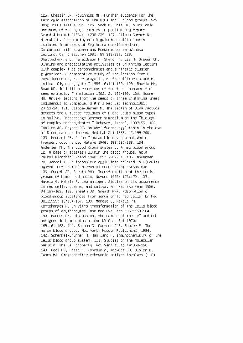
125. Chessin LN, McGinniss MH. Further evidence for theserologic association of the O(H) and I blood groups. VoxSang 1968; 14:194-201. 126. Voak D. Anti-HI, a new coldantibody of the H,O,I complex. A preliminary report.Scand J Haematol1964; 1:238-239. 127. Gilboa-Garber N,Mizrahi L. A new mitogenic D-galactosephilic lectinisolated from seeds of Erythrina corallodendron.Comparison with soybean and Pseudomonas aeruginosalectins. Can J Biochem 1981; 59:315-320. 128.Bhattacharyya L, Haraldsson M, Sharon N, Lis H, Brewer CF.Binding and precipitating activities of Erythrina lectinswith complex type carbohydrates and synthetic clusterglycosides. A comparative study of the lectins from E.corallodendron, E. cristagalli, E. f/abelliformis and E.indica. Glycoconjugate J 1989; 6:141-150. 129. Bhatia HM,Boyd WC. Inhibition reactions of fourteen "nonspecific"seed extracts. Transfusion 1962; 2: 106-109. 130. MooreHH. Anti-H lectins from the seeds of three Erythrina treesindigenous to Zimbabwe. S Afr J Med Lab Technol1981;27:33-34. 131. Gilboa-Garber N. The lectin of Ulva /actucadetects the L-fucose residues of H and Lewis blood typesin saliva. Proceedings Gentner symposium on the "biologyof complex carbohydrates." Rehovot, Israel, 1987:55. 132.Topliss JA, Rogers DJ. An anti-fucose agglutinin in the ovaof Dicentrarchus labrax. Med Lab Sci 1985; 42:199-200.133. Mourant AE. A "new" human blood group antigen offrequent occurrence. Nature 1946; 158:237-238. 134.Andersen PH. The blood group system L. A new blood groupL2. A case of epistasy within the blood groups. ActaPathol Microbiol Scand 1948; 25: 728-731. 135. AndersenPH, Jordal K. An incomplete agglutinin related to L(Lewis)system. Acta Pathol Microbiol Scand 1949; 26:636-638.136. Sneath JS, Sneath PHA. Transformation of the Lewisgroups of human red cells. Nature 1955; 176:172. 137.Makela 0, Makela P. Leb antigen. Studies on its occurrencein red cells, plasma, and saliva. Ann Med Exp Fenn 1956;34:157-162. 138. Sneath JS, Sneath PHA. Adsorption ofblood-group substances from serum on to red cells. Br MedBull1959; 15:154-157. 139. Makela 0, Makela PH,Kortekangas A. In vitro transformation of the Lewis bloodgroups of erythrocytes. Ann Med Exp Fenn 1967:159-164.140. Marcus DM. Discussion: the nature of the Le" and Lebantigens in human plasma. Ann NY Acad Sci 1970;169:161-163. 141. Salmon C, Cartron J-P, Rouger P. Thehuman blood groups. New York: Masson Publishing, 1984.142. Schenkel-Brunner H, Hanfland P. Immunochemistry of theLewis blood group system. III. Studies on the molecularbasis of the Le' property. Vox Sang 1981; 40:358-366.143. Gooi HC, Feizi T, Kapadia A, Knowles BB, Sloter D,Evans MJ. Stagespecific embryonic antigen involves (1-3)

fucosylated type 2 blood group chains. Nature 1981;292:156-158. 144. Hakomori S, Kannagi R.Glycosphingolipids as tumor-associated and differentiationmarkers. JNCI 1983; 71:231-251.
145. Lloyd KO. Blood group antigens as markers for normaldifferentiation and malignant changes in human tissues. AmJ Clin Patholl987; 87:129-139.
146. Fox N, Damjanov I, Knowles BB, Solter D.Immunohistochemical localization of the mousestage-specific embryonic antigen 1 in human tissues andtumors. Cancer Res 1983; 43:669-678.
147. Garin-Chesa P, Rettig WJ. Immunohistochemical analysisof LNT, NeuAc 2-3 LNT, and Le" carbohydrate antigens inhuman tumors and normal tissues. Am J Pathol1989;134:1315-1327.
148. Spohr U, Hindsgaul 0, Lemieux RU. Molecularrecognition II. The binding of the Lewis b and Y humanblood group determinants by the lectin IV of Grifjoniasimplicifolia. Can J Chern 1985; 63:2644-2652.
149. Spohr U, Lemieux RU. Synthetic, conformational, andimmunochemical studies of modified Lewis b and Y humanblood-group determinants to serve as probes for thecombining site of the lectin IV of Griffonia simplicifolia.Carbohydr Res 1988; 174:211-237.
150. Vandonselaar M, Delbaere LTJ, Spohr U, Lemieux RU.Crystallization of the lectin IV of Grifjoniasimplicifolia and its complexes with the Lewis b and Yhuman blood group determinants. J Bioi Chern 1987;262:1084810849.
151. Vicari G, Kabat EA. Immunochemical studies on bloodgroups. XLII. Isolation and characterization from ovariancyst fluid of a blood group substance lacking A, B, H,Le" and Leb specificity. J lmmunoll969; 102:821-825.
152. Hakomori S, Wang M, Young WW. Isoantigenic expressionof Forssman glycolipid in human gastric and colonicmucosa: its possible identity with "Alike antigen" inhuman cancer. Proc Natl Acad Sci USA 1977; 72:3023-3027.
153. Baker DA, Sugii S, Kabat EA, Ratcliffe RM, HermentinP, Lemieux RU. lmmunochemical studies on the combiningsites of Forssman hapten reactive hemagglutinins fromDolichos biflorus and Helix pomatia, and Wistaria

jloribunda. Biochemistry 1983; 22:2741-2750.
154. Prokop 0, Uhlenbruck G, Kohler W. A new source ofantibody-like substances having anti-blood groupspecificity. A discussion on the specificity of Helixagglutinins. Vox Sang 1968; 14:321-323.
155. Galbraith W, Goldstein IJ. Phytohemagglutinin of thelima bean (Phaseolus lunatus). Isolation,characterization, and interaction with type A blood groupsubstance. Biochemistry 1972; 11:3976-3984.
156. Hammarstrom S, Kabat EA. Purification andcharacterization of a bloodgroup A reactive hemagglutininfrom the snail Helix pomatia and a study of its combiningsite. Biochemistry 1969; 8:2696-2705.
157. Hammarstrom S, Kabat EA. Studies on specificity andbinding properties of the blood group A reactivehemagglutinin from Helix pomatia. Biochemistry 1971;10:1684-1692.
158. Hammarstrom S, Westoo A, Bjork I. Subunit structure ofHelix pomatia A hemagglutinin. Scand J Immunol1972;1:295-309.
159. Johnson HM. Human blood group A 1 specific agglutininof the butter clam Saxidomus giganteus. Science 1964;146:548-549.
160. Nguyen OK, Guillaume J-L, Hoebeke J. A blood group Aspecific lectin from the seeds of Crotalaria striata.Biochim Biophys Acta 1990; 1033:210213. 161. Judd WJ.Lectins and polyagglutination. In: Petz LD, Swisher SN,eds. Clinical practice of blood transfusion, 2nd ed. NewYork: Churchill Livingstone, 1989:137-151. 162. Rogers DJ,Fish BC. Marine algallectins. Lectin Rev 1991; 1:129-142.163. Etzler ME, Kabat EA. Purification and characterizationof a lectin (plant hemagglutinin) with blood group Aspecificity from Dolichos bif/orus. Biochemistry 1970;9:869-877. 164. Etzler ME. Horse gram (Dolichos biflorus)lectin. Methods Enzymol 1972; 28:340-344. 165. Sung LA,Kabat EA, Chien S. Interactions of lectins with membranereceptors on erythrocyte surfaces. J Cell Biol1985;101:646-651. 166. Garratty G, Willbanks E, Petz LD.Acquired-B antigen associated with Proteus vulgarisinfection. Vox Sang 1971; 21:45-46. 167. Judd WJ.Microbial-associated forms of polyagglutination (T, Tk andacquired-B). In: Beck ML, Judd WJ. eds. Polyagglutination.Washington DC: American Association of Blood Banks,

1980:23-53. 168. Gerbal A, Ropars C, Gerbal R, Cartron JP,Maslet C, Salmon C. Acquired B antigen disappearance by invitro acetylation associated with A 1 activityrestoration. Vox Sang 1976; 31:64-66. 169. Bird GWG.Erythrocyte polyagglutination. In: Greenwalt TJ, Steane EA,eds. CRC handbook. Series in clinical laboratory science,section D; blood banks. Cleveland: CRC Press,1977:443-454. 170. Beck ML. Blood group antigens acquiredde novo. In: Garratty G, ed. Blood group antigens anddisease. Arlington, Va: American Association of BloodBanks, 1983:45-47. 171. Rogers DJ, Blunden G, Evans PR.Ptilota plumosa, a new source of a bloodgroup B specificlectin. Med Lab Sci 1977; 34:193-200. 172. Anstee DJ,Holt PDJ, Pardoe GI. Agglutinins from fish ova definingblood groups Band P. Vox Sang 1973; 25:347-360. 173.Sakakibara F, Kawauchi H, Takayanagi G. Blood groupB-specific lectin of Plecoglossus altivelis (ayu fish)eggs. Biochim Biophys Acta 1985; 841:103111. 174. DownieDM, Madin DF, Voak D. An evaluation of salmon anti-Breagent in manual and automated blood grouping. Med LabSci 1977; 34:319-324. 175. Umetsu K, Yamashita K, SuzukiT. Purification and carbohydrate-binding specificities ofa blood type B binding lectin from hemolymph of a crab(Charybdisjaponica). J Biochem 1991; 109:718-721. 176.Poretz, RD, Riss H, Timberlake JW, Chien S-M. Purificationand properties of the hemagglutinin from Sophora japonicaseeds. Biochemistry 1974; 13: 250-256. 177. Wiener AS,Unger LJ, Cohen L, Feldman J. Type-specific coldautoantibodies as a cause of acquired hemolytic anemia andhemolytic transfusion reactions: biologic test with bovinered cells. Ann Intern Med 1956; 44:221-240.
178. Horstmann DM, Tatlock H. Cold agglutinins: adiagnostic aid in certain types of primary atypicalpneumonia. JAmMed Assoc 1943; 122:369-370.
179. Peterson OL, Ham TH, Finland M. Cold agglutinins(autohaemagglutinins) in primary atypical pneumonia.Science 1943; 97:167.
180. Beck ML. The I blood group collection. In: Moulds JM,Woods LL, eds. Blood groups: P,I,sd• and Pr. Arlington,Va: American Association of Blood Banks, 1991:23-52.
181. Marsh WL, Jenkins WJ. Anti-i: a new cold antibody.Nature 1960; 188:753.
182. Tippett P, Noades J, Sanger R, Race RR, Laima S,Holman CA, Buttimer RJ. Further studies of the I antigenand antibody. Vox Sang 1960; 5:107121.

183. Gold ER. Observations on the specificity of anti-0and anti-A 1 sera. Vox Sang 1964; 9:153-159.
184. Lewis M, Anstee DJ, Bird GWG, Brodheim E, Cartron J-P,Contreras M, Crookston M, Dahr W, Daniels GL, EngelfrietCP, Giles CM, Issitt PD, Jorgensen J, Kornstad L, LubenkoA, Marsh WL, McCreary J, Moore BPL, Morel P, Moulds JJ,Nevanlinna H, Nordhagen R, Okubo Y, Rosenfield RE, RougerP, Rubenstein P, Salmon C, Seidl P, Sistonen P, Tippett P,Walker G, YoungS. Blood group terminology 1990. Vox Sang1990; 58:152169.
185. Feizi T. The blood group Ii system: a carbohydrateantigen system defined by naturally monoclonal oroligoclonal autoantibodies of man. Immunol Commun 1981;10:127-156.
186. Anstee DJ. Blood group-active surface molecules of thehuman red blood cell. Vox Sang 1990; 58:1-20.
187. Childs RA, Feizi T, Fukuda M, Hakomori S-1. Bloodgroup I activity associated with band 3, the majorintrinsic membrane protein of human erythrocytes. Biochem J1978; 173:333-336.
188. Yokoyama M. Close relationship between A and I bloodgroups. Nature 1965; 206:411-412.
189. Joshi SR, Bahtia HM. A new red cell phenotype I-, i-:red cells lacking both I and i antigens. Vox Sang 1979;36:34-38.
190. Thomas AB. The i antigen complex: a new specificityunique to dividing human cells. Eur J Immunol1974;4:819-824.
191. Moore HH. A lectin displaying anti-i like specificityfrom the seeds of Pterocarpus angolensis. S Afr J Med LabTechnol1983; 29:43-44.
192. Doinel C, Ropars C, Salmon C. Effects of proteolyticenzymes and neuraminidase on the I and i erythrocyteantigen sites. Quantitative and thermodynamic studies.Immunology 1978; 34:653-662.
193. Landsteiner K, Levine P. Further observations onindividual differences of human blood. Proc Soc Exp BiolMed 1927; 24:941-942.

194. Marcus DM, Kundu SK, Suzuki A. The P group system:recent progress in immunochemistry and genetics. SeminHematol1981; 18:63-71.
195. Anstall HB, Blaylock RC. The P blood group system:biochemistry, genetics, and clinical significance. In:Moulds JM, Woods LL, eds. Blood groups: P, I, sd• and Pr.Arlington, Va: American Association of Blood Banks, 1991:1-22. 196. Levine P, Bobbitt OB, Walker RK, Kuhmichel A.Isoimmunization of a new blood factor in tumor cells. ProcSoc Exp Bioi Med 1951; 77:403-405. 197. Matson GA, SwansonJ, Noades J, Sanger R, Race RR. A "new" antigen andantibody belonging to the P blood group system. Am J HumGenet 1959; 11:26-34. 198. Kortekangas AE, Kaarsalo E,Melartin L, Tippett P, Gavin J, Noades J, Sanger R, RaceRR. The red cell antigen pk and its relationship to the Psystem. The evidence of three more pk families. Vox Sang1965; 10:385-404. 199. Voak D, Anstee DJ, Pardoe GI. Thea-galactose specificity of anti-Pk. Vox Sang 1973;25:263-270. 200. Issitt PD. The P blood group system. In:Issitt PD. ed. Applied blood group serology, 3rd ed.Miami: Montgomery Scientific Publication, 1985:203-218.201. Cameron GL, Staveley JM. Blood group P substance inhydatid cyst fluids. Nature 1957; 179:147-148. 202.Morgan WTJ, Watkins WM. Blood group P 1 substance. I.Chemical properties. Proceedings 9th congress InternationalSociety Blood Transfusion. Mexico, 1962. 1964:225-229.203. Naiki M, Kato M. Immunological identification ofblood group pk antigen on normal human erythrocytes andisolation of anti-Pk with different affinity. Vox Sang1979; 37:30-38. 204. Tippett P, Sanger R, Race RR,Swanson J, Busch S. An agglutinin associated with the Pand the ABO blood systems. Vox Sang 1965; 10:269-280. 205.Tippett P. Contributions of monoclonal antibodies tounderstanding one new and some old blood group systems.In: Garratty G, ed. Red cell antigens and antibodies.Arlington, Va: American Association of Blood Banks,1986:8398. 206. Iseki S, Masaki S, Levine P. A remarkablefamily with the rare human isoantibody anti Tj• in foursiblings: anti Tj• and habitual abortion. Nature 1954;173:1192-1193. 207. Allen FH Jr, Marsh WL, Jensen L, FinkJ. Anti-IP: an antibody defining another product ofinteraction between the genes of the I and P blood groupsystems. Vox Sang 1974; 27:422-426. 208. Booth PB. AntiITP 1 : an antibody showing a further association betweenthe I and P blood group systems. Vox Sang 1970; 19:85-90.209. Landsteiner K, Levine P. A new agglutinable factordifferentiating individual human bloods. Proc Soc ExpBioi Med 1927; 24:600-602. 210. Walsh RJ, Montgomery C. Anew human isoagglutinin subdividing the MN blood groups.

Nature 1947; 160:504. 211. Levine P, Kuhmichel AB, WigodM, Koch E. A new blood factor, s, allelic to S. Proc SocExp Bio Med 1951; 78:218-220. 212. Adamany AM, Kathan RH.Isolation of a tetrasaccharide common to MM, NN and MNantigens. Biochem Biophys Res Commun 1969; 37:171-178.213. German J, Metaxas MN, Metaxas-Buehler M, Louie E,Chaganti RSK. Further evaluation of a child with the Mkphenotype and a translocation affecting the long arms ofchromosomes 2 and 4. Cytogenet Cell Genet 1979; 25:160.
214. Holliman SM. The MN blood group system: distribution,serology and genetics. In: Unger PJ, Laird-Fryer B, eds.Blood group systems: MN and Gerbich. Arlington, Va:American Association of Blood Banks, 1989:1-29.
215. Wiener AS, Unger LJ, Gordon EB. Fatal hemolytictransfusion reaction caused by sensitization to a newblood factor U. J Am Med Assoc 1953; 153: 1444-1446.
216. Tomita M, Marchesi VT. Amino-acid sequence andoligosaccharide attachment sites of human erythrocyteglycophorin. Proc Natl Acad Sci USA 1975; 72:2964-2968.
217. Furthmayr H. Glycophorins A, B and C: a family ofsialoglycoproteins. Isolation and preliminarycharacterization of trypsin derived peptides. J SupramolStruct 1978; 9:79-95.
218. Tomita M, Furthmayr H, Marchesi VT. Primary structureof human erythrocyte glycophorin A: isolation andcharacterization of peptides and complete amino acidsequence. Biochemistry 1978; 17:4756-4770.
219. Rolih SD. Erythrocyte antigens of the MN system andrelated structures. In: Bell CA, ed. A seminar onantigens on blood cells and body fluids. 33rd annualmeeting of the American Association of Blood Banks.Washington, DC. 1980:209-247.
220. Issitt PD. The MN blood group system. Cincinnati,Ohio: Montgomery Scientific Publications, 1981.
221. Rolih S. Biochemistry of MN antigens. In: Blood groupsystems: MN and Gerbich. Arlington, Va: AmericanAssociation of Blood Banks, 1989.
222. Issitt PD. Applied blood group serology, 3rd ed.Miami: Montgomery Scientific Publications, 1985.
223. Metaxas MN, Metaxas-Buhler M. Mk: an apparent silent

allele at the MN locus. Nature 1964; 202:1123.
224. Metaxas MN, Metaxas-Buhler M, Romanski Y. Theinheritance of the blood group gene Mk and someconsiderations on its possible nature. Vox Sang 1971;20:509-518.
225. Tokunaga E, Sasakawa S, Tamaka K, Kawamata H, GilesCM, Ikin EW, Poole J, Anstee DJ, Mawby WJ, Tanner MJA.Two apparently healthy Japanese individuals of type MkMkhave erythrocytes which lack both the blood group MN andSs active sialoglycoproteins. J Immunogenet 1979; 6:383-390.
226. Stoltz JF, Streiff F, Genetet B. Demonstration of Mantigen on human lymphocytes by liquid phaseelectrophoresis. Vox Sang 1974; 26:467-469.
227. Simpson MB, Dunstan RA, Rosse WF, Munro AC, Fraser R,Nichols ME. Status of the MNSs antigens on humanplatelets. Transfusion 1987; 27:1518.
228. Wasniowaska K, Drzeniek Z, Lisowska E. The amino acidsof M and N blood group glycopeptides are different.Biochem Biophys Res Commun 1977; 76:385-390. 229. AnsteeDJ. Blood group MNSs-active sialoglycoproteins of the humanerythrocyte membrane. In: Sander SG, Nusbacher J,Schanfield MS, eds. Immunobiology of the erythrocyte. NewYork: Alan R Liss, 1980:67-98. 230. Springer GF, HuprikarSV. On the biochemical and genetic basis of the humanblood-group MN specificities. Haematologia 1972; 6:81-92.231. Springer GF, Desai PR. Common precursors of humanblood group MN specificities. Biochem Biophys Res Commun1974; 61:470-475. 232. Thomas DB, Winzler RJ. Structuralstudies on human erythrocyte sialoglycoproteins:alkali-labile oligosaccharides. J Bioi Chern 1969;244:5943-5946. 233. Springer GF, Ansell NJ. Inactivationof human erythrocyte agglutinogens M and N by influenzaviruses and receptor destroying enzyme. Proc Natl Acad SciUSA 1958; 44:182-189. 234. Kim Z, Uhlenbruck G.Untersuchungen iiber T-antigen and T-agglutinin. ZImmunitatsforsch Exp Klin Immunol1966; 130:88-99. 235.Lisowska E. Reaction of erythrocyte mucoproteins withanti-N phytagglutinins from Vicia graminea seeds. Nature1963; 198:865-866. 236. Lisowska E, Duk M. Effect ofmodification of amino groups of human erythrocytes on M,Nand N. 1 blood group specificities. Vox Sang 1975; 28:392-397. 237. Dahr W, Uhlenbruck G, Knott H.Immunochemical aspects of the MNSsblood group system. JImmunogenet 1975; 2:87-100. 238. Blanchard D, Dahr W,

Hummel M, Latron F, Beyreuther K, Cartron J-P.Glycophorins Band C from human erythrocyte membranes:purification and sequence analysis. J Bioi Chern 1987;262:5808-5811. 239. Siebert PD, Fukuda M. Isolation andcharacterisation of human glycophorin A eDNA clones by asynthetic oligonucleotide approach: nucleotide sequenceand mRNA structure. Proc Natl Acad Sci USA 1986;83:1665-1669. 240. Siebert PD, Fukuda M. Molecular cloningof a human glycophorin B eDNA nucleotide sequence andgenomic relationship to glycophorin A. Proc Natl Acad SciUSA 1987; 84:6735-6739. 241. Rahuel C, London J, d'AuriolL, Mattei MG, Tournamille C, Skrzynia C, Lebouc Y,Galibert F, Cartron J-P. Characterisation of eDNA clonesfor human glycophorin A: use for gene localisation andfor analysis of normal and glycophorin A-deficient(Finnish type) genomic DNA. Eur J Biochem 1988;172:147-153. 242. Tate CG, Tanner MJA. Isolation of eDNAclones for human erythrocyte membrane sialoglycoproteinsa and~. Biochem J 1988; 254:743-750. 243. Tanner MJA,Anstee DJ. The membrane change in En( a-) humanerythrocytes. Absence of the major erythrocytesialoglycoprotein. Biochem J 1976; 153:271-277. 244. DahrW, Uhlenbruck G, Leikola J, Wagstaff W, Landfriet K.Studies on the membrane glycoprotein defect of En(a-)erythrocytes. I. Biochemical aspects. J Immunogenet 1976;3:329-346. 245. Winzler RJ. A glycoprotein in humanerythrocyte membranes. In: Red cell membrane.Philadelphia: JB Lippincott, 1969:157-171.
246. Alloway GP, Burness AT. Site of attachment ofencephalomyocarditis virus on human erythrocytes. JVirol1986; 59:768-770.
247. Jokinen M, Ehnholm C. Vaisanen-Rhen V, Korhonen T,Pipkorn R, Kalkinen N, Gahmberg CG. Identification of themajor human sialoglycoprotein from red cells, glycophorinA, as the receptor for Escherichia coli IH 11165 andcharacterization of the receptor site. Eur J Biochem 1985;147:47-52.
248. Pasvol G. Receptors on red cells forPlasmodiumfalciparum and their interaction with merozoites.Philos Trans R Soc Lond 1984; B307:189-200.
249. Dahr W, Uhlenbruck G, Bird GWG. Cryptic A-likereceptor sites in human erythrocyte glycoproteins:proposed nature of Tn-antigen. Vox Sang 1974; 27:29-42.
250. Dahr W, Uhlenbruck G, Gunson HH, VanDer Hart M.Molecular basis of Tn-polyagglutinatinability. Vox Sang

1975; 29:36-50.
251. Ottensooser F, Silberschmidt K. Haemagglutinin anti-Nin plant seeds. Nature 1953; 172:914.
252. Duk M, Lisowska E, Kordowicz M, Wasnioska K. Studieson the specificity of the binding site of Vicia gramineaanti-N lectin. Eur J Biochem 1982; 123: 105-112.
253. Lisowska E. Reaction of erythrocyte mucoproteins withanti-N phytohemagglutinins from Vicia graminea seeds.Nature 1963; 198:865-866.
254. Romanowska E. Reaction of M and N blood-groupsubstances natural and degraded with specific reagents ofhuman and plant origin. Vox Sang 1964; 8:578-588.
255. Prigent MJ, Blanchard D, Cartron J-P. Membranereceptors for Vicia graminea anti-N lectin and its bindingto native and neuraminidase-treated erythrocytes. ArchBiochem Biophys 1983; 222:231-244.
256. Yanagi K, Ohyama K, Yamakawa T, Ohkuma S. Isolationand characterization of blood group N antigen precursorglycoproteins with Thomsen-Friedenreich (T) activity, Nantigen precursor glycoproteins and T -active glycoproteinsfrom cyst fluids of malignant ovarian clear cellcarcinoma. Int J Biochem 1990; 22:1015-1028.
257. Yanagi K, Ohyama K, Yamakawa T, Watanabe H, HirakawaS, Ohkuma S. Biochemical characterization of glycoproteincomponents in human ovarian cyst fluids by lectins. Int JBiochem 1990; 22:659-663.
258. Moon GJ, Wiener AS. A new source of anti-N lectin:leaves of the Korean Vicia unijuga. Vox Sang 1974;26:167-170.
259. Ohkuma S, Ohyama K, Akitoshi Y, Okada Y, Kawanishi M,Matsuda K. Immunochemical property of anti-N hemagglutininof leaves of Vicia unijuga. J Clin Exp Med 1978;107:631-633.
260. Yanagi K, Ohyama K, Yamakawa T, Hashimoto K, OhkumaS. Purification and characterization of anti-N lectinfrom Vicia unijuga leaves. Int J Biochem 1990; 22:43-52.
261. Fletcher G. The anti-N phytoagglutinin of Bauhiniavariegata. Aust J Sci 1959; 22:167.

262. Bird GWG, Wingham J. Agglutinins for antigens of twodifferent human blood group systems in the seeds ofMoluccella /aevis. Vox Sang 1970; 18: 235-239. 263. JuddWJ, Murphy LA, Goldstein IJ, Campbell L, Nichols ME. Ananti-B reagent prepared from a-o-galactopyranosyl-bindingisolectins from Bandeiraea simplicifolia seeds.Transfusion 1978; 18:274-280. 264. Allen NK, BrilliantineL. A survey of hemagglutinins in various seeds. Jlmmunol1969; 102:1295-1299. 265. Nakajima H, Nakayama T.Anti-M lectin of Iberis amana L. Rep Jpn Natl Res lnstPolice Sci 1968; 21:111. 266. Okada T, Nakajima H. Anti-Mlectin of Japanese radish. Rep Jpn Natl Res lnst Res lnstPolice Sci 1970; 23:207. 267. Vaisiinen, V, Korhonen T,Jokinen M, Gahmberg CG, Ehnholm C. Blood group M specifichaemagglutination in pyelonephritogenic Escherichia coli.Lancet 1982; 1:1192. 268. Bird GWG, Wingham J.N-Acetyl-neuraminic (sialic) acid and human blood groupantigen structure. Vox Sang 1970; 18:240-243. 269.Furthmayr H. Structural comparison of glycophorins andimmunochemical analysis of genetic variants. Nature 1978;271:519-524. 270. Darnborough J, Dunsford I, Wallace JA.The En• antigen and antibody. A genetical modification ofhuman red cells affecting their blood grouping reactions.Vox Sang 1969; 17:241-255. 271. Uhlenbruck G. Action ofproteolytic enzymes on the human erythrocyte surface.Nature 1961; 190:181. 272. Bird GWG, Wingham J. The actionof seed and other reagents on En(a-) erythrocytes. VoxSang 1973; 24:48-57. 273. lssitt PD, Pavone BG, Rolih SD.New concepts of the structure of En• abstracted.Transfusion 1979; 19:662 274. Bird GWG. Specificity ofGlycine soja agglutinins. Experientia 1963; 19: 625. 275.Furuhjelm U, Myllyla G, Nevanlinna HR, Nordling S, PirkolaA, Gavin J, Gooch A, Sanger R, Tippett P. The red cellphenotype En( a-) and anti En•: serological andphysicochemical aspects. Vox Sang 1969; 17:256-278. 276.Hiibener G. Untersuchungen uber isoagglutination mitbesonderer beriicksichtigung scheinbarer abweichungen vomgruppenschema. lmmunobiology 1925; 45:223-226. 277.Thomsen 0. Ein vermehrungsfiihiges agens als veriindererdes isoagglutinatorischen verhaltens der rotenblutkorperchen eine bisher unbekannte quelle derfehlbestimmung. lmmunobiology 1927; 52:85-90. 278.Friedenreich V. The Thomsen hemagglutination phenomenon.Copenhagen: Levin and Munskgaard, 1930. 279. Levine P,Katzin EM. Temporary agglutinability of red blood cells.Proc Soc Exp Bioi Med 1938; 39:167-169. 280. Bird GWG.Clinical aspects of red blood cell polyagglutinability ofmicrobial origin. In: Salmon C, ed. Blood groups and otherred cell surface markers in health and disease. New York:Masson, 1982:55-64. 281. Springer GF, Desai PR, Murthy

MS, Yang HJ, Scanlon EF. Precursors of blood group MNantigens as carcinoma associated antigens. Transfusion1979; 19:233-249.
282. Chorpenning FW, Dodd MC. Polyagglutinable erythrocytesassociated with bacteriogenic transfusion reactions. VoxSang 1965; 10:460-471.
283. Bird GWG. Anti-Tin peanuts. Vox Sang 1964; 9:748-749.
284. Bird GWG. Lectins and red cell polyagglutinability:history, comments and recent developments. In: Beck ML,Judd WJ, eds. Polyagglutination. Washington DC: AmericanAssociation of Blood Banks, 1980:71-90.
285. Rickard KA, Robinson RJ, Worlledge SM. Acute acquiredhaemolytic anaemia associated with polyagglutination. ArchDis Child 1969; 44:102-105.
286. Moores P, Pudfin D, Patel PL. Severe hemolytic anemiain an adult associated with anti-T. Transfusion 1975;15:329-333.
287. Levene C, Sela R, Blat J, Friedlaender M, Manny N.Intravascular hemolysis and renal failure in a patientwith T polyagglutination. Transfusion 1986; 26: 243-245.
288. Van Loghem JJ Jr, Vander Hart M, Land ME.Polyagglutinability of red cells as a cause of a severehemolytic transfusion reaction. Vox Sang 1955; 5: 125-128.
289. Gray JM, Beck ML, Oberman HA. Clostridial-induced typeI polyagglutinability with associated intravascularhemolysis. Vox Sang 1972; 22:379-383.
290. Judd WJ, Oberman HA, Flynn S. Fatal intravascularhemolysis associated with T-polyagglutination. Transfusion1982; 22:345-346.
291. Klein PJ, Bulla M, Newman RA, Muller P, Uhlenbruck G,Schaefer HE, Kruger G, Fisher R. Thomsen-Friedenreichantigen in haemolytic-uraemic syndrome. Lancet 1974;2:1024-1025.
292. Lotan R, Skutelsky E, Danon D, Sharon N. Thepurification, composition and specificity of the anti-Tlectin from peanut (Arachis hypogaea). J Bioi Chern 1975;250:8518-8523.
293. Bird GWG, Wingham J. Tk: a new form of red cell

polyagglutination. Br J Haematol1972; 23:759-763.
294. Doinel C, Andreu G, Cartron J-P, Salmon C, Fukuda MN.Tk polyagglutination produced in vitro by anendo-~-galactosidase from Escherichia jreundii. Vox Sang1980; 38:94-98.
295. Judd WJ. The role of exo-~-galactosidases inTk-activation. Transfusion 1980; 20:622.
296. Bird GWG, Wingham J, Beck ML, Pierce SR, Oates GD,Pollock A. Th a new form of erythrocyte polyagglutination.Lancet 1978; 1:1215-1216.
297. Veneziano G, Rasore-Quartino A, Sansone G. Therythrocyte polyagglutination. Lancet 1978; 2:483.
298. Rawlinson V, Stratton F. Incidence ofT activation in ahospital population. Vox Sang 1984; 46:306-317.
299. Levene NA. Polyagglutination in Israel; MD thesisHebrew University, Hadassah Medical School [in Hebrew].1984:1-68.
300. Herman JH, Whiteheart W, Shirey RS, Johnson RJ,Kickier TS, Ness PN. Red cell Th activation: biochemicalstudies. Br J Haematol 1987; 65:205209. 301. Okubu Y, SenoT, Yamaguchi H. A persistent type of erythrocytepolyagglutinability Th. Transfusion 1984; 24:277-278.302. Levene NA, Levene C, Gecker K, Sigler E, Merhav H,Berrebi A. Th polyagglutination with fatal outcome in apatient with massive intravascular hemolysis and perforatedtumor of colon. Am J Hematol1990; 35:215-216. 303.Sondag-Thull D, Levene NA, Levene C, Manny N, Yew WahL,Bird GWG, Schechter Y, Francois-Gerard C, Huet M,Blanchard D. Characterization of a neuraminidase fromCorynebacterium aquaticum responsible for Thpolyagglutination. Vox Sang 1989; 57:193-198. 304. BirdGWG, Wingham J. Vicia cretica a powerful lectin forT and Thbut not for Tk or other polyagglutinable erythrocytes. JClin Pathol1981; 34:69-70. 305. Bird GWG, Wingham J, SegerR, Kenny AB. Tx a "new" red cell cryptantigen exposed bypneumococcal enzymes. Blood Transfus Immunohematol 1982;25:215-216. 306. Wolach B, Nahum S, Bird GWG, Moulds JJ,Bar-Shany S, Ben-Porath D, Levene NA, Sela R, Levene C.Tx polyagglutination in three members of one family. ActaHaematol1987; 78:45-47. 307. Yang E, Moore BPL, Liew YW,Bird GWG. Further observations on the Vicia creticalectin. Transfusion 1986; 26:306. 308. Gerbal A, Maslet C,Salmon C. Immunologic aspects of the acquired-B antigen.

Vox Sang 1975; 28:398-405. 309. Graninger W, Rameis H,Fischer K, Poschmann A, Bird GWG, Wingham J, Neumann E."VA," a new type of erythrocyte polyagglutinationcharacterized by depressed H receptors and associated withhemolytic anemia. I. Serological and hematologicalobservations. Vox Sang 1977; 32:195-200. 310. Moreau R,Dausset J, Bernard J, Moullec J. Anemie hemolytique acquiseavec polyagglutinabilite hematies par un nouveau facteurpresent dans le serum humain normal (anti-Tn). Bull SocMed Hop Paris 1957; 73:569-587. 311. Cartron JP, Andreu G,Cartron J, Bird GWG, Salmon C. Demonstration ofT-transferase deficiency in Tn-polyagglutinable bloodsamples. Eur J Biochem 1978; 92:111-119. 312. Myllyla G,Furhjelm U, Nordling S, Pirkola A, Tippett P, Gavin J.Sanger R. Persistent mixed field polyagglutinability:electrokinetic and serological aspects. Vox Sang 1971;20:7-23. 313. Bird GWG. Significant advances in lectinsand polyagglutinable red cells. Presented at the XVIIcongress of the International Society of Blood Transfusion.Paris, Libraire Arnette. 1978:87-98. 314. Bird GWG,Wingham J, Richardson SON. Myelofibrosis, autoimmunehemolytic anemia and Tn polyagglutinability. Haematologia1985; 18:99-102. 315. Bird GWG, Wingham J, Pippard MJ,Hoult JG, Melikian V. Erythrocyte membrane modification inmalignant disease of myeloid and lymphoreticular tissuesI. Tn-polyagglutination in acute myelocytic leukemia. Br JHaematol 1976; 33:289-294. 316. Wilson MH, Cott ME, SotusPC. Probable Tn-activation in utero. [abstract].Transfusion 1980; 20:622. 317. Rose RR, Skradski KJ,Polesky HF, Beck ML, Judd WJ, Rogers E. Tran391 sientneonatal Tn-activation: another example [abstract].Transfusion 1983; 23:422.
318. Schultz M, Fortes P, Brewer L, Miller A, Beck M. "Inutero" exposure of Tn and Th cryptantigens [abstract].Transfusion 1983; 23:422.
319. Beck ML. Tn: a nonmicrobial form of polyagglutination.In: Beck ML, Judd WJ, eds. Polyagglutination. WashingtonDC: American Association of Blood Banks, 1980:55-70.
320. Ness PM, Oarratty 0, Morel PE, Perkins HA.Tn-po1yagglutination preceding acute leukemia. Blood 1979;54:30-34.
321. Crookston JH, Crookston MC, Burnie KL, Francombe WH,Dacie JV, Davis JA, Lewis SM. Hereditary erythroblasticmultinuclearity with a positive acidified serum test: atype of congenital dyserythropoeitic anemia. Br JHaematol1969; 17:11-26.

322. Rochant H, Oerbal A. Polyagglutinabilitie due aL'antigen Hempas. Rev Fr Transfus Immunohematol1976;19:239-245.
323. Harris PA, Kaczmarski OR, Moulds JJ, Bird OWO, ShahaNO. An inherited RBC characteristic NOR, resulting inerythrocyte polyagglutination. Transfusion 1979; 19:664.
324. Cazal P, Monis M, Caubel J, Brives J.Polyagglutinabilite hereditaire dominante: antigene prive(Cad) correspondent a un anticorps public et a une lectinede Dolichos bif/orus. Rev Fr Transfus Immunohematol1968;11:209221.
325. Blanchard D, Cartron JP, Fournet B, Montreuil J, vanHalbeek H, Vliegenthart JFO. Primary structure of theoligosaccharide determinant of blood group Cadspecificity. J Bioi Chern 1983; 358:7691-7695.
326. Bird OWO. Human blood group mosaics and chimaeras.Biotest Bull 1985; 2:215-224.
327. Booth PB, Plaut 0, James JD, Ikin EW, Moores P, SangerR, Race RR. Blood chimerism in a pair of twins. Br Med J1957; 1:1456-1458.
328. Bird OWO, Wingham J. TheM, N, and N. 8 receptors ofTn-erythrocytes. Vox Sang 1974; 26:171-175.
329. Bird OWO. Haemagglutinins in Indian plants. Army MedCorps J (India) 1955; 9:17-25.
330. Roque-Barreira MC, Campos-Neto A. Jacalin: anlgA-binding lectin. JImmunol1985; 134:1740-1743.
331. Kondoh H, Kobayashi K, Hagiwara K, Kajii T. Jacalin, ajackfruit lectin precipitates lgA 1 not lgA 2 subclasson gel diffusion reaction. J Immunol Methods 1986;88:171-173.
332. Autcouturier P, Mihaesco E, Mihaesco C, Preud'hoommeJL. Characterization of jacalin, the human lgA and lgDbinding lectin from jack fruit. Mol lmmunol187;24:503-511.
333. Vyas ON, Holmdahl L, Perkins HA, Fudenberg HH.Serologic specificity of human anti-lgA and itssignificance in transfusion. Blood 1969; 34:573-581.

334. Hodson C. The incidence and characterisation ofvariant red cell sialoglycoproteins in a UK population.Thesis, Lancaster, 1985. Quoted in: Bird OWO. Lectins inimmunohematology. Transfus Med Rev 1989; 3:55-62. 335.Schnebli HP, Dukor P. Plant agglutinins used to distinguishbetween different classes of mouse lymphocytes. Bur JImmuno11972; 2:607-609. 336. Reisner Y, Ravid A, Sharon N.Use of soybean agglutinin for the separation of mouse Band T lymphocytes. Biochem Biophy Res Commun 1976; 72:1585-1591. 337. Reisner Y, Itzicovitch L, Meshorer A,Sharon N. Hemopoietic stem cell transplantation usingmouse bone-marrow and spleen cells fractionated bylectins. Proc Natl Acad Sci USA 1978; 75:2933-2936. 338.Reisner Y, O'Reilly RJ, Kapoor N, Good RA. Allogeneic bonemarrow transplantation using stem cells fractionated bylectins: VI. In vitro anlaysis of human and monkey bonemarrow cells fractionated by sheep red blood cells andsoybean agglutinin. Lancet 1980; 2:1320-1324. 339. ReisnerY, Hodes MB, O'Reilly RJ, Good RA. Enrichment for CRU-Cfrom murine and human bone marrow using soybeanagglutinin. Blood 1982; 59: 360-363. 340. Reisner Y,Kapoor N, Kirkpatrick D, Pollack MS, Dupont B, Good RA,O'Reilly RJ. Transplantation for acute leukemia with HLA-Aand B nonidentical parental marrow cells factionated withsoybean agglutinin and sheep red blood cells. Lancet1981; 2:327-331. 341. Reisner Y, Kapoor RA, Good RA,O'Reilly RJ. Allogeneic bone marrow transplantation inmouse, monkey and man using lectin-separated grafts. In:Slavin S, ed. Tolerance in bone marrow and organtransplantation. Amsterdam: Elsevier Scientific,1984:293-308. 342. Krolick KA, Villemez C, Isakson P, UhrJW, Vitetta ES. Selective killing of normal or neoplasticB cells by antibodies coupled to the A chain of ricin.Proc Natl Acad Sci USA 1980; 77:5419-5423. 343. Raso V,Ritz J, Basala M, Schlossman SF. Monoclonal antibody-ricinchain conjugation selectively cytotoxic for cells bearingthe common acute lymphoblastic leukemia antigen. CancerR~s 1982; 42:457-464. 344. Seon BK. Specific killing ofhuman T-leukemia cells by immunotoxins prepared with ricinA chain and monoclonal anti-human T -cell leukemiaantibodies. Cancer Res 1984; 44:259-264. 345. Sienna S,VillaS, Bonadonna GU, Bregni M, Gianni AM. Ex vivodepletion of human bone marrow T lymphocytes by soybeanlectin fractionation followed by treatment with anti-pan-T-cell (CD5) ricin Achain immunotoxin. TransplantationProc 1987; 19:2735-2737. 346. Morecki S, Pavlotzky F,Margel S, Slavin S. Purging breast cell cancer cells inpreparation for autologous bone marrow transplantation.Bone Marrow Transplantation 1987; 1:357-363. 347.Springer GF, Desai PR, Robinson MK, Tegtmeyer H, Scanlon

EF. The fundamental and diagnostic role of T and Tnantigens in breast carcinoma at the earliest histologicstage and throughout. In: Dao T, Brodie A, Ip C, eds.Tumor markers and their significance in the management ofbreast cancer. Prog Clin Bioi Res 1986; 204:47-70. 348.Springer GF, Desai PR, Wise W, Carlstedt SC, Tegtmeyer H,Stein R, Scanlon EF. Pancarcinoma T and Tn epitopes:autoimmunogens and diagnostic 393 markers that revealincipient carcinomas and help establish prognosis. In:Herberman RB, Mercer DW, eds. Immunodiagnosis of cancer,2nd ed. New York: Marcel Dekker, 1990:587-612.
349. Levene C, Levene NA, Buskila D, Manny N. Red cellpolyagglutination. Transfus Med Rev 1988; 2:176-185.
350. Anglin JH Jr, Lerner MP, Nordquist RE. Bloodgroup-like activity released by human mammary carcinomacells in culture. Nature 1977; 269:254-255.
351. Buskila D, Levene C, Bird GWG, Levene NA.Polyagglutination in hospitalized patients: a prospectivestudy. Vox Sang 1987; 52:99-102.
352. Buskila D, Levene C, Bird GWG, Levene NA. Exposure ofcryptantigens on erythrocytes in patients with breastcancer. Cancer 1988; 61:2455-2459.
353. Tibebu M. Polyagglutination in lymphomas, bone marrowtransplants and beta thalassemia major. Thesis forHaematology specialization: Hadassah University MedicalSchool, Jerusalem, 1987.
354. Levene NA. Polyagglutination in patients with lymphoma[Hebrew]. Thesis for basic sciences, Internal Medicine,Ben Gurion University, Beer Sheva, 1988.