Antibacterial and Antioxidant Activities of a Food Lectin Isolated from the Seeds of Lablab ...
10
www.ajethno.com American Journal of Ethnomedicine, 2014, Vol. 1, No. 1, 008-017 Available online at http://www.ajethno.com © American Journal of Ethnomedicine Antibacterial and Antioxidant Activities of a Food Lectin Isolated from the Seeds of Lablab purpureous Repon Kumer Saha* 1 , Syed Hossain Mahmood Tuhin 1 , Nishat Jahan 1 , Ajoy Roy 1 and Priyanka Roy 2 1 Department of Pharmacy, East West University, Dhaka, Bangladesh. 2 Dhaka Medical College, Dhaka, Bangladesh ABSTRACT Lectins were precipitated by ammonium sulphate precipitation method. Carbohydrate and proteins were detected by thin layer chromatography and infrared spectroscopy techniques. Crude lectins were partially characterized by its binding affinity with human erythrocytes by hemagglutination inhibition assay and their molecular weights were determined by SDS-page gel electrophoresis. The amount of proteins was quantitatively measured by Lowry method. Antibacterial and antifungal activities were investigated by disk diffusion assay. Salmonella induced hemagglutination inhibition activities were observed. Isolated lectins contained carbohydrates and protein conjugates in their structures. The isolated lectins showed molecular weight was about 45 kD and 18 kD and seemed as a dimeric. It showed weaker antibacterial and antifungal activities. Conclusions: The seed of Lablab purpureous is food stuff and it may be considered as good source of proteins and as an antimicrobial protective agent. Keywords- Musa sapientum var. sylvesteris, Hemolysis activity, Flavanoids, Antifungal, Hemagglutination, Protein, Carbohydrate. INTRODUCTION Lectins are becoming very popular with a widespread use and application in biological science. Because of the various application and uses of lectins, it is of utmost importance to isolate and purify lectins from abundant and less expensive sources, such as Hyacinth bean (Lablab purpureus). Moreover the most abundant lectins can be found in the seeds of leguminous plants. Legumes such as hyacinth bean seed contain nearly 10% more fiber while winged bean contains three times more fiber than common bean. The potential breast cancer fighting chemical known as kievitone is found in hyacinth bean 1 . The most abundant lectins can be found in the seeds of leguminous plants but they can also be obtained from other plant parts (leaves, roots, stems). Studies have shown that lectins play an important role in the mechanism of binding Rhizobium to root hairs of legume plants 2 . Toxicological
-
Upload
independent -
Category
Documents
-
view
1 -
download
0
Transcript of Antibacterial and Antioxidant Activities of a Food Lectin Isolated from the Seeds of Lablab ...

www.ajethno.com
American Journal of Ethnomedicine, 2014, Vol. 1, No. 1, 008-017 Available online at http://www.ajethno.com © American Journal of Ethnomedicine
Antibacterial and Antioxidant Activities of a
Food Lectin Isolated from the Seeds of Lablab
purpureous
Repon Kumer Saha*1, Syed Hossain Mahmood Tuhin1, Nishat Jahan1, Ajoy
Roy1 and Priyanka Roy2
1Department of Pharmacy, East West University, Dhaka, Bangladesh. 2Dhaka Medical College, Dhaka, Bangladesh ABSTRACT
Lectins were precipitated by ammonium sulphate precipitation method.
Carbohydrate and proteins were detected by thin layer chromatography and
infrared spectroscopy techniques. Crude lectins were partially characterized by its
binding affinity with human erythrocytes by hemagglutination inhibition assay
and their molecular weights were determined by SDS-page gel electrophoresis.
The amount of proteins was quantitatively measured by Lowry method.
Antibacterial and antifungal activities were investigated by disk diffusion assay.
Salmonella induced hemagglutination inhibition activities were observed. Isolated
lectins contained carbohydrates and protein conjugates in their structures. The
isolated lectins showed molecular weight was about 45 kD and 18 kD and seemed
as a dimeric. It showed weaker antibacterial and antifungal activities.
Conclusions: The seed of Lablab purpureous is food stuff and it may be
considered as good source of proteins and as an antimicrobial protective agent.
Keywords- Musa sapientum var. sylvesteris, Hemolysis activity, Flavanoids,
Antifungal, Hemagglutination, Protein, Carbohydrate.
INTRODUCTION
Lectins are becoming very popular with a
widespread use and application in biological science. Because of the various application and uses of lectins, it is of utmost importance to isolate and purify lectins from abundant and less expensive sources, such as Hyacinth bean (Lablab purpureus). Moreover the most abundant lectins can be found in the seeds of leguminous plants. Legumes such as hyacinth bean seed contain
nearly 10% more fiber while winged bean contains three times more fiber than common bean. The potential breast cancer fighting chemical known as kievitone is found in hyacinth bean1. The most abundant lectins can be found in the seeds of leguminous plants but they can also be obtained from other plant parts (leaves, roots, stems). Studies have shown that lectins play an important role in the mechanism of binding Rhizobium to root hairs of legume plants2. Toxicological

American Journal of Ethnomedicine
Page 008-017
studies of extracts showed bioefficacy against causal agents of malaria, dracunculiasis, and amoebiasis3. In an investigation about the lectins of L. purpureus, it was found that the lectins are nonspecific for human blood groups and they agglutinate a variety of other erythrocytes. Among a number of sugars, D-glucose and D-mannose inhibited the haemagglutinating activity of the lectins4.
The aim of this research project was to carry out the characterization of the isolated lectin from this plant seed and also to find out the different pharmacological and medicinal use that it may possess. The objective of this work is also to explore the possibility of developing new drug candidates from this plant for the treatment of various diseases and using this lectin protein as a potential tool to be used for blood typing, bacterial typing indicated by assays like hemagglutination. MATERIALS AND METHODS
Isolation The fresh seeds were collected from
the surrounding of Dhaka, Bangladesh during January, 2010 and identified by the taxonomist of the Bangladesh National Herbarium, Mirpur, Dhaka as Lablab Purpureus (Family: Fabaceae). A voucher specimen of the plant has been deposited (Accession No.: DACB 37899) in the herbarium for further reference. The seeds were dried and grinded. Lectins were isolated as previously described method with some modification 5. Seeds of Lablab purpureus were ground to a powder in an electric mill and filtered through 80 mesh grit. The powder (5g) was mixed with 0.15M NaCl (1:8, w/v) for 48 h at 4°C, and filtered through 80 mesh grid. Subsequently, the filtrate was centrifuged at 10000×g for 30 minutes, and the supernatant was fractionally precipitated with ammonium sulfate at 40%, 50%, 60%, and 70%
saturation, respectively. The four pellets were combined, dissolved in a minimal volume of water, and separated using ultra centrifugation at at 10000×g for 30 minutes at 4°C.
Determination of Protein and Carbohydrate Concentration Lowry's method6 was used for protein quantification, using bovine serum albumin (BSA) as the standard. The relative protein concentration of the eluted fractions was determined by measuring the absorbance at 280nm. Hemagglutinating Inhibition Activity Assay
Hemagglutination inhibition activity was measured as previously described 5 with little modification. Briefly, serial two-fold dilutions of the lectin solution in microtiter v-plates (25 μL) was mixed with 25 μL 0.5% human red blood cell suspension in saline (pH 7.2). Readings were recorded after about 30 minutes at room temperature, when the blank had fully sedimented. The hemagglutination titer, defined as the reciprocal of the highest dilution exhibiting hemagglutination, was treated as one hemagglutination unit. Specific activity was expressed as the number of hemagglutination units per mg protein. SDS-PAGE analysis
Sodium dodecyl sulfate polyacry-lamide gel electrophoresis (SDS-PAGE) was performed in accordance with the method of Laemmli7, using a 15% separating and a 5% stacking gel. Detection of carbohydrate and protein using TLC method
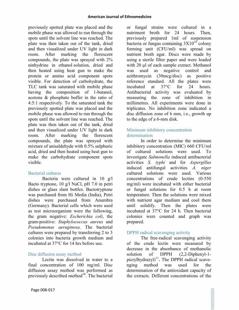
The presence of carbohydrate and protein was detected using TLC method as previously described method8,9 with little modifications. For detection of proteins, TLC tank was saturated with mobile phase having the composition of 1-butanol, glacial acetic acid and water in the ratio of 4:1:1 respectively. To the saturated tank the
American Journal of Ethnomedicine
Page 008-017
previously spotted plate was placed and the mobile phase was allowed to run through the spots until the solvent line was reached. The plate was then taken out of the tank, dried and then visualized under UV light in dark room. After marking the florescent compounds, the plate was sprayed with 2% ninhydrine in ethanol solution, dried and then heated using heat gun to make the protein or amino acid component spots visible. For detection of carbohydrate, the TLC tank was saturated with mobile phase having the composition of 1-butanol, acetone & phosphate buffer in the ratio of 4:5:1 respectively. To the saturated tank the previously spotted plate was placed and the mobile phase was allowed to run through the spots until the solvent line was reached. The plate was then taken out of the tank, dried and then visualized under UV light in dark room. After marking the florescent compounds, the plate was sprayed with mixture of anisaldehyde with 0.5% sulphuric acid, dried and then heated using heat gun to make the carbohydrate component spots visible.
Bacterial cultures
Bacteria were cultured in 10 g/l Bacto tryptone, 10 g/l NaCl, pH 7.0 in petri dishes or glass slant bottles. Bactotryptone was purchased from Hi Media (India), Petri dishes were purchased from Anumbra (Germany). Bacterial cells which were used as test microorganism were the following, the gram negative: Escherichia coli, the gram-positive: Staphylococcus aureus and Pseudomonas aeruginosa. The bacterial cultures were prepared by transferring 2 to 3 colonies into bacteria growth medium and incubated at 37°C for 14 hrs before use.
Disc diffusion assay method
Lectin was dissolved in water to a final concentration of 100 mg/ml. Disc diffusion assay method was performed as previously described method10. The bacterial
or fungal strains were cultured in a nutriment broth for 24 hours. Then, previously prepared 1ml of suspension bacteria or fungus containing 3X10¹⁰ colony forming unit (CFU/ml) was spread on nutrient broth agar. Discs were made by using a sterile filter paper and were loaded with 20 μl of each sample extract. Methanol was used as negative control and azithromycin (30mcg/disc) as positive reference standard. All the plates were incubated at 37°C for 24 hours. Antibacterial activity was evaluated by measuring the zone of inhibition in millimetres. All experiments were done in triplicates. No inhibition zone indicated a disc diffusion zone of 6 mm, i.e., growth up to the edge of a 6-mm disk. Minimum inhibitory concentration determination
In order to determine the minimum inhibitory concentration (MIC) 660 CFU/ml of cultured solutions were used. To investigate Salmonella induced antibacterial activities S. typhi and for Aspergillus induced antifungal activities A. niger cultured solutions were used. Various concentrations of crude lectins (0-550 mg/ml) were incubated with either bacterial or fungal solutions for 0.5 h at room temperature. Then the solutions were mixed with nutrient agar medium and cool them until solidify. Then the plates were incubated at 37°C for 24 h. Then bacterial colonies were counted and graph was prepared.
DPPH radical scavenging activity
The free-radical scavenging activity of the crude lectin were measured by decrease in the absorbance of methanolic solution of DPPH (2,2-Diphenyl-1-picrylhydrazyl)11. The DPPH radical scave-nging method was used for the determination of the antioxidant capacity of the extracts. Different concentrations of the

American Journal of Ethnomedicine
Page 008-017
plant extract (2, 4, 6, 8 & 10μg/ml, in water were added at an equal volume (10ml) to aqueous solution of DPPH (400μg/ml). Different concentrations of L-Ascorbic acid (2-10 mg/ml) were used as the standard antioxidant. After 30 min at room temperature, the absorbance values were measured at 517 nm on a spectrophotometer and converted into the percentage antioxidant activity using the following equation: DPPH antiradical scavenging capacity (%) = (Absorbance of sample – Absorbance of blank) × 100/Absorbance of blank. Methanol plus different concentration of plant extract solution was used as a blank, while DPPH solution plus methanol was used as a control. IC50 values denote the concentration of the sample required to scavenge 50% of DPPH radicals.
RESULTS AND DISCUSSION
Detection of Carbohydrate and Protein We separated the carbohydrate like
molecules and protein like molecules present in the isolated lectin by TLC as described in the method section. Then, we examine the plates under ultra-violet light and normal light. For detection of carbohydrate the dried plates were sprayed with anisaldehyde with 0.5% sulphuric acid and heated and other sugars used in the experiment as control (Fig 1). Such a result indicates the presence of carbohydrate like residue. Charring the dried plates with 0.2% ninhydrin solution formed a brown spot on the TLC plate (Fig. 1B) indicates the presence of protein. It was found in this study that the spectral region between 1500 and 800 cm−1 would cover most of characteristic absorption bands relevant to major sugars. Absorption peaks of glucose include 902, 1036, 1360, and 1431 cm−1 with a key peak at 1032 cm−1 was found (data not shown). Absorption bands of fructose includes 923, 978, 1067, 1250, 1346, and 1418 cm−1 with a key peak at
1053 cm−1 which corresponds to C-O bending and C-OH stretching12. Therefore lectin showed the presence of carbohydrate and protein residues. Lowry's method also showed the presence of protein in the lectin (data not shown). Hemagglutinating Activity Assay
The human blood types have different sugar moieties on the surface of the cell. Type A has N-acetyl- D-galactosamine, D-galactose for type B and L- fructose in type O. Blood type AB contains the sugar determinants for both A and B. Agglutination occurs when the lectin interacts with these sugar moieties. Non-blood type specificity of the lectin may be due to the presence of multiple binding sites where it can recognize all the determinants for each blood type.
In this study we found the concentrations of lectin required for the hemagglutination of blood-group-AB (+), B(+), A(+), and O(+) at 2.53, 5.06, 0.28, and 0.55 mg/well respectively as shown in Fig.3. Therefore, it showed the activity as like as the bryony lectin, which is not a blood-group-specific and Eranthis hyemalis lectin with group-O erythrocytes specificity. Crude lectins from Lablab purpureus preferentially agglutinates group-A erythrocytes, thus exhibiting presence of lectins13,14. Electrophoretic analysis
Reducing SDS PAGE of TTML showed double band with a molecular weight of approximately 45 kDa and 18 kDa was seen as shown in Fig. 2. Therefore, it resembles some of plant lectin with respect to sugar binding specificity and some structural aspects, namely that from bryony (Bryonia ioica) root stocks and Eranthis hyemalis lectin which is also a Nacetylgalactosamine- specific lectin composed of two non-identical subunits of MW 30 and 32 kDa held together by disulphide bridges14,15.
American Journal of Ethnomedicine
Page 008-017
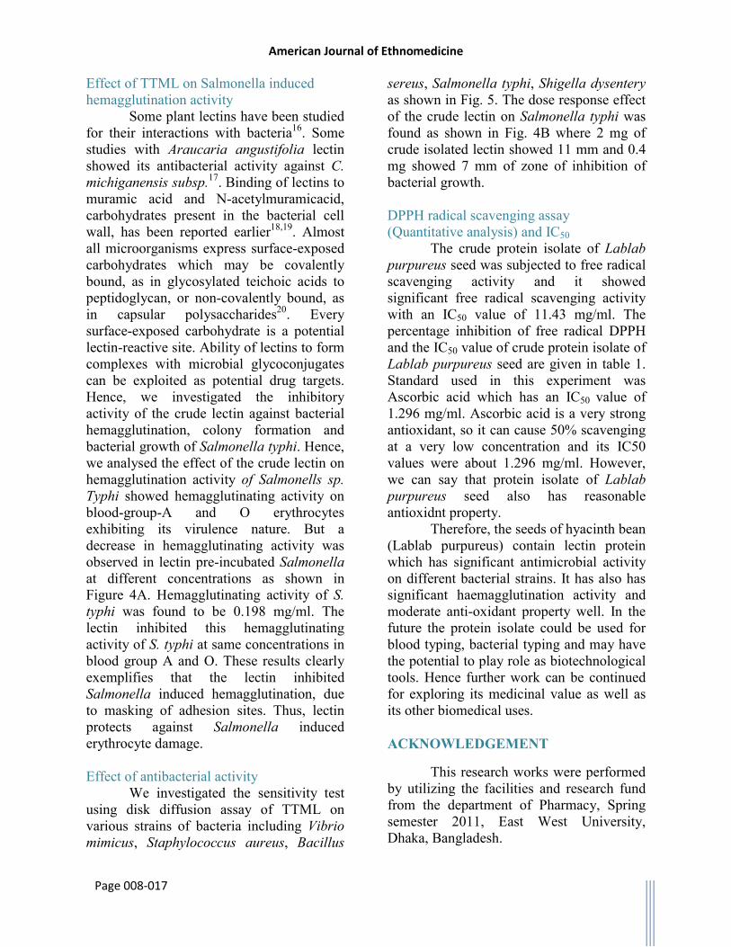
Effect of TTML on Salmonella induced hemagglutination activity
Some plant lectins have been studied for their interactions with bacteria16. Some studies with Araucaria angustifolia lectin showed its antibacterial activity against C. michiganensis subsp.17. Binding of lectins to muramic acid and N-acetylmuramicacid, carbohydrates present in the bacterial cell wall, has been reported earlier18,19. Almost all microorganisms express surface-exposed carbohydrates which may be covalently bound, as in glycosylated teichoic acids to peptidoglycan, or non-covalently bound, as in capsular polysaccharides20. Every surface-exposed carbohydrate is a potential lectin-reactive site. Ability of lectins to form complexes with microbial glycoconjugates can be exploited as potential drug targets. Hence, we investigated the inhibitory activity of the crude lectin against bacterial hemagglutination, colony formation and bacterial growth of Salmonella typhi. Hence, we analysed the effect of the crude lectin on hemagglutination activity of Salmonells sp. Typhi showed hemagglutinating activity on blood-group-A and O erythrocytes exhibiting its virulence nature. But a decrease in hemagglutinating activity was observed in lectin pre-incubated Salmonella at different concentrations as shown in Figure 4A. Hemagglutinating activity of S. typhi was found to be 0.198 mg/ml. The lectin inhibited this hemagglutinating activity of S. typhi at same concentrations in blood group A and O. These results clearly exemplifies that the lectin inhibited Salmonella induced hemagglutination, due to masking of adhesion sites. Thus, lectin protects against Salmonella induced erythrocyte damage.
Effect of antibacterial activity
We investigated the sensitivity test using disk diffusion assay of TTML on various strains of bacteria including Vibrio mimicus, Staphylococcus aureus, Bacillus
sereus, Salmonella typhi, Shigella dysentery as shown in Fig. 5. The dose response effect of the crude lectin on Salmonella typhi was found as shown in Fig. 4B where 2 mg of crude isolated lectin showed 11 mm and 0.4 mg showed 7 mm of zone of inhibition of bacterial growth.
DPPH radical scavenging assay (Quantitative analysis) and IC50
The crude protein isolate of Lablab purpureus seed was subjected to free radical scavenging activity and it showed significant free radical scavenging activity with an IC50 value of 11.43 mg/ml. The percentage inhibition of free radical DPPH and the IC50 value of crude protein isolate of Lablab purpureus seed are given in table 1. Standard used in this experiment was Ascorbic acid which has an IC50 value of 1.296 mg/ml. Ascorbic acid is a very strong antioxidant, so it can cause 50% scavenging at a very low concentration and its IC50 values were about 1.296 mg/ml. However, we can say that protein isolate of Lablab purpureus seed also has reasonable antioxidnt property.
Therefore, the seeds of hyacinth bean (Lablab purpureus) contain lectin protein which has significant antimicrobial activity on different bacterial strains. It has also has significant haemagglutination activity and moderate anti-oxidant property well. In the future the protein isolate could be used for blood typing, bacterial typing and may have the potential to play role as biotechnological tools. Hence further work can be continued for exploring its medicinal value as well as its other biomedical uses. ACKNOWLEDGEMENT
This research works were performed by utilizing the facilities and research fund from the department of Pharmacy, Spring semester 2011, East West University, Dhaka, Bangladesh.

American Journal of Ethnomedicine
Page 008-017
REFERENCES
1. Morris, B. 2003. Bio-Functional Legumes with Nutraceutical, Pharmaceutical, and Industrial Uses. Economic Botany, 57.
2. occena, I.V., Mojica, E.E. & Merca, F.E. 2007. Isolation and Partial Characterization of a Lectin from the Seeds of Artocarpus camansi Blanco. Asian Journal of Plant Sciences, 6(5), 757-764.
3. Kamal, R. & Mathur, N. 2008. Rotenoids from Lablab purpureus L. and their bioefficacy against human disease vectors. Parasitol Res (2010) 107, 1481–1488.
4. Kumar, N. S. & Rao, D. R. 1986. The nature of lectins from Dolichos lablab. J. Biosci., 10(1), 95-109.
5. Hou, Y., Hou, Y., Yanyan, L., Qin, G., LI, J. 2010. Extraction and purification of a lectin from red kidney bean and preliminary immune function studies of the lectin and four Chinese herbal polysaccharides. J Biomed Biotechnol. 217342.
6. Lowry, O. H., Rosebrough, N. J., FARR, A. L., and Randall, R. J. 1951. Protein measurement with the Folin-Phenol reagents. J. Biol. Chem. 193, 265-275.
7. Laemmli, U.K., Favre, M. 1973. Gel electrophoresis of proteins. Journal of Molecular Biology.80, 575–599.
8. Hammann, R., Werner, I. 1980. Thin layer chromatography for rapid detection of carbohydrate utilization by Bacteroides strains. Zentralbl Bakteriol A. 247, 424-429.
9. BASAK, B., BANDYOPADHYAY, D., PATRA, M., BANERJI, A., CHATTERJEE, A., BANERJI, J. 2005. Role of sulfur compounds in the detection of amino acids by ninhydrin on TLC plate. J Chromatogr Sci. 43,104-105.
10. Lehtopolku, M., Nakari, U.M., Kotilainen, P., Huovinen, P., Siitonen, A., Hakanen, A.J. 2010. Antimicrobial susceptibilities of multidrug-resistant Campylobacter jejuni and C. coli strains: in vitro activities of 20 antimicrobial agents. Antimicrob Agents Chemother. 54, 1232-1236.
11. Milena N. 2011. Screening of radical scavenging activity and polyphenol content of Bulgarian plant species. Journal of Phrmacognosy Research, 3(4): 256-259.
12. Kacurakova M, Mathlouthi M. 1996. Ftir and laser-Raman spectra of oligosaccharides in water: characterization of the glycosidic bond. Carbohydr Res 284(2):145–57.
13. Cammue, B.P., Peeters, B., Peumans, W.J. 1985. Isolation and partial characterization of an N-acetylgalactosamine-specific lectin from winter-aconite (Eranthis hyemalis) root tubers. Biochem J 227, 949–955.
14. Pereira, M.E., KABAT, E.A. 1974. Specificity of purified hemagglutinin (lectin) from Lotus tetragonolobus. Biochemistry 13, 3184–3192.
15. Pereira, M.E., Gruezo, F., Kabat, E.A, 1979. Purification and characterization of lectin II from Ulex europaeus seeds and an immunochemical study of its combining site. Arch Biochem Biophys 194, 511–525.
16. Santi-Gadelha, T., DE Almeida Gadelha, C.A., Aragao, K.S., DE Oliveira, C.C., Lima, et al. 2006. Purification and biological effects of Araucaria angustifolia (Araucariaceae) seed lectin. Biochemical and Biophysical Research Communications 350, 1050–1055.
17. Avouba, A., Chatelain, C., Rouge, P. 1991. Legume lectins interact with muramic acid and N-acetylmuramic acid. FEBS Lett 289, 102–104.
18. Himo, S., UT, M., Wadstrom, T, 1997. Sialylglycoconjugate an proteoglycan-binding microbial lectins. The Electronic Lectin Journal: Lectins, Biology, Biochemistry, Clinical Biochemistry 12, (87-984583-0-2).
19. Caldeon, A.M., Buck, G., Doyle, R.J. 1997. Lectin-microorganism complexes. The Electronic Lectin Journal: Lectins, Biology, Biochemistry, Clinical Biochemistry 12, (87-984583-0-2).
20. Utsunomiya, A., Nakamura, M., Hamamoto, A. 2000. Expression of fimbriae and hemagglutination activity in Shigella boydi. Microbiol Immunol. 44, 529–533.

American Journal of Ethnomedicine
Page 008-017
Table 1. IC50 values of antioxidant activities of the lectin isolated from Lablab purpureus seed
Sl No. ABlank Concentration
(mg/ml) ASample
% inhibition of free radical DPPH = (1 – ASample/ABlank) X 100
IC50 mg/ml
1
0.366
0.8 0.352 3.83
11.43 2 1.6 0.341 6.83
3 2.4 0.324 11.48
4 4 0.302 17.49
Here, ABlank and ASample are the absorbance of blank and sample respectively.
A. Carbohydrate B. Protein
Fig. 1. TLC analysis of carbohydeare (A) and protein (B) in crude lectin. Spot has been analyzed in comparison with a reference standard shown in left side of each TLC plate

American Journal of Ethnomedicine
Page 008-017
Fig. 2. SDS-PAGE analysis of crude lectin
Fig. 3. Hemagglutination inhibition assay of crude lectin

American Journal of Ethnomedicine
Page 008-017
Fig. 4. Bacterial hemagglutination inhibition assay (A) and disc diffusion assay of crude lectin (B)

American Journal of Ethnomedicine
Page 008-017
Fig. 5. Antibacterial activity of the crude isolated lectin. Open column indicates the zone of inhibition caused by a positive control (Azithromycin 30 microgram/disk); solid black column and
dotted gradient filled column indicates zone of inhibition caused by indicates the zone of inhibition caused by isolated crude lectin 200 microgram/disk and 20 microgram/disk respectively; Negative
control showed no zone of inhibition.