Glycan-Lectin Interactions as Novel Immunosuppression ...
24
Citation: Pace, A.; Scirocchi, F.; Napoletano, C.; Zizzari, I.G.; D’Angelo, L.; Santoro, A.; Nuti, M.; Rahimi, H.; Rughetti, A. Glycan-Lectin Interactions as Novel Immunosuppression Drivers in Glioblastoma. Int. J. Mol. Sci. 2022, 23, 6312. https://doi.org/10.3390/ ijms23116312 Academic Editor: Giovanni Luca Gravina Received: 14 April 2022 Accepted: 3 June 2022 Published: 5 June 2022 Publisher’s Note: MDPI stays neutral with regard to jurisdictional claims in published maps and institutional affil- iations. Copyright: © 2022 by the authors. Licensee MDPI, Basel, Switzerland. This article is an open access article distributed under the terms and conditions of the Creative Commons Attribution (CC BY) license (https:// creativecommons.org/licenses/by/ 4.0/). International Journal of Molecular Sciences Review Glycan-Lectin Interactions as Novel Immunosuppression Drivers in Glioblastoma Angelica Pace 1 , Fabio Scirocchi 1 , Chiara Napoletano 1 , Ilaria Grazia Zizzari 1 , Luca D’Angelo 2 , Antonio Santoro 2 , Marianna Nuti 1 , Hassan Rahimi 1 and Aurelia Rughetti 1, * 1 Laboratory of Tumor Immunology and Cell Therapy, Department of Experimental Medicine, “Sapienza” University of Rome, Viale Regina Elena 324, 00161 Rome, Italy; [email protected] (A.P.); [email protected] (F.S.); [email protected] (C.N.); [email protected] (I.G.Z.); [email protected] (M.N.); [email protected] (H.R.) 2 Department of Neurology and Psychiatry, Neurosurgery, “Sapienza” University of Rome, Viale dell’ Università 30, 00185 Rome, Italy; [email protected] (L.D.); [email protected] (A.S.) * Correspondence: [email protected]; Tel.: +39-06-499473029 Abstract: Despite diagnostic and therapeutic improvements, glioblastoma (GB) remains one of the most threatening brain tumor in adults, underlining the urgent need of new therapeutic targets. Lectins are glycan-binding proteins that regulate several biological processes through the recognition of specific sugar motifs. Lectins and their ligands are found on immune cells, endothelial cells and, also, tumor cells, pointing out a strong correlation among immunity, tumor microenvironment and vascularization. In GB, altered glycans and lectins contribute to tumor progression and immune evasion, shaping the tumor-immune landscape promoting immunosuppressive cell subsets, such as myeloid-derived suppressor cells (MDSCs) and M2-macrophages, and affecting immunoeffector populations, such as CD8 + T cells and dendritic cells (DCs). Here, we discuss the latest knowledge on the immune cells, immune related lectin receptors (C-type lectins, Siglecs, galectins) and changes in glycosylation that are involved in immunosuppressive mechanisms in GB, highlighting their interest as possible novel therapeutical targets. Keywords: glioblastoma; immunosuppression; C-type lectins; galectins; Siglecs; MGL/CLEC10A; galectin-9; extracellular vesicles (EVs); O-glycosylation; N-glycosylation 1. Introduction Gliomas are malignant brain tumors arising from transformed primitive neural stem cells, glial progenitor cells or dedifferentiated mature cell type [1]. Recently, the World Health Organization (WHO) released the newest 2021 classification of brain tumors, based on not only canonical criteria, such as histological and molecular features, but also novel diagnostic technologies, such as DNA methylome profiling. According to that, glioblastoma (GB) is characterized by the IDH-wildtype phenotype, while his mutant counterpart is identified as Astrocytoma IDH-mutant (grades 2–4) [2]. GB is the most common malignant brain tumor in adults, mostly in males compared to females. Nowadays, the first line of therapy requires maximal safe resection followed by radiotherapy (RT) and a high-dose of chemotherapy with temozolomide (TMZ). Despite the heavy treatment, most of the patients experience a recurrence, and the median overall survival remains below 18 months [3]. At recurrence, the standard of care is less well de- fined. Indeed, therapeutic options include afresh surgery and reirradiation in combination with systemic therapies, such as chemotherapy (TMZ, lomustine, carmustine) or anti-VEGF monoclonal antibody Bevacizumab [3–5]. Emerging therapies, such as immunotherapy and tumor-treating fields (TTFields), are available. TTFields and their ability to disrupt specifically mitotic process in cancer cells had shown promising results in prolonging the Int. J. Mol. Sci. 2022, 23, 6312. https://doi.org/10.3390/ijms23116312 https://www.mdpi.com/journal/ijms
-
Upload
khangminh22 -
Category
Documents
-
view
4 -
download
0
Transcript of Glycan-Lectin Interactions as Novel Immunosuppression ...

Citation: Pace, A.; Scirocchi, F.;
Napoletano, C.; Zizzari, I.G.;
D’Angelo, L.; Santoro, A.; Nuti, M.;
Rahimi, H.; Rughetti, A.
Glycan-Lectin Interactions as Novel
Immunosuppression Drivers in
Glioblastoma. Int. J. Mol. Sci. 2022,
23, 6312. https://doi.org/10.3390/
ijms23116312
Academic Editor: Giovanni
Luca Gravina
Received: 14 April 2022
Accepted: 3 June 2022
Published: 5 June 2022
Publisher’s Note: MDPI stays neutral
with regard to jurisdictional claims in
published maps and institutional affil-
iations.
Copyright: © 2022 by the authors.
Licensee MDPI, Basel, Switzerland.
This article is an open access article
distributed under the terms and
conditions of the Creative Commons
Attribution (CC BY) license (https://
creativecommons.org/licenses/by/
4.0/).
International Journal of
Molecular Sciences
Review
Glycan-Lectin Interactions as Novel ImmunosuppressionDrivers in GlioblastomaAngelica Pace 1, Fabio Scirocchi 1 , Chiara Napoletano 1 , Ilaria Grazia Zizzari 1 , Luca D’Angelo 2 ,Antonio Santoro 2, Marianna Nuti 1 , Hassan Rahimi 1 and Aurelia Rughetti 1,*
1 Laboratory of Tumor Immunology and Cell Therapy, Department of Experimental Medicine,“Sapienza” University of Rome, Viale Regina Elena 324, 00161 Rome, Italy; [email protected] (A.P.);[email protected] (F.S.); [email protected] (C.N.); [email protected] (I.G.Z.);[email protected] (M.N.); [email protected] (H.R.)
2 Department of Neurology and Psychiatry, Neurosurgery, “Sapienza” University of Rome,Viale dell’ Università 30, 00185 Rome, Italy; [email protected] (L.D.);[email protected] (A.S.)
* Correspondence: [email protected]; Tel.: +39-06-499473029
Abstract: Despite diagnostic and therapeutic improvements, glioblastoma (GB) remains one of themost threatening brain tumor in adults, underlining the urgent need of new therapeutic targets.Lectins are glycan-binding proteins that regulate several biological processes through the recognitionof specific sugar motifs. Lectins and their ligands are found on immune cells, endothelial cells and,also, tumor cells, pointing out a strong correlation among immunity, tumor microenvironment andvascularization. In GB, altered glycans and lectins contribute to tumor progression and immuneevasion, shaping the tumor-immune landscape promoting immunosuppressive cell subsets, suchas myeloid-derived suppressor cells (MDSCs) and M2-macrophages, and affecting immunoeffectorpopulations, such as CD8+ T cells and dendritic cells (DCs). Here, we discuss the latest knowledge onthe immune cells, immune related lectin receptors (C-type lectins, Siglecs, galectins) and changes inglycosylation that are involved in immunosuppressive mechanisms in GB, highlighting their interestas possible novel therapeutical targets.
Keywords: glioblastoma; immunosuppression; C-type lectins; galectins; Siglecs; MGL/CLEC10A;galectin-9; extracellular vesicles (EVs); O-glycosylation; N-glycosylation
1. Introduction
Gliomas are malignant brain tumors arising from transformed primitive neural stemcells, glial progenitor cells or dedifferentiated mature cell type [1]. Recently, the WorldHealth Organization (WHO) released the newest 2021 classification of brain tumors, basedon not only canonical criteria, such as histological and molecular features, but also noveldiagnostic technologies, such as DNA methylome profiling. According to that, glioblastoma(GB) is characterized by the IDH-wildtype phenotype, while his mutant counterpart isidentified as Astrocytoma IDH-mutant (grades 2–4) [2].
GB is the most common malignant brain tumor in adults, mostly in males comparedto females. Nowadays, the first line of therapy requires maximal safe resection followed byradiotherapy (RT) and a high-dose of chemotherapy with temozolomide (TMZ). Despitethe heavy treatment, most of the patients experience a recurrence, and the median overallsurvival remains below 18 months [3]. At recurrence, the standard of care is less well de-fined. Indeed, therapeutic options include afresh surgery and reirradiation in combinationwith systemic therapies, such as chemotherapy (TMZ, lomustine, carmustine) or anti-VEGFmonoclonal antibody Bevacizumab [3–5]. Emerging therapies, such as immunotherapyand tumor-treating fields (TTFields), are available. TTFields and their ability to disruptspecifically mitotic process in cancer cells had shown promising results in prolonging the
Int. J. Mol. Sci. 2022, 23, 6312. https://doi.org/10.3390/ijms23116312 https://www.mdpi.com/journal/ijms
Int. J. Mol. Sci. 2022, 23, 6312 2 of 24
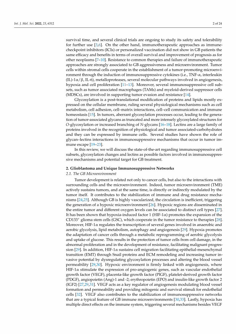
survival time, and several clinical trials are ongoing to study its safety and tolerabilityfor further use [3,6]. On the other hand, immunotherapeutic approaches as immune-checkpoint inhibitors (ICIs) or personalized vaccination did not show in GB patients thesame efficacy and benefits in terms of overall survival and improvement of prognosis as forother neoplasms [7–10]. Resistance to common therapies and failure of immunotherapeuticapproaches are strongly associated to GB aggressiveness and microenvironment. Tumorcells within stromal cells cooperate in the establishment of a tumor-promoting microenvi-ronment through the induction of immunosuppressive cytokines (i.e., TNF-α, interleukin(IL)-1α/β, IL-6), metalloproteases, several molecular pathways involved in angiogenesis,hypoxia and cell proliferation [11–13]. Moreover, several immunosuppressive cell sub-sets, such as tumor associated macrophages (TAMs) and myeloid-derived suppressor cells(MDSCs), are involved in supporting tumor evasion and resistance [14].
Glycosylation is a post-translational modification of proteins and lipids mostly ex-pressed on the cellular membrane, ruling several physiological mechanisms such as cellmetabolism, cell adhesion, cell–matrix interactions, cell–cell communication and immunehomeostasis [15]. In tumors, aberrant glycosylation processes occur, leading to the genera-tion of tumor-associated glycans as truncated and more intensely glycosylated structures forO-glycosylation or increased branching of N-glycans [16–18]. Lectins are a large family ofproteins involved in the recognition of physiological and tumor associated-carbohydratesand they can be expressed by immune cells. Several studies have shown the role ofglycan–lectins interactions in immunosuppressive mechanisms that occur in tumor im-mune escape [19–23].
In this review, we will discuss the state-of-the-art regarding immunosuppressive cellsubsets, glycosylation changes and lectins as possible factors involved in immunosuppres-sive mechanisms and potential target for GB treatment.
2. Glioblastoma and Unique Immunosuppressive Networks2.1. The GB Microenvironment
Tumor development is related not only to cancer cells, but also to the interactions withsurrounding cells and the microenvironment. Indeed, tumor microenvironment (TME)actively sustains tumors, and at the same time, is directly or indirectly modulated by thetumor itself. It contributes to the stabilization of immune and drug resistance mecha-nisms [24,25]. Although GB is highly vascularized, the circulation is inefficient, triggeringthe generation of a hypoxic microenvironment [26]. Hypoxic regions are disseminated inthe entire tumor and different oxygen levels can be associated to distinct cell types [27].It has been shown that hypoxia-induced factor 1 (HIF-1α) promotes the expansion of theCD133+ glioma stem cells (GSC), which cooperate in the tumor resistance to therapies [28].Moreover, HIF-1α regulates the transcription of several genes involved in anaerobic andaerobic glycolysis, lipid metabolism, autophagy and angiogenesis [29]. Hypoxia promotesthe adaptation of cancer cells through a metabolic reprogramming of aerobic glycolysisand uptake of glucose. This results in the protection of tumor cells from cell damage, in theabnormal proliferation and in the development of resistance, facilitating malignant progres-sion [29]. In addition, HIF-1α sustains cell migration facilitating epithelial-mesenchymaltransition (EMT) through Snail proteins and ECM remodeling and increasing tumor in-vasive potential by dysregulating glycosylation processes and altering the blood vesselpermeability [29,30]. Hypoxic environment is firmly linked with angiogenesis, whereHIF-1α stimulate the expression of pro-angiogenic genes, such as vascular endothelialgrowth factor (VEGF), placenta-like growth factor (PlGF), platelet-derived growth factor(PDGF), angiopoietin (Ang)-1 and -2; erythropoietin (EPO) and insulin-like growth factor 2(IGF2) [27,29,31]. VEGF acts as a key regulator of angiogenesis modulating blood vesselformation and permeability and providing mitogenic and survival stimuli for endothelialcells [32]. VEGF also contributes to the stabilization of immunosuppressive networksthat are a typical feature of GB immune microenvironments [30,33]. Lastly, hypoxia hasmultiple direct effects on the immune system, triggering several mechanisms besides VEGF

Int. J. Mol. Sci. 2022, 23, 6312 3 of 24
production, such as immune suppressive myeloid cell shift and recruitment, impairing Tcell response, thus promoting a complex immunosuppressive network, fostering tumorprogression and poor prognosis for patients [29,31,33].
2.2. Immune Cells in GB Microenvironment
Notoriously, GB has been considered a “cold” tumor due to its privileged locationand the presence of few immunoregulatory cells, mostly immunosuppressive actors [34].Actually, the “privileged term” has been reconsidered after the discovery of lymphaticvessels outlining the dural sinuses in mice and potentially analogous structures can exist inthe human brain, underlining how it is possible for the immune system to enter and interactwith the central nervous system (CNS) [35]. Moreover, the blood–brain-barrier (BBB), whichnaturally separates the brain from the systemic circulation, is an obstacle for the deliveryof therapeutic agents. In a tumor context, BBB can lose its integrity due to microvascularsprouting, new vasculature branching and dysfunction of tight junctions, allowing theinfiltration of inflammatory cells [36]. Several studies have shown the presence of differentimmune subsets with distinct localization and function in GB microenvironments [37,38].Within the TME composition, GB cells release a variety of chemoattractive factors such asCCL2, CXCL12, and SDF-1 that actively promote the recruitment of immune cells [39].
Myeloid cells are the most represented cell type in GB microenvironment (Figure 1) [14,40].Among them, TAMs and MDSCs are the most diffused myeloid cells that cooperate in
immunosuppressive mechanisms and tumor resistance. TAM population includes infiltrat-ing bone marrow-derived macrophages (BMDMs) and microglia (MG), that make up to30–50% of the total tumor mass [39,41]. MG are brain resident and resting macrophages thatare involved in CNS development and homeostasis and immune surveillance [42]. MG andBMDMs are distinguished by the expression of surface markers, as CD49d and CD45: MGis usually identified as CD45low CD11b+ CX3CR1+ P2RY12+ CD49d− while macrophagesare CD45high CD11b+ CX3CR1+ CD49d+ P2RY12− [43]. Recently, the transmembrane pro-tein 119 (TMEM119) has been reported as novel unique marker of resident MG both inhumans and mice [44]. Moreover, MG and BMDMs differs for their localization underliningtheir distinct contribution to immunosuppression. MG cells appears stationary, large andbranched and infiltrate the white matter, outlining the edge of the tumor, while BMDMs areusually localized near blood vessels in central and necrotic area of the tumor [14,43]. MGtakes part in the tumorigenesis process through TGF-β release, enhancing the immunosup-pression in the TME by blocking T-cells and NK cells activity and promoting T regulatorycells (Tregs) [38,45]. In response to chemokines and cytokines released by GB cells, BMDMsare recruited from the periphery to the TME. By interacting with tumor cells and TME,BMDMs change their expression profiles and mostly acquire a M2 pro-tumor phenotypethrough a modulation of immunosuppressive and phagocytic mechanisms and oxidativeand iron metabolisms [14,46]. A recent study on patients-derived GB sections revealedthe high infiltration of CD163+ cells (M2-like TAMs) in both the tumor core (TC) andperitumoral area (PTA). Interestingly the high frequency of these cells in the TC correlatewith a significant prevalence of immunosuppressive markers, such as IDO and PD-L1,underlining a pro-tumoral immune system within GB TME [47]. Moreover, in a recentstudy the selective targeting of TAMs by inhibiting CSF-1R resulted in the alteration ofmacrophage polarization and block of glioma progression, proposing TAMs as potentialtherapeutic targets [48].

Int. J. Mol. Sci. 2022, 23, 6312 4 of 24
Int. J. Mol. Sci. 2022, 23, x FOR PEER REVIEW 3 of 24
vessel formation and permeability and providing mitogenic and survival stimuli for en-
dothelial cells [32]. VEGF also contributes to the stabilization of immunosuppressive net-
works that are a typical feature of GB immune microenvironments [30,33]. Lastly, hypoxia
has multiple direct effects on the immune system, triggering several mechanisms besides
VEGF production, such as immune suppressive myeloid cell shift and recruitment, im-
pairing T cell response, thus promoting a complex immunosuppressive network, foster-
ing tumor progression and poor prognosis for patients [29,31,33].
2.2. Immune Cells in GB Microenvironment
Notoriously, GB has been considered a “cold” tumor due to its privileged location
and the presence of few immunoregulatory cells, mostly immunosuppressive actors [34].
Actually, the “privileged term” has been reconsidered after the discovery of lymphatic
vessels outlining the dural sinuses in mice and potentially analogous structures can exist
in the human brain, underlining how it is possible for the immune system to enter and
interact with the central nervous system (CNS) [35]. Moreover, the blood–brain-barrier
(BBB), which naturally separates the brain from the systemic circulation, is an obstacle for
the delivery of therapeutic agents. In a tumor context, BBB can lose its integrity due to
microvascular sprouting, new vasculature branching and dysfunction of tight junctions,
allowing the infiltration of inflammatory cells [36]. Several studies have shown the pres-
ence of different immune subsets with distinct localization and function in GB microenvi-
ronments [37,38]. Within the TME composition, GB cells release a variety of chemoattrac-
tive factors such as CCL2, CXCL12, and SDF-1 that actively promote the recruitment of
immune cells [39].
Myeloid cells are the most represented cell type in GB microenvironment (Figure 1)
[14,40].
Figure 1. Schematic representation of glioblastoma immune microenvironment. Glioblastoma ishighly vascularized with impaired circulation and hypoxic regions. Several soluble factors, as HIF-1αand VEGF, are involved in GB development and progression promoting EMT, ECM remodelingand immunosuppression through TAM and MDSC recruitment. Within tumor cells and cancerstem cells, myeloid cells are the most represented cell type in GB TME. TAM population includesmacrophages (CD45high CD11b+ CX3CR1+ CD49d+ P2RY12−) and MG (CD45low CD11b+ CX3CR1+
P2RY12+ TMEM119+ CD49d−) while MDSC are mainly represented by PMN-MDSC (CD11b+ CD14−
CD15+ CD66b+). Created with BioRender.com, accessed on 2 June 2022.
MDSCs play a fundamental role in promoting tumor progression impairing T-cellfunctions and inducing Treg cells [49]. MDSCs consist of two large group of cells, i.e.,monocytic MDSCs (M-MDSCs) and polymorphonuclear MDSCs (PMN-MDSCs), whichare differentiated by molecular and phenotypical features [14].
In the tumor site, MDSCs functions are strongly controlled by several soluble factors,such as IL-6, IL-10, TGF-β, CCR2, VEGF secreted by GB cells and immune cells, leading tothe modulation of molecular pathways mediated by Janus tyrosine kinase (JAK) and signaltransducer and activator of transcription 1, 3 and 6 (STAT1; STAT3; STAT6). This results inthe promotion of MDSCs proliferation and survival and induction of immunosuppressivemolecules, such as arginase 1 (ARG1), reactive oxygen species (ROS) and inducible nitricoxide synthase (iNOS). Thus, high levels of circulating ARG1 in GB patients’ blood havebeen considered as markers of MDSCs activation [50].
Several studies have reported the contribution of both MDSCs subpopulations tothe progression of gliomas and high levels of circulating MDSCs are associated with theworst prognosis for GB patients [50–54]. Indeed, PMN-MDSCs have been identified asthe main circulating MDSC subpopulation in GB patients’ blood and their proportion ishigher than M-MDSC in GB tumors [55–57]. Moreover, Gielen et al., showed that MDSC

Int. J. Mol. Sci. 2022, 23, 6312 5 of 24
levels of GB patients’ blood were significantly higher than healthy controls and found thatPMN-MDSCs were representative of MDSC infiltrating tumor mass [58]. The discoveryof novel markers, such as LOX1, helps to further distinguish MDSC subpopulations andelucidate their role and mechanisms in tumor progression [59]. Indeed, a recent studyshowed that LOX1+ identifies a specific PMN-MDSC subpopulation that is increased in GBpatients’ blood and tumor tissue and is characterized by a high expression of ARG1 andiNOS and strong immunosuppressive ability to inhibit CD3+ T cells [52].
3. GB and Glycosylation Pathways3.1. Glycosylation Pathways
Glycosylation is the main post-translational modification of proteins and lipids, thatrequires a highly monitored enzymatic complex network from nucleus to Golgi appara-tus [60,61]. Indeed, specific enzymes participate to the finely tuned phases of the glycosyla-tion process, i.e., core extension, elongation and branching, and capping of the carbohydratechains. This generates an astonishing diversity in the glycome that corresponds to highlyspecific and selective biological functions [60,61].
Most of the glycoconjugates and glycans are exposed on the cellular membrane andplay a crucial role in tissue lubrification and protection, cell–matrix interactions and cellsignaling [62]. Glycans are also involved in immunomodulation mechanisms through theinteraction with glycan-binding proteins of lectins family, regulating immune homeostasisand host–pathogen interactions [63].
N- and O-linked glycans are the most common post-translational modification (PTMs)in extracellular components (Figure 2).
O-linked glycosylation is characterized by the addition of monosaccharides, as such asN-acetyl-D-galactosamine (GalNAc), D-glucose (Glc) or D-mannose (Man), to serin or thre-onin residues (Ser, Thr). O-linked Glc, Fucose (Fuc) or Man are found on the EGF-relatedO-glycans. O-linked GalNAc is the substrate for the extension of long polylactosamineside chains in the so called “Mucin-type glycosylation”. In this pathway, the synthesis ofGalNAc α1-O-Ser/Thr (the Tn antigen) is required for the next synthesis of the T antigen(Galβ1-3GalNAcα1-O-Ser/Thr) and further extension of long polylactosamine side chainsthat decor the protein backbone of mucins. These molecular structures function as pro-tection and lubrification of epithelia and become truncated in carcinoma. Additionally,O-linked xylose may trigger the elongation of glycosaminoglycans (GAGs) on the back-bone of proteoglycans. In response to nutrient and stress response, addition of O-linkedN-Acetyl-D-glucosamine (O-GlcNAcylation) can occur on many intracellular proteins [64].
N-linked glycosylation consists in the covalent binding of the oligosaccharideN-Acetyl-D-glucosamine (GlcNAc) to the asparagine (Asn) residue of polypeptide chainsmediated by the oligosaccharyltransferase (OST) complex. Elongation processes give riseto a common pentasaccharide core region composed by GlcNAc and Man residues. ThisN-core structure is the substrate for the branching of the side chains that are further diversi-fied in carbohydrate composition. On the basis of that, N-glycoconjugates are classifiedin high mannose, hybrid or complex [65,66]. High mannose structures are composed onlyby Man, while complex structures are made also by other monosaccarides (GalNAc, Fuc,Galactose, Sialic Acid (Sia)). Hybrid structures are characterized by the presence of bothhigh mannose and complex structures on distinct branches [65,66].

Int. J. Mol. Sci. 2022, 23, 6312 6 of 24
Int. J. Mol. Sci. 2022, 23, x FOR PEER REVIEW 5 of 24
their role and mechanisms in tumor progression [59]. Indeed, a recent study showed that
LOX1+ identifies a specific PMN-MDSC subpopulation that is increased in GB patients’
blood and tumor tissue and is characterized by a high expression of ARG1 and iNOS and
strong immunosuppressive ability to inhibit CD3+ T cells [52].
3. GB and Glycosylation Pathways
3.1. Glycosylation Pathways
Glycosylation is the main post-translational modification of proteins and lipids, that
requires a highly monitored enzymatic complex network from nucleus to Golgi apparatus
[60,61]. Indeed, specific enzymes participate to the finely tuned phases of the glycosyla-
tion process, i.e., core extension, elongation and branching, and capping of the carbohy-
drate chains. This generates an astonishing diversity in the glycome that corresponds to
highly specific and selective biological functions [60,61].
Most of the glycoconjugates and glycans are exposed on the cellular membrane and
play a crucial role in tissue lubrification and protection, cell–matrix interactions and cell
signaling [62]. Glycans are also involved in immunomodulation mechanisms through the
interaction with glycan-binding proteins of lectins family, regulating immune homeosta-
sis and host–pathogen interactions [63].
N- and O-linked glycans are the most common post-translational modification
(PTMs) in extracellular components (Figure 2).
Figure 2. Common N- and O-glycan structures. Extracellular proteins are mainly glycosylated by the addition of monosaccha-
rides in N- or O-linkage. N-glycans share a common core region in which the oligosaccharide N-Acetyl-D-glucosamine (GlcNAc) is
covalently linked to the asparagine (Asn) residue. The elongation process through the linkage of D-mannose (Man) and the addition
of other motifs, as such as Sia and Gal, led to the generation of High mannose, hybrid and complex N-glycoconjugates. O-glycosyla-
tion starts from the binding of monosaccharides, as such as N-acetyl-D-galactosamine (GalNAc) to serin/threonin (Ser/Thr) residues.
GalNAc represents the substrate for further elongation of Mucin-type O- glycans. Other O-glycan structures include O-xylose (Xyl)
for glycosaminoglycans, O- D-glucose (Glc) for glycosphingolipids, and O-Glc, fucose (Fuc) or Man for the EGF-related O-glycans.
Created with BioRender.com
Figure 2. Common N- and O-glycan structures. Extracellular proteins are mainly glycosylated bythe addition of monosaccharides in N- or O-linkage. N-glycans share a common core region inwhich the oligosaccharide N-Acetyl-D-glucosamine (GlcNAc) is covalently linked to the asparagine(Asn) residue. The elongation process through the linkage of D-mannose (Man) and the addition ofother motifs, as such as Sia and Gal, led to the generation of High mannose, hybrid and complexN-glycoconjugates. O-glycosylation starts from the binding of monosaccharides, as such as N-acetyl-D-galactosamine (GalNAc) to serin/threonin (Ser/Thr) residues. GalNAc represents the substrate forfurther elongation of Mucin-type O-glycans. Other O-glycan structures include O-xylose (Xyl) forglycosaminoglycans, O- D-glucose (Glc) for glycosphingolipids, and O-Glc, fucose (Fuc) or Man forthe EGF-related O-glycans. Created with BioRender.com, accessed on 2 June 2022.
Lipid glycosylation is also crucial for the biology of cells. In particular, glycosphin-golipids (GLS), which are the major component of the outer cell plasmamembrane, arecomposed by a hydrophobic ceramide backbone linked to a first glycan moiety (Gal or Glc)through a glycosid linkage [67]. A complex enzyme network located among the intracel-lular compartments mediates the subsequent addition of other carbohydrate residues assuch as GlcNAc, GalNAc, Fuc and Sia generating structurally different molecules, whichpotentially correspond to a distinct function. Gangliosides (GG) are acidic GLS containingsialic acid residues, enriched in cell membrane microdomains, and are more abundantlyfound in the nervous system [68,69].
Despite the diversity of glycosylation pathways, the carbohydrate chains can undergoto non-specific modifications thus generating molecular structures shared by distinctglycoconjugates. Sulfation, the addition of SO3 group to the carbohydrate backbone, is themost abundant glycan modification generating a large pattern of sulfated structures thatplay wide essential biological role. Moreover, the terminal addition of Sia (Sialylation) orFuc (Fucosylation) are crucial events in the capping of the glycan chains, thus blockingthe elongation process and generating glycan structures specifically recognized by glycan-binding proteins [60,70].
The variety and complexity of the glycome is shaped by the cellular stress and becomesa strategy for the cell to modulate its function and interactions with the microenvironment.
Int. J. Mol. Sci. 2022, 23, 6312 7 of 24
Indeed, glycosylation alterations may be regarded as key features of diseases and a keyfactor for immune recognition [71].
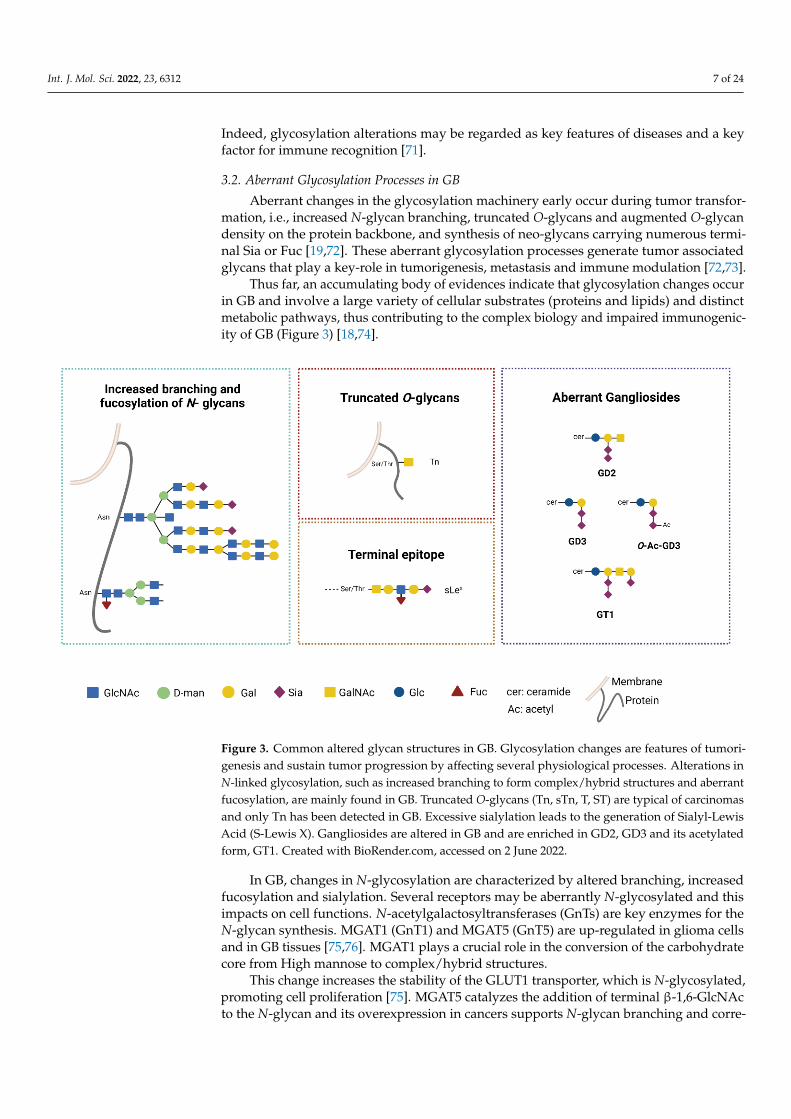
3.2. Aberrant Glycosylation Processes in GB
Aberrant changes in the glycosylation machinery early occur during tumor transfor-mation, i.e., increased N-glycan branching, truncated O-glycans and augmented O-glycandensity on the protein backbone, and synthesis of neo-glycans carrying numerous termi-nal Sia or Fuc [19,72]. These aberrant glycosylation processes generate tumor associatedglycans that play a key-role in tumorigenesis, metastasis and immune modulation [72,73].
Thus far, an accumulating body of evidences indicate that glycosylation changes occurin GB and involve a large variety of cellular substrates (proteins and lipids) and distinctmetabolic pathways, thus contributing to the complex biology and impaired immunogenic-ity of GB (Figure 3) [18,74].
Int. J. Mol. Sci. 2022, 23, x FOR PEER REVIEW 7 of 24
Thus far, an accumulating body of evidences indicate that glycosylation changes occur in
GB and involve a large variety of cellular substrates (proteins and lipids) and distinct met-
abolic pathways, thus contributing to the complex biology and impaired immunogenicity
of GB (Figure 3) [18,74].
Figure 3. Common altered glycan structures in GB. Glycosylation changes are features of tumorigenesis and sustain tumor
progression by affecting several physiological processes. Alterations in N-linked glycosylation, such as increased branching to form
complex/hybrid structures and aberrant fucosylation, are mainly found in GB. Truncated O-glycans (Tn, sTn, T, ST) are typical of
carcinomas and only Tn has been detected in GB. Excessive sialylation leads to the generation of Sialyl-Lewis Acid (S-Lewis X).
Gangliosides are altered in GB and are enriched in GD2, GD3 and its acetylated form, GT1. Created with BioRender.com
In GB, changes in N-glycosylation are characterized by altered branching, increased
fucosylation and sialylation. Several receptors may be aberrantly N-glycosylated and this
impacts on cell functions. N-acetylgalactosyltransferases (GnTs) are key enzymes for the
N-glycan synthesis. MGAT1 (GnT1) and MGAT5 (GnT5) are up-regulated in glioma cells
and in GB tissues [75,76]. MGAT1 plays a crucial role in the conversion of the carbohy-
drate core from High mannose to complex/hybrid structures.
This change increases the stability of the GLUT1 transporter, which is N-glycosyl-
ated, promoting cell proliferation [75]. MGAT5 catalyzes the addition of terminal β-1,6-
GlcNAc to the N-glycan and its overexpression in cancers supports N-glycan branching
and correlates with tumor invasion [76,77]. Increased N-branching augments terminal si-
alylation and fucosylation processes that contributes to tumor spreading [78].
The tumor associated Sialyl-Lewis Acid (S-Lewis X) glycan generated by these
changes is recognized by immune lectins [79]. Dysregulated fucosylation is a feature as-
sociated to GB aggressiveness. Fucosylation of the first GlcNAc residue of the N-core
structure (Figure 3) is catalyzed by the fucosyltrasferases 8 (FUT8). In human GB FUT8,
overexpression is associated with high tumor grade and aggressive disease. Interestingly,
MET and EGFR molecules are substrate for FUT8 and the altered glycosylation increases
their function with an overall gain of motility and proliferation for the tumor cells [80].
Indeed, inhibition of N-linked glycosylation has been proposed as an appealing phar-
macological strategy to dampen receptor kinase activity and enhance radiosensitivity of
GB cells [81].
The increased sialylation levels observed in glioma cells are mainly due to the over-
expression of the α-2,3-sialyl transferase (ST3GAL1). Its up-regulation correlates with
Figure 3. Common altered glycan structures in GB. Glycosylation changes are features of tumori-genesis and sustain tumor progression by affecting several physiological processes. Alterations inN-linked glycosylation, such as increased branching to form complex/hybrid structures and aberrantfucosylation, are mainly found in GB. Truncated O-glycans (Tn, sTn, T, ST) are typical of carcinomasand only Tn has been detected in GB. Excessive sialylation leads to the generation of Sialyl-LewisAcid (S-Lewis X). Gangliosides are altered in GB and are enriched in GD2, GD3 and its acetylatedform, GT1. Created with BioRender.com, accessed on 2 June 2022.
In GB, changes in N-glycosylation are characterized by altered branching, increasedfucosylation and sialylation. Several receptors may be aberrantly N-glycosylated and thisimpacts on cell functions. N-acetylgalactosyltransferases (GnTs) are key enzymes for theN-glycan synthesis. MGAT1 (GnT1) and MGAT5 (GnT5) are up-regulated in glioma cellsand in GB tissues [75,76]. MGAT1 plays a crucial role in the conversion of the carbohydratecore from High mannose to complex/hybrid structures.
This change increases the stability of the GLUT1 transporter, which is N-glycosylated,promoting cell proliferation [75]. MGAT5 catalyzes the addition of terminal β-1,6-GlcNActo the N-glycan and its overexpression in cancers supports N-glycan branching and corre-

Int. J. Mol. Sci. 2022, 23, 6312 8 of 24
lates with tumor invasion [76,77]. Increased N-branching augments terminal sialylationand fucosylation processes that contributes to tumor spreading [78].
The tumor associated Sialyl-Lewis Acid (S-Lewis X) glycan generated by these changesis recognized by immune lectins [79]. Dysregulated fucosylation is a feature associatedto GB aggressiveness. Fucosylation of the first GlcNAc residue of the N-core structure(Figure 3) is catalyzed by the fucosyltrasferases 8 (FUT8). In human GB FUT8, overex-pression is associated with high tumor grade and aggressive disease. Interestingly, METand EGFR molecules are substrate for FUT8 and the altered glycosylation increases theirfunction with an overall gain of motility and proliferation for the tumor cells [80].
Indeed, inhibition of N-linked glycosylation has been proposed as an appealingpharmacological strategy to dampen receptor kinase activity and enhance radiosensitivityof GB cells [81].
The increased sialylation levels observed in glioma cells are mainly due to the over-expression of the α-2,3-sialyl transferase (ST3GAL1). Its up-regulation correlates withpoor prognosis in GB patients and is associated to enhanced invasive potential [82]. Onthe contrary, α-2,6-sialylation is downregulated in the tumor cells, but it is upregulatedin the GB vasculature, suggesting its crucial role for endothelial survival [83–85]. There-fore, two distinct sialylation pathways underlie distinct biological mechanisms for GBprogression [74]. Little is known about changes in the O-linked mucin type glycosyla-tion. In carcinoma, alterations of this enzymatic pathway generate truncated O- glycans,i.e., Tn and T and their sialylated version STn (NeuAcα2-6GalNAcα1-O-Ser/Thr) andST (NeuAcα2-3Galβ1-3GalNAcα1-O-Ser/Thr) [86]. In GB, alteration of the enzymaticO-glycosylation pathway was found and Tn glycan expression was revealed by the bindingof anti-Tn lectins in mouse model as well as in human tumor cells [87,88]. In GB mousemodel, the overexpression of Tn has been associated to strong immunosuppression (asdescribed in Section 4.1) [88].
Additionally, glycolipid components are altered in GB. Early work had shown changesin GLS components in human gliomas, in particular ganglioside GD3 was shown tobe increased in gliomas and correlated with malignancy [89,90]. Thus far, increase ofGD2 and GD3 in GB has been associated with resistance to therapy and increase oninvasiveness [91,92]. The aberrant GG pattern alters the organization of membrane mi-crodomains with huge impact on the activity of tyrosin-kinase receptors and downstreamcell signaling [93]. Indeed, GD3 plays a crucial role in the stemness and tumorigenicity ofGB by activating c-MET signaling [94]. At the same time, GD3 has been shown to modulateinnate immune response by specifically interacting with Siglec7 (see Section 4.2). Morerecently, increase of O-Ac-GD3 (GD3 carrying acetylated Sia) and the ganglioside GT1 werefound in human GB samples, while GM1 was found in peritumoral brain tissue [95,96].These differential patterns may be relevant to both tumor biology and immunomodulationmediated by GGs [97].
Another crucial source of glycan diversity is the matrisome. In GB, the ECM is in-creased due to the up-regulated secretion of PGs and the associated O-linked GAGs [98–100].These ECM components can undergo to aberrant sulfation processes that appear to berelevant for the glycan alteration in GB ECM. Modulation of sulfation among distinct PGswas found and several core matrisome PGs (as such as decorin, agrin, glypcan-1), andlaminin, tenascin, fibronectin among the others, were differentially regulated in tumorvs. control tissues. Interestingly, changes in PG composition were also found within theGB samples in accordance to the IDH mutant status. From this analysis, O-glycosylatedpeptides from PGs were found only in the GB tissue and not in the control, again indicatingthat dysregulated glycosylation process generates specific glyco-profile [101].
Indeed, a glycoproteomic fingerprinting would be of great interest to in depth char-acterize the ECM components to unveil biological mechanisms aiding cell growth andmetastatic spreading that could be useful as potential therapeutic targets [74,102].

Int. J. Mol. Sci. 2022, 23, 6312 9 of 24
4. Glycan-Lectin Interactions and Immunosuppressive Networks in GB
The de novo exposed tumor glycan array triggers novel immune interactions mediatedby lectins that are able to skew the immune system function with high impact on theoverall tumor progression [103]. Lectins constitute a large protein family that share acommon carbohydrate recognition domain (CRD) for the binding of specific glycosylatedstructures [19].
Human lectins are classified by considering their subcellular location and their struc-tures. Lectins can be incorporate in the cellular membrane or have a soluble form and basedon their form they can be divided into groups as C-type lectins, I-type lectins (includingSiglecs), S-type lectins (also known as galectins), P-type lectins (known as Selectins) andpentraxins [104]. In tumor, they have been proposed to decipher the tumor glycocode thatmodulate immune tolerance/suppression [103].
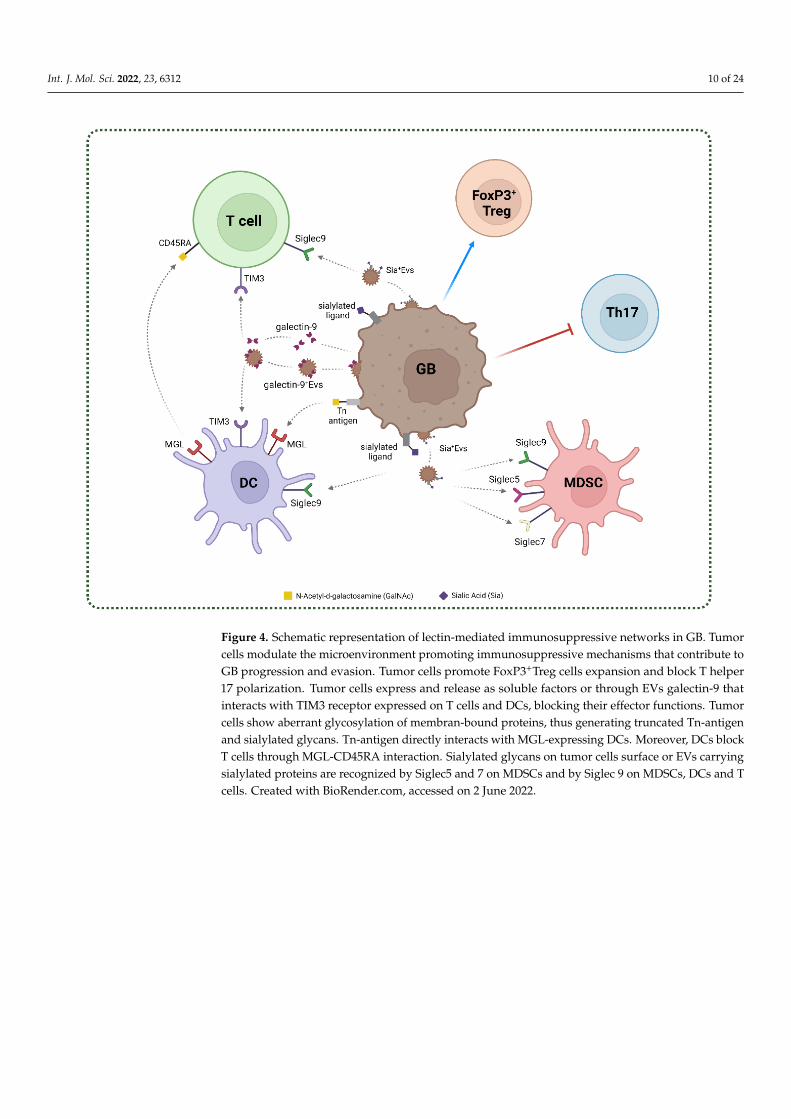
In GB, the expression of lectins from the C-type, Siglec and galectin family has beendescribed and their possible role in the stabilization of immune networks has been pro-posed [105–107]. The main glycan–lectin interactions found in GB are represented inFigure 4.
4.1. C-Type Lectins in GB
C-type lectins (CTLs) are a large family of proteins that share one or more conservedC-type lectin-like domain (CTLD) able to recognize a broad range of carbohydrate motifs.CTLs are mainly expressed on myeloid cells, and their expression is carefully regulated orinduced under specific conditions, as inflammation. CTLs are mainly known for their rolein antimicrobial immunity and tissue homeostasis, but they can both promote or suppressthe immune response in disease context, such as cancer [23,108,109].
For example, alteration of MHC class I molecules due to oncogenic transforma-tion or infection can be sensed by natural killer (NK) cells and can activate the C-typelectin NKG2D-mediated cytotoxicity. However, several C-type lectins, such as DC-SIGN,CLEC14A, macrophage galactose C-type lectin (MGL) contribute to tumor progressioninducing immunosuppressive responses by sensing abnormal or altered tumor-associatedcarbohydrates [110]. Thus far, the few data on the expression and role of CTLs in braindiseases suggest they may play an immunosuppressive role in the immunological net-works. CTLs expression associated to microglia has been observed in neuroinflammatorydegenerative diseases as Alzheimer disease (AD) and Multiple Sclerosis [111,112].
In these pathological settings, expression of CTL by microglia has been observed tomitigate the inflammatory reaction suggesting a possible protective role in inflammatorymediated neurodegenerative diseases. Expression of MGL by macrophages and microgliain experimental autoimmune encephalomyelitis (EAE) mouse model, induced apoptosisof autoreactive T cells and release of immunosuppressive IL-10, thus proposing a role ofMGL as negative regulator of autoimmune-driven neuroinflammation [112]. MGL is aType II C-type lectin, with specificity for the truncated Tn O-glycan (GalNAc-α-Ser/Thr)(Table 1) [113].
MGL engagement by its ligand triggers phenotypic and metabolic changes in theantigen presenting cells [114] that are finely tuned by the structure of the Tn carryingligand [113]. In physiological context, MGL expressing APCs appears to act as negativeregulator for naïve T cell homeostasis and Treg cells through the interaction with theTn-glycosylated CD45RA [115]. Expression of MGL and its Tn O-glycan ligand has beendescribed in GB human tissues, preferentially associated to tumor infiltrating CD163+
cells [88]. Results from a GB syngenic mouse model highly expressing the Tn O-glycanshowed that distinct myeloid infiltrating cell subsets heterogeneously expressed MGL.Interestingly, in the MGL+ tissue areas, a strong infiltration of PD-L1+ TAMs was found [88].These results support the hypothesis that the MGL-Tn axis may contribute to the immuno-suppressive network in GB. It is interesting to recall that the Tn-MGL axis has been vali-dated as a therapeutic target in an ovarian cancer mouse model by means of glycomimeticpeptides [116,117].
Int. J. Mol. Sci. 2022, 23, 6312 10 of 24
Int. J. Mol. Sci. 2022, 23, x FOR PEER REVIEW 9 of 24
Figure 4. Schematic representation of lectin-mediated immunosuppressive networks in GB. Tumor cells modulate the microenviron-
ment promoting immunosuppressive mechanisms that contribute to GB progression and evasion. Tumor cells promote FoxP3+Treg
cells expansion and block T helper 17 polarization. Tumor cells express and release as soluble factors or through EVs galectin-9 that
interacts with TIM3 receptor expressed on T cells and DCs, blocking their effector functions. Tumor cells show aberrant glycosylation
of membran-bound proteins, thus generating truncated Tn-antigen and sialylated glycans. Tn-antigen directly interacts with MGL-
expressing DCs. Moreover, DCs block T cells through MGL-CD45RA interaction. Sialylated glycans on tumor cells surface or EVs
carrying sialylated proteins are recognized by Siglec5 and 7 on MDSCs and by Siglec 9 on MDSCs, DCs and T cells. Created with
BioRender.com.
4.1. C-Type Lectins in GB
C-type lectins (CTLs) are a large family of proteins that share one or more conserved C-
type lectin-like domain (CTLD) able to recognize a broad range of carbohydrate motifs.
CTLs are mainly expressed on myeloid cells, and their expression is carefully regulated
or induced under specific conditions, as inflammation. CTLs are mainly known for their
role in antimicrobial immunity and tissue homeostasis, but they can both promote or sup-
press the immune response in disease context, such as cancer [23,108,109].
For example, alteration of MHC class I molecules due to oncogenic transformation or
infection can be sensed by natural killer (NK) cells and can activate the C-type lectin
NKG2D-mediated cytotoxicity. However, several C-type lectins, such as DC-SIGN,
CLEC14A, macrophage galactose C-type lectin (MGL) contribute to tumor progression
inducing immunosuppressive responses by sensing abnormal or altered tumor-associated
carbohydrates [110].Thus far, the few data on the expression and role of CTLs in brain
diseases suggest they may play an immunosuppressive role in the immunological net-
works. CTLs expression associated to microglia has been observed in neuroinflammatory
degenerative diseases as Alzheimer disease (AD) and Multiple Sclerosis [111,112].
In these pathological settings, expression of CTL by microglia has been observed to
mitigate the inflammatory reaction suggesting a possible protective role in inflammatory
mediated neurodegenerative diseases. Expression of MGL by macrophages and microglia
in experimental autoimmune encephalomyelitis (EAE) mouse model, induced apoptosis
of autoreactive T cells and release of immunosuppressive IL-10, thus proposing a role of
Figure 4. Schematic representation of lectin-mediated immunosuppressive networks in GB. Tumorcells modulate the microenvironment promoting immunosuppressive mechanisms that contribute toGB progression and evasion. Tumor cells promote FoxP3+Treg cells expansion and block T helper17 polarization. Tumor cells express and release as soluble factors or through EVs galectin-9 thatinteracts with TIM3 receptor expressed on T cells and DCs, blocking their effector functions. Tumorcells show aberrant glycosylation of membran-bound proteins, thus generating truncated Tn-antigenand sialylated glycans. Tn-antigen directly interacts with MGL-expressing DCs. Moreover, DCs blockT cells through MGL-CD45RA interaction. Sialylated glycans on tumor cells surface or EVs carryingsialylated proteins are recognized by Siglec5 and 7 on MDSCs and by Siglec 9 on MDSCs, DCs and Tcells. Created with BioRender.com, accessed on 2 June 2022.
Int. J. Mol. Sci. 2022, 23, 6312 11 of 24
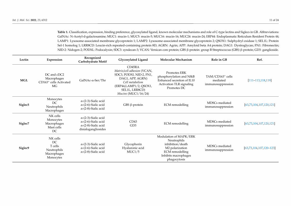
Table 1. Classification, expression, binding preference, glycosylated ligand, known molecular mechanisms and role of C-type lectins and Siglecs in GB. Abbreviations:GalNAc: N-Acetyl-d-galactosamine; MUC1: mucin-1; MUC5: mucin-5; MUC16: mucin-16; MUC24: mucin-24; ERP44: Endoplasmatic Reticulum Resident Protein 44;LAMP1: Lysosome-associated membrane glycoprotein 1; LAMP2: Lysosome-associated membrane glycoprotein 2; QSOX1: Sulphydryl oxidase 1; SEL1L: ProteinSel-1 homolog 1; LRR8CD: Leucin-rich repeated-containing protein 8D; AGRN: Agrin; APP: Amyloid beta A4 protein; DAG1: Dystroglycan; FN1: Fibronectin;NID-2: Nidogen-2; PODXL: Podocalyxin; SDC3: syndecan-3; VCAN: Versican core protein; GBS β-protein: group B Streptococcus (GBS) β-protein; GD3: ganglioside.
Lectin Expression RecognizedCarbohydrate Motif Glycosylated Ligand Molecular Mechanism Role in GB Ref.
MGL
DC and cDC2Macrophages
CD163+ cells ActivatedMG
GalNAc-α-Ser/Thr
CD45RAMatrix/cell adhesion (VCAN,SDC3, PODXL NID-2, FN1,
DAG1, APP, AGRN)Cell metabolism
(ERP44,LAMP1/2, QSOX1,SEL1L, LRR8CD)
Mucins (MUC1/16/24)
Promotes ERKphosphorylation and NfkBEnhanced secretion of IL10Activation TLR signaling
Promotes DC
TAM/CD163+ cellsmediated
immunosuppression[111–113,118,119]
Siglec5
MonocytesDC
NeutrophilsMacrophages
α-(2-3)-Sialic acidα-(2-6)-Sialic acidα-(2-8)-Sialic acid
GBS β-protein ECM remodelling MDSCs mediatedimmunosuppression [63,73,104,107,120,121]
Siglec7
NK cellsMonocytes
MacrophagesMast cells
DC
α-(2-3)-Sialic acidα-(2-6)-Sialic acidα-(2-8)-Sialic acid
disialogangliosides
CD43GD3 ECM remodelling MDSCs mediated
immunosuppression [63,73,104,107,120,121]
Siglec9
NK cellsDC
T cellsNeutrophils
MacrophagesMonocytes
α-(2-3)-Sialic acidα-(2-6)-Sialic acidα-(2-8)-Sialic acid
GlycophorinHyaluronic acid
MUC1/5
Modulation of MAPK/ERKNeutrophils
inhibition/deathM2 polarization
ECM remodellingInhibits macrophages
phagocytosis
MDSCs mediatedimmunosuppression [63,73,104,107,120–123]
Int. J. Mol. Sci. 2022, 23, 6312 12 of 24
4.2. Siglecs in GB
Siglecs belong to the I-type lectins immunoglobulin superfamily that mainly recognizesialic acids (Sia) and other carbohydrate residues. They are classified based on theirstructure in CD33-related Siglecs (Siglec3 or CD33; Siglecs 5-11; Siglecs14 and 16) and otherSiglecs (Siglec1 or CD169; Siglec2 or CD22; Siglec4a or MAG and Siglec15).
Siglecs function as immunomodulatory receptors that mainly mediate immunosup-pressive responses through the phosphorylation of the immunoreceptor tyrosine-basedinhibition motif (ITIM) depending on the cell which they are expressed by and on theligand they interact with. All Siglecs are expressed on immune cells, except for themyelin-associated glycoprotein (MAG) [104]. Siglec1 and 12 expression is restricted onmacrophages, CD22 on B lymphocytes, Siglec16 on MG while other Siglecs, such as CD33and Siglec7, can be found on several immune cell subsets [121–124].
MAG is selectively expressed in the nervous system and is known to have an importantrole in the maintenance of myelinated axons, the physiology of oligodendrocytes and thesuppression of axonal regeneration after injury [125]. MAG exerts its function throughthe interaction with Nogo receptor and gangliosides containing 2,3-linked sialic acid [125].Interestingly, in U87MG glioma cells, it has been shown that MAG is able to reduce themigratory capacity of these cells in culture [126].
It has been shown that in tumor models, sialoglycan–Siglecs interactions are able toimpair and suppress the effector immune cells, such as NK and CD8+ T cells. Moreover,Siglec7 and 9 glycosylated-ligands have been found in several cancers, including GB [127].In a recent work, the authors characterized the expression of Siglec5, 7, 9 on MDSC bothfrom peripheral blood and glioma infiltrating cells and found the respective ligands bothon GB cell lines and patient-derived glioma tissue samples (Table 1). These evidencesunderline the possible interaction between glioma cells and MDSC via the sialic acid–siglecaxis, modulating their function and shaping the immunosuppressive TME [107].
Interestingly, Dusoswa et al. showed a high presence of N-linked and O-linked α-2,3 sialic acids and N-linked α-2,6 sialic acids on GB-derived extracellular vesicles (EVs).GB-derived EVs express Sia-glycoconjugates which are ligands for Siglec9, whereas noligand for the other Siglecs was detected [128]. Siglec9 is mainly found on immune cells, asNK, T and DC and can mediate immunosuppressive functions through the activation ofthe two ITIM sequences comprise in the intracellular domain. In this work, the authorsshowed that the modification of EVs-glycosylation profile through enzymatic desialylationenhanced EV’s internalization by DCs, thus suggesting that glycosylation degree may indi-rectly modulate antigen processing, as shown for other glycan carrying EV systems [129].Although, these results sustain the relevance of that Siglecs as sensors of sialylated carbo-hydrates in GB, further studies are required to elucidate their functional contribution to theimmunosuppressive networks.
4.3. Galectins in GB
S-type lectins, or galectins, are defined by the presence of at least one conserved carbo-hydrate recognition domain (CRD) with high affinity for lactose or N-acetyllactosamine(LacNAc; Galβ1-4GlcNAc)-containing ligands. Currently, galectins are structurally differ-entiated in three groups, as they can exist as dimeric form (galectin-1, -2, -5, -7, -10, -11,-13, -14, -15), tandem-repeat form (galectin-4, -6, -8, -9) and the monomer or multivalentchimera type (galectin-3) [130].
Int. J. Mol. Sci. 2022, 23, 6312 13 of 24
Galectins can be retained in the cytosol or secreted both by transformed and normalcells, such as fibroblasts, endothelial and immune cells [131,132]. Galectins are involved ina wide range of basic cellular functions, as apoptosis, proliferation, cell adhesion and sig-naling, but also in immune system modulation and cancer development [130]. Interestingly,galectins exert their function both intracellularly and extracellularly [133].
Galectins have been shown to play a key role in tumor progression, promoting an-giogenesis and ECM remodeling through the binding with highly glycosylated ligandsexpressed by several cell types. Tumoral galectins, interacting with glycosylated immune-related ligands, induce immunosuppression promoting tolerogenic DCs, CD4+ and CD8+
T cell apoptosis and exhaustion, FoxP3+ Treg cells expansion and NK impairment [21].Upon interaction with galectins, glycoproteins and glycolipids undergo structural rear-rangement, thus modulating their function and the downstream signaling pathways [133].Galectin-1 shapes the immune compartment, promoting the proliferation of tolerogenicDCs and M2-macrophages, the apoptosis of Th1 and Th17 T cells and the expansion ofTreg cells. Moreover, galectin-3 exerts its immunomodulatory functions affecting effectorT cells activity by altering the immunological synapses organization and distancing TCRfrom CD8. These molecular events induce anergy, exhaustion and suppression of T cells.At the same time, galectin-3 promotes immunosuppressive cells, as Tregs and MDSCs, thusdampening the anti-tumor immune response [133–135].
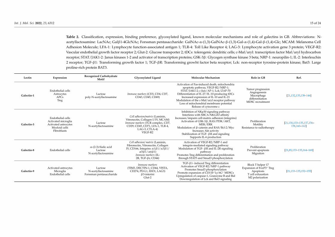
The abundant galectins levels both in TME and peripheral blood have been proposedas prognostic factors and associated to a negative prognosis for oncologic patients [130,136].Several galectins have been found in both neurons and glial cells and physiologicallycontribute to CNS development and functions. Alterations in galectins levels are observedin neuroinflammatory and neurodegenerative disease, and also in CNS tumors [105,137].Indeed galectin-1, -3, -8 and -9 are expressed in GB and the interaction with their respectiveglycan-carrying molecules blocks the anti-tumor response, thus promoting immunosup-pression (Table 2) [138].
Galectin-1 and -9 are the two galectins studied for their association with immuno-suppressive mechanisms [21]. In vitro and mice model studies have shown that galectin-1promotes tumor cells’ migratory capability and invasiveness through modification of cy-toskeleton and small GTPases [138–142]. The selective silencing of glioma-derived galectin-1 delays tumor progression and prolongs the survival of glioma-bearing mice impairingangiogenesis and the recruitment of myeloid cells as MDSCs and macrophages [143].Gou et al., showed that the new putative oncogene FAM92A1-289 is involved in DNAmethylation in GB cell line and interacts with galectin-1 contributing to tumor progres-sion [144]. Recently, LGALS1 gene, coding for the galectin-1, has been included among thegenes that define a glioma microenvironmental gene signature and has been identified as akey immunosuppressive gene in GB [145].
Galectin-1 levels directly correlated with the grade of GB samples and its selective knock-down impaired immunosuppressive mechanisms by down regulating M2 macrophages andMDSC cells and by inhibiting immunosuppressive cytokines [145]. Expression of galectin-1was also associated with a clinical response: GB patients treated with radiotherapy witha low expression of galectin-1 showed a better median survival than patients with highlevels [146]. The silencing or knockdown of galectin-1 in vivo via intranasal administrationor through siRNA-loaded chitosan lipid nanocapsule showed promising results by alteringthe GB TME and immune compartment and reducing TMZ resistance, unraveling galectin-1importance in immunosuppression and tumor promotion [147–149].
Int. J. Mol. Sci. 2022, 23, 6312 14 of 24
Galectin-9 is known as eosinophil chemoattractant and, more recently, as a negativemodulator of adaptative immune response. The major mechanism through which galectin-9 exerts its immunosuppressive mechanisms involves the binding to TIM3, that is widelyexpressed on immune cells, especially on T-cells [21]. Moreover, other immune checkpointsare recognized by galectin-9, therefore galectin-9 is extremely relevant for immunosup-pressive mechanisms (see Table 2) [21]. Through a comprehensive analysis of 1027 gliomapatients, Yuan et al., found a strong common upregulation of galectin-9 in GB compared tonormal brain tissue. In addition, high galectin-9 expression levels correlate with a shorteroverall survival in lower-grade glioma patients. In GB tissue samples, galectin-9 expressionis also associated to immunosuppressive M2-macrophages and its levels positively correlatewith immune checkpoint molecules [150,151]. Notably, galectin-9 also exerts its functionswhen release in EVs. A fascinating study underlined that GB derived-exosomes (GB-Exos),enriched in galectin-9, anontribute to tumor progression by impairing DCs and CD8+ T cellsfunction by TIM3 binding. Indeed, exosomal galectin-9 activity on DCs is TIM3 dependentand knockout of TIM3 in DCs, restore DCs function and activation [152]. Taken together,these studies suggest that galectin-9/TIM3 can be one of the key mechanisms that sustainsimmunosuppression in GB TME and it has been proposed as therapeutical intervention fortargeting cell–cell interaction and exosomes communication [153–155].
Galectin-3 and -8 role in GB progression is less well defined. Little is known abouttheir immune-related functions, while a better knowledge on their interplay with ECM isavailable (see Table 2) [130,137]. Depending on its expression, galectin-3 is able to reinforceor weak cell adhesion and cell–matrix interactions promoting the activation of pathwayinvolved in ECM remodeling and EMT [156,157]. Moreover, galectin-3 upregulation underhypoxic and nutrient deprivation has been correlated to tumor aggressiveness and poorprognosis in several cancer type [21,158]. Galectin-3 has been also proposed as biomarkerfor differential grading and diagnosis of gliomas [159], resistance to standard therapies andhigh levels are associated with a poor survival [160–162].
Lastly, galectin-8 contribution to GB is very poorly understood. It is interesting to notethat galectin-8 is the only one of this family that shows an high affinity for α-2-3-sialylatedglycans, of which GB is rich [85,163]. In a recent study was showed that galectin-8 promotesproliferation and prevents apoptosis of GB cell lines [164]. Indeed, galectin-8 is stronglyimplicated in cell adhesion, growth and cytoskeletal organization, thus underlining apossible favorable function in tumor development and progression [165]. Moreover, highlevels of galectin-8 had been found in oncologic patients and had been often consideredas a poor prognostic factor in several malignancy, as multiple myeloma and prostatecancer [166–168]. Interestingly the selective knockdown of galectin-8 in a mouse modelshowed a notable reduction of the tumor size and a minor metastatic process, highlightingits importance as potential therapeutical target. However, high levels of galectin-8 havebeen also associated to a better survival for gastric cancer patients, pointing out the need tobetter clarify galectins role in carcinogenesis [169].
Int. J. Mol. Sci. 2022, 23, 6312 15 of 24
Table 2. Classification, expression, binding preference, glycosylated ligand, known molecular mechanisms and role of galectins in GB. Abbreviations: N-acetyllactosamine: LacNAc; Galβ1-4GlcNAc; Forssman pentasaccharide: GalNAc-α-(1,3)-GalNAc-β-(1,3)-Gal-α-(1,4)-Gal-β-(1,4)-Glc; MCAM: Melanoma CellAdhesion Molecule; LFA-1: Lymphocyte function-associated antigen 1; TLR-4: Toll Like Receptor 4; LAG-3: Lymphocyte activation gene 3 protein; VEGF-R2:Vascular endothelial growth factor receptor 2; Glut-2: Glucose transporter 2; tDCs: tolerogenic dendritic cells; c-Maf/aryl: transcription factor Maf/aryl hydrocarbonreceptor; STAT/JAK1-2: Janus kinases 1-2 and activator of transcription proteins; GSK-3β: Glycogen synthase kinase 3 beta; NRP-1: neuropilin-1; IL-2: Interleuchin2 receptor; TGF-β1: Transforming growth factor 1; TGF-βR: Transforming growht factor beta receptor; Lck: non-receptor tyrosine-protein kinase; Bat3: Largeproline-rich protein BAT3.
Lectin Expression Recognized CarbohydrateMotif Glycosylated Ligand Molecular Mechanism Role in GB Ref.
Galectin-1
Endothelial cellsAstrocytes
APCsTreg
Lactosepoly-N-acetyllactosamine
Immune markers (CD3, CD4, CD7,CD43, CD45, CD69)
Activation of Fas-induced death, mitochondriaapoptotic pathway, VEGF-R2/NRP-1;
STAT/JAK1-2; c-Jun/AP-1; Lck/ZAP-70Differentiation of IL-27/IL-10-producing tDCs
Increased expression of IL-10 and IL-21Modulation of the c-Maf/aryl receptor pathway
Loss of mitochondrial membrane potentialRelease of cytocrome c
Tumor progressionAngiogenesisMacrophage
differentiationMDSC recruitment
[21,132,135,138–146]
Galectin-3
Endothelial cellsActivated microgliaActivated astrocytes
Myeloid cellsFibroblasts
LactoseN-acetyllactosamine
Cell adhesion/matrix (Laminin,Vitronectin, Collagen I/IV, MCAM)Immune markers (TCR complex, CD7,
CD29, CD45, CD71, LFA-1, TLR-4,LAG-3, CTLA-4)
VEGF-R2
Inhibition of NKp30 signaling pathwayInterferes with MICA-NKG2D affinity
Increases/impairs cell-matrix adhesion (integrins)Activation of GSK-3β, RAS/PI3K/AKT,
MEK/ERKModulation of β-catenin and RAS/Bcl-2/Myc
Increases Akt activityStabilization of TGF- βR and signaling
Supports IL-6 production
ProliferationMotility
Resistance to radiotherapy
[21,130,133–135,137,156–158,160–162]
Galectin-8 Endothelial cellsα-(2-3)-Sialic acid
LactoseN-acetyllactosamine
Cell adhesion/ matrix (Laminin,Fibronectin, Vitronectin, Collagen
IV, CD166, Integrins α1β1/α3β1/α5β1/α6β1)
Immune markers (IL-2R, TGF-βr, CD44)
Activation of VEGF-R2/NRP-1 andintegrin-mediated signaling pathway
Modulation of TGF- βR and IL-2R signalingpathway
Promotes Treg differentiation and proliferationthrough STAT5 and Smad3 phosphorylation
ProliferationPrevent apoptosis
Migration[21,85,133–135,164–169]
Galectin-9Activated astrocytes
MicrogliaEndothelial cells
LactoseN-acetyllactosamine
Forssman pentasaccharide
Immune markers(TIM3, DECTIN-1, CD44, VISTA,
CD274, PD-L1, IDO1, LAG3)β3-interini
Glut-2
TGF-β1- induced Treg differentiationActivation of VEGF-R2/NRP-1 pathway
Promotes Smad3 phosphorylationPromote expansion of CD11b+Ly-6G+ MDSCs
Upregulation of caspase-1, Granzyme B and BidDownregulation of Lck and Bat3 signaling
Block T helper 17Expansion of FoxP3+ Treg
ApoptosisT cell exhaustionM2 polarization
[21,133–135,152–155]
Int. J. Mol. Sci. 2022, 23, 6312 16 of 24
5. Conclusions
Despite medical innovation, GB remains one of the most fatal malignant tumors [1].Novel therapeutic strategies targeting GB cell self-renewal and growth as well as
immunotherapies potentiating/eliciting anti-tumor immune response are currently in-vestigated. However, unsatisfactory results have been reached with little impact on theavailability of novel therapies [170,171].
Thus far, it has become clear that most therapeutic interventions aimed to dampentumor growth, promote anti-tumor response as a bystander effect by reducing immunesuppression and/or enhancing antigen presentation and eliciting T cell responses [172–174].This indirect immune modulation contributes to the overall clinical benefit induced by thetherapy [175].
It is also clear that the immunosuppressive, tumor-promoting microenvironmenthalts the drug-induced immunological benefits or the efficacy of immunotherapeuticinterventions as such as Immune checkpoint blockade, now standard of therapy for severaltumors, or more experimental immunotherapies as vaccination, adoptive T cell therapy,CAR-T [176–178].
In this scientific and biological framework, the tumor associated glycans that aregenerated by aberrant tumor glycosylation acquire a crucial importance for their immune-coding significance, and not just as markers of tumor phenotype and invasiveness [79,103].
Indeed, the lectin immune receptors that selectively sense and recognize these tumorassociated structures have emerged as markers and possible therapeutic targets to dismantlethe immunosuppressive networks [179].
The GB TME is characterized by a peculiar array of diverse myeloid cell types, whosemolecular profile and functions are a challenging research field [180].
The diverse expression profiles of lectins displayed by these cell subsets is stronglysuggestive of their role in the triggering and fueling of immunosuppression. Indeed, severalcompounds have been developed to block galectin- and Siglec-mediated interactions fortherapeutic purposes in tumor and other pathological settings [181–183]. Glycomimeticpeptides have been successfully employed for such purpose [182,184]; indeed the MGL-Tnglycan axis can be targeted with therapeutic benefit in ovarian cancer settings [116]. Theavailability of novel glycoproteomic approaches may allow in depth glycosylation profilingand mapping to identify the glycoconjugated ligands that in vivo participate to the lectinmediated immune networks in GB. Therefore, further investigations are required in orderto better understand and clarify the contribution of these interactions in GB developmentand progression.
Glycan–lectin interactions mediate also immune cell migration [185]. Indeed, thematrisome has emerged as a key factor in the motility of tumor as well as immune cellsand its modulation has been regarded as potential therapeutic option [101,186]. From animmunological point of view, the knowledge of the mechanisms that regulate immune cellsinfiltration is essential to switch the immune tumor contexture and improve the efficacy oftreatments [187,188].
The characterization of the changes in glycan composition and the unveiling of themechanisms of glycan-lectin interactions that occur in GB can critically contribute to identifynovel therapeutic strategies and ameliorate already available immunotherapeutic treatmentin GB.
Author Contributions: Conceptualization, A.P. and A.R.; writing—original draft preparation, A.P.,A.R. and F.S.; Drawings, F.S. and H.R.; writing—review and editing, M.N., A.P., F.S., H.R., C.N., I.G.Z.,L.D., A.S. and A.R.; funding acquisition and supervision, A.R. and M.N. All authors have read andagreed to the published version of the manuscript.
Funding: This research and APC were funded by MUR/”Sapienza” University of Rome, grantsnumber RM1201772B803DB14, RM12117A7B767D0D and RM12117A85361029.

Int. J. Mol. Sci. 2022, 23, 6312 17 of 24
Acknowledgments: We are most grateful to M. Cristiani and G.J.F. for their collaboration throughoutthe preparation of the manuscript. We thank A. Di Filippo for helpful discussion and suggestions.A.P. and F.S. are fellows of the PhD course “Network Oncology and Precision Medicine”; Dpt.Experimental Medicine, “Sapienza” University of Rome.
Conflicts of Interest: The authors declare no conflict of interest. The funders had no role in the designof the study; in the collection, analyses, or interpretation of data; in the writing of the manuscript, orin the decision to publish the results.
References1. Ferris, S.P.; Hofmann, J.W.; Solomon, D.A.; Perry, A. Characterization of Gliomas: From Morphology to Molecules. Virchows Arch.
2017, 471, 257–269. [CrossRef] [PubMed]2. Louis, D.N.; Perry, A.; Wesseling, P.; Brat, D.J.; Cree, I.A.; Figarella-Branger, D.; Hawkins, C.; Ng, H.K.; Pfister, S.M.; Reifenberger,
G.; et al. The 2021 WHO Classification of Tumors of the Central Nervous System: A Summary. Neuro Oncol. 2021, 23, 1231–1251.[CrossRef] [PubMed]
3. Tan, A.C.; Ashley, D.M.; López, G.Y.; Malinzak, M.; Friedman, H.S.; Khasraw, M. Management of Glioblastoma: State of the Artand Future Directions. CA Cancer J. Clin. 2020, 70, 299–312. [CrossRef] [PubMed]
4. Friedman, H.S.; Prados, M.D.; Wen, P.Y.; Mikkelsen, T.; Schiff, D.; Abrey, L.E.; Yung, W.K.A.; Paleologos, N.; Nicholas, M.K.;Jensen, R.; et al. Bevacizumab Alone and in Combination with Irinotecan in Recurrent Glioblastoma. J. Clin. Oncol. 2009,27, 4733–4740. [CrossRef]
5. Yang, K.; Wu, Z.; Zhang, H.; Zhang, N.; Wu, W.; Wang, Z.; Dai, Z.; Zhang, X.; Zhang, L.; Peng, Y.; et al. Glioma Targeted Therapy:Insight into Future of Molecular Approaches. Mol. Cancer 2022, 21, 39. [CrossRef]
6. Liu, S.; Shi, W.; Zhao, Q.; Zheng, Z.; Liu, Z.; Meng, L.; Dong, L.; Jiang, X. Progress and Prospect in Tumor Treating FieldsTreatment of Glioblastoma. Biomed. Pharmacother. 2021, 141, 111810. [CrossRef]
7. Sampson, J.H.; Gunn, M.; Fecci, P.E.; Ashley, D.M. Brain Immunology and Immunotherapy in Brain Tumours John. Nat. Rev.Cancer 2020, 20, 12–25. [CrossRef]
8. Tomaszewski, W.; Al, E. Brain Tumor Micro-Environment and Host State—Implications for Immunotherapy. Clin. Cancer Res.2019, 25, 4202–4210. [CrossRef]
9. Keskin, D.B.; Anandappa, A.J.; Sun, J.; Tirosh, I.; Mathewson, N.D.; Li, S.; Oliveira, G.; Giobbie-Hurder, A.; Felt, K.; Gjini, E.;et al. Neoantigen Vaccine Generates Intratumoral T Cell Responses in Phase Ib Glioblastoma Trial. Nature 2019, 565, 234–239.[CrossRef]
10. Jackson, C.M.; Choi, J.; Lim, M. Mechanisms of Immunotherapy Resistance: Lessons from Glioblastoma. Nat. Immunol. 2019,20, 1100–1109. [CrossRef]
11. Basheer, A.S.; Abas, F.; Othman, I.; Naidu, R. Role of Inflammatory Mediators, Macrophages, and Neutrophils in GliomaMaintenance and Progression: Mechanistic Understanding and Potential Therapeutic Applications. Cancers 2021, 13, 4226.[CrossRef] [PubMed]
12. Yeo, E.C.F.; Brown, M.P.; Gargett, T.; Ebert, L.M. The Role of Cytokines and Chemokines in Shaping the Immune Microenvironmentof Glioblastoma: Implications for Immunotherapy. Cells 2021, 10, 607. [CrossRef] [PubMed]
13. Brown, N.F.; Carter, T.J.; Ottaviani, D.; Mulholland, P. Harnessing the Immune System in Glioblastoma. Br. J. Cancer 2018,119, 1171–1181. [CrossRef] [PubMed]
14. De Leo, A.; Ugolini, A.; Veglia, F. Myeloid Cells in Glioblastoma Microenvironment. Cells 2020, 10, 18. [CrossRef] [PubMed]15. Rodrigues, J.G.; Balmaña, M.; Macedo, J.A.; Al, E. Glycosylation in Cancer: Selected Roles in Tumour Progression, Immune
Modulation and Metastasis. Cell. Immunol. 2018, 333, 46–57. [CrossRef] [PubMed]16. Ho, W.L.; Hsu, W.M.; Huang, M.C.; Kadomatsu, K.; Nakagawara, A. Protein Glycosylation in Cancers and Its Potential
Therapeutic Applications in Neuroblastoma. J. Hematol. Oncol. 2016, 9, 1–15. [CrossRef]17. Fu, C.; Zhao, H.; Wang, Y.; Cai, H.; Xiao, Y.; Zeng, Y.; Chen, H. Tumor-Associated Antigens: Tn Antigen, STn Antigen, and T
Antigen. Hla 2016, 88, 275–286. [CrossRef]18. Veillon, L.; Fakih, C.; Abou-El-Hassan, H.; Kobeissy, F.; Mechref, Y. Glycosylation Changes in Brain Cancer. ASC Chem. Neurosci.
2018, 9, 51–52. [CrossRef]19. Kremsreiter, S.M.; Kroell, A.H.; Weinberger, K.; Boehm, H. Glycan—Lectin Interactions in Cancer and Viral Infections and How to
Disrupt Them. Int. J. Mol. Sci. 2021, 22, 10577. [CrossRef]20. Pillai, S.; Netravali, I.A.; Cariappa, A.; Mattoo, H. Siglecs and Immune Regulation. Annu. Rev. Immunol. 2012, 30, 357–392.
[CrossRef]21. Cedeno-Laurent, F. Galectins and Their Ligands: Negative Regulators of Anti-Tumor Immunity. Glycoconj J. 2012, 29, 619–625.
[CrossRef] [PubMed]22. Drickamer, K.; Taylor, M.E. Recent Insights into Structures and Functions of C-Type Lectins in the Immune System. Curr. Opin.
Struct. Biol. 2015, 34, 26–34. [CrossRef] [PubMed]23. Mayer, S.; Raulf, M.K.; Lepenies, B. C-Type Lectins: Their Network and Roles in Pathogen Recognition and Immunity. Histochem.
Cell Biol. 2017, 147, 223–237. [CrossRef] [PubMed]

Int. J. Mol. Sci. 2022, 23, 6312 18 of 24
24. Spill, F.; Reynolds, D.S.; Kamm, R.D.; Zaman, M.H. Impact of the Physical Microenvironment on Tumor Progression andMetastasis. Curr. Opin. Biotechnol. 2016, 40, 41–48. [CrossRef]
25. Khalaf, K.; Hana, D.; Chou, J.T.-T.; Singh, C.; Mackiewicz, A.; Kaczmarek, M. Aspects of the Tumor Microenvironment Involvedin Immune Resistance and Drug Resistance. Front. Immunol. 2021, 12, 656364. [CrossRef]
26. Di Cintio, F.; Dal Bo, M.; Baboci, L.; De Mattia, E.; Polano, M.; Toffoli, G. The Molecular and Microenvironmental Landscape ofGlioblastomas: Implications for the Novel Treatment Choices. Front. Neurosci. 2020, 14, 603647. [CrossRef]
27. Colwell, N.; Larion, M.; Giles, A.J.; Seldomridge, A.N.; Sizdahkhani, S.; Gilbert, M.R.; Park, D.M. Hypoxia in the GlioblastomaMicroenvironment: Shaping the Phenotype of Cancer Stem-like Cells. Neuro Oncol. 2017, 19, 887–896. [CrossRef]
28. Soeda, A.; Park, M.; Lee, D.; Mintz, A.; Androutsellis-Theotokis, A.; McKay, R.D.; Engh, J.; Iwama, T.; Kunisada, T.; Kassam,A.B.; et al. Hypoxia Promotes Expansion of the CD133-Positive Glioma Stem Cells through Activation of HIF-1α. Oncogene 2009,28, 3949–3959. [CrossRef]
29. Dom, M.; Hern, A.; Plaja, A.; Mart, E. Hypoxia: The Cornerstone of Glioblastoma. Int. J. Mol. Sci. 2021, 22, 12608.30. Silva-Filho, A.F.; Sena, W.L.B.; Lima, L.R.A.; Carvalho, L.V.N.; Pereira, M.C.; Santos, L.G.S.; Santos, R.V.C.; Tavares, L.B.; Pitta,
M.G.R.; Rêgo, M.J.B.M. Glycobiology Modifications in Intratumoral Hypoxia: The Breathless Side of Glycans Interaction. Cell.Physiol. Biochem. 2017, 41, 1801–1829. [CrossRef]
31. Kaur, B.; Khwaja, F.W.; Severson, E.A.; Matheny, S.L.; Brat, D.J.; Van Meir, E.G. Hypoxia and the Hypoxia-Inducible-FactorPathway in Glioma Growth and Angiogenesis. Neuro Oncol. 2005, 7, 134–153. [CrossRef] [PubMed]
32. Carmeliet, P. VEGF as a Key Mediator of Angiogenesis in Cancer. Oncology 2005, 69, 4–10. [CrossRef] [PubMed]33. Duru, G.; van Egmond, M.; Heemskerk, N. A Window of Opportunity: Targeting Cancer Endothelium to Enhance Immunotherapy.
Front. Immunol. 2020, 11, 584723. [CrossRef] [PubMed]34. Yang, F.; He, Z.; Duan, H.; Zhang, D.; Li, J.; Yang, H.; Dorsey, J.F.; Zou, W.; Ali Nabavizadeh, S.; Bagley, S.J.; et al. Synergistic
Immunotherapy of Glioblastoma by Dual Targeting of IL-6 and CD40. Nat. Commun. 2021, 12, 1–15. [CrossRef] [PubMed]35. Louveau, A.; Smirnov, I.; Keyes, T.J.; Eccles, J.D.; Sherin, J.; Peske, J.D.; Derecki, N.C.; Castle, D.; Mandell, J.W.; Kevin, L.; et al.
Structural and Functional Features of Central Nervous System Lymphatics. Nature 2015, 523, 337–341. [CrossRef]36. Eder, K.; Kalman, B. The Dynamics of Interactions Among Immune and Glioblastoma Cells. Neuro Mol. Med. 2015, 17, 335–352.
[CrossRef]37. Zhang, B.; Shen, R.; Cheng, S.; Feng, L. Immune Microenvironments Differ in Immune Characteristics and Outcome of Glioblas-
toma Multiforme. Cancer Med. 2019, 8, 2897–2907. [CrossRef]38. DeCordova, S.; Shastri, A.; Tsolaki, A.G.; Yasmin, H.; Klein, L.; Singh, S.K.; Kishore, U. Molecular Heterogeneity and Immunosup-
pressive Microenvironment in Glioblastoma. Front. Immunol. 2020, 11, 1402. [CrossRef]39. Chen, Z.; Feng, X.; Herting, C.J.; Garcia, V.A.; Nie, K.; Pong, W.W.; Rasmussen, R.; Dwivedi, B.; Seby, S.; Wolf, S.A.; et al. Cellular
and Molecular Identity of Tumor-Associated Macrophages in Glioblastoma. Cancer Res. 2017, 77, 2266–2278. [CrossRef]40. Pinton, L.; Masetto, E.; Vettore, M.; Solito, S.; Magri, S.; D’Andolfi, M.; Del Bianco, P.; Lollo, G.; Benoit, J.P.; Okada, H.; et al.
The Immune Suppressive Microenvironment of Human Gliomas Depends on the Accumulation of Bone Marrow-DerivedMacrophages in the Center of the Lesion. J. Immunother. Cancer 2019, 7, 58. [CrossRef]
41. Brandenburg, S.; Blank, A.; Bungert, A.D.; Vajkoczy, P. Distinction of Microglia and Macrophages in Glioblastoma: Close Relatives,Different Tasks? Int. J. Mol. Sci. 2021, 22, 194. [CrossRef] [PubMed]
42. Geribaldi-Doldán, N.; Fernández-Ponce, C.; Quiroz, R.N.; Sánchez-Gomar, I.; Escorcia, L.G.; Velásquez, E.P.; Quiroz, E.N. TheRole of Microglia in Glioblastoma. Front. Oncol. 2021, 10, 603495. [CrossRef] [PubMed]
43. Daubon, T.; Hemadou, A.; Romero Garmendia, I.; Saleh, M. Glioblastoma Immune Landscape and the Potential of NewImmunotherapies. Front. Immunol. 2020, 11, 585616. [CrossRef] [PubMed]
44. Satoh, J.; Kino, Y.; Asahina, N.; Takitani, M.; Miyoshi, J.; Ishida, T.; Saito, Y. TMEM119 Marks a Subset of Microglia in the HumanBrain. Neuropathology 2016, 36, 39–49. [CrossRef] [PubMed]
45. Crane, C.A.; Ahn, B.J.; Han, S.J.; Parsa, A.T. Soluble Factors Secreted by Glioblastoma Cell Lines Facilitate Recruitment, Survival,and Expansion of Regulatory T Cells: Implications for Immunotherapy. Neuro. Oncol. 2012, 14, 584–595. [CrossRef] [PubMed]
46. Buonfiglioli, A.; Hambardzumyan, D. Macrophages and Microglia: The Cerberus of Glioblastoma. Acta Neuropathol. Commun.2021, 9, 1–21. [CrossRef] [PubMed]
47. Koshkaki, H.R.; Minasi, S.; Ugolini, A.; Trevisi, G.; Napoletano, C.; Zizzari, I.G.; Gessi, M.; Giangaspero, F.; Mangiola, A.; Nuti,M.; et al. Immunohistochemical Characterization of Immune Infiltrate in Tumor Microenvironment of Glioblastoma. J. Pers. Med.2020, 10, 112. [CrossRef]
48. Pyonteck, S.M.; Akkari, L.; Schuhmacher, A.J.; Bowman, R.L.; Sevenich, L.; Quail, D.F.; Olson, O.C.; Quick, M.L.; Huse, J.T.;Teijeiro, V.; et al. CSF-1R Inhibition Alters Macrophage Polarization and Blocks Glioma Progression. Nat. Med. 2013, 19, 1264–1272.[CrossRef]
49. Lindau, D.; Gielen, P.; Kroesen, M.; Wesseling, P.; Adema, G.J. The Immunosuppressive Tumour Network: Myeloid-DerivedSuppressor Cells, Regulatory T Cells and Natural Killer T Cells. Immunology 2013, 138, 105–115. [CrossRef]
50. Lakshmanachetty, S.; Cruz-Cruz, J.; Hoffmeyer, E.; Cole, A.P.; Mitra, S.S. New Insights into the Multifaceted Role of Myeloid-Derived Suppressor Cells (MDSCs) in High-Grade Gliomas: From Metabolic Reprograming, Immunosuppression, and Therapeu-tic Resistance to Current Strategies for Targeting MDSCs. Cells 2021, 10, 893. [CrossRef]
Int. J. Mol. Sci. 2022, 23, 6312 19 of 24
51. Bayik, D.; Zhou, Y.; Park, C.; Hong, C.; Vail, D.; Silver, D.J.; Lauko, A.; Roversi, G.; Watson, D.C.; Lo, A.; et al. Myeloid-DerivedSuppressor Cell Subsets Drive Glioblastoma Growth in a Sex.-Specific Manner. Cancer Discov. 2020, 10, 1210–1225. [CrossRef][PubMed]
52. Chai, E.; Zhang, L.; Li, C. LOX-1+ PMN-MDSC Enhances Immune Suppression Which Promotes Glioblastoma MultiformeProgression. Cancer Manag. Res. 2019, 11, 7307–7315. [CrossRef] [PubMed]
53. Dubinski, D.; Wölfer, J.; Hasselblatt, M.; Schneider-Hohendorf, T.; Bogdahn, U.; Stummer, W.; Wiendl, H.; Grauer, O.M. CD4+ TEffector Memory Cell Dysfunction Is Associated with the Accumulation of Granulocytic Myeloid-Derived Suppressor Cells inGlioblastoma Patients. Neuro Oncol. 2016, 18, 807–818. [CrossRef] [PubMed]
54. Parizi, M.S.; Parizi, F.S.; Abdolhosseini, S.; Vanaei, S.; Manzouri, A.; Ebrahimzadeh, F. Myeloid-Derived Suppressor Cells (MDSCs)in Brain Cancer: Challenges and Therapeutic Strategies. Inflammopharmacology 2021, 29, 1613–1624. [CrossRef]
55. Raychaudhuri, B.; Rayman, P.; Huang, P.; Grabowski, M.; Hambardzumyan, D.; Finke, J.H.; Vogelbaum, M.A. Myeloid DerivedSuppressor Cell Infiltration of Murine and Human Gliomas Is Associated with Reduction of Tumor Infiltrating Lymphocytes. J.Neurooncol. 2015, 122, 293–301. [CrossRef]
56. Raychaudhuri, B.; Ireland, P.R.J.; Ko, J.; Rini, B.; Borden, E.C.; Garcia, J.; Vogelbaum, M.A.; Finke, J. Myeloid-Derived SuppressorCell Accumulation and Function in Patients with Newly Diagnosed Glioblastoma. Neuro Oncol. 2011, 13, 591–599. [CrossRef]
57. Del Bianco, P.; Pinton, L.; Magri, S.; Canè, S.; Masetto, E.; Basso, D.; Padovan, M.; Volpin, F.; d’Avella, D.; Lombardi, G.; et al.Myeloid Diagnostic and Prognostic Markers of Immune Suppression in the Blood of Glioma Patients. Front. Immunol. 2022,12, 809826. [CrossRef]
58. Gielen, P.R.; Schulte, B.M.; Kers-Rebel, E.D.; Verrijp, K.; Petersen-Baltussen, H.M.J.M.; Ter Laan, M.; Wesseling, P.; Adema, G.J.Increase in Both CD14-Positive and CD15-Positive Myeloid-Derived Suppressor Cell Subpopulations in the Blood of Patientswith Glioma but Predominance of CD15-Positive Myeloid-Derived Suppressor Cells in Glioma Tissue. J. Neuropathol. Exp. Neurol.2015, 74, 390–400. [CrossRef]
59. Condamine, T.; Gabrilovich, D.I.; Dominguez, G.A.; Youn, J.-I.; Kossenkov, A.V.; Mony, S.; Alicea-Torres, K.T.; Tcyganov, E.;Hashimoto, A.; Nefedova, Y.; et al. Lectin-Type Oxidized LDL Receptor-1 Distinguishes Population of Human PolymorphonuclearMyeloid-Derived Suppressor Cells in Cancer Patients. Sci. Immunol. 2016, 1, aaf8943. [CrossRef]
60. Schjoldager, K.T.; Narimatsu, Y.; Joshi, H.J.; Clausen, H. Global View of Human Protein Glycosylation Pathways and Functions.Nat. Rev. Mol. Cell Biol. 2020, 21, 729–749. [CrossRef]
61. Zhang, X.; Wang, Y. Glycosylation Quality Control by the Golgi Structure. J. Mol. Biol. 2016, 428, 3183–3193. [CrossRef] [PubMed]62. Varki, A. Biological Roles of Glycans. Glycobiology 2017, 27, 3–49. [CrossRef] [PubMed]63. Johannssen, T.; Lepenies, B. Glycan-Based Cell Targeting to Modulate Immune Responses. Trends Biotechnol. 2017, 35, 334–346.
[CrossRef] [PubMed]64. Yang, X.; Qian, K. Protein O-GlcNAcylation: Emerging Mechanisms and Functions. Nat. Rev. Mol. Cell Biol. 2017, 18, 452–465.
[CrossRef]65. Schwarz, F.; Aebi, M. Mechanisms and Principles of N-Linked Protein Glycosylation. Curr. Opin. Struct. Biol. 2011, 21, 576–582.
[CrossRef]66. Cherepanova, N.; Al, E. N-Linked Glycosylation and Homeostasis of the Endoplasmic Reticulum. Curr. Opin. Cell Biol. 2016,
41, 57–65. [CrossRef]67. Angelo, G.D.; Capasso, S.; Sticco, L.; Russo, D. Glycosphingolipids: Synthesis and Functions. FEBS J. 2013, 280, 6338–6353.
[CrossRef]68. Yu, R.K.; Tsai, Y.T.; Ariga, T.; Yanagisawa, M. Structures, Biosynthesis, and Functions of Gangliosides-an Overview. J. Oleo Sci.
2011, 60, 537–544. [CrossRef]69. Sipione, S.; Monyror, J.; Galleguillos, D.; Steinberg, N.; Kadam, V. Gangliosides in the Brain: Physiology, Pathophysiology and
Therapeutic Applications. Front. Neurosci. 2020, 14, 572965. [CrossRef]70. Varki, A. Glycan-Based Interactions Involving Vertebrate Sialic-Acid-Recognizing Proteins. Nature 2007, 446, 1023–1029. [CrossRef]71. Alves, I.; Fernandes, Â.; Santos-Pereira, B.; Azevedo, C.M.; Pinho, S.S. Glycans as a Key Factor in Self and Non-self Discrimination:
Impact on the Breach of Immune Tolerance. FEBS Lett. 2022, 1–18. [CrossRef]72. Reily, C.; Stewart, T.J.; Renfrow, M.B.; Novak, J. Glycosylation in Health and Disease. Nat. Rev. Nephrol 2019, 15, 346–366.
[CrossRef] [PubMed]73. Mantuano, N.R.; Natoli, M.; Zippelius, A.; Läubli, H. Tumor-Associated Carbohydrates and Immunomodulatory Lectins as
Targets for Cancer Immunotherapy. J. Immunother. Cancer 2020, 8, e001222. [CrossRef] [PubMed]74. Tondepu, C.; Karumbaiah, L. Glycomaterials to Investigate the Functional Role of Aberrant Glycosylation in Glioblastoma. Adv.
Healthc. Mater. 2022, 11, 2101956. [CrossRef]75. Li, Y.; Liu, Y.; Zhu, H.; Chen, X.; Tian, M.; Wei, Y.; Gong, Y.; Jiang, J. N-Acetylglucosaminyltransferase I Promotes Glioma Cell
Proliferation and Migration through Increasing the Stability of the Glucose Transporter GLUT1. FEBS Lett. 2020, 594, 358–366.[CrossRef]
76. Marhuenda, E.; Fabre, C.; Zhang, C.; Martin-Fernandez, M.; Iskratsch, T.; Saleh, A.; Bauchet, L.; Cambedouzou, J.; Hugnot, J.P.;Duffau, H.; et al. Glioma Stem Cells Invasive Phenotype at Optimal Stiffness Is Driven by MGAT5 Dependent Mechanosensing. J.Exp. Clin. Cancer Res. 2021, 40, 1–14. [CrossRef]
Int. J. Mol. Sci. 2022, 23, 6312 20 of 24
77. Cuello, H.A.; Ferreira, G.M.; Gulino, C.A.; Toledo, A.G.; Segatori, V.I.; Gabri, M.R. Terminally Sialylated and Fucosylated ComplexN-Glycans Are Involved in the Malignant Behavior of High-Grade Glioma. Oncotarget 2021, 11, 4822–4835. [CrossRef]
78. Magalhães, A.; Duarte, H.O.; Reis, C.A. Aberrant Glycosylation in Cancer: A Novel Molecular Mechanism Controlling Metastasis.Cancer Cell 2017, 31, 733–735. [CrossRef]
79. Olio, F.D.; Pucci, M.; Malagolini, N. The Cancer-Associated Antigens Sialyl Lewis a/x and Sd a: Two Opposite Faces of TerminalGlycosylation. Cancers 2021, 13, 5273.
80. Wei, K.-C.; Lin, Y.-C.; Chen, C.-H.; Chu, Y.-H.; Huang, C.-Y.; Liao, W.-C.; Liu, C.-H. Fucosyltransferase 8 Modulates ReceptorTyrosine Kinase Activation and Temozolomide Resistance in Glioblastoma Cells. Am. J. Cancer Res. 2021, 11, 5472–5484.
81. Granta, B.D.; Smithb, C.A.; Castlec, P.E.; Al, E. Oligosaccharyltransferase Inhibition Reduces Receptor Tyrosine Kinase Activationand Enhances Glioma Radiosensitivity. Clin. Cancer Red. 2019, 25, 784–795. [CrossRef]
82. Chong, Y.K.; Sandanaraj, E.; Koh, L.W.H.; Thangaveloo, M.; Tan, M.S.Y.; Koh, G.R.H.; Toh, T.B.; Lim, G.G.Y.; Holbrook, J.D.; Kon,O.L.; et al. ST3GAL1-Associated Transcriptomic Program in Glioblastoma Tumor Growth, Invasion, and Prognosis. J. Natl. CancerInst. 2016, 108, djv326. [CrossRef] [PubMed]
83. Imamaki, R.; Ogawa, K.; Kizuka, Y.; Komi, Y.; Kojima, S.; Kotani, N.; Honke, K.; Honda, T.; Taniguchi, N.; Kitazume, S.Glycosylation Controls Cooperative PECAM-VEGFR2-B3 Integrin Functions at the Endothelial Surface for Tumor Angiogenesis.Oncogene 2018, 37, 4287–4299. [CrossRef] [PubMed]
84. Kroes, R.A.; He, H.; Emmett, M.R.; Nilsson, C.L.; Leach, F.E.; Amster, I.J.; Marshall, A.G.; Moskal, J.R. Overexpression ofST6GalNAcV, a Ganglioside-Specific A2,6- Sialyltransferase, Inhibits Glioma Growth in Vivo. Proc. Natl. Acad. Sci. USA 2010,107, 12646–12651. [CrossRef]
85. Yamamoto, H.; Oviedo, A.; Sweeley, C.; Saito, T.; Moskal, J.R. A2, 6-Sialylation of Cell-Surface N-Glycans Inhibits GliomaFormation in Vivo. Cancer Res. 2001, 61, 6822–6829.
86. Burchell, J.M.; Beatson, R.; Graham, R.; Taylor-Papadimitriou, J.; Tajadura-Ortega, V. O-Linked Mucin-Type Glycosylation inBreast Cancer. Biochem. Soc. Trans. 2018, 46, 779–788. [CrossRef]
87. Lou, Y.W.; Wang, P.Y.; Yeh, S.C.; Chuang, P.K.; Li, S.T.; Wu, C.Y.; Khoo, K.H.; Hsiao, M.; Hsu, T.L.; Wong, C.H. Stage-SpecificEmbryonic Antigen-4 as a Potential Therapeutic Target in Glioblastoma Multiforme and Other Cancers. Proc. Natl. Acad. Sci.USA 2014, 111, 2482–2487. [CrossRef]
88. Dusoswa, S.A.; Verhoeff, J.; Abels, E.; Méndez-Huergo, S.P.; Croci, D.O.; Kuijper, L.H.; de Miguel, E.; Wouters, V.M.C.J.; Best,M.G.; Rodriguez, E.; et al. Glioblastomas Exploit Truncated O-Linked Glycans for Local and Distant Immune Modulation via theMacrophage Galactose-Type Lectin. Proc. Natl. Acad. Sci. USA 2020, 117, 3693–3703. [CrossRef]
89. Mennel, H.D.; Wiegandt, H.; Bauer, B.L.; Jennemann, R.; Rodden, A.F.; Schachenmayr, W. Tissue Architecture and Glycosphin-golipid Content in Human Gliomas II–IV. Pathol. Res. Pract. 1991, 187, 157–165. [CrossRef]
90. Nakamura, O.; Iwamori, M.; Matsutani, M.; Takakura, K. Ganglioside GD3 Shedding by Human Gliomas. Acta Neurochir. 1991,109, 34–36. [CrossRef]
91. Iwasawa, T.; Zhang, P.; Ohkawa, Y.; Momota, H.; Wakabayashi, T.; Ohmi, Y.; Bhuiyan, R.H.; Furukawa, K.; Furukawa, K.Enhancement of Malignant Properties of Human Glioma Cells by Ganglioside GD3/GD2. Int. J. Oncol. 2018, 52, 1255–1266.[CrossRef] [PubMed]
92. Spaeth, E.L.; Dembinski, J.L.; Sasser, A.K.; Watson, K.; Klopp, A.; Hall, B.; Andreeff, M.; Marini, F. Mesenchymal Stem CellTransition to Tumor-Associated Fibroblasts Contributes to Fibrovascular Network Expansion and Tumor Progression. PLoS ONE2009, 4, e4992. [CrossRef] [PubMed]
93. Head, B.P.; Al, E. Interaction of Membrane/Lipid Rafts with the Cytoskeleton: Impact on Signaling and Function: Mem-brane/Lipid Rafts, Mediators of Cytoskeletal Arrangement and Cell Signaling. Biochim. Biophys. Acta 2014, 1838. [CrossRef]
94. Yeh, S.C.; Wang, P.Y.; Lou, Y.W.; Khoo, K.H.; Hsiao, M.; Hsu, T.L.; Wong, C.H. Glycolipid GD3 and GD3 Synthase Are Key Driversfor Glioblastoma Stem Cells and Tumorigenicity. Proc. Natl. Acad. Sci. USA 2016, 113, 5592–5597. [CrossRef] [PubMed]
95. Fabris, D.; Rožman, M.; Sajko, T.; Vukelic, Ž. Aberrant Ganglioside Composition in Glioblastoma Multiforme and PeritumoralTissue: A Mass Spectrometry Characterization. Biochimie 2017, 137, 56–68. [CrossRef] [PubMed]
96. Sarbu, M.; Petrica, L.; Clemmer, D.E.; Vukelic, Ž.; Zamfir, A.D. Gangliosides of Human Glioblastoma Multiforme: A Comprehen-sive Mapping and Structural Analysis by Ion Mobility Tandem Mass Spectrometry. J. Am. Soc. Mass Spectrom. 2021, 32, 1249–1257.[CrossRef] [PubMed]
97. Groux-Degroote, S.; Delannoy, P. Cancer-Associated Glycosphingolipids as Tumor Markers and Targets for Cancer Immunother-apy. Int. J. Mol. Sci. 2021, 22, 6145. [CrossRef]
98. Belousov, A.; Titov, S.; Shved, N.; Garbuz, M.; Malykin, G.; Gulaia, V.; Kagansky, A.; Kumeiko, V. The Extracellular Matrix andBiocompatible Materials in Glioblastoma Treatment. Front. Bioeng. Biotechnol. 2019, 7, 341. [CrossRef]
99. Barnes, J.M.; Kaushik, S.; Bainer, R.O.; Sa, J.K.; Woods, E.C.; Kai, F.B.; Przybyla, L.; Lee, M.; Lee, H.W.; Tung, J.C.; et al. A Tension-Mediated Glycocalyx–Integrin Feedback Loop Promotes Mesenchymal-like Glioblastoma. Nat. Cell Biol. 2018, 20, 1203–1214.[CrossRef]
100. Ma, D.; Liu, S.; Lal, B.; Al, E. Extracellular Matrix Protein Tenascin C Increases Phagocytosis Mediated by CD47 Loss of Functionin Glioblastoma. Cancer Res. 2019, 79, 2697–2708. [CrossRef]
101. Sethi, M.K.; Downs, M.; Shao, C.; Hackett, W.E.; Phillips, J.J.; Zaia, J. In-Depth Matrisome and Glycoproteomic Analysis of HumanBrain Glioblastoma Versus Control Tissue. Mol. Cell. Proteom. 2022, 21, 100216. [CrossRef] [PubMed]

Int. J. Mol. Sci. 2022, 23, 6312 21 of 24
102. Berois, N.; Pittini, A.; Osinaga, E. Targeting Tumor Glycans for Cancer Therapy: Successes, Limitations, and Perspectives. Cancers2022, 14, 645. [CrossRef] [PubMed]
103. Rodriguez, E.; Schetters, S.T.T.; van Kooyk, Y. The Tumour Glyco-Code as a Novel Immune Checkpoint for Immunotherapy. Nat.Rev. Immunol. 2018, 18, 204–211. [CrossRef] [PubMed]
104. Raposo, C.D.; Canelas, A.B.; Barros, M.T. Human Lectins, Their Carbohydrate Affinities and Where to Find Them. Biomolecules2021, 11, 188. [CrossRef] [PubMed]
105. Le Mercier, M.; Fortin, S.; Mathieu, V.; Kiss, R.; Lefranc, F. Galectins and Gliomas. Brain Pathol. 2010, 20, 17–27. [CrossRef]106. Fan, H.W.; Ni, Q.; Fan, Y.N.; Ma, Z.X.; Li, Y. Bin C-Type Lectin Domain Family 5, Member A (CLEC5A, MDL-1) Promotes Brain
Glioblastoma Tumorigenesis by Regulating PI3K/Akt Signalling. Cell Prolif. 2019, 52, e12584. [CrossRef]107. Santegoets, K.C.M.; Gielen, P.R.; Büll, C.; Schulte, B.M.; Kers-Rebel, E.D.; Küsters, B.; Bossman, S.A.J.F.H.; ter Laan, M.; Wesseling,
P.; Adema, G.J. Expression Profiling of Immune Inhibitory Siglecs and Their Ligands in Patients with Glioma. Cancer Immunol.Immunother. 2019, 68, 937–949. [CrossRef]
108. Dambuza, I.M.; Brown, G.D. C-Type Lectins in Immunity: Recent Developments. Curr. Opin. Immunol. 2015, 32, 21–27. [CrossRef]109. Sancho, D.; Sousa, C.R. Signaling by Myeloid C-Type Lectin Receptors in Immunity and Homeostasis. Annu. Rev. Immunol. 2012,
30, 491–529. [CrossRef]110. Brown, G.D.; Willment, J.A.; Whitehead, L. C-Type Lectins in Immunity and Homeostasis. Nat. Rev. Immunol. 2018, 18, 374–389.
[CrossRef]111. Wu, X.; Saito, T.; Saido, T.C.; Barron, A.M.; Ruedl, C. Microglia and CD206+ Border-Associated Mouse Macrophages Maintain
Their Embryonic Origin during Alzheimer’s Disease. eLife 2021, 10, e71879. [CrossRef] [PubMed]112. Ilarregui, J.M.; Kooij, G.; Rodríguez, E.; Van Der Pol, S.M.A.; Koning, N.; Kalay, H.; Van Der Horst, J.C.; Van Vliet, S.J.; García-
Vallejo, J.J.; De Vries, H.E.; et al. Macrophage Galactose-Type Lectin (MGL) Is Induced on M2 Microglia and Participates in theResolution Phase of Autoimmune Neuroinflammation. J. Neuroinflammation 2019, 16, 1–14. [CrossRef] [PubMed]
113. Zizzari, I.G.; Napoletano, C.; Battisti, F.; Rahimi, H.; Caponnetto, S.; Pierelli, L.; Nuti, M.; Rughetti, A. MGL Receptor andImmunity: When the Ligand Can Make the Difference. J. Immunol. Res. 2015, 2015, 450695. [CrossRef] [PubMed]
114. Napoletano, C.; Rughetti, A.; Agervig Tarp, M.P.; Coleman, J.; Bennett, E.P.; Picco, G.; Sale, P.; Denda-Nagai, K.; Irimura, T.;Mandel, U.; et al. Tumor-Associated Tn-MUC1 Glycoform Is Internalized through the Macrophage Galactose-Type C-Type Lectinand Delivered to the HLA Class I and II Compartments in Dendritic Cells. Cancer Res. 2007, 67, 8358–8367. [CrossRef] [PubMed]
115. Zizzari, I.G.; Martufi, P.; Battisti, F.; Rahimi, H.; Caponnetto, S.; Bellati, F.; Nuti, M.; Rughetti, A.; Napoletano, C. The MacrophageGalactose-Type C-Type Lectin (MGL) Modulates Regulatory T Cell Functions. PLoS ONE 2015, 10, e0132617. [CrossRef]
116. Eggink, L.L.; Roby, K.F.; Cote, R.; Kenneth Hoober, J. An Innovative Immunotherapeutic Strategy for Ovarian Cancer: CLEC10Aand Glycomimetic Peptides. J. Immunother. Cancer 2018, 6, 28. [CrossRef]
117. Napoletano, C.; Steentoff, C.; Battisti, F.; Ye, Z.; Rahimi, H.; Zizzari, I.G.; Dionisi, M.; Cerbelli, B.; Tomao, F.; French, D.; et al.Investigating Patterns of Immune Interaction in Ovarian Cancer: Probing the O-glycoproteome by the Macrophage Galactose-likeC-type Lectin (Mgl). Cancers 2020, 12, 2841. [CrossRef]
118. Heger, L.; Balk, S.; Lühr, J.J.; Heidkamp, G.F.; Lehmann, C.H.K.; Hatscher, L.; Purbojo, A.; Hartmann, A.; Garcia-Martin, F.;Nishimura, S.; et al. CLEC10A Is a specific marker for human CD1c+ dendritic cells and enhances their toll-like receptor7/8-Induced cytokine secretion. Front. Immunol. 2018, 9, 744. [CrossRef]
119. Van Vliet, S.J.; Bay, S.; Vuist, I.M.; Kalay, H.; García-Vallejo, J.J.; Leclerc, C.; van Kooyk, Y. MGL signaling augments TLR2-mediatedresponses for enhanced IL-10 and TNF-α secretion. J. Leukoc. Biol. 2013, 94, 315–323. [CrossRef]
120. Pearce, O.M.T.; Läubli, H. Sialic acids in cancer biology and immunity. Glycobiology 2015, 26, 111–128. [CrossRef]121. Gianchecchi, E.; Arena, A.; Fierabracci, A. Sialic acid-siglec axis in human immune regulation, involvement in autoimmunity and
cancer and potential therapeutic treatments. Int. J. Mol. Sci. 2021, 22, 5774. [CrossRef] [PubMed]122. Wielgat, P.; Rogowski, K.; Niemirowicz-Laskowska, K. Sialic Acid-Siglec Axis as Molecular Checkpoints Targeting of Immune
System: Smart Players in Pathology and Conventional Therapy. Int. J. Mol. Sci. 2020, 21, 4361. [CrossRef] [PubMed]123. Gonzalez-Gil, A.; Schnaar, R.L. Siglec Ligands. Cells 2021, 10, 1260. [CrossRef]124. Bochner, B.S.; Zimmermann, N. Role of Siglecs and Related Glycan-Binding Proteins in Immune Responses and Immunoregulation.
J. Allergy Clin. Immunol. 2015, 135, 598–608. [CrossRef]125. Quarles, R.H. Myelin-Associated Glycoprotein (MAG): Past, Present and Beyond. J. Neurochem. 2007, 100, 1431–1448. [CrossRef]126. Liao, H.; Duka, T.; Teng, F.Y.H.; Sun, L.; Bu, W.Y.; Ahmed, S.; Tang, B.L.; Xiao, Z.C. Nogo-66 and Myelin-Associated Glycopro-
tein (MAG) Inhibit the Adhesion and Migration of Nogo-66 Receptor Expressing Human Glioma Cells. J. Neurochem. 2004,90, 1156–1162. [CrossRef] [PubMed]
127. Van de Wall, S.; Santegoets, K.C.M.; van Houtum, E.J.H.; Büll, C.; Adema, G.J. Sialoglycans and Siglecs Can Shape the TumorImmune Microenvironment. Trends Immunol. 2020, 41, 274–285. [CrossRef] [PubMed]
128. Dusoswa, S.A.; Horrevorts, S.K.; Ambrosini, M.; Kalay, H.; Paauw, N.J.; Nieuwland, R.; Pegtel, M.D.; Würdinger, T.; Van Kooyk,Y.; Garcia-Vallejo, J.J. Glycan Modification of Glioblastoma-Derived Extracellular Vesicles Enhances Receptor-Mediated Targetingof Dendritic Cells. J. Extracell. Vesicles 2019, 8, 1648995. [CrossRef]
129. Battisti, F.; Napoletano, C.; Koshkaki, H.R.; Belleudi, F.; Zizzari, I.G.; Ruscito, I.; Palchetti, S.; Bellati, F.; Panici, P.B.; Torrisi, M.R.;et al. Tumor-Derived Microvesicles Modulate Antigen Cross-Processing via Reactive Oxygen Species-Mediated Alkalinization ofPhagosomal Compartment in Dendritic Cells. Front. Immunol. 2017, 8, 1179. [CrossRef]
Int. J. Mol. Sci. 2022, 23, 6312 22 of 24
130. Li, C.-H.; Chang, Y.-C.; Chan, M.-H.; Yang, Y.-F.; Liang, S.-M.; Hsiao, M. Galectins in Cancer and the Microenvironment: FunctionalRoles, Therapeutic Developments, and Perspectives. Biomedicines 2021, 9, 1159. [CrossRef]
131. Thiemann, S.; Baum, L.G. Galectins and Immune Responses-Just How Do They Do Those Things They Do? Annu. Rev. Immunol.2016, 34, 243–264. [CrossRef] [PubMed]
132. Elola, M.T.; Ferragut, F.; Méndez-Huergo, S.P.; Croci, D.O.; Bracalente, C.; Rabinovich, G.A. Galectins: Multitask SignalingMolecules Linking Fibroblast, Endothelial and Immune Cell Programs in the Tumor Microenvironment. Cell. Immunol. 2018,333, 34–45. [CrossRef] [PubMed]
133. Johannes, L.; Jacob, R.; Leffler, H. Galectins at a Glance. J. Cell Sci. 2018, 131, jcs208884. [CrossRef] [PubMed]134. Gilson, R.C.; Gunasinghe, S.D.; Johannes, L.; Gaus, K. Galectin-3 Modulation of T-Cell Activation: Mechanisms of Membrane
Remodelling. Prog. Lipid Res. 2019, 76, 101010. [CrossRef] [PubMed]135. Méndez-Huergo, S.P.; Blidner, A.G.; Rabinovich, G.A. Galectins: Emerging Regulatory Checkpoints Linking Tumor Immunity
and Angiogenesis. Curr. Opin. Immunol. 2017, 45, 8–15. [CrossRef] [PubMed]136. Laderach, D.J.; Compagno, D. Unraveling How Tumor-Derived Galectins Contribute to Anti-Cancer Immunity Failure. Cancers
2021, 13, 4529. [CrossRef] [PubMed]137. Nio-Kobayashi, J.; Itabashi, T. Galectins and Their Ligand Glycoconjugates in the Central Nervous System Under Physiological
and Pathological Conditions. Front. Neuroanat. 2021, 15, 767330. [CrossRef]138. Huang, Y.; Wang, H.C.; Zhao, J.; Wu, M.H.; Shih, T.C. Immunosuppressive Roles of Galectin-1 in the Tumor Microenvironment.
Biomolecules 2021, 11, 1398. [CrossRef]139. Jung, T.Y.; Jung, S.; Ryu, H.H.; Jeong, Y.; Jin, Y.H.; Jin, S.G.; Kim, I.Y.; Kang, S.S.; Kim, H.S. Role of Galectin-1 in Migration and
Invasion of Human Glioblastoma Multiforme Cell Lines: Laboratory Investigation. J. Neurosurg. 2008, 109, 273–284. [CrossRef]140. Fortin, S.; Le Mercier, M.; Camby, I.; Spiegl-Kreinecker, S.; Berger, W.; Lefranc, F.; Kiss, R. Galectin-1 Is Implicated in the Protein
Kinase c ε/Vimentin-Controlled Trafficking of Integrin-B1 in Glioblastoma Cells. Brain Pathol. 2010, 20, 39–49. [CrossRef]141. Camby, I.; Belot, N.; Lefranc, F.; Sadeghi, N.; De Launoit, Y.; Kaltner, H.; Musette, S.; Darro, F.; Danguy, A.; Salmon, I.; et al.
Galectin-1 Modulates Human Glioblastoma Cell Migration into the Brain through Modifications to the Actin Cytoskeleton andLevels of Expression of Small GTPases. J. Neuropathol. Exp. Neurol. 2002, 61, 585–596. [CrossRef] [PubMed]
142. Toussaint, L.G.; Nilson, A.E.; Goble, J.M.; Ballman, K.V.; James, C.D.; Lefranc, F.; Kiss, R.; Uhm, J.H. Galectin-1, a GenePreferentially Expressed at the Tumor Margin, Promotes Glioblastoma Cell Invasion. Mol. Cancer 2012, 11, 32. [CrossRef][PubMed]
143. Verschuere, T.; Toelen, J.; Maes, W.; Poirier, F.; Boon, L.; Tousseyn, T.; Mathivet, T.; Gerhardt, H.; Mathieu, V.; Kiss, R.; et al.Glioma-Derived Galectin-1 Regulates Innate and Adaptive Antitumor Immunity. Int. J. Cancer 2014, 134, 873–884. [CrossRef][PubMed]
144. Guo, X.R.; Wu, M.Y.; Dai, L.J.; Huang, Y.; Shan, M.Y.; Ma, S.N.; Wang, J.; Peng, H.; Ding, Y.; Zhang, Q.F.; et al. NuclearFAM289-Galectin-1 Interaction Controls FAM289-Mediated Tumor Promotion in Malignant Glioma. J. Exp. Clin. Cancer Res. 2019,38, 394. [CrossRef]
145. Chen, Q.; Han, B.; Meng, X.; Duan, C.; Yang, C.; Wu, Z.; Magafurov, D.; Zhao, S.; Safin, S.; Jiang, C.; et al. ImmunogenomicAnalysis Reveals LGALS1 Contributes to the Immune Heterogeneity and Immunosuppression in Glioma. Int. J. Cancer 2019,145, 517–530. [CrossRef] [PubMed]
146. Chou, S.Y.; Yen, S.L.; Huang, C.C.; Huang, E.Y. Galectin-1 Is a Poor Prognostic Factor in Patients with Glioblastoma Multiformeafter Radiotherapy. BMC Cancer 2018, 18, 105. [CrossRef] [PubMed]
147. Van Woensel, M.; Wauthoz, N.; Rosière, R.; Mathieu, V.; Kiss, R.; Lefranc, F.; Steelant, B.; Dilissen, E.; Van Gool, S.W.; Mathivet, T.;et al. Development of SiRNA-Loaded Chitosan Nanoparticles Targeting Galectin-1 for the Treatment of Glioblastoma Multiformevia Intranasal Administration. J. Control. Release 2016, 227, 71–81. [CrossRef]
148. Van Woensel, M.; Mathivet, T.; Wauthoz, N.; Rosière, R.; Garg, A.D.; Agostinis, P.; Mathieu, V.; Kiss, R.; Lefranc, F.; Boon, L.; et al.Sensitization of Glioblastoma Tumor Micro-Environment to Chemo- and Immunotherapy by Galectin-1 Intranasal Knock-downStrategy. Sci. Rep. 2017, 7, 1217. [CrossRef]
149. Danhier, F.; Messaoudi, K.; Lemaire, L.; Benoit, J.P.; Lagarce, F. Combined Anti-Galectin-1 and Anti-EGFR SiRNA-LoadedChitosan-Lipid Nanocapsules Decrease Temozolomide Resistance in Glioblastoma: In Vivo Evaluation. Int. J. Pharm. 2015,481, 154–161. [CrossRef]
150. Yuan, F.; Ming, H.; Wang, Y.; Yang, Y.; Yi, L.; Li, T.; Ma, H.; Tong, L.; Zhang, L.; Liu, P.; et al. Molecular and Clinical Characterizationof Galectin-9 in Glioma through 1027 Samples. J. Cell. Physiol. 2020, 235, 4326–4334. [CrossRef]
151. Knudsen, A.M.; Rudkjøbing, S.J.; Sørensen, M.D.; Dahlrot, R.H.; Kristensen, B.W. Expression and Prognostic Value of the ImmuneCheckpoints Galectin-9 and PD-L1 in Glioblastomas. J. Neuropathol. Exp. Neurol. 2021. [CrossRef] [PubMed]
152. Wang, M.; Cai, Y.; Peng, Y.; Xu, B.; Hui, W.; Jiang, Y. Exosomal LGALS9 in the Cerebrospinal Fluid of Glioblastoma PatientsSuppressed Dendritic Cell Antigen Presentation and Cytotoxic T-Cell Immunity. Cell Death Dis. 2020, 11, 896. [CrossRef][PubMed]
153. Liu, Z.; Han, H.; He, X.; Li, S.; Wu, C.; Yu, C.; Wang, S. Expression of the Galectin-9-Tim-3 Pathway in Glioma Tissues Is Associatedwith the Clinical Manifestations of Glioma. Oncol. Lett. 2016, 11, 1829–1834. [CrossRef] [PubMed]
154. Jin, Q.Y.; Li, Y.S.; Qiao, X.H.; Yang, J.W.; Guo, X.L. Targeting Galectins in T Cell-Based Immunotherapy within Tumor Microenvi-ronment. Life Sci. 2021, 277, 119426. [CrossRef]

Int. J. Mol. Sci. 2022, 23, 6312 23 of 24
155. Videla-Richardson, G.A.; Morris-Hanon, O.; Torres, N.I.; Esquivel, M.I.; Vera, M.B.; Ripari, L.B.; Croci, D.O.; Sevlever, G.E.;Rabinovich, G.A. Galectins as Emerging Glyco-Checkpoints and Therapeutic Targets in Glioblastoma. Int. J. Mol. Sci. 2022,23, 316. [CrossRef]
156. Colomb, F.; Wang, W.; Simpson, D.; Zafar, M.; Beynon, R.; Rhodes, J.M.; Yu, L.G. Galectin-3 Interacts with the Cell-SurfaceGlycoprotein CD146 (MCAM, MUC18) and Induces Secretion of Metastasispromoting Cytokines from Vascular Endothelial Cells.J. Biol. Chem. 2017, 292, 8381–8389. [CrossRef]
157. Nangia-Makker, P.; Hogan, V.; Raz, A. Galectin-3 and Cancer Stemness. Glycobiology 2018, 28, 172–181. [CrossRef]158. Ikemori, R.Y.; Machado, C.M.L.; Furuzawa, K.M.; Nonogaki, S.; Osinaga, E.; Umezawa, K.; De Carvalho, M.A.; Verinaud, L.;
Chammas, R. Galectin-3 up-Regulation in Hypoxic and Nutrient Deprived Microenvironments Promotes Cell Survival. PLoSONE 2014, 9, e111592. [CrossRef]
159. Park, S.H.; Min, H.S.; Kim, B.; Myung, J.; Paek, S.H. Galectin-3: A Useful Biomarker for Differential Diagnosis of Brain Tumors.Neuropathology 2008, 28, 497–506. [CrossRef]
160. Wang, H.; Song, X.; Huang, Q.; Xu, T.; Yun, D.; Wang, Y.; Hu, L.; Yan, Y.; Chen, H.; Lu, D.; et al. LGALS3 Promotes TreatmentResistance in Glioblastoma and Is Associated with Tumor Risk and Prognosis. Cancer Epidemiol. Biomark. Prev. 2019, 28, 760–769.[CrossRef]
161. Seguin, L.; Odouard, S.; Corlazzoli, F.; Haddad, S.; Moindrot, L.; Calvo Tardón, M.; Yebra, M.; Koval, A.; Marinari, E.; Bes, V.; et al.Macropinocytosis Requires Gal-3 in a Subset of Patient-Derived Glioblastoma Stem Cells. Commun. Biol. 2021, 4, 718. [CrossRef][PubMed]
162. He, X.; Zhang, S.; Chen, J.; Li, D. Increased LGALS3 Expression Independently Predicts Shorter Overall Survival in Patients withthe Proneural Subtype of Glioblastoma. Cancer Med. 2019, 8, 2031–2040. [CrossRef] [PubMed]
163. Ideo, H.; Matsuzaka, T.; Nonaka, T.; Seko, A.; Yamashita, K. Galectin-8-N-Domain Recognition Mechanism for Sialylated andSulfated Glycans. J. Biol. Chem. 2011, 286, 11346–11355. [CrossRef] [PubMed]
164. Metz, C.; Döger, R.; Riquelme, E.; Cortés, P.; Holmes, C.; Shaughnessy, R.; Oyanadel, C.; Grabowski, C.; González, A.; Soza,A. Galectin-8 Promotes Migration and Proliferation and Prevents Apoptosis in U87 Glioblastoma Cells. Biol. Res. 2016, 49, 33.[CrossRef] [PubMed]
165. Zick, Y.; Eisenstein, M.; Goren, R.A.; Hadari, Y.R.; Levy, Y.; Ronen, D. Role of Galectin-8 as a Modulator of Cell Adhesion and CellGrowth. Glycoconj. J. 2002, 19, 517–526. [CrossRef] [PubMed]
166. Gentilini, L.D.; Jaworski, F.M.; Tiraboschi, C.; Pérez, I.G.; Kotler, M.L.; Chauchereau, A.; Laderach, D.J.; Compagno, D. Stableand High Expression of Galectin-8 Tightly Controls Metastatic Progression of Prostate Cancer. Oncotarget 2017, 8, 44654–44668.[CrossRef]
167. Friedel, M.; André, S.; Goldschmidt, H.; Gabius, H.J.; Schwartz-Albiez, R. Galectin-8 Enhances Adhesion of Multiple MyelomaCells to Vascular Endothelium and Is an Adverse Prognostic Factor. Glycobiology 2016, 26, 1048–1058. [CrossRef]
168. Cludts, S.; Decaestecker, C.; Mahillon, V.; Chevalier, D.; Kaltner, H.; André, S.; Remmelink, M.; Leroy, X.; Gabius, H.J.; Saussez, S.Galectin-8 up-Regulation during Hypopharyngeal and Laryngeal Tumor Progression and Comparison with Galectin-1, -3 and -7.Anticancer Res. 2009, 29, 4933–4940.
169. Wu, S.; Liu, H.; Zhang, H.; Lin, C.; Li, R.; Cao, Y.; He, H.; Li, H.; Shen, Z.; Qin, J.; et al. Galectin-8 Is Associated with Recurrenceand Survival of Patients with Non-Metastatic Gastric Cancer after Surgery. Tumor Biol. 2016, 37, 12635–12642. [CrossRef]
170. Wu, S.; Yang, W.; Zhang, H.; Ren, Y.; Fang, Z.; Yuan, C.; Yao, Z. The Prognostic Landscape of Tumor-Infiltrating Immune Cellsand Immune Checkpoints in Glioblastoma. Technol. Cancer Res. Treat. 2019, 18, 1–10. [CrossRef]
171. Erhart, F.; Buchroithner, J.; Reitermaier, R.; Fischhuber, K.; Klingenbrunner, S.; Sloma, I.; Hibsh, D.; Kozol, R.; Efroni, S.;Ricken, G.; et al. Immunological Analysis of Phase II Glioblastoma Dendritic Cell Vaccine (Audencel) Trial: Immune SystemCharacteristics Influence Outcome and Audencel up-Regulates Th1-Related Immunovariables. Acta Neuropathol. Commun. 2018,6, 135. [CrossRef] [PubMed]
172. Fucikova, J.; Kepp, O.; Kasikova, L.; Petroni, G.; Yamazaki, T.; Liu, P.; Zhao, L.; Spisek, R.; Kroemer, G.; Galluzzi, L. Detection ofImmunogenic Cell Death and Its Relevance for Cancer Therapy. Cell Death Dis. 2020, 11, 1013. [CrossRef] [PubMed]
173. Scirocchi, F.; Napoletano, C.; Pace, A.; Rahimi Koshkaki, H.; Di Filippo, A.; Zizzari, I.G.; Nuti, M.; Rughetti, A. Immunogenic CellDeath and Immunomodulatory Effects of Cabozantinib. Front. Oncol. 2021, 11, 755433. [CrossRef]
174. Wargo, J.A.; Reuben, A.; Cooper, Z.A.; Oh, K.S.; Sullivan, R.J. Immune Effects of Chemoterapy, Radiation, and Targeted Rapy andOpportunities for Combination with Immunorapy. Semin. Oncol. 2015, 42, 601–616. [CrossRef] [PubMed]
175. Scirocchi, F.; Scagnoli, S.; Botticelli, A.; Filippo, D.; Napoletano, C.; Zizzari, G.; Strigari, L.; Tomao, S.; Cortesi, E.; Rughetti, A.; et al.Immune Effects of CDK4/6 Inhibitors in Patients with HR+/HER2- Metastatic Breast Cancer: Relief from Immunosuppression IsAssociated with Clinical Response. eBioMedicine 2022, 79, 104010. [CrossRef]
176. Ponterio, E.; De Maria, R.; Haas, T.L. Identification of Targets to Redirect CAR T Cells in Glioblastoma and Colorectal Cancer: AnArduous Venture. Front. Immunol. 2020, 11, 565631. [CrossRef] [PubMed]
177. Kakimi, K.; Karasaki, T.; Matsushita, H.; Sugie, T. Advances in Personalized Cancer Immunotherapy. Breast Cancer 2017, 24, 16–24.[CrossRef]
178. Wang, H.C.; Chan, L.P.; Cho, S.F. Targeting the Immune Microenvironment in the Treatment of Head and Neck Squamous CellCarcinoma. Front. Oncol. 2019, 9, 1084. [CrossRef]
Int. J. Mol. Sci. 2022, 23, 6312 24 of 24
179. Derosiers, N.; Aguilar, W.; DeGaramo, D.A.; Posey, A.D. Sweet Immune Checkpoint Targets to Enhance T Cell Therapy. J.Immunol. 2022, 208, 278–285. [CrossRef]
180. Chen, Z.; Hambardzumyan, D. Immune Microenvironment in Glioblastoma Subtypes. Front. Immunol. 2018, 9, 1004. [CrossRef]181. Sethi, A.; Sanam, S.; Alvala, M. Non-Carbohydrate Strategies to Inhibit Lectin Proteins with Special Emphasis on Galectins. Eur. J.
Med. Chem. 2021, 222, 113561. [CrossRef] [PubMed]182. Vrbata, D.; Filipová, M.; Tavares, M.R.; Cervený, J.; Vlachová, M.; Šírová, M.; Pelantová, H.; Petrásková, L.; Bumba, L.; Konefał, R.;
et al. Glycopolymers Decorated with 3-O-Substituted Thiodigalactosides as Potent Multivalent Inhibitors of Galectin-3. J. Med.Chem. 2022, 65, 3866–3878. [CrossRef] [PubMed]
183. Choi, H.; Ho, M.; Adeniji, O.S.; Giron, L.; Bordoloi, D.; Kulkarni, A.J.; Puchalt, A.P.; Abdel-Mohsen, M.; Muthumani, K.Development of Siglec-9 Blocking Antibody to Enhance Anti-Tumor Immunity. Front. Oncol. 2021, 11, 778989. [CrossRef][PubMed]
184. Jiménez, V.A.; Navarrete, K.R.; Duque-Noreña, M.; Marrugo, K.P.; Contreras, M.A.; Campos, C.H.; Alderete, J.B. Rational Designof Novel Glycomimetic Peptides for E-Selectin Targeting. J. Chem. Inf. Model. 2021, 61, 2463–2474. [CrossRef] [PubMed]
185. Van Vliet, S.J.; Paessens, L.C.; Broks-van den Berg, V.C.M.; Geijtenbeek, T.B.H.; van Kooyk, Y. The C-Type Lectin MacrophageGalactose-Type Lectin Impedes Migration of Immature APCs. J. Immunol. 2008, 181, 3148–3155. [CrossRef] [PubMed]
186. Tian, C.; Öhlund, D.; Rickelt, S.; Lidström, T.; Huang, Y.; Hao, L.; Zhao, R.T.; Franklin, O.; Bhatia, S.N.; Tuveson, D.A.; et al. CancerCell–Derived Matrisome Proteins Promote Metastasis in Pancreatic Ductal Adenocarcinoma. Cancer Res. 2020, 80, 1461–1474.[CrossRef]
187. Galon, J.; Bruni, D. Tumor Immunology and Tumor Evolution: Intertwined Histories. Immunity 2020, 52, 55–81. [CrossRef]188. Bruni, D.; Angell, H.K.; Galon, J. The Immune Contexture and Immunoscore in Cancer Prognosis and Therapeutic Efficacy. Nat.
Rev. Cancer 2020, 20, 662–680. [CrossRef]