Lebecetin, a potent antiplatelet C-type lectin from Macrovipera lebetina venom
11
Lebecetin, a potent antiplatelet C-type lectin from Macrovipera lebetina venom Sameh Sarray a, * , Najet Srairi a , Mohamed Hatmi b , Jose Luis c , Hechmi Louzir d , Imed Regaya a , Hmida Slema e , Jacques Marvaldi c , Mohamed El Ayeb a , Naziha Marrakchi a,f a Laboratoire des Venins et Toxines, Institut Pasteurde Tunis, 13, place Pasteur, BP 74, 1002 Belve ´de `re, Tunis, Tunisia b Unite ´ de Pharmacologie Cellulaire/INSERM U485, Institut Pasteur de Paris, 25 rue du Dr. Roux, 75724 Paris Cedex 15, France c CNRS UPRESA 6032, Faculte ´ de Pharmacie, Laboratoire de Biochimie Cellulaire, 27 Bd Jean Moulin, 13385 Marseille CEDEX 5, France d Laboratoire d’Immunologie, Institut Pasteur de Tunis, BP 74-1002, Tunisia e Centre National de Transfusion Sanguine, 11 Bis, Rue Djebel Lakhdar, Bab Sa ¨adoun, 1006 Tunisia f Faculte ´ de Me ´decine de Tunis, 9 Rue Professeur Zouheir Essafi, Tunisia Received 13 November 2002; received in revised form 9 July 2003; accepted 14 July 2003 Abstract A novel C-type lectin protein (CLP), lebecetin, was purified to homogeneity from the venom of Macrovipera lebetina by gel filtration on a Sephadex G75 column and ion exchange chromatography on Mono S column. Lebecetin is a basic protein with a pHi = 9.9 and migrates in SDS-PAGE as a single band or two distinct bands under nonreducing and reducing conditions, respectively. These results are further confirmed by MALDI-TOF mass spectrometry that indicates a molecular mass of 29 779 Da for native lebecetin and molecular masses of 15015 and 16296 Da for a and h subunits, respectively. The N-terminal amino acid sequences of lebecetin subunits show a high degree of similarity with those of C-type lectin-like proteins. In addition, functional studies showed that lebecetin has a potent inhibitory effect on platelet aggregation induced by thrombin in a concentration-dependent manner. In contrast, no inhibitory effect is observed when platelets are exposed to thromboxane A2 (TxA2) mimetic (U46619) or arachidonic acid. Moreover, there was no effect either on blood coagulation or A, B and O washed human erythrocytes agglutination. Furthermore, flow cytometric analysis revealed that fluoro-isothiocyanate (FITC)- labelled lebecetin bound to human formalin fixed platelets in a saturable and concentration manner and this binding was specifically prevented by anti-glycoprotein Ib (GPIb) mAb. These observations suggest that lebecetin is a C-type lectin-like protein that selectively binds to platelet GPIb. D 2003 Elsevier B.V. All rights reserved. Keywords: Macrovipera lebetina; C-type lectin-like venom protein; anti-aggregating protein; glycoprotein Ib antagonist 1. Introduction Snake venoms contain a variety of proteins that affect mammalian proteins such as GPIb-IX-V, von Willebrand factor (vWF), a2h1 and aIIbh3. These proteins are of opposite functions: some of them promote while other block platelet aggregation. Interestingly, a number of proteins belong to two classes: disintegrins and C-lectin proteins (CLPs). Disintegrins represent a family of low molecular mass, cysteine-rich, RGD-containing, or not, polypeptides. They inhibit integrins aIIb h3, avh3, a5h1, a4h1/a4h7 and a5h1 [1–5]. The CLPs represent a separate large family of snake venom proteins that exhibit different biological activi- ties. They have many similar features, in particular their inhibitory effects on both vWF binding to glyco- protein Ib (GPIb) and vWF-mediated platelet aggrega- tion. However, their specific binding sites on GPIb and their actions are not necessarily identical. For example, Botrocetin from Bothrops jararaca venom modifies vWF, increasing the binding affinity of vWF for GPIb 1570-9639/$ - see front matter D 2003 Elsevier B.V. All rights reserved. doi:10.1016/S1570-9639(03)00232-2 Abbreviations: CLP, C-lectin protein; vWF, von Willebrand factor; ADP, adenosine diphosphate; TxA2, thromboxane A2; IC50, concentration leading to the half inhibitory effect; FITC, fluoro-isothiocyanate; GPIb, glycoprotein Ib; TFA, trifluoroacetic acid; PRP, platelet-rich plasma * Corresponding author. Tel: +216-71-843-755; fax: +216-71-791-833. E-mail address: [email protected] (S. Sarray). www.bba-direct.com Biochimica et Biophysica Acta 1651 (2003) 30 – 40
-
Upload
independent -
Category
Documents
-
view
0 -
download
0
Transcript of Lebecetin, a potent antiplatelet C-type lectin from Macrovipera lebetina venom

www.bba-direct.com
Biochimica et Biophysica Acta 1651 (2003) 30–40
Lebecetin, a potent antiplatelet C-type lectin from
Macrovipera lebetina venom
Sameh Sarraya,*, Najet Srairia, Mohamed Hatmib, Jose Luisc, Hechmi Louzird, Imed Regayaa,Hmida Slemae, Jacques Marvaldic, Mohamed El Ayeba, Naziha Marrakchia,f
aLaboratoire des Venins et Toxines, Institut Pasteur de Tunis, 13, place Pasteur, BP 74, 1002 Belvedere, Tunis, TunisiabUnite de Pharmacologie Cellulaire/INSERM U485, Institut Pasteur de Paris, 25 rue du Dr. Roux, 75724 Paris Cedex 15, France
cCNRS UPRESA 6032, Faculte de Pharmacie, Laboratoire de Biochimie Cellulaire, 27 Bd Jean Moulin, 13385 Marseille CEDEX 5, FrancedLaboratoire d’Immunologie, Institut Pasteur de Tunis, BP 74-1002, Tunisia
eCentre National de Transfusion Sanguine, 11 Bis, Rue Djebel Lakhdar, Bab Saadoun, 1006 TunisiafFaculte de Medecine de Tunis, 9 Rue Professeur Zouheir Essafi, Tunisia
Received 13 November 2002; received in revised form 9 July 2003; accepted 14 July 2003
Abstract
A novel C-type lectin protein (CLP), lebecetin, was purified to homogeneity from the venom of Macrovipera lebetina by gel filtration on
a Sephadex G75 column and ion exchange chromatography on Mono S column. Lebecetin is a basic protein with a pHi = 9.9 and migrates in
SDS-PAGE as a single band or two distinct bands under nonreducing and reducing conditions, respectively. These results are further
confirmed by MALDI-TOF mass spectrometry that indicates a molecular mass of 29779 Da for native lebecetin and molecular masses of
15015 and 16296 Da for a and h subunits, respectively. The N-terminal amino acid sequences of lebecetin subunits show a high degree of
similarity with those of C-type lectin-like proteins. In addition, functional studies showed that lebecetin has a potent inhibitory effect on
platelet aggregation induced by thrombin in a concentration-dependent manner. In contrast, no inhibitory effect is observed when platelets are
exposed to thromboxane A2 (TxA2) mimetic (U46619) or arachidonic acid. Moreover, there was no effect either on blood coagulation or A,
B and O washed human erythrocytes agglutination. Furthermore, flow cytometric analysis revealed that fluoro-isothiocyanate (FITC)-
labelled lebecetin bound to human formalin fixed platelets in a saturable and concentration manner and this binding was specifically
prevented by anti-glycoprotein Ib (GPIb) mAb. These observations suggest that lebecetin is a C-type lectin-like protein that selectively binds
to platelet GPIb.
D 2003 Elsevier B.V. All rights reserved.
Keywords: Macrovipera lebetina; C-type lectin-like venom protein; anti-aggregating protein; glycoprotein Ib antagonist
1. Introduction belong to two classes: disintegrins and C-lectin proteins
Snake venoms contain a variety of proteins that affect
mammalian proteins such as GPIb-IX-V, von Willebrand
factor (vWF), a2h1 and aIIbh3. These proteins are of
opposite functions: some of them promote while other block
platelet aggregation. Interestingly, a number of proteins
1570-9639/$ - see front matter D 2003 Elsevier B.V. All rights reserved.
doi:10.1016/S1570-9639(03)00232-2
Abbreviations: CLP, C-lectin protein; vWF, von Willebrand factor;
ADP, adenosine diphosphate; TxA2, thromboxane A2; IC50, concentration
leading to the half inhibitory effect; FITC, fluoro-isothiocyanate; GPIb,
glycoprotein Ib; TFA, trifluoroacetic acid; PRP, platelet-rich plasma
* Corresponding author. Tel: +216-71-843-755; fax: +216-71-791-833.
E-mail address: [email protected] (S. Sarray).
(CLPs). Disintegrins represent a family of low molecular
mass, cysteine-rich, RGD-containing, or not, polypeptides.
They inhibit integrins aIIb h3, avh3, a5h1, a4h1/a4h7and a5h1 [1–5].
The CLPs represent a separate large family of snake
venom proteins that exhibit different biological activi-
ties. They have many similar features, in particular
their inhibitory effects on both vWF binding to glyco-
protein Ib (GPIb) and vWF-mediated platelet aggrega-
tion. However, their specific binding sites on GPIb and
their actions are not necessarily identical. For example,
Botrocetin from Bothrops jararaca venom modifies
vWF, increasing the binding affinity of vWF for GPIb

S. Sarray et al. / Biochimica et Biophysica Acta 1651 (2003) 30–40 31
complex and promotes platelet aggregation [6,7]. Echi-
cetin from Echis carinatus venom binds to GPIb and
inhibits platelet aggregation [8]. Alboaggregins A and
B from Trimeresurus albolabris venom are also ago-
nists of GPIb, causing platelet agglutination without
any cofactors [9–11]. Agkicetin from Agkistrodon acu-
tus venom specifically blocks human umbilical vein
endothelial GPIb and inhibits angiogenesis in vivo
[12]. Other platelet GPIb-binding proteins include fla-
vocetins A and B from Trimeresurus flavoviridis ven-
om [13], tokaracetin from Trimeresurus tokarensis venom
[14] and EMS16 isolated from Echis multisquamatus
venom which is a selective inhibitor of the a2h1 integrin
[15].
Despite their different functions, these proteins represent
a structurally homologous group of proteins. They exist as a
multimeric form of two subunits, a and h, linked by a singleinter-chain disulfide bond. Each subunit of these proteins
constitutes a structural domain known as a carbohydrate
recognition domain (CRD). This structure was identified as
the minimum functional motif of Ca+ +-dependent animal
lectins [16].
In this article, we report the purification of a novel anti-
platelet aggregation protein termed lebecetin from the ven-
om of Macrovipera lebetina. As a result, this venom
component is of a heterodimeric structure and acts probably
as GPIb ligand.
2. Materials and methods
2.1. Materials
Venom was collected from M. lebetina snake of both
sexes in the serpentarium (Institut Pasteur, Tunis) and
immediately kept at � 18 jC. Collagen and adenosine
diphosphate (ADP) were a product of Chrono-Par. Human
thrombin was from Hoffman la Roche Diagnostica
(R-3235)l; Ca+ +-ionophore was purchased from Sigma.
U46619, a thromboxane A2 (TxA2) mimetic, was obtained
from Caymon Chemical. Luciferin and luciferase solution
was from Lumac (The Netherlands). Monoclonal antibod-
ies LJIb1 and LJCP8 were a generous gift from Dr.
Zavario M. Ruggeri (Room Research Center for Arterio-
sclerosis and thrombosis, Division of Experimental Hae-
mostasis and Thrombosis, Departments of Molecular and
Experimental Medicine and Vascular Biology, The Scripps
Research Institutes La Jolla, California 92037). vWF was
from immuno AG, Vienna, Austria. Applied Biosystem
supplied the reagents used for protein sequencing. MonoS
(HR 5/5) column for Fast Protein Liquid Chroma-
tography and Sephadex G-75 were purchased from Phar-
macia (Sweden) and reverse phase C8 column (5 Am;
4.6� 250 mm) was obtained from Beckman (USA). All
buffers and organic solvents were from standard commer-
cial sources.
2.2. Purification of lebecetin
Crude venom (267 mg) of M. lebetina was dissolved in
0.2 M ammonium acetate pH 6.8, applied to Sephadex G-
75 column equilibrated with the same buffer. The column
was eluted at a rate flow of 20 ml/h and 3-ml aliquots
were collected. Fraction II, containing anti-platelet aggre-
gation, was collected and lyophilised for further purifica-
tion. It was loaded on a Mono S (HR5/5) column
previously equilibrated with 50 mM HEPES/HCl pH 7.5
and eluted with linear 0–1 M NaCl gradient at a flow rate
of 1 ml/min. The fraction, containing the highest anti-
aggregation activity, was found in the last peak and has
been collected manually and dialysed against 5 mM
HEPES/HCl pH 7.5. Finally, this fraction was purified
on C8 reversed phase HPLC column equilibrated in 0.1%
trifluoroacetic acid (TFA), 10% acetonitrile and eluted with
a 60-min linear gradient of 10–80% acetonitrile at a flow
rate of 1 ml/min.
2.3. Gel electrophoresis
The homogeneity and apparent mass of the purified
lebecetin and its subunits were determined by SDS-PAGE
according to the method of Laemmli [17] using 15%
acrylamide gel without or with reduction by 2% h-mercaptoethanol. Molecular weight standards consisted
of phosphorylase b (93.3 kDa), BSA (67.6 kDa), oval-
bumin (42.7 kDa), carbonic anhydrase (30.2 kDa), soy-
bean trypsin inhibitor (20 kDa) and a-lactalbumine (14.4
kDa). Proteins were stained with Coomassie blue. Iso-
electric focusing was performed with a Phast system
(Pharmacia-LKB).
2.4. Protein assay
Protein concentration was determined by the BCA protein
assay (Pierce Chemical Co.) using bovine albumin as a
standard.
2.5. Carbohydrate analysis
Neutral sugars were determined as described by Dubois
et al. [18] using a standard solution containing D-glucose.
2.6. Separation of lebecetin subunits
Five nanomoles of purified lebecetin was dissolved in 40
Al of alkylation buffer (6 M guanidine–HCl; 0.5 M Tris/
HCl, 2 mM EDTA, pH 7.5) containing 1.4 Amol dithio-
threitol (DTT). After 1 h of reduction at 37 jC, 9 Amol of 4-
vinylpyridine (4VP) was added for alkylation. The reaction
was stopped after 5 min by addition of 14 Amol of DTT. The
S-alkylated protein chains were then desalted and separated
by reverse phase HPLC on a C8 column as described above
for protein purification.

S. Sarray et al. / Biochimica et Biophysica Acta 1651 (2003) 30–4032
2.7. NH2-terminal amino acid sequence analysis and
sequence identity search of lebecetin
The N-terminal amino acid sequences of S-alkylated
subunits were determined by Edman degradation in an
Applied Biosystem 476 A protein sequencer and phenyl-
thiohydantoin derivative of amino acids were identified by
on-line reverse HPLC using an RP-18 column. Sequence
homology was evaluated by a computer search in the protein
sequence database (BLAST search).
2.8. Mass spectrometry analysis
Samples were analyzed on a Voyager DE RP MALDI-
TOF mass spectrometer (perspective Biosystem, Inc., Fra-
mingham, MA). Lebecetin and its subunits were dissolved in
CH3CN/H2O (30:70) with 0.3% TFA. The matrix was pre-
pared as follows: alpha-cyano 4-hydroxycinnamic acid was
dissolved in 50% CH3CN in 0.3% TFA/H2O to obtain a
saturated solution at 10 Ag/Al. Peptide solution, 0.5 Al, wasplaced on the sample plate before addition of a 0.5-Al matrix
solution, and the mixture was then allowed to dry. Mass
spectra recorded in linear mode were externally calibrated
with suitable standards and then analyzed by the GRAMS/386
software.
2.9. Hemagglutination assay
Hemagglutination activity was assayed using human
erythrocytes and micro-agglutination slides according to the
Song method [19]. Serial dilutions of lebecetin in physiolog-
ical saline (9x NaCl) were used. Slides were placed on a
mini-shaker and 25 Al of a four-time washed 0.5% suspension
of human erythrocytes was added to each dilution. The slides
shacked for 1 min after erythrocyte addition were subse-
quently incubated for 1 h at 4 jC and the agglutination was
then read macroscopically.
2.10. Plasma clot retraction
As described by Li et al. [20], human platelet-rich plasma
(PRP; 0.6 ml) was mixed with 0.3 ml of lebecetin at various
concentrations of phosphate-buffered saline in noncoated
glass tubes. After incubation at 37 jC for 30 min, 0.1 ml of
thrombin (5 IU/ml final concentration) was added to each
tube, gently mixed and left for 2 h at 37 jC. The serum
volume of each tube was then measured and compared to a
control.
2.11. Platelet preparation
Platelets were prepared from whole blood obtained from a
3–4-kg male rabbit (HY/CR), with addition of 5 mM EDTA
as anticoagulant. Platelets were then separated from blood
and washed as previously described [21]. PRP was collected
after blood centrifugation at 375� g for 20 min and centri-
fuged for 15 min at 1800� g at 20 jC. The pellet was
washed twice with calcium-free Tyrode’s buffer (137 mM
NaCl, 2.7 mM KCl, 33 mM NaHCO3, 0.42 mM NaH2PO4, 1
mMMgCl2, 0.2 mM EGTA and 1% glucose or 2.5% gelatin)
adjusted to pH 7.38. Platelets were resuspended in a mod-
ified Tyrode’s buffer (pH 7.38) where EGTAwas substituted
by 2 mM CaCl2 and 1 mM MgCl2.
2.12. Platelet aggregation and ATP release
Platelet aggregation was measured by the turbidimetric
method [22] using a chronolog aggregometer with contin-
uous stirring at 1100 rpm. Platelets (0.4 ml) were pre-
incubated at 37 jC for 2 min with various concentrations
of lebecetin or its separated subunits prior stimulation by
various agents. Percent inhibition was calculated by com-
paring the value of the aggregation curves 5 min after the
addition of platelet agonists, taking the value of the control
curve as 100% aggregation. ATP release was measured by
bioluminescence technique [23] using a lumi aggregometer
(Coulter-France).
2.13. Preparation of formalin-fixed platelet suspension and
platelet agglutination
Formalin-fixed platelets were prepared as described by
Kirby and Mills [24]. Human platelet suspensions were
incubated with an equal volume of 2% formalin in Tris-
saline buffer for 1 h at room temperature. Platelets were then
washed twice with phosphate-buffered saline (PBS). Finally,
platelets were resuspended to a final concentration of
3� 108 platelets/ml in PBS containing 2 mg/ml BSA.
Platelets were pre-incubated for 2 min with variable
amounts of lebecetin in the presence of vWF (1 U/ml) and
ristocetine (20 Ag/ml), before recording the agglutination
with a chronolog aggregometer under continuous stirring at
1100 rpm. The effect of anti GPIb mAb on lebecetin activity
was tested after 1-min pre-incubation of the antibody with
fixed platelets.
2.14. Fluoro-isothiocyanate (FITC) conjugation of lebecetin
Lebecetin was conjugated with FITC according to Liu et
al. [25]. Briefly, protein dissolved in 0.1 M sodium bicar-
bonate was mixed continuously with FITC solution for 4
h and the reaction was stopped by addition of 1.5 M
hydroxylamine. The conjugated protein was separated from
free FITC using a Sephadex G-25 column at room temper-
ature. The concentration of FITC-protein was determined by
BCA protein assay kit. FITC-lebecetin retained an anti-
aggregating activity equivalent to unlabelled lebecetin.
2.15. Binding assay of FITC-lebecetin by flow cytometry
Thirty microliters of fixed platelets (2� 105 platelets/
ml) premixed with 65 Al of Tyrode’s solution was

S. Sarray et al. / Biochimica et Biophysica Acta 1651 (2003) 30–40 33
incubated with 5 Al of FITC-lebecetin for 30 min at
room temperature. After incubation, mixtures were
centrifuged at 1000� g for 6 min and pellets were
Fig. 1. Purification of lebecetin. (A) Freshly milked M. lebetina venom was dilu
Sephadex G-75 column. Five fractions were collected and anti-aggregation activity
II, possessing anti-platelet aggregation, was lyophilised and applied to mono S co
performed at a flow rate of 1 ml/min by increasing the NaCl concentration in
absorbance at 280 nm. Anti-aggregation activity was determined as indicated
aggregation activity, was applied to a reverse phase C8 column. Elution was achi
insert is the SDS-PAGE pattern of the purified lebecetin. SDS-PAGE (15%) was p
with Coomassie blue. The standard proteins used for calibration of the apparent m
(67.6 kDa), ovalbumin (42.7 kDa), carbonic anhydrase (30.2 kDa), soybean tryp
subsequently resuspended in 600 Al of PBS before flow
cytometric analysis. The platelet’s mean fluorescence
intensity was determined by fluorescence activated cell
ted twice with ammonium acetate 200 mM, pH 6.8, and then applied on
was tested. The overall yield of the column is about 85%. (B) The fraction
lumn (HR 5/5) equilibrated with HEPES 50 mM pH 7.5. The elution was
the same buffer as indicated (--). The protein content was monitored by
in Materials and methods. (C) Fraction II4, containing the highest anti-
eved in 60 min, using a gradient of acetonitrile (--) from 10% to 80%. The
erformed under nonreducing (NR) and reducing (R) conditions and stained
olecular weight were phosphorylase b (93.3 kDa), bovine serum albumin
sin inhibitor (20 kDa) and a-lactoalbumin (14.4 kDa).

S. Sarray et al. / Biochimica et Biophysica Acta 1651 (2003) 30–4034
sorter using excitation and emission of 488 and 525
nm, respectively. To determine the relative inhibitory
effect, anti-GPIb (LJIb1) and anti-GPIIb/IIIa (LJCP8)
Fig. 2. Chromatographic profile of reduced and alkylated lebecetin on an RP-H
chromatographic conditions were used: isocratic (10% B) for 10 min, followed by
Components A and B were 0.1% TFA (v/v) and 0.1% TFA (v/v) in 100% acetonitr
subunits.
were respectively added to fixed platelets during 30
min prior to the interaction of FITC-protein with
platelets.
PLC C8 column and MALDI-TOF mass spectrometry. (A) The following
10–80% B for 60 min and 80–100% for 2 min at a flow rate of 1 ml/min.
ile, respectively. (B) Mass spectrometry of lebecetin (b1), a (b2) and h (b3)
S. Sarray et al. / Biochimica et Biophysica Acta 1651 (2003) 30–40 35
4. Results
4.1. Purification and characterization of lebecetin
The crude venom of M. lebetina was separated into five
fractions by gel filtration in a Sephadex G-75 column
yielding about 85% (Fig. 1A). Among these, fraction II
exhibiting anti platelet aggregation activity was collected
and fractionated again by ion exchange chromatography in
mono S column (Fig. 1B). Four fractions (II1, II2, II3 and
II4) were purified and showed to posses a molecular mass of
about 30 kDa, as estimated by SDS-PAGE. These fractions
exhibited an anti-aggregating activity (data not shown).
Fraction II4, containing the main anti-aggregating activity,
was purified by reversed phase HPLC in a C8 column. The
component eluted at a retention time of 38.53 min was
designated as lebecetin (Fig. 1C). The yields of purified
lebecetin after RP-HPLC were about 1% of the starting
material. Lebecetin is a basic protein with an isoelectric
point of about 9.9 (data not shown). The neutral sugar
content is about 20% for the purified protein. The reduced
and alkylated lebecetin was separated by chromatography on
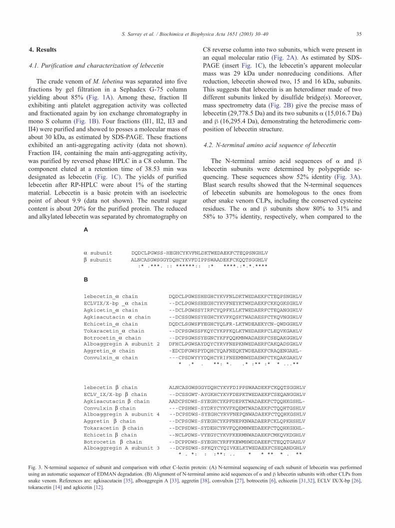
Fig. 3. N-terminal sequence of subunit and comparison with other C-lectin prote
using an automatic sequencer of EDMAN degradation. (B) Alignment of N-termin
snake venom. References are: agkisacutacin [35], alboaggregin A [33], aggretin [
tokaracetin [14] and agkicetin [12].
C8 reverse column into two subunits, which were present in
an equal molecular ratio (Fig. 2A). As estimated by SDS-
PAGE (insert Fig. 1C), the lebecetin’s apparent molecular
mass was 29 kDa under nonreducing conditions. After
reduction, lebecetin showed two, 15 and 16 kDa, subunits.
This suggests that lebecetin is an heterodimer made of two
different subunits linked by disulfide bridge(s). Moreover,
mass spectrometry data (Fig. 2B) give the precise mass of
lebecetin (29,778.5 Da) and its two subunits a (15,016.7 Da)
and h (16,295.4 Da), demonstrating the heterodimeric com-
position of lebecetin structure.
4.2. N-terminal amino acid sequence of lebecetin
The N-terminal amino acid sequences of a and hlebecetin subunits were determined by polypeptide se-
quencing. These sequences show 52% identity (Fig. 3A).
Blast search results showed that the N-terminal sequences
of lebecetin subunits are homologous to the ones from
other snake venom CLPs, including the conserved cysteine
residues. The a and h subunits show 80% to 31% and
58% to 37% identity, respectively, when compared to the
in: (A) N-terminal sequencing of each subunit of lebecetin was performed
al amino acid sequences of a and h lebecetin subunits with other CLPs from
38], convulxin [27], botrocetin [6], echicetin [31,32], ECLV IX/X-bp [26],

S. Sarray et al. / Biochimica et Biophysica Acta 1651 (2003) 30–4036
sequences of the corresponding subunits of the other
CLPs. The lebecetin’s h subunit shares a highest identity
with the h chain of ECLV IX/X-bp (80%) [26]. However,
the N-terminal sequence of a lebecetin’s subunit exhibits
only 48% identity to that of convulxin a chain [27] (Fig.
3B). Consequently, we may assume that lebecetin is one of
the CLP superfamily.
4.3. Effect of lebecetin on enzymatic, hemagglutination and
thrombin-induced clot retraction and coagulation activities
Lebecetin was devoid of enzymatic activities such as
phospholipase A2, fibrinogenolytic and amidolytic activities
(data not shown). Moreover, lebecetin did not inhibit the
procoagulant activity of thrombin in the presence of human
plasma or fibrinogen. Also, it is unable to inhibit thrombin-
Fig. 4. Inhibition of platelet aggregation by lebecetin. (A) Inhibitory effect of leb
incubated with lebecetin for 2 min at 37 jC before adding 1 mM ADP (-o-) or 10
with lebecetin for 2 min before the addition of 0.04 IU/ml thrombin (-x-) or
experiments. (C) Effect of lebecetin on U46619- (1 AM) and collagen- (1 mg/ml
cells/ml) pre-incubated with different concentrations of lebecetin were exposed to
were determined; shown traces are results of experiments realized with 1.6 nM
platelets were incubated with a (-o-) or h (-x-) lebecetin subunits for 2 min befo
separate experiments.
induced clot retraction in PRP and the agglutination of
human erythrocytes (data not shown).
4.4. Effect of lebecetin on platelet aggregation
Lebecetin showed a potent inhibitory effect on platelet
aggregation in PRP induced by ADP or Ca+ +-ionophore
(Fig. 4A). The concentrations of lebecetin leading to 50% of
the inhibition of platelet aggregation (IC50) were 3.6 and
4.4 nM, respectively.
Lebecetin also inhibited either the thrombin- or the
collagen-induced aggregation of washed platelets in a con-
centration-dependent manner. The IC50 were about of 0.25
and 0.35 nM, respectively (Fig. 4B). However, under same
conditions, lebecetin did not inhibit the aggregation induced
by U46619.
ecetin on rabbit whole blood platelet aggregation: Platelet-rich plasma was� 3 mM Ca+ +-ionophore (-x-). (B) Rabbit washed platelets were incubated
1 mg/ml of collagen (-o-). Results are representative of three separate
) induced platelet aggregation and ATP release: Platelet suspensions (3.108
collagen (left panel) or U46619 (right panel); aggregation and ATP released
of lebecetin. Values in brackets indicate ATP amounts. (D) Rabbit washed
re the addition of 0.04 IU/ml thrombin. Results are representative of three

S. Sarray et al. / Biochimica et Biophysica Acta 1651 (2003) 30–40 37
Rabbit washed platelets which aggregated and released
their granular contents in the presence of collagen and
U46619 were used to study the effects of lebecetin. As
shown in Fig. 4C, lebecetin failed to block both aggregation
and ATP release in response to U46619. In contrast, when
platelets were exposed to collagen, lebecetin inhibited in a
concentration-dependent manner both aggregation and ATP
release.
Reduced and alkylated lebecetin subunits have exhibited
separately an anti-aggregating activity when tested on
washed platelets in the presence of low concentration of
thrombin (0.04 IU/ml). The h subunit appears to be more
efficient than the a subunit. In fact, the IC50 were 1.55 and
0.7 nM for a and h subunits, respectively (Fig. 4D).
4.5. Effect of lebecetin on platelet agglutination
Lebecetin inhibited the agglutination of human platelets
by ristocetin in PRP. This effect was concentration-depen-
dent with an IC50 of about 0.45 nM (Fig. 5A). Furthermore,
Fig. 5. Inhibition of platelet agglutination by lebecetin. (A) Inhibition of agglutin
incubated with different concentrations of lebecetin for 2 min at 37 jC before add
lebecetin was incubated with fixed platelets for 2 min at 37 jC. Ristocetin (20 Agdetermined. A representative experiment is shown (n= 3). (C) Binding studies o
human platelets. Fixed platelets (2.105/ml) were incubated with various concentr
lebecetin concentration (nonspecific binding) (-o-) and then analysed by flow cy
the agglutination of washed human formalin-fixed platelets
induced by vWF (1 IU/ml) in the presence of ristocetin was
inhibited by lebecetin (Fig. 5B), with an IC50 of 0.65 nM.
In order to evaluate the importance of Ca+ + in this
process, we have tested the effect of EDTA and EGTA on
lebecetin-induced fixed platelet agglutination. Indeed,
EDTA and EGTA significantly reduced lebecetin activity
to 40% on platelet agglutination (data not shown).
4.6. Effect of FITC-lebecetin binding to formalin-fixed
platelets
Using FITC-conjugated lebecetin as a probe, we have
started to study the molecular mechanism of lebecetin. The
nonspecific binding was evaluated using high concentration
of native lebecetin. FITC-lebecetin is a potent anti-platelet
aggregating protein as native lebecetin (data not shown).
FITC-lebecetin bound in a concentration-dependent manner
to human fixed platelets and almost reached a saturable
binding at a concentration of 90 Ag/ml (3.1 AM).
ation of human whole blood by lebecetin: human platelet-rich plasma was
ing ristocetin (20 Ag/ml). (B) Effect of lebecetin on formalin fixed platelets:
/ml) and vWF (1 U/ml) were then added for 5 min and inhibition was then
f lebecetin to fixed platelets: binding isotherm of FITC-lebecetin to fixed
ation of FITC-lebecetin (-x-) in the presence or absence of higher native
tometry. Each point is the average of triplicate determinations.

S. Sarray et al. / Biochimica et Biophysica Acta 1651 (2003) 30–4038
4.7. Effect of various inhibitors on FITC-lebecetin binding
The concentration of FITC-lebecetin (3.1 AM) used in a
competition binding studies induces the maximal antipla-
telet aggregation. At 50 Ag/ml, anti GPIb mAb LJIb1 and
anti-GPIIb/IIIa (LJCP8) are used in separate experiments
using FITC lebecetin at 3.1 AM. Anti-GPIIb/IIIa (LJCP8)
did not compete with FITC-labelled lebecetin. In the
contrary, anti-GPIb mAb LJIb1, used at the same concen-
tration, was able to inhibit 50% of FITC-labelled lebecetin
to fixed platelets (data not shown). This result indicates
that lebecetin binds specifically with high affinity on GPIb
receptor.
5. Discussion
Ca+ +-dependent CLPs isolated from snake venom are
defined as nonenzymatic proteins. They are heterodimers
comprising of a and h subunits with a molecular mass value
of about 14–16000 Da. The subunits share a high degree of
sequence similarity with each other and with the other snake
venom CLPs. These proteins are recognized to contain a
CRD characterized by several conserved amino acid residues
in their structure. Despite their striking structural similarity,
the proteins of this group show a variety of biological effects
that depend on the structure of the carbohydrate recognition
site’s nature [28]. We report here the purification and the
characterization of a CLP, called lebecetin, isolated from the
venom of M. lebetina.
The crude venom of M. lebetina prevents platelet aggre-
gation induced by various agonists including thrombin. This
venom is known to contain small lebetin peptides with an
anti-platelet activity [29,30]. Besides them, we have identi-
fied at least three proteins with the same molecular mass of
about 29 kDa. One of them, lebecetin, has been purified. It
is a potent anti-platelet and anti-agglutination protein as
revealed by IC50 in the nanomolar range. Lebecetin was
homogeneous as examined by SDS-PAGE and mass spec-
trometry. It behaved as a molecule of 29,779 Da under
nonreducing conditions and separated into two bands, a and
h, of 15,016 and 16,296 Da, respectively, under reducing
conditions. Lebecetin exists as an heterodimeric glycopro-
tein composed of a and h subunits linked probably with a
disulfide bridge, as observed for other CLPs [6,9,31–35].
The putative role of the glycosylation in the biological
properties of lebecetin is not so far elucidated. Thus, it will
be interesting to elucidate the contribution of glucidic motif
to its biological activities. Lebecetin subunits were separated
by HPLC after reduction and alkylation and then sequenced.
Comparing the alignment of amino acid sequences of
lebecetin’s subunits, we found that the N-terminal sequences
of a and h subunits of lebecetin are highly homologous to
one another, and with those of CLPs, including GPIb-
binding proteins [6–8,12]. Thus, lebecetin may belong to
the CLP family.
Lebecetin inhibits platelet aggregation and ATP release
triggered by ADP, Ca+ +-ionophore, collagen or thrombin
but not by arachidonic acid or the TxA2 mimetic, U46619.
This selective inhibition rules out the possibility that,
whatever agonists used, lebecetin may inhibit GPIIb/IIIa–
fibrinogen interaction required for platelet aggregation [36].
One of the hurdles in understanding the structure–function
relationships of snake venom CLPs is to recognize whether
one or both subunits are required for biological activity.
Kawasaki et al. [37] suggested that the platelet GPIb-
binding site of these GPIb-BPs resides on the h subunit
but not the a subunit. On the other hand, Polgar et al. [32]
proposed that the a subunit may play a major role on the
cross-linking GPIb molecules in the same way as the natural
ligand, vWF, based on the observation that the amino acid
sequencing of h subunits is highly homologous to each
other and only GPIb ligand, such as AL-B (25-kDa alboag-
gregin), can agglutinate platelets. However, the reduced and
alkylated a and h subunits of lebecetin retained their
biological activities, suggesting the importance of the pri-
mary structure of lebecetin’s subunits. Therefore, the exact
participation of each subunit in binding is not clearly
elucidated.
The specific binding of lebecetin to GPIb is supported
by several lines of evidence. Lebecetin prevents selective-
ly a further aggregation induced by ristocetin in the
presence of human vWF. Flow cytometric analysis reveals
that lebecetin binds to fixed platelets in a dose-dependent
and saturable manner and that this binding is blocked
specifically by an anti-GPIb mAb (LJIb1). However, this
binding is unaffected by an anti-GPIIb-IIIa mAb (LJCP8),
which acts as a fibrinogen receptor (GPIIb/IIIa complex)
antagonist.
Further studies, which include the GPIb-binding site of
lebecetin, by using a panel of monoclonal antibodies
presenting different specificities with GPIb, will allow
us to elucidate the molecular mechanism of platelet
aggregation in terms of GPIb-vWF and provide a molec-
ular design suitable for the development of future anti-
thrombotic agents targeting at blocking vWF binding to
GPIb.
Acknowledgements
We thank Prof. Koussay Dellagi (Institut Pasteur de
Tunis) for his continuous interest in this study and for his
support. Prof. Zavario M. Ruggeri (Department of Molecular
and Experimental Medicine and Vascular Biology, The
Scripps Research Institutes La Jolla, California) is greatfully
acknowledged for his generous gift of monoclonal anti-
bodies. We would also like to thank Dr. Elalamy Ismaıl
(Institut Pasteur de Paris) for helpful advices. Dr. Benlasfar
Zakaria and Ben Zakour Lotfi (laboratoire veterinaire,
Institut Pasteur de Tunis) are acknowledged for providing
viper venoms.

S. Sarray et al. / Biochimica et Biophysica Acta 1651 (2003) 30–40 39
References
[1] S. Niewiarowski, M.A. McLane, M. Kloczewiak, G.J. Stewart, Dis-
integrins and other naturally occurring antagonists of platelet fibrino-
gen receptors, Semin. Hematol. 31 (1994) 289–300.
[2] M.A. McLane, C. Marcinkiewicz, S. Vijay-Kumar, I. Wierzbicka-Pa-
tynowski, S. Niewiarowski, Viper venom disintegrin and related mol-
ecules, Proc. Soc. Exp. Biol. Med. 219 (1998) 109–119.
[3] C. Marcinkiewicz, L.A. Rosenthal, D.M. Mosser, T.J. Kunicki, S.
Niewiarowski, Immunological characterization of eristostatin and
echistatin binding sites on alpha IIb beta 3 and alpha v beta 3 integ-
rins, Biochem. J. 317 (1996) 817–825.
[4] C. Marcinkiewicz, J.J. Calvete, M.M. Marcinkiewicz, M. Raida, S.
Vijay-Kumar, Z. Huang, R.R. Lobb, S. Niewiarowski, EC3, a novel
heterodimeric disintegrin from Echis carinatus venom, inhibits alpha4
and alpha5 in an RGD-independent manner, J. Biol. Chem. 274
(1999) 12468–12473.
[5] C. Marcinkiewicz, J.J. Calvete, S. Vijay-Kumar, M.M. Marcinkie-
wicz, M. Raida, P. Shick, R.R. Lobb, S. Niewiarowski, Structural
and functional characterization of EMF10, a heterodimeric disintegrin
from Eristocophis macmahoni venom that selectively inhibits alpha 5
beta 1 integrins, Biochemistry 38 (1999) 13302–13309.
[6] Y. Fujimura, K. Titani, Y. Usami, M. Suzuki, R. Oyama, T. Matsui, H.
Fukui, M. Sugimoto, Z.M. Ruggeri, Isolation and characterization of
two structurally and functionally distinct from botrocetin, the platelet
co agglutinin from the venom of Bothrops jararaca, Biochemistry 30
(1991) 1957–1964.
[7] M.S. Read, S.V. Smith, M.A. Lamb, K.M. Brinkhous, Role of
botrocetin in platelet agglutination: formation of an activated com-
plex of botrocetin and von Willebrand factor, Blood 74 (1989)
1031–1035.
[8] M. Peng, F.A. Emig, A. Mao, W. Lu, E.P. Kirby, S. Niewiarowski,
M.A. Kowalska, Interaction of echicetin with a high affinity throm-
bin-binding site on platelet glycoprotein GPIb, Thromb. Haemost. 74
(1995) 954–957.
[9] M. Peng, W. Lu, L. Beviglia, S. Niewiarowski, E.P. Kirby, A
snake venom protein that inhibits binding of von Willebrand Fac-
tor and alboaggregins to platelet glycoprotein Ib, Blood 81 (1993)
2321–2328.
[10] M. Peng, W. Lu, E.P. Kirby, Alboaggregins-B: a new platelet agonist
that binds to platelet membrane glycoprotein Ib, Biochemistry 30
(1991) 11529–11536.
[11] M. Peng, W. Lu, E.P. Kirby, Characterization of three alboaggregins
purified from Trimeresurus albolabris venom, Thromb. Haemost. 67
(1992) 702–707.
[12] C.H. Yeh, W.C. Wang, T.T. Hsieh, T.F. Huang, Agkicetin, a snake
venom-derived glycoprotein Ib antagonist, disrupts von Willebrand
factor–endothelial cell interaction and inhibits angiogenesis, J. Biol.
Chem. 275 (2000) 18615–18618.
[13] Y. Taniuchi, T. Kawasaki, Y. Fujimura, M. Suzuki, K. Titani, K.
Sakai, S. Kaku, N. Hisamichi, V. Satoh, T. Takenaka, M. Handa, Y.
Swai, Flavocetin-A and -B, two high molecular mass glycoprotein Ib
binding proteins with high affinity purified from Trimeresurus flavor-
idis venom, inhibit platelet aggregation at high shear stress, Biochim.
Biophys. Acta 1244 (1995) 331–338.
[14] T. Kawasaki, Y. Taniuchi, N. Hisamichi, Y. Fujimura, M. Suzuki, K.
Titani, S. Kaku, N. Satoh, T. Takenaka, M. Handa, Y. Swai, Tokar-
acetin, a new platelet antagonist that binds to platelet glycoprotein Ib
and inhibits von Willebrand factor-dependent shear-induced platelet
aggregation, Biochem. J. 308 (1995) 947–953.
[15] C. Marcinkiewicz, R.R. Lobb, M.M. Marcinkiewicz, J.L. Daniel, J.
Bryan Smith, C. Dangelmair, P.H. Weinreb, D.A. Beacham, S.
Niewiarowski, Isolation and characterization of EM16, a C-lectin
type protein from Echis multisquamatus venom, a potent and se-
lective inhibitor of the a2h1 integrin, Biochemistry 39 (2000)
9859–9867.
[16] K. Drickamer, M.E. Taylor, Biology of animal lectins, Annu. Rev.
Cell Biol. 9 (1993) 237–264.
[17] U.K. Laemmli, Cleavage of structural proteins during the assembly of
the head of bacteriophage T4, Nature 277 (1970) 680–685.
[18] M. Dubois, K.A. Gilles, J.K. Hamilton, P.A. Rebers, F. Smith, Col-
orimetric method for determination of sugars and related substances,
Anal. Chem. (1956) 350–356.
[19] P.L. Song, P. Xin, A lectin-like peptide isolated from the venom
of the Chinese spider Selenocosmia huwena, Toxicon 33 (1995)
875–882.
[20] Y.S. Li, K.F. Liu, Q.C. Wang, Mechanism of action of the platelet
function inhibitor from Vipera russelli siamensis snake venom, Tox-
icon 24 (1986) 875–883.
[21] N.G. Ardlie, D.W. Perry, M.A. Packham, J.F. Mustard, Adenosine
diphosphate induced platelet aggregation in suspensions of washed
rabbit platelet, Proc. Soc. Exp. Biol. Med. 13 (1970) 1021–1023.
[22] G.V.R. Born, M.J. Cross, The aggregation of blood platelets, J. Phys-
iol. 168 (1963) 178–195.
[23] H. Holmsen, E. Storm, H.J. Day, Determination of ATP and ADP in
blood platelets: a modification of the firefly luciferase assay for plas-
ma, Anal. Biochem. 46 (1972) 489–501.
[24] E.P. Kirby, D.C.B. Mills, The interaction of bovine factor VIII with
human platelets, J. Clin. Invest. 56 (1975) 491–502.
[25] C.Z. Liu, Y.W. Wang, M.C. Shen, T.F. Huang, Analysis of human
platelet glycoprotein IIb– IIIa by fluorescein isothiocyanate-conju-
gated disintegrins with flow cytometry, Thromb. Haemost. 39
(1994) 1915–1923.
[26] Y.L. Chen, I.H. Tsai, Functional and sequence characterization of
coagulation factor X-binding protein from the venom of Echis car-
inatus leucogaster, Biochemistry 35 (1996) 5264–5271.
[27] M. Leduc, C. Bon, Cloning of subunits of convulxin, a collagen-like
platelet aggregating protein from Crotalus durissus terrificus venom,
Biochem. J. 333 (1998) 389–393.
[28] Y. Komori, T. Nikai, T. Tohkai, H. Sugihara, Primary structure
and biological activity of snake venom lectin (APL) from Agkis-
trodon p. piscivorus (Eastern cottonmouth), Toxicon 37 (1999)
1053–1064.
[29] R. Barbouche, N. Marrakchi, P. Mansuelle, M. Krifi, E. Fenouillet, H.
Rochat, M. El Ayeb, Novel anti-platelet aggregation polypeptides
from Vipera lebetina venom: isolation and characterization, FEBS
Lett. 392 (1996) 6–10.
[30] R. Barbouche, N.Marrakchi, K.Mabrouk,M. Krifi, J. VanRietschoten,
E. Fenouillet, M. El Ayeb, H. Rochat, Anti-platelet activity of the
peptides composing the lebetin 1 family, a new cl of inhibitors of
platelet aggregation, Toxicon 36 (1998) 1939–1947.
[31] M. Peng, J.C. Holt, S. Niewiarowski, Isolation, characterization and
amino acid sequence of echicetin beta subunit, a specific inhibitor of
von Willebrand factor and thrombin interaction with glycoprotein Ib,
Biochem. Biophys. Res. Commun. 205 (1994) 68–72.
[32] J. Polgar, E.M. Magnenat, M.C. Peitsch, T.N.C. Wells, M.S.A. Saqi,
K.J. Clemetson, Amino acid sequence of the alpha subunit and com-
puter modelling of the alpha and beta subunits of echicetin from the
venom of Echis carinatus (saw-scaled viper), Biochem. J. 323 (1997)
533–537.
[33] M.A. Kowalska, L. Tan, J.C. Holt, M. Peng, J. Karczewski, J.J.
Calvete, S. Niewiarowski, Alboaggregins A and B. Structure and
interaction with human platelets, Thromb. Haemost. 79 (1998)
609–613.
[34] Y. Usami, Y. Fujimura, M. Suzuki, Y. Ozeki, K. Nishio, H. Fukui, K.
Titani, Primary structure of two chain botrocetin, a von Willebrand
factor modulator purified from the venom of Bothrops jararaca, Proc.
Natl. Acad. Sci. U. S. A. 90 (1993) 928–932.
[35] X. Cheng, Y. Qian, Q. Liu, B.X. Li, M. Zhang, J. Liu, Purification,
characterization, and cDNA cloning of a new fibrinogenolytic venom
protein, Agkisacutacin, from Agkistrodon acutus venom, Biochem.
Biophys. Res. Commun. 265 (1999) 530–535.
[36] J.J. Calvete, Platelet integrin GPIIb/IIIa structure– function correla-

S. Sarray et al. / Biochimica et Biophysica Acta 1651 (2003) 30–4040
tions. An update and lessons from other integrins, Proc. Soc. Exp.
Biol. Med. 222 (1999) 29–38.
[37] T. Kawasaki, Y. Fujimura, Y. Usami, M. Suzuki, S. Miura, Y. Sakurai,
K. Makita, Y. Taniuchi, K. Hirano, K. Titani, Complete amino acid
sequence and identification of the platelet glycoprotein Ib-binding site
of jararaca GPIb-BP, a snake venom protein isolated from Bothrops
jararaca, J. Biol. Chem. 271 (1996) 10635–10639.
[38] C.H. Chung, L.C. Au, T.F. Huang, Molecular cloning and sequence
analysis of aggretin, a collagen-like platelet aggregation inducer, Bio-
chem. Biophys. Res. Commun. 263 (1999) 723–727.