Infestation of mice and voles with Ixodes ricinus ticks in Lithuania and Norway
Upload
independentCategory
view
0download
0
530
Lectinochemical Studies on the Binding Properties of a ToxicLectin (Ricin) Isolated from the Seeds of Ricinus communis
Albert M.Wu, PhD; June H. Wu1, PhD; Tanuja Singh, PhD; Pei-Yi Hwang; Ming-Sung Tsai, PhD; Anthony Herp, PhD
Background: Ricin (RCA2 or RCA60) is a highly toxic heterodimeric protein found in theseeds of the castor plant Ricinus communis. It is a potential biohazard. In thepresent study, the fine specificity of ricin was defined.
Methods: The combining site of ricin was characterized by quantitative precipitin(QPA) and precipitin inhibition assays (QPIA).
Results: Of 31 glycoproteins and pneumococcus type XIV capsular polysaccharidetested, only twelve of them precipitated over 50% of the toxin N added,reflecting poor precipitability of the lectin with the compounds tested. Thiscan be explained by only a single chain (B chain of the molecules) participat-ing in binding. The blood group active glycoproteins after mild acid hydroly-sis or Smith degradation, as well as sialic-acid containing glycoproteins afterremoval of sialic acid, in general, had substantially increased activity. Of themonosaccharides tested for inhibition of precipitation of ricin, p-nitrophenyl
Gal was the best; this compound was 1.3-fold better than its -anomer.While methyl Gal was twice as active as its anomer, Gal and bloodgroup B active disaccharides (Gal 1-3Gal) were 2.5 times more active thanGalNAc. Among the oligosaccharides tested, Gal 1-3GalNAc (T) Gal 1-3/4GlcNAc (I/II), Gal 1-4Glc (L) and human blood group I Ma trisaccha-ride (Gal 1-4GlcNAc 1-6Gal) were about equally active and the bestinhibitors. They were about 2.0 and 2.4 more active than Gal 1-4Gal (E)sequence and B determinant, respectively.
Conclusion: From the present results, it is concluded that: (a) this toxin has a broad rangeof affinity for the -anomer of Gal; (b) its combining site is probably of ashallow groove type and as large as a trisaccharide; (c) Gal - is the majorcombining site of the lectin; and (d) hydrophobic interaction gives a signifi-cant contribution for binding. This information should facilitate future usageof this lectin in glycobiological research and medical applications.(Chang Gung Med J 2005;28:530-42)
Key words: carbohydrate specificities, glycoprotein binding, combining site, toxin, ricin.
From the Glyco-Immunochemistry Research Laboratory, Institute of Molecular and Cellular Biology, 1Department of Microbiologyand Immunology, Chang Gung University, Taoyuan.Received: May. 9, 2005; Accepted: Jun. 7, 2005Address for reprints: Prof. Albert M. Wu, Glyco-Immunochemistry Research Laboratory, Institute of Molecular and CellularBiology, Chang-Gung University, No. 259, Wen-hwa 1st Rd., Gueishan Shiang, Taoyuan, Taiwan 333, R.O.C. Tel.: 886-3-211-8966(Lab.); Fax: 886-3-211-8456 (Lab.), 886-3-211-8700 (Col.); E-mail: [email protected]
Ricin (RCA2 or RCA60) is the highly toxic frac-tion of the carbohydrate binding proteins found
in aqueous extracts of the castor bean (Ricinus com-munis). It is a potential biohazard. It agglutinated
Original Article

Chang Gung Med J Vol. 28 No. 8August 2005
Albert M.Wu, et alRicinus communis agglutinin 2
531
weakly human red blood cells regardless of ABOblood group type, with or without neuraminidasetreatment. Ricin is a dimeric protein Mr = 60-65kDa, composed of two different polypeptide chainsA and B linked by a single disulphide bridge.(1,2) Itwas found that the A chain is a catalytic unit capableof inactivating the 60S ribosomal subunit of eukary-otic cells resulting in inhibition of protein synthe-sis.(3) The B chain binds to cell surface glycoconju-gate and thereby permits the entry of the toxic sub-unit into the cell. The combining site of this toxinhas not yet been investigated, especially the bindingrelationship among many important mammalianoligosaccharide units, such as I/II (Gal 1-3/4GlcNAc), T determinant (Gal 1-3GalNAc), E(Gal 1-4Gal, human blood group P1 disaccharide),and B (Gal 1-3Gal, human blood group B activedisaccharide) as well as anomers of p-nitrophenylGal have not yet been investigated. Therefore, in thepresent study, we have examined the glycan affinityof ricin by quantitative precipitin (QPA) and precip-itin inhibition assays (QPIA).(4,5) Comparison of theinhibition profile of ricin (RCA2) with RCA1 is alsoincluded.(6,7) From the present results, it is concludedthat: (a) this toxin has a broad range of affinity forthe -anomer of Gal; (b) its combining site is proba-
bly of a shallow groove type and its size is as largeas a trisaccharide; (c) Gal - should be the majorcombining site of the lectin; and (d) hydrophobicinteraction provide a significant contribution forbinding.
METHODS
LectinRicin, purified from the aqueous extracts of the
castor bean (seeds of Ricinus communis) by affinitycolumn chromatography using the method of Lin etal.(8) was purchased from Sigma Chemical Company(St. Louis, MO, USA).
Glycoproteins and polysaccharidesCarbohydrate structural units in mammalian
glycoprotein are shown in Table 1. The glycoproteinstested were prepared from human ovarian cyst fluid,saliva, and hog gastric mucosa.(9-15) The blood groupA, B, H, Lea, Leb and Ii active substances were puri-fied from human ovarian cyst fluid (HOC) by diges-tion with pepsin, precipitation with increasing con-centrations of ethanol and extraction of the driedethanol precipitates with 90% phenol. The insolublefraction is named after the blood group substance
Table 1. Carbohydrate Structural Units in Mammalian Glycoproteins (46)
Codes* Structural units Sources
1 Tn GalNAc 1 Ser/Thr of protein core
2 T Gal 1 3GalNAc 1 Ser/Thr of protein core
3 I Gal 1 3GlcNAc
4 II Gal 1 4GlcNAc
II Gal 1 4GlcNAc 1
Tri-II Triantennary Gal 1 4GlcNAc
mII Multivalent Gal 1 4GlcNAc
5 A GalNAc 1 3Gal
Ah GalNAc 1 3[LFuc 1 2]Gal
6 B Gal 1 3Gal
Bh Gal 1 3[LFuc 1 2]Gal
7 E Gal 1 4Gal
8 L Gal 1 4Glc
L Gal 1 4Glc 1
* , : anomer of sugars; m: multivalent.
Tn antigen, only in glycoproteins.
The mucin-type sugar sequence on the human erythrocyte membrane.
Human blood group type I and II carbohydrate sequences. Branched or linearrepeated II sequence is part of blood group I and i epitopes. I and II are precur-sors of ABH and Lea, Leb, Lex, Ley blood group active antigens. Most of thelectins reactive with II are also reactive with I. Tri-II and mII determinants arepresent at the nonreducing end of the carbohydrate chains derived from N- andO-glycans.
Human blood group A active di-saccharide.
Human blood group A active tri-saccharide.
Human blood group B active di-saccharide.
Human blood group B active tri-saccharide.
Blood group pk and P1 active disaccharide. Sheep hydatid cyst glycoproteins,salivary glycoproteins of the Chinese swiftlet, glycosphingolipids in human ery-throcytes, and small intestine.
Constituent of mammalian milk.
Lactosyl ceramides in brain and part of carbohydrate structures in gangliosides.

Chang Gung Med J Vol. 28 No. 8August 2005
Albert M.Wu, et alRicinus communis agglutinin 2
532
(e.g. Cyst Tighe phenol insoluble, where “Tighe”denotes the HOC sample code). A similar principlewas applied to other HOC collections (e.g. Beach,MSS, and N-1, etc). The supernatant was fractionallyprecipitated by addition of 50% ethanol in 90% phe-nol to the indicated concentrations.(9) The designation“10 (or 20)% (ppt)” denotes a fraction precipitatedfrom phenol at an ethanol concentration of 10 (or20)%; “2x” signifies that a second round of phenolextraction and ethanol precipitation was carried out(e.g. Cyst MSS 10% 2x and Cyst N-1 Lea 20% 2x).The carbohydrate chains of HOC consist of multiplesaccharide branches attached by O-glycosidic link-ages at their internal reducing ends to serine or threo-nine residues of the polypeptide backbone.(11-13,16) Ingeneral, the “P-1” fractions (e.g. Cyst Beach P-1 andCyst Tighe P-1) represent the nondialyzable portionof the blood group substances after mild hydrolysisat pH 1.5-2.0 for 2 h which removes most of the L-fucopyranosyl end groups, as well as some bloodgroup A and B active oligosaccharide side-chains.(10,17,18) P-1 fractions from HOC gps whichexpose the internal structures equivalent to those onthe blood group precursors are defined as “precursorequivalent gps” (Structure 1).
Human 1-acid glycoprotein and porcine thy-roglobulin were purchased from Sigma. Human 1-acid glycoprotein contains tetra-, tri- and di-anten-nary complex type glycans in the ratio of 2:2:1.(19,20)
Fetuin (Gibco Laboratories, Grand Island, NY,USA), which is the major glycoprotein in fetal calfserum and has six oligosaccharide side chains permolecule, three O-glycosidically-linked to Ser/Thrand three N-glycosidically-linked to Asn.(21) that con-tains tri- and di-antennary complex type glycans inthe ratio of 1:2.
The rat sublingual glycoprotein (RSL) was pre-pared by the method of Moschera and Pigman.(22)
The established carbohydrate side chains were foundto be composed of 9, 10, 12, 13, and 15-sugarresidues, respectively, and contain sialic acid, N-acetylglucosamine, galactose, and N-acetylgalac-tosaminitol.(23) RSL may also contain the Tn reactivedeterminants.(24)
Hog gastric mucin #4, a blood group A + H sub-stance (Structure 2), was derived from crude hogstomach mucin as described previously.(25) Treatmentof mucin #4 with HCl (pH 2, 100°C, 90 min) yieldshog gastric mucin #9, while acid hydrolysis (pH 1.5,
100°C, 2 and 5 hrs) gives hog gastric mucins #14and 21, respectively. Extensive hydrolysis leads todestruction of blood group activities.(4)
Structure 1. Proposed representative carbohydrate sidechains of blood group active glycoproteins, prepared fromhuman ovarian cyst fluid. This structure represents the inter-nal portion of carbohydrate chains to which various humanblood group determinants are attached. The four-branchedstructure (1 to 4) shown is the representative internal portionof the carbohydrate moiety of blood group substances towhich the residues responsible for A, B, H, Lea, and Leb activ-ities are attached. This structure also represents precursorblood group active glycoproteins(13) and can be prepared bySmith degradation of A, B, H active glycoproteins, purifiedfrom human ovarian cyst fluids.(11-13,17) Numbers in parenthesesindicate the site of attachment for the human blood group A,B, H, Lea, and Leb determinants. These determinants as wellas the structural units at the nonreducing end are the sourcesof lectin reactive A/Ah, B, I/II, T, and Tn determinants.(4) Thismegalo-saccharide of twenty-four sugars has not been isolat-ed. However, most of the carbohydrate chains isolated areparts of this structure.
Structure 2. Proposed structure of carbohydrate side chainsof Hog A + H gastric glycoproteins.(25) Blood group A and Hactive key sugars for ricin. Most carbohydrate side chains arepart of this structure.

Chang Gung Med J Vol. 28 No. 8August 2005
Albert M.Wu, et alRicinus communis agglutinin 2
533
Ovine, bovine, and porcine submandibular/sali-vary glycoproteins were purified according to themethod of Tettamanti and Pigman(26) with modifica-tions.(27,28) About 75% of the carbohydrate side chainsof asialo OSM were GalNAc 1-Ser/Thr (Tn). AsialoPSM contains Gal 1-3GalNAc 1- (T ) togetherwith Tn and GalNAc 1-3Gal (A) sequences, as mostof the outer fucosyl residues and sialic acids arecleaved by mild acid hydrolysis (Structure 3). NativeASG-Tn,(29) a salivary glycoprotein of nine-bandedarmadillo (Dasypus novemcinctus mexicanus) con-taining only Tn (GalNAc 1-Ser/Thr) as carbohy-drate side chains, was isolated from 0.01 M PBS pH6.8 gland extract after removal of ASG-A, which isone of the sialoglycoproteins in armadillo glands.(30)
Desialylation of sialoglycoproteins was per-formed by mild acid hydrolysis in 0.01 N HCl at80°C for 90 min and dialyzed against distilled H2Ofor 2 d to remove small fragments.(26,30)
The anti-freeze glycoprotein from the Antarcticfish (Trematomus borchgrevinki) which containsonly T as carbohydrate chains.(31) was provided byDr. R. E. Feeney (Department of Food Science andTechnology, University of California, Davis, CA,USA) through the late Dr. E. A. Kabat (ColumbiaMedical Center, New York, USA).
The Pneumococcus type XIV polysaccharide,isolated from Streptococcus pneumoniae capsule,(32)
was a generous gift from the late Dr. E.A. Kabat.
Sugars used for inhibition studiesMono-, di-, oligo-saccharides and their deriva-
tives were purchased from or prepared by Dextra(Berkshire, UK) and Sigma.
Immunochemical AssaysQuantitative precipitin and precipitin-inhibition
assays were performed by a microprecipitin tech-nique(33) using 6.0 µg of lectin nitrogen (N) for eachdetermination: total N in the washed precipitates wasestimated by the ninhydrin method.(34)
RESULTS
Quantitative Precipitin Assays (QPA)Quantitative precipitin reactions of the purified
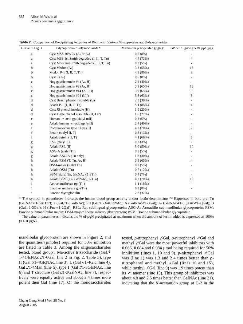
ricin with various blood group active substances andglycoproteins are seen in Figure 1 and Table 2.Reactions were carried out in 250 µl and some of thedifferences reflect solubility effects (Fig. 1). Themost reactive glycoproteins, cyst Mcdon P-1 andBeach P-1 precipitated over 80% of the lectin N withless than 4 µg being required for 50% precipitation(Fig. 1b and 1d). 1st Smith degraded product of cystMSS 10% 2x (Fig. 1a), hog gastric mucin before andafter mild acid treatment (Fig. 1c), Pneumococcustype XIV capsular polysaccharide (Fig. 1f), asialofetuin (Fig. 1f), asialo RSL (Fig. 1g) and desializedPSM and BSM (Fig. 1h) gave intermediate reactionand precipitated about 2/3 of the lectin nitrogen. Ofthe soluble complex carbohydrates tested, onlytwelve of them precipitated over 50% of the toxin Nadded. This result indicates that RCA2 has a poorprecipitability. This can be explained by only a singlechain (B chain of the molecules) responsible forbinding. The blood group active glycoproteins aftermild acid hydrolysis or Smith degradation as well assialic acid containing glycoprotein after removal ofsialic acid, in general, had substantially increasedactivity. This indicates that ricin-glycoprotein inter-action was strongly masked by sialic acid or Gal sub-stituted residues at non-reducing end.
Quantitative precipitin inhibition assays(QPIA)
The abilities of various sugars to inhibit the pre-cipitation of ricin with desialized porcine sub-
Structure 3. Porcine salivary mucin gp (PSM). The carbo-hydrate side chains of this glycoprotein are O-glycosidicallylinked through GalNAc at the reducing end of the carbohy-drate side chain to Ser or Thr of the protein core.(28) Twelvekinds of carbohydrate side chains have been isolated. Theyare composed of one to five sugar residues with Gal 1-3GalNAc -1-O-Thr or Ser as the carbohydrate core region.Substitution by NeuNAc, NeuNGc, GalNAc, or LFuc exhibitsblood group A and H activities, respectively. The mild acidtreated product of porcine salivary glycoprotein containsmainly mixtures of the above sequences. H, LFuc 1-2Gal.

Chang Gung Med J Vol. 28 No. 8August 2005
Albert M.Wu, et alRicinus communis agglutinin 2
534
Fig. 1 Quantitative precipitin curves of ricin with glycoproteins and Pneumococcus type XIV capsular polysaccharide. Conditions:6.0 µg ricin nitrogen; total volume was 250 µl.
Cyst MSS 10% 2x (Ah[A2])Cyst MSS 1st Smith degradedCyst MSS 2nd Smith degraded
Cyst McdonCyst Mcdon P-1Cyst 9 phenol insoluble
Hog gastric mucin #4Hog gastric mucin #9Hog gastric mucin #21Hog gastric mucin #14
Pneumococus type 14 psAsialo fetuinFetuin
Human 1-acid gp
Asialo human 1-acid gp
Cyst Beach phenol insolubleCyst Beach P-1Cyst JS phenol insolubleCyst Tighe phenol insoluble
RSL
Asialo RSL
ASG-A
Asialo ASG-A
Asialo PSM
BSM
Asialo BSM
OSM
Asialo OSM
Porcine thyroglobulin
Active antifreeze gp
Inactive antifreeze gp
Chang Gung Med J Vol. 28 No. 8August 2005
Albert M.Wu, et alRicinus communis agglutinin 2
535
mandibular glycoprotein are shown in Figure 2, andthe quantities (µmoles) required for 50% inhibitionare listed in Table 3. Among the oligosaccharidestested, blood group I Ma-active trisaccharide (Gal1-4GlcNAc 1-6Gal, line 2 in Fig. 2, Table 3), typeII (Gal 1-4GlcNAc, line 3), L (Gal 1-4Glc, line 4),Gal 1-4Man (line 5), type I (Gal 1-3GlcNAc, line6) and T structure (Gal 1-3GalNAc, line 7), respec-tively were equally active and about 2.4 times morepotent then Gal (line 17). Of the monosaccharides
tested, p-nitrophenyl Gal, p-nitrophenyl Gal andmethyl Gal were the most powerful inhibitors with0.066, 0.084 and 0.084 µmol being required for 50%inhibition (lines 1, 10 and 9). p-nitrophenyl Galwas (line 1) was 1.3 and 2.4 times better than p-nitrophenyl and methyl Gal (lines 10 and 15),while methyl Gal (line 9) was 1.9 times potent thanits anomer (line 15). This group of inhibitors wasabout 4.8 and 2.5 times better than GalNAc (line 21),indicating that the N-acetamido group at C-2 in the
Table 2. Comparison of Precipitating Activities of Ricin with Various Glycoproteins and Polysaccharides
Curve in Fig. 1 Glycoprotein / Polysaccharide* Maximum precipitated (µgN)† GP or PS giving 50% ppt (µg)
a Cyst MSS 10% 2x (A1 or Ah) 0.5 (8%) -a Cyst MSS 1st Smith degraded (I, II, T, Tn) 4.4 (73%) 4a Cyst MSS 2nd Smith degraded (I, II, T, Tn) 0.3 (5%) -b Cyst Mcdon (Ah) 3.3 (55%) 13b Mcdon P-1 (I, II, T, Tn) 4.8 (80%) 3b Cyst 9 (Ah) 0.5 (8%) -c Hog gastric mucin #4 (Ah, H) 2.4 (40%) -c Hog gastric mucin #9 (Ah, H) 3.9 (65%) 13c Hog gastric mucin #14 (A, I/II) 3.9 (65%) 9c Hog gastric mucin #21 (I/II) 3.8 (63%) 6d Cyst Beach phenol insoluble (B) 2.3 (38%) -d Beach P-1 (I, II, T, Tn) 5.1 (85%) 4d Cyst JS phenol insoluble (H) 1.5 (25%) -d Cyst Tighe phenol insoluble (H, Leb) 1.6 (27%) -e Human 1-acid gp (sialyl mII) 0.3 (5%) -e Asialo human 1-acid gp (mII) 2.4 (40%) -f Pneumococcus type 14 ps (II) 4.2 (70%) 2f Fetuin (sialyl II, T) 0.8 (13%) -f Asialo fetuin (II, T) 4.1 (68%) 6g RSL (sialyl II) 0.2 (3%) -g Asialo RSL (II) 3.0 (50%) 10g ASG-A (sialyl Tn) 0.3 (5%) -g Asialo ASG-A (Tn only) 1.8 (30%) -h Asialo PSM (T, Tn, Ah, H) 3.9 (65%) 4h OSM-major (sialyl Tn) 0.3 (5%) -h Asialo OSM (Tn) 0.7 (12%) -h BSM (sialyl Tn, GlcNAc 1-3Tn) 0.4 (7%) -h Asialo BSM (Tn, GlcNAc 1-3Tn) 4.2 (70%) 15i Active antifreeze gp (T ) 1.1 (18%) -i Inactive antifreeze gp (T ) 0.5 (8%) -i Porcine thyroglobulin 2.2 (37%) -
* The symbol in parentheses indicates the human blood group activity and/or lectin determinants.(46) Expressed in bold are: Tn(GalNAc 1-Ser/Thr); T (Gal 1-3GalNAc); I/II (Gal 1-3/4GlcNAc); A (GalNAc 1-3Gal); Ah (GalNAc 1-3 [LFuc 1-2]Gal); B(Gal 1-3Gal); H (LFuc 1-2Gal); RSL: Rat sublingual glycoprotein; ASG-A: Armadillo submandibular glycoprotein; PSM:Porcine submandibular mucin: OSM-major: Ovine salivary glycoprotein; BSM: Bovine submandibular glycoprotein.† The value in parantheses indicates the % of µgN precipitated at maximum when the amount of lectin added is expressed as 100%(= 6.0 µgN).

Chang Gung Med J Vol. 28 No. 8August 2005
Albert M.Wu, et alRicinus communis agglutinin 2
536
Fig. 2 Inhibition by various monosaccharides and oligosaccharides of the precipitation of ricin with desialized porcine salivaryglycoprotein. Total volume was 250 µl.
Gal 1 4GlcNAc 1 6Gal (I Ma)Gal 1 4GlcNAc (II)Gal 1 4Glc (L)Gal 1 3GlcNAc (L)
Gal 1 3GalNAc (T)
Gal 1 4Gal 1-Me (E)
Gal 1 3Gal 1-Me (B)
Gal
GalGal 1 6Glc (Melibiose)Gal 1 6Gl 1 2Fruf (Raffinose)DFuc
GalNAcLRhamnoseLAraGlcManMethyl DGlcMethyl DGlc
p-NO2phenyl DGalGal 1 4ManLactuloseMethyl- DGalp-NO2phenyl DGalGal 1 6GlcNAcGal 1 3DAraGal 1 6Gal 1 6Glc 1 2Fruf(Stachyose)Methyl- DGal

Chang Gung Med J Vol. 28 No. 8August 2005
Albert M.Wu, et alRicinus communis agglutinin 2
537
pyranose ring blocks reactivity. DFuc (line 20) was1/2 as active as Gal (line 17), implying that the OHgroup on C-6 participates in binding; LArabinosewhich has the same configuration as DGal but lacksthe CH2OH of C6 was about 1/5 as active as Gal, anindication that the CH2OH of C6 is necessary forbinding. Glc (line 24) and Man (line 25) showed1/200 and 1/333 of Gal activity, demonstrating thatthe configuration of carbon-4 and carbon-2 in Galare essential for binding. Melibiose (Gal 1-6Glc)and raffinose (Gal 1-6Glc 1-2DFruf) were equallyactive but 1.5 and 2.0 times less active thanstachyose (Gal 1-6Gal 1-6Glc 1-2Dfruf) (lines 18
and 19 vs. line 14), demonstrating that the carbohy-drate-binding site of the lectin for the -anomer ofGal can accommodate up to trisaccharide structure.
DISCUSSION
Ricin, like other plant toxins such as abrin fromthe seeds of Jequiriti beans (Abrus precatorious),mistletoe lectin from mistletoe plant (Viscum album)and modeccin from the roots of Adenia digitata aregalactose specific lectins,(35) has played interestingand important roles in the history of clinical medi-cine and biomedical research.(36) During the past two
Table 3. Amount of Various Saccharides Giving 50% Inhibition of Precipitation of Ricin by Desialized Porcine Salivary Glycoprotein*
Line No. Inhibitor
Quantity giving Ka Relative Potency†
in Fig. 2 50% inhibition (µmol) (103 M-1) Ricin (RCA2) RCA1(6,7)‡
1 p-NO2phenyl Gal 0.066 8.5 2.4 14.62 Gal 1-4GlcNAc 1-6Gal 0.066 8.5 2.4 9.1
(Human blood group I Ma trisaccharide)3 Gal 1-4GlcNAc (II) 0.066 8.5 2.4 6.4 4 Gal 1-4Glc (L) 0.066 8.5 2.4 4.15 Gal 1-4Man 0.066 8.5 2.4 3.76 Gal 1-3GlcNAc (I) 0.066 8.5 2.4 3.27 Gal 1-3GalNAc (T) 0.066 8.5 2.4 3.28 Lactulose 0.066 8.5 2.4 ND9 Methyl Gal 0.084 6.7 1.9 2.310 p-NO2phenyl Gal 0.084 6.7 1.9 0.611 Gal 1-6GlcNAc 0.084 6.7 1.9 3.312 Gal 1-3DAra 0.10 5.6 1.6 3.313 Gal 1-4Gal (E) 0.13 4.3 1.2 1.714 Gal 1-6Gal 1-6Glc 1-2Fruf (Stachyose) 0.13 4.3 1.2 0.615 Methyl Gal 0.16 3.5 1.0 0.816 Gal 1-3Gal 1-Me (B) 0.16 3.5 1.0 1.517 Gal 0.16 3.5 1.0 1.018 Gal 1-6Glc (Melibiose) 0.20 2.8 0.8 1.519 Gal 1-6Glc 1-2Fruf (Raffinose) 0.28 2.0 0.6 1.120 DFuc 0.28 2.0 0.6 ND21 GalNAc 0.37 1.5 0.4 0.0222 LRhamnose 0.54 1.0 0.3 0.823 LAra 1.0 0.41 0.2 0.224 Glc 30.0 0.019 0.005 0.00625 Man 56.0 0.010 0.003 ND26 Methyl Glc 56.0 0.010 0.003 ND27 Methyl Glc 100.0 0.006 0.001 ND
* 6.0 µg N of ricin + 5 µg of desialized porcine salivary glycoprotein. Total volume was 250 µl.† Relative potency of Gal taken as 1.0.‡ Data taken from references 6 and 7.Lectin abbreviation: RCA1 = Ricinus communis agglutinin 1; ND = not determined.
Chang Gung Med J Vol. 28 No. 8August 2005
Albert M.Wu, et alRicinus communis agglutinin 2
538
decades, this cytotoxic lectin has been cloned andsequenced.(37-39) and its protein crystal structureresolved,(40,41) but its fine specificity profile and theprecise combining sites have not been established.Furthermore, the binding relationship of lectin deter-minants among II, T and Tn in ricin was obscure.(42-44)
Therefore, in the present study, we characterized thebinding properties of ricin by quantitative precipitin(QPA) and precipitin-inhibition assays (QPIA) whichcan provide insight into the specificities and sizeparameters for its combining site. During the pasttwo to three decades, this system has been used as animportant tool to characterize the affinity of lectins.(4)
In this study, the blood group precursor equiva-lent gps (Structure 1), such as cyst Mcdon P-1 andBeach P-1 were the best and precipitated over 80%of the lectin N. Of the soluble complex carbohy-drates tested, only twelve of them precipitated over50% of the toxin N added. This result indicates thatricin has a poor precipitability. This can be explainedby only a single chain (B chain of the molecules)responsible for binding. The blood group active gly-coproteins after mild acid hydrolysis or Smith degra-dation as well as sialic acid containing glycoproteinafter removal of sialic acid, in general, had substan-tially increased binding activity. This result is ingood agreement with Baenziger and Fiete(43) whoreported that the presence of sialic acid substituentson Gal of I/II or T/Tn markedly reduced the bindingreactivity. Thus, sialic acid plays an important mask-ing effect on cell interactions. The poor precipitationof ricin with the active and inactive antifreeze glyco-proteins, which are composed of repeat units of Gal
1-3GalNAc can be ascribed to the glycotopesmasked by steric effects or GalNAc for reaction.This case may be similar to the result obtained byanother T-specific lectin, amaranthin of the seeds ofAmaranthus caudatus.(45) The poor reactivity of ricinwith desialized ovine submandibular gps, which con-tains over 75% of Tn determinants and asialoarmadillo salivary gps whose carbohydrate sidechains are exclusively made up of Tn determinants(Table 2) can be explained by the high-density of Tnresidues along the peptide chains which prevents thelectin domains to access the Tn glycotopes.
Among the monosaccharides tested for inhibi-tion of precipitation of ricin, p-nitrophenyl Gal wasthe best; this compound was 1.3-fold better than its
anomer. While methyl Gal was twice as active
as its anomer, Gal and blood group B active dis-accharides (Gal 1-3Gal) were 2.5 times more activethan GalNAc. Among the oligosaccharides tested,Gal 1-3GalNAc (T) Gal 1-3/4GlcNAc (I/II), Gal
1-4Glc (L) and human blood group I Ma trisaccha-ride (Gal 1-4GlcNAc 1-6Gal) were about equallyactive and the best inhibitors. They were about 2.0and 2.4 more active than Gal 1-4Gal (E) sequenceand B determinant, respectively.
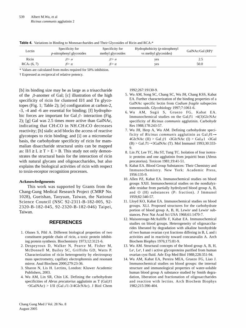
It is of interest to compare ricin (RCA2) withRCA1 (Ricinus communis agglutinin 1; Tables 3 and4).(6,7) Ricin is a potent cytotoxin but a weak hemag-glutinin, whereas RCA1 is a weak cytotoxin and apowerful hemagglutinin. As illustrated in Table 3,the interaction of ricin with simple sugars is similar,but not identical to that of the agglutinin (RCA1).Melibiose and raffinose were 80% and 60% lessactive than Gal in causing inhibition of the precipitinreactions of glycoproteins by ricin; both results aredifferent from those found with the agglutinin.Though N-acetyllactosamine (Gal 1-4GlcNAc) andhuman blood group I Ma trisaccharide (Gal 1-4GlcNAc 1-6Gal) are 6.4 and 9.1 times more effec-tive as inhibitors of the agglutinin than Gal, p-NO2
phenyl Gal was the best inhibitor of the agglutininand 6.3 times more potent than the methyl analogue.On the otherhand, ricin showed equal potency withtype II, L (Gal 1-4Glc), T structure (Gal 1-3GalNAc), blood group I Ma-active trisaccharideand Gal 1-4Man. Besides, in both toxin and agglu-tinin, the specificity for p-nitrophenyl and methylglycosides, the -anomer of Gal is preferred overthe -anomer, and that hydrophobic forces areimportant for interaction. However, the ratio ofGalNAc/Gal in ricin was 20 times less than that ofagglutinin (Table 4). Furthermore, the relativeinhibitory power of some important oligosaccharideswith ricin and RCA1 differs considerably. Forinstance, the values of the relative potencies of Gal
1-4GlcNAc (II), Gal 1-4GlcNAc 1-6Gal (I Ma),p-NO2 phenyl Gal, and Gal 1-3GalNAc (T) arestrikingly different. These variations support the con-cept that every lectin with its unique amino acidsequence has its own binding characteristics.(46)
Overall, the conclusions of this study are thefollowing: [a] from the present and previous results,it can be deduced that the combining site of lectindomains in ricin should be a shallow groove-type,recognizing Gal 1-4Glc as the major binding site;
Chang Gung Med J Vol. 28 No. 8August 2005
Albert M.Wu, et alRicinus communis agglutinin 2
539
[b] its binding size may be as large as a trisaccharideof the -anomer of Gal; [c] illustration of the highspecificity of ricin for clustered II/I and Tn glyco-topes (Fig. 1; Table 2); [e] configuration at carbon-2,-3, -4 and -6 are essential for binding; [f] hydropho-bic forces are important for Gal - interaction (Fig.2); [g] Gal was 2.5 times more active than GalNAc,indicating that CH3CO in NH.CH3CO decreasesreactivity; [h] sialic acid blocks the access of reactiveglycotopes to ricin binding; and [i] on a micromolarbasis, the carbohydrate specificity of ricin for mam-malian disaccharide structural units can be mappedas: II/I ≥ L ≥ T > E > B. This study not only demon-strates the structural basis for the interaction of ricinwith natural glycans and oligosaccharides, but alsoexplains the biological activities of ricin with respectto toxin-receptor recognition processes.
AcknowledgmentsThis work was supported by Grants from the
Chang-Gung Medical Research Project (CMRP No.1028), Gueishan, Taoyuan, Taiwan, the NationalScience Council (NSC 92-2311-B-182-005, 92-2320-B-182-045, 92-2320-B-182-046) Taipei,Taiwan.
REFERENCES
1. Olsnes S, Pihl A. Different biological properties of twoconstituent peptide chain of ricin, a toxic protein inhibit-ing protein synthesis. Biochemistry 1973;12:3121-6.
2. Despeyroux D, Walker N, Pearce M, Fisher M,McDonnell M, Bailey SC, Griffiths GD, Watts P.Characterization of ricin heterogeneity by electrospraymass spectrometry, capillary electrophoresis and resonantmirror. Anal Biochem 2000;279:23-36.
3. Sharon N, Lis H. Lectins, London: Kluwer AcademicPublishers, 2003.
4. Wu AM, Lin SR, Chin LK. Defining the carbohydratespecificities of Abrus precatorius agglutinin as T (Gal 1
3GalNAc) > I/II (Gal 1-3/4GlcNAc). J Biol Chem
1992;267:19130-9.5. Wu AM, Song SC, Chang SC, Wu JH, Chang KSS, Kabat
EA. Further characterization of the binding properties of aGalNAc specific lectin from Codium fragile subspeciestomentosoids. Glycobiology 1997;7:1061-6.
6. Wu AM, Sugii S, Gruezo FG, Kabat EA.Immunochemical studies on the Gal 1 4(3)GlcNAcspecificity of Ricinus communis agglutinin. CarbohydrRes 1988;178:243-57.
7. Wu JH, Herp A, Wu AM. Defining carbohydrate speci-ficity of Ricinus communis agglutinin as Gal 14GlcNAc (II) > Gal 1 3GlcNAc (I) > Gal 1 3Gal(B) > Gal 1 3GalNAc (T). Mol Immunol 1993;30:333-9.
8. Lin JY, Lee TC, Hu ST, Tung TC. Isolation of four isotox-ic proteins and one agglutinin from jequiriti bean (Abrusprecatorius). Toxicon 1981;19:41-51.
9. Kabat EA. Blood Group Substances: Their Chemistry andImmunochemistry. New York: Academic Press,1956:135-9.
10. Allen PZ, Kabat EA. Immunochemical studies on bloodgroups XXII. Immunochemical studies on the nondialyz-able residue from partially hydrolyzed blood group A, B,and O (H) substances (P1 fraction). J Immunol1959;82:340-57.
11. Lloyd KO, Kabat EA. Immunochemical studies on bloodgroups. XLI. Proposed structures for the carbohydrateportion of blood group A, B, H, Lewisa and Lewisb sub-stances. Proc Nat Acad Sci USA 1968;61:1470-7.
12. Maisonrouge-McAuliffe F, Kabat EA. Immunochemicalstudies on blood groups. Heterogeneity of oligosaccha-rides liberated by degradation with alkaline borohydrideof two human ovarian cyst fractions differing in B, I, and iactivities and in reactivity toward concanavalin A. ArchBiochem Biophys 1976;175:81-9.
13. Wu AM. Structural concepts of the blood group A, B, H,Lea, Leb, I and i active glycoproteins purified from humanovarian cyst fluid. Adv Exp Med Biol 1988;228:351-94.
14. Wu AM, Kabat EA, Pereira MEA, Gruezo FG, Liao J.Immunochemical studies on blood groups: the internalstructure and immunological properties of water-solublehuman blood group A substance studied by Smith degra-dation, liberation and fractionation of oligosaccharidesand reaction with lectins. Arch Biochem Biophys1982;215:390-404.
Table 4. Variations in Binding to Monosaccharides and Their Glycosides of Ricin and RCA1*
LectinSpecificity for Specificity for Hydrophobicity (p-nitrophenyl
GalNAc/Gal (RP)†
p-nitrophenyl glycosides methyl glycosides vs methyl glycosides)
Ricin > > yes 2.5RCA1 (6, 7) > > yes 50.0
* Values are calculated from moles required for 50% inhibition.† Expressed as reciprocal of relative potency.

Chang Gung Med J Vol. 28 No. 8August 2005
Albert M.Wu, et alRicinus communis agglutinin 2
540
15. Wu AM, Kabat EA, Nilsson B, Zopf DA, Gruezo FG,Liao J. Immunochemical studies on blood groups.Purification and characterization of radioactive 3H-reduced di- to hexasaccharides produced by alkaline beta-elimination-borohydride 3H-reduced of Smith degradedblood group A active glycoproteins. J Biol Chem1984;259:7178-86.
16. Rovis L, Anderson B, Kabat EA, Gruezo FG, Liao J.Structures of oligosaccharides produced by base-borohy-dride degradation of human ovarian cyst blood group H,Leb, and Lea active glycoproteins. Biochemistry1973;12:5340-54.
17. Kabat EA, Baer H, Bezer AE, Knaub V. Immunochemicalstudies on blood groups VII. Chemical changes associatedwith destruction of blood group activity and enhancementof the type XIV cross reactivity by partial hydrolysis ofhog and human blood group A, B, and O substances. JExp Med 1948;88:43-57.
18. Leskowitz S, Kabat EA. Immunochemical studies onblood groups XV. The effect of mild acid hydrolysis onthe glucosamine and galactosamine in blood group sub-stances. J Amer Chem Soc 1954;76:5060-5.
19. Fournet B, Montreuil J, Strecker G, Dorland L,Haverkamp J, Vliegenthart JFG, Binette JP, Schmid K.Determination of the primary structures of 16 asialo-car-bohydrate units derived from human plasma 1-acid gly-coprotein by 360-MHz 1H-NMR spectroscopy and perme-thylation analysis. Biochemistry 1978;17:5206-14.
20. Fournier T, Medjoubi NN, Porquet D. Alpha-1-acid gly-coprotein. Biochim Biophys Acta 2000;1482:157-71.
21. Nilsson B, Norden NE, Svensson S. Structural studies onthe carbohydrate portion of fetuin. J Biol Chem1979;254:4545-53.
22. Moschera J, Pigman W. The isolation and characterizationof rat sublingual mucus-glycoprotein. Carbohydr Res1975;40:53-67.
23. Slomiany A, Slomiany BL. Structures of the acidicoligosaccharides isolated from rat sublingual glycopro-tein. J Biol Chem 1978;253:7301-6.
24. Wu AM, Herp A, Song SC, Wu JH, Chang KSS.Interaction of native and asialo rat sublingual glycopro-teins with lectins. Life Sci 1995;57:1841-52.
25. van Halbeek H, Dorland L, Vliegenthart JFG, KochetkovNK, Arbatsky NP, Derevitskaya VA. Characterization ofthe primary structure and the microheterogeneity of thecarbohydrate chains of porcine blood-group H substanceby 500-MHz 1H-NMR spectroscopy. Eur J Biochem1982;127:21-9.
26. Tettamanti G, Pigman W. Purification and characteriza-tion of bovine and ovine submaxillary mucins. ArchBiochem Biophys 1968;124:41-50.
27. Herp A, Wu AM, Moschera J. Current concepts of thestructure and nature of mammalian salivary mucous gly-coproteins. Mol Cell Biochem 1979;23:27-44.
28. Herp A, Borelli C, Wu AM. Biochemistry and lectin bind-
ing properties of mammalian salivary mucous glycopro-teins. Adv Exp Med Biol 1988;228:395-435.
29. Wu AM, Wu JH, Shen F. Interaction of a novel Tn(GalNAc alpha 1 Ser/Thr) glycoprotein with Gal,GalNAc and GlcNAc specific lectins. Biochem BiophysRes Commun 1994;198:251-6.
30. Wu AM, Pigman W. Preparation and characterization ofArmadillo Submandibular Glycoproteins. Biochem J1977;161:37-47.
31. De Vries AL, Komatsu SK, Feeney RE. Chemical andphysical properties of freezing point-depressing glycopro-teins from antarctic fishes. J Biol Chem 1970;245:2901-8.
32. Lindberg A, Lönngren J, Powell DA. Structural studies onthe specific type-14 pneumococcal polysaccharide.Carbohydr Res 1977;58:177-86.
33. Kabat EA, Mayer MM. Experimental Immunochemistry(2nd Ed). Thomas, Springerfield, IL , 1961.
34. Schiffman G., Kabat EA, Thompson W. Immunochemicalstudies on blood groups. XXX. Cleavage of A, B and Hblood group substance by alkali. Biochemistry1964;3:113-20.
35. Goldstein IJ, Poretz RD. Isolation, physiochemical char-acterization, and carbohydrate -binding specificity oflectins. In: Liener IE, Sharon N, Goldstein IJ, eds. TheLectins, Properties, Functions and Applications inBiology and Medicine. New York: Academic press,1986:33-247.
36. Olsnes S. The history of ricin, abrin and related toxins.Toxicon 2004;44:361-70.
37. Halling KC, Halling AC, Murray EE, Ladin BF, HoustonLL, Weaver RF. Genomic cloning and characterization ofa ricin gene from Ricinus communis. Nucl Acid Res1985;13:8019-33.
38. Lamb FI, Roberts LM, Lord JM. Nucleotide sequence ofcloned cDNA coding for preproricin. Eur J Biochem1985;148:265-70.
39. Montfort W, Villafranca JE, Monzingo AF, Ernst S,Katzin B, Rutenber E, Xuong NH, Hamlin R, RobertusJD. The three-dimensional structure of ricin at 2.8A. JBiol Chem 1987;262:5398-403.
40. Rutenber E, Katzin BJ, Collins EJ, Mlsna D, Ernst S,Ready MP, Robertus JD. The crystallographic refinementof ricin at 2.5A resolution. Proteins 1991;10:240-50.
41. Lord JM, Roberts LM, Robertus JD. Ricin: structure,mode of action, and some current applications. FASEB J1994;8:201-8.
42. Nicolson GL, Blaustein J, Etzler ME. Characterization oftwo plant lectins from Ricinus communis and their quanti-tative interaction with a murine lymphoma. Biochemistry1974;13:196-204.
43. Baenziger JU, Fiete D. Structural determinants of Ricinuscommunis agglutinin and toxin specificity for oligosac-charides. J Biol Chem 1979;254:9795-9.
44. Debray H, Decout D, Strecker G, Spik G, Montreuil J.Specificity of twelve lectins towards oligosaccharides and

Chang Gung Med J Vol. 28 No. 8August 2005
Albert M.Wu, et alRicinus communis agglutinin 2
541
glycoeptides related to N-glycosylproteins. Eur J Biochem1981;117:41-55.
45. Rinderle SJ, Goldstein IJ, Matta, KL, Ratcliffe MR.Isolation and characterization of Amaranthin, a lectin pre-sent in the seeds of Amaranthus caudatus, that recognizes
the T- (or Cryptic T)-antigen. J Biol Chem1989;264:16123-131.
46. Wu AM. Carbohydrate Structural Units in glycoproteinsand polysaccharides as important ligands for Gal andGalNAc reactive lectins. J Biomed Sci 2003;10:676-88.

542
(Ricin)1 Tanuja Singh Anthony Herp
(QPA) (QPIA) 31 XIV
(RCA1) 12 50% (N) -
Gal B GalNAc 2.5 Gal 1-3GalNAc(T) Gal 1-3/4GlcNAc (I/II) Gal 1-4Glc (L) I Ma
Gal 1-4Gal (E) B 2 2.4 (1) ( (2)
(3)Gal - (4) ( 2005;28:530-42)
1
94 5 9 94 6 7333 259 Tel.:
(03)2118966 (Lab.); Fax: (03)2118456 (Lab.), (03) 2118700 (Col.); E-mail: [email protected]
Copyright © 2022 FDOKUMEN