The Epididymis - Research Group - Reproductive Biology
78
Introduction, 1072 Historical Perspective, 1072 Development of the Epididymis, 1073 Formation of the Mesonephric/Wolffian/Nephric Duct and Tubules, 1073 Postnatal Development, 1074 Structural Organization of the Epididymis, 1076 Anatomy, 1076 Epididymal Cell Types and Specific Markers, 1076 The Blood–Epididymis Barrier, 1080 Functions Taking Place in the Luminal Compartment, 1081 Transport of Spermatozoa, 1081 Maturation of Spermatozoa, 1083 Storage of Spermatozoa, 1087 Protection of Spermatozoa, 1088 Microenvironment for Maturation, Protection, and Storage, 1089 Regulation of Epididymal Functions, 1092 Hormones, 1092 Testicular Factors, 1093 Major Protein Families in the Epididymis and Their Regulation, 1095 General Principles, 1095 Functional Families, 1098 Aging, 1114 Effects of Aging on the Appearance of the Epididymal Epithelium, 1114 Molecular and Functional Effects of Aging in the Epididymis, 1115 Mechanisms Underlying Aging of the Epididymis, 1115 Changes in Spermatozoa during Aging, 1116 The Epididymis as Target for Xenobiotics, 1117 Chemicals for Which the Epididymis is an Explicit Target, 1117 Chemicals Targeted Principally at Other Organs that Also Act on the Epididymis, 1119 Perspective and Future Directions, 1120 Acknowledgments, 1120 References, 1120 Knobil and Neill’s Physiology of Reproduction, Third Edition edited by Jimmy D. Neill, Elsevier © 2006 CHAPTER 22 The Epididymis Bernard Robaire, 1 Barry T. Hinton, 2 and Marie-Claire Orgebin-Crist 3 ABSTRACT Throughout embryonic and early postnatal devel- opment, the mammalian epididymis changes from a straight tube to a highly coiled, complex duct that links the efferent ducts to the vas deferens. Overwhelming evidence points to the importance of this tissue in transforming spermatozoa leaving the testis as immotile cells, unable to fertilize oocytes, into fully mature cells that have the ability both to swim and to recognize and fertilize eggs. Under normal condi- tions, the acquisition of these functions is essentially completed by the time sperm enter the proximal cauda epididymidis. In addition to sperm maturation, the epididymis also plays an important role in sperm transport, concentration, protection, and storage. A highly specialized and region-specific microenvi- ronment is created along the epididymal lumen by active secretion and absorption of water, ions, organic solutes, and proteins as well as by the blood–epididymis barrier. The primary factor regulating epididymal function is androgens, but there is mounting evidence that estrogens, retinoids, and other factors coming directly into the epididymis from the testis through the efferent ducts, such as growth factors, also play specific regulatory roles. Several epithelial cell types, 1071 1 Department of Pharmacology and Therapeutics, McGill University, Montreal, Quebec, Canada; 2 Department of Cell Biology, University of Virginia Health System, School of Medicine, Charlottesville, Virginia; 3 Department of Obstetrics and Gynecology, Vanderbilt University School of Medicine, Center for Reproductive Biology Research, Nashville, Tennessee.
-
Upload
khangminh22 -
Category
Documents
-
view
1 -
download
0
Transcript of The Epididymis - Research Group - Reproductive Biology
Introduction, 1072Historical Perspective, 1072Development of the Epididymis, 1073
Formation of the Mesonephric/Wolffian/Nephric Ductand Tubules, 1073
Postnatal Development, 1074Structural Organization of the Epididymis, 1076
Anatomy, 1076Epididymal Cell Types and Specific Markers, 1076The Blood–Epididymis Barrier, 1080
Functions Taking Place in the LuminalCompartment, 1081Transport of Spermatozoa, 1081Maturation of Spermatozoa, 1083Storage of Spermatozoa, 1087Protection of Spermatozoa, 1088Microenvironment for Maturation, Protection,
and Storage, 1089Regulation of Epididymal Functions, 1092
Hormones, 1092Testicular Factors, 1093
Major Protein Families in the Epididymis and Their Regulation, 1095General Principles, 1095Functional Families, 1098
Aging, 1114Effects of Aging on the Appearance of the
Epididymal Epithelium, 1114Molecular and Functional Effects of Aging
in the Epididymis, 1115Mechanisms Underlying Aging of the
Epididymis, 1115Changes in Spermatozoa during Aging, 1116
The Epididymis as Target for Xenobiotics, 1117Chemicals for Which the Epididymis is an
Explicit Target, 1117Chemicals Targeted Principally at Other
Organs that Also Act on the Epididymis, 1119
Perspective and Future Directions, 1120Acknowledgments, 1120References, 1120
Knobil and Neill’s Physiology of Reproduction,Third Editionedited by Jimmy D. Neill,Elsevier © 2006
CHAPTER 22
The Epididymis
Bernard Robaire,1 Barry T. Hinton,2 and Marie-Claire Orgebin-Crist3
ABSTRACT
Throughout embryonic and early postnatal devel-opment, the mammalian epididymis changes from astraight tube to a highly coiled, complex duct that linksthe efferent ducts to the vas deferens. Overwhelmingevidence points to the importance of this tissue intransforming spermatozoa leaving the testis asimmotile cells, unable to fertilize oocytes, into fullymature cells that have the ability both to swim andto recognize and fertilize eggs. Under normal condi-tions, the acquisition of these functions is essentiallycompleted by the time sperm enter the proximal
cauda epididymidis. In addition to sperm maturation,the epididymis also plays an important role in spermtransport, concentration, protection, and storage. A highly specialized and region-specific microenvi-ronment is created along the epididymal lumen byactive secretion and absorption of water, ions, organicsolutes, and proteins as well as by the blood–epididymisbarrier. The primary factor regulating epididymalfunction is androgens, but there is mounting evidencethat estrogens, retinoids, and other factors comingdirectly into the epididymis from the testis throughthe efferent ducts, such as growth factors, also playspecific regulatory roles. Several epithelial cell types,
1071
1Department of Pharmacology and Therapeutics, McGill University, Montreal, Quebec, Canada;2Department of Cell Biology, University of Virginia Health System, School of Medicine, Charlottesville, Virginia;3Department of Obstetrics and Gynecology, Vanderbilt University School of Medicine, Center for Reproductive Biology Research, Nashville, Tennessee.
P515401_22 10/24/05 8:14 PM Page 1071

each showing selective expression of genes and pro-teins, are differentially distributed along the duct;each cell type shows highly regionalized expression ofa wide array of markers. Both epididymal epithelialcells and spermatozoa in the lumen are targets forxenobiotics; such exposures can result in undesirabletoxic effects or may provide the basis for the develop-ment of novel male contraceptive agents. During aging,both the epididymal epithelium and the germ cells inthe lumen undergo a series of dramatic changes. Theexplosion of knowledge we are witnessing regardingall aspects of epididymal structure and function islikely to lay the basis for a new fundamental under-standing of epididymal cell biology and novel thera-peutic approaches targeted at this organ.
INTRODUCTION
Since the publication of the comprehensive reviewon the male excurrent duct system (efferent ducts,epididymis, and vas deferens) in the first edition ofPhysiology of Reproduction in 1988 (1), a remarkableseries of events has marked the growth in our knowl-edge and understanding of this duct system. At thattime, the epididymis, a long, complex, convoluted ductconnecting the efferent ducts to the vas deferens, wasviewed as having moved from being “an abandonedchild” of the male reproductive system (2), to a pointof maturity, where its basic structures, functions,and regulation were beginning to be understood.
Since 1988, three international conferences dedi-cated to this tissue have been held, with proceedingsensuing for two of these (3,4), and the first compre-hensive, multiauthored volume dedicated to the epi-didymis has been published (5). The yearly numberof research publications on this tissue has increasedby more than one order of magnitude (from less than500 to more than 6,000) since the review appeared inthe first edition of Physiology of Reproduction.Therefore, in preparing the current review, we havechosen first to narrow the scope by placing most ofthe emphasis on the epididymis itself, as opposed tothe other components of the excurrent duct system;second, to exclude some topics that have been exten-sively reviewed recently, such as changes in sperma-tozoa during epididymal transit, innervation andvasculature of the duct system, or pathology of theepididymis; and third, to focus on some of the moreexciting recent developments in this field and providesupplemental information in an alternative format.Several of the subjects covered in the first edition areupdated, but, because of space limitations, we havechosen not to include most of the plates that depictthe histological organization and structure of thevarious epididymal epithelial cells from the first
edition, but rather to place them on a web site(www.medicine.mcgill.ca/PhysiolReprodThirdEd/Epididymis). Along with these plates, this web sitecontains color versions of plates from the currentchapter and a number of tables, including some con-taining detailed information about specific epididy-mal proteins and genes as well as their regulation.We have intentionally chosen not to include suchtables in the chapter not only because of space limi-tations but because of the need to update this infor-mation regularly. We also felt that the review shouldfocus on our ideas, philosophy, and biased judgmenton what we believe is important and where the fieldshould go.
HISTORICAL PERSPECTIVE
As early as the 4th century B.C., the epididymis wasdescribed by Aristotle in his Historia Animalium,whereas the first recorded description of a dissectedepididymis was made by de Graaf in 1668 in hismonograph Tractatus de Virorum Organis GenerationiInservientibus [reviewed in Orgebin-Crist (6)]. DeGraaf noted that “the semen … was watery and ash-like in the testis and becomes milky and thick in theepididymis” (7). Between 1888 and 1928, a numberof scientists described the histological features of theepididymal epithelium, and surmised, from the imagesof secretion they observed, that epididymal secretions“nourished” the spermatozoa in the epididymallumen. In 1913, Tournade (8) showed that spermatozoareleased from the proximal epididymis were not motilewhen diluted in saline, but spermatozoa releasedfrom the distal epididymis were fully motile. In thebat, spermatozoa were observed to survive for severalmonths in the cauda epididymidis during hibernation,presumably protected by epididymal secretions (9),whereas rabbit spermatozoa survived in the epi-didymis for 30 to 60 days (10), and bull spermatozoafor 2 months (11), after ligation of the efferent ducts.The implication of these observations was that theepididymis conditioned the development of spermmotility and was important for sperm survival.
The consensus of these early investigations can besummarized by the last sentence of Benoit’s classic1926 (12) monograph: “The role of epididymal secre-tions is to maintain sperm vitality, to permit thedevelopment of sperm motility, and possibly to pro-tect them against noxious agents.” In a series of fourpapers between 1929 and 1931, Young (13–16) showedthat during epididymal transit spermatozoa not onlyacquire a mature motility pattern but become fertile;based on some poorly designed and interpreted studies,he concluded, erroneously, that the changes sperma-tozoa undergo during their transit through the
1072 / CHAPTER 22
P515401_22 10/24/05 8:14 PM Page 1072
epididymis represent a continuation of changes thatstart while spermatozoa are still attached to the germinal epithelium, and are not conditioned bysome specific action of the epididymal secretion (6).Nevertheless, by 1931 most of the problems andquestions relating to epididymal physiology had beenrecognized, and most of the work done since then hasbeen to provide the experimental evidence to fleshout the insights of earlier investigators.
Few studies were published on epididymal physi-ology until the 1960s. Those that appeared focusedprimarily on establishing the length of time necessaryfor spermatozoa to transit through the epididymis(17,18). The apparent lack of interest in the epi-didymis during this 20- to 30-year span is puzzling.After the work of Benoit in 1926 (12) and Young in1931 (13–16), there was a clear controversy thatneeded to be resolved. In 1965, only 43 papers on theepididymis were published. Whatever the reason forthis disaffection, in 1964 Thaddeus Mann, in his bookThe Biochemistry of Semen (2), refers to the epi-didymis as the “abandoned child” of the reproductivesystem.
In the mid- and late 1960s, a resurgence of interestin the epididymis was spearheaded independently byOrgebin-Crist and Bedford; they demonstrated thatthe key event in sperm maturation was not the pas-sage of time, as proposed by Young, but exposure tothe luminal environment of the epididymis (19,20).Thanks to these and other studies, by the end of the1960s it was established that the potential for spermmotility and fertilizing ability is acquired as spermato-zoa pass from the proximal to the distal epididymis;that the maturation process does not end with theacquisition of fertilizing ability, because spermatozoathat have just become fertile induce a higher rate ofembryonic mortality when inseminated in vivo; thatthe maturation process depends on an androgen-stimulated epididymis; and that the maturation processincludes changes in sperm organelles [reviewed inOrgebin-Crist (21)].
Since the 1980s, we have seen an explosion ofstudies on the presence, characterization, immunolo-calization, and regulation of a large number of pro-teins and their RNAs known to be specific to theepididymis, to be expressed at particularly high levelsin some segments of the tissue, or to control key molecules that regulate epididymal function. Theobjective of many of these studies has been to developways of modifying epididymal function with respectto rendering spermatozoa fully mature and motile,thus providing potential leads for male contraceptionor the management of male infertility. In the courseof these studies, it has become clear that this tissuepresents a novel model for studying cell-, segment-,and region-specific gene expression, for understanding
mechanisms of aging, and for identifying processesconferring selective protection from infections andcancer.
DEVELOPMENT OF THE EPIDIDYMIS
Formation of theMesonephric/Wolffian/Nephric Duct and Tubules
Many of the studies focusing on the specificationand regulation of mesonephric duct and tubule for-mation have been conducted on chick, frog, andzebrafish embryos, with some more recent studiesusing the mouse embryo. Space limitations prevent acomprehensive review of the development of the uro-genital system, but there are several excellentresources with detailed descriptions of these events(22,23). What follows is a basic summary of the embry-onic and postnatal development of the epididymis.
The urogenital system is derived from the inter-mediate mesoderm, which, in the chick and mouse, isthe result of a complex interaction between the inter-mediate and paraxial mesoderm. The expression oftwo key transcription factors, Pax 2 and Pax 8, playsa vital role in this process because mice that lack thesetwo genes fail to form a mesonephros and, therefore,later structures of the urogenital system; Bouchardet al. (2002) (24) suggest that Pax2 and Pax8 are critical regulators that specify the nephric lineage. At approximately gestational days 8 to 9 in the mouseand stage 16 in the developing chick, the mesonephric(Wolffian/nephric) duct develops as a cord of epithelialcells that express c-Sim-1 (chick) and Pax2 (25,26),undergoes the formation of a lumen, and rapidlyextends throughout the length of the embryo in acranial-to-caudal direction. There is considerable evi-dence to suggest that, in some species, elongation ofthe nephric duct is the result of cell rearrangementsrather than proliferation or changes in cell shape(27). Interestingly, for proper nephric duct formationbut not elongation, bone morphogenetic protein-4expression in the surface ectoderm appears to be critical (28). Retinoic acid is also crucial for nephricduct formation because the duct does not form inRALDH2, a retinoic acid synthetic enzyme, knockoutmice (29). As the mesonephric duct elongates, it inducesthe nearby mesenchyme to form the mesonephrictubules (30). The tubules have a characteristic J- orS-shape and resemble developing nephrons. Withoutthe Wolffian duct, the mesonephric tubules do notform (31).
It is clear that those cranial mesonephric tubulesthat lie close to the testis survive, and the distal endgrows toward the gonad, whereas the proximal end
THE EPIDIDYMIS / 1073
P515401_22 10/24/05 8:14 PM Page 1073

contacts the wolffian/mesonephric duct. However,several fundamental questions remain. What are mech-anisms by which the mesonephric tubules are inducedby the mesonephric duct? This process involves a complex mesenchyme-to-epithelial transition, andthere is evidence to suggest that leukemia inhibitoryfactor, members of the Wnt family (Wnt6 and Wnt4),and fibroblast growth factor (FGF)-2 are responsiblefor mesenchymal cell aggregation and the formationof the renal nephron [reviewed in Gilbert (22)].Whether this is recapitulated during the formation ofthe mesonephric tubules is unclear. Alarid et al.(1991) (32) have shown that FGF-2 is important forepididymal development because the epididymis failsto develop when embryonic urogenital ridges andsinuses are cultured under the kidney capsule in thepresence of anti–FGF-2 antibodies. Studies by Sainioet al. (1997) (33) showed that the Wilms tumor-1(WT1) gene is important in the formation of the mostcaudal mesonephric tubules.
What are the mechanisms by which only thosetubules close to the gonad survive, yet others cranialand caudal to the gonad degenerate? Evidence suggeststhat the caudal tubules normally undergo apoptosis,so presumably those tubules close to the gonad expressthe antiapoptotic genes required to survive, but thishas not been established.
What controls the migration and differentiation ofcells of the distal tubule into the rete testis? Earlierstudies by Upadhyay et al. (34) provided evidencethat in the mouse, the cells in the distal end of themesonephric tubules contribute directly to the retetestis and undergo differentiation, and that fusionbetween the mesonephric tubules and the testis didnot occur. This finding is perhaps not too surpris-ing in light of the extensive contribution of themesonephros to the developing testis.
Which portions of the mesonephric tubule and ductform the discrete regions of the initial segment andthe first part of the caput epididymidis? There issome evidence to suggest that cells of the wolffianduct contribute to a small part of the proximal portionof the mesonephric tubule but, again, it is not clearwhether this represents the junction between thecaput and the initial segment. For a more completeoverview of these processes, the reader should referto the reviews by Sainio (2003) (31), Vazquez et al.(2003) (35), and Jones (2003) (36).
It is intriguing that the development of themesonephric tubules may recapitulate the develop-ment of the renal vesicle into the renal proximal anddistal tubules. It would not be too surprising to findthat many genes that regulate this process also playa role in the formation of the efferent ducts or initialsegment. For example, members of the Wnt family[Wnt 4 (37)] and cadherins (38) play a role in theproximal/distal patterning of the renal proximal and
distal tubules. Cadherin 6 is expressed in the proximaltubule progenitors, E-cadherin is expressed in thedistal tubule, and P-cadherin is expressed in theglomerulus (39). The initial segment fails to developin c-Ros mutants (40) and in Sxr (XXSxr) mice (41),suggesting that the defect may lie in the origin ordevelopment of the mesonephric tubules. Although itis well recognized that androgens play an importantrole in epididymal development, the initial segmentfails to develop in a normal androgen environment inthe Sxr mouse (41). This would suggest that addi-tional factors, such as growth factors, may be importantfor mesonephric tubule/initial segment development.In both of these mutants the prominent vascularsystem that supplies the initial segment also fails todevelop (40,42), indicating that there is an intimaterelationship between epithelial and endothelialdevelopment. Again, this is perhaps not surprising inview of the formation of the renal glomerulus at thetip of the renal proximal tubule where endothelialcells are recruited to this site.
The embryonic origins of the different regions of the epididymis have received some interest; itappears that the efferent ducts are derived from themesonephric tubules, whereas the caput epididymidisto the vas deferens regions are derived from themesonephric duct [reviewed in Jones (36)]. However,the origins of the initial segment warrant furtherinvestigation because it is not entirely clear if thecells are of mesonephric tubule or duct origin. The cellsof the initial segment are quite distinct from eitherthe efferent ducts or the distal epididymal regions,but function similarly to the efferent ducts in thatthey are actively involved in water reabsorption.Hence, it is possible that the initial segment may beof mesonephric tubule origin.
Postnatal Development
A more complete picture of the postnatal develop-ment of the epididymis has emerged over the years,with the majority of studies focused on the mouse,rat, and human, although other species have beenstudied, including the bull, dog, rabbit, marmoset, andboar [reviewed in Rodriguez et al. (43)]. Perhaps themost detailed description of postnatal epididymaldevelopment is that of the rat, and the studies by Sunand Flickinger (1979) (44) and Hermo et al. (1992)(45) provide the basis for the following brief overview.There appear to be three major stages of epididymaldevelopment: the undifferentiated period, the periodof differentiation, and the period of expansion.
By birth, the epididymis has undergone consider-able coiling in the proximal regions (initial segmentto corpus) and the cauda has yet to complete coil-ing (Fig. 1). In the next 1 to 2 days, individual
1074 / CHAPTER 22
P515401_22 10/24/05 8:14 PM Page 1074

septa are seen delineating the tubule into segments(D. Bomgardner and B. T. Hinton, unpublishedobservations). The epithelium is undifferentiated andis characterized by columnar cells containing numer-ous mitotic figures. There is considerable growth of
the rat epididymal duct from its embryological originsto postnatal day 15, when it reaches almost 2 m inlength (45,46). The period of differentiation has beenexamined extensively. Undifferentiated cells differ-entiate into the classically described cells of the adultepididymis: principal, halo, narrow (pencil), basal,and clear (light) cells; these cells are described later.Key changes in development are following: at postnatalday 14 the halo cells appear, on day 15 one observesnarrow and columnar cells, by day 28 the columnarcells differentiate into basal and principal cells, fromday 36 onward the narrow and clear cells appear, andby approximately day 49 all epididymal cells are fullydifferentiated. Figure 2 outlines the differentiation ofthe rat epididymal epithelium from birth to adulthood.The period of expansion describes the continuedgrowth of the duct and the appearance of spermatozoain the lumen.
The mechanisms that regulate the growth and differentiation of the epididymal duct are unknown,although it is clear that the expression of many epi-didymal genes is developmentally regulated. Certainly,luminal and circulating androgens play a critical role,but luminal fluid factors other than androgens alsomay be important. Early studies by Alexander (1972)(47) and Abe et al. (1984) (48) provided evidence thatluminal contents produced by the testis or proximalregions of the epididymal duct may regulate epididymalepithelial differentiation, although the candidate reg-ulators were not identified. Because there is some evi-dence to suggest that testicular luminal fluid growth
THE EPIDIDYMIS / 1075
A B
C D
FIG. 1. Coiling of the epididymal duct from mouse E14 to P1. Notethat coiling proceeds in a caput-to-cauda direction and is completedin the early postnatal period. The efferent duct–initial segment coilingis not shown here, but proceeds independently of the coiling of themain epididymal duct. To help visualize the changes, the size of thetestes and epididymides at different ages are not drawn to scale.(From D. Bomgardner and B. T. Hinton, unpublished observations.)
no apparentchange
Appearanceof halo cells
columnar cells
basalcells clear
cells
narrow/clearcells
narrowcellsinitialsegmentonly
principalcells
Fully differentiatedclear cells
undifferentiated cells
narrow cells columnar cells principal cells- Golgi expansion- ER cisternae- secretory vesicles
7 14 21 28 35 49
Undifferentiatedperiod
Period ofdifferentiation
Period ofexpansion
postnatal daysbirth
FIG. 2. Diagram outlining the differentiation of the rat epididymal epithelium from birth until adulthood. Epididymal epithelial cells are undifferentiated until approximately day 21, when bothnarrow and columnar cells are first observed. At approximately day 28, the columnar cells differentiateinto principal cells and basal cells. Narrow cells are seen in the initial segment only, but clear cells areobserved throughout the epithelium from approximately day 36 onward. By day 49, all epididymalepithelial cells are fully differentiated. (Adapted from Rodriguez, C. M., Kirby, J. L., and Hinton, B. T.[2002]. The development of the epididymis. In The Epididymis: From Molecules to Clinical Practice[B. Robaire and B. T. Hinton, Eds.], pp. 251–267. Kluwer Academic/Plenum, New York.)
P515401_22 10/24/05 8:14 PM Page 1075

factors (e.g., FGFs) may regulate initial segmentfunction and that the same growth factors regulateembryonic wolffian duct development, it is temptingto speculate that luminal growth factors (e.g., bonemorphogenetic proteins, FGFs, nerve growth factors)may also regulate epididymal epithelial differentiation.One might also envisage a more complex cell–cellinteraction in which multiple luminal factors aresecreted in the proximal regions of the duct (andtestis), which in turn cause the cells in the mid-distalregions (e.g., corpus) to secrete factors, which in turnregulate cell function in the more distal epididymalregion (e.g., cauda). Such multiple cascades may actto coordinate proper differentiation along the duct.
STRUCTURAL ORGANIZATIONOF THE EPIDIDYMIS
Anatomy
Seminiferous tubules converge to form the retetestis, which in turn gives rise to the efferent ducts(ductuli efferentes), a series of 4 to 20 tubules, thenumber depending on the species (49,50). Thesetubules converge to form a single highly coiled duct,the epididymis (from the Greek meaning “on or adjacent to the testis”), which is extremely long andvaries in length from 1 m in mice, (51), 3 m in rats, (52),3 to 6 m in humans, (53), and up to 80 m in horses, (54).
The epididymis is usually divided into four grossanatomical regions: the initial segment, head (caput),body (corpus), and tail (cauda), as first described byBenoit (1926) (12). Subsequently, a number of otherschemes have been proposed for dividing the epi-didymis into different regions or segments, includinga number of zones and the demonstration of thepresence of an intermediate zone between the initialsegment and caput epididymidis that has characteristiccells (55–59). However, in this review, we retain themost commonly used nomenclature for the four regionsdescribed previously. In all mammalian speciesexamined to date, each region of the epididymis isfurther organized into lobules separated by connectivetissue septa. These septa not only as serve internalsupport for the organ but have been proposed to pro-vide a functional separation between lobules thatallows selective expression of genes and proteinswithin individual lobules (60). The extension of theepididymis is a straight tube, the vas deferens, whichis surrounded by a very thick muscular layer. Thevas deferens connects with the urethra, which emp-ties to the outside of the body. A schematic represen-tation of the testis, efferent ducts, epididymis, andvas deferens is shown in Fig. 3.
Epididymal Cell Types and Specific Markers
There are several types of epithelial cells that linethe epididymis; some are located throughout the duct(e.g., principal cells), whereas others are found eitherexclusively or primarily in specific regions (e.g., narrowcells). We provide a brief description of the major celltypes along the epididymis, discuss the potentialfunctions of these cells, and demonstrate the pres-ence of specific markers for each cell type. Severaldetailed reviews of the histology of the epididymis fora number of species ranging from mouse to human,and including dog, camel, elephant, opossum, bull, ram,hamster, mouse, and monkey, have been published(1,12,48,56,57,61–73). Together, these publications pro-vide increasing evidence that similar regions and celltypes are present in most mammals, including humans.
1076 / CHAPTER 22
TESTIS
EPIDIDYMIS
Ductuli efferentes
Rete testis
Initialsegment IZ
Caput
Vasdeferens
Corpus
Distal
Proximal
Cauda
Seminiferous tubule
FIG. 3. Diagrammatic representation of the testis showing a seminiferous tubule and the rete testis, the ductuli efferentes, theepididymis, and vas deferens. The major regions of the epididymis(i.e., the initial segment, intermediate zone, caput, corpus, and proximal and distal cauda) are indicated. (Adapted from Robaire, B.,and Hermo, L. [1988]. Efferent ducts, epididymis and vas deferens:structure, functions and their regulation. In The Physiology ofReproduction [E. Knobil and J. D. Neill, Eds.], pp. 999–1080. RavenPress, New York.)
P515401_22 10/24/05 8:14 PM Page 1076
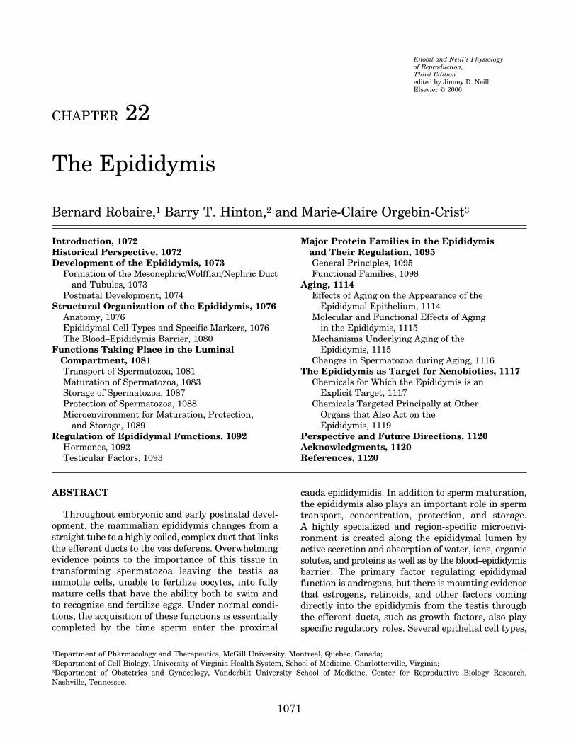
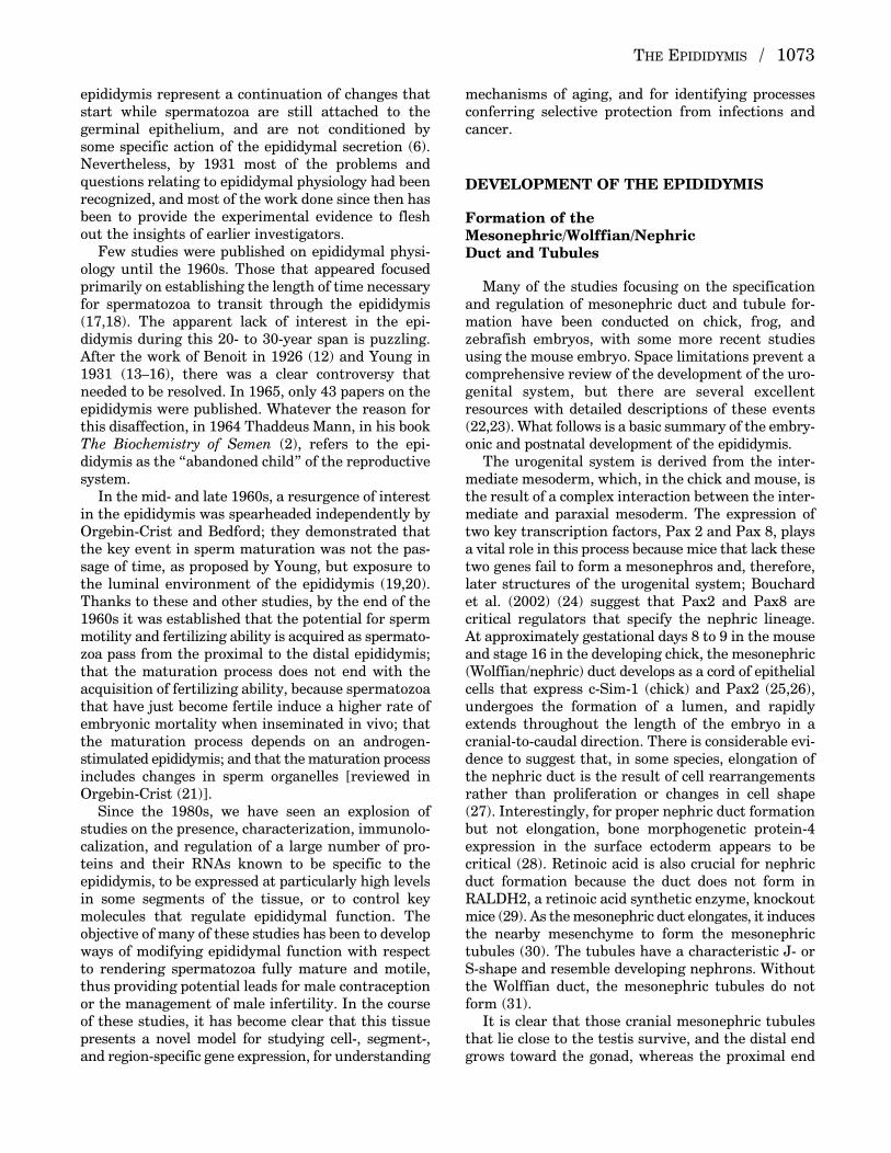
These cell types appear in the appropriate regions,share similar structural features and functions, includ-ing regional differences, and show very similar pat-terns of expression of some secretory proteins(74–79). The comprehensive set of light and electronmicroscopy plates prepared for this volume are availableat www.medicine.mcgill.ca/PhysiolReprodThirdEd/-Epididymis. Schematic representations of the organ-ization of all the cell types and their known functionsare shown in Fig. 4. Key components of epithelialcells in the initial segment and in the remainder ofthe epididymis as observed at the electron microscopeare depicted in Figs. 5 and 6, respectively.
Principal Cells
The main cell type in the epididymis of all mammalsis referred to as the principal cell. These cells appearalong the entire duct but show structural differencesin each region (1,63). The most striking feature ofthese cells is their highly developed secretory andendocytic machinery and their basally aligned nuclei.Depending on the segment examined, principal cellscomprise approximately 65% to 80% of the totalepithelial cell population of the epididymis (80). Boththeir structure and functions vary dramaticallybetween the different segments (1,63,81). These dif-ferences are reflected in the appearance and organi-zation of their secretory apparatus (endoplasmicreticulum, Golgi apparatus, and secretory granules)and endocytic apparatus (coated pits, endosomes,
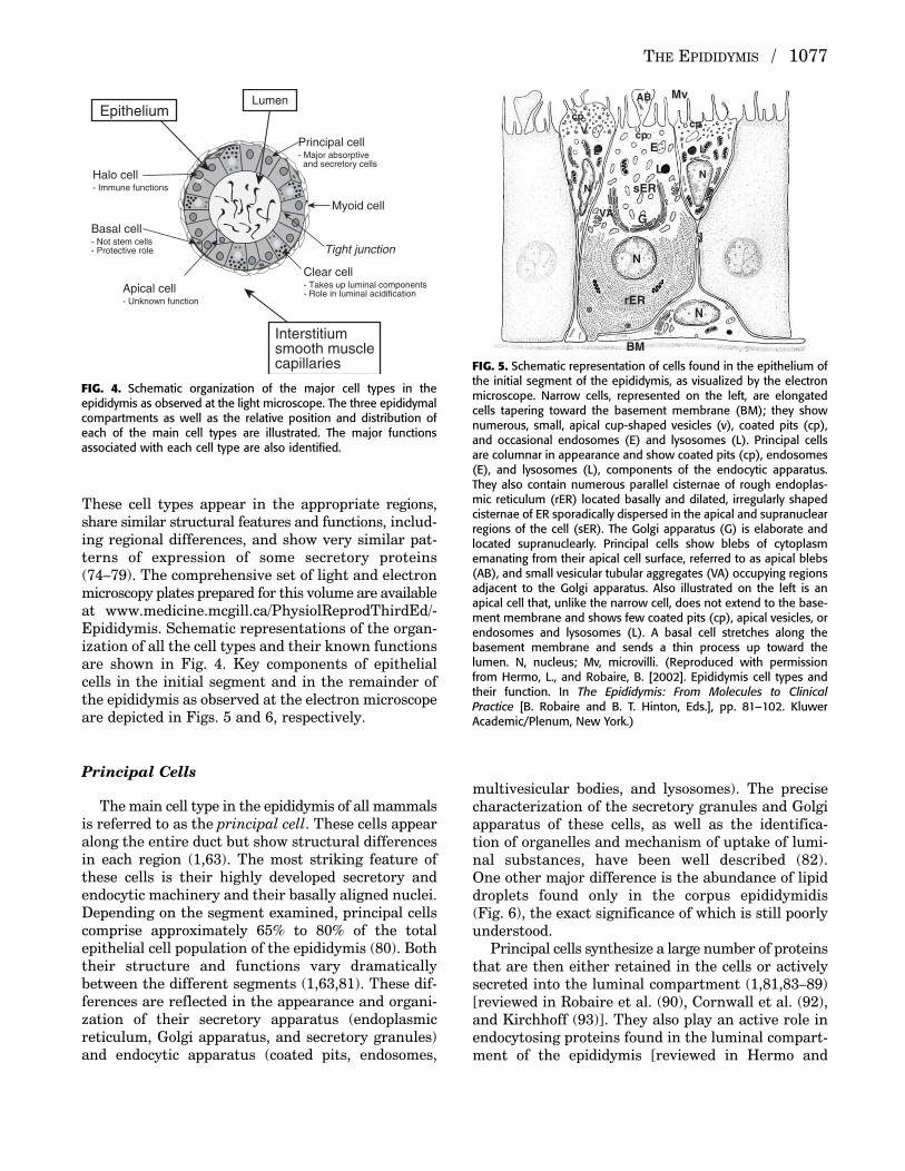
multivesicular bodies, and lysosomes). The precisecharacterization of the secretory granules and Golgiapparatus of these cells, as well as the identifica-tion of organelles and mechanism of uptake of lumi-nal substances, have been well described (82). One other major difference is the abundance of lipiddroplets found only in the corpus epididymidis (Fig. 6), the exact significance of which is still poorlyunderstood.
Principal cells synthesize a large number of proteinsthat are then either retained in the cells or activelysecreted into the luminal compartment (1,81,83–89)[reviewed in Robaire et al. (90), Cornwall et al. (92),and Kirchhoff (93)]. They also play an active role inendocytosing proteins found in the luminal compart-ment of the epididymis [reviewed in Hermo and
THE EPIDIDYMIS / 1077
Principal cell- Major absorptive and secretory cells
Halo cell- Immune functions
Apical cell- Unknown function
Basal cell- Not stem cells- Protective role
Clear cell- Takes up luminal components- Role in luminal acidification
Myoid cell
Tight junction
Interstitiumsmooth musclecapillaries
EpitheliumLumen
FIG. 4. Schematic organization of the major cell types in the epididymis as observed at the light microscope. The three epididymalcompartments as well as the relative position and distribution ofeach of the main cell types are illustrated. The major functions associated with each cell type are also identified.
Mv
E
L
LL
sER
rER
G
N
N
BM
cp
cpcp
NN
VA
V
AB
FIG. 5. Schematic representation of cells found in the epithelium ofthe initial segment of the epididymis, as visualized by the electronmicroscope. Narrow cells, represented on the left, are elongatedcells tapering toward the basement membrane (BM); they shownumerous, small, apical cup-shaped vesicles (v), coated pits (cp),and occasional endosomes (E) and lysosomes (L). Principal cellsare columnar in appearance and show coated pits (cp), endosomes(E), and lysosomes (L), components of the endocytic apparatus.They also contain numerous parallel cisternae of rough endoplas-mic reticulum (rER) located basally and dilated, irregularly shapedcisternae of ER sporadically dispersed in the apical and supranuclearregions of the cell (sER). The Golgi apparatus (G) is elaborate andlocated supranuclearly. Principal cells show blebs of cytoplasmemanating from their apical cell surface, referred to as apical blebs(AB), and small vesicular tubular aggregates (VA) occupying regionsadjacent to the Golgi apparatus. Also illustrated on the left is anapical cell that, unlike the narrow cell, does not extend to the base-ment membrane and shows few coated pits (cp), apical vesicles, orendosomes and lysosomes (L). A basal cell stretches along thebasement membrane and sends a thin process up toward thelumen. N, nucleus; Mv, microvilli. (Reproduced with permissionfrom Hermo, L., and Robaire, B. [2002]. Epididymis cell types andtheir function. In The Epididymis: From Molecules to ClinicalPractice [B. Robaire and B. T. Hinton, Eds.], pp. 81–102. KluwerAcademic/Plenum, New York.)
P515401_22 10/24/05 8:14 PM Page 1077

Robaire (2002) (82)]. A schematic representation ofthese processes is depicted in Fig. 7.
Apical Cells
Apical cells are found primarily in the epitheliumof the initial segment and intermediate zone (94,95),although they have been seen occasionally in othersegments in aging rats (96). These cells have a char-acteristic apically located spherical nucleus and donot contact the basement membrane (Fig. 5). Theydiffer clearly from adjacent narrow and principalcells in terms of their protein expression profile (95).However, little is known about the specific functionsof these cells, aside from their ability to endocytosesubstances from the lumen, as revealed by the exami-nation of β-hexosaminidase A knockout mice (69), andthe observation that they contain many proteolyticenzymes (95).
Narrow Cells
In the rat and mouse, narrow (pencil) cells of theadult epididymis appear only within the epitheliumof the initial segment and intermediate zone (94,95).
These cells are narrower than the adjacent principalcells, attenuated, and send a thin process of cyto-plasm to reach the basement membrane (Fig. 5).They are characterized by numerous apically locatedcup-shaped vesicles that are involved in endocytosisand function in secreting H+ ions into the lumen byrecycling to and from the apical plasma membrane (97).Similar cells have also been reported in the sameregions in numerous other species, including bovine,hamster, echidna, and human (61,65,66,76,95,98).Narrow cells are distinct from apical cells in theirmorphological appearance, relative distribution, andexpression of different proteins. They also differ dra-matically from neighboring principal cells and displayregion-specific expression of proteins such as the glu-tathione S-transferases and lysosomal enzymes (95).
Clear Cells
Clear cells are large, active endocytic cells presentonly in the caput, corpus, and cauda regions of theepididymis and are found in many species, includinghumans (1,63,99). These cells are characterized byan apical region containing numerous coated pits,vesicles, endosomes, multivesicular bodies, and lyso-somes and a basal region containing the nucleus and
1078 / CHAPTER 22
Mv
BM
M
N
N
NNN
N
LL L
E
EE
rER
Lip
GG
G
cp cp
cvv
g
FIG. 6. Schematic representation of a principal cell of the caput epididymidis on the left and a principal cell of the corpus epididymidis on the right, with a clear cell in between, as visualized by theelectron microscope. Also represented is a halo cell and a basal cell. Principal cells of both regionscontain coated pits (cp), endosomes (E) and lysosomes (L), and an elaborate Golgi apparatus (G).Rough endoplasmic reticulum (rER) occupies the basal region of the principal cell of the caput,whereas numerous lipid droplets (lip) occupy the cytoplasm of the principal cells of the corpus region.The clear cell shows few microvilli (Mv), but numerous coated pits (cp), small apical vesicles (v),endosomes (E), and lysosomes (L), all involved in endocytosis. The halo cell is inserted between adjacent principal cells, is located basally, and contains small dense core granules (g), whereas thebasal cell stretches itself along the basement membrane (BM). N, nucleus. (Reproduced with permissionfrom Hermo, L., and Robaire, B. [2002]. Epididymis cell types and their function. In The Epididymis:From Molecules to Clinical Practice [B. Robaire and B. T. Hinton, Eds.], pp. 81–102. Kluwer Academic/Plenum, New York.)
P515401_22 10/24/05 8:14 PM Page 1078
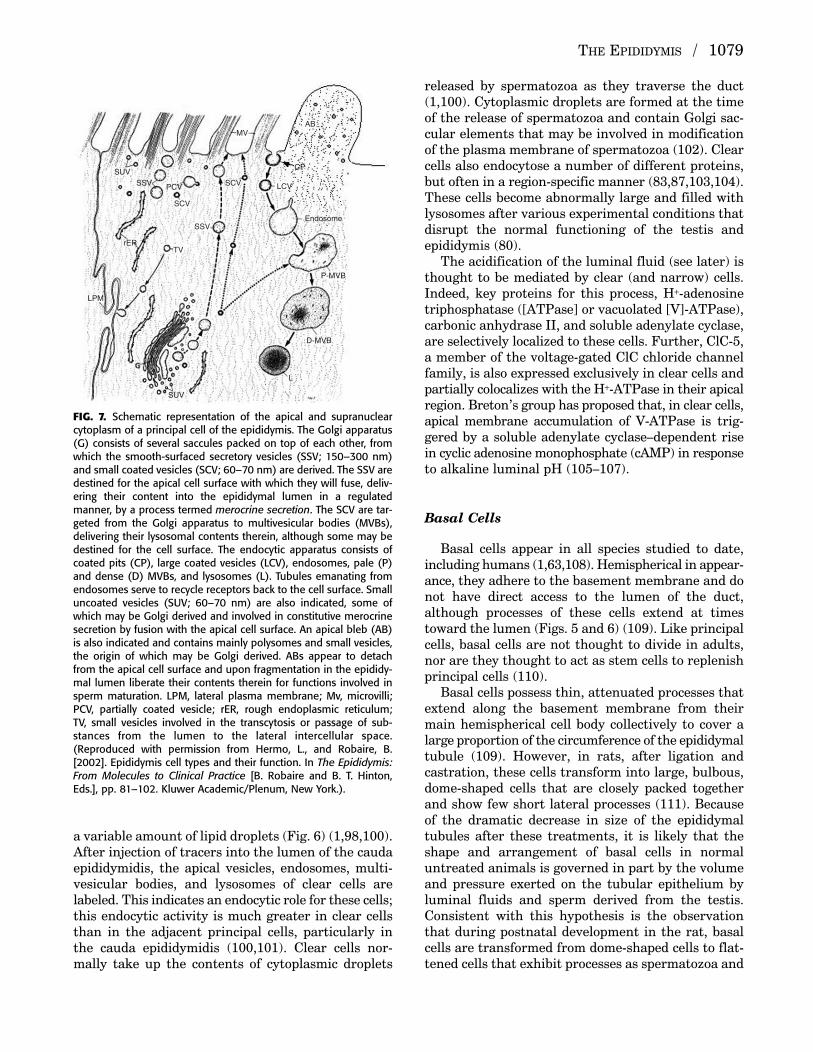
a variable amount of lipid droplets (Fig. 6) (1,98,100).After injection of tracers into the lumen of the caudaepididymidis, the apical vesicles, endosomes, multi-vesicular bodies, and lysosomes of clear cells arelabeled. This indicates an endocytic role for these cells;this endocytic activity is much greater in clear cellsthan in the adjacent principal cells, particularly inthe cauda epididymidis (100,101). Clear cells nor-mally take up the contents of cytoplasmic droplets
released by spermatozoa as they traverse the duct(1,100). Cytoplasmic droplets are formed at the timeof the release of spermatozoa and contain Golgi sac-cular elements that may be involved in modificationof the plasma membrane of spermatozoa (102). Clearcells also endocytose a number of different proteins,but often in a region-specific manner (83,87,103,104).These cells become abnormally large and filled withlysosomes after various experimental conditions thatdisrupt the normal functioning of the testis and epididymis (80).
The acidification of the luminal fluid (see later) isthought to be mediated by clear (and narrow) cells.Indeed, key proteins for this process, H+-adenosinetriphosphatase ([ATPase] or vacuolated [V]-ATPase),carbonic anhydrase II, and soluble adenylate cyclase,are selectively localized to these cells. Further, ClC-5,a member of the voltage-gated ClC chloride channelfamily, is also expressed exclusively in clear cells andpartially colocalizes with the H+-ATPase in their apicalregion. Breton’s group has proposed that, in clear cells,apical membrane accumulation of V-ATPase is trig-gered by a soluble adenylate cyclase–dependent risein cyclic adenosine monophosphate (cAMP) in responseto alkaline luminal pH (105–107).
Basal Cells
Basal cells appear in all species studied to date,including humans (1,63,108). Hemispherical in appear-ance, they adhere to the basement membrane and donot have direct access to the lumen of the duct,although processes of these cells extend at timestoward the lumen (Figs. 5 and 6) (109). Like principalcells, basal cells are not thought to divide in adults,nor are they thought to act as stem cells to replenishprincipal cells (110).
Basal cells possess thin, attenuated processes thatextend along the basement membrane from theirmain hemispherical cell body collectively to cover alarge proportion of the circumference of the epididymaltubule (109). However, in rats, after ligation and castration, these cells transform into large, bulbous,dome-shaped cells that are closely packed togetherand show few short lateral processes (111). Becauseof the dramatic decrease in size of the epididymaltubules after these treatments, it is likely that theshape and arrangement of basal cells in normaluntreated animals is governed in part by the volumeand pressure exerted on the tubular epithelium byluminal fluids and sperm derived from the testis.Consistent with this hypothesis is the observationthat during postnatal development in the rat, basalcells are transformed from dome-shaped cells to flat-tened cells that exhibit processes as spermatozoa and
THE EPIDIDYMIS / 1079
AB
Endosome
P-MVB
D-MVB
CP
LCVPCV SCV
SCV
SUV
SUV
SSV
SSV
TV
LPM
MV
rER
L
FIG. 7. Schematic representation of the apical and supranuclearcytoplasm of a principal cell of the epididymis. The Golgi apparatus(G) consists of several saccules packed on top of each other, fromwhich the smooth-surfaced secretory vesicles (SSV; 150–300 nm)and small coated vesicles (SCV; 60–70 nm) are derived. The SSV aredestined for the apical cell surface with which they will fuse, deliv-ering their content into the epididymal lumen in a regulatedmanner, by a process termed merocrine secretion. The SCV are tar-geted from the Golgi apparatus to multivesicular bodies (MVBs),delivering their lysosomal contents therein, although some may bedestined for the cell surface. The endocytic apparatus consists ofcoated pits (CP), large coated vesicles (LCV), endosomes, pale (P)and dense (D) MVBs, and lysosomes (L). Tubules emanating fromendosomes serve to recycle receptors back to the cell surface. Smalluncoated vesicles (SUV; 60–70 nm) are also indicated, some ofwhich may be Golgi derived and involved in constitutive merocrinesecretion by fusion with the apical cell surface. An apical bleb (AB)is also indicated and contains mainly polysomes and small vesicles,the origin of which may be Golgi derived. ABs appear to detachfrom the apical cell surface and upon fragmentation in the epididy-mal lumen liberate their contents therein for functions involved insperm maturation. LPM, lateral plasma membrane; Mv, microvilli;PCV, partially coated vesicle; rER, rough endoplasmic reticulum; TV, small vesicles involved in the transcytosis or passage of sub-stances from the lumen to the lateral intercellular space.(Reproduced with permission from Hermo, L., and Robaire, B.[2002]. Epididymis cell types and their function. In The Epididymis:From Molecules to Clinical Practice [B. Robaire and B. T. Hinton,Eds.], pp. 81–102. Kluwer Academic/Plenum, New York.).
P515401_22 10/24/05 8:14 PM Page 1079
fluids arrive in the corpus and cauda epididymidis bydays 49 and 56, respectively (108).
Basal cells possess coated pits on the plasma mem-brane face opposing the basement membrane andoverlying principal cells, suggesting the receptor-mediated endocytosis of factors derived from theblood or principal cells. Basal cells also show an accu-mulation of a secretory material in Golgi saccules,and distinct secretory granules appear next to theGolgi apparatus (L. Hermo and B. Robaire, unpub-lished results), as seen in other typical secretory cells(112). The destiny of the secretory material may be toregulate principal cell function or enter the circula-tion for functions as yet to be determined. Basal cellshave been shown also to express apolipoprotein Eand alcohol dehydrogenases (113).
It has also been proposed that basal cells may havea role as immune cells because of their ability torespond in number and macrophage antigen expressionto the presence of sperm autoantigens in the lumen(114), and it has been postulated that these cells mayhave an extratubular origin (115).
Studies from Wong’s group [reviewed in Leung et al.(116)] have proposed an additional function for basalcells; they suggest that these cells may have a role inregulating electrolyte and water transport by principalcells. This process is proposed to be mediated by thelocal formation of prostaglandins (PGs) and requirethe participation of the transient receptor potential(Trp) proteins. The latter serve as transmembranepathways for Ca2+ influx, whereas cyclooxygenase-1(COX-1) is a key enzyme in the formation of PGs.Both of these proteins are exclusively expressed inbasal cells.
Halo Cells
Halo cells are small cells with a narrow rim of clearcytoplasm that are present throughout the epididymalepithelium (Fig. 6) (1). These cells are usually locatedat the base of the epithelium and contain variablenumbers of dense core granules. Halo cells have beendescribed either as lymphocytes (1) or monocytes(117); these two cell types are difficult to distinguishby light microscopy because of their similarity in sizeand nuclear morphology. Although the exact natureof halo cells has been controversial since they werefirst described by Reid and Cleland (1957) (57), studiesby Flickinger et al. (1997) (118) and Serre andRobaire (1999) (119) have resolved this issue byimmunolabeling the main types of immunocompetentcells. It is now clear that, in young adult animals,halo cells consist of helper T lymphocytes, cytotoxicT lymphocytes, and monocytes, but not B lymphocytes.With age, there is a region-specific increase in thenumber of each of these immune cell types, as well
as the occasional appearance of eosinophils (96) andB lymphocytes. In the epididymal epithelium of youngrats, the number of cells that stain for antibodiesagainst monocytes–macrophages (ED1+), helper Tlymphocytes (CD4+), and cytotoxic T lymphocytes(CD8+) is equivalent to the number of halo cells (119),suggesting that halo cells are, under normal conditions,the primary immune cell in the epididymis.
The Blood–Epididymis Barrier
Given the presence in spermatozoa of proteins thatare recognized by the body as foreign, it stands toreason that there should be a continuation beyond thetestis of a functional barrier. The probable existenceof a blood–epididymis barrier was discussed as earlyas 1976 (120), and several reviews describing differentaspects of the barrier have appeared (121–123).
Structure
The junctional complex between adjacent epididymalprincipal cells is composed of apically located gap,adherens, and tight junctions. Tight junctions betweenadjacent principal epithelial cells at their luminalsurface form the blood–epididymis barrier (122,124),whereas gap junctions allow communication betweenadjacent principal cells. These tight junctions form acontinuous zonule around the cell, sealing the spacesbetween the epithelial cells, so that the luminal compartment and the intercellular spaces becomeseparate physiological compartments (125). Thetight junctions begin to form at the time of differen-tiation of the Wolffian duct (126). Using lanthanumnitrate as an electron-opaque tracer that is blockedat tight junctions, the postnatal development of theblood–epididymis barrier was shown to be gradual;its formation is virtually complete by postnatal day 21in rats (125).
Electron microscopic changes in the structure ofthe junctional complex of the initial segment havebeen observed compared with the other segments ofthe epididymis. In the initial segment, the tight junc-tions span a considerable length of the apical plasmamembrane and have few desmosomes (122). Withprogress toward the caudal end of the epididymis, ageneral decrease in the number of tight junctionalstrands is noted; the span of merging plasma mem-branes is considerably reduced, but numerousdesmosomes are found in the apical region (122,126).
Junctional Proteins
Adherens junctions form a continuous belt andhold neighboring cells together through a family of
1080 / CHAPTER 22
P515401_22 10/24/05 8:14 PM Page 1080

calcium-dependent cell–cell adhesion molecules calledcadherins, which mediate calcium-dependent homo-typic interactions (127). The cadherins have also beenimplicated in the formation and maintenance of tightjunctions (127–133). The cytoplasmic domain of cadherins forms a tight complex with several pro-teins, which either link cadherins to the cytoskeletonor are involved in signal transduction pathways.These include catenins, actinin, vinculin, and zonulaoccludens-1. The cadherin–catenin complex is essen-tial for cadherin-mediated cell adhesion. Cyr et al.(1992) (134) reported the presence of E-cadherin andP-cadherin messenger RNA (mRNA) in the rat epididymis. Electron microscopy with immunogoldlabeling indicates that E-cadherin is localized in theextracellular space between the lateral plasma mem-branes of adjacent principal cells at the level of apicaljunctional complexes in the adult rat epididymis(122,135,136), as well as in the deeper underlyingregions of the extracellular space between the lateralplasma membranes. Similar observations were notedin the human and mouse epididymis (137–139). Thecomposition of the catenin-adhering junctional familyof proteins and their relationship with cadherinsremain to be established in the epididymis; however,in one study it was shown that, in the normal adultrat epididymis, there was immunostaining for threeanti-catenin antibodies (alpha-, beta-, and p120ctn)along the lateral plasma membranes between adjacentepithelial cells (140).
In addition to adherens and tight junctions, the epi-didymal junctional complex also contains gap junctions(124,141). Gap junctions, made up of proteins termedconnexins, mediate communication between cells byallowing small molecules to pass from cytoplasm tocytoplasm of neighboring cells, thereby metabolicallyand electrically coupling them (142). Connexin sub-units oligomerize in the trans-Golgi network to formhemichannels or connexons. In the epididymis, gapjunctions containing connexin 43 were first localizedbetween principal and basal cells (143). Using a reversetranscriptase-polymerase chain reaction (RT-PCR)strategy, Finnson and Cyr (unpublished observations)have identified at least seven different connexin tran-scripts in the rat epididymis. Although the presenceof multiple connexins in specific cell types is notunique to the epididymis, the large number of differentconnexins is suggestive of complex communicationbetween epididymal cells.
Functions
The composition of epididymal luminal fluid is distinctly different from that of blood plasma. Theblood–epididymis barrier keeps the two fluids in separate compartments (144). The blood–epididymis
barrier also maintains a specialized luminal microen-vironment for the maturing spermatozoa by restrictingthe passage of a number of ions, solutes, and macro-molecules across the epididymal epithelium (121,144).For instance, molecules such as inositol and carnitinecan be concentrated ten- to 100-fold in the lumen ofthe caput epididymidis, whereas others, such as inulin,L-glucose, and bovine serum albumin, are effectivelyexcluded [reviewed in Robaire and Hermo (1) andTurner (85)]. The blood–epididymis barrier carefullycontrols the microenvironment so that the spermatozoaare bathed in an appropriate fluid milieu at eachstage of maturation as they travel through each segment of the epididymis (85).
This barrier also serves as an extension of theblood–testis barrier. Spermatozoa are immunogenic;they contain proteins on their surfaces that would be recognized as foreign if they were to leave the epi-didymis (1). The exact function of the blood–epididymis barrier in protecting spermatozoa fromthe immune system is unclear at this time. The barrierprevents the passage of spermatozoa between epithelialcells, but cellular elements of spermatozoa can be takenup by epithelial cells. However, additional studies areneeded to clarify whether, in the adult, any epididymalepithelial cell is capable of acting as an antigen-presenting cell.
Although it would appear that the blood–epididymisbarrier is resistant to some foreign substances [e.g.,gossypol (144a), estradiol, (144b)], administration ofcyclophosphamide to efferent duct–ligated rats resultedin the production of damaged spermatozoa, suggestingthat this drug could enter the epididymal lumen andmodify spermatozoa. However, very little is knownabout the role played by this barrier in protecting sper-matozoa from toxic substances and immunoglobulins(145). The inability of this barrier to maintain its tight-ness under conditions of stress, such as aging (136),may play help explain some of the deleterious effects ofstressors on sperm function and fertility.
FUNCTIONS TAKING PLACE IN THELUMINAL COMPARTMENT
The four main functions of the epididymis are trans-port of spermatozoa, development of sperm motility,development of sperm fertilizing ability, and the cre-ation of a specialized luminal environment conduciveof the maturation process through the absorptiveand secretory activities of the epididymal epithelium.
Transport of Spermatozoa
Once released in the lumen of the seminiferoustubule, spermatozoa are transported through the
THE EPIDIDYMIS / 1081
P515401_22 10/24/05 8:14 PM Page 1081
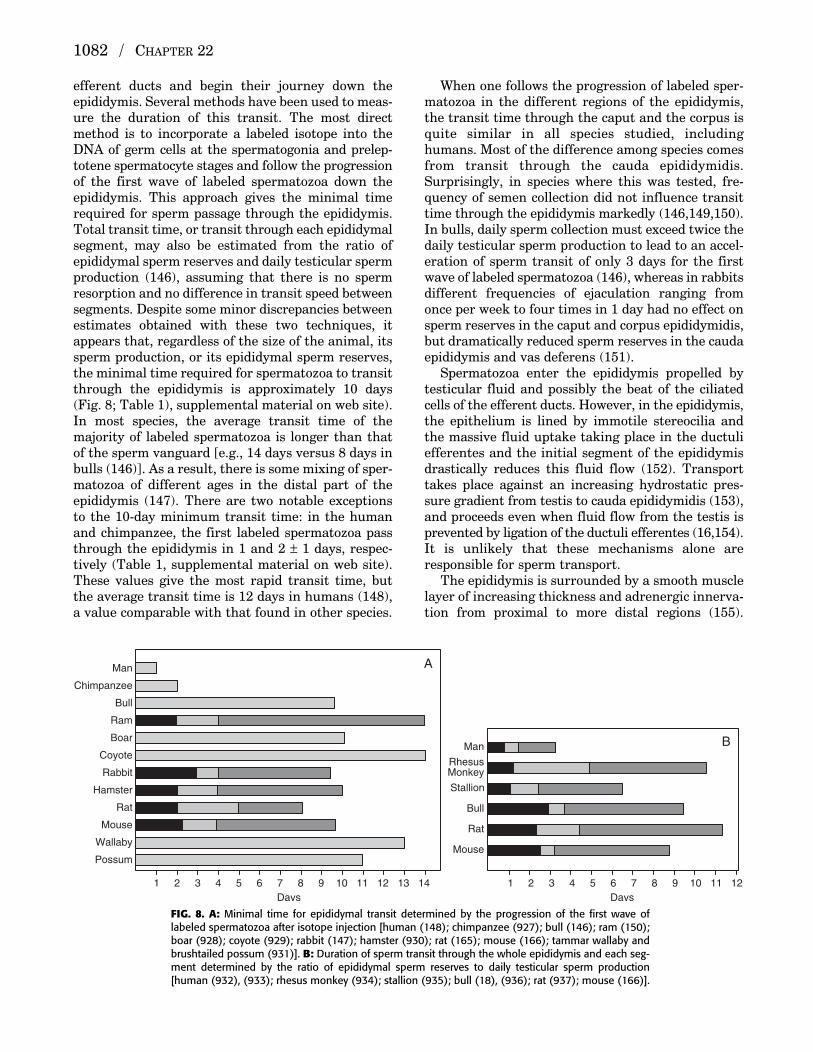
efferent ducts and begin their journey down the epididymis. Several methods have been used to meas-ure the duration of this transit. The most directmethod is to incorporate a labeled isotope into theDNA of germ cells at the spermatogonia and prelep-totene spermatocyte stages and follow the progressionof the first wave of labeled spermatozoa down theepididymis. This approach gives the minimal timerequired for sperm passage through the epididymis.Total transit time, or transit through each epididymalsegment, may also be estimated from the ratio of epididymal sperm reserves and daily testicular spermproduction (146), assuming that there is no spermresorption and no difference in transit speed betweensegments. Despite some minor discrepancies betweenestimates obtained with these two techniques, itappears that, regardless of the size of the animal, itssperm production, or its epididymal sperm reserves,the minimal time required for spermatozoa to transitthrough the epididymis is approximately 10 days(Fig. 8; Table 1), supplemental material on web site).In most species, the average transit time of themajority of labeled spermatozoa is longer than thatof the sperm vanguard [e.g., 14 days versus 8 days inbulls (146)]. As a result, there is some mixing of sper-matozoa of different ages in the distal part of the epididymis (147). There are two notable exceptionsto the 10-day minimum transit time: in the humanand chimpanzee, the first labeled spermatozoa passthrough the epididymis in 1 and 2 ± 1 days, respec-tively (Table 1, supplemental material on web site).These values give the most rapid transit time, butthe average transit time is 12 days in humans (148),a value comparable with that found in other species.
When one follows the progression of labeled sper-matozoa in the different regions of the epididymis,the transit time through the caput and the corpus isquite similar in all species studied, includinghumans. Most of the difference among species comesfrom transit through the cauda epididymidis.Surprisingly, in species where this was tested, fre-quency of semen collection did not influence transittime through the epididymis markedly (146,149,150).In bulls, daily sperm collection must exceed twice thedaily testicular sperm production to lead to an accel-eration of sperm transit of only 3 days for the firstwave of labeled spermatozoa (146), whereas in rabbitsdifferent frequencies of ejaculation ranging fromonce per week to four times in 1 day had no effect onsperm reserves in the caput and corpus epididymidis,but dramatically reduced sperm reserves in the caudaepididymis and vas deferens (151).
Spermatozoa enter the epididymis propelled bytesticular fluid and possibly the beat of the ciliatedcells of the efferent ducts. However, in the epididymis,the epithelium is lined by immotile stereocilia andthe massive fluid uptake taking place in the ductuliefferentes and the initial segment of the epididymisdrastically reduces this fluid flow (152). Transporttakes place against an increasing hydrostatic pres-sure gradient from testis to cauda epididymidis (153),and proceeds even when fluid flow from the testis isprevented by ligation of the ductuli efferentes (16,154).It is unlikely that these mechanisms alone areresponsible for sperm transport.
The epididymis is surrounded by a smooth musclelayer of increasing thickness and adrenergic innerva-tion from proximal to more distal regions (155).
1082 / CHAPTER 22
1 2 3 4 5 6 7Days
8 9 10 11 12 13 14 1 2 3 4 5 6 7Days
8 9 10 11 12
Man
Chimpanzee
Bull
Ram
Boar
Coyote
Rabbit
Hamster
Rat
Mouse
Wallaby
Possum
Man
RhesusMonkey
Bull
Stallion
Rat
Mouse
A
B
FIG. 8. A: Minimal time for epididymal transit determined by the progression of the first wave oflabeled spermatozoa after isotope injection [human (148); chimpanzee (927); bull (146); ram (150);boar (928); coyote (929); rabbit (147); hamster (930); rat (165); mouse (166); tammar wallaby andbrushtailed possum (931)]. B: Duration of sperm transit through the whole epididymis and each seg-ment determined by the ratio of epididymal sperm reserves to daily testicular sperm production[human (932), (933); rhesus monkey (934); stallion (935); bull (18), (936); rat (937); mouse (166)].
P515401_22 10/24/05 8:14 PM Page 1082
Therefore, the mechanism responsible for drivingthe contents through the lumen of the resting epi-didymis has been attributed primarily to the rhythmicmuscular contractions of the smooth muscle liningthe epididymal tubule (156–159). These elegant studieshave shown a relationship between the electrical andcontractile activities of the epididymal tubule andthe progression of oil droplets injected in the lumen.The droplets do not move in a linear fashion, butback and forth. The movement forward starts whenthe spread of electrical activity approaches thedroplet and stops or reverses when the electricalactivity wanes or changes direction. Because the elec-trical pacemakers are randomly distributed and theelectrical pulse spreads in both directions, dropletsinjected at the same time may spread in the lumenand follow different courses. The net distance coveredduring each pendular movement is small. Nevertheless,the droplets progress downward towards the vas def-erens, where the frequency of electrical activity islower than in the caput epididymidis (156,158). Therate of luminal flow is not uniform in the differentsegments of the epididymis (158–160). The progres-sion of droplets decreases from 420 mm/2 hours inthe initial segment to 64 mm/2 hours in the distalcaput and 25 mm/2 hours in the cauda epididymidisand vas deferens (158). It is likely that the progres-sion of oil droplets mimics that of spermatozoa in theepididymis. In bulls, labeled gold-coated beadsinjected in the rete testis are grouped in the distalcaput epididymidis 2 days after injection and in thecauda 5 days after injection, a transit time compara-ble with that of labeled spermatozoa, but 6 days afterinjection, beads are spread throughout the cauda (146).Therefore, the progression of the epididymal luminalcontent is a dynamic process, resulting in a mixing ofluminal content, and is controlled by the electricaland contractile activity of the epididymal tubule.
The smooth muscle contractions of the epididymaltubule and transit of spermatozoa therein are influ-enced by several factors, both hormonal and neuronal.Castration depletes epididymal sperm reserves(161–163) and increases intraluminal pressure, con-tractility of the epididymis (164), and sperm transport(165). Testosterone treatment reverses the effect ofcastration, indicating that androgens control the con-tractility of the epididymal tubule to ensure an opti-mal rate of sperm transport. Estrogen, on the otherhand, speeds up murine sperm transport drasticallyfrom 9.7 to 2.1 days (166).
Contractility of the epididymal tubule is also influ-enced by PGs (167,168). PGF2α increases the frequencyand amplitude of contractions in proximal epididymistubules in vitro, whereas PGE2 decreases these contractions (168). The endogenous levels of PGs areconsistent with their regulation of basal contractilityof the proximal epididymis (168).
Neurohypophysial peptides, such as oxytocin orvasopressin, mediated by receptors present in theepididymis (169–173), also increase epididymal con-tractility both in vitro (174–176) and in vivo (177–180).In several species, including humans (169), this resultsin an increase in the number of ejaculated spermatozoaor spermatozoa transported through the epididymis(179,181–187). Although the relative effect of oxytocinand vasopressin may vary among species, dependingon the dose of the peptide or on the epididymal seg-ment used in the in vitro studies (180,187), it is clearthat neurohypophysial peptides regulate both basalcontractility of the epididymis and, on release in theperipheral circulation around the time of ejaculation(188–192), transport of spermatozoa through the vasdeferens. Interestingly, the effect of oxytocin on epi-didymal motility is regulated by estrogens in part byan upregulation of the oxytocin receptor gene andprotein (175). This may account for the estrogen-induced accelerated sperm transport (166), and suggests an interplay between steroids and neuro-hypophysial hormones in the regulation of epididymalcontractility.
Neuronal regulation is also involved in epididymalcontractility and sperm transport. This was firstdemonstrated by Simeone in 1933 (193) using surgicalsympathectomy and confirmed in later studies usingeither surgical (168) or guanethidine-induced chemicalsympathectomy (194–197). Surgical removal of a singleneuronal ganglion, the inferior mesenteric ganglionthat provides sympathetic innervation to the caudaepididymidis, is sufficient to slow sperm transportthrough the epididymis (198). Indeed, adrenergic andcholinergic drugs affect contractility of the epididymisboth in vitro (199) and in vivo (179,200–203).Temperature also affects epididymal contractility: aswitch from scrotal to body temperature increasesthe frequency and spread of electrical activity of thesmooth muscle of the epididymis (204), and signifi-cantly speeds up sperm transport through the epi-didymis (205), thus having potential deleterious effectson sperm maturation and fertility.
Although a neuromuscular mechanism may not bethe only one responsible for sperm transport in thevarious segments of the epididymis and in all species,it appears to be the main mechanism responsible for sperm transport through the epididymis in mammals.
Maturation of Spermatozoa
Fertilizing Ability
In lower vertebrates, such as cyclostomes, fish, andamphibians, spermatozoa released from the testisare fully motile and competent to fertilize. In contrast,
THE EPIDIDYMIS / 1083
P515401_22 10/24/05 8:14 PM Page 1083
in higher vertebrates, spermatozoa become functionallymature as they pass through the epididymis. Theyacquire the ability to move forward when releasedfrom the epididymis, ascend the female genital tract,undergo the acrosome reaction, bind to and pene-trate the egg vestments, and achieve syngamy withthe female gamete.
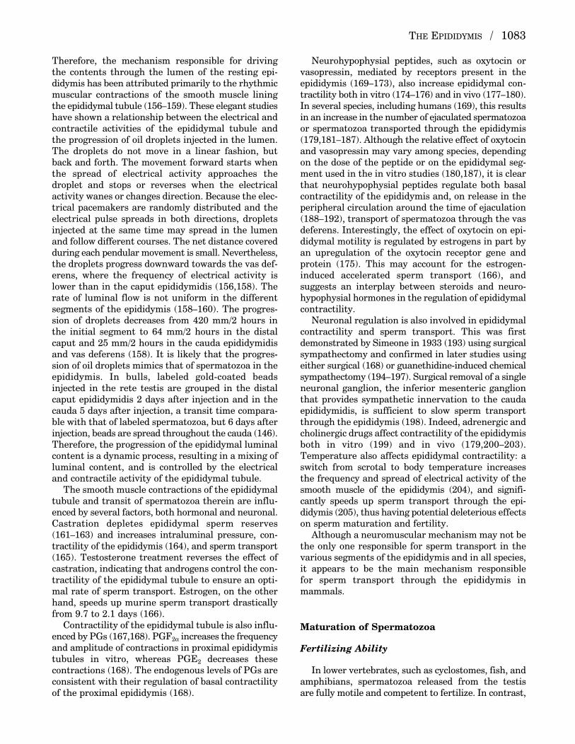
In all species examined thus far, a gradient of fer-tilizing potential is observed as spermatozoa traversethe epididymis (Fig. 9). Although there is some speciesvariation with respect to the exact site at which sper-matozoa first gain their fertilizing potential, it isclear that to be competent to fertilize in vivo, sper-matozoa leaving the testis have to pass through somepart of the proximal epididymis.
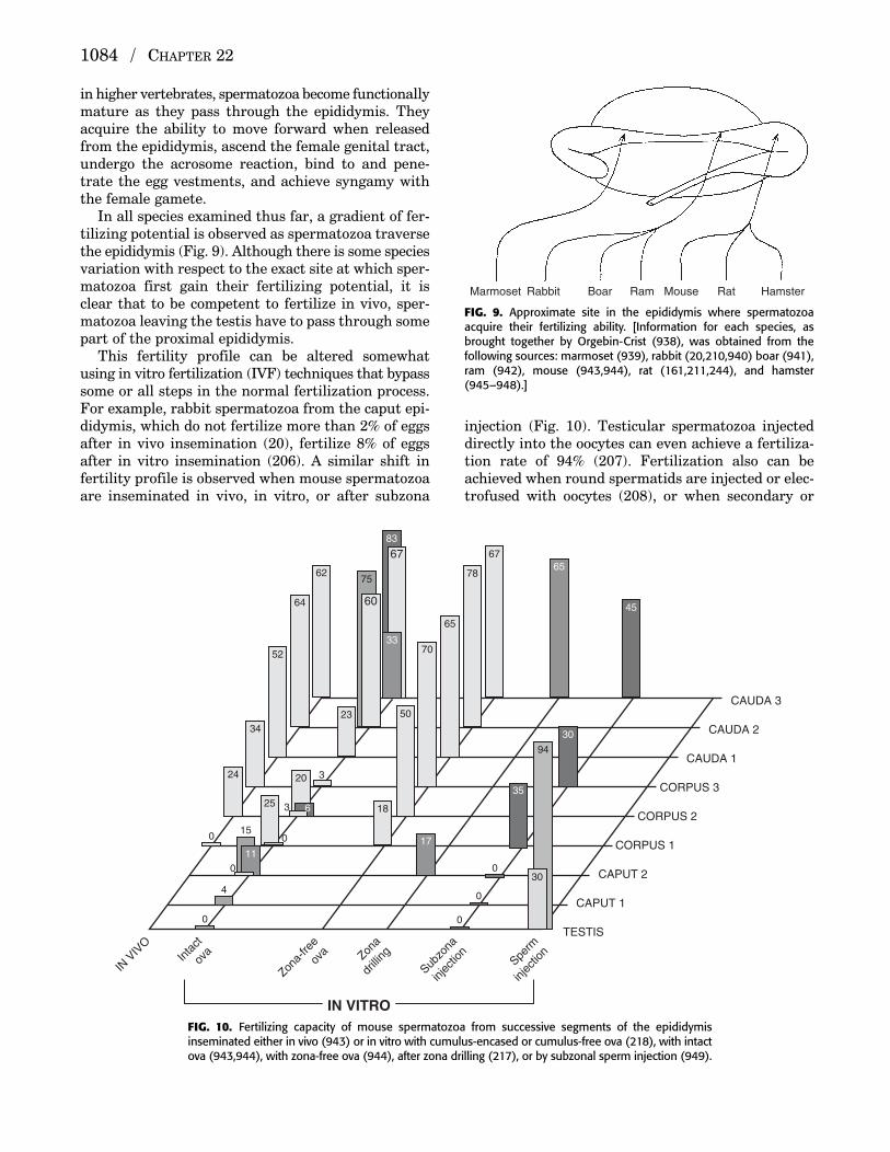
This fertility profile can be altered somewhatusing in vitro fertilization (IVF) techniques that bypasssome or all steps in the normal fertilization process.For example, rabbit spermatozoa from the caput epi-didymis, which do not fertilize more than 2% of eggsafter in vivo insemination (20), fertilize 8% of eggsafter in vitro insemination (206). A similar shift infertility profile is observed when mouse spermatozoaare inseminated in vivo, in vitro, or after subzona
injection (Fig. 10). Testicular spermatozoa injecteddirectly into the oocytes can even achieve a fertiliza-tion rate of 94% (207). Fertilization also can beachieved when round spermatids are injected or elec-trofused with oocytes (208), or when secondary or
1084 / CHAPTER 22
Marmoset Rabbit Boar Ram Mouse Rat Hamster
FIG. 9. Approximate site in the epididymis where spermatozoaacquire their fertilizing ability. [Information for each species, asbrought together by Orgebin-Crist (938), was obtained from the following sources: marmoset (939), rabbit (20,210,940) boar (941),ram (942), mouse (943,944), rat (161,211,244), and hamster(945–948).]
52
64
62
67
78
65
70
50
18
34
35
45
83
30
24
25
20
23
0
0
4
15
0
0
0
0
6
3
11
3
67
33
75
60
94
30
65
17
0
IN V
IVO
Inta
ct
ova
Zona-
free
ova Zon
a
drilli
ng
Subzo
na
injec
tion
Sperm
injec
tion
CAUDA 3
CAUDA 2
CAUDA 1
CORPUS 3
CORPUS 2
CORPUS 1
CAPUT 2
CAPUT 1
TESTIS
IN VITROFIG. 10. Fertilizing capacity of mouse spermatozoa from successive segments of the epididymisinseminated either in vivo (943) or in vitro with cumulus-encased or cumulus-free ova (218), with intactova (943,944), with zona-free ova (944), after zona drilling (217), or by subzonal sperm injection (949).
P515401_22 10/24/05 8:14 PM Page 1084

even primary spermatocytes are injected into theooplasm (207,209). However, the fertilization ratesare lower than after injection of testicular spermatozoa.
Moreover, it appears that the acquisition of fertil-izing potential is not a simple “on” or “off” state.Initial studies with rabbits (20,210), rats (211), andrams (212,213) showed that spermatozoa first gainthe ability to fertilize eggs, and only after furthertransit through the epididymis acquire the ability toproduce complete litters of viable offspring. Orgebin-Crist noted an increase in both preimplantation andpostimplantation loss when rabbits were inseminatedwith corpus versus ejaculated spermatozoa (20); thisis apparently due to a delay in the fertilization ofeggs (20,214) and in the first zygotic division (215).Although Overstreet and Bedford (216) were unableto confirm these observations, more recent studies inmice (217) reported that only 8% of oocytes fertilizedby caput epididymal spermatozoa were capable ofdeveloping into blastocysts in vitro, compared with48% of oocytes fertilized by cauda spermatozoa;Lacham-Kaplan and Trounson (218) confirmed thata high rate of embryonic arrest and retarded develop-ment occurs in mouse oocytes fertilized by epididymalspermatozoa that have just gained their fertilizingcapacity. Although testicular spermatozoa and sper-matids fertilize 94% and 37% of eggs, respectively,after injection into the oocytes, only 54% and 28%,respectively, of the fertilized eggs develop into liveoffspring when transferred into foster mothers (207).Collectively, these experiments show that passagethrough the epididymis endows spermatozoa with theability to ascend the female genital tract and interactwith the egg. This maturation process can be circum-vented because spermatid-injected oocytes developinto live offspring, but the chances of normal devel-opment are higher after injection of more maturespermatozoa.
The experimental protocol of insemination ofequal numbers of spermatozoa from succeeding seg-ments of the epididymis from one individual is obvi-ously not possible in humans. However, in cases ofobstructive azoospermia, epididymovasostomieshave been done to recanalize the epididymis. Thecumulative results show a low pregnancy rate whenthe anastomosis is done proximally and an increasedincidence of pregnancies with more distal connections(219,220). These results, although not directly com-parable with the animal studies, imply that in human,as in other species, there is a progressive maturationof sperm fertilizing ability in the epididymis. Theyalso suggest that the fertility profile may be shiftedmore proximally compared with other species.Pregnancies have been achieved even after reanasto-mosis of the efferent ducts to the vas, but the postop-erative interval before pregnancy occurred was longer
(2 years) (221) than after corpus–vas anastomosis (6 to 13 months) (220). If spermatozoa from theefferent ducts had the same level of maturity as sper-matozoa from the corpus, one would expect the samepostoperative interval, but the small number of casesprecludes any generalization.
In cases of obstructive azoospermia or congenitalvas agenesis, spermatozoa can also be aspirated fromthe epididymis and inseminated in vitro (IVF). Thereis a statistical difference in fertilization rates withspermatozoa retrieved from different levels of the epi-didymis (222), confirming the progressive maturationof human spermatozoa in the epididymis observedafter reanastomosis (219). After IVF and transfer,pregnancies have been reported with spermatozoarecovered from the corpus epididymidis (223–225),and even the proximal caput epididymidis (226,227).However, the consequences of either obstructiveazoospermia or congenital vas agenesis on the struc-ture and functions of the epididymal epithelium havenot been investigated.
If immature human spermatozoa bypass normalsperm ascent in the female genital tract andsperm–egg interaction, and are injected directly intothe ooplasm (intracytoplasmic sperm injection[ICSI]), they can form zygotes. As in other species,the overall fertilization rate is higher than after conventional IVF (45% versus 6.9%) (228). Even tes-ticular spermatozoa achieve fertilization rates ofapproximately 50%. Nevertheless, in all studies, thereis a small but consistent difference in fertilizationrates after injection of testicular and epididymal ejac-ulated spermatozoa (229–236). In all studies but one(234) that reported statistical analyses, the differ-ences in fertilization rates were significant.
Delayed fertilization and cleavage arrest werereported in one case of IVF with spermatozoa fromthe corpus epididymidis (224). In a large series (236)comparing outcome after ICSI with testicular, epi-didymal, and ejaculated spermatozoa, testicular sper-matozoa had not only statistically significantly lowerfertilization rates (53.4%) than epididymal (58.5%)or ejaculated spermatozoa (64.8%), but statisticallylower yields of good-quality embryos (47%, 40%, and59%, respectively). The embryos chosen for transferyielded a similar pregnancy rate per transfer (23.1%,31.3%, and 27.4%, respectively). The congenital mal-formation rate and the developmental outcome ofchildren born after ICSI with testicular, epididymal,or ejaculated spermatozoa are similar to that of chil-dren born after conventional IVF (237–239) or in thegeneral population. Collectively, these clinical studiesindicate that, as in other mammalian species, humansperm capacity for optimal fertility in vivo increasesduring epididymal transit, and procedures bypassingcritical steps of the natural process of fertilization
THE EPIDIDYMIS / 1085
P515401_22 10/24/05 8:14 PM Page 1085

permit fertilization with immature spermatozoa.However, unlike other species, the fertility profile of human epididymal spermatozoa appears to beshifted more proximally because pregnancies havebeen reported with spermatozoa from the proximalepididymis.
Motility
Concomitant with the acquisition of fertilizingability, a number of sperm characteristics undergomaturational changes. The first one to be recognizedlast century, by Tournade (8), was the epididymalmaturation of the sperm potential for motility. Thiswas subsequently confirmed in numerous species ofhigher vertebrates, from lizard (240,241), to mouse(242), rat (211,243–251), hamster (252,253,253a),guinea pig (254,255), rabbit (20,251,256,257), boar(258,259), goat (260), ram (261–263), bull (264,265),monkey (266–268), and human (75,269–271). Theacquisition of motility is observed whether the testisand epididymis are located in the scrotum or in theabdomen, as in the elephant (272), the hyrax (272,273),and the armadillo (273). The maturation of spermmotility potential involves both a quantitativeincrease in the percentage of motile spermatozoa anda qualitative difference in motility pattern. Testicularspermatozoa are either immotile or display only afaint twitch of the flagellum. Spermatozoa releasedfrom the caput epididymidis swim in a circular pattern,whereas spermatozoa released from the cauda moveprogressively and vigorously forward.
Other Maturational Changes
In addition to the capacity for progressive motility,epididymal spermatozoa develop the capacity toundergo the acrosome reaction [mouse (274), ram(275), pig (276), dog (277), monkey (278), and human(279)], recognize and bind to the zona pellucida[mouse (280), pig (276)], and fuse with the vitellinemembrane as tested with zona-free hamster eggs [pig (281), human (281a,282)]. Concomitant withthese functional changes, spermatozoa undergo struc-tural changes during epididymal transit: migration ofthe cytoplasmic droplet along the sperm flagellum,acrosomal reshaping, changes in the sperm nuclearchromatin and some tail organelles, and changes inthe sperm plasma membrane [reviewed in Bedford(283,284)]. Collectively, these changes underpin thefunctional maturation and subsequent storage ofspermatozoa in the epididymis, but none of thesechanges has been shown to be the only determinantof the acquisition of fertilizing ability.
Regulation of Sperm Maturation
Benoit (12) demonstrated that the function of theepididymis, and the maturation and survival of sper-matozoa within it, depended on hormones secretedby the testis. After bilateral castration, not only didthe epididymal epithelium dedifferentiate, but epi-didymal spermatozoa died quickly, whereas afterunilateral castration spermatozoa maintained theirpotential for motility on the castrated side 2 monthsafter the operation. Experiments conducted byBedford (19) and Orgebin-Crist (21,285) showed thatalthough immature rabbit spermatozoa develop thecapacity for motility and fertility when retained byligatures in the proximal corpus, they do not becomefertile when retained in the caput, although theydevelop a mature pattern of forward motility. Thisindicates that sperm maturation is a multistep processand shows that although the development of a maturepattern of motility is necessary, it is not sufficient torender a spermatozoon capable of fertilizing. It alsoshows that whereas some of the maturationalchanges, like motility, may be intrinsic to the spermcell and develop with time, others, such as the abilityto interact with the egg, depend on the epididymalenvironment. The latter is conditioned by testicularandrogens because sperm maturation occurs aftercastration, hypophysectomy, or in organ culture onlywhen androgen is administered in vivo or included inthe culture medium (161,286–288).
Studies of coincubation of epididymal spermatozoawith epididymal epithelial cell cultures have con-firmed the role of the epididymis in promoting spermmaturation. In such cultures, brushtail possum andtammar wallaby spermatozoa from the proximal caputunderwent the morphological maturational changein head orientation accompanied by the developmentof progressive motility, normally only observed in vivo (289,290). In hamsters, mice, and humans,coculturing of immature spermatozoa from the caputor corpus epididymis with cauda cells not onlyincreased their motility (291–293), but increased theircapacity to bind to salt-stored zona pellucida (293), tofertilize (291,292), and to support the development ofembryos (sired by corpus spermatozoa) (292). Thesematurational changes are promoted by androgen-dependent factors from epididymal principal cellsbecause cocultures maintained in the absence ofandrogens fail to induce maturation (292,293). Theoverwhelming evidence from all these experimentalstudies, both in vivo and in vitro, is that the finalstages of the sperm maturation process in all mam-malian species studied to date, including humans,depend on the epididymis.
However, reports of human pregnancies in twocases where the vasa efferentia was anastomosed to
1086 / CHAPTER 22
P515401_22 10/24/05 8:14 PM Page 1086
the vas deferens, thereby bypassing the entire epi-didymis, led to a challenge to the concept that theepididymis is necessary for the acquisition of fertilizingability (221). As discussed previously, subsequentreports noted that the more distal the anastomoticsite, the greater the chances of fertility, confirmingthat the epididymis provides the milieu necessary foroptimal fertility. Nevertheless, Temple-Smith (294)revisited the issue in elegant experiments using epididymovasostomies at different levels of the epi-didymis in rabbits and rats to assess the fertility ofthese animals. In both of these species, anastomosisof the distal initial segment to the vas, bypassing thedistal caput, corpus, and cauda epididymis, resulted ina significant reduction in sperm viability, motility, andfertility, confirming that spermatozoa require expo-sure to the epididymal environment for normal deve-lopment of fertilizing ability. Interestingly, in rats,bypass of the distal caput and proximal corpus, eitherby epididymo-epididymostomy or graft of a vas bridge,had no effect on sperm motility, viability, or fertility.These elegant studies show clearly that the epididymisplays a major role in the post-testicular maturationof spermatozoa, but they also show that exposure tothe initial segment may be sufficient or that the lackof exposure to the epididymal regions where sperm-atozoa normally become fertile may be compensatedby secretions from other epididymal regions. As dis-cussed later, epididymal spermatozoa are exposed toa highly complex environment, but the nature of thechanges induced by epididymal transit that arerequired to promote optimal fertilization remains tobe established.
Storage of Spermatozoa
The major site for storage of spermatozoa in theexcurrent duct system of mammals is the cauda epi-didymidis. Although normal transit time in mammalsthrough the cauda epididymidis is in the range of 3 to10 days (Fig. 8; Table 1, supplemental material onweb site), spermatozoa can be stored in this tissue forperiods extending beyond 30 days (295); in bats,spermatozoa may be stored in this tissue for manymonths and retain their function (296). With storagein the cauda epididymidis, a loss in fertilizing abilitywas found to occur before a loss in motility (297).Interestingly, spermatozoa that were aged in themale reproductive tract of rabbits, presumably in thecauda epididymidis, induced a tenfold higher inci-dence of chromosomal abnormalities in the resultingblastocysts than did their fresh counterparts (298).
Based on studies in a number of mammalian species[reviewed in Amann (299)], it was noted that 50% to80% of spermatozoa present in the excurrent ducts
were found in the cauda epididymidis and thatapproximately 50% of these spermatozoa were avail-able for ejaculation (299). When animals (rabbits,stallions, and bulls) were at sexual rest (i.e., 7 ormore days without ejaculation), the number of storedspermatozoa that were available for ejaculation wasthree- to fivefold greater than the daily sperm pro-duction rate and two- to threefold greater than thatfound in a “typical” ejaculate; frequent ejaculation,that is, one ejaculate every 1 to 2 days, did not resultin a change in sperm production rate but didmarkedly decrease caudal epididymal sperm reservesand the number of spermatozoa in the ejaculate (299).In contrast, the human, whose sperm productionrate is well below that of most other mammals, has asperm reserve that is only approximately equal to thenumber of spermatozoa found in an ejaculate, whetherat sexual rest or not (299).
In a series of studies on the evolution of the scro-tum, Bedford proposed that the prime moving forcebehind the formation of the scrotum was the need tostore spermatozoa at a low temperature (205,300,301);he suggested “that migration to a scrotal site hasbeen ordained primarily by the sperm storageregion—the cauda epididymidis—and that the func-tion of the testis has only been an incidental factor inthis evolutionary development” (301). This proposalwas based on anatomical observations and on somephysiological studies where the epididymis wasplaced abdominally while the testis remained in thescrotum; the rate of sperm passage through thecauda epididymidis was found to increase approxi-mately twofold in the rabbit after such a procedure.
One would expect that the luminal environmentrequired for the storage of spermatozoa in the caudaepididymidis versus that required for the acquisitionof fertilizing ability would be different. Althoughmany differences have been described in the luminalmakeup of ions, small organic molecules, proteins,and glycoproteins between the cauda and the rest ofthe epididymis (see later), those special conditionsthat allow for the storage of spermatozoa in a quies-cent state for long periods in this tissue have not yetbeen elucidated, although changes in pH (302) in sev-eral species and the presence of immobilin in the ratand hamster (302,303) have been proposed to bemajor factors.
Although it is clear that the major function of thedistal segments of the epididymis is to store mature,live spermatozoa, other functions have been ascribedto this tissue. Abnormal-appearing or dead sperma-tozoa have often been seen in the cauda epididymidis(304,305), and the ability to recognize such sperma-tozoa and then develop a mechanism to neutralize ordestroy them has been proposed recently by severalgroups.
THE EPIDIDYMIS / 1087
P515401_22 10/24/05 8:14 PM Page 1087

Sutovsky et al. (2001) (306) proposed that the cell-surface ubiquitination of defective spermatozoasupplies the necessary signal for these cells to bephagocytosed by epididymal principal cells, thus providing a mechanism for sperm quality control.However, based on extensive histological observationsof the normal epididymal epithelium and the raritywith which sperm phagocytosis is observed, it isapparent that phagocytosis of spermatozoa is anunlikely mechanism for the disposal of the largenumber of spermatozoa produced daily (307). If a qual-ity control system is present in the epididymis, thenit is likely to be more subtle than the phagocytosis ofmarked spermatozoa.
Studies from Olson’s group (308,309), have identi-fied a fibrinogen-like protein (fgl2) that is secretedselectively in the proximal cauda epididymidis of thehamster and that binds to and coats nonviable, butnot viable, luminal spermatozoa. Unlike the hypoth-esis of Sutovsky’s group, these investigators proposethat the epididymis possesses a specific mechanismto identify and envelop defective spermatozoa with aprotein complex containing fgl2 that remains withinthe epididymal lumen. Both of these novel mecha-nisms require that epididymal epithelial cells havethe ability somehow to recognize and tag defectivespermatozoa. Although conceptually appealing, moreextensive studies using different species and meansof creating “abnormal” spermatozoa, such as drugtreatments, are required to establish this as a functionof the normal epididymis.
In a review, Jones (2004) (310) has hypothesizedthatafter androgen withdrawal, either by orchidectomy oras a result of seasonal variation in seasonal breeders,a death pathway is activated that leads to the disso-lution of spermatozoa. Such a mechanism wouldensure that the epididymis is cleansed of defectiveand dead spermatozoa in preparation for the nextbreeding period. How androgen withdrawal wouldactivate such a pathway remains to be determined,but it is interesting to note that many genes are acti-vated in the epididymis after orchidectomy (311).
Protection of Spermatozoa
It is clear that the blood–epididymis barrier allowsfor the production of a specialized luminal fluidmicroenvironment that is important for sperm maturation. However, another critical role played bythis barrier is protection of the maturing spermatozoa.In addition, a series of elaborate defense mechanismsthat help to protect spermatozoa from the immunesystem, harmful xenobiotics, and reactive oxygenspecies (ROS) have been developed in the epididymis.
The defense mechanisms include restricting thetypes of compounds that can enter the epididymallumen, the synthesis and secretion of specific proteinssuch as the defensins and defensin-like molecules,rapid elimination of potential harmful agents throughthe synthesis and secretion of antioxidant and conju-gating enzymes, and the synthesis and secretion ofantioxidant compounds such as glutathione and taurine. Because spermatozoa mature in a hyperos-motic environment, the epididymis also ensures thatthe spermatozoa are protected from potential rapidchanges in osmolality and can regulate their cellvolume.
It was shown some 50 years ago that spermatozoaare highly susceptible to oxidative damage and thathydrogen peroxide was responsible for loss of motilityand cell death (312,313). Later, through a series ofwell-designed studies, Jones and Mann clearlyshowed that when spermatozoa were incubatedunder aerobic conditions, they produced an organicperoxide and released a substance that was believedto be a lipid (314–317). When incubated in the presenceof lipid peroxides, spermatozoa became irreversiblyimmotile and released numerous intracellularenzymes. From this series of studies, it was sug-gested that incubation of spermatozoa under aerobicconditions resulted in lipid peroxidation by peroxidesor lipid radicals that were responsible for structuraldamage, loss of motility, decline in metabolic activity,and release of intracellular enzymes. Because mam-malian spermatozoa have a high content of polyun-saturated fatty acids in their membranes, they arehighly susceptible to lipid peroxidation by ROS(317–320). It is crucial that spermatozoa be protectedfrom the deleterious effects of ROS as they progressalong the epididymal duct; lipid peroxidation of mem-branes has been correlated with midpiece defects(321), decreased motility due to axonemal defects,and reduced intracellular ATP levels (322–324), aswell as impaired capacity for fertilization [reviewedin Vernet et al. (2004) (320)]. Spermatozoa are protected from ROS by superoxide dismutase(325–328). This enzyme protects spermatozoa fromlipid peroxidation by ROS through dismutation ofthe reactive oxygen to hydrogen peroxide and water.Hydrogen peroxide is then rapidly converted towater by the enzymes catalase or glutathione perox-idase (328,329,330).
In a continuing effort to prevent the oxidativedamage of spermatozoa, the epididymis has developedan elaborate system to ensure that spermatozoa areprotected as they mature along the epididymal duct.Each region or segment of the epididymis has devel-oped its own sperm protective mechanisms primarilybecause (a) the metabolic activity differs from one
1088 / CHAPTER 22
P515401_22 10/24/05 8:14 PM Page 1088

region to the next, thereby producing different ROSspecies that need to be eliminated appropriately; and(b) spermatozoa are in a different state of maturityin each region, and therefore differ with respect tosusceptibility to oxidative damage. For example,spermatozoa need to be especially protected fromROS as they enter the initial segment. The luminalfluid in this region is highly oxygenated (331), theepithelial cells are surrounded by a dense capillarynetwork (332), with blood flow exceeding that of thedistal epididymal regions (333,334), and the initialsegment cells are highly metabolically active[reviewed in Hinton et al. (335)]. This unique combi-nation results in the generation of ROS from severalsources, including the endothelial and initial seg-ment epithelial cells [reviewed in Vernet et al. (320)].Therefore, it is not surprising that the initial seg-ment expresses antioxidant enzymes. In the distalepididymal regions, the epithelial cells are still meta-bolically active, and the spermatozoa are continuallyexposed to an oxygen-rich environment; however,there is less vascularization compared with the initialsegment. Hence, spermatozoa are still subjected toROS production, but the type of ROS may well be dif-ferent from the types produced in the initial segment.Hence, the distal epididymal regions have alternativestrategies to protect spermatozoa.
The major antioxidant enzymes present in the epi-didymis (320) include superoxide dismutase (336,337),γ-glutamyl transpeptidase (338–344), glutathioneperoxidases (345–349), glutathione transferases(109,350–354), and indolamine dioxygenase (355). In addition, the lumen of the epididymis containsantioxidant molecules such as glutathione, taurine,and tryptophan as the substrate for indolamine dioxy-genase. Each of these antioxidant enzymes and mol-ecules is found to varying degrees throughout thelength of the epididymis.
Hence, the epididymis plays a critical role in theprotection of spermatozoa from oxidative stress andharmful xenobiotics. The processes by which the epi-didymis protects spermatozoa have also been consid-ered to be a prime target for the development of amale contraceptive (320).
Microenvironment for Maturation,Protection, and Storage
From the previous discussion it can be seen thatthe epididymis plays an important role in sperm maturation. Although the actual time spent in theepididymis may be important for the maturationprocess, spermatozoa must still undergo considerableremodeling and be protected and stored in a specialized
luminal fluid milieu. Crabo (1965) (152) first recog-nized the importance of the epididymal microen-vironment: “The consensus today is doubtless thatthe epididymis actively promotes maturation of spermatozoa. The natural way for the epididymis toexert this influence would be via regulation of thesperm cells’ environment, i.e., epididymal plasma:the environment in the caudal portion should favormaximal survival time of the spermatozoa whichmay be stored there for relatively long periods, with-out losing their fertilizing power and motility.” In viewof several published comprehensive reviews on theformation of the epididymal luminal microenviron-ment (144,356–361), this section summarizes thecontributions of ions, organic solutes, and proteins tothe specialized luminal fluid milieu. Although muchis known concerning the composition of the epididymalluminal fluid, more studies are needed to understandthe precise contribution of each component to spermmaturation.
Perhaps the most underappreciated role of theepididymis is the manner by which it forms themicroenvironment. To accomplish this formidabletask, the epithelium must be structured in a waysuch that (a) it prevents and regulates the entry ofblood-borne substances into the lumen; (b) it has theability to synthesize, secrete, and absorb components;(c) it is arranged so that spermatozoa come into contact with the appropriate environment at theappropriate time—this is the most challengingaspect to understand because it implies that each cellalong the duct knows precisely what its neighborsare doing; and (d) it can respond to spermatozoa,which in turn regulate the microenvironment.Hence, there is a series of complex and ever-changinginteractions between the epididymal epithelium, themicroenvironment, and spermatozoa from the begin-ning to the end of this tube that is several meters inlength.
Water Movement and Its Influence on theFormation of the Luminal Microenvironment
Before discussing the composition of the luminalfluid microenvironment, it is important to considerwater movement. The efferent ducts and the epi-didymis are water-transporting epithelia. Hence, theconcentrations of ions, organic solutes, and proteinsdepend on the movement of water into and out of thelumen. Thus, the measured concentrations of anyion, solute, or protein in epididymal luminal fluid arenot only due to direct secretion into the lumen butalso to the amount of water that is being reabsorbed.Water movement out of the duct is quite extensive.
THE EPIDIDYMIS / 1089
P515401_22 10/24/05 8:14 PM Page 1089
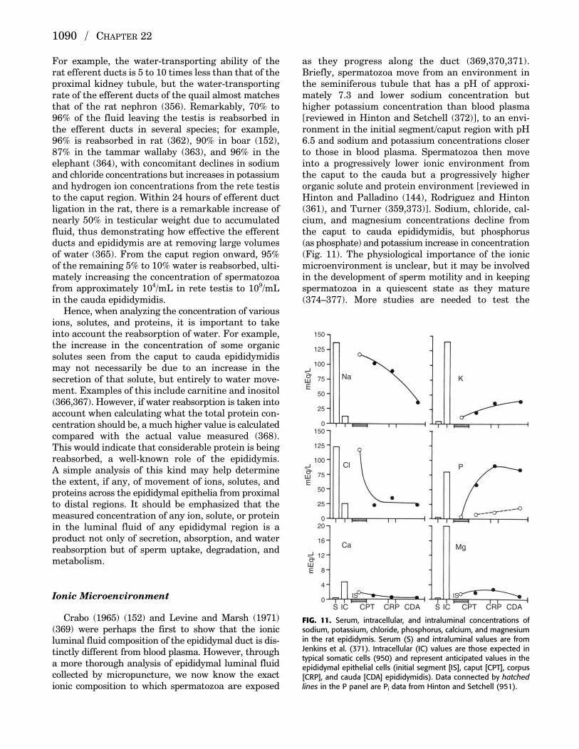
For example, the water-transporting ability of therat efferent ducts is 5 to 10 times less than that of theproximal kidney tubule, but the water-transportingrate of the efferent ducts of the quail almost matchesthat of the rat nephron (356). Remarkably, 70% to96% of the fluid leaving the testis is reabsorbed inthe efferent ducts in several species; for example,96% is reabsorbed in rat (362), 90% in boar (152),87% in the tammar wallaby (363), and 96% in the elephant (364), with concomitant declines in sodiumand chloride concentrations but increases in potassiumand hydrogen ion concentrations from the rete testisto the caput region. Within 24 hours of efferent ductligation in the rat, there is a remarkable increase ofnearly 50% in testicular weight due to accumulatedfluid, thus demonstrating how effective the efferentducts and epididymis are at removing large volumesof water (365). From the caput region onward, 95%of the remaining 5% to 10% water is reabsorbed, ulti-mately increasing the concentration of spermatozoafrom approximately 104/mL in rete testis to 109/mLin the cauda epididymidis.
Hence, when analyzing the concentration of variousions, solutes, and proteins, it is important to takeinto account the reabsorption of water. For example,the increase in the concentration of some organicsolutes seen from the caput to cauda epididymidismay not necessarily be due to an increase in thesecretion of that solute, but entirely to water move-ment. Examples of this include carnitine and inositol(366,367). However, if water reabsorption is taken intoaccount when calculating what the total protein con-centration should be, a much higher value is calculatedcompared with the actual value measured (368). This would indicate that considerable protein is beingreabsorbed, a well-known role of the epididymis. A simple analysis of this kind may help determinethe extent, if any, of movement of ions, solutes, andproteins across the epididymal epithelia from proximalto distal regions. It should be emphasized that themeasured concentration of any ion, solute, or proteinin the luminal fluid of any epididymal region is aproduct not only of secretion, absorption, and waterreabsorption but of sperm uptake, degradation, andmetabolism.
Ionic Microenvironment
Crabo (1965) (152) and Levine and Marsh (1971)(369) were perhaps the first to show that the ionicluminal fluid composition of the epididymal duct is dis-tinctly different from blood plasma. However, througha more thorough analysis of epididymal luminal fluidcollected by micropuncture, we now know the exactionic composition to which spermatozoa are exposed
as they progress along the duct (369,370,371).Briefly, spermatozoa move from an environment inthe seminiferous tubule that has a pH of approxi-mately 7.3 and lower sodium concentration buthigher potassium concentration than blood plasma[reviewed in Hinton and Setchell (372)], to an envi-ronment in the initial segment/caput region with pH6.5 and sodium and potassium concentrations closerto those in blood plasma. Spermatozoa then moveinto a progressively lower ionic environment fromthe caput to the cauda but a progressively higherorganic solute and protein environment [reviewed inHinton and Palladino (144), Rodriguez and Hinton(361), and Turner (359,373)]. Sodium, chloride, cal-cium, and magnesium concentrations decline fromthe caput to cauda epididymidis, but phosphorus (as phosphate) and potassium increase in concentration(Fig. 11). The physiological importance of the ionicmicroenvironment is unclear, but it may be involvedin the development of sperm motility and in keepingspermatozoa in a quiescent state as they mature(374–377). More studies are needed to test the
1090 / CHAPTER 22
S IC CPT CRP CDA S IC CPT CRP CDA
Ca Mg
Cl P
Na K
0
4
8
12
16
20
mE
q/L
0
25
50
75
100
125
150
mE
q/L
0
25
50
75
100
125
150
mE
q/L
IS IS
FIG. 11. Serum, intracellular, and intraluminal concentrations ofsodium, potassium, chloride, phosphorus, calcium, and magnesiumin the rat epididymis. Serum (S) and intraluminal values are fromJenkins et al. (371). Intracellular (IC) values are those expected intypical somatic cells (950) and represent anticipated values in theepididymal epithelial cells (initial segment [IS], caput [CPT], corpus[CRP], and cauda [CDA] epididymidis). Data connected by hatchedlines in the P panel are Pi data from Hinton and Setchell (951).
P515401_22 10/24/05 8:14 PM Page 1090
hypothesis that ions play a critical role in sperm maturation.
Organic Solute Microenvironment
As well as analyzing the luminal fluid ionicmicroenvironment, Levine and Marsh (1971) (369)also measured the osmolality of fluid collected fromeach epididymal region and then compared thesevalues with the sum of osmotically active ions at eachregion. They noted a difference. This osmotic differ-ence increased from the caput to cauda epididymidisto reach over 250 mOsm/kg water; the investigatorssuggested that this difference was due to the pres-ence of organic solutes. Later studies by severalgroups showed that this was indeed the case, with theidentification of solutes such as glutamate, taurine,L-carnitine, myoinositol, glycerylphosphorylcholine,phosphorylcholine, sialic acids, and many otheramino acids present in different concentrationsthrough the epididymal duct of many species, includingthe human [reviewed in Turner (85,359), Hinton andPalladino (144), and Rodriguez and Hinton (361)].
The total concentration of organic solutes in theluminal fluid of the rat testis and epididymisincreases from approximately 10 to 20 mM in theseminiferous tubule to 100 to 150 mM in the caputand to over 200 mM in the cauda epididymidis.Although virtually nothing is known about the rolesof each solute, evidence suggest that they may play acritical role in the protection of spermatozoa and theepididymal epithelium from osmotic stress, similar tothe role played by these solutes in the kidney (377a).The manner by which the epididymal epitheliumforms an organic solute–rich environment is notcompletely known, but there is considerable evidenceto suggest that at least several transporters areinvolved in moving the solute from the blood into thelumen; in addition, the epididymis synthesizes andsecretes solutes into the lumen. For example, L-carnitine is transported into the epididymal cellsand lumen, reaching concentrations as high as 50 to60 mM in some species (378–382). The high intralu-minal concentration of L-carnitine translates into aremarkable 2,000-fold gradient of L-carnitine acrossthe epididymal epithelium. Studies suggest that thedistal caput, corpus, and proximal cauda epididymidisare the regions involved in the active transport of L-carnitine (383–388). The mechanism of L-carnitinetransport in the epididymis is unknown, but hasbeen suggested to involve an active transport system(383,384,386,389) that is androgen dependent(378,380,390,391).
More recent studies have suggested that L-carnitineis transported from blood into cells by the organic
cation/carnitine transporter OCTN2 (392), and thenpresumably from cells into the lumen by the carni-tine transporter CT2, which has been identified inthe human epididymis (393). Several transporters,such as OCTN2 and SMIT (a sodium–myoinositoltransporter), have TonE binding motifs on their pro-moters that bind a transcription factor, TonE bind-ing protein (TonEBP; Rodriguez and Hinton,unpublished observations), that is expressed duringperiods of hypertonicity; these data suggest that theorganic solutes play a role in the regulation of watermovement across the epididymal cell membranes.This is particularly important because in the epi-didymis of most species, spermatozoa are exposed toa hyperosmotic environment. Disruption of the abil-ity of spermatozoa to regulate their cell volume hasbeen suggested to cause male infertility [reviewed inCooper et al. (394)].
The high levels of organic solutes measured in thecauda epididymal fluid of several species suggest thatthese solutes play a similar role in the long-term stor-age of spermatozoa in this region. Likewise, in batsthat store spermatozoa in the cauda epididymidis forup to 6 months, the spermatozoa are stored in a sup-raphysiologically high osmotic environment with aluminal fluid osmolality measured at greater than1,000 mOsm/kg water (395); the mechanisms forgenerating and maintaining such a high osmolalityare unknown. Taurine and glutathione are bothfound in epididymal luminal fluid (396,397), andbecause both of these compounds have been impli-cated in protecting cells from oxidative stress, itwould not be surprising if they have a similar role inthe epididymis.
The transport of sugars and amino acids, includinginositol, 3-O-methyl-D-glucose and 2-deoxy-D-glucose(nonmetabolizable forms of glucose), and α-amino-isobutyric acid (AIB; a nonmetabolizable neutralamino acid), has been studied in the rat epididymis(398–403), yielding several observations. First, glucoseentered by a facilitated diffusion mechanism, andthis uptake appeared to be similar across all epididy-mal regions. Second, glucose transport was inhibitedby the male infertility agents α-chlorhydrin, 5-thio-D-glucose, and 6-chloro-6-deoxy-glucose. Third, trans-port of inositol and AIB was highest in the initialsegment compared with the rest of the epididymis.Finally, transport of inositol and AIB was saturableand, presumably, transport of inositol was throughan active mechanism, whereas that of AIB wasthrough a facilitated diffusion mechanism. Althoughthe GLUT family of glucose transporters has beenwell studied in numerous tissues, only GLUT8 hasbeen identified in epididymal spermatozoa (404) andtissue (43). Presumably, the glucose and amino acidneeds of the epididymis reflect the metabolic needs of
THE EPIDIDYMIS / 1091
P515401_22 10/24/05 8:14 PM Page 1091

the epididymis and maturing spermatozoa. However,more studies are needed to examine the roles playedby each solute. The role of known specific ion andorganic solute transporters that have been identifiedin the epididymis is discussed in greater detail laterin this chapter.
The androgen luminal fluid microenvironmenthas received little attention recently, but past studiesrevealed that the spermatozoa are exposed to a richand complex androgen environment as they progressalong the duct [reviewed in Turner (359)]. In severalspecies, including rat, human, hamster, stallion, andbull, the epididymal luminal fluid concentrations oftestosterone and dihydrotestosterone (DHT) exceedthe blood concentrations (405–407), with DHT con-centration being very high in the caput region. Thehigh luminal fluid androgen concentrations aremaintained by androgen-binding protein (ABP)because studies by Turner et al. (1984) (407) showedthat intraluminal ABP concentrations are equimolarwith total androgen levels. It has been proposed thatthe ABP–androgen complex plays a role in epididymalfunction (408), but confirmatory experiments haveyet to be designed to test this hypothesis.
Protein Microenvironment
The protein composition of epididymal luminalfluid has been well studied for a number of species[reviewed in Dacheux et al. (93,409)]. Spermatozoaare exposed to varying protein concentrations as theymove along the seminiferous tubule (approximately 6 to 10 µg/µL), to the rete testis (approximately 1 µg/µL), to the caput and cauda epididymidis(approximately 25 to 30 µg/µL). Because water isbeing absorbed along the epididymal duct, a higherprotein concentration would have been expected inthe cauda compared with the caput epididymidis. To account for this difference, proteins are presum-ably being absorbed or degraded, or are being utilizedby the spermatozoa. However, even though the netmovement of protein is by absorption, many proteinsare still being synthesized and secreted into the duct.
The proteins in epididymal luminal fluid havebeen analyzed by one- and two-dimensional gel elec-trophoresis (410), generating characteristic patternsof protein profiles for several species; many of theproteins have now been identified using mass spec-trophotometric methods. Using an in vivo split-drop,stopped-flow microperfusion technique together withtwo-dimensional gel electrophoresis and both silverstaining and Western blot analyses, Hinton and colleagues (411,412) showed in vivo protein secretioninto the lumen of discrete epididymal segments andregions. Later studies by Turner and colleagues
(413,414) obtained similar results when the intersti-tium was perfused with radiolabeled methionine andluminal fluid collected by micropuncture and ana-lyzed for radiolabeled proteins.
Epididymal luminal fluid contains a large repertoireof proteins, including enzymes, growth factors, lipid-binding proteins, iron-binding proteins, proteins thatmay be involved in protection or in sperm–egg binding,and other proteins such as the lipocalins, clusterin,lactoferrin, and the cholesterol transport proteinCTP/HE1 [reviewed in Dacheux and Dacheux (93)].Many of these families of proteins are discussed inlater sections.
REGULATION OF EPIDIDYMAL FUNCTIONS
Hormones
In 1926, pioneering studies by Benoit demon-strated that the epididymis depends on an unknowntesticular substance for maintenance of its structureand functions; he showed that the ratio of nucleus tocytoplasm in the mouse epididymis decreased afterbirth as the epididymis differentiated, and increaseddramatically after orchidectomy, as the epididymisdedifferentiated (12). This regulatory substance wasidentified 5 years later as testosterone (415). Sincethen, over a thousand papers have appeared on theresponse of the epididymis in numerous mammals to androgen withdrawal and replacement on a widevariety of end points, ranging from morphological tobiochemical and molecular.
The main approach to understanding the effects ofandrogen withdrawal on the epididymis has beenremoval of the testis. It is clear that this approachcauses loss not only of androgens but of estrogensand any other testicular factor that may affect theepididymis.
Orchidectomy causes a decrease in epididymalweight that is less marked than that of sex accessorytissues such as the prostate or seminal vesicles(416,417). Unlike the case with other androgen-dependent male reproductive tissues, testosteronereplacement, even at supraphysiological levels, onlypartially restores epididymal weight; this is presum-ably due to the large proportion (nearly half) of epididymal weight that is attributable to spermato-zoa and the luminal fluid bathing them (417,418). In the androgen-deprived state, spermatozoa becomeimmotile, lose the ability to fertilize, and die(12,161,419). After orchidectomy, the luminal diame-ter and epithelial cell height decrease and the inter-tubular stroma increases (420). The smoothendoplasmic reticulum content is dramaticallyreduced, whereas the extent of decline in the Golgi
1092 / CHAPTER 22
P515401_22 10/24/05 8:14 PM Page 1092

apparatus is less pronounced (421–423). Morphologicalchanges in principal cells suggest that these cells areparticularly sensitive to androgen levels, in contrastto the other epithelial cell types, which appear to beless affected by orchidectomy (101). The secretoryfunction of principal cells becomes compromised inthe androgen-deprived state. In addition to the virtualdisappearance of endoplasmic reticulum from theirapical cytoplasm, principal cells undergo a strikingloss of apical microvilli from their surface, as well aslysosome accumulation, vacuolization, disappearanceof vesicles from the cell apex, and increased endocy-tosis (101,287). Epididymal androgen receptors and5α-reductase activity are both decreased in theandrogen-deficient state (417,424–426), suggestingthat the mechanisms of androgen action are compro-mised by androgen withdrawal. Total epididymalprotein, RNA, and DNA content are reduced after orchidectomy, but the DNA concentration is apparently increased (427). The increase in DNA concentration is attributed to the concomitant declinein cell volume; this is thought to be the principalmechanism by which the epididymal epitheliumregresses after orchidectomy.
Restoration of circulating testosterone levelsappears sufficient to reverse regressive changes inthe caput, corpus, and cauda epididymidis afterorchidectomy but not in the initial segment, evenwhen supraphysiological doses of testosterone areadministered (417,423,428–432). At 3 days afterorchidectomy with testosterone or DHT replacementto circulating levels, a time at which the prostateshows maximum rates of DNA synthesis, the epi-didymal labeling index, measured by [3H]-thymidineincorporation and mitotic index, is low except in thecorpus region (433). The lack of effect of androgenson mitotic rate in the epididymis of the adult animaldistinguishes this tissue from other androgen-dependent tissues such as the prostate and seminalvesicles. This characteristic of the epididymal epithe-lium suggests that it may contain antiproliferativesignals that inhibit cellular proliferative capacity inresponse to androgen stimulation or other stimuli.Interestingly, B-myc, a transcription factor known toinhibit cellular proliferation, is highly expressed inthe epididymal epithelium (434).
Withdrawal of androgen stimulation by orchidec-tomy induces a wave of apoptotic cell death in theepididymis, beginning in the initial segment andmoving over several days to the cauda epididymidis(435,436). Apoptosis in the initial segment seems tobe caused by withdrawal of androgens and luminalcomponents from the testis, and appears to be p53independent. Using the entire epididymis, Bcl-2, anantiapoptotic factor (437), was found to be sup-pressed by 36 hours after orchidectomy; this was
followed by the appearance of Fas and DNA fragmen-tation in the epididymal epithelium at 48 hours pos-torchidectomy (438). Mutant mice null for Fas (lpr)showed no epididymal regression or DNA fragmenta-tion after orchidectomy. These data suggest that theregression of the epididymal epithelium afterorchidectomy may be regulated through the Faspathway (438).
In addition to androgens, many other hormonal fac-tors have been postulated to play a role in regulatingepididymal function; these are listed in Table 4 (supplemental material on web site). Of special note,however, is estradiol. It has been known for manyyears that the administration of estrogens could affectthe male reproductive system in general (439–448)and the epididymis in particular (166,449). With theadvent of aromatase-specific inhibitors and nullmutant mice for the two isoforms of the estrogenreceptor (discussed later), it is likely that estrogenswill be shown to act as regulators of some specificepididymal functions; however, much remains to bedone to resolve exactly what role this steroid familyplays.
Testicular Factors
In addition to depending on the presence of circu-lating androgens, the epididymis also depends on thepresence of luminal fluid factors originating from thetestis or the epididymis itself. Without testicularluminal fluid factors, many cells in the initial segmentundergo apoptosis within 24 hours (435,436,450).Therefore, it is not surprising that ligating the effer-ent ducts results in changes in morphology and geneexpression in the initial segment [reviewed inRobaire and Hermo (1), Cornwall et al. (91), andHinton et al. (451,452)]. From these studies, it hasbeen postulated that a factor or factors originatingfrom the testis is responsible for maintaining theintegrity and survival of cells in the initial segment.This type of paracrine secretion has been termedlumicrine because this mode of regulation occurs in aduct/tubal system (452). A general scheme depictinghow factors secreted directly into the epididymisthrough the efferent ducts can affect activities ofcells in the initial segment and how such effects cancascade down the duct is shown in Fig. 12. Perhapsthe first study demonstrating lumicrine regulation inthe male excurrent duct system was that by Skinnerand Rowson (1967; 1968) (453,454). These investiga-tors showed that the ampulla of the vas deferensweighed less, contained less fructose, and wassmaller in diameter after unilateral vasectomy com-pared with the ampulla on the intact control side.Skinner and Rowson (454) suggested that vasectomy
THE EPIDIDYMIS / 1093
P515401_22 10/24/05 8:14 PM Page 1093
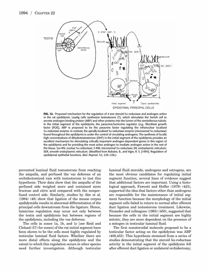
prevented luminal fluid testosterone from reachingthe ampulla, and perfused the vas deferens of anorchidectomized ram with testosterone to test thishypothesis. Their data show that the ampulla of theperfused side weighed more and contained morefructose and citric acid compared with the nonper-fused control side. Similarly, studies by Abe et al.(1984) (48) show that ligation of the mouse corpusepididymidis results in abnormal differentiation of theprincipal cells downstream from the ligature. Hence,lumicrine regulation may occur not only betweenthe testis and epididymis but between regions of the epididymis, including the vas deferens.
The cells in zones 1a, 1b, and 1c [see Reid andCleland (57) for zones] of the rat initial segment havebeen shown to be the cells most highly regulated bytesticular luminal fluid factors. Whether there aremore distal effects along the epididymis and theextent to which this regulation occurs in other speciesneed further investigation. Although testicular
luminal fluid steroids, androgens and estrogens, arethe most obvious candidates for regulating initialsegment function, several lines of evidence suggestthat additional factors are important. Using a histo-logical approach, Fawcett and Hoffer (1979) (423),supported the idea that factors other than androgensare responsible for the maintenance of initial seg-ment function because the morphology of the initialsegment cells failed to return to normal after efferentduct ligation and testosterone treatment. Likewise,Nicander and colleagues (1983) (450), suggested thatbecause the cells in the initial segment are highlymitotic, they are more dependent on the presence ofa mitogen in testicular luminal fluid.
The first nonsteroidal molecule proposed to be atesticular factor acting on the epididymis was ABP(408,455). This hypothesis stemmed from a series ofstudies demonstrating that the steroid 5α-reductaseactivity in the initial segment of the epididymis fellafter efferent duct ligation or unilateral orchidectomy;
1094 / CHAPTER 22
TESTIS
EPIDIDYMAL PRINCIPAL CELLS
Sertolicell
Seminiferoustubule
ABPFGF
X
X
T
T
TTc Tc
T
T
T
TLeydig
cellI
Initial segment Caput epididymidis
X (FGF, ABP, ?)
Microvilli
Microvilli
DHTDHT
DHT
DHT
DHTAR
AR
Nucleus
5α-R5α-R
5α-R
5α-R5α-R
5α-R
5α-R5α-R
5α-R
RoughER
ER, SER,?
ER, SER,?
FIG. 12. Proposed mechanism for the regulation of 4-ene steroid-5α-reductase and androgen actionin the rat epididymis. Leydig cells synthesize testosterone (T), which stimulates the Sertoli cell tosecrete androgen-binding protein (ABP) and other proteins into the lumen of the seminiferous tubule.In the initial segment of the epididymis, the paracrine/lumicrine regulator (e.g., fibroblast growthfactor [FGF]), ABP is proposed to be the paracrine factor regulating the infranuclear localized 5α-reductase enzyme. In contrast, the apically localized 5α-reductase enzyme (microsomal 5α-reductase)found throughout the epididymis is under the control of circulating androgens. The synthesis of locallyhigh concentrations of dihydrotestosterone (DHT) in the initial segment of the epididymis provides anexcellent mechanism for stimulating critically important androgen-dependent genes in this region ofthe epididymis and for providing the most active androgen to mediate androgen action in the rest ofthe tissue. 5ur-RN, nuclear 5α-reductase; 5-RM, microsomal 5α-reductase; ER, endoplasmic reticulum;SER, smooth endoplasmic reticulum. (Modified from Robaire, B., and Viger, R. S. [1995]. Regulation ofepididymal epithelial functions. Biol. Reprod. 52, 226–236.)
P515401_22 10/24/05 8:14 PM Page 1094

it could not be maintained by administration oftestosterone, even at very high doses. Luminal fluidABP was also proposed as the factor that enhances the function of ovine epididymal principal cells inculture (456), and appears to play a role in protein syn-thesis in the caput region (457). However, although γ-glutamyl transpeptidase IV mRNA is also con-trolled in a lumicrine manner in the initial segmentof the epididymis, ABP does not play a role in itsexpression (458). Instead, FGFs may be partlyresponsible for regulation of the expression of thisgene (458,459). It has been postulated that luminalfluid growth factors interact with their cognate recep-tors on the apical cell surface of initial segment cells,whereupon a signal transduction pathway is initi-ated (e.g., mitogen-activated protein kinase [MAPK],phosphatidylinositol-3 kinase); transcription factorsare then activated, presumably by phosphorylation,leading to transcription of target genes (452,459). Insupport of this hypothesis, soluble FGFs (2, 4, and 8)have been identified in testicular luminal fluid(458,459), and FGF receptor (FGFR)-1 IIIc (bothlong and short forms) has been identified in the prin-cipal cells in the initial segment. Further, the activ-ity of the MAPK pathway and the expression ofmembers of the polyomavirus enhancer activator 3(PEA3) transcription factor family (PEA3, ERM,ER81) are decreased after loss of testicular factors[(460); J. L. Kirby, B. V. Troan, and B. T. Hinton,unpublished observations]. Interestingly, several genes,including steroid 5α-reductase (461), expressed in therat initial segment contain multiple PEA3 familymember–binding motifs, and most of these genes areregulated by testicular luminal factors. In addition,neurotrophins (nerve growth factor, NT-3) and theircognate receptors, Trks and p75, may also play a rolein the regulation of initial segment function (S. Crenshaw and B. T. Hinton, unpublished obser-vations). Crucial to the coordinated functions of theFGFR and Trk receptor pathways may be adaptorproteins such as FRS2 and Shc (S. Crenshaw and B. T. Hinton, unpublished observation).
Another “factor” that has been postulated to reg-ulate initial segment function is the presence of sper-matozoa (462). At first, cells seem to be an unusual“ligand,” yet, on closer inspection, there may be asound basis for this hypothesis. Spermatozoa them-selves may not be the ligand, but it is possible thatthe ligands they carry and transport to the differentepididymal regions are important; many moleculesare known to bind to spermatozoa and potentiallyshed in the epididymis. Interestingly, testicular sper-matozoa stain positively for growth factor receptors(463); it is tempting to speculate that on entering theinitial segment, growth factors dissociate from thesperm surface and become available to stimulate
their cognate receptors on the epididymal apical cellsurface.
If it is assumed that the lumicrine mode of regula-tion occurs throughout the epididymis, there are several potential key points that emerge regardingthe regulation of the initial segment, and possibly theentire epididymis. First, the cells in the adult initialsegment are being constantly stimulated by FGFs,yet the cells are not proliferating; likewise, theMAPK pathway is constitutively switched on in thesecells. However, not all cells in the initial segmentshow MAPK pathway activity (J. L. Kirby et al.,unpublished observation). Hence, this raises a secondpoint that cells may be in communication with eachother such that they cycle between “on” and “off”states with respect to the response to growth factors.This may explain the characteristic checkerboardpattern seen when the tissue is stained for many different proteins; it would enable the initial seg-ment to function normally as a whole, yet with eachcell autonomous with respect to the regulation of itsrepertoire of target genes but susceptible to influenceby the state of its neighboring cells. This theory mayalso explain why cells do not proliferate in the initialsegment after constant stimulation with growth factors. At any time, there is sufficient stimulation ofgrowth factors to upregulate target genes, yet, becauseof complex negative feedback systems (e.g., involvingthe brain-derived neurotrophic factor/NT-3–Trkreceptors and adaptor proteins), those cells do not enterthe cell cycle pathway. However, the cells need to beresponsive to growth factors fairly rapidly after the“off” state because lack of stimulation by testicular fac-tors leads to apoptosis (S. Crenshaw and B. T. Hinton,unpublished observations). Future experiments willbe needed to test this hypothesis, but it will also beinteresting to know whether the recycling hypothesisfor many of the genes applies in all the cells through-out the epididymis. This hypothesis may also haveimplications with respect to the low rate of primaryor secondary cancers observed in the epididymis.
MAJOR PROTEIN FAMILIES IN THEEPIDIDYMIS AND THEIR REGULATION
General Principles
The division of the epididymis into segments hasmore than anatomical implications. For many years,the differential expression of proteins and mRNAalong the epididymis has become a hallmark of thistissue. Indeed, the large number of gene expressionprofiling studies (311,353,464–468a), proteomic analy-ses [reviewed in Dacheux and Dacheux (93), Gatti et al.(469), Chaurand et al. (470), and Umar et al. (471)],
THE EPIDIDYMIS / 1095
P515401_22 10/24/05 8:14 PM Page 1095

and immunohistochemical studies [e.g., (107,109,472–474)] have all revealed that nearly all moleculeshave characteristic longitudinal expression profilesin the epididymis of all species examined. Data on thegene expression profiles of the different segments of rat and mouse epididymis are available at the website for this article (www.ttuhsc.edu/cbb/faculty/cornwall/default.asp), and at www.wsu.edu/%7Egriswold/microarray/epididymis_dht/. In addition,tables of genes encoding distinct classes of proteins(enzymes, signaling, DNA binding) that exhibitregionalized expression in the epididymis are pro-vided in the supplemental material on this chapter’sweb site. Table 2 lists genes encoding secretory pro-teins that may play direct or indirect roles in thesperm maturation process, whereas Table 3 providesa list of genes that encode cellular proteins thatlikely perform regulatory functions.
Several comprehensive reviews describing the longitudinal profiles of both proteins and mRNAhave been published recently. Although all of thesestudies clearly demonstrate that there is a highlyregionalized activation and deactivation of gene
expression, the underlying mechanisms that allowfor this regulation are unclear. Regionalized expres-sion of transcription factors (see later) is beginningto provide some insight into this question, but therelationship between the luminal, endocrine, and cellular components that allow for such highlyregionalized expression of genes and proteins willrequire many more years to resolve.
Perhaps as challenging to understand is thecheckerboard pattern of expression observed in crosssections of individual tubules. This characteristicpattern is usually seen when a given protein ormRNA is first expressed along the duct or when theexpression terminates. It must be assumed that adja-cent principal cells either use different sensing mech-anisms for the triggering signal, or more likelyfunction as a syncytium to optimize coordinately thedegree of expression of a given gene or protein.Examples of this checkerboard pattern of expressionare shown in Fig. 13.
As might be expected based on their appearance andproposed functions, the different cells of the epididy-mal epithelium express different genes and proteins.
1096 / CHAPTER 22
Lu
Lu
Lu
Lu
P
P
P
P
P IT
B
B
n
nIT
Mv
A B C
FIG. 13. Representative examples of the checkerboard pattern of expression seen immunohisto-chemically along the epididymis. A: High-power light micrograph of tubules of the distal area of theinitial segment immunostained with anti-immobilin. Although many principal cells are reactive, theyshow a variable staining pattern. Several principal cells are intensely stained throughout their cytoplasm(arrows), whereas others are moderately or weakly stained (arrowheads); a few are unreactive (P).The luminal content (Lu) is well stained. IT, intertubular space. (Magnification ×350; modified fromHermo, L., Oko, R., and Robaire, B. [1992]. Epithelial cells of the epididymis show regional variationswith respect to the secretion or endocytosis of immobilin as revealed by light and electron microscopeimmunocytochemistry. Anat. Rec. 232, 202–220.) B: High-power light micrographs of the proximalinitial segment of the epididymis. Epithelial principal cells (P), nuclei of principal cells (n), basal cells(B), and lumen of the duct (Lu) are shown. Intense staining in the infranuclear region of the principalcells is designated by the arrowheads. Immunostaining reaction is confined to an oval region presentabove the nuclei of the principal cells (large arrows). (Magnification ×400; modified from Robaire, B.,and Viger, R. S. [1995]. Regulation of epididymal epithelial functions. Biol. Reprod. 52, 226–236.) C: Higher-power micrograph of portions of adjacent tubules of the caput epididymis immunostainedwith anti–SGP-2. Note intense immunoperoxidase staining reaction product over some epithelial principal cells (P). Other principal cells are moderately (arrowheads) or weakly (curved arrows)immunoreactive. A few principal cells are virtually unreactive (arrow). Note that the reaction productextends throughout the entire cell cytoplasm (i.e., basal, supranuclear, and apical regions), leavingonly the nucleus (n) unstained. The microvillar (Mv) border of the principal cells and sperm in thelumen (Lu) of the tubules are reactive. IT, intertubular space. (Magnification ×600; modified fromHermo, L., Wright, J., Oko, R., and Morales, C. R. [1991]. Role of epithelial cells of the male excurrentduct system of the rat in the endocytosis or secretion of sulfated glycoprotein-2 (clusterin). Biol. Reprod. 44, 1113–1131.)
P515401_22 10/24/05 8:14 PM Page 1096

Isolating each of the different cell types has been achallenge, but with the development of tools such aslaser capture microscopy, it should become possibleto resolve the profiles of genes and proteins expressedin each type of epithelial cell. Using immunohisto-chemistry, however, it has been possible to demon-strate the highly specific expression of certainproteins in principal, basal, clear, narrow, and halo
cells. Figure 14 presents examples of the selectivemarking of each of these cell types. To provide poten-tial tools to help identify cell types in vivo and invitro, representative proteins expressed selectively inindividual cell types of different regions of the epi-didymis are listed in Table 4 (supplemental materialon web site); some of these proteins are secreted,whereas others are retained in the cell.
THE EPIDIDYMIS / 1097
A B C
D E F
P
P
P
P
P
C
C C
C
Lu
lu
p
p
B
IT
IT
IT
i
FIG. 14. Cell-specific expression of proteins along the rat epididymis. Examples of immunolocalizationof proteins in each of the major cell types are presented. A: Principal cells (junction of thecaput–corpus of the epididymis) are immunostained for the glutathione S-transferase (GST) Yo subunit.The epithelial principal cells (P) are well stained, but basal (white arrow) and clear (C) cells are unreactive.Cytoplasmic droplets of spermatozoa (arrows) in the lumen are intensely reactive. (Magnification × 365;modified from Veri, J. P., Hermo, L., and Robaire, B. [1994]. Immunocytochemical localization of glutathione S-transferase Yo subunit in the rat testis and epididymis. J. Androl. 15, 415–434.) B: Apicalcells (arrowheads) and narrow cells (large arrows) of the proximal initial segment of the epididymisare intensely reactive when immunostained with anti-Yo GST antibody, whereas principal cells (P) andbasal cells (small arrows) are unreactive. Lu, lumen. (Magnification ×320; modified from Adamali, H. I.,and Hermo, L. [1996]. Apical and narrow cells are distinct cell types differing in their structure, distri-bution, and functions in the adult rat epididymis. J. Androl. 17, 208–222.) C: Basal cells. The only celltype of the epithelium of the corpus epididymidis that is intensely reactive is the basal cell (B) whenimmunostained with antibodies to the Yf (Yp) subunit of GST. These cells often show slenderprocesses (arrows) extending from the expanded main cell body along the basement membrane and,occasionally, toward the lumen (arrowheads). Principal cells (P) appear unreactive. Spermatozoa inthe lumen are unreactive. IT, intertubular space. (Magnification ×440; modified from Veri, J. P., Hermo, L.,and Robaire, B. [1993]. Immunocytochemical localization of the Yf subunit of glutathione S-transferaseP shows regional variation in the staining of epithelial cells of the testis, efferent ducts, and epididymisof the male rat. J. Androl. 14, 23–44.) D: Narrow cells. Initial segment of epididymides immunostainedwith anti–carbonic anhydrase II antibody. Intense reaction is localized over narrow cells. P, principalcell; IT, intertubular space; arrows, narrow cells. (Magnification ×350; modified from Hermo, L.,Adamali, H. I., and Andonian, S. [2000]. Immunolocalization of carbonic anhydrase (CA) II and H+ V-ATPase in epithelial cells of the mouse and rat epididymis. J. Androl. 21, 376–391.) E: Clear cells.Tubules of the distal area of the cauda epididymidis immunostained with anti-immobilin. Note thatseveral epithelial clear cells (C) are intensely reactive throughout their cytoplasm, whereas other cellsremain unreactive. IT, intertubular space; P, principal cells. (Magnification ×375; modified from Hermo,L., Oko, R., and Robaire, B. [1992]. Epithelial cells of the epididymis show regional variations withrespect to the secretion or endocytosis of immobilin as revealed by light and electron microscopeimmunocytochemistry. Anat. Rec. 232, 202–220.) F: Halo cells. Immunostaining with an antibody forhelper T lymphocytes (CD4) in the cauda epididymidis. Arrows indicate CD4+ cells. lu, lumen.(Magnification ×450; modified from Serre, V., and Robaire, B. [1999]. The distribution of immune cellsin the epithelium of the epididymis of the aging brown Norway rat is segment-specific and related tothe luminal content. Biol. Reprod. 61, 705–714.)
P515401_22 10/24/05 8:14 PM Page 1097

Functional Families
Receptors and Their Ligands
Androgen Receptor, Androgen-binding Protein, and Steroid 5α-Reductases. The limitingfactor in determining the action of testosterone oncells is not clear. The presence of both the androgenreceptor and an agonist are obviously necessary, butthere are several lines of evidence that indicate thatit is not primarily the absolute number of androgenreceptors present but rather the amount of the ligandthat is limiting. In addition, testosterone, a highlylipophilic molecule, is carried to sites distant fromwhere it is made by binding proteins such as ABP.Testosterone can act directly on the androgen recep-tor in some tissues, such as muscle; in other tissues,such as some brain nuclei, testosterone needs to bearomatized to estradiol to mediate its action throughthe estrogen receptor; whereas in still other tissues,it is converted to a 5α-reduced metabolite, DHT.DHT binds to the androgen receptor with higheraffinity than testosterone [reviewed in Blanchard andRobaire (475)]. The latter mode operates in manyandrogen-dependent tissues such as the epididymis,prostate, seminal vesicles, and skin. The rate-limitingenzyme in the pathway leading from testosterone toits 5α-reduced metabolites is steroid 5α-reductase(5α-R, EC 1.3.1.22).
Androgen Receptor. The first report that exoge-nously administered testosterone was converted toDHT and bound to a cytosolic protein in the epi-didymis (the androgen receptor) (476) was nearlycoincident with the discovery of a second androgen-binding component (ABP) in testis and epididymalcytoplasmic fractions (476a). Although both of theseproteins bind androgen with high affinity, they aredistinct molecules. For example, although both ofthese proteins have high affinity for DHT, the andro-gen receptor has a slow dissociation rate for DHT(t1/2 0°C > 4 days), whereas ABP dissociates at amuch faster rate (t1/2 0°C = 6 minutes). Androgenreceptors have been found in the epididymis of allspecies examined to date (rat, rabbit, dog, ram,monkey, and human) (476,476b–476h). In othermammalian tissues, a single gene for the androgenreceptor has been identified. In humans, the genespans approximately 90 kilobases, and it consists of 8 exons on chromosome Xq11-12. The coded proteinis approximately 110 kDa, and multiple transcriptsare produced with variable lengths of a polymorphicregion of CAG repeats in exon 1 (477,478). Recently,a variant of the human androgen receptor (AR45),lacking the entire region encoded by exon 1 of thegene but retaining the DNA-binding and steroid-binding domains, has been identified in heart and
muscle tissues (479). The potential presence of furthervariants in other tissues may help explain the tissuespecificity of androgen action.
There are relatively small changes in the concen-tration of mRNA for the androgen receptor along theepididymis (480,481). When changes are noted, theyoften reflect a decline in the concentration of androgenreceptors in the progression toward the distal end ofthe epididymis. For example, in humans, both themRNA protein for the androgen receptor are foundthroughout the tissue, with high levels in the distalcaput and proximal corpus and declining in the caudalregion, including the vas deferens. Interestingly theefferent ducts do not show any immunostaining,whereas the vas deferens is only weakly positive(476d). An exception is the ram, where androgenreceptor concentrations are similar in the caput andcauda epididymidis but are less than 25% of theselevels in the corpus epididymidis (482). In both thegoat and the rat, epithelial cells stain fairly evenly forandrogen receptors throughout the duct (483,484).
It is clear that principal cells stain heavily forandrogen receptors, but the relative concentrationsof this receptor in basal, clear, narrow, and apicalcells are not known. However, the immunolocalizationof androgen receptors in all epididymal epithelialcells has been reported for monkey, mouse, and goat(476h,485,486), whereas in the rat, it is apparent thatclear cells do not stain for the androgen receptor (426).Because of their origin, and as one might anticipate,halo cells do not stain for the androgen receptor inany species examined. Peritubular cells occasionallydisplay weak signals, whereas interstitial cells orblood vessels are consistently negative. When noted,the intracellular localization of the androgen receptoris distinctly nuclear.
Very few studies have focused directly on themechanism of action of the androgen receptor in theepididymis; however, with the advent of severalstable cell lines from this tissue (487–489), such studiesare likely to be undertaken in the near future. Thecharacteristics and mode of action of the androgenreceptor as a transcription factor (490,491) in theepididymis seem to resemble those in other tissues.However, androgen signaling, although essential, isnot sufficient for epididymis-specific gene expressionbecause the androgen receptor is expressed in manytissues. One mechanism proposed to explain thespecificity of transcriptional responses to androgen isthe tissue-specific combination of transcription factorsbinding to the promoter region of androgen-regulated genes (492). For example, the epididymaland prostatic lipocalin genes for epididymal retinoicacid–binding protein (E-RABP) and probasin are regulated by the androgen receptor. In the prostate,the transcription factor Foxa1 (forkhead box A1) is
1098 / CHAPTER 22
P515401_22 10/24/05 8:14 PM Page 1098
required for probasin gene expression (493). In theepididymis, Foxa2, but not Foxa1, is expressed, inter-acts with the androgen receptor, and binds to thepromoter of the E-RABP gene. Interestingly, over-expression of Foxa2 suppresses androgen activationof the E-RABP promoter, but increases activation ofthe prostate-specific promoter in an androgen-independent manner (494). Because Foxa1 and Foxa2expression is restricted to the prostate and epididymis,respectively, it suggests that these transcription factors have distinct and different action on androgenreceptor–regulated genes in different male reproductiveorgans. It is tempting to speculate that these differ-ential responses play a role in the different responsesof these two organs to oncogenic transformation.
Little is known about the regulation of expressionof the androgen receptor in the epididymis. In thegoat, circulating androgens seem to be sufficient toregulate the expression of both androgen and estrogenreceptors (486). In the rat, androgen withdrawal bythe administration of a luteinizing hormone–releasinghormone antagonist results in a more extensive andrapid decline in androgen receptors in stromal thanin epithelial cells; androgen replacement results in acomplete restoration of androgen receptors, indicatingthat stromal cells are more sensitive than epithelialcells to the regulation of androgen receptor concen-trations by androgen (426).
Androgen-binding Protein and Its Receptor. ABPis one of the first proteins shown to be synthesized bySertoli cells of the testis (495) and has been found inseveral species, including the rat (476a), rabbit (496),guinea pig (497), ram (476b), monkey (498), andhuman (499). It is secreted in both the luminal and basal compartments of the seminiferous tubules,but the overwhelming majority, more than 80%, isdirected toward the epididymis (500). Plasma sexhormone–binding globulin (SHBG) shares the sameamino acid sequence with ABP; these molecules differonly in the types of oligosaccharides associated withthem (501). The biological significance of these dif-ferences is not clear in light of the fact that SHBGand a glycosylated mutant have similar binding characteristics to sex steroids (502). ABP concentra-tions are highest in the caput and lowest in the caudaepididymal segments of most species examined,including primates; this distribution correlates wellwith DHT content (85,503,504). ABP is synthesizedand secreted by epididymal principal cells all alongthe epididymis (505), but in several species it is alsotaken up into the epididymal principal cells of theinitial segment and caput epididymidis (495,506,507)through a receptor-mediated process (505,508–511).Testicular ABP production is regulated by androgens(512), whereas that of the epididymis seems to beunder the control of a testicular factor (505).
Several functions have been ascribed to ABP. It hasbeen proposed to act as a carrier of androgens to theepididymis (513). In the principal cells, testosteroneis released from ABP and converted to the morepotent androgen, DHT, by the enzyme 5α-reductase.ABP has high affinity for 5α-DHT (Ka = 1.25 × 10⁹
M−1) and testosterone (Ka = 0.5 × 10⁹ M−1); rapid dissociation of testosterone from ABP (6 minutes) isconsistent with a role for ABP as a carrier moleculefor testosterone. In addition to its role as a carriermolecule, ABP has been suggested to be an androgensink in the seminiferous tubules of the testis (513)because the concentration of androgens in the semi-niferous tubules needs to be higher than that in theperipheral circulation (514–516) to maintain sper-matogenesis. ABP has also been proposed to regulatesteroid 5α-reductase type 1 (408,456,517).
To demonstrate unequivocally a function for ABPin spermatogenesis or in the epididymis would requirethe development of a null mutation model; this hasnot yet been accomplished. However, mice overex-pressing ABP (518) show fertility defects stemmingfrom spermatogenic dysfunction in the testis, whichresults in reduced numbers of spermatozoa in thecauda epididymidis. Although in these transgeniclines, ABP levels are increased in the epididymis, theconsequence of this for epididymal function is notevident.
Although it is clear that testicular ABP enters theprincipal cells of the proximal segments of the epididymis, the mechanism of internalization is stillnot fully understood. The presence of high-affinitybinding sites (Kass = 3.7 nM−1; 4.5 × 10¹¹ sites/mg protein) on the apical surface of epididymal principalcells and the demonstration that the number of thesereceptors is fivefold lower in the cauda than in thecaput of the epididymis have led to the proposal thatABP internalization is a receptor-mediated processin both rats and monkeys (504,509,510).
Steroid 5α-Reductases. The selective presence of radioactive DHT in epididymal cell nuclei afterinjection of radiolabeled testosterone (519), and thedemonstration using micropuncture studies that,beyond the efferent ducts, the predominant androgensin epididymal fluid are 5α-reduced metabolites oftestosterone [reviewed in Turner (85)], are two of thekey findings establishing the central role of DHT,and hence steroid 5α-reductase, in mediating andro-gen action in the epididymis. The discovery thattestosterone is rapidly converted by the epididymisto 5α-reduced products was first made almost simul-taneously by two different groups (520,521). The epi-didymis is one of the tissues with the highest level ofactivity anywhere in the body (521).
In the mouse, rat, dog, monkey, and human, two 5α-reductases (isozymes), with different tissue
THE EPIDIDYMIS / 1099
P515401_22 10/24/05 8:14 PM Page 1099
distributions, have been identified. Although severaltheories have been put forward regarding the relativerole of these two isozymes (522,523), there are fewsolid data to allow resolution of their relative roles.The consequences on the female of a null mutation in5α-reductase 1 have been described (524). Null muta-tions for these two genes are compatible with malefertility; however, no detailed studies on spermatozoaor the epididymis of mice carrying null mutations forthese genes have yet appeared. To understand theregulation of this enzyme activity in the epididymis,it is necessary to determine where and how the proteins are expressed, as well as the translationand, ultimately, the factors regulating transcriptionof the genes for this enzymatic activity.
Enzyme activity is expressed in a striking positionalgradient as well as in different subcellular fractionsin the adult rat epididymis (455,525). The activityassociated with the nuclear fraction is highest in theinitial segment, where it is higher than in any othermale reproductive tissue, and declines dramaticallyin more distal segments of the tissue. The hormonalregulation of epididymal 5α-reductase activity iscomplex [reviewed in Robaire and Viger (517) andEzer and Robaire (526)]. This was the first enzymeshown to be regulated by testicular factors as well asby circulating testosterone, depending on which segment and subcellular fractions were examined.Whereas nuclear fraction epididymal 5α-reductaseactivity is regulated in a paracrine or lumicrinemanner by a substance directly entering the epi-didymis through the efferent ducts, microsomal 5α-reductase activity is found throughout the epi-didymis but is present at a lower level and is regu-lated by circulating androgens.
The two mRNAs for 5α-reductase types 1 and 2 arethe products of separate genes (527,528). The type 1mRNA isozyme is most abundantly expressed in theinitial segment of the epididymis, with a positionalgradient that is similar to the enzyme activity. Studiesof the endocrine and developmental regulation of the5α-reductase type 1 mRNA show that (a) orchidectomyresults in a decrease in type 1 mRNA levels in all epi-didymal segments; (b) after orchidectomy, high-doseexogenous testosterone maintains 5α-reductase type 1mRNA levels at control levels in all regions of the epi-didymis except the initial segment; (c) unilateralorchidectomy and efferent duct ligation cause a dra-matic decrease in type 1 5α-reductase, selectively inthe initial segment of the epididymis; and (d) onlythe type 1 transcript is developmentally regulated.Therefore, with regard to enzyme activity, the primaryregulator of 5α-reductase type 1 mRNA expression inthe initial segment is a paracrine/lumicrine factor oftesticular origin [reviewed in Robaire and Viger (517)and Ezer and Robaire (526)].
The 5α-reductase type 2 mRNA is found in highconcentrations throughout the epididymis, with asomewhat elevated concentration in the caput epi-didymal segment (523). Although 5α-reductase type2 mRNA is present in abundance in the epididymisrelative to the type 1 mRNA, its activity as a func-tional enzyme appears to be relatively poor. Unlike thedevelopmental pattern of 5α-reductase type 1 mRNAexpression, characterized by dramatic increasesoccurring just before the first appearance of sperma-tozoa in the epididymis, 5α-reductase type 2 mRNAexpression does not show any significant develop-mental changes in any epididymal segment (523).Efferent duct ligation results in a dramatic decreasein 5α-reductase type 1 mRNA levels in the initial segment, but causes a near doubling in type 2 mRNAlevels. This differential regulation of 5α-reductasetype 1 and type 2 transcripts in one tissue has notbeen observed in other rat tissues such as theprostate (523).
In comparing the regulation of epididymal 5α-reductase enzyme activity and the expression ofits mRNAs, it is apparent that a potential regulatorystep is at the level of the protein itself. Post-transcriptional regulation has been proposed for severalother epididymal proteins (79,135,345,466,529,530).In the adult rat, 5α-reductase type 1 protein expressionis intensely immunolocalized in discrete lobules ofthe proximal initial segment of the epididymis; asharp decline in staining intensity occurs betweenthe proximal initial segment and its adjacent region,followed by a progressive decrease in intensity beyondthat point [reviewed in Robaire and Viger (517)]. In all epididymal regions, 5α-reductase immunoreac-tive protein is localized specifically in the epithelialprincipal cells and is uniquely associated with mem-branous cytoplasmic elements; the intracellularlocalization of the 5α-reductase protein changes withmovement down the epididymis. Savory et al. (1995)(531) have localized type 1 5α-reductase to the nuclearenvelope of both transfected cells and rat tissues.The infranuclear localized form of the 5α-reductasetype 1 protein is regulated in a similar fashion to thetype 1 mRNA and enzyme activity, that is, by aparacrine factor of Sertoli cell origin (517,532).
The genomic regulation of the expression of both5α-reductase isozyme genes is still poorly understood.Cloning and characterization of the proximal 2.2 kilo-bases of the 5′ upstream region of rat 5α-reductasetype 2 have revealed that there are regions withstrong repressor and enhancer activity; potentialtranscription factors (Sp1 and Sp3) regulating geneexpression have been identified and immunolocalizedto principal cells of the epididymis. The promoterregion of rat 5α-reductase type 2 is GC-rich with anoncanonical TATA box located upstream of the
1100 / CHAPTER 22
P515401_22 10/24/05 8:14 PM Page 1100

transcriptional start site (461). The potential role ofthis GC-rich region is intriguing in light of the obser-vation that the methylation pattern of the two genesappears to differ not only between each other butbetween reproductive and nonreproductive tissues (533). The genes for 5α-reductase types 1 and 2are also differentially methylated in lymphocytes fromnormal and 5α-reductase–deficient patients (534).
Estrogen Receptors and Cytochrome P450Aromatase
Estrogen Receptors. Starting in the 1970s, sub-stantial evidence accumulated for the direct action ofestrogens on androgen target tissues. Using bindingstudies, specific, high-affinity cytosolic and nuclearestrogen-binding proteins, presumably receptors, wereidentified by a number of investigators in mice (535),rats (536–538), guinea pigs (539), rabbits (540–543),dogs (472,544), rams (545), monkeys (546,547), andmen (548). Through a comprehensive series of studies,Danzo’s group established the presence of estrogenreceptors in the rabbit epididymis and demonstratedboth its developmental (549) and regional distribution(539); however, enthusiasm for further studies onthe estrogen receptor in the epididymis diminishedwhen this same group demonstrated the presence ofa protease, in the adult, that cleaves the DNA bindingregion of the estrogen receptor (550).
The histolocalization of tritiated DHT and estradiolin the adult mouse epididymis revealed differentialdistribution of grains over nuclei in different seg-ments of the excurrent duct system, as well as overdifferent cell types (535,551). These studies con-firmed the earlier binding experiments and provideda more detailed localization of the putative receptors.Very strong labeling of the efferent ducts and initialsegment of the epididymis was noted, with lesserbinding in the more distal segments of the epididymis;nuclei of clear cells were more densely labeled withestradiol than with DHT, in sharp contrast to whatwas seen over nuclei of principal cells. Based on suchdata, and before the studies with null mutant mice,specific functions for estradiol in the epididymis wereproposed (1,552).
With the discovery of the existence of two estrogenreceptors (ERα and ERβ), and the availability of specific antibodies for these receptors as well as micewith null mutations for either or both receptors, ourunderstanding of the localization of estrogen receptorsand their potential role(s) has grown rapidly[reviewed in Hess (553)]. ERα and ERβ are remarkablysimilar to one another in some respects (e.g., 95%identity in the DNA binding domain), but they haveindividual characteristics (e.g., <55% homology inthe ligand binding domain) that clearly indicate func-tional differences (554,555). The two isoreceptorshave different binding affinity for estrogenic and
antiestrogenic compounds (537,556), different tissuedistributions (557), and different response elements(558,559).
Although there are some conflicting data on theimmunolocalization of ERα and ERβ along the effer-ent ducts and the epididymis, a general pattern isemerging. In the mouse, the epithelium of the effer-ent ducts show strong ERα and weaker ERβimmunoreaction (553,560,561). The initial segmentof the epididymis shows some reactivity for ERα, pri-marily in narrow, apical, and some basal cells (485);in the caput epididymidis, principal cells and clearcells stain positively, whereas in the distal regionsonly clear cells are immunoreactive. In contrast, ERβimmunoreactivity is clearly observed along the entireepididymis, with more intense staining in the moredistal regions (corpus and cauda) (553,562). In therat, in spite of some earlier controversies relating tothe method of section preparation and antibody usedfor immunolocalization studies (551,563), it appearsthat ERα and ERβ immunolocalize in patterns simi-lar to those of the mouse. The presence of ERα andthe strong immunoreactivity of ERβ all along the epi-didymis have been confirmed using RT-PCR data(551,564) [reviewed in Hess (553)]. In monkeys,baboons, and humans, both forms of the estrogenreceptor are present along the epididymis (565–568).
The first clear insight into the functional role ofERα in the efferent ducts and epididymis camethrough null mutation studies. The ERα knockoutmouse was developed by Lubahn et al. (1993) (569)and is infertile in spite of the presence until pubertyof apparently normal seminiferous tubules; in theadult, testicular atrophy becomes evident (570).Through an elegant series of studies, Hess’s group(553,571,572) has demonstrated that the cause ofinfertility is back-pressure atrophy of the seminifer-ous tubules owing to the inability of the efferentducts and initial segment of the epididymis to reab-sorb the large volume of fluid secreted by the testis.This reabsorption process is under the control ofestradiol, acting as a paracrine regulator of the efferentducts. Further, it has been proposed that the actionof estrogen on fluid reabsorption is mediated by thecystic fibrosis transmembrane conductance regulator(CFTR) receptor (573). In contrast to the clear role ofthe ERα receptor, function of ERβ in the male repro-ductive tract awaits further investigation becausethe ERβ knockout mouse is fertile and appears tohave a normal testis and epididymis (574).
Cytochrome P450 Aromatase. An ongoing sourceof ligand is necessary to activate estrogen receptors.Cytochrome P450 aromatase, localized to the smoothendoplasmic reticulum, is a terminal enzyme thatirreversibly catalyzes the conversion of testosteroneto estradiol. Both the absence of aromatase and an
THE EPIDIDYMIS / 1101
P515401_22 10/24/05 8:14 PM Page 1101

excess of estrogen result in male infertility [reviewedin Carreau (575)]. There are several sources of estradiol in the adult male reproductive tract. In thetestis of adults, both Leydig cells (576,577) and germcells (578) have been reported actively to synthesizeestradiol. In germ cells, the enzyme is localized in theGolgi of round spermatids and throughout the cytoplasm of elongating and late spermatids[reviewed in Hess (553), Carreau (575), and O’Donnellet al. (579)].
In the epididymis, aromatization of the very highconcentrations of testosterone to produce estradiol,particularly in the proximal segments, could occureither in the luminal or the epithelial compartments.The presence of cytochrome P450 aromatase in epididymal spermatozoa has been demonstrated inseveral species, including mouse, rat, bear, and rooster(578,580–582). The enzyme is localized to cytoplasmicdroplets, and the staining becomes less intense asspermatozoa traverse the epididymis (583).
Early investigations of the presence of cytochromeP450 aromatase in epididymal epithelial cells indi-cated that this activity was absent (584); however, nodirect assessment of the enzyme was done in thesestudies. In several subsequent investigations, directdetection of the P450 aromatase mRNA, protein, andenzyme activity (formation of estradiol from testos-terone), as well as the ability of specific aromataseinhibitors to inhibit estradiol formation, all led to theconclusion that epididymal principal cells, but notthe smooth muscle cells surrounding the duct, havethe ability to synthesize estradiol from testosteronein a number of species, including the rat (585), monkey(586), and human (587).
Retinoic Acid Receptors. Retinoids are highlypotent molecules with pleiotropic action in a widerange of biological processes during development andin the adult (588). Vitamin A deficiency results inboth keratinizing and nonkeratinizing squamousmetaplasia of numerous epithelia, including the epididymal epithelium (589,590).
Given the importance of retinoids, it is not sur-prising that a complex system has evolved to controlretinoic acid biosynthesis and metabolism (591–593).Because retinoids are hydrophobic molecules insolu-ble in water, they require chaperones to ensureproper storage, transport, and uptake in the tissues.These chaperones, the retinoid-binding proteins, aremembers of either a large superfamily of small intra-cellular proteins that includes numerous fattyacid–binding proteins or another superfamily ofsecreted proteins, the lipocalins, which bindhydrophobic molecules. There are four intracellularretinoid-binding proteins that are members of thefatty acid–binding protein family (cellular retinol-binding protein [CRBP] I and II, and cellular retinoic
acid–binding protein [CRABP] I and II), and twoextracellular retinoid-binding proteins that aremembers of the lipocalin family (retinol-binding proteins and epididymal retinoic acid–binding protein [E-RABP]).
Within the nucleus, two classes of nuclear retinoicacid receptors, RAR and RXR, have been identifiedand cloned. Each receptor class consists of threereceptor subtypes, α, β, and γ, and their isoforms.RXRs form heterodimers with RARs that arethought to be the functional units transducing theretinoid signal. The binding of RARs/RXRs toretinoic acid response elements modulates geneexpression and is likely to be responsible for theobserved biological effects of retinoids (594).
Most of the components of the retinoid signalingpathway have been identified in the epididymis.Retinoids were quantified in the rat epididymis byhigh-performance liquid chromatography (HPLC)followed by derivation of the HPLC-purifiedretinoids to confirm their identities (595). In the epi-didymal tissue, the major retinoids identifiedincluded retinol, retinyl ester, all-trans retinoic acid,and 9-cis-retinoic acid (9-cis-RA). The concentrationof retinol and retinyl esters decreased from the caputto the cauda epididymidis from 2.7 to 1.7 nmole/g oftissue and from 6.2 to 1.4 nmole/g tissue, respec-tively. The active retinoids, all-trans retinoic acidand 9-cis-RA, showed an inverse gradient, withlowest concentrations in the caput (all-trans retinoicacid: 13 pmole/g tissue; 9-cis-RA: 2.9 pmole/g tissue)and highest concentrations in the cauda (all-transretinoic acid: 35 pmole/g tissue; 9-cis-RA: 6.8 pmole/gtissue). Using a bioassay, levels of 7 pmole/g of tissuehave been reported in the murine epididymis (595a).
The CRBPs and CRABPs are also present in theepididymis, but their localization is regionalized.CRBP is most abundant in the columnar epithelialcells of the proximal caput epididymidis of the rat;this tissue has one of the highest CRBP expressionsin the body (596–598). The mouse has the highestconcentrations of CRBP in the proximal caput epithe-lium, but also has significant levels in the epitheliumthroughout the epididymis. CRABP II is found in theepithelium of the cauda epididymidis coexpressedwith CRBP in both the mouse and the rat, whereasCRABP I is present in the smooth muscle of thedistal cauda epididymidis (599).
The retinoid signaling pathway in the epididymisincludes the retinoic acid receptors and the retinoicacid carrier protein E-RABP, synthesized and secretedinto the lumen in the distal caput and found in thelumen from the caput to the cauda epididymidis. Inthe rat epididymis, RARα, β, and γ transcripts havebeen detected by RT-PCR (600). In situ hybridizationand immunolocalization revealed region-specific
1102 / CHAPTER 22
P515401_22 10/24/05 8:14 PM Page 1102

patterns of RARα mRNA and protein expression (601).The highest levels of RARα transcripts and RARαimmunostaining were seen in the proximal caput epididymidis. The levels of RARα transcripts andRARα protein staining were higher in the initial seg-ment, proximal caput, and distal cauda. The lowestlevels were seen in distal caput, corpus, and proximalcauda epididymidis.
It is clear that a remarkable retinoid homeostasissystem has evolved to permit the transport of thesehydrophobic molecules and their metabolism, extra-cellular and intracellular sequestration, and deliveryto the nuclear chromatin for optimum retinoid sig-naling in the epididymis. However, knowledge of thepattern of expression of the various components ofthe retinoid biosynthesis and metabolism pathway,although suggestive, is not sufficient to deduce theprecise role of these regulatory factors or the site ofaction of retinoic acid in the epididymis. Vitamin Adeficiency has pervasive effects, affecting manyorgans during prenatal and postnatal developmentand adult life. Reproduction is affected both at thelevel of the testis, where spermatogenesis is arrested,and at the level of the epididymis, where the epithe-lium develops squamous metaplasia. Because of thisdual effect, it is difficult to ascertain from vitamin Adeficiency studies the physiological role of retinoidsin epididymal function.
Gene inactivation has produced surprising results.The transthyretin null mutant has a virtual loss ofserum retinol-binding protein, yet the mice seemnormal (602). CRABP I and CRABP II null mutantmice also appear normal (603,604). The doubleknockouts of CRABP I and CRABP II have a higherdeath rate at 6 weeks than the wild-type mice, but thesurvivors appear normal (604). Although each retinoid-binding protein separately appears to be dispensable,the lack of phenotype may simply reflect that redun-dant mechanisms are built into the organism.
The functional redundancy is well illustrated inthe case of the retinoic acid nuclear receptors, ofwhich six RXR isoforms and six RAR isoforms havebeen identified (605). Null mutations of RARβ,RARα1, RARβ2, or RARγ2 appear normal. Null muta-tions of all RARα isoforms or all RARγ isoforms dis-play some of the defects of the postnatal vitamin Adeficiency syndrome. Only double-null mutants ofthe RAR subtypes (RARα/γ, RARα/β2, RARα1/β2,RARβ2/γ) reproduce most of the features of vitamin Adeficiency syndrome. Null mutation of RXRα inducessome abnormalities, but double inactivation of RXRαand RARβ or RARγ increases the severity of the RXRphenotype. It appears that even though the differentreceptor isoforms are not truly functionally equivalent,they can substitute for one another under certainconditions. This functional redundancy may not be
surprising in any gene family that has been conserved throughout vertebrate evolution and hasevolved by duplications from an ancestral gene.
Even with these caveats, an epididymal phenotypehas been observed in transgenic mice after disruptionof the RAR gene. Expression of a dominant negativemutant of RARα induced loss of organization of thecolumnar epithelium lining the cauda and its trans-formation by squamous metaplasia. The aberranttransformation resulted in blockage or rupture of theduct with concomitant inflammatory response and,ultimately, infertility (606). The RARα null mutantmice display not only aspermatogenesis, but vacuoliza-tion of the epididymal epithelium (607), and the RARα/γdouble-null mutants display either severe dysplasia orcomplete agenesis of the epididymis and the vas deferens (608).
In summary, given the pathological changes seenin the epididymal epithelium in vitamin A deficiency,the epididymal phenotypes seen in RAR null mutantmice, and the presence of most of the components ofthe retinoid signaling pathway, it is highly likely thatretinoids play a regulatory role in epididymal function.
Oxytocin Receptors. The first demonstration ofthe presence of oxytocin receptors in the epididymiswas by Maggi et al. (1987) (170). Since then, severalreports on the localization, distribution, regulation,and potential functions of these receptors in a varietyof species have appeared (169,171–173).
Although in the marmoset monkey, the oxytocinreceptor seems to be primarily expressed in peritubularcells of the epididymis, in the human, macaquemonkey, and ram, its localization is in the epididymalepithelium and varies along the duct (169,172). Theexpression in principal cells is seen only in the initialsegment of the epididymis of primates and has acheckerboard appearance; throughout the rest of theduct, it is confined to peritubular cells of the epi-didymis (172). In the ram, however, oxytocin receptorsare found in both principal and basal cells throughoutthe epididymal duct, whereas peritubular staining isseen only in the more distal segments (173).
Two functions have been ascribed to the oxytocin/-oxytocin receptor system in the epididymis. As dis-cussed previously, the first is that of promotingcontractility of the duct, as demonstrated by both invitro (174) and in vivo (179) studies, thus promotingsperm transport through the epididymis (187). Thesecond function of this system involves the promotionof the formation of DHT by stimulation of 5α-reductaseactivity in the initial segment of the epididymis(609). This function is of particular interest, giventhe very high 5α-reductase enzymatic activity in thissegment of the epididymis and the presence of oxy-tocin in the luminal fluid of the ram and the rat(610,611). The mechanism for this type of stimulation
THE EPIDIDYMIS / 1103
P515401_22 10/24/05 8:14 PM Page 1103

is not clear but has been postulated to be mediatedby phosphorylation and activation of existing 5α-reductase due to tyrosine kinase activation through acoupling to the oxytocin receptor by Gi signal trans-duction (612).
Growth Factor Receptors. FGF receptors(FGFR), platelet-derived growth factor (PDGF) recep-tors, and vascular endothelial growth factor (VEGF)receptors have all been identified in the epididymis.For FGFR, two splice variants, FGFR-1 IIIc (longand short forms), have been identified in the principalcells of the initial segment (459). Although all fourmajor splice variants of FGFR (1 through 4) are pres-ent in initial segment tissue, only the FGFR-I IIIcvariants are present in principal cells. The remainingsplice variants are presumably localized to theendothelial and interstitial cells. FGFs 2, 4 and 8,present in rete testis and epididymal luminal fluid(458,459), are potential ligands for the apically placedreceptor (S. Crenshaw and B. T. Hinton, unpublishedobservations).
PDGF α and β receptors and their ligands (PDGF-Athrough D) have been localized in the rat and mouseepididymis (613). In view of the specific localizationof PDGF-A in the epithelium and the PDGF α recep-tor in the mesenchyme, the authors suggested thatthe PDGF-A/α receptor system may be involved inepithelial–mesenchymal interactions during epididymaldevelopment. In the adult epididymis, PDGF-A andB and α and β receptors were identified in the epithe-lium only. Interestingly, in PDGF-A null mutants, anepididymal phenotype was not observed duringdevelopment, but epididymal epithelial disruptionwas observed at postnatal day 25 (613).
VEGF and its receptors are highly localized inboth the testis and epididymis, with fms-like tyrosinekinase (flt-1) and fetal liver kinase (designated askinase insert domain–containing receptor [KDR])VEGF receptors being identified by immunohisto-chemistry in the ciliated cells of the efferent ductsand vascular endothelial cells, respectively, of thehuman epididymis (614). Flt-1 was also observed inthe lymphatics. Furthermore, addition of VEGF28 to epididymal tissue in culture resulted in endothelialfenestrations and opening of interendothelial junc-tions. Because the ligand, VEGF, was localized to theperitubular and ciliated cells of the efferent ductsand to the peritubular and basal cells of the epi-didymis, it was suggested that it may play aparacrine role in the permeability of blood vesselsthrough KDR and lymphatics through flt-1 (614).Overexpression of VEGF in the testis and epididymisresults in male infertility due to spermatogenic arrestand increased capillary density in the testis and epididymis (615).
Although not specifically expressed in the epididy-mal epithelium, the hepatocyte growth factor receptor,c-met, is localized to epididymal spermatozoa, withthe ligand, hepatocyte growth factor, being secretedinto the lumen by the epididymal epithelium. Theputative action of hepatocyte growth factor is main-tenance of sperm motility (616). Interestingly, thehepatocyte growth factor–like protein receptor, RonmRNA, which is a receptor tyrosine kinase, isexpressed in the epididymal epithelium (617).Additional growth factor–binding proteins andgrowth factor receptors that are expressed in epi-didymal epithelial cells include insulin-like growthfactor–binding protein-rP1/mac25 (618), latent trans-forming growth factor-β–binding protein 2 (619), epi-dermal growth factor receptor (620), and growthhormone receptor–binding protein (621).
c-Ros. c-Ros is an orphan tyrosine kinase receptorthat has received considerable attention since it wasshown that c-Ros null male mice were infertile (40).The kinase and extracellular domains of the c-Rosgene show similarities to the Drosophila sevenless(sev) tyrosine kinase receptor (622). The sev genewas originally discovered in a Drosophila mutantthat failed to express the R7 photoreceptor cell in theommatidium (623,624). Further studies demon-strated that the adjacent R8 photoreceptor cellexpressed boss, the ligand for sev. Interactionbetween these two cell types was crucial for thedevelopment of these photoreceptor cells. After thiscell interaction, a signal transduction cascade involvingRas and activation of downstream target genes wasinitiated in the R7 photoreceptor cell. Interestingly,ectopic expression of boss or of constitutively activeforms of sev causes the formation of ectopic R7 cells(625,626). The overall sequence homology between c-Ros and sev is quite low, but there are some veryinteresting structural similarities between the two.For example, (a) both genes encode a large extracel-lular domain and a kinase domain; (b) sequencehomology extends into the putative extracellularligand-binding domain; (c) there is a short insert offive to seven amino acids in the same position in thekinase domain of both genes that is not found inother receptor tyrosine kinases; (d) there are severalcommon exon–intron junction points between thetwo genes; (e) the kinase domains of both genes are70% homologous (622,627–630); and (f) particularlystriking is the analysis of Springer (1998) (631), whoshowed a colinearity and spacing of β-propeller andepidermal growth factor-repeat structures in c-Rosand sev. Interaction of ligand with β-propellers hasbeen shown for other proteins (632). The ligand ofsev is bride of sevenless (boss) (626,633), a seven-transmembrane G-coupled protein kinase, which has
1104 / CHAPTER 22
P515401_22 10/24/05 8:14 PM Page 1104

signal transduction properties of its own. Presumably,a boss homolog exists in mammals if c-Ros has a sim-ilar function to sev.
Although c-Ros is expressed in several tissuesthroughout development, including the lung, kidney,and epididymis, it remains highly expressed in theinitial segment of the adult mouse epididymis(40,634). A clue to the function of c-Ros came from c-Ros knockout mice: the animals displayed anunderdeveloped initial segment and male infertility.The infertility phenotype was due to spermatozoahaving an angulated flagellum at the end of the mid-piece (635); it was suggested that this defect wascaused by the failure of spermatozoa to regulate theircell volume (635). This infertility phenotype was alsoshown to be cell autonomous; the defect in the spermphenotype was due to the passage of normal sperma-tozoa through the underdeveloped initial segment,and not to some inherent defect in the spermatozoathemselves. Further, the infertility phenotype wasobserved if either the kinase domain or the extracel-lular domain of c-Ros was lacking (40).
Although the functional role of c-Ros is notknown, it has been suggested that it plays a role inthe regulation of cell volume (636,637), and althoughc-Ros itself may not have a direct effect on cellvolume regulation, gene expression profiling suggeststhat several osmolyte transporters are compromisedin the initial segment of the c-Ros null animals (467).It also appears that at least inositol and glutamatesperm concentrations are lower in the c-Ros knock-out mice compared with controls, even though thereare no differences in luminal fluid concentrations ofthe two osmolytes between the two groups (377).Whether the lower intracellular levels of theseosmolytes in spermatozoa is the cause of the infertilityphenotype remains to be determined. Adding to thecomplexity of the problem is that the infertility phe-notype may be related to an underdeveloped initialsegment rather than directly to c-Ros. Because theadult mouse initial segment still expresses c-Ros, thechallenge will be to silence this gene in the adult andthen examine the fertility phenotype. This experimentneeds to be done, especially if c-Ros and its down-stream signaling pathways are to be considered aspotential targets for the development of a male contraceptive. An additional important finding is thatthe adult human epididymis also expresses c-Rosalong its length (638).
In an attempt to identify downstream targets of c-Ros, Keilhack and colleagues (2001) (639) used ayeast two-hybrid assay and identified several proteinsthat associated with the kinase domain of c-Ros.These included the phosphatase SHP-1 (Src homologyregion 2–containing protein tyrosine phosphatase-1),
phospholipase Cγ, the SH2 domain of c-Abl, Grb2,the phosphoinositide-3 kinase subunit p85α and theSH2 domains of SHP-2. To demonstrate the poten-tial importance of the SHP-1 phosphatase in malefertility, viable motheaten (mev) mice were tested forfertility because these mice contain a mutation in theSH2 domain of SHP-1. The male mice were infertile.Hence, downstream targets of c-Ros are clearly excel-lent candidates for male contraceptive development.Although Keilhack et al. (2001) (639) claim that theinfertility phenotype is similar to that seen in the c-Ros knockout animals, the authors do not presentconvincing data that there is a reduction in epithelialcell height in the initial segment of the SHP-1 knock-out animals, similar to that seen in the initial segment of c-Ros knockout mice. Their data suggestthat the initial segment has developed normally inthis knockout. Hence, although male infertility isobserved in both the c-Ros and SHP-1 knockouts, themechanisms by which infertility is achieved in eachcase are probably different.
HE6. The HE6 gene encodes a novel human epididymal protein that comprises a seven transmem-brane component with a large extracellular domainand shows high homology to the secretin/pituitaryadenylate cyclase–activating polypeptide (PACAP)superfamily of G-protein–coupled receptors (GPCRs)(640). In view of the large ectodomain, HE6 has alsobeen considered to be a member of the LNB-TM7proteins (family-B GPCR–related TM7 receptorswith long N-terminal extensions) (641). This largedomain also contains a GPCR proteolysis site, andtherefore HE6 may exist as a two-subunit receptor.HE6 is located on the human X chromosome, and bothrat and mouse homologs of HE6 have been cloned(640,641). Immunolocalization studies show humanand mouse HE6 localized to the apical surface ofhuman epididymal cells, with staining more pro-nounced in the efferent ducts–caput region comparedwith the distal epididymal regions. A mosaic patternof staining was also observed, as is often seen withlocalization of other epididymal proteins. Mouse HE6 was localized to the apical cell surface buthighly restricted to the initial segment region (641).The function of HE6 is yet to be determined,although a knockout study suggests that the proteinmay be involved in fluid reabsorption in the efferentducts (642).
Other Receptors. In addition to those receptorsdiscussed in the preceding sections, the epididymisalso expresses orexin receptors (643–645), the urokinase-type plasminogen activator receptor (646),apolipoprotein E receptor-2 (647), adrenomedullinreceptors (648), purinergic receptors (649), andguanylyl cyclase C receptor (650).
THE EPIDIDYMIS / 1105
P515401_22 10/24/05 8:14 PM Page 1105

Transcription Factors
An understanding of transcriptional regulation ofepididymal genes has been limited to a few genes[reviewed in Robaire et al. (90), Cornwall et al. (91),Kirchhoff (92), Rodriguez et al. (651), Rao andWilkinson (652), and Suzuki et al. (653)]. However,in these studies, several key transcription factorshave been identified that may play an important rolein the tissue-specific, region-specific, or cell-specificexpression of epididymal genes. For the sake of sim-plicity, steroid hormone receptor coregulators arenot discussed here, but are discussed in the sectionon Receptors and Their Ligands.
PEA3 Family. One of the first major epididymaltranscription factors to be examined was poly-omavirus enhancer activator 3 (PEA3), which is amember of the Ets transcription factor family. Thisfamily is characterized by an 85–amino acid Etsdomain (654) that encodes for a helix-turn-helixDNA binding motif. It is also part of a subfamily,referred to as the PEA3 family, that comprisesPEA3, ERM, and ER81 (655–660). The consensussite for PEA3 is 5′-AGGAAG-3′, with GGAA beingthe core binding motif for the Ets family. Xin et al.(1992) (655) were the first to show expression ofPEA3 in the epididymis, and later studies by Lan etal. (1997) (460) and Drevet et al. (1998) (661) clearlyshowed a role for members of the PEA3 family in regulating GGTIV and GPX5 gene expression in theepididymis. Standard in vitro promoter analyses ofboth GGTIV and GPX5 revealed that PEA3 acts asboth a repressor and an activator of transcription.This was confirmed more recently by Kirby et al.(2004) (662) using a novel in vivo electroporationtechnique. There were differences in promoter activitybetween the in vivo and in vitro studies when thesame constructs were used, highlighting the impor-tance of performing such an analysis under as near-physiological conditions as possible. The arrangementof PEA3 binding sites along the GGTIV promotermay be unique to this gene because of the absence ofbinding motifs (e.g., AP-1) that are known to regulatethe activity of PEA3 in many other gene promoters(e.g., urokinase plasminogen activator and matrixmetalloproteinase) (663,664).
There appears to be redundancy among the PEA3family members because SouthWestern analysis (460)showed that multiple proteins identified as PEA3,and possibly ERM and ER81, bound to GGTIVmRNA PEA3 binding motifs. Further, PEA3 knock-outs had a normal epididymal phenotype, supportingthe idea that ERM and ER81 may take over the func-tions of PEA3. PEA3 knockout male mice are infertile,but their infertility is due to impotence or failure toejaculate spermatozoa (665). Interestingly, the
promoters of several genes that are highly expressedin the initial segment, including 5α-reductase type 2(461), have PEA3 family binding motifs, and eachgene, including PEA3 family members, is regulatedspecifically by testicular factors. It will be importantto generate initial segment-specific PEA3 familymember knockouts to determine whether PEA3family members play an important role in the func-tion of the initial segment and hence male fertility.
CCAAT/Enhancer-binding Protein ββ. CCAAT/enhancer-binding protein β (C/EBPβ) is a member ofthe CCAAT/enhancer-binding proteins that regulategenes that have various functions, including cell cyclecontrol, fatty acid metabolism, cellular differentiation,growth, tumorigenesis, and apoptosis [reviewed inRanji and Foka (666)]. This six-member transcriptionfactor family is characterized by a highly conservedbasic-leucine zipper domain at the carboxyl terminusthat is involved in dimerization and DNA binding.Interestingly, although heterodimerization canoccur, the heterodimer C/EBPβ-C/EBPζ does notbind to the standard C/EBP consensus sequence, butbinds to a subset of promoters in genes involved incellular stress. Hence, this family of transcriptionfactors can function as either repressors or enhancersdepending on the physiological state of the cell (667).
C/EBPβ is expressed in the proximal regions of themouse epididymis and plays a role in the regulationof expression of the CRES (cystatin-related epididymalspermatogenic) gene (668). Two putative C/EBPDNA binding motifs were identified in the first 135base pairs of the CRES promoter. Data from gel shiftassays, in vitro and in vivo promoter analyses, andanalysis of C/EBPβ knockout mice suggest thatexpression and activation of C/EBP is necessary forcomplete activation of CRES in the proximal caputregion (662,668). Further analysis showed thatalthough C/EBPβ was the predominant formexpressed in the epididymis, C/EBPα and δ were alsoexpressed, but to a lesser degree. C/EBPε was notidentified (668). Because the expression of membersof the CRES gene family depends on the presence oftesticular luminal fluid factors (669), it is assumedthat C/EBP family members are also regulated in asimilar manner. Because members of C/EBP familyplay a role during cellular stress, it would be interest-ing to examine their potential role during apoptosisof the initial segment after loss of testicular factors.
Homeodomain-containing TranscriptionFactors. Several classes of homeodomain-containingproteins are expressed in the mammalian epididymis;these include hox (homeobox), PEPP-family (PEM),Prd-class, and Pou-class genes [reviewed in Rao andWilkinson (652) and Bomgardner et al. (670)].Homeodomain-containing proteins are transcriptionfactors that contain a highly conserved 60–amino acid
1106 / CHAPTER 22
P515401_22 10/24/05 8:14 PM Page 1106

DNA binding domain composed of three α-helices,with the third helix interacting closely with DNA.The major role of hox genes is in organ developmentand the establishment of segment identity along theanteroposterior body axis (671,672). The epididymisis a highly segmented organ, and therefore it is notsurprising that many hox genes are expressed in thedeveloping epididymis (670,673–675). Because theepididymis forms later in development, it is the 5′hox genes that appear to play a major role in epididy-mal development compared to the earlier expressed3′ hox genes. Interestingly, hox genes are also expressedin the adult epididymis (675); their precise functionin the adult warrants further study.
A first indication that hox genes were expressed inthe epididymis and played a role in epididymal devel-opment came from an analysis of reproductive tissuesfrom hoxa10 and hoxa11 knockout animals (673,676).One of the most intriguing phenotypes to be pro-duced from these knockouts was the homeotic trans-formation of the distal epididymal regions. Hoxa11was expressed in the initial segment and in the moredistal epididymal regions (675), so the observation ofa distal epididymal phenotype in the hoxa11 knock-out was not surprising. However, it is not clear if theepididymis developed appropriately in this mutantbecause the initial segments of these animals werenot analyzed. The regulation of hox genes in the epi-didymis is poorly understood; however, it is knownthat in many tissues cofactors must be involved inthe transcriptional activation of genes by hox tran-scription factors. Two examples include Meis familymembers and PBX (677), both of which contain ahomeodomain; Meis 1a and 1b are expressed in themammalian epididymis (675).
More recently, a new homeobox cluster termedRhox has been identified on the X chromosome.Although most of the members of this family areexpressed in the testis, several are also expressed inthe epididymis (678). A putative homeodomain tran-scription factor called Phtf1 has been identified inthe principal cells of the initial segment and caputregion (679). Although this protein contains domainsthat suggest it is a transcription factor, immunolo-calization studies show it to be localized to the endo-plasmic reticulum saccules applied to the trans faceof the Golgi system.
One the best-studied homeodomain-containingtranscription factors is PEM, a member of the PEPPhomeobox subfamily. Rao and Wilkinson (2002)(652) suggested that the PEPP genes were derivedfrom the Drosophila aristaless gene. Of the PEPPgene family members, only PEM was expressed inthe epididymis as well as in the testis (652). PEM,like hoxa11, was expressed throughout developmentand into adulthood (680,681) from a “male-specific”
proximal promoter (Pp). The expression of PEM inthe epididymis was species dependent. In the mouse,PEM was found mainly in the caput, whereas in therat it is expressed throughout the duct, increasingfrom caput to cauda. However, PEM was not expressedin the initial segment of either the rat or mouse (652).Further, expression of PEM was androgen dependent(680,682). The function of PEM is not knownbecause PEM knockout animals did not display anepididymal phenotype (683), although Rao andWilkinson (2002) (652) suggested that the function ofPEM may be context dependent (e.g., under differentenvironmental conditions).
Other transcription factors that have been identi-fied in the epididymis include sp1 and sp3 (461,684),c-Fos (91), Tst-1 (685), Emx-2 (686), Lbx-2 (687),pax-2 (688), zfy (689), and nor-1 (690).
Transporters and Channels
Water and Ion Transport. Although the efferentducts serve as a conduit for spermatozoa from thetestis to the epididymis, a major function of the efferentducts is the reabsorption of water. The major drivingforce for water movement across the efferent ductsand the cauda epididymidis is the Na+/K+-ATPaselocated on the basolateral membrane (691,692).However, because the activity of the Na+/K+-ATPasein the efferent ducts is less than that in the cauda ofthe epididymis, the paracellular pathway also plays acritical role in sodium and water movement (356)across the epithelium of the efferent ducts.
The involvement of an apically placed Na+-H+
exchanger that drives sodium out of the lumen withwater following, was proposed by Wong and colleagues(2002) (358) because fluid absorption is amilorideinhibitable, isosmotic, and flow dependent. So far,four Na+-H+ exchangers (NHE1 through 4) have beenidentified in several tissues, and three of them havebeen found in the epididymis (693–696); such trans-porters appear to play a role in sodium and bicarbon-ate reabsorption and proton secretion. NHE1 andNHE2 are expressed on the basolateral surface andapical surface, respectively, throughout the epididy-mal duct, except that NHE2 is not expressed in the initial segment. Cheng-Chew et al. (2000) (694)suggested that NHE1 is involved in bicarbonatesecretion, whereas NHE2 may be involved in sodiumreabsorption. Studies by Breton et al. (695) andothers (693,696) have shown that the expression ofNHE3 varies throughout the length of the epi-didymis and is localized to the apical surface of princi-pal cells only. Important to bicarbonate reabsorptionis the presence of apical and cytoplasmic carbonic anhy-drases. NHE3 appears to participate in bicarbonate
THE EPIDIDYMIS / 1107
P515401_22 10/24/05 8:14 PM Page 1107

reabsorption, similar to its role in the renal proxi-mal tubule. Through the activity of apically placedNHE3, protons are secreted and sodium is reabsorbed.Carbonic anhydrase IV, which is also apically posi-tioned, then converts the luminal protons into waterand carbon dioxide, allowing the carbon dioxide toenter the cells. Cytosolic carbonic anhydrase II cat-alyzes the production of new intracellular bicarbonateand generates a proton. The proton is recycled backinto the lumen by NHE3. Intracellular bicarbonate is then transported out of the cell across the basolat-eral membrane by the bicarbonate exchanger (AE2)(473), or the electrogenic sodium-bicarbonate cotrans-porter, NBC1 (473). The driving force for this wholeprocess is provided by the low intracellular sodium con-centration achieved by the basolateral Na+/K+-ATPase.In addition, maintenance of a high potassium concen-tration by the Na+/K+-ATPase induces an intracellularnegative potential of approximately −70 mV. Thisdrives bicarbonate across the basolateral membranethrough the NBC1 transporter, which transportsthree molecules of bicarbonate for every sodium molecule, resulting in the net transfer of two nega-tive charges.
A major player in hydrogen ion secretion is vacuolar-type H+-ATPase, which is highly expressed in apical/-narrow cells in the proximal regions of the epididymisand in the clear cells throughout the epididymis(97,697–700). Using a proton-selective electrode,Breton and colleagues (1996) (699) showed that theepithelium of the vas deferens secreted protons andthat this was inhibited by bafilomycin, an inhibitorof H+-ATPase. This study clearly demonstrated thatacidification of the luminal fluid in the vas deferenswas due to this transporter. To maintain an acidicluminal fluid microenvironment, clear cells probablyrespond to a luminal increase in bicarbonate byincreasing their rate of proton secretion (106). Further,the rapid recycling of the H+-ATPase in clear cellssuggests that the regulation of proton secretion mayinvolve unique clathrin- and caveolin-independentmechanisms, as well as members of the SNARE proteinfamily; using colchicine, a microtubule network hasbeen found to be important [reviewed in Breton (360)].
The aquaporins also play a role in water reabsorp-tion and, of the 10 isoforms, AQP1, 2, 3, 8, and 9 havebeen identified throughout the epididymis, includingthe efferent ducts (701–708), but not necessarily inevery region and epithelial cells. Although very fewfunctional studies have been done on the aquaporins,studies by Wong and colleagues (707) have shown thatthe cystic fibrosis transmembrane conductance regu-lator (CFTR) potentiates water permeability of AQP9in oocytes and in the rat epididymis. CFTR is expressedin the rat and human epididymis. Besides CFTR’sknown role as a cAMP-activated chloride channel,
its role as a regulator of AQP9 may have ramifica-tions in men with cystic fibrosis (707). Other chloridechannels have received little attention. However, astudy by Isnard-Bagnis et al. (2003) (107) showed thatthe epididymis expresses members of the CIC genefamily of chloride channels. Studies from this groupsuggest that CIC-3 may be involved in chloride trans-port by principal cells, and CIC-5 might play a role in the acidification of H+-ATPase–containing vesiclesin narrow and clear cells.
Although net water movement is from lumen to cell,water is still secreted and plays an important role in ion and water homeostasis. Chloride and bicarbon-ate are again responsible for driving water from thecell into the lumen, and Wong and colleagues (2002)(358) have undertaken extensive investigations rec-ognizing the importance of peptide hormones in thisprocess. These investigators have presented a novelmodel to explain anion secretion by multiple peptidehormones. For example, angiotensin II, bradykinin,endothelin, and arginine vasopressin interact withtheir cognate receptors on the basolateral membrane,which in turn activates phospholipase A2 on mem-brane phospholipids, resulting in the release of arachi-donic acid. PGs are subsequently produced throughthe actions of COX-1 and isomerases. This processoccurs in the basal cells. PGs then diffuse out of thebasal cells and interact with their receptors on thebasolateral membranes of principal cells, which in turncauses an increase in cAMP resulting in activation ofthe apically placed CFTR channel and chloride secre-tion (Wong et al., 2002) (358). With the identificationof the family of Trp channels in the epididymis,Leung et al. (2004) (116) suggest that Trp3 providesthe increase in intracellular calcium levels, whichtranslocates phospholipase A2 from the cytosol tomembrane phospholipids, where COX-1 has beenidentified (Fig. 15).
In summary, water secretion is driven by chloridetransport, whereas fluid reabsorption is principallydriven by sodium transport. As suggested by Wongand colleagues (2002) (358), secretion of water mayplay a role in fine-tuning water movement across the epididymal epithelium. This may be particularlyimportant because for now very few hormones appearto play a role in regulating water reabsorption.However, estrogen has been implicated as a major reg-ulator of water reabsorption in the efferent ducts(551,571,709). As discussed previously, the efferentducts in an ERα knockout mouse failed to reabsorbwater sufficiently, resulting in the accumulation offluid and spermatozoa, testicular blockage, and atro-phy. Later studies by Hess and others suggest thatandrogens may also play a role in the regulation of water reabsorption by the efferent ducts (553). If hormones such as estrogen and androgen play a
1108 / CHAPTER 22
P515401_22 10/24/05 8:14 PM Page 1108

role in water reabsorption in the efferent ducts, it willbe of interest to identify their downstream targets touncover the mechanisms by which these hormonesregulate water movement.
Organic Solute Transporters. Few studies havefocused on the physiology of transport, although someorganic transporters, such as ion transporters, havebeen identified in gene arrays (353,467–468a). Thisaspect of the review focuses only on those transpor-ters that have been identified as functioning in theepididymis.
L-Carnitine Transporters. In 1998, two inde-pendent laboratories reported the cloning of a high-affinity L-carnitine transporter, OCTN2, isolated fromhuman and rat small intestine complementary DNA(cDNA) libraries (710,711). OCTN2 is a member ofthe organic cation transporter family. Thus far, onlythree members of this family, OCTN1, OCTN2, andOCTN3, have been shown to transport L-carnitine.OCTN2 transports L-carnitine against a concentrationgradient in a sodium-dependent manner. In contrast,OCTN1 is a low-affinity, sodium-independent carnitinetransporter (712,713), and OCTN3 is a high-affinity,sodium-independent carnitine transporter (714,715).
The OCTN2 cDNA encodes a 557–amino acidprotein with a predicted molecular mass of 63 kDa.Mutations in OCTN2 are responsible for primary car-nitine deficiency, an autosomal recessive disorder char-acterized by cardiomyopathy and muscle weakness(716–718). Hence, OCTN2 is a key protein involved inL-carnitine transport. OCTN2 is localized to the baso-lateral membranes of the rat epididymis in a region-specific manner, with highest mRNA and proteinexpression observed in epididymal regions involvedin L-carnitine transport (392). Therefore, OCTN2 isthe prime candidate for transporting L-carnitine intoepididymal cells.
The function of L-carnitine in the epididymis isunknown, but the hypothesis that OCTN2 plays a rolein protection against osmotic stress in the epididymisis supported by an intriguing observation. Toshimoriet al. (1999) (719) described an animal model for pri-mary carnitine deficiency, the juvenile visceral steato-sis (jvs) mouse. These mice exhibit symptoms similarto those of patients with primary carnitine deficiency.Studies by Lu et al. (1998) (720) demonstrated thatmutations in OCTN2 were responsible for the jvsphenotype, and complementation with human OCTN2,
THE EPIDIDYMIS / 1109
PC
EP
2/4
BC
Cl−
Ca2
+
Ca2+P
GE
2
IP3
IP3-R
Cl−
PK
AcA
MP
AT
P
Basolateral
Apical
CytosolicPLA
PLA PLC
TRPC3
AA
COX-1
Gq
DAG PIP2
Ang
iote
nsin
IIB
rady
kini
nE
ndot
helin
AV
P?
FIG. 15. Schematic diagram illustrating the roles of cyclooxygenase (COX)-1 and transient receptorpotential 3 (Trp3) in the regulation of principal cell functions by basal cells in the rat epididymis.(Modified from Leung, G. P. H., Cheung, K. H., Leung, T., Tsabg, M. W., and Wong, P. Y. D. [2004].Regulation of epididymal principal cell functions by basal cells: role of transient receptor potential[Trp] proteins and cyclooxygenase-1 [COX-1]. Mol. Cell. Endocrinol. 216, 5–13; kindly supplied by Dr. Patrick Wong.)
P515401_22 10/24/05 8:14 PM Page 1109

but not OCTN1, rescues the phenotype in the jvsmice (721). Loss of OCTN2 results in the loss ofintegrity of the epididymal epithelium in jvs mice, espe-cially in the corpus region, a site known to expresshigh levels of OCTN2 and that readily transports L-carnitine. Therefore, it would seem plausible thatloss of epididymal epithelial integrity in jvs mice maybe the result of osmotic stress.
CT2, the putative protein that transports L-carnitinefrom cells into the lumen, has been identified (393).Although initially thought to be a testis-specific trans-porter, immunohistochemical studies clearly demon-strate its presence on the apical surface of humanepididymal cells (393). The transporting characteris-tics and kinetics of CT2 are similar to those previouslyreported for an apical L-carnitine transporter in therat (722); we propose that they are, in fact, the sametransporter. CT2 is a somewhat unusual in that itcan transport L-carnitine in either direction; transportpartially depends on the presence of sodium (in bothdirections), and the transporter has limited substratespecificity (393). CT2 may have multiple functionsdespite its narrow substrate specificity. Certainly,from the distal caput to proximal cauda epididymidis,it functions to transport L-carnitine from cells intothe lumen. However, CT2 can also transport solutesfrom the lumen into cells (722), suggesting that it ensures that any L-carnitine that does enter thelumen proximal to the distal caput is rapidly removedor that it can transport other solutes yet to be identi-fied. Studies have revealed that flipt2 is the mousehomolog of human CT2, and that the mRNA and protein are expressed throughout the length of the mouse epididymis (B. T. Hinton, unpublishedobservations).
Taurine Transporters. Although not shownspecifically to transport taurine into the epididymis,the taurine transporter TauT and the putative taurinechannel phospholemman are expressed in the epi-didymis, with the more distal epididymal regions show-ing higher expression (723). Although these studieswere performed using the mouse epididymis, thesedata coincide well with the measurement of intralumi-nal concentrations of taurine in the rat epididymis;the values measured were corpus (~6 mM) > cauda(~3mM) > caput (~2 mM) (396).
Glutamate Transporters. Expression of glutamatetransporter EAAC1 and the glutamate transporter–associated protein (GTRAP) has been identified in theepididymis (724). Although the expression of both ofthese genes is high in the caput region, where thehighest concentration of glutamate is observed [~50 mM (396)], expression is also high in the distal epididymal regions. This suggests that the protein is transporting glutamate into the lumen in the proximal epididymal regions, that the protein is
transporting glutamate out of the lumen in the distalepididymal regions because the intraluminal concen-tration of glutamate rapidly declines from the caputto cauda epididymidis (396), or that the protein maybe transporting other amino acids. Because manyamino acid transporters are not specific for individ-ual amino acids and some are bidirectional, all three possibilities are plausible. Definitive functional stud-ies are needed to confirm that either EAAC1 orGTRAP (or both) is responsible for the high intralu-minal concentrations of glutamate. An alternativepossibility for the high concentrations of glutamateis the metabolism of intraluminal glutathione by γ-glutamyl transpeptidase (335).
CE11. More recently, a transporter called CE11has been identified in the canine epididymis. This 12 transmembrane cotransporter has homology to thethymic stromal cotransporter (TSCOT), with somesimilarity to a sugar transporter (725). The transporterhas been localized to the apical membranes of thecanine epididymis and its function is not known.
Antimicrobial Peptides
Groups of epididymal proteins belonging to thedefensin and defensin-like family and other proteinshaving antimicrobial properties have been identifiedin the epididymis. The presence of antimicrobial pep-tides in both reproductive and nonreproductive tissueshas long been acknowledged, but more recently severalantimicrobial peptides have been shown to be epi-didymis specific. Some of these are members of the β-defensin family, β-defensin–like peptides (e.g., bin1b,EP2, and HE2 variants), lactoferrin, cystatins, Eppin,and the cathelicidins (human cathelicidin antimicrobialprotein-18 [hCAP18], cathelin-related antimicrobialpeptide [CRAMP]) (726–745).
The vertebrate defensin family of genes comprisestwo major groups, the α- and β-defensins. The classicdefensin motif consists of six cysteines linked bythree intramolecular cysteine disulfide bonds, whichdiffer in topology in the α and β groups. α-Defensinsare produced mainly in neutrophils and Paneth cells,whereas the β-defensins are widely expressed inepithelial tissues, including those of the respiratory,gastrointestinal, and reproductive systems and theskin. Defensins are synthesized as preproproteins thatare subsequently cleaved to generate the active cationicpeptides of approximately 3 to 5 kDa [reviewed inLehrer and Ganz (732)]. It has been suggested that,in the epididymis, a furin-like proprotein convertaseis responsible for the cleavage (733). The defensingenes are also clustered on certain chromosomes—chromosome 8 in humans (746,747). A recent studyby Zaballos et al. (2004) (741) using the Celera mouse
1110 / CHAPTER 22
P515401_22 10/24/05 8:14 PM Page 1110

genome database found exons encoding 23 differentβ-defensins. Using a combination of RT-PCR and insitu hybridization, these workers found nine of thesegenes to be highly specific for the epididymis andexpressed in distinct epididymal regions.
Members of the β-defensin and β-defensin–likefamily are expressed in the epididymal epithelium in a region-specific manner and appear to be secretedinto the epididymal fluid as demonstrated by varietyof techniques, including RT-PCR, in situ hybridization,Western blot analysis, and immunolocalization(726,730,733–739,741–744,748). The androgen regu-lation of epididymal β-defensins is still unclear. Theexpression of bin1b in the rat epididymis (730) and HE2 expression in the nonhuman primate epi-didymis are androgen dependent (726). Although ratβ-defensin-1 is androgen regulated in the initial segment and caput regions, this may not be the casein the more distal epididymal regions. To add to thecomplexity, rat β-defensin-1 expression in the initialsegment/caput regions appears to respond slowly toandrogen deprivation, suggesting that the action ofandrogens may be indirect (736). HE2 expression inthe human epididymis does not appear to be androgenregulated, but the subcellular distribution of theHE2 variants is androgen regulated (733). Expressionof human β-defensin-118 (previously called ESC42)in the nonhuman primate is primarily in the caputregion and is androgen regulated. Hence, more studiesare warranted to define clearly the androgen regula-tion of these genes. Although one would suspect thatmembers of the β-defensin family are upregulatedduring inflammation, it turns out that, at least forrat β-defensin-1, this gene was not upregulatedduring inflammation of the epididymis induced bylipopolysaccharide from Pseudomonas aeruginosa orEscherichia coli (736).
The structure–function relationship of thedefensins is complex. Outside of the defensin motif,their sequences vary greatly (748a). The antimi-crobial activities of human β-defensin-3 (749) andHE2 (733,743) depend very much on both charge and structure. In the case of HE2 expressed in thechimpanzee and the human (726,733), two major iso-forms are found in the epididymis, HE2α1 and HE2β1,each related to the β-defensin gene family. HE2β1clearly has the β-defensin motif and antibacterialactivity, yet the processed form of HE2α1 does nothave the β-defensin motif but does have antibacterialactivity. This suggests that the human epididymissecretes a novel class of antimicrobial peptides (733).
Cathelicidins are another group of antimicrobialpeptides that has received attention recently. Thehuman cathelicidin hCAP18 is expressed in the epi-didymis and associates with prostasomes (exosomes)in seminal fluid and with spermatozoa (727,728,750).
Although hCAP18 is processed in neutrophils by proteinase 3 (751), it is processed in seminal fluid by a prostate-derived protease, gastricsin, to gener-ate a 38–amino acid antimicrobial peptide ALL-38.Presumably, this antimicrobial peptide plays an impor-tant role in preventing vaginal bacterial infectionsafter intercourse. Cathelicidins have also been iden-tified in other species, for example, rat and mouseCRAMP, and have been shown to have antimicro-bial properties (752–754). Members of the family of lipocalins are actively synthesized and secreted by the epididymis of most mammals and have been proposed to play an active role in immune andinflammatory responses (755).
Several of these peptides have functions in additionto their antimicrobial activity. For example, bin1b is potentially involved in the initiation of spermmotility in the epididymis (745), and Eppin (756) maybe involved in the ability of spermatozoa to fertilizean egg. It is intriguing that these peptides have evolvedto play multiple roles in the epididymis. Future stud-ies should enhance our understanding of these bifunc-tional peptides and the mechanisms by which theypromote sperm maturation and maintain antimicrobialactivity.
Lipocalins
Several proteins binding and transporting smallhydrophobic ligands and belonging to the lipocalinfamily have been identified in the epididymis.
Epididymal Retinoic Acid–binding Protein.E-RABP is one of the major epididymal-secreted proteins. It was first identified as one of the four pre-albumin proteins migrating after polyacrylamide gelelectrophoresis of rat and mouse epididymal cytosol(757) and luminal fluid (88,758), with an apparentmolecular weight of 18,500 Da. The mouse and ratproteins were subsequently purified (529,759) andthe cDNA isolated (529,760,761). In other studies,two retinoic acid–binding activities were found afterseparation by ion exchange chromatography of rat andmouse caudal epididymal fluid (762–764). After purifi-cation and N-termini sequencing (763,765), theseproteins were found to be identical to proteins iden-tified by Brooks et al. (529) and Rankin et al. (764).The predicted amino acid sequence, the ligand-bindingspecificity, and the x-ray crystal structure showingthat the protein has a β-barrel structure and ahydrophobic ligand pocket, indicated that E-RABPbelonged to the lipocalin family of small extracellularhydrophobic binding proteins (766,767).
E-RABP is present as a single-copy gene in boththe rat and mouse. The gene structures are similarbut not identical. The rat E-RABP gene is unusual
THE EPIDIDYMIS / 1111
P515401_22 10/24/05 8:14 PM Page 1111

compared with other lipocalins in that it has eightexons instead of seven, the more usual number. ThecDNA encodes a protein of 188 residues with alter-native sites for signal sequence cleavage to givesecreted proteins composed of 166 and 169 aminoacid residues. In contrast, the murine gene has sevenexons, as seen in other lipocalins (767). The cDNAencodes a protein of 185 residues, also with alter-native sites for cleavage of the signal sequence to givesecreted proteins of 163 and 166 residues. The exon–intron boundaries of the first seven exons are identi-cal for rat and mouse, and the first five exons arequite similar in size to the exons of other members of the lipocalin family. The murine gene is located in the (A3) region of chromosome 2.
A computer analysis of the nucleotide sequence of the flanking regions of the mouse E-RABP gene(Lcn 5) showed the presence of 6 new genes, Lcn 8,Lcn 9, Lcn 10, Lcn 11, Lcn 12, and Lcn 13, that evolvedby gene duplication. The new genes encode proteinsof 175, 178, 182, 170, 193, and 176 amino acids,respectively, with a signal peptide of 16 to 21 aminoacids, consistent with these proteins being secretedproteins (768,769). Within this cluster of genes, Lcn 5, Lcn 8, Lcn 9, Lcn 10, Lcn 12, and Lcn 13 arespecifically expressed in the mouse epididymis.However, each gene has a distinct spatial expressionin the epididymis and a different regulation. Lcn 8 and Lcn 9 are expressed in the initial segment only, Lcn 5 is expressed in the distal caput only, whereasLcn 10 is expressed in the initial segment but also inthe upper margin of the distal caput. The Lcn 8, Lcn9, and Lcn 13 genes depend strictly on testicular fac-tors circulating in the luminal fluid. The Lcn 5 andLcn 12 genes depend on androgens circulating in the serum, whereas Lcn 10 gene expression is reg-ulated both by circulating androgens and by testicularfactors supplied from the luminal fluid.
A computational analysis of the human genomeidentified five putative genes (E-RABP [LCN5],LCN8, LCN9, LCN10, and LCN12) encoding homolo-gous lipocalins. These genes were localized on humanchromosome 9q34 (769,770). The genomic organiza-tion, chromosomal arrangement, and orientation ofthese human genes were similar to those of the murinegenes.
The cluster of lipocalin genes expressed specificallyin the epididymis is localized in an area rich inlipocalin genes on mouse chromosome 2 and humanchromosome 9 (771). Interestingly, this epididymis-specific lipocalin cluster is flanked by other membersof the lipocalin family, such as prostaglandin D syn-thase (PGDS) and Lcn 2 (769,771), that are expressedin other tissues besides the epididymis (772,773). Thissuggests that the epididymis-specific lipocalin clustermay contain a locus control region that enhances
lipocalin gene expression only in the epididymis,whereas genes flanking this epididymal gene clustershow expression in other tissues besides the epididymis.
Lipocalin-type Prostaglandin D Synthase.PGDS, formerly identified as β-trace, was first shownto be localized in the central nervous system andsecreted in cerebrospinal fluid (774–779). PGDS is also localized in the male genital tract, where itconstitutes one of the major secreted epididymal proteins in rodents, domestic animals, and humans[mouse (780–782), rat (772,782,783), hamster (781),ram, stallion (784,785), bull (786), monkey (787), and man (788)]. The cDNA for PGDS was first iso-lated from a rat brain library (789), and subsequentlyfrom many species, including humans and zebrafish(784,786,790–794). The cDNA for PGDS encodes aprotein of 180 to 190 amino acid residues with a signalpeptide of approximately 20 amino acids removed inthe mature protein and 2 N-glycosylation sites. Thetertiary structure of the rat and mouse recombinantproteins identified PGDS as a lipocalin (787,795).The gene organization is similar to that of otherlipocalins in terms of number and size of exons andphase of splicing introns (796–798).
In the epididymis, PGDS distribution varies amongspecies. In the mouse, PGDS mRNA is not detectedin the initial segment, but is expressed in all otherregions of the epididymis (781), whereas in the rat(772,799), ram, stallion (784), and bull (800), there isa higher expression of PGDS mRNA in the caputthan in the cauda epididymidis. In rodents, the secretedprotein accumulates in the caudal fluid of mouse, rat,and hamster (781,786), whereas in ram and stallionthe highest concentration of the protein is found inthe fluid of the proximal epididymis (784).
PGDS expression is regulated by androgens; itincreases in parallel with endogenous androgen levelsduring sexual maturation, decreases after castration,and is restored with exogenous androgens (772,782,784,799).
Lipocalin 2. Lipocalin 2 (Lcn 2), also known as NGAL (neutrophil gelatinase-associated lipocalinprecursor), uterocalin, neu-related lipocalin, and 24p3,was originally identified as a component of neutrophilgranules, but is also expressed in epithelia in responseto inflammatory signals (801). It is present in epi-didymal fluid, although it is not one of its major components. This protein is expressed in variousorgans but, in the male reproductive tract, is restrictedto the epididymis (773). Lcn 2 is a 25-kDa proteinoriginally purified from human neutrophils (802). It is a 178–amino acid protein with glycosylationsites and a 20–amino acid signal peptide (803–806).X-ray crystallography revealed that the protein hasthe typical lipocalin structure, but the calyx is shallower and broader than in other lipocalins (807).
1112 / CHAPTER 22
P515401_22 10/24/05 8:14 PM Page 1112

In the epididymis, Lcn 2 is expressed in the caput(773) and is under androgen control (769); the proteinaccumulates in the caudal fluid (773). Unlike E-RABP,sequential washing of spermatozoa with low salt andhigh salt left a substantial amount of Lcn2 immunore-activity in the Triton X-100 and the sodium dodecylsulfate (SDS) sperm extract, suggesting that Lcn 2 isbound to spermatozoa (773).
Lipocalin Function. Lipocalins comprise anancient family of proteins widely distributed amongspecies, with the phylogenetic tree rooted in bacteria(808). L-PGDS gene is found in fish, amphibians, andmany mammalian species (787), and is believed to bethe ancestral gene of the epididymal lipocalin cluster(769), whereas the E-RABP gene is found in reptiles(809) and mammals, including humans (769). Genespreserved in different lineages are likely to possessimportant biological functions that are conserved fromreptiles to primates. In addition, gene duplication pro-vides genetic redundancy and improves the fitness ofthe organism (810). However, gene duplication,through neofunctionalization or subfunctionalization,also allows adaptation and functional innovation(811,812). Species comparison, even over short evolu-tionary distances, reveals dramatic expansions andcontractions of gene families (813–817). It is all themore remarkable that the organization of the lipocalingene cluster is conserved in mouse and human, withsimilar gene number, order, and orientation (769). Thisimplies that functional constraints have maintainedthe integrity of the cluster since the last commonancestor of the two species, 75 million years ago.
The basic function of lipocalins, determined by x-ray crystallography, is to bind small hydrophobicligands. The folding of the protein into a barrel struc-ture with a deep calyx is well suited for this function.As extracellular secreted proteins, lipocalins functionas carriers for these ligands, and their biological prop-erties depend on their respective ligand. However,endogenous ligands are known for few lipocalinsbecause the methods of isolation used disrupt thecomplexes. The endogenous ligands of LCN 1 (humantear lipocalin) belong to a class of lipophilic mole-cules such as fatty acids, phospholipids, glycolipids,and cholesterols (818). By scavenging these molecules,LCN 1 may act as a general protection factor forepithelial surfaces. In vitro, LCN 1 also bindsretinol (819). In addition, LCN 1 binds to microbialsiderophores with high affinities and effectively inhibitsbacterial and fungal growth under iron-limiting conditions. Therefore, as discussed previously, LCN 1appears to be part of the complex defense systeminvolved in protection against harmful molecules and microbial and fungal infections (820). LCN 2, theonly lipocalin with a known endogenous ligand, ispart of the lipocalin cluster on human chromosome 9.
Its ligand is a breakdown product of enterobactin, asiderophore that is used by bacteria for uptake of theessential nutrient iron (821). This is consistent withthe potent antimicrobial action of LCN 2 (822). LCN 2is also upregulated in response to inflammatory stimuli (823,824). This suggests that LCN 2 mayhave a role in the innate immune response to bacte-rial challenges in the epididymis. Mouse Lcn 2 hasalso been reported to deliver ferric iron to spermato-zoa through internalization (825), and to enhancesperm motility through an elevation in intracellularpH and increased intracellular cAMP accumulation(826). Lcn 2 may have a dual function in the epididymis.
Ligand affinity of the other lipocalins has beendetermined in vitro. L-PGDS binds all-trans or 9-cisretinoic acid and all-trans or 13-cis retinaldehyde,but not all-trans retinol (827). It also binds thyroidhormone, biliverdin, and bilirubin (828). However,similar to bin1b and Eppin, discussed earlier, PGDSis a dual-function protein. In the cell, it acts as anenzyme producing PGD2, a potent endogenous somno-gen and pain modulator as well as anti-inflammatoryagent; after secretion, it functions as a lipophilicligand carrier. This dual function may reflect sub-functionalization in the lipocalin cluster. After inac-tivation of the PGDS gene, the main abnormalitiesthat occur are in the regulation of sleep and painresponses, but no obvious phenotype was reported inthe male genital tract, reflecting perhaps a built-inredundancy in the lipocalin cluster (829). Nevertheless,PGDS has been reported to be a biochemical markerof sperm quality in human semen (830) and to be correlated with fertility in bulls (786). Another studyfailed to find the correlation statistically significant,although the lowest PGDS contents were found in rams and bulls with the lowest fertility and thehighest content in animals with normal or high fertility (781). This suggests that PGDS affects malefertility, but its role is either not essential or can becompensated by other lipocalins in the cluster.
It is likely that retinoic acid is the endogenousligand for E-RABP, which binds in vitro all-trans and9-cis retinoic acid, but not retinol. E-RABP inactiva-tion produced a phenotype similar to that seen intransgenic mice expressing a dominant negative muta-tion of RARα, albeit with a lower severity and pene-trance [K. Suzuki, unpublished observations, cited inCosta et al. (606)]. The transgenic mice expressingthe dominant negative RARα were either infertile orhad reduced fertility. The epithelium of the caudaepididymidis had undergone squamous metaplasiawith tubule rupture, sperm leakage into the connectivetissue, and inflammatory reaction. This indicates thatinteraction of retinoic acid with its receptor is essen-tial for the maintenance of the epithelium of the
THE EPIDIDYMIS / 1113
P515401_22 10/24/05 8:14 PM Page 1113
cauda epididymidis. These observations are consistentwith E-RABP functioning as an intraluminal carrierprotein delivering retinoic acid to the epithelium of the cauda, and the fact that the epithelium of the cauda epididymidis is most affected in vitamin Adeficiency (831).
In summary, gene duplication of a PGDS-likelipocalin gave rise to a new epididymal lipocalin familyas amniotes appeared during evolution. Internal fer-tilization is a common feature of amniotes and thisprocess leads to complex and changing environmentalconditions for male gametes. Gene duplication, whichled to an epididymal lipocalin gene cluster, may haveallowed an early adaptive response to a new environ-ment; its conservation through 250 million yearsstrongly suggests that the epididymal lipocalins haveimportant functions.
Lipocalins on mouse chromosome 2 and humanchromosome 9 are involved in immunoregulatory, anti-inflammatory, and antibacterial responses (755). Asdiscussed previously, the epididymis has a complex hostdefense system involving, in particular, the defensins.It is likely that the lipocalins of the epididymal clus-ter are part of this network, either through theirantimicrobial action, as with Lcn 2, or by preservingthe integrity of the luminal compartment of the epididymis, as with E-RABP (Lcn 5).
AGING
Effects of Aging on the Appearanceof the Epididymal Epithelium
As discussed previously, numerous studies havefocused on the structure, function, and regulation of the epididymis during prenatal and postnataldevelopment and in the adult, yet very few studieshave investigated the effects of senescence on epididy-mal structure and function. Less than 20 publica-tions have either directly or indirectly addressed this question. In the aging rabbit, Cran and Jones(1980) (832) showed that there was an absence ofspermatozoa in the epididymal lumen, an accumula-tion of lipofuscin pigmented bodies in principal andbasal cells, cytoplasmic vacuoles in the distal segments,and intranuclear inclusions. In the aging cat epi-didymis, Elcock and Scoeming (833) observed occa-sional intraepithelial cysts and hyperplasia of theepithelium, as well as eosinophilic to amphophilicround cytoplasmic bodies in interstitial cells, andproteinaceous luminal debris. In the hamster, Calvoet al. (833a) found that the cauda epididymidis wasparticularly affected by aging. Aged hamsters presentedinvolutive changes in the epididymis, including adecrease in tubular diameter in the cauda epididymidis.Principal cell ultrastructure showed the appearance
of damaged mitochondria and bundles of filaments.The presence of large, electron-dense vacuoles in someclear cells was unusual. Decreases in sperm concen-tration and an apparent decrease in motility werenoted also. Decreased epididymal sperm counts, brownpatches or lipofuscin, arteriosclerosis, lymphocyteinfiltration, and the emergence of sperm autoantibod-ies have been reported in other studies [rat (834,835),human (836,837), monkey (838)].
Results from many of these studies are difficult to interpret owing to the difficulty in establishingwhether the age-dependent effects observed are dueto aging of the tissue per se or to other conditions,such as arthroscleroses or carcinoma in other tissuesimpinging on the epididymis. The brown Norway rathas been established as a valuable model for the studyof the aging male reproductive system (90,839–842).This strain of rat has a long life span, does notexhibit many of the age-related pathologies found inother rat strains, such as pituitary and Leydig celltumors, does not become obese, and is the first ratstrain to have had its genome sequenced (839,840,843);similarly, in humans, aging in men leads to dra-matic changes in the seminiferous epithelium andto decreases in spermatogenesis and steroidogene-sis [reviewed in Neaves et al. (844), Johnson et al. (845), Zirkin and Chen (846), and Viger and Robaire(847)], with no decrease in the concentrations ofgonadotropins.
Using the brown Norway rat as a model to studyaging of the epididymis, several clear observationshave emerged (96,119,136). Although the luminaldiameter and epithelial height were not affected byage, there was a marked progressive increase in thethickness of the basement membrane during aging.In young rats, the relative contribution of clear cellsincreased in the distal segments, resulting in a rela-tive decrease in the numbers of principal cells, whereashalo cells were rare. With increasing age, there was adecrease in the proportion of principal cells and basalcells in all segments of the epididymis, of narrow cellsin the initial segment, and of clear cells in the corpusepididymidis. This decrease was accompanied by aproportional striking increase in the number of halocells in each segment (96).
Several dramatic changes occurred along the epi-didymis in specific cellular structures during aging (96).In the initial segment, basal cells emitted an exten-sive network of pseudopods extending downwardtoward the basement membrane, and the numberand size of halo cells increased progressively duringaging. In the caput epididymidis, principal and basalcells did not appear to change, whereas clear cellschanged dramatically: they appeared swollen andbulged into the lumen; their nuclei were irregularlyshaped and displaced to the upper half of the cell;lysosomes, enlarged and dense, were often fused
1114 / CHAPTER 22
P515401_22 10/24/05 8:14 PM Page 1114

with lipid droplets; and the presence of lipofuscinbecame evident. In the corpus epididymidis, a remark-able increase in the size and number of lysosomeswas seen in principal cells. The emergence of a local-ized region with large vacuoles reflected the majoreffect of age in the proximal distal corpus/proximalcauda epididymidis. Although some principal cells inold animals had a normal morphology, many otherscontained extremely large vacuoles. Endosomes andlysosomes often appeared to be emptying their con-tents into the large vacuoles, and debris from sperma-tozoa was found in giant vacuoles. Serial sections ofthese vacuoles revealed that entire spermatozoa werefound in the vacuoles, thus leading to the hypothesisthat mechanisms controlling intracellular traffickingare impaired as animals age (96).
There was a dramatic increase with increasing agein the number of halo cells (composed of monocytes,helper T lymphocytes, and cytotoxic T lymphocytes)throughout the epididymis in a segment-specificmanner (119). There was a segment-specific recruit-ment of cytotoxic T lymphocytes and monocytes–macrophages in the epididymal epithelium of aged ratswhose epididymal lumen contained few spermatozoa.Thus, accumulation of damaged epithelial cells andantigens of germ cell origin, leaking through a dys-functional blood–epididymis barrier, may contributeto the active recruitment of immune cells with age(136). This age-dependent increase in the number ofhalo cells in the epididymis, in conjunction with thefact that the blood–epididymis barrier should pro-tect from immunological attack, led to investigationof whether there are changes in the structure andfunction of this barrier with age in the brown Norwayrat model (136). Based on changes in the distributionof several immunocytochemical markers of this bar-rier (occludin, zonula occludens-1, and E-cadherin),as well as in lanthanum nitrate permeability, a clearbreakdown of the barrier during aging was shown tooccur selectively in the corpus epididymidis.
Molecular and Functional Effectsof Aging in the Epididymis
Using both gene expression profiling (cDNAmicroarrays) and Northern blot analysis, segment-specific changes have been found to occur along theepididymis during aging (464,847). The overwhelmingeffect was a decrease in gene expression during aging.In the initial segment, corpus, and cauda epididymidis,more genes decreased in expression with age than did not change. Interestingly, the magnitude of thedecreases in expression was considerably larger in thecorpus and cauda epididymidis, where expression of83% (211 of 254) and 62% (157 of 254), respectively,of the genes decreased by greater than 50% with age.
This is in contrast to the initial segment, in whichonly 31% of the genes (78 of 254) expressed decreasedby at least 50%; the caput epididymidis was the onlysegment in which the expression of a large propor-tion of the genes did not change with age (less than33% changed between young and old). No genes hadincreased expression with age in any of the four segments of the tissue; however, the expression offour transcripts was increased in a segment-specificmanner (464).
Changes in the expression of specific genes/genefamilies are of particular interest. Expression of themRNAs for 5α-reductases (particularly the type 1isozyme) was among the first to decline during aging,whereas the message for the androgen receptor wasnot affected (847). Changes in the expression of components of intracellular degradation pathways(i.e., proteasome components) were most dramatic in the distal epididymis. Although in the initial seg-ment and caput epididymidis, either no change orsmall decreases (<33%) were seen with age, in thecorpus and cauda epididymidis essentially all pro-teasome components showed remarkable decreasesin intensity of expression (>67%). Cathepsins, a familyof cysteine proteases implicated in proteolyticprocesses, are abundantly expressed in the epididymis(353,474,847). Of the six cathepsin transcripts detectedin the epididymis, the relative intensity of cathepsinK was affected by age to the greatest extent in theinitial segment, whereas that of cathepsin H and Bdecreased most in the more distal segments of the epididymis; cathepsin E transcripts decreased to below the level of detection in all regions of agedepididymides. Oxidative stress–related genes, such as copper-zinc superoxide dismutase, showed a smalldecrease with age in the initial segment, but a verylarge decrease in the corpus epididymidis. The rela-tive intensity of expression of glutathione synthetasedecreased dramatically with age in the initial segment,corpus and cauda epididymides, whereas epididymalsecretory glutathione peroxidase (GPX5) showed thelargest decrease in relative intensity in the initialsegment and GPX4 showed the largest decrease inrelative intensity in the cauda epididymidis (464).The changes seen with age for many of these genetranscripts were consistent with what has beenobserved by others on the effects of aging in varioustissues in a wide spectrum of animals, from flies (848)to mice [muscle (849); brain (850)].
Mechanisms Underlying Agingof the Epididymis
Numerous hypotheses have been put forwardregarding the underlying cause or causes of aging.These include the specific gene theory (851), such as
THE EPIDIDYMIS / 1115
P515401_22 10/24/05 8:14 PM Page 1115
the klotho gene (852), the telomere theory (853),altered gene expression due either to mutations (854)or altered DNA methylation status (855), the networkor immune theory of aging (856), and the free radical(oxidative stress) theory (857). However, there is noconsensus regarding any one hypothesis, and too fewstudies have been done on the epididymis to demon-strate the primary role of any one or combination ofthese mechanisms. There is a significant recruitmentof immune cells into the epididymis as animals age,and this recruitment is greatest in parts of the tissueexhibiting greatest damage (119,136). Although it ispossible that this immune response is a cause of theepithelial damage, it is perhaps more likely that thedamaged tissue and ruptured blood– epididymis bar-rier are the attractants of immune cells. It was alsoshown that the amount of apoptosis seen in epi-didymides was higher in aged than in young mice,and that this increased rate of apoptosis could be pre-vented by the administration of testosterone (858).
There are several lines of suggestive evidence that point to the association of oxidative stress withchanges induced by aging in the epididymis. Theseinclude region-specific accumulation of lipofuscin(859), changes in the distribution of glutathione S-transferases (860), formation of intracellular vac-uoles (96), and altered expression of genes associatedwith oxidative stress (464). Therefore, it is reasonableto speculate that oxidative stress is a key factor inmediating the dramatic response of the epididymis to age. However, little is known about potentialchanges with age in the ability of the epididymis torespond to stress or altered endocrine environments.
In addition, two issues have been addressed withrespect to the effects of advancing age on epididymalfunctions: the first relates to tissue weight, spermnumber, and the effect of sexual experience on spermnumber in this tissue (861), and the second to theeffects of testosterone administration on sperm numberand sperm maturation status (“sperm profile”) (40).Sexual experience in rats was shown to lead to anincrease in epididymal sperm counts, which peakedat 9 months but returned to within 5% of that at 3 months when animals were 24 months old. Onlythe weight of the epididymides increased between 3 and 24 months, reaching a value at 24 months that was 28% greater than that of the 3-month-oldanimals. However, sexually inexperienced rats had a31% decrease in epididymal spermatozoa between theages of 3 and 24 months, although epididymal weightstill increased by 25% (861). The lack of correlationbetween epididymal weight and epididymal spermcontent is completely unexpected and provides an important clue to potential histological changestaking place in the tissue. The administration of
exogenous testosterone could not prevent the loss insperm number, but did prevent the changes seen insperm profile, suggesting that the epididymis ofaged animals retains its ability to respond to endo-crine manipulations (834).
Changes in Spermatozoa during Aging
Several clinical studies, as well as studies usingmodels of animal aging, indicate that significantchanges occur in spermatozoa as males enter advancedage and that such changes have consequences for prog-eny (862–866). Determining whether these changesare due to alterations taking place in the testis or theepididymis is challenging. The existence of a paternalage effect on semen parameters, fertility, and anom-alies in children demonstrates that the quality ofspermatozoa and the genetic integrity of otherwiseapparently healthy spermatozoa are not immune tothe effects of time.
Although genetic changes in spermatozoa duringaging, such as nondysjunction and autosomal domi-nant disorders that exhibit paternal age effects [e.g., Down’s (867) and Klinefelter’s syndromes (868)and achondroplasia (869)] are clearly of testicularorigin, others, such as changes in the DNA methy-lation pattern (epigenetic changes) (870), alteredsperm motility, and retention of cytoplasmic droplets,are more likely to have an origin in the epididymis.Using the brown Norway rat as an model for malereproductive aging, it was shown that there is amarked increase with age in the number of abnormalflagellar midpieces of spermatozoa, that the percentageof motile spermatozoa was significantly decreased inthe cauda epididymidis of old rats, and that the pro-portion of spermatozoa that retained their cytoplas-mic droplet was markedly elevated (871). The originof these changes may still be ascribable to alteredproteins made during spermatogenesis, but the pos-sibility that altered epididymal epithelial functionscontribute to such changes must be considered.
Another mechanism that may allow for spermquality control has been observed to act at the levelof the epididymis and involves the cell surface ubiq-uitination and subsequent phagocytosis of abnormalspermatozoa (306). However, large-scale phagocytosisof spermatozoa by the epididymal epithelium is notobserved in young or old animals (307). To date, it hasnot been clearly demonstrated that genetic damage–specific apoptosis or phagocytosis does occur or thatthere is an age-dependent increase in this process.
The multiple effects of aging on progeny out-come, sperm morphology, the acquisition of motil-ity, and the shedding of the cytoplasmic droplet
1116 / CHAPTER 22
P515401_22 10/24/05 8:14 PM Page 1116

all indicate that the quality of spermatozoa is affectedby aging.
THE EPIDIDYMIS AS TARGETFOR XENOBIOTICS
Xenobiotics that have been shown to act on theepididymis can be broadly divided into two major,somewhat overlapping, categories. The first consistsof those chemicals for which the epididymis is theexplicit target; a number of compounds that have the potential to act as part of a male contraceptiveformulation fall into this group. The second is madeup of therapeutic agents or known toxicants that aretargeted principally at other organs but also act, as a side effect, on the epididymis. For some of thesecompounds, their effects on the epididymis are at thelevel of the epididymal epithelium; for others, effectshave been demonstrated on sperm traversing the epididymis; and for others still the site of action ismixed or not resolved. Various aspects of the actionof drugs on the epididymis have been thoroughlyreviewed (872–877).
Demonstrating the direct action of chemicals on the epididymis is challenging because if, subsequent to administration or exposure to a chemical, there isan effect on the epididymal epithelium or on epidi-dymal spermatozoa, it becomes essential to establishthat such an effect is not mediated at the level of thehypothalamus, pituitary, or testis. Furthermore,because epididymal histopathological study and spermfunction tests have not been standard in drug devel-opment and the assessment of toxicants, the actionof xenobiotics on this tissue may often have beenoverlooked.
Chemicals for Which the Epididymisis an Explicit Target
Selective modification of functions carried out eitherin or by the epididymis should be of major value notonly in the development of male contraceptives, butfor the treatment of male fertility problems with an etiology that arises in the epididymis. Becausespermatozoa reside a relatively short time in the epididymis and because no genetic transformation isknown to occur as spermatozoa undergo epididymalmaturation, the epididymis can be viewed as an idealtarget for the development of male contraceptives or drugs that can enhance the maturation process.Although no therapeutic agent has yet been devel-oped that meets these objectives, a large number ofapproaches have been pursued to attain these goals.
Most of these agents can be grouped into drugs thatact by modulating hormonal dependence, neuronalactivity, or metabolism.
Hormonal Regulation
Although, as described previously, many hormonesand hormone receptors have been found in the epi-didymis in various species, we focus on androgens andestrogens, hormones for which the consequences ofmanipulating the endocrine signal have been reported.
Blocking Androgen Action. Near the beginningof the 20th century it was established that the epi-didymis is highly dependent on androgens (12). Severallaboratories have determined the effects on the epididymis of agents that act as androgen receptorantagonists, such as cyproterone acetate or hydroxy-flutamide (878,879). These compounds successfullyblock epididymal functions and cause a reduction intissue weight. Interestingly, unlike the case with otherandrogen-dependent tissues, such as the prostate orseminal vesicles, exposure to androgen receptor antag-onists does not result in changes in intracellular andro-gen receptor localization in the epididymis (880). Thetherapeutic potential of these compounds as male con-traceptives targeting epididymal functions is limited,however, because, as expected, the action of androgensin other target tissues was also blocked, producingside effects equivalent to a chemical orchidectomy.
Blocking the Formation of Dihydrotestos-terone. Testosterone is normally not synthesizedde novo in the epididymis but is converted to themore biologically active androgen, DHT, by steroid5α-reductase (517). There are two isozymes of 5α-reductase (types 1 and 2), both of which are abundantly expressed in the epididymis (523,881). It has been established that DHT is essential for the maturation of spermatozoa and that the andro-gen found in the nuclei of epididymal cells is DHT(476,882). Thus, one approach to block androgenaction in the epididymis is to inhibit DHT formation.Several compounds have been reported to inhibiteither one or both forms of 5α-reductase in many tissues, including the epididymis (883–889). The firstreport was of a family of 5,10-secosteroids that are suicide substrates (irreversible inhibitors) of theenzyme in vitro and have been shown to inhibit the epididymal enzyme (884). In a series of studies,Rasmusson’s group (885,890) found that 4-aza steroidswere effective inhibitors of steroid-5α-reductase in theprostate and a variety of other tissues. The lead com-pound (diethyl-4-methyl-3-oxo-4-aza-5α-androstane-17 β-carboxamide [4-MA]), a competitive inhibitor ofthe epididymal enzyme (886), ushered in a new era
THE EPIDIDYMIS / 1117
P515401_22 10/24/05 8:14 PM Page 1117

in potent inhibitors of this enzyme activity; thisinhibitor was shown in one study to alter the fertilityof male rats (891). The first commercially availableinhibitor of 5α-reductase, finasteride, also a memberof the azasteroid family, has a predominant effect onthe type 2 isozyme (892), whereas more recently devel-oped agents, such as dutasteride or PNU157706, act as dual inhibitors (887,893). Using PNU157706as a model inhibitor, it was shown that treatment of adult rats with this agent results in pronouncedeffects on the expression of genes involved in signaltransduction, fatty acid and lipid metabolism, regula-tion of ion and fluid transport, luminal acidification,oxidative defense, and protein processing and degra-dation, all processes essential to the formation of the optimal luminal microenvironment required forproper sperm maturation (889). Treatment of adultrats with this dual inhibitor had effects on both spermquality and pregnancy outcome but did not result inan arrest of fertility (894).
Blocking Estrogen Action. Although the pres-ence and selective localization of both ERα and ERβalong the epididymis has now been established inseveral species (485,567,568), few studies have focusedon elaborating the consequences of blocking estrogenaction in the epididymis. It is clear that the removalof estrogen action (null mutations) in the efferentducts resulted in a dramatic reduction in the fluiduptake capacity of that tissue and consequent infer-tility (571), but less is known about the consequencesof withdrawing estrogen from the epididymis. Sometantalizing lines of evidence are that selective block-ade of estrogen action, whether by treating with an aromatase inhibitor or an ER antagonist, resultedin blocking the oxytocin receptor in the rabbit epi-didymis (175), and that by using an ERα blocker, ICI182,780, the apical cytoplasm of narrow and clearcells was affected (895).
Neuronal Regulation
An alternate means by which xenobiotics can affectepididymal functions is by increasing or decreasingsperm transit time. Either administration of guanethi-dine, a drug that causes chemical sympathetectomy, or surgical sympathetectomy resulted in a markedincrease in sperm transit time as well as in cauda epididymal weight without other apparent effects(896,896a,897). In contrast, several drugs acceleratetransport of spermatozoa through the epididymis. In 1975, Meistrich et al. (166) demonstrated that treat-ment of mice with estradiol resulted in an increasedrate of transport of sperm through the epididymis.Administration of chloroethylmethanesulfonate, achemical that causes a reduction in serum testosterone,or hydroxyflutamide also led to accelerated sperm
transit (897). Sulfapyridine, a metabolite of the antibi-otic sulfasalazine, caused a marked reduction in fer-tility in both rat and human; although its mechanismof action has yet to be elucidated, it was also found toaccelerate sperm transit through the epididymis (898).
Metabolism
Several compounds that act on enzymes in the cellular metabolic pathways have been tested aspotential male contraceptives. The ones that hold the greatest potential are reviewed in the following.
αα-Chlorohydrin. In the late 1960s, a series ofstudies appeared on the ability of α-chlorohydrin, a monochloro derivative of glycerol, to inhibit fer-tility reversibly in an array of species (899,900). This drug is converted by glycerol dehydrogenase to 3-phosphoglyceraldehyde, an inhibitory analog ofthe substrate for glyceraldehyde-3-phosphate dehy-drogenase (GADPH), and acts, therefore, at low dosesto inhibit this key metabolic enzyme in sperma-tozoa residing in the cauda epididymidis (901) andreduces sperm motility (902). At higher doses, how-ever, α-chlorohydrin caused the formation of sperma-toceles in the caput epididymidis of rodents, resultingin the blockade of sperm transport through the tissueand the consequent atrophy of the seminiferoustubules due to increases in back pressure (903).Wong and colleagues (1977) (904) demonstrated that α-chlorohydrin blocked the resorption of sodiumand water from the cauda epididymidis. Although α-chlorohydrin inhibits a ubiquitous metabolicenzyme, its selectivity of action on spermatozoa isbased on the fact that spermatozoa have a GADPHisoenzyme with a higher molecular weight that is not cytosolic (905). Nevertheless, severe toxicity (bonemarrow depression, kidney, liver) was associatedwith this compound when it was tested in primates(900,906), and therefore it will not be developed as a contraceptive in humans.
6-Chloro-6-Deoxy-Glucose. In 1978, Ford andWaites (907) described another antifertility compound,6-chloro-6-deoxy-glucose, with the ability to inactivatespermatozoa from the cauda epididymides; it alsoacts by blocking GADPH (903). This compound wasalso found to affect different functions of the epi-didymis, but because of its neurotoxicity in primates(associated with its effects on glucose transport andinhibition of GADPH), it is not likely to be acceptedas a male contraceptive in humans.
Gossypol. Although gossypol, a chemical found incotton seed oil, has been studied extensively as a poten-tial male contraceptive agent (908,909), its mechanismof action remains poorly understood. Although it hasbeen shown to have a high contraceptive efficacy, its toxicity, probably mediated through a hypokalemic
1118 / CHAPTER 22
P515401_22 10/24/05 8:14 PM Page 1118

action, is likely to preclude the clinical use of gossy-pol as a “stand-alone” contraceptive (910); it may havea role at low does in combination with steroidal con-traception (911). Gossypol caused shedding of sper-matozoa from the seminiferous tubules, separationof sperm heads from tails, and a decrease in spermnumber in the cauda epididymides (908). Studies byRomualdo et al. (2002) (912) have also shown thattreatment of rats during the pubertal time windowresulted in an increase in the appearance of roundcells in the epididymal lumen; these cells were mostlikely of epididymal epithelial origin because theystained for cysteine-rich secretory protein (CRISP)-1,also known as protein E, a marker of epididymalprincipal cells. CRISP-1 staining was not found intesticular cells, providing further evidence that theepididymis is indeed a target organ for gossypol (912).
Chemicals Targeted Principally at OtherOrgans that Also Act on the Epididymis
The epididymis has not usually been studied asone of the target tissues for the toxicological effectsof drugs or toxicants, and consequently little is knownabout the importance of this tissue as a target for thetoxic effects of xenobiotics. However, there are a fewinstances where the epididymis has been demonstratedto be a target of drug action.
Cyclophosphamide
Cyclophosphamide, a widely used anticancer andimmunosuppressive drug, has been shown to act as amale-mediated developmental toxicant [reviewed inRobaire and Hales (913)]. Administration of thisdrug resulted in an increase in the number of clearcells and in the number of spermatozoa with abnor-mal tails in the epididymis of rats (80). By using sev-eral complementary approaches, Qiu et al. (1992)(145) demonstrated that the increase in postimplan-tation embryo loss observed among progeny sired bymale rats subjected to short-term (4 or 7 days) treat-ment with cyclophosphamide was mediated by theaction of this drug on spermatozoa while transitingthrough the epididymis. Whether the drug actionwas mediated by an effect on the epididymal epithe-lium or acted directly on spermatozoa is unresolved.
Fungicides
Several fungicides, including mancozeb, ornidazole,carbendazim, and benzimidazole, have effects on malefertility (914–917). For some of these compounds,sufficiently detailed studies have been undertaken
to indicate that there is a selective action at the levelof the epididymis. For example, benzimidazole causesocclusion of the epididymis and the efferent ducts,and ornidazole causes a decrease in sperm motility.Whether the members of this family of drugs have a similar mechanism of action is not clear, but treatment with ornidazole caused an inhibition ofGADPH, a similar action to that of 6-chloro,6-deoxyfructose and α-chlorohydrin (918).
Methyl Chloride
Another mechanism by which a xenobiotic canaffect the epididymis is by increasing the inflamma-tory response. Exposure of male rats to methyl chlo-ride, an industrial gas, resulted in infertility (a highrate of dominant lethal mutations) that was associatedwith testicular degeneration, epididymal inflamma-tion, and sperm granuloma formation. Chellman et al.(1986) (919) demonstrated that the infertility wasassociated with epididymal inflammation and thattreatment with an anti-inflammatory drug resulted ina reversal of these effects.
Sulfonates
Exposure to either ethyldimethylsulfonate orchloroethylmethanesulfonate resulted in what is commonly viewed as a selective destruction of Leydigcells and commensurate reduction in testosterone pro-duction (920). These chemicals also caused a reductionin fertility. Although the effect on fertility may beascribed to the reduction in testosterone, Klinefelteret al. (1994) (921) clearly demonstrated that thesechemicals have, at similar doses, a direct effect on theepididymis, independent of their action on Leydig cells.The height of epithelial cells along the corpus and proximal cauda epididymidis was reduced, there was areduction in the fertilizing ability of cauda epididymalspermatozoa, and these spermatozoa had a reductionin protein content. Although the specific mechanismof action of this family of chemicals on the epididymisis still unresolved, a selective reduction in SP22, aprotein that is specifically acquired by spermatozoa asthey traverse the epididymis, has been reported (922).
In addition to the effects of xenobiotics seen afterthe exposure of adults, a number of studies havedemonstrated that exposure during fetal, early post-natal, or pubertal time windows can result in abnor-malities in the epididymis (weight, histology) or in thespermatozoa (number, motility) present in this tissue.Some of these chemicals include dibutyl phthalate, a plasticizer (923,924), 2,3,7,8-tetrachlorodibenzo-rho-dioxin (925), and pesticides such as PCB 169 andmethoxychlor (926).
THE EPIDIDYMIS / 1119
P515401_22 10/24/05 8:14 PM Page 1119
PERSPECTIVE AND FUTURE DIRECTIONS
Since the early 1990s, we have witnessed an explo-sion of information on a wide range of facets of epididymal gene and protein expression and theirregulation, effects of null mutation and targeted trans-genic expression of specific molecules, secretion ofproteins by epididymal epithelial cells and their inter-actions with spermatozoa, and mechanisms for creat-ing an ever-changing luminal microenvironment inthe fluid surrounding spermatozoa in the duct lumen.Many of these advances have been attained thanks to the application of novel technologies that rangefrom laser capture microscopy to gene expression pro-filing by microarrays, from in situ electroporation totissue-targeted gene deletions and the developmentof immortalized cell lines.
We are beginning to grasp the remarkable complex-ity of the epithelium lining the epididymal duct andthe milieu it creates in the lumen that allows sperma-tozoa to mature and be stored and protected. However,in spite of these great strides, some of the major ques-tions relating to the epididymis remain unanswered.What mechanisms regulate the lobe-/segment-/region-specific expression of genes and proteins along theepididymis? How can adjacent principal cells havesuch dramatically different levels of expression ofvarious proteins? What are the actual functions ofeach of the highly differentiated cell types in the duct?Why have we not yet succeeded in isolating andimmortalizing each of these cell types? Is there a specific protein secreted by the epididymis that allowsspermatozoa to attain a mature state, or is there aseries of multiple, inter-related steps involving changesin proteins, sugars, and lipids? What aspects of thecauda epididymal fluid allow sperm to remain dor-mant and functional for protracted periods? What isthe role of the assortment of antimicrobials beingdiscovered in this tissue? Why is primary cancer ofthe epididymis so rare? These and other questionshave been challenging reproductive biologists formany decades. With the plethora of new tools thathave been developed and that will certainly continueto emerge, we are now poised to elucidate the workingof this remarkable tissue.
ACKNOWLEDGMENTS
We acknowledge the support of the CanadianInstitutes for Health Research, the National Institutesof Health (NICHD HD 32979, HD 042089, and HD045890), The Ernst Schering Research Foundation,and CONRAD for support of our ongoing research.We also express our thanks to Charles Flickinger,Sylvie Breton, Louis Hermo, and Barbara Hales for
helpful comments and suggestions, and to Elise Boivin-Ford for excellent assistance in preparation of thefinal manuscript.
REFERENCES
1. Robaire, B., and Hermo, L. (1988). Efferent ducts, epididymisand vas deferens: structure, functions and their regulation. In The Physiology of Reproduction (E. Knobil and J. Neill,Eds.), pp. 999–1080. Raven Press, New York.
2. Mann, T. (1964). The Biochemistry of Semen and of the MaleReproductive Tract. Methuen, London.
3. Jones, R. C., Holland, M. K., and Doberska, C. (1998). TheEpididymis: Cellular and Molecular Aspects. J. Reprod. Fertil.Suppl. 53, 1–292.
4. Hinton, B. T., and Turner, T. T. (2003). The Third InternationalConference on the Epididymis. Van Doren, Charlottesville, VA.
5. Robaire, B., and Hinton, B. T. (Eds.) (2002). The Epididymis:From Molecules to Clinical Practice: A Comprehensive Surveyof the Efferent Ducts, the Epididymis and Vas Deferens. KluwerAcademic/Plenum, New York.
6. Orgebin-Crist, M.-C. (1998). The epididymis across 24 centuries.J. Reprod. Fertil. Suppl. 53, 285–292.
7. Jocelyn, H. D., and Setchell, B. P. (1972). A treatise concerningthe generative organs of men. An annotated translation of“Tractatus de Virorum Organis Generationi Inservientibus”(1968). J. Reprod. Fertil. Suppl. 17, 1–76.
8. Tournade, A. (1913). Différence de motilité des spermatozoïdesprélevés dans les divers segments de l’épididyme. C. R. Soc.Biol. 74, 738–739.
9. Courrier, R. (1920). Sur l’existence d’une sécrétion épididymairechez la Chauve-Souris hibernante et sa signification. C. R. Soc.Biol. 83, 67–69.
10. Hammond, J., and Asdell, S. A. (1926). The vitality of thespermatozoa in the male and female reproductive tracts. Br.J. Exp. Biol. 4, 155–185.
11. Kirillov, V. S., and Morozov, V. A. (1936). Durée de conservationde la vitalité des spermatozoides de Taureau dans les épididymesisolés du testicule. Uspekhi zootekh, Nauk 2, 19–22.
12. Benoit, J. (1926). Recherches anatomiques, cytologiques et histo-physiologiques sur les voies excrétrices du testicule chez lesmammifères. Arch. Anat. Histol. Embryol. (Strasb). 5, 173–412.
13. Young, W. C. (1929). A study of the function of the epi-didymis: I. Is the attainment of full spermatozoon maturityattributable to some specific action of the epididymal secretion?J. Morphol. Physiol. 47, 479–495.
14. Young, W. C. (1929). A study of the function of the epi-didymis: II. The importance of an aging process in sperm for the length of the period during which fertilizing capacityis retained by sperm isolated in the epididymis of the guineapig. J. Morphol. Physiol. 48, 475–491.
15. Young, W. C. (1931). A study of the function of the epi-didymis: III. Functional changes undergone by spermatozoaduring their passage through the epididymis and vas deferensin the guinea pig. J. Exp. Biol. 8, 151–160.
16. Toothill, M. C., and Young, W. C. (1931). The time consumed byspermatozoa in passing through the epididymis of the guinea-pig determined by India-ink injections. Anat. Rec. 50, 95–107.
17. Ortavant, R. (1954). Détermination de la vitesse de transfertdes spermatozoïdes dans l’épididyme de Bélier à l’aide de 32P.C. R. Soc. Biol. 143, 866–871.
18. Orgebin-Crist, M.-C. (1961). Étude du transit épididymairedes spermatozoïdes de Taureau marqués à l’aide du 32P. Ann. Biol. Anim. Biochem. Biophys. 1, 117–120.
1120 / CHAPTER 22
P515401_22 10/24/05 8:14 PM Page 1120

19. Bedford, J. M. (1967). Effect of duct ligation on the fertilizingability of spermatozoa from different regions of the rabbit epididymis. J. Exp. Zool. 166, 271–281.
20. Orgebin-Crist, M.-C. (1967). Maturation of spermatozoa inthe rabbit epididymis: fertilizing ability and embryonic mortality in does inseminated with epididymal spermatozoa.Ann. Biol. Anim. Biochem. Biophys. 7, 373–389.
21. Orgebin-Crist, M.-C. (1969) Studies on the function of the epididymis. Biol. Reprod. 1, 155–175.
22. Gilbert, S. F. (2003). Developmental Biology. Sinauer,Sunderland, MA.
23. Vize, P. D., Woolf, A. S., and Bard, J. B. L. (Eds.) (2003). TheKidney: From Normal Development to Congenital Disease.Academic Press, New York.
24. Bouchard, M., Souabni, A., Mandler, M., Neubuser, A., andBusslinger, M. (2002). Nephric lineage specification by Pax2and Pax8. Genes Dev. 16, 2958–2970.
25. Dressler, G. R., Deutsch, U., Chowdhury, K., Nornes, H. O., andGruss, P. (1990). Pax2: a new murine paired-box-containinggene and its expression in the developing excretory system.Development 109, 787–795.
26. Pourquie, O., Fan, C. M., Coltey, M., Hirsinger, E., Watanabe, Y.,Breant, C., Francis-West, P., Brickell, P., Tessier-Lavigne, M.,and Le Douarin, N. M., (1996). Lateral and axial signals involvedin avian somite patterning: a role for BMP4. Cell 84, 461–471.
27. Schultheiss, T. M., James, R. G., Listopadova, A., andHerzlinger, D. (2003). Formation of the nephric duct. In TheKidney: From Normal Development to Congenital Disease (P. D.Vize, A. S. Woolfe, and J. B. L. Bard, Eds.), pp. 51–60. AcademicPress, New York.
28. Obara-Ishihara, T., Kuhlman, J., Niswander, L., andHerzlinger, D. (1999). The surface ectoderm is essential fornephric duct formation in intermediate mesoderm. Development126, 1103–1108.
29. Niederreither, K., Subbarayan, V., Dolle, P., and Chambon, P.(1999). Embryonic retinoic acid synthesis is essential for earlymouse post-implantation development. Nat. Genet. 21, 444–448.
30. Saxen, L. (1987). Organogenesis of the Kidney. CambridgeUniversity Press, Cambridge.
31. Sainio, K. (2003). Development of the mesonephric kidney. In The Kidney: From Normal Development to CongenitalDisease (P. D. Vize, A. S. Woolfe, and J. B. L. Bard, Eds.), pp. 75–86. Academic Press, New York.
32. Alarid, E. T., Cunha, G. R., Young, P., and Nicoll, C. S. (1991).Evidence for an organ- and sex-specific role of basic fibroblastgrowth factor in the development of the fetal mammalianreproductive tract. Endocrinology 129, 2148–2154.
33. Sainio, K., Hellstedt, P., Kreidberg, J., Saxen, L., and Sariola, H.(1997). Differential regulation of two sets of mesonephrictubules by WT-1. Development 124, 1293–1299.
34. Upadhyay, S., Luciani, J.-M., and Zamboni, L. (1981) The roleof the mesonephros in the development of the mouse testisand its excurrent pathways. In Development and Function of Reproductive Organs (A. G. Byskov and H. Peters, Eds.),pp. 18–27. Excerpta Medica, Amsterdam.
35. Vazquez, M. D., Bouchet, P., and Vize, P. D. (2003). Three-dimensional anatomy of mammalian mesonephroi. In TheKidney: From Normal Development to Congenital Disease (P. D. Vize, A. S. Woolfe, and J. B. L. Bard, Eds.), pp. 87–92.Academic Press, New York.
36. Jones, E. A. (2003). Molecular control of pronephric develop-ment: an overview. In The Kidney: From Normal Developmentto Congenital Disease (P. D. Vize, A. S. Woolfe, and J. B. L.Bard, Eds.), pp. 93–118. Academic Press, New York.
37. Stark, K., Vainio, S., Vassileva, G., and McMahon, A. P. (1994).Epithelial transformation of metanephric mesenchyme in thedeveloping kidney regulated by Wnt-4. Nature 372, 679–683.
38. Dahl, U., Sjodin, A., Larue, L., Radice, G. L., Cajander, S.,Takeichi, M., Kemler, R., and Semb, H. (2002). Genetic dissec-tion of cadherin function during nephrogenesis. Mol. Cell Biol.22, 1474–1487.
39. Cho, E. A., Patterson, L. T., Brookhiser, W. T., Mah, S.,Kintner, C., and Dressler, G. R. (1998). Differential expressionand function of cadherin-6 during renal epithelium development.Development 125, 4806–4815.
40. Sonnenberg-Riethmacher, E., Walter, B., Riethmacher, D.,Godecke, S., and Birchmeier, C. (1996). The c-ros tyrosinekinase receptor controls regionalization and differentiation ofepithelial cells in the epididymis. Genes Dev. 10, 1184–1193.
41. Le Barr, D. K., Blecher, S. R., and Moger, W. H. (1986). Androgenlevels and androgenization in sex-reversed (XXSxr pseudomale)mouse: absence of initial segment of epididymis is independentof androgens. Arch. Androl. 17, 195–205.
42. Le Barr, D. K., and Blecher, S. R. (1987). Decreased arterialvasculature of the epididymal head in XXSxr pseudomale(“sex-reversed”) mice. Acta Anat. 129, 123–126.
43. Rodriguez, C. M., Kirby, J. L., and Hinton, B. T. (2002). Thedevelopment of the epididymis. In The Epididymis: FromMolecules to Clinical Practice (B. Robaire and B. T. Hinton,Eds.), pp. 251–267. Kluwer Academic/Plenum, New York.
44. Sun, E. L., and Flickinger, C. J. (1979). Development of cell typesand of regional differences in the postnatal rat epididymis.Am. J. Anat. 154, 27–55.
45. Hermo, L., Barin, K., and Robaire, B. (1992). Structural dif-ferentiation of the epithelial cells of the testicular excurrentduct system of rats during postnatal development. Anat. Rec.233, 205–228.
46. Jiang, F. X., Temple-Smith, P., and Wreford, N. G. (1994).Postnatal differentiation and development of the rat epididymis:a stereological study. Anat. Rec. 238, 191–198.
47. Alexander, N. J. (1972). Prenatal development of the ductusepididymis in the rhesus monkey: the effects of fetal castration.Am. J. Anat. 135, 119–134.
48. Abe, K., Takano, H., and Ito, T. (1984). Interruption of theluminal flow in the epididymal duct of the corpus epididymidisin the mouse, with special reference to differentiation of theepididymal epithelium. Arch. Histol. Jpn. 47, 137–147.
49. Hemeida, N. A., Sack, W., and McEntee, K. (1978). Ductuliefferentes in the epididymis of boar, goat, ram, bull, and stallion.Am. J. Vet. Res. 39, 1892–1900.
50. Nistal, M., and Paniagua, R. (1984). Development of the malegenital tract. In Testicular and Epididymal Pathology(M. Nistal and R. Paniagua, Eds.), pp. 1–13. Thieme-Stratton,New York.
51. Takano, H., Abe, K., and Ito, T. (1981). Changes in the mouse epididymis after ligation of the ductuli efferentes or proximal epididymal duct: qualitative and quantitative histological studies [author’s translation]. Kaibogaku Zasshi56, 79–90.
52. Turner, T. T., Gleavy, J. L., and Harris, J. M. (1990). Fluidmovement in the lumen of the rat epididymis: effect of vasec-tomy and subsequent vasovasostomy. J. Androl. 11, 422–428.
53. von Lanz, T., and Neuhauser, G. (1964). MorphometrischeAnalyse des menschlichen Nebenhodens. Z. Anat. Entwickl.124, 126–152.
54. Maneely, R. B. (1959). Epididymal structure and function: ahistorical and critical review. Acta Zool. 40, 1–21.
55. Nicander, L. (1956–58). Studies on the regional histology andcytochemistry of the ductus epididymis in stallions, rams, andbulls. Acta Morphol. Neerl. Scand. 1, 337–362.
56. Hoffer, A. P., and Karnovsky, M. L. (1981). Studies on zona-tion in the epididymis of the guinea pig: I. Ultrastructural andbiochemical analysis of the zone rich in large lipid droplets(zone II). Anat. Rec. 201, 623–633.
THE EPIDIDYMIS / 1121
P515401_22 10/24/05 8:14 PM Page 1121
57. Reid, B. L., and Cleland, K. W. (1957). The structure andfunction of the epididymis: histology of the rat epididymis.Aust. J. Zool. 5, 223–246.
58. Hermo, L., (1995). Structural features and functions of principalcells of the intermediate zone of the epididymis of adult rats.Anat. Rec. 242, 515–530.
59. Holstein, A. F. (1969). Morphologische studien am Nebenhodendes Menschen. In Zwanglose Abhandlungen aus dem Gebietder normalen un pathologischen Anatomie (W. Bargmann andW. Doerr, Eds.), Heft 20. Georg Thieme, Stuttgart.
60. Turner, T. T., Bomgardner, D., Jacobs, J. P., and Nguyen, Q. A.(2003). Association of segmentation of the epididymal inter-stitium with segmented tubule function in rats and mice.Reproduction 125, 871–878.
61. Flickinger, C. J., Howards, S. S., and English, H. F. (1978).Ultrastructural differences in efferent ducts and several regionsof the epididymis of the hamster. Am. J. Anat. 152, 557–586.
62. Ramos, A. S. Jr. (1979). Morphologic variations along the lengthof the monkey vas deferens. Arch. Androl. 3, 187–196.
63. Hamilton, D. W. (1975). Structure and function of the epitheliumlining the ductuli efferentes, ductus epididymidis and ductusdeferens in the rat. In Handbook of Physiology (R. O. Greepand E. B. Astwood, Eds.), Sec. 7, Vol. 5, pp. 259–301. AmericanPhysiological Society, Washington, DC.
64. Nilnophakoon, N. (1978). Histological studies on regionalpostnatal differentiation of the epididymis in the ram. Zbl.Vet. Med. C. Anat. Histol. Embryol. 7, 253–272.
65. Djakiew, D., and Jones, R. C. (1982). Ultrastructure of theductus epididymidis of the echidna, Tachyglossus aculeatus.J. Anat. 135, 625–634.
66. Goyal, H. O. (1985). Morphology of the bovine epididymis.Am. J. Anat. 172, 155–172.
67. Tingari, M. D., and Moniem, K. A. (1979). On the regional histology and histochemistry of the epididymis of the camel(Camelus dromedarius). J. Reprod. Fertil. 57, 11–20.
68. Orsi, A. M., DeMelo, V. R., Ferreira, A. L., and Campos, V. J. M.(1980). Morphology of the epithelial cells of the epididymalduct of the South American opossum (Didelphis azarae). Anat.Anz. 148, 7–13.
69. Adamali, H. I., Somani, I. H., Huang, J. Q., Mahuran, D., Gravel,R. A., Trasler, J. M., and Hermo, L. (1999). I: Abnormalitiesin cells of the testis, efferent ducts, and epididymis in juvenileand adult mice with beta-hexosaminidase A and B deficiency.J. Androl. 20, 779–802.
70. Orsi, A. M. (1983). Regional histology of the epididymis of thedog: a light microscope study. Anat. Anz. 153, 441–445.
71. Jones, R. C., and Brosman, M. F. (1981). Studies of the deferentducts from the testis of the African elephant, Loxodontaafricana: I. Structural differentiation. J. Anat. 132, 376–386.
72. Vendrely, E. (1981). Histology of the epididymis in the humanadult. In Epididymis and Fertility: Biology and Pathology(C. Bollack and A. Clavert, Eds.), pp. 21–33. S. Karger, Basel.
73. Smithwick, E. B., and Young, L. G. (1997). Sequential his-tology of the adult chimpanzee epididymis. Tissue Cell 29,383–412.
74. Yeung, C. H., Cooper, T. G., Bergmann, M., and Schulze, H.(1991). Organization of tubules in the human caput epidi-dymidis and the ultrastructure of their epithelia. Am. J. Anat.191, 261–279.
75. Yeung, C. H., Cooper, T. G., Oberpenning, F., Schulze, H., andNieschlag, E. (1993). Changes in movement characteristics ofhuman spermatozoa along the length of the epididymis. Biol.Reprod. 49, 274–280.
76. Palacios, J., Regadera, J., Nistal, M., and Paniagua, R. (1991).Apical mitochondria-rich cells in the human epididymis: anultrastructural, enzymohistochemical, and immunohisto-chemical study. Anat. Rec. 231, 82–88.
77. Krull, N., Ivell, R., Osterhoff, C., and Kirchhoff, C. (1993).Region-specific variation of gene expression in the human epididymis as revealed by in situ hybridization with tissue-specific cDNAs. Mol. Reprod. Dev. 34, 16–24.
78. O’Bryan, M. K., Mallidis, C., Murphy, B. F., and Baker, H. W.(1994). Immunohistological localization of clusterin in the malegenital tract in humans and marmosets. Biol. Reprod. 50,502–509.
79. Kirchhoff, C. (1999). Gene expression in the epididymis. Int.Rev. Cytol. 188, 133–202.
80. Trasler, J. M., Hermo, L., and Robaire, B. (1988). Morphologicalchanges in the testis and epididymis of rats treated withcyclophosphamide: a quantitative approach. Biol. Reprod. 38,463–479.
81. Hermo, L., Oko, R., and Morales, C. R. (1994). Secretion andendocytosis in the male reproductive tract: a role in spermmaturation. Int. Rev. Cytol. 154, 106–189.
82. Hermo, L., and Robaire, B. (2002). Epididymis cell types andtheir function. In The Epididymis: From Molecules to ClinicalPractice (B. Robaire and B. T. Hinton, Eds.), pp. 81–102. KluwerAcademic/Plenum, New York.
83. Lea, O. A., Petrusz, P., and French, F. S. (1978). Purificationand localization of acidic epididymal glycoprotein (AEG). A sperm coating protein secreted by the rat epididymis. Int. J.Androl. (Suppl. 2), 592–607.
84. Holland, M. K., and Orgebin-Crist, M.-C. (1988). Characteriza-tion and hormonal regulation of protein synthesis by themurine epididymis. Biol. Reprod. 38, 487–496.
85. Turner, T. T. (1991). Spermatozoa are exposed to a complexmicroenvironment as they traverse the epididymis. Ann. N. Y.Acad. Sci. 637, 364–383.
86. Garrett, S. H., Garrett, J. E., and Douglass, J. (1991). In situhistochemical analysis of region-specific gene expression inthe adult rat epididymis. Mol. Reprod. Dev. 30, 1–17.
87. Hermo, L., Oko, R., and Robaire, B. (1992). Epithelial cells of theepididymis show regional variations with respect to the secretionor endocytosis of immobilin as revealed by light and electronmicroscope immunocytochemistry. Anat. Rec. 232, 202–220.
88. Rankin, T. L., Tsuruta, K. J., Holland, M. K., Griswold, M. D.,and Orgebin-Crist, M.-C. (1992). Isolation, immunolocalization,and sperm-association of three proteins of 18, 25, and 29 kilo-daltons secreted by the mouse epididymis. Biol. Reprod. 46,747–766.
89. Vierula, M. E., Araki, Y., Rankin, T. L., Tulsiani, D. R., andOrgebin-Crist, M.-C. (1992). Immunolocalization of a 25-kilo-dalton protein in mouse testis and epididymis. Biol. Reprod.47, 844–856.
90. Robaire, B., Syntin, P., and Jervis, K. (2000). The coming ofage of the epididymis. In Testis, Epididymis and Technologiesin the Year 2000 (B. Jégou, C. Pineau, and J. Saez, Eds.), pp. 229–262. Springer-Verlag, New York.
91. Cornwall, G. A., Lareyre, J.-J., Matusik, R. J., Hinton, B. T., andOrgebin-Crist, M.-C. (2002). Gene expression and epididymalfunction. In The Epididymis: From Molecules to ClinicalPractice (B. Robaire and B. T. Hinton, Eds.), pp. 169–199.Kluwer Academic/Plenum, New York.
92. Kirchhoff, C. (2002). Specific gene expression in the humanand non-human primate epididymis. In The Epididymis: FromMolecules to Clinical Practice (B. Robaire and B. T. Hinton,Eds.), pp. 201–218. Kluwer Academic/Plenum, New York.
93. Dacheux, J.-L., and Dacheux, F. (2002). Protein secretion inthe epididymis. In The Epididymis: From Molecules toClinical Practice (B. Robaire and B. T. Hinton, Eds.), pp. 151–168. Kluwer Academic/Plenum, New York.
94. Sun, E. L, and Flickinger, C. J. (1980). Morphological charac-teristics of cells with apical nuclei in the initial segment of theadult rat epididymis. Anat. Rec. 196, 285–293.
1122 / CHAPTER 22
P515401_22 10/24/05 8:14 PM Page 1122

95. Adamali, H. I., and Hermo, L. (1996). Apical and narrowcells are distinct cell types differing in their structure, distri-bution, and functions in the adult rat epididymis. J. Androl.17, 208–222.
96. Serre, V., and Robaire, B. (1998). Segment specific morpho-logical changes in the aging brown Norway rat epididymis.Biol. Reprod. 58, 497–513.
97. Hermo, L., Adamali, H. I., and Andonian, S. (2000).Immunolocalization of CA II and H+ V-ATPase in epithelialcells of the mouse and rat epididymis. J. Androl. 21, 376–391.
98. Abou-Haila, A., and Fain-Maurel, M. A. (1984). Regional differences of the proximal part of mouse epididymis: morphological and histochemical characterization. Anat.Rec. 209, 197–208.
99. Cooper, T. G. (1986). The Epididymis, Sperm Maturationand Fertilisation. Springer-Verlag, New York.
100. Hermo, L., Dworkin, J., and Oko, R. (1988). Role of epithe-lial clear cells of the rat epididymis in the disposal of the contents of cytoplasmic droplets detached from spermatozoa.Am. J. Anat. 183, 107–124.
101. Moore, H. D. M., and Bedford, J. M. (1979). Short-termeffects of androgen withdrawal on the structure of differentepithelial cells in the rat epididymis. Anat. Rec. 193, 293–311.
102. Oko, R., Hermo, L., Chan, P. T., Fazel, A., and Bergeron, J. J. (1993). The cytoplasmic droplet of rat epididymal spermatozoa contains saccular elements with Golgi charac-teristics. J. Cell. Biol. 123, 809–821.
103. Flickinger, C. J., Herr, J. C., and Klotz, K. L. (1988). Immuno-cytochemical localization of the major glycoprotein of epididy-mal fluid from the cauda in the epithelium of the mouseepididymis. Cell. Tissue Res. 251, 603–610.
104. Vierula, M. E., Rankin, T. L., and Orgebin-Crist, M.-C. (1995).Electron microscopic immunolocalization of the 18 and 29 kilodalton secretory proteins in the mouse epididymis:evidence for differential uptake by clear cells. Microsc. Res.Tech. 30, 24–36.
105. Jensen, L. J., Stuart-Tilley, A. K., Peters, L. L., Lux, S. E.,Alper, S. L., and Breton, S. (1999). Immunolocalization ofAE2 anion exchanger in rat and mouse epididymis. Biol.Reprod. 61, 973–980.
106. Pastor-Soler, N., Beaulieu, V., Litvin, T. N., Da Silva, N.,Chen, Y., Brown, D., Buck, J., Levin, L. R., and Breton, S.(2003). Bicarbonate-regulated adenylyl cyclase (sAC) is asensor that regulates pH-dependent V-ATPase recycling. J. Biol. Chem. 278, 49523–49529.
107. Isnard-Bagnis, C., Da Silva, N., Beaulieu, V., Yu, A. S.,Brown, D., and Breton, S. (2003). Detection of ClC-3 andClC-5 in epididymal epithelium: immunofluorescence andRT-PCR after LCM. Am. J. Physiol. Cell. Physiol. 284,C220–C232.
108. Hermo, L., Papp, S., and Robaire, B. (1994). Developmentalexpression of the Yf subunit of glutathione S-transferase Pin epithelial cells of the testis, efferent ducts, and epididymisof the rat. Anat. Rec. 239, 421–440.
109. Veri, J. P., Hermo, L., and Robaire, B. (1993). Immuno-cytochemical localization of the Yf subunit of glutathione S-transferase P shows regional variation in the staining of epithelial cells of the testis, efferent ducts, and epididymisof the male rat. J. Androl. 14, 23–44.
110. Clermont, Y., and Flannery, J. (1970). Mitotic activity in theepithelium of the epididymis in young and old adult rats.Biol. Reprod. 3, 283–292.
111. Hermo, L., and Papp, S. (1996). Effects of ligation, orchidec-tomy, and hypophysectomy on expression of the Yf subunitof GST-P in principal and basal cells of the adult rat epididymis and on basal cell shape and overall arrangement.Anat. Rec. 244, 59–69.
112. Clermont, Y., Rambourg, A., and Hermo, L. (1995). Trans-Golginetwork (TGN) of different cell types: three-dimensionalstructural characteristics and variability. Anat. Rec. 242,289–301.
113. Olson, L. M., Zhou, X., and Schreiber, J. R. (1995). Cell-specific localization of apolipoprotein E messenger ribo-nucleic acid in the testis and epididymis of the rat. Biol.Reprod. 52, 1003–1011.
114. Seiler, P., Cooper, T. G., and Nieschlag, E. (2000). Spermnumber and condition affect the number of basal cells and their expression of macrophage antigen in the murineepididymis. Int. J. Androl. 23, 65–76.
115. Holschbach, C., and Cooper, T. G. (2002). A possible extratubu-lar origin of epididymal basal cells in mice. Reproduction123, 517–525.
116. Leung, G. P. H., Cheung, K. H., Leung, T., Tsabg, M. W., andWong, P. Y. D. (2004). Regulation of epididymal principalcell functions by basal cells: role of transient receptor potential (Trp) proteins and cyclooxygenase-1 (COX-1). Mol.Cell. Endocrinol. 216, 5–13.
117. Hamilton, D. W. (1972). The mammalian epididymis. In Reproductive Biology (H. Balin and S. Glassner, Eds.), pp. 268–337. Excerpta Medica, Amsterdam.
118. Flickinger, C. J., Bush, L. A., Howards, S. S., and Herr, J. C.(1997). Distribution of leucocytes in the epithelium andinterstitium of four regions of the Lewis rat epididymis.Anat. Rec. 248, 380–390.
119. Serre, V., and Robaire, B. (1999). The distribution of immunecells in the epithelium of the epididymis of the aging brownNorway rat is segment-specific and related to the luminalcontent. Biol. Reprod. 61, 705–714.
120. Howards, S. S., Jessee, S. J., and Johnson, A. L. (1976).Micropuncture studies of the blood–seminiferous tubule barrier. Biol. Reprod. 14, 264–269.
121. Hinton, B. T. (1985). Physiological aspects of the blood–epididymis barrier. In Male Fertility and its Regulation(T. J. Lobl and E. S. E. Hafez, Eds.), pp. 371–382. MTPPress, Boston.
122. Cyr, D. G., Robaire, B., and Hermo, L. (1995). Structure andturnover of junctional complexes between principal cells ofthe rat epididymis. Microsc. Res. Tech. 30, 54–66.
123. Cyr, D. G., Finnson, K., Dufresne, J., and Gregory, M. (2002).Cellular interactions and the blood–epididymal barrier. In The Epididymis: From Molecules to Clinical Practice (B. Robaire and B. T. Hinton, Eds.), pp. 103–118. KluwerAcademic/Plenum, New York.
124. Friend, D. S., and Gilula, N. B. (1972). Variations in tight andgap junctions in mammalian tissues. J. Cell. Biol. 53, 758–776.
125. Agarwal, A., and Hoffer, A. P. (1989). Ultrastructural studieson the development of the blood–epididymis barrier inimmature rats. J. Androl. 10, 425–431.
126. Suzuki, F., and Nagano, T. (1978). Development of tightjunctions in the caput epididymal epithelium of the mouse.Dev. Biol. 63, 321–334.
127. Gumbiner, B. M. (1996). Cell adhesion: the molecular basisof tissue architecture and morphogenesis. Cell 84, 345–357.
128. Takeichi, M. (1988). The cadherins: cell–cell adhesion moleculescontrolling animal morphogenesis. Development 102, 639–655.
129. Takeichi, M. (1990). Cadherins: a molecular family impor-tant in selective cell–cell adhesion. Annu. Rev. Biochem. 59,237–252.
130. Vestweber, D., Kemler, R., and Ekblom, P. (1985). Cell-adhesion molecule uvomorulin during kidney development.Dev. Biol. 112, 213–221.
131. Bollier, K., Vestweber, D., and Kemler, R. (1985). Cell-adhesionmolecule uvomorulin is localized in the intermediate junctionsof adult intestinal epithelial cells. J. Cell. Biol. 100, 327–332.
THE EPIDIDYMIS / 1123
P515401_22 10/24/05 8:14 PM Page 1123
132. Ogou, S. I., Yoshida-Noro, C., and Takeichi, M. (1983). Calcium-dependent cell–cell adhesion molecules common to hepatocytesand teratocarcinoma stem cells. J. Cell. Biol. 97, 944–948.
133. Cyr, D. G., Blaschuk, O. W., and Robaire, B. (1992). Identifi-cation and developmental regulation of cadherin messengerribonucleic acids in the rat testis. Endocrinology 131, 139.
134. Cyr, D. G., and Robaire, B. (1991). Developmental regulationof epithelial and placental-cadherin mRNA in the rat epi-didymis. Ann. N. Y. Acad. Sci. 637, 399–408.
135. Cyr, D. G., Hermo, L., and Robaire, B. (1993). Developmentalchanges in epithelial cadherin messenger ribonucleic acid andimmunocytochemical localization of epithelial cadherin duringpostnatal epididymal development in the rat. Endocrinology132, 1115–1124.
136. Levy, S., and Robaire, B. (1999). Segment-specific changes inthe expression of junctional proteins and the permeability of theblood–epididymis barrier with age. Biol. Reprod. 60, 1392–1401.
137. Andersson, A. M., Edvardsen, K., and Skakkebaek, N. E.(1994). Expression and localization of N- and E-cadherin inthe human testis and epididymis. Int. J. Androl. 17, 174–180.
138. Byers, S. W., Citi, S., Anderson, J. M., and Hoxter, B. (1992).Polarized functions and permeability properties of rat epi-didymal epithelial cells in vitro. J. Reprod. Fertil. 95, 385–396.
139. Byers, S., Jegou, B., MacCalman, C., and Blaschuk, O. (1993).Sertoli cell adhesion molecules and the collective organiza-tion of the testis. In The Sertoli Cell (L. D. Russell and M. D.Griswold, Eds.), p. 461. Cache River Press, Clearwater, FL.
140. DeBellefeuille, S., Hermo, L., Gregory, M., Dufresne, J., andCyr, D. G. (2003). Catenins in the rat epididymis: their expres-sion and regulation in adulthood and during postnatal devel-opment. Endocrinology 144, 5040–5049.
141. Pelletier, R.-M. (1995). Freeze-fracture study of cell junctionsin the epididymis and vas deferens of a seasonal breeder: themink (Mustela vison). Microsc. Res. Tech. 30, 37–53.
142. Soranzo, L., Dadoune, J.-P., and Fain-Maurel, M.-A. (1982).Segmentation of the epididymal duct in mouse: an ultra-structural study. Reprod. Nutr. Dev. 22, 999–1012.
143. Cyr, D. G., Hermo, L., and Laird, D. W. (1996). Immunocyto-chemical localization and regulation of connexin43 in theadult rat epididymis. Endocrinology 137, 1474–1484.
144. Hinton, B. T., and Palladino, M. A. (1995). Epididymal epithe-lium: its contribution to the formation of a luminal fluidmicroenvironment. Microsc. Res. Tech. 30, 67–81.
144a.Hoffer, A. P., and Hinton, B. T. (1984). Morphological evidencefor a blood–epididymis barrier and the effects of gossypol onits integrity. Biol. Reprod. 30, 991–1004.
144b.Turner, T. T., Giles, R. D., and Howards, S. S. (1981). Effect ofoestradiol valerate on the rat blood–testis and blood–epididymalbarriers to [3H]inulin. J. Reprod. Fertil. 63, 355–358.
145. Qiu, J., Hales, B. F., and Robaire, B. (1992). Adverse effectsof cyclophosphamide on progeny outcome can be mediatedthrough post-testicular mechanisms in the rat. Biol. Reprod.46, 926–931.
146. Orgebin-Crist, M.-C. (1962). Recherches expérimentales surla durée de passage des spermatozoïdes dans l’épididyme dutaureau. Ann. Biol. Anim. Biochem. Biophys. 2, 51–108.
147. Orgebin-Crist, M.-C. (1965). Passage of spermatozoa labelledwith thymidine-3H through the ductus epididymidis of therabbit. J. Reprod. Fertil. 10, 241–251.
148. Rowley, M. J., Teshima, F., and Heller, C. G. (1970). Durationof transit of spermatozoa through the human male ductularsystem. Fertil. Steril. 21, 390–396.
149. Koefoed-Johnsen, H. H. (1960). Influence of ejaculation frequency on the time required for sperm formation and epididymal passage in the full. Nature 185, 49–50.
150. Amir, D., and Ortavant, R. (1968). Influence de la fréquencedes collectes sur la durée du transit des spermatozoïdes dans
le canal épididymaire du bélier. Ann. Biol. Anim. Biochem.Biophys. 8, 195–207.
151. Kirton, K. T., Desjardins, C., and Hafs, H. D. (1967). Distri-bution of sperm in male rabbits after various ejaculation frequencies. Anat. Rec. 158, 287–292.
152. Crabo, B. (1965). Studies on the composition of epididymalcontent in bulls and boars. Acta Vet. Scand. Suppl. 5, 1–94.
153. Johnson, A. L., and Howards, S. S. (1975). Intratubular hydro-static pressure in testis and epididymis before and aftervasectomy. Am. J. Physiol. 228, 556–564.
154. Macmillan, E. W., and Aukland, J. (1960). The transport ofradiopaque medium through the initial segment of the ratepididymis. J. Reprod. Fertil. 1, 139–145.
155. Baumgarten, H. G., Holstein, A. F., and Rosengren, E. (1971).Arrangement, ultrastructure and adrenergic innervation of smooth musculature of the ductuli efferentes, ductus epi-didymis and ductus deferens of man. Z. Zellforsch. Mikrosk.Anat. 120, 37–79.
156. Talo, A., Jaakkola, U.-M., and Markkula-Viitanen, M. (1979).Spontaneous electrical activity of the rat epididymis in vitro.J. Reprod. Fertil. 57, 423–429.
157. Markkula-Viitanen, M., Nikkanen, V., and Talo, A. (1979).Electrical activity and intraluminal pressure of the caudaepididymidis of the rat. J. Reprod. Fertil. 57, 431–435.
158. Jaakkola, U.-M., and Talo, A. (1982). Relation of electricalactivity to luminal transport in the cauda epididymidis of therat in vitro. J. Reprod. Fertil. 64, 121–126.
159. Jaakkola, U.-M. (1983). Regional variations in transport ofthe luminal contents of the rat epididymis in vivo. J. Reprod.Fertil. 68, 465–470.
160. Jaakkola, U.-M., and Talo, A. (1983). Movements of theluminal contents in two different regions of the caput epi-didymidis of the rat in vitro. J. Physiol. (Lond.) 336, 453–463.
161. Dyson, A. L. M. B., and Orgebin-Crist, M.-C. (1973). Effect ofhypophysectomy, castration and androgen replacementupon the fertilizing ability of rat epididymal spermatozoa.Endocrinology 93, 391–402.
162. Lubicz-Nawrocki, C. M. (1974). Effects of castration andtestosterone replacement on the number of spermatozoa in thecauda epididymidis of hamsters. J. Reprod. Fertil. 39, 97–100.
163. Foldesy, R. G., and Bedford, J. M. (1982). Biology of the scrotum: I. Temperature and androgen as determinants ofthe sperm storage capacity of the rat cauda epididymidis.Biol. Reprod. 26, 673–682.
164. Din-Udom, A., Sujarit, S., and Pholpramool, C. (1985). Short-term effect of androgen deprivation on intraluminal pressureand contractility of the rat epididymis. J. Reprod. Fertil. 73,405–410.
165. Sujarit, S., and Pholpramool, C. (1985). Enhancement of spermtransport through the rat epididymis after castration. J.Reprod. Fertil. 74, 497–502.
166. Meistrich, M. L., Hughes, T. H., and Bruce, W. R. (1975).Alteration of epididymal sperm transport and maturation inmice by oestrogen and testosterone. Nature 258, 145–147.
167. Hib, J., and Oscar, P. (1978). Effects of prostaglandins andindomethacin on rat epididymal responses to norepinephrineand acetylcholine. Arch. Androl. 1, 43–47.
168. Cosentino, M. J., Takihara, H., Burhop, J. W., and Cockett,A. T. K. (1984). Regulation of rat caput epididymidis contrac-tility by prostaglandins. J. Androl. 5, 216–222.
169. Filippi, S., Vannelli, G. B., Granchi, S., Luconi, M., Crescioli, C.,Mancina, R., Natali, A., Brocchi, S., Vignozzi, L., Bencini, E.,Noci, I., Ledda, F., Forti, G., and Maggi, M. (2002). Identifi-cation, localization and functional activity of oxytocin recep-tors in epididymis. Mol. Cell. Endocrinol. 193, 89–100.
170. Maggi, M., Malozowski, S., Kassis, S., Guardabasso, V., andRodbard, D. (1987). Identification and characterization of
1124 / CHAPTER 22
P515401_22 10/24/05 8:14 PM Page 1124

two classes of receptors for oxytocin and vasopressin in porcinetunica albuginea, epididymis, and vas deferens. Endocrinology120, 986–994.
171. Einspanier, A., and Ivell, R. (1997). Oxytocin and oxytocinreceptor expression in reproductive tissues of the male marmoset monkey. Biol. Reprod. 56, 416–422.
172. Frayne, J., and Nicholson, H. D. (1998). Localization of oxytocin receptors in the human and macaque monkey malereproductive tracts: evidence for a physiological role of oxytocin in the male. Mol. Hum. Reprod. 4, 527–532.
173. Whittington, K., Assinder, S. J., Parkinson, T., Lapwood, K. R.,and Nicholson, H. D. (2001). Function and localization of oxytocin receptors in the reproductive tissue of rams.Reproduction 122, 317–325.
174. Hib, J. (1974). The in vitro effects of oxytocin and vasopressinon spontaneous contractility of the mouse cauda epididymidis.Biol. Reprod. 11, 436–439.
175. Filippi, S., Luconi, M., Granchi, S., Vignozzi, L., Bettuzzi, S.,Tozzi, P., Ledda, F., Forti, G., and Maggi, M. (2002). Estrogens,but not androgens, regulate expression and functional activityof oxytocin receptor in rabbit epididymis. Endocrinology 143,4271–4280.
176. Studdard, P. W., Stein, J. L., and Cosentino, M. J. (2002).The effects of oxytocin and arginine vasopressin in vitro onepididymal contractility in the rat. Int. J. Androl. 25, 65–71.
177. Melin, P. (1970). Effects in vivo of neurohypophysial hormoneson the contractile activity of accessory sex organs in malerabbits. J. Reprod. Fertil. 22, 283–292.
178. Hibb, J. (1977). The in vivo effects of oxytocin and vasopressinon spontaneous contractility of the rat epididymis. Int. J.Fertil. 22, 63–64.
179. Knight, T. W. (1974). A qualitative study of factors affectingthe contractions of the epididymis and ductus deferens of theram. J. Reprod. Fertil. 40, 19–29.
180. Jaakkola, U.-M., and Talo, A. (1981). Effects of oxytocin andvasopressin on electrical and mechanical activity of the ratepididymis in vitro. J. Reprod. Fertil. 63, 47–51.
181. Kihlstrom, J. M., and Melin, P. (1963). The influence of oxytocin upon some seminal characteristics in the rabbit.Acta Physiol. Scand. 59, 363–369.
182. Knight, T. W., and Lindsay, D. R. (1970). Short- and long-termeffects of oxytocin on quality and quantity of semen fromrams. J. Reprod. Fertil. 21, 523–529.
183. Voglmayr, J. K. (1975). Output of spermatozoa and fluid bythe testis of the ram and its response to oxytocin. J. Reprod.Fertil. 43, 119–122.
184. Sharma, O. P., and Hays, R. L. A. (1976). A possible role foroxytocin in sperm transport in the male rabbit. J. Endocrinol.68, 43–47.
185. Agmo, A., Andersson, R., and Johansson, C. (1978). Effect ofoxytocin on sperm numbers in spontaneous rat ejaculates.Biol. Reprod. 18, 346–349.
186. Berndtson, W. E., and Igboeli, G. (1988). Spermatogenesis,sperm output and seminal quality of Holstein bulls electro-ejaculated after administration of oxytocin. J. Reprod. Fertil.82, 467–475.
187. Nicholson, H. D., Parkinson, T. J., and Lapwood, K. R. (1999).Effects of oxytocin and vasopressin on sperm transport from the cauda epididymis in sheep. J. Reprod. Fertil. 117,299–305.
188. Murphy, M. R., Seckl, J. R., Burton, S., Checkley, S. A., andLightman, S. L. (1987). Changes in oxytocin and vasopressinsecretion during sexual activity in men. J. Clin. Endocrinol.Metab. 65, 738–741.
189. Sharma, S. C., Fitzpatrick, R. J., and Ward, W. R. (1972).Coital-induced release of oxytocin in the ram. J. Reprod.Fertil. 31, 488–489.
190. Ogawa, S., Kudo, S., Kitsunai, Y., and Fukuchi, S. (1980).Increase in oxytocin secretion at ejaculation in male. Clin.Endocrinol. 13, 95–97.
191. Peeters, G., Legros, J. J., Piron-Bossuyt, C., Reynaert, R.,Vanden Driessche, R., and Vannieuwenhuyse, E. (1983).Release of neurophysin I and oxytocin by stimulation of thegenital organs in bulls. J. Endocrinol. 99, 161–171.
192. Stoneham, M. D., Everitt, B. J., Hansen, S., Lightman, S. L.,and Todd, K. (1985). Oxytocin and sexual behavior in themale rat and rabbit. J. Endocrinol. 107, 97–106.
193. Simeone, F. A. (1933). A neuromuscular mechanism in theductus epididymidis and its impairment by sympathetic denervation. Am. J. Physiol. 103, 582–591.
194. Zankl, H., and Leidl, W. (1969). Effect of vasoligation and a sympatholytic agent on the number of sperm cells in theepididymis in rabbit. J. Reprod. Fertil. 18, 181–182.
195. Evans, B., Gannon, B. J., Heath, J. W., and Burnstock, G.(1972). Long-lasting damage to the internal male genital organsand their adrenergic innervation in rats following chronictreatment with the anthihypertensive drug guanethidine.Fertil. Steril. 23, 657–667.
196. Bhathal, P. S., Gerkens, J. F., and Mashford, M. L. (1974).Spermatic granuloma of the epididymis in rats treated withguanethidine. J. Pathol. 112, 19–26.
197. Hib, J., Ponzio, R. O., and Vilar, O. (1979). Contractile behav-iour of rat epididymis after sympathectomy produced by theadministration of guanethidine. Andrologia 11, 461–465.
198. Ricker, D. D., and Chang, T. S. K. (1996). Neuronal input fromthe inferior mesenteric ganglion (IMG) affects sperm transportwithin the rat cauda epididymis. Int. J. Androl. 19, 371–376.
199. Laitinen, L., and Talo, A. (1981). Effects of adrenergic andcholinergic drugs on electrical and mechanical activities of therat cauda epididymidis in vitro. J. Reprod. Fertil. 63, 205–209.
200. Pholpramool, C., and Triphrom, N. (1984). Effects of choliner-gic and adrenergic drugs on intraluminal pressures and con-tractility of the rat testis and epididymis in vivo. J. Reprod.Fertil. 71, 181–188.
201. Da Silva e Souza, M. C., Gimeno, M. F., and Gimeno, A. L.(1975). Physiologic and pharmacologic studies on the motilityof isolated guinea pig cauda epididymidis. Fertil. Steril. 26,1250–1256.
202. Hib, J. (1976). Effects of autonomic drugs on epididymal contractions. Fertil. Steril. 27, 951–956.
203. Yamamoto, M., Hibi, H., and Miyake, K. (1995). Effects ofalpha-blocker on daily testicular sperm production and spermconcentration, motility, intraluminal pressure and fluidmovement in the rat epididymis. Tohoku J. Exp. Med. 177,25–37.
204. Jaakkola, U.-M., and Talo, A. (1980). Effect of temperatureon the electrical activity of the rat epididymis in vitro. J. Therm. Biol. 5, 207–210.
205. Bedford, J. M. (1978). Influence of abdominal temperatureon epididymal function in the rat and rabbit. Am. J. Anat.152, 509–522.
206. Brackett, B. G., Hall, J. L., and Oh, Y.-K. (1978). In vitro fertilizing ability of testicular, epididymal, and ejaculatedrabbit spermatozoa. Fertil. Steril. 29, 571–582.
207. Kimura, Y., and Yanagimachi, R. (1995). Development ofnormal mice from oocytes injected with secondary spermato-cyte nuclei. Biol. Reprod. 53, 855–862.
208. Ogura, A., Matsuda, J., and Yanagimachi, R. (1994). Birth ofnormal young after electrofusion of mouse oocytes with roundspermatids. Proc. Natl. Acad. Sci. U S A 91, 7460–7462.
209. Ogura, A., Suzuki, O., Tanemura, K., Mochida, K., Kobayashi,Y., and Matsuda, J. (1998). Development of normal mice frommetaphase I oocytes fertilized with primary spermatocytes.Proc. Natl. Acad. Sci. U S A 95, 5611–5615.
THE EPIDIDYMIS / 1125
P515401_22 10/24/05 8:14 PM Page 1125

210. Nishikawa, Y., and Waide, Y. (1952). Studies on the matura-tion of spermatozoa. I. Mechanism and speed of transition ofspermatozoa in the epididymis and their functional changes.Bull. Natl. Inst. Agr. Sci. (G) 3, 69–81.
211. Paz (Frenkel), G., Kaplan, R., Yedwab, G., Homonnai, Z. T., andKraicer, P. F. (1978). The effect of caffeine on rat epididymalspermatozoa: motility, metabolism and fertilizing capacity.Int. J. Androl. 1, 145–152.
212. Fournier-Delpech, S., Colas, G., Courot, M., Ortavant, R.,and Brice, G. (1979). Epididymal sperm maturation in theram: motility, fertilizing ability and embryonic survival afteruterine artificial insemination in the ewe. Ann. Biol. Anim.Biochem. Biophys. 19, 597–605.
213. Fournier-Delpech, S., Colas, G., and Courot, M. (1981).Observations sur les premiers clivages des œufs intratubairesde brebis après fécondation avec des spermatozoides épididy-maires ou éjaculés. C. R. Séances Acad. Sci. (D) (Paris) 292:515–517.
214. Orgebin-Crist, M.-C. (1968). Maturation of spermatozoa inthe rabbit epididymis: delayed fertilization in does inseminatedwith epididymal spermatozoa. J. Reprod. Fertil. 16, 29–33.
215. Orgebin-Crist, M.-C., and Jahad, N. (1977). Delayed cleavageof rabbit ova after fertilization by young epididymal sperma-tozoa. Biol. Reprod. 16, 358–362.
216. Overstreet, J. W., and Bedford, J. M. (1976). Embryonic mortality in the rabbit is not increased after fertilization byyoung epididymal spermatozoa. Biol. Reprod. 15, 54–57.
217. Wazzan, W. C., Gwatkin, R. B. L., and Thomas, A. J., Jr. (1990).Zona drilling enhances fertilization by mouse caput epididymalsperm. Mol. Reprod. Dev. 27, 332–336.
218. Lacham-Kaplan, O., and Trounson, A. O. (1994). Embryo devel-opment capacity of oocytes fertilized by immature sperm andsperm treated with motility stimulants. Reprod. Fertil. Dev.6, 113–116.
219. Schoysman, R. J., and Bedford, J. M. (1986). The role of thehuman epididymis in sperm maturation and sperm storageas reflected in the consequences of epididymovasostomy. Fertil.Steril. 46, 293–299.
220. Silber, S. J. (1989). Results of microsurgical vasoepididy-mostomy: role of epididymis in sperm maturation. Hum.Reprod. 4, 298–303.
221. Silber, S. J. (1988). Pregnancy caused by sperm from vasaefferentia. Fertil. Steril. 49, 373–375.
222. Patrizio, P., Ord, T., Silber, S. J., and Asch, R. H. (1994).Correlation between epididymal length and fertilization ratein men with congenital absence of the vas deferens. Fertil.Steril. 61, 265–268.
223. Pryor, J. P. (1987). Surgical retrieval of epididymal sperma-tozoa. Lancet 2, 1341.
224. Mahadevan, M. M., and Trounson, A. O. (1985). Removal ofthe cumulus oophorus from the human oocyte for in vitrofertilization. Fertil. Steril. 43, 263–267.
225. Temple-Smith, P. D., Southwick, G. J., Yates, C. A., Trounson,A. O., and De Kretser, D. M. (1985). Human pregnancy byIVF using sperm aspirated from the epididymis. J. In VitroFertil. Embryo Transf. 2, 119–122.
226. Silber, S. J., Balmaceda, J., Borrero, C., Ord, T., and Asch, R.(1988). Pregnancy with sperm aspiration from the proximalhead of the epididymis: a new treatment for congenitalabsence of the vas deferens. Fertil. Steril. 50, 525–528.
227. Jequier, A. M., Cummins, J. M., Gearon, C., Apted, S. L.,Yovich, J. M., and Yovich, J. L. (1990). A pregnancy achievedusing sperm from the epididymal caput in idiopathicobstructive azoospermia. Fertil. Steril. 53, 1104–1105.
228. Silber, S. J., Nagy, Z. P., Liu, J., Godoy, H., Devroey, P., andVan Steirteghem, A. C. (1994). Conventional in-vitro fertil-ization versus intracytoplasmic sperm injection for patients
requiring microsurgical sperm aspiration. Hum. Reprod.9, 1705–1709.
229. Devroey, P., Liu, J., Nagy, Z., Tournaye, H., Silber, S. J., andVan Steirteghem, A. C. (1994). Normal fertilization of humanoocytes after testicular sperm extraction and intracytoplasmicsperm injection. Fertil. Steril. 62, 639–641.
230. Tournaye, H., Devroey, P., Liu, J., Nagy, Z., Lissens, W., andVan Steirteghem, A. (1994). Microsurgical epididymal spermaspiration and intracytoplasmic sperm injection: a new effectiveapproach to infertility as a result of congenital bilateral absenceof the vas deferens. Fertil. Steril. 61, 1045–1051.
231. Nagy, Z., Liu, J., Cecile, J., Silber, S., Devroey, P., and VanSteirteghem, A. (1995). Using ejaculated, fresh, and frozen-thawed epididymal and testicular spermatozoa gives rise tocomparable results after intracytoplasmic sperm injection.Fertil. Steril. 63, 808–815.
232. Tarlatzis, B. C. (1996). Report on the activities of the ESHREtask force on intracytoplasmic sperm injection. Hum. Reprod.11 (Suppl. 4), 160–186.
233. Watkins, W., Nieto, F., Bourne, H., Wutthiphan, B., Speirs, A.,and Baker, H. W. G. (1997). Testicular and epididymal spermin a microinjection program: methods of retrieval and results.Fertil. Steril. 67, 527–535.
234. Ghazzawi, I. M., Sarraf, M. G., Taher, M. R., and Khalifa, F. A.(1998). Comparison of the fertilizing capability of spermatozoafrom ejaculates, epididymal aspirates and testicular biopsiesusing intracytoplasmic sperm injection. Hum. Reprod. 13,348–352.
235. Tarlatzis, B. C., and Bili, H. (1998). Survey on intracytoplas-mic sperm injection: report from the ESHRE ICSI task force.Hum. Reprod. 13 (Suppl. 1), 165–177.
236. Van Steirteghem, A., Nagy, P., Joris, H., Janssenswillen, C.,Staessen, C., Verheyen, G., Camus, M., Tournaye, H., andDevroey, P. (1998). Results of intracytoplasmic sperm injection with ejaculated, fresh and frozen-thawed epididy-mal and testicular spermatozoa. Hum. Reprod. 13 (Suppl. 1),134–142.
237. Bonduelle, M., Wilikens, A., Buysse, A., Van Assche, E.,Wisanto, A., Devroey, P., Van Steirteghem, A. C., andLiebaers, I. (1996). Prospective follow-up study of 877 childrenborn after intracytoplasmic sperm injection (ICSI), withejaculated epididymal and testicular spermatozoa and afterreplacement of cryopreserved embryos obtained after ICSI.Hum. Reprod. 11 (Suppl. 4), 131–155.
238. Bonduelle, M., Wilikens, A., Buysse, A., Van Assche, E.,Devroey, P., Van Steirteghem, A. C., and Liebaers, I. (1998).A follow-up study of children born after intracytoplasmicsperm injection (ICSI) with epididymal and testicular sper-matozoa and after replacement of cryopreserved embryosobtained after ICSI. Hum. Reprod. 13 (Suppl. 1), 196–207.
239. Devroey, P., and Van Steirteghem, A. (2004). A review of tenyears experience of ICSI. Hum. Reprod. Update 10, 19–28.
240. Depeiges, A., and Dacheux, J. L. (1985). Acquisition of spermmotility and its maintenance during storage in the lizard,Lacerta vivipara. J. Reprod. Fertil. 74, 23–27.
241. Nirmal, B. K., and Rai, U. (1997). Epididymal influence on acquisition of sperm motility in the gekkonid lizardHemidactylus flaviviridis. Arch. Androl. 39, 105–110.
242. Soler, C., Yeung, C. H., and Cooper, T. G. (1994). Developmentof sperm motility patterns in the murine epididymis. Int. J.Androl. 17, 271–278.
243. Yochem, D. E. (1930). A study of the motility and resistanceof rat spermatozoa at different levels in the reproductivetract. Physiol. Zool. 3, 309–329.
244. Blandau, R. J., and Rumery, R. E. (1964). The relationship ofswimming movements of epididymal spermatozoa to theirfertilizing capacity. Fertil. Steril. 15, 571–579.
1126 / CHAPTER 22
P515401_22 10/24/05 8:14 PM Page 1126

245. Fray, C. S., Hoffer, A. P., and Fawcett, D. W. (1972). Reexam-ination of motility patterns of rat epididymal spermatozoa.Anat. Rec. 173, 301–308.
246. Wyker, R., and Howards, S. S. (1977). Micropuncture studiesof the motility of rete testis and epididymal spermatozoa.Fertil. Steril. 28, 108–112.
247. Hinton, B. T., Dott, H. M., and Setchell, B. P. (1979). Meas-urement of the motility of rat spermatozoa collected bymicropuncture from the testis and from different regionsalong the epididymis. J. Reprod. Fertil. 55, 167–172.
248. Turner, T. T., and Giles, R. D. (1981). The effects of carnitine,glycerylphosphorylcholine, caffeine, and egg yolk on the motil-ity of rat epididymal spermatozoa. Gamete Res. 4, 283–295.
249. Pholpramool, C., Lea, O. A., Burrow, P. V., Dott, H. M., andSetchell, B. P. (1983). The effects of acidic epididymal glyco-protein (AEG) and some other proteins on the motility of ratepididymal spermatozoa. Int. J. Androl. 6, 240–248.
250. Jeulin, C., Lewin, L. M., Chevrier, C., and Schoevaert-Brossault, D. (1996). Changes in flagellar movement of ratspermatozoa along the length of the epididymis: manual and computer-aided image analysis. Cell Motil. Cytoskeleton35, 147–161.
251. Pholpramool, C., and Chaturapanich, G. (1979). Effect ofsodium and potassium concentrations and pH in the mainte-nance of motility of rabbit and rat epididymal spermatozoa.J. Reprod. Fertil. 57, 245–251.
252. Kann, M.-L., and Serres, C. (1980). Development and initia-tion of sperm motility in the hamster epididymis. Reprod.Nutr. Dev. 20, 1739–1749.
253. Cornwall, G. A., Smyth, T. B., Vindivich, D., Harter, C.,Robinson, J., and Chang, T. S. K. (1986). Induction andenhancement of progressive motility in hamster caput epi-didymal spermatozoa. Biol. Reprod. 35, 1065–1074.
253a.Yeung, C. H., Oberländer, G., and Cooper, T. G. (1994).Maturation of hamster epididymal sperm motility and influ-ence of the thiol status of hamster and rat spermatozoa ontheir motility patterns. Mol. Reprod. Dev. 38, 347–355.
254. Frenkel, G., Peterson, R. N., and Freund, M. (1973). Changesin the metabolism of guinea pig sperm from different seg-ments of the epididymis (37508). Proc. Soc. Exp. Biol. Med.143, 1231–1236.
255. Shilon, M., Paz (Frenkel), G., Homonnai, Z. T., andSchoenbaum, M. (1978). The effect of caffeine on guinea pigepididymal spermatozoa: motility and fertilizing capacity.Int. J. Androl. 1, 416–423.
256. Gaddum, P. (1968). Sperm maturation in the male reproduc-tive tract: development of motility. Anat. Rec. 161, 471–482.
257. Pérez-Sánchez, F., Tablado, L., Yeung, C.-H., Cooper, T. G.,and Soler, C. (1996). Changes in the motility patterns of sper-matozoa from the rabbit epididymis as assessed by computer-aided sperm motion analysis. Mol. Reprod. Dev. 45, 364–371.
258. Dacheux, J. L., O’Shea, T., and Paquignon, M. (1979). Effectsof osmolality, bicarbonate and buffer on the metabolism andmotility of testicular, epididymal and ejaculated spermatozoaof boars. J. Reprod. Fertil. 55, 287–296.
259. Bork, K., Chevrier, C. Paquignon, M., Jouannet, P., andDacheux, J. L. (1988). Analyse de la motilité et du mouve-ment flagellaire des spermatozoides de verrat au cours dutransit epididymaire. Reprod. Nutr. Dev. 28, 1307–1315.
260. Jaiswal, B. S., and Majumder, G. C. (1996). Cyclic AMP phosphodiesterase: a regulator of forward motility initiationduring epididymal sperm maturation. Biochem. Cell. Biol.74, 669–674.
261. Amann, R. P., Hay, S. R., and Hammerstedt, R. H. (1982).Yield, characteristics, motility and cAMP content of spermisolated from seven regions of ram epididymis. Biol. Reprod.27, 723–733.
262. Pariset, C. C., Feinberg, J. M. F., Dacheux, J. L., and Weinman,S. J. (1985). Changes in calmodulin level and cAMP-dependentprotein kinase activity during epididymal maturation of ramspermatozoa. J. Reprod. Fertil. 74, 105–112.
263. Chevrier, C., and Dacheux, J.-L. (1992). Evolution of the flagellar waveform of ram spermatozoa in relation to the degreeof epididymal maturation. Cell. Motil. Cytoskeleton 23, 8–18.
264. Acott, T. S., Katz, D. F., and Hoskins, D. D. (1983). Movementcharacteristics of bovine epididymal spermatozoa: effects offorward motility protein and epididymal maturation. Biol.Reprod. 29, 389–399.
265. Pholpramool, C., Zupp, J. L., and Setchell, B. P. (1985).Motility of undiluted bull epididymal spermatozoa collectedby micropuncture. J. Reprod. Fertil. 75, 413–420.
266. Yeung, C. H., Morrell, J. M., Cooper, T. G., Weinbauer, G. F.,Hodges, J. K., and Nieschlag, E. (1996). Maturation of spermmotility in the epididymis of the common marmoset (Callithrixjacchus) and the cynomolgus monkey (Macaca fascicularis).Int. J. Androl. 19, 113–121.
267. Van der Horst, G., Seier, J. V., Spinks, A. C., and Hendricks, S.(1999). The maturation of sperm motility in the epididymisand vas deferens of the vervet monkey, Cercopithecus aethiops.Int. J. Androl. 22, 197–207.
268. Mahony, M. C., Oehninger, S., Doncel, G., Morshedi, M.,Acosta, A., and Hodgen, G. D. (1993). Functional and morphological features of spermatozoa microaspirated fromthe epididymal regions of cynomolgus monkeys (Macaca fascicularis). Biol. Reprod. 48, 613–620.
269. Mooney, J. K., Horan, A. H., and Lattimer, J. K. (1972). Motilityof spermatozoa in the human epididymis. J. Urol. 108, 443–445.
270. Bedford, J. M., Calvin, H., and Cooper, G. W. (1973). The mat-uration of spermatozoa in the human epididymis. J. Reprod.Fertil. Suppl. 18, 199–213.
271. Dacheux, J. L., Chevrier, C., and Lanson, Y. (1987). Motilityand surface transformations of human spermatozoa duringepididymal transit. Proc. Natl. Acad. Sci. U S A 513, 560–563.
272. Glover, T. D. (1973). Aspects of sperm production in some EastAfrican mammals. J. Reprod. Fertil. 35, 45–53.
273. Bedford, J. M., and Millar, R. P. (1978). The character ofsperm maturation in the epididymis of the ascrotal hyrax,Procavia capensis and armadillo, Dasypus novemcinctus.Biol. Reprod. 19, 396–406.
274. Lakoski, K. A., Carron, C. P., Cabot, C. L., and Saling, P. M.(1988). Epididymal maturation and the acrosome reaction inmouse sperm: response to zona pellucida develops coincidentwith modification of M42 antigen. Biol. Reprod. 38, 221–233.
275. Williams, R. M., Graham, J. K., and Hammerstedt, R. H.(1991). Determination of the capacity of ram epididymal andejaculated sperm to undergo the acrosome reaction and penetrate ova. Biol. Reprod. 44, 1080–1091.
276. Burkin, H., and Miller, D. J. (2000). Zona pellucida proteinbinding ability of porcine sperm during epididymal matura-tion and the acrosome reaction. Dev. Biol. 222, 99–109.
277. Sirivaidyapong, S., Bevers, M. M., Gadella, B. M., andColenbrander, B. (2001). Induction of the acrosome reactionin dog sperm cells is dependent on epididymal maturation: thegeneration of a functional progesterone receptor is involved.Mol. Reprod. Dev. 58, 451–459.
278. Yeung, C. H., Cooper, T. G., and Weinbauer, G. F. (1996).Maturation of monkey spermatozoa in the epididymis withrespect to their ability to undergo the acrosome reaction. J. Androl. 17, 427–432.
279. Yeung, C. H., Perez-Sanchez, F., Soler, C., Poser, D., Kliesch, S.,and Cooper, T. G. (1997). Maturation of human spermatozoa(from selected epididymides of prostatic carcinoma patients)with respect to their morphology and ability to undergo theacrosome reaction. Hum. Reprod. Update 3, 205–213.
THE EPIDIDYMIS / 1127
P515401_22 10/24/05 8:14 PM Page 1127

280. Saling, P. M. (1982). Development of the ability to bind tozonae pellucidae during epididymal maturation: reversibleimmobilization of mouse spermatozoa by lanthanum. Biol.Reprod. 26, 429–436.
281. Harayama, H., Kusunoki, H., and Kato, S. (1993). Capacityof rete testicular and cauda epididymal boar spermatozoa toundergo the acrosome reaction and subsequent fusion withegg plasma membrane. Mol. Reprod. Dev., 35, 62–68.
281a.Hinrichsen, M. J., and Blaquier, J. A. (1980). Evidence supporting the existence of sperm maturation in the humanepididymis. J. Reprod. Fertil. 60, 291–294.
282. Moore, H. D. M., Hartman, T. D., and Pryor, J. P. (1983).Development of the oocyte-penetrating capacity of sperma-tozoa in the human epididymis. Int. J. Androl. 6, 310–318.
283. Bedford, J. M. (1973). Components of sperm maturation inthe human epididymis. Adv. Biosci. 10, 145–155.
284. Bedford, J. M. (2004). Enigmas of mammalian gamete formand function. Biol. Rev. 79, 429–460.
285. Orgebin-Crist, M.-C. (1967). Sperm maturation in rabbit epididymis. Nature 216, 816–818.
286. Orgebin-Crist, M.-C. (1973). Maturation of spermatozoa inthe rabbit epididymis: effect of castration and testosteronereplacement. J. Exp. Zool. 185, 301–309.
287. Orgebin-Crist, M.-C., and Davies, J. (1974). Functional andmorphological effects of hypophysectomy and androgen replace-ment in the rabbit epididymis. Cell. Tissue Res. 148, 183–201.
288. Orgebin-Crist, M.-C., and Tichenor, P. L. (1973). Effect oftestosterone on sperm maturation in vitro. Nature 245,328–329.
289. Lin, M., Zhang, X., Murdoch, R., and Aitken, R. J. (2000). In vitro culture of brushtail possum (Trichosurus vulpecula)epididymal epithelium and induction of epididymal spermmaturation in co-culture. J. Reprod. Fertil. 119, 1–4.
290. Lin, M., Hess, R., and Aitken, R. J. (2002). Induction ofsperm maturation in vitro in epididymal cell cultures of thetammar wallaby (Macropus eugenii): disruption of motilityinitiation and sperm morphogenesis by inhibition of actinpolymerization. Reproduction 124, 107–117.
291. Moore, H. D. M., and Hartman, T. D. (1986). In-vitro devel-opment of the fertilizing ability of hamster epididymal sper-matozoa after co-culture with epithelium from the proximalcauda epididymidis. J. Reprod. Fertil. 78, 347–352.
292. Bongso, A., and Trounson, A. (1996). Evaluation of motility,fertilizing ability and embryonic development of murine epididymal sperm after coculture with epididymal epithelium.Hum. Reprod. 11, 1451–1456.
293. Moore, H. D. M., Curry, M. R., Penfold, L. M., and Pryor, J. P.(1992). The culture of human epididymal epithelium and in vitro maturation of epididymal spermatozoa. Fertil. Steril.58, 776–783.
294. Temple-Smith, P. D., Zheng, S. S., Kadioglu, T., andSouthwick, G. J. (1998). Development and use of surgicalprocedures to bypass selected regions of the mammalian epididymis: effects on sperm maturation. J. Reprod. Fertil.Suppl. 53, 183–195.
295. Orgebin-Crist, M.-C., Danzo, B. J., and Davies, J. (1975).Endocrine control of the development and maintenance ofsperm fertilizing ability in the epididymis. In Handbook ofPhysiology (R. O. Greep and E. B. Astwood, Eds.), Section 7,Vol. 5, pp. 319–338. American Physiological Society,Washington, DC.
296. Gopalakrishna, A., and Bhatia, D. (1980). Storage of sper-matozoa in the epididymis of the bat, Hipposideros speoris(Schneider). Curr. Sci. 49, 951–953.
297. Depeiges, A., Betail, G., Coulet, M., and Dufaure, J.-P. (1985):Histochemical study of epididymal secretions in the lizard,Lacerta vivipara. Cell. Tissue Res. 239, 463–466.
298. Martin-DeLeon, P. A., Shaver, E. L., and Gammal, E. B. (1973).Chromosome abnormalities in rabbit blastocysts resultingfrom spermatozoa aged in the male tract. Fertil. Steril. 24,212–220.
299. Amann, R. P. (1981). A critical review of methods for evaluationof spermatogenesis from seminal characteristics. J. Androl.2, 37–58.
300. Bedford, J. M. (1977). Evolution of the scrotum: the epididymisas the prime mover. In Reproduction and Evolution (J. H.Calaby, Ed.), pp. 171–182. Australian Academy of Science,Canberra.
301. Bedford, J. M. (1978). Anatomical evidence for the epididymisas the prime mover in the evolution of the scrotum. Am. J.Anat. 152, 483–508.
302. Carr, D. W., Usselman, M. C., and Acott, T. S. (1985). Effectsof pH, lactate, and viscoelastic drag on sperm motility: aspecies comparison. Biol. Reprod. 33, 588–595.
303. Usselman, M. C., and Cone, R. A. (1983). Rat sperm aremechanically immobilized in the caudal epididymis by“immobilin,” a high molecular weight glycoprotein. Biol.Reprod. 29, 1241–1253.
304. Cooper, T. G., and Hamilton, D. W. (1977). Observations on destruction of spermatozoa in the cauda epididymidis and proximal vas deferens of non-seasonal male mammals. Am. J. Anat. 149, 93–110.
305. Weissenberg, R., Yossefi, S., Oschry, Y., Madgar, I., andLewin, L. M. (1995). Investigation of epididymal sperm maturation in the golden hamster. Int. J. Androl. 18, 55.
306. Sutovsky, P., Moreno, R., Ramalho-Santos, J., Dominko, T,Thompson, W. E., and Schatten, G. (2001). A putative, ubiquitin-dependent mechanism for the recognition andelimination of defective spermatozoa in the mammalian epididymis. J. Cell. Sci. 114, 1665–1675.
307. Cooper, T. G., Yeung, C. H., Jones, R., Orgebin-Crist, M.-C.,and Robaire, B. (2002). Rebuttal of a role for the epididymisin sperm quality control by phagocytosis of defective sperm.J. Cell. Sci. 115, 5–7.
308. NagDas, S. K., Winfrey, V. P., and Olson, G. E. (2000). Iden-tification of a hamster epididymal region-specific secretoryglycoprotein that binds nonviable spermatozoa. Biol. Reprod.63, 1428–1436
309. Olson, G. E., Winfrey, V. P., NagDas, S. K., and Melner, M. H.(2004). Region-specific expression and secretion of the fibrinogen-related protein, fgl2, by epithelial cells of thehamster epididymis and its role in disposal of defective spermatozoa. J. Biol. Chem. 279, 51266–51274.
310. Jones, R. (2004). Sperm survival versus degradation in themammalian epididymis: a hypothesis. Biol. Reprod. 71,1405–1411.
311. Ezer, N., and Robaire, B. (2003). Gene expression is differ-entially regulated in the epididymis after orchidectomy.Endocrinology 144, 975–988.
312. MacLeod, J. (1943). The role of oxygen in the metabolism andmotility of human spermatozoa. Am. J. Physiol. 138, 512–518.
313. Tosic, J., and Walton, A. (1950). Metabolism of spermatozoa:formation of hydrogen peroxide by spermatozoa and its effectson motility and survival. J. Biochem. (Tokyo) 47, 199–212.
314. Jones, R., and Mann, T. (1973). Lipid peroxidation in spermatozoa. Proc. R. Soc. Lond. B. Biol. Sci. 184, 103–107.
315. Jones, R., and Mann, T. (1977). Toxicity of exogenous fatty acid peroxides towards spermatozoa. J. Reprod. Fertil.50, 255–260.
316. Jones, R., Mann, T., and Sherins, R. J. (1978). Adverseeffects of peroxidized lipid on human spermatozoa. Proc. R.Soc. Lond. B. Biol. Sci. 201, 413–417.
317. Jones, R., Mann, T., and Sherins, R. J. (1979). Peroxidativebreakdown of phospholipids by human spermatozoa,
1128 / CHAPTER 22
P515401_22 10/24/05 8:14 PM Page 1128

spermicidal properties of fatty acid peroxides and protectiveaction of seminal plasma. Fertil. Steril. 31, 531–537.
318. Poulos, A., Darin-Bennett, A., and White, I. G. (1973). Thephospholipids-bound fatty acids and aldehydes of mammalianspermatozoa. Comp. Biochem. Physiol. B. Biochem. Mol. Biol.46, 541–549.
319. Aitken, R. J., and Clarkson, J. S. (1987). Cellular basis ofdefective sperm function and its association with the genesisof reactive oxygen species by human spermatozoa. Biol.Reprod. 40, 183–197.
320. Vernet, P., Aitken, R. J., and Drevet, J. R. (2004). Antioxidantstrategies in the epididymis. Mol. Cell. Endocrinol. 216, 31–39.
321. Rao, B., Soufir, J. C., Martin, M., and David, G. (1989). Lipidperoxidation in human spermatozoa as related to midpieceabnormalities and motility. Gamete Res. 24, 127–134.
322. Alvarez, J. G., and Storey, B. T. (1982). Spontaneous lipidperoxidation in rabbit epididymal spermatozoa: its effects onsperm motility. Biol. Reprod. 27, 1102–1108.
323. De Lamirande, E., and Gagnon, C. (1992). Reactive oxygenspecies and human spermatozoa: I. Effects on the motility of intact spermatozoa and on sperm axonemes. J. Androl. 13,368–378.
324. De Lamirande, E., and Gagnon, C. (1992). Reactive oxygenspecies and human spermatozoa: II. Depletion of adenosinetriphosphate plays an important role in the inhibition ofsperm motility. J. Androl. 13, 379–386.
325. Holland, M. K., and Storey, B. T. (1981). Oxygen metabolismof mammalian spermatozoa: generation of hydrogen peroxideby rabbit spermatozoa. Biochem. J. 198, 273–280.
326. Holland, M. K., Alvarez, J. G., and Storey, B. T. (1982).Production of superoxide and activity of superoxide dismu-tase in rabbit epididymal spermatozoa. Biol. Reprod. 27,1109–1118.
327. Alvarez, J. G., and Storey, B. T. (1983). Role of superoxidedismutase in protecting rabbit spermatozoa from O2 toxicitydue to lipid peroxidation. Biol. Reprod. 28, 1129–1108.
328. Alvarez, J. G., and Storey, B. T. (1989). Role of glutathioneperoxidase in protecting mammalian spermatozoa from lossof motility caused by spontaneous lipid peroxidation. GameteRes. 23, 77–90.
329. Jeulin, C., Soufir, J. C., Weber, P., Laval-Martin, D., andCalvayrac, R. (1989). Catalase activity in human spermatozoaand seminal plasma. Gamete Res. 24, 185–196.
330. Alvarez, J. G., Touchstone, J. C., Blasco, L., and Storey, B. T.(1987). Spontaneous lipid peroxidation and production ofhydrogen peroxide and superoxide in human spermatozoa:superoxide dismutase as major enzyme protectant againstoxygen toxicity. J. Androl. 8, 338–348.
331. Free, M. J., Schluntz, G. A., and Jaffe, R. A. (1976). Respiratorygas tensions in tissues and fluids of the male reproductivetract. Biol. Reprod. 14, 481–488.
332. Suzuki, F. (1982). Microvasculature of the mouse testis andexcurrent duct system. Am. J. Anat. 163, 309–325.
333. Setchell, B. P., Waites, G. M. H., and Till, A. R. (1964).Variations in blood flow within the epididymis and testis ofthe sheep and rat. Nature 203, 317–318.
334. Waites, G. M. H., Setchell, B. P., and Quinlan, D. (1973).Effects of local heating of the scrotum, testis and epi-didymides of rats on cardiac output and regional blood flow.J. Reprod. Fertil. 34, 41–49.
335. Hinton, B. T. Palladino, M. A., Rudolph, D., Lan, Z. J., andLabus, J. C. (1996). The role of the epididymis in the protec-tion of spermatozoa. Curr. Topics Dev. Biol. 33, 61–102.
336. Nonogaki, T., Noda, Y., Narimoto, K., Shiotani, M., Mori, T.,Matsuda, T., and Yoshida, O. (1992). Localization of CuZn-superoxide dismutase in the human male genital organs.Hum. Reprod. 7, 81–85.
337. Perry, A. C., Jones, R., and Hall, L. (1993). Isolation andcharacterization of a rat cDNA clone encoding a secretedsuperoxide dismutase reveals the epididymis to be a majorsite of its expression. Biochem. J. 293(Pt. 1), 21–25.
338. DeLap, L. W., Tate, S. S., and Meister, A. (1977). γ-Glutamyltranspeptidase and related enzyme activities in the reproduc-tive system of the male rat. Life Sci. 20, 673–680.
339. Kozak, E. M., and Tate, S. S. (1982). Glutathione-degradingenzymes of microvillus membranes. J. Biol. Chem. 257,6322–6327.
340. Kohdaira, T., Kinoshita, Y., Konno, M., and Oshima, H.(1986). Distribution of γ-glutamyl transpeptidase in malereproductive system of rats and its age-related changes.Andrologia 18, 610–617.
341. Agrawal, Y. P., Peura, T., and Vanha-Perttula, T. (1989).Distribution of γ-glutamyl transpeptidase in the mouse epi-didymis and its response to acivicin. J. Reprod. Fertil. 86,185–193.
342. Agrawal, Y. P., and Vanha-Perttula, T. (1989). γ-glutamyltranspeptidase in the rat epididymis: effects of castration,hemicastration and efferent duct ligation. Int. J. Androl. 12,321–328.
343. Hinton, B. T., Palladino, M. A., Mattmueller, D. R., Bard, D.,and Good, K. (1991). Expression and activity of gamma-glutamyl transpeptidase in the rat epididymis. Mol. Reprod.Dev. 28, 40–46.
344. Palladino, M. A., Laperche, Y., and Hinton, B. T. (1994).Multiple forms of gamma-glutamyl transpeptidase messengerribonucleic acid are expressed in the adult rat testis and epididymis. Biol. Reprod. 50, 320–328.
345. Ghyselinck, N. B., Jimenez, C., Lefrancois, A. M., and Dufaure,J. P. (1990). Molecular cloning of a cDNA for androgen-regulated proteins secreted by the mouse epididymis. J. Mol.Endocrinol. 4, 5–12.
346. Perry, A. C., Jones, R., Niang, L. S., Jackson, R. M., and Hall, L.(1992). Genetic evidence for an androgen-regulated epididymalsecretory glutathione peroxidase whose transcript does notcontain a selenocysteine codon. Biochem. J. 285, 863–870.
347. Schwaab, V., Baud, E., Ghyselinck, H., Mattei, M. G., Dufaure,J. P. and Drevet, J. R. (1995). Cloning of the mouse geneencoding plasma glutathione peroxidase: organization. Gene167, 25–31.
348. Vernet, P., Faure, J., Dufaure, J. P., and Drevet, J. R. (1997).Tissue and developmental distribution dependence upon testicular factors and attachment to spermatozoa of GPX5 a murine epididymis-specific glutathione peroxidase. Mol.Reprod. Dev. 47, 87–98.
349. Rejraji, H., Vernet, P., and Drevet, J. R. (2002). GPX5 ispresent in the mouse caput and cauda epididymidis lumen atthree different locations. Mol. Reprod. Dev. 63, 96–103.
350. Robaire, B., and Hales, B. F. (1982). Regulation of epididy-mal glutathione S-transferases: effects of orchidectomy andandrogen replacement. Biol. Reprod. 26, 559–565.
351. Papp, S., Robaire, B., and Hermo, L. (1995). Immunocyto-chemical localization of the Ya, Yc, Yb1, and Yb2 subunits of glutathione S-transferases in the testis and epididymis ofadult rats. Microsc. Res. Tech. 30, 1–23.
352. Gandy, J., Primiano, T., Novak, R. F., Kelce, W. R., andYork, J. L. (1996). Differential expression of glutathione S-transferase isoforms in compartments of the testis andsegments of the epididymis of the rat. Drug Metab. Dispos.7, 725–733.
353. Jervis, K. M., and Robaire, B. (2001). Dynamic changes in geneexpression along the rat epididymis. Biol. Reprod. 65, 696–703.
354. Montiel, E. E., Huidobro, C. C., and Castellon, E. A. (2003).Glutathione-related enzymes in cell cultures from differentregions of human epididymis. Arch. Androl. 49, 95–105.
THE EPIDIDYMIS / 1129
P515401_22 10/24/05 8:14 PM Page 1129

355. Yoshida, R., Nukiwa, T., Watanabe, Y., Fujiwara, M., Hirata, F.,and Hayaishi, O. (1980). Regulation of indolamine 2,3-dioxygenase activity in the small intestine and the epididymisof mice. Arch. Biochem. Biophys. 203, 343–351.
356. Clulow, J., Jones, R. C., Hansen, L. A., and Man, S. Y. (1998).Fluid and electrolyte reabsorption in the ductuli efferentestestis. J. Reprod. Fertil. Suppl. 53, 1–14.
357. Leung, P. S., Chan, H. C., Chung, Y. W., Wong, T. P., andWong, P. Y. D. (1998). The role of local angiotensins andprostaglandins in the control of anion secretion by the ratepididymis. J. Reprod. Fertil. Suppl. 53, 15–22.
358. Wong, P. Y. D., Gong, X. D., Leung, G. P. H., and Cheuk, B. L. Y. (2002). Formation of the epididymal fluid microen-vironment. In The Epididymis: From Molecules to ClinicalPractice (B. Robaire and B. T. Hinton, Eds.), pp. 119–130.Kluwer Academic/Plenum, New York.
359. Turner, T. T. (2002). Necessity’s potion: inorganic ions andsmall organic molecules in the epididymal lumen. In TheEpididymis: From Molecules to Clinical Practice (B. Robaireand B. T. Hinton, Eds.), pp. 131–150. Kluwer Academic/Plenum, New York.
360. Breton, S. (2003). Luminal acidification in the epididymisand vas deferens. In Third International Conference on theEpididymis (B. T. Hinton and T. T. Turner, Eds.), pp. 60–72.Van Doren, Charlottesville, VA.
361. Rodriguez, C. M., and Hinton, B. T. (2003). The testicular andepididymal luminal fluid microenvironment. In Introductionto Mammalian Reproduction (D. Tulsiani, Ed.), pp. 61–77.Kluwer Academic, Norwell, MA.
362. Clulow, J., Jones, R. C., and Hansen, L. A. (1994). Micro-puncture and cannulation studies of fluid composition andtransport in the ductuli efferentes testis of the rat: compar-isons with the homologous metanephric proximal tubule.Exp. Physiol. 79, 915–928.
363. Jones, R. C., and Clulow, J. (1987). Regulation of the elementalcomposition of the epididymal fluids in the tammar. Macropuseugenii. J. Reprod. Fertil. 81, 583–590.
364. Jones, R. C. (1980). Luminal composition and maturation ofspermatozoa in the genital ducts of the African elephant.Loxodonta africana. J. Reprod. Fertil. 60, 87–93.
365. Tao, L., Zupp, J. L., and Setchell, B. P. (2000). Effect of efferent duct ligation on the function of the blood–testis barrier in rats. J. Reprod. Fertil. 120, 13–18.
366. Hinton, B. T., and Setchell, B. P. (1978). Fluid movement inthe seminiferous tubules and the epididymal duct of the rat[Proceedings]. J. Physiol. (Lond.) 284, 16P-17P.
367. Hinton, B. T., White, R. W., and Setchell, B. P. (1980). Theconcentration of free myo-inositol in the luminal fluid of the mammalian testis and epididymis. J. Reprod. Fertil. 58,395–399.
368. Hinton, B. T., and Turner, T. T. (1988). Is the epididymis akidney analog? News Phys. Sci. 3, 28–31.
369. Levine, N., and Marsh, D. J. (1971). Micropuncture studiesof the electrochemical aspects of fluid and electrolyte trans-port in individual seminiferous tubules, the epididymis andvas deferens in rats. J. Physiol. (Lond.) 213, 557–570.
370. Jessee, S. J., and Howards, S. S. (1976). A survey of sperm,potassium, and sodium concentrations in the tubular fluid ofthe hamster epididymis. Biol. Reprod. 15, 626–631.
371. Jenkins, A. D., Lechene, C. P., and Howards, S. S. (1980).Concentrations of seven elements in the intraluminal fluidsof the rat seminiferous tubules, rate testis, and epididymis.Biol. Reprod. 23, 981–987.
372. Hinton, B. T., and Setchell. B. P. (1993). Fluid secretion and movement. In The Sertoli Cell (L. D. Russell and M. D. Groswold, Eds.), pp. 249–267. Cache River Press,Clearwater, FL.
373. Turner, T. T. (1984). Resorption versus secretion in the ratepididymis. J. Reprod. Fertil. 72, 509–514.
374. Morton, B., Harrigan-Lum, J., Albagi, L., and Jooss, T. (1974).The activation of motility in quiescent hamster sperm fromthe epididymis by calcium and cyclic nucleotides. Biochem.Biophys. Res. Commun. 56, 372–379.
375. Turner, T. T., and Howards, S. S. (1978). Factors involved inthe initiation of sperm motility. Biol. Reprod. 18, 571–578.
376. Pholpramool, C., and Chaturapanich, G. (1979). Effect ofsodium and potassium concentrations and pH in the mainte-nance of motility of rabbit and rat epididymal spermatozoa.J. Reprod. Fertil. 57, 245–251.
377. Wong, P. Y. D., Lee, W. M., and Tsang, A. Y. F. (1981). Theeffects of sodium and amiloride on the motility of caudal epididymal spermatozoa of the rat. Experientia 37, 69–71.
377a.Yeung, C. H., Anapolski, M., Setiawan, I., and Cooper, T. G.(2004). Effects of putative epididymal osmolytes on spermvolume regulation of fertile and infertile c-ros transgenic mice.J. Androl. 25, 216–223.
378. Marquis, N. R., and Fritz, I. B. (1965). Effects of testosteroneon the distribution of carnitine, acetylcarnitine, and carni-tine acetyltransferase in tissues of the reproductive systemof the male rat. J. Biol. Chem. 240, 2197–2200.
379. Casillas, E. R. (1972). The distribution of carnitine in malereproductive tissues and its effect on palmitate oxidation byspermatozoal particles. Biochim. Biophys. Acta 280, 545–551.
380. Brooks, D. E., Hamilton, D. W., and Mallek, A. H. (1974).Carnitine and glycerylphosphorylcholine in the reproductivetract of the male rat. J. Reprod. Fertil. 36, 141–160.
381. Hinton, B. T., Pryor, J. P., Hirsh, A. V., and Setchell, B. P.(1981). The concentration of some inorganic ions and organiccompounds in the luminal fluid of the human ductus deferens.Int. J. Androl. 4, 457–461.
382. Casillas, E. R., Villalobos, P., and Gonzales, R. (1984). Distri-bution of carnitine and acetylcarnitine in the hamster epididymis and in epididymal spermatozoa during maturation.J. Reprod. Fertil. 72, 197–201.
383. Brooks, D. E. (1980). Carnitine in the male reproductivetract and its relation to the metabolism of the epididymisand spermatozoa. In Carnitine Biosynthesis, Metabolism andFunctions (R. A. Frenkel and J. D. McGary, Eds.), pp.219–235. Academic Press, New York.
384. Hinton, B. T., and Setchell, B. P. (1980). Concentration anduptake of carnitine in the rat epididymis: a micropuncturestudy. In Carnitine Biosynthesis, Metabolism and Functions(R. A. Frenkel and J. D. McGary, Eds.), pp. 237–250. AcademicPress, New York.
385. Yeung, C. H., Cooper, T. G., and Waites, G. M. H. (1980).Carnitine transport into the perfused epididymis of the rat:regional differences, stereospecificity, stimulation by choline,and the effect of other luminal factors. Biol. Reprod. 23,294–304.
386. James, M. J., Brooks, D. E., and Snoswell, A. M. (1981). Kineticsof carnitine uptake by rat epididymal cells: androgen-dependence and lack of stereospecificity. FEBS Lett. 126, 53–56.
387. Cooper, T. G., Gudermann, T. W., and Yeung, C-H. (1986).Characteristics of the transport of carnitine into the caudaepididymidis of the rat as ascertained by luminal perfusionin vitro. Int. J. Androl. 9, 348–358.
388. Cooper, T. G., Yeung, C-H., and Weinbauer, G. F. (1986).Transport of carnitine by the epididymis of the cynomolgusmacaque (Macaca fasicularis). J. Reprod. Fertil. 77, 297–301.
389. Brooks, D. E., Hamilton, D. W., and Mallek, A. H. (1975).The uptake of L-[methyl3H] carnitine by the rat epididymis.Biochem. Biophys. Res. Commun. 52, 1354–1360.
390. Bohmer, T., and Hansson, V. (1975). Androgen-dependentaccumulation of carnitine by rat epididymis after injection
1130 / CHAPTER 22
P515401_22 10/24/05 8:14 PM Page 1130

of [3H]butyrobetaine in vivo. Mol. Cell. Endocrinol. 3,103–115.
391. Bohmer, T. (1978). Accumulation of carnitine in rat epididymisafter injection of [3H] butyrobetaine in vivo: quantitativeaspects and the effects of androgens and antiandrogens. Mol.Cell. Endocrinol. 11, 213–223.
392. Rodriguez, C. M., Labus, J. C., and Hinton, B. T. (2002).Organic cation/carnitine transporter, OCTN2, is differen-tially expressed in the adult rat epididymis. Biol. Reprod. 67,314–319.
393. Enomoto, A., Wempe, M. F., Tsuchida, H., Shin, H. J., Cha,S. H., Anzai, N., Goto, A., Sakamoto, A., Niwa, T., Kanai, Y.,Anders, M. W., and Endou, H. (2002). Molecular identificationof a novel carnitine transporter specific to human testis.Insights into the mechanism of carnitine recognition. J. Biol.Chem. 277, 36262–36271.
394. Cooper, T. G., Yeung, C-H., Wagenfeld, A., Nieschlag, E.,Poutanen, M., Huhtaniemi, I., and Sipila, P. (2004). Mousemodels of infertility due to swollen spermatozoa. Mol. Cell.Endocrinol. 216, 55–63.
395. Crichton, E. G., Hinton, B. T., Pallone, T. L., andHammerstedt, R. H. (1994). Hyperosmolality and sperm storage in hibernating bast: prolongation of sperm life bydehydration. Am. J. Physiol. 267, 1363–1370.
396. Hinton, B. T. (1990). The testicular and epididymal luminalamino acid microenvironment in the rat. J. Androl. 11, 498–505.
397. Hinton, B. T., Palladino, M. A., Rudolph, D., and Labus, J. C.(1995). The epididymis as protector of maturing spermatozoa.Reprod. Fertil. Dev. 7, 731–745.
398. Brooks, D. E. (1979). Carbohydrate metabolism in the ratepididymis: Evidence that glucose is taken up by tissue slicesand isolated cells by a process of facilitated diffusion. Biol.Reprod. 21, 19–26.
399. Cooper, T. G., and Waites, G. M. H. (1979). Investigation byluminal perfusion of the transfer of compounds into the epi-didymis of the anaesthetized rat. J. Reprod. Fertil. 56, 159–164.
400. Turner, T. T., D’Addario, D. A., and Howards, S. S. (1980).[3H]-3-O-methyl-D-glucose transport from blood to thelumina of the seminiferous and epididymal tubules in intactand vasectomized hamsters. J. Reprod. Fertil. 60, 285–289.
401. Cooper, T. G. (1982). Secretion of inositol and glucose by theperfused rat cauda epididymis. J. Reprod. Fertil. 64, 373–379.
402. Hinton, B. T., and Howards, S. S. (1982). Rat testis and epididymis can transport [3H] 3-O-methyl-D-glucose, [3H]inositol and [3H] α-aminoisobutyric acid across its epitheliain vivo. Biol. Reprod. 27, 1181–1189.
403. Hinton, B. T., Hernandez, H., and Howards, S. S. (1983). Themale antifertility agents alpha chlorhydrin, 5-thio-D-glucoseand 6-chloro-6-deoxy-D-glucose interfere with sugar transportacross the epithelium of the rat caput epididymidis. J. Androl.4, 216–221.
404. Schurmann, A., Axer, H., Scheepers, A., Doege, H., andJoost, H. G. (2002). The glucose transport facilitator GLUT8is predominantly associated with the acrosomal region ofmature spermatozoa. Cell. Tissue Res. 307, 237–242.
405. Ganjam, V. K., and Amann, R. P. (1976). Steroids in fluids andsperm entering and leaving the bovine epididymis, epididymaltissue, and accessory sex gland secretions. Endocrinology 99,1618–1630.
406. Ganjam, V. K., and Amann, R. P. (1973). Testosterone anddihydrotestosterone concentrations in the fluid milieu ofspermatozoa in the reproductive tract of the bull. ActaEndocrinol. (Copenh.) 74, 186–200.
407. Turner, T. T., Jones, C. E., Howards, S. S., Ewing, L. L.,Zegeye, B., and Gunsalus, G. L. (1984). On the androgenmicroenvironment of maturing spermatozoa. Endocrinology115, 1925–1932.
408. Scheer, H., and Robaire, B. (1980). Steroid ∆4-5α-reductaseand 3α-hydroxysteroid dehydrogenase in the rat epididymisduring development. Endocrinology 107, 948–953.
409. Dacheux, J-L., Gatti, J-L., Castella, S., Metayer, S.,Fouchecourt, S., and Dacheux, F. (2003). The epididymalproteome. In Third International Conference on the Epididymis(B. T. Hinton and T. T. Turner, Eds.), pp. 115–122. VanDoren, Charlottesville, VA.
410. Dacheux, J. L., and Voglmayr, J. K. (1983). Sequence of spermcell surface differentiation and its relationship to exogenousfluid proteins in the ram epididymis. Biol. Reprod. 29,1033–1046.
411. Hinton, B. T., Olson, G. E., and Good, K. (1987). A noveltechnique for studying the in vivo secretion of epididymalproteins. Ann. N. Y. Acad. Sci. 513, 559.
412. Mattmueller, D. R., and Hinton, B. T. (1991). In vivo secre-tion and association of clusterin (SGP-2) in luminal fluidwith spermatozoa in the rat testis and epididymis. Mol.Reprod. Dev. 30, 62–69.
413. Turner, T. T., Avery, E. A., and Sawchuk, T. J. (1994).Assessment of protein synthesis and secretion by rat semi-niferous and epididymal tubules in vivo. Int. J. Androl. 17,205–213.
414. Turner, T. T., Riley, T. A., Vagnetti, M., Flickinger, C. J.,Caldwell, J. A., and Hunt, D. F. (2000). Postvasectomy alter-ations in protein synthesis and secretion in the rat caput epididymidis are not repaired after vasovasostomy. J. Androl.21, 276–290.
415. Butenandt, A. (1931). Uber die chemisch Untersuchung derSexualhormone. Z. Angnew. Chem. 44, 905–908.
416. Brooks, D. E. (1976). Control of glycolytic enzymes by andro-gens in the rat epididymis. J. Endocrinol. 71, 355–365.
417. Robaire, B., Ewing L. L., Zirkin, B. R., and Irby, D. C. (1977).Steroid ∆4-5α-reductase and 3α-hydroxysteroid dehydroge-nase in the rat epididymis. Endocrinology 101, 1379–1390.
418. Brooks, D. E. (1979). Influence of androgens on the weightsof the male accessory reproductive organs and on the activi-ties of mitochondrial enzymes in the epididymis of the rat. J. Endocrinol. 82, 293–303.
419. White, W. E. (1932). The effect of hypophysectomy on the sur-vival of spermatozoa in the male rat. Anat. Rec. 54, 253–273.
420. Delongeas, J., Gelly, J., Leheup, B., and Grignon, G. (1987).Influence of testicular secretions on differentiation of the ratepididymis: ultrastructural studies after castration, efferentduct ligation and cryptorchidism. Exp. Cell. Biol. 55, 74–82.
421. Moore, H. D. M., and Bedford, J. M. (1979). The differentialabsorptive activity of epithelial cells of the rat epididymisbefore and after castration. Anat. Rec. 193, 313–328.
422. Bartsch, G., Oberholzer, M., Hollinger, O., Weber, J., Weber, A.,and Rohr, P. R. (1978). Stereology: a new and quantita-tive morphological method to study epididymal function.Andrologia 10, 31–42.
423. Fawcett, D. W., and Hoffer, A. P. (1979). Failure of exogenousandrogen to prevent regression of the initial segments of ratepididymis after efferent duct ligation or orchidectomy. Biol.Reprod. 20, 162–181.
424. De Larminat, M., Monsalve, A., Charreau, E., Calandra, R.,and Blaquier, J. (1978). Hormonal regulation of 5α-reductaseactivity in rat epididymis. J. Endocrinol. 79, 157–165.
425. Pujol, A., and Bayard, F. (1979). Androgen receptors in the ratepididymis and their hormonal control. J. Reprod. Fertil. 56,217–222.
426. Zhu, L. J., Hardy, M. P., Inigo, I. V., Huhtaniemi, I., Bardin,C. W., and Moo-Young, A. J. (2000). Effects of androgen onandrogen receptor expression in rat testicular and epididymalcells: a quantitative immunohistochemical study. Biol. Reprod.63, 368–376.
THE EPIDIDYMIS / 1131
P515401_22 10/24/05 8:14 PM Page 1131

427. Brooks, D. E. (1977). The androgenic control of the compositionof the rat epididymis determined by efferent duct ligation orcastration. J. Reprod. Fertil. 49, 383–385.
428. Moniem, K., Glover, T., and Lubicz-Nawrocki, C. (1978). Effectsof duct ligation and orchidectomy on histochemical reactionsin the hamster epididymis. J. Reprod. Fertil. 54, 173–176.
429. Ruiz-Bravo, N. (1988). Tissue and cell specificity of immobilinbiosynthesis. Biol. Reprod. 39, 901–911.
430. Holland, M. K., Vreeburg, J. T., and Orgebin-Crist, M.-C.(1992). Testicular regulation of epididymal protein secretion.J. Androl. 13, 266–273.
431. Schwaab, V., Lareyre, J. J., Vernet, P., Pons, E., Faure, J.,Dufaure, J. P., and Drevet, J. R. (1998). Characterization,regulation of the expression and putative roles of two glu-tathione peroxidase proteins found in the mouse epididymis.J. Reprod. Fertil. Suppl. 53, 157–162.
432. Palladino, M. A., and Hinton, B. T. (1994). Expression ofmultiple gamma-glutamyl transpeptidase messenger ribo-nucleic acid transcripts in the adult rat epididymis is differ-entially regulated by androgens and testicular factors in aregion-specific manner. Endocrinology 135, 1146–1156.
433. Niemi, M., and Tuohimaa, P. (1971). The mitogenic activity oftestosterone in the accessory sex glands of the rat in relationto its conversion to dihydrotestosterone. In Basic Actions ofSex Steroids on Target Organs (P. O. Hubinot and F. Leroy,Eds.), pp. 258–264. S. Karger, Basel.
434. Gregory, M., Xiao, Q., Cornwall, G., Lutterbach, B., and Hann, S. (2000). B-myc is preferentially expressed inhormonally-controlled tissues and inhibits cellular prolifera-tion. Oncogene 19, 4886–4895.
435. Fan, X. P., and Robaire, B. (1998). Orchiectomy induces awave of apoptotic cell death in the epididymis. Endocrinology139, 2128–2136.
436. Turner, T. T., and Riley, T. A. (1999) p53 independent,region-specific epithelial apoptosis in induced in the rat epididymis by deprivation of luminal factors. Mol. Reprod.Dev. 53, 188–197.
437. Jara, M., Esponda, P., Carballada, R. (2002). Abdominal tem-perature induces region-specific p53-independent apoptosisin the cauda epididymidis of the mouse. Biol. Reprod. 67,1189–1196.
438. Suzuki, A., Matsuzawa, A., and Iguchi, T. (1996). Down reg-ulation of Bcl-2 is the first step on Fas-mediated apoptosis ofmale reproductive tract. Oncogene 13, 31–37.
439. Grayhack, J. T. (1965). Effect of testosterone-estradiol admin-istration on citric acid and fructose content of rat prostate.Endocrinology 76, 1168–1174.
440. Steinberger,E., and Duckett, G. E. (1965). The effect ofestrogen or testosterone on the initiation and maintenanceof spermatogenesis in the rat. Endocrinology 76, 1184–1189.
441. Gay, V. L., and Dever, N. W. (1971). Effects of testosteronepropionate and estradiol benzoate—alone or in combination—on serum LH and FSH in orchidectomized rats. Endocrinology89, 161–168.
442. Oshima, H., Wakabayashi, K., and Tamaoki, I. (1967). Theeffect of synthetic estrogen on the biosynthesis in vitro of androgen and LH in the rat. Biochim. Biophys. Acta 137,356–366.
443. Andersson, M., and Muntzing, J. (1972). The effect of a long-acting estrogen on the activity and distribution of some hydrolases in the ventral prostate of intact, castrated,and androgen treated castrated adult rats. Invest. Urol. 9,401–407.
444. Swerdloff, R. S., and Walsh, P. C. (1973). Testosterone andoestradiol suppression of LH and FSH in adult male rats:duration of castration, duration of treatment and combinedtreatment. Acta Endocrinol. 73, 11–21.
445. Chowdhury, M., Tcholakian, R., and Steinberger, E. (1974).An unexpected effect of oestradiol-17 on LH and testosterone.J. Endocrinol. 60, 375–376.
446. Karr, J. P., Kirdani, R. Y., Murphy, G. P., and Sandberg, A. A.(1974). Effects of testosterone and estradiol on ventral prostateand body weights of castrated rats. Life Sci. 15, 501–513.
447. Verjans, H. L., DeJong, F. H., Cooke, B. A., Van der Molen,H. J., and Eik-Nes, K. B. (1974). Effect of oestradiol benzoateon pituitary and testis function in the normal and adult malerat. Acta Endocrinol. 77, 636–642.
448. Ewing, L. L., Desjardins, C., Irby, D. C., and Robaire, B.(1977). Synergistic interaction of testosterone and estradiolinhibits spermatogenesis in rats. Nature 269, 409–411.
449. Orgebin-Crist, M.-C., Eller, B. C., and Danzo, B. J. (1983).The effects of estradiol, tamoxifen, and testosterone on theweights and histology of the epididymis and accessory sexorgans of sexually immature rabbits. Endocrinology 113,1703–1715.
450. Nicander, L., Osman, D. I., Ploen, L., Bugge, H. P., andKvisgaard, K. N. (1983). Early effects of efferent ductile ligation on the proximal segment of the rat epididymis. Int. J. Androl. 6, 91–102.
451. Hinton, B. T., Lan, Z.-J., Rudolph, D. B., Labus, J. C., andLye, R. J. (1998). Testicular regulation of epididymal geneexpression. J. Reprod. Fertil. Suppl. 53, 47–57.
452. Hinton, B. T., Lan, Z.-J., Lye, R. J., and Labus, J. C. (2000).Regulation of epididymal function by testicular factors: The lumicrine hypothesis. In The Testis: From Stem Cell toSperm Function (E. Goldberg, Ed.), pp. 163–173. SeronoSymposia USA. Springer, New York.
453. Skinner, J. D., and Rowson, L. E. A. (1967). Effect of unilat-eral cryptorchidism on sexual development in the pubescentmale animal. J. Reprod. Fertil. 14, 349–350.
454. Skinner, J. D., and Rowson, L. E. A. (1968). Some effects ofunilateral cryptorchidism and vasectomy on sexual develop-ment of the pubescent ram and bull. J. Reprod. Fertil. 42,311–321.
455. Robaire, B., Scheer, H., and Hachey, C. (1981). Regulation ofepididymal steroid metabolizing enzymes. In Bioregulators ofReproduction (G. Jagiello and H. J. Vogel, Eds.), pp. 487–498.Academic Press, New York.
456. Brown, D. V., Amann, R. P., and Wagley, L. M. (1983).Influence of rete testis fluid on the metabolism of testosteroneby cultured principal cells isolated from the proximal ordistal caput of the rat epididymis. Biol. Reprod. 28, 1257–1268.
457. Turner, T. T., Miller, D. W., and Avery, E. A. (1995). Proteinsynthesis and secretion by the rat caput epididymidis in vivo:influence of the luminal microenvironment. Biol. Reprod. 52,1012–1019.
458. Lan, Z.-J., Labus, J. C., and Hinton, B. T. (1998). Regulationof gamma-glutamyl transpeptidase catalytic activity and protein level in the initial segment of the rat epididymis bytesticular factors: role of basic fibroblast growth factor. Biol.Reprod. 58, 197–206.
459. Kirby, J. L., Yang, L., Labus, J. C., and Hinton, B. T. (2003).Characterization of fibroblast growth factor receptorsexpressed in principal cells in the initial segment of the ratepididymis. Biol. Reprod. 68, 2314–2321.
460. Lan, Z.-J., Palladino, M. A., Rudolph, D. B., Labus, J. C., andHinton, B. T. (1997). Identification, expression and regulationof the transcription factor polyomavirus enhancer activator3 and its putative role in regulating the expression of gamma-glutamyltranspeptidase mRNA-IV in the rat epididymis.Biol. Reprod. 57, 186–193.
461. Seenundun, S., and Robaire, B. (2005). Cloning and charac-terization of the 5α-reductase type 2 promoter in the rat epididymis. Biol. Reprod. 72, 851–861.
1132 / CHAPTER 22
P515401_22 10/24/05 8:14 PM Page 1132

462. Garrett, J. E., Garrett, S. H., and Douglass, J. A. (1990). A spermatozoa-associated factor regulates proenkephalingene expression in the rat epididymis. Mol. Endocrinol. 4,108–118.
463. Cancilla, B., Davies, A., Ford-Perriss, M., and Risbridger, G. P.(2000). Discrete cell- and stage-specific localization of fibro-blast growth factors and receptor expression during testisdevelopment. J. Endocrinol. 164, 149–159.
464. Jervis, K. M., and Robaire B. (2002). Changes in gene expres-sion during aging in the brown Norway rat epididymis. Exp.Gerontol. 37, 897–906
465. Jervis, K. M., and Robaire, B. (2003). Effects of caloricrestriction on gene expression along the epididymis of thebrown Norway rat during aging. Exp. Gerontol. 38, 549–560.
466. Cornwall, G. A., and Hann, S. R. (1995). Specialized geneexpression in the epididymis. J. Androl. 16, 379–383.
467. Cooper, T. G., Wagenfeld, A., Cornwall, G. A., Hsia, N., Chu,S. T., Orgebin-Crist, M.-C., Drevet, J., Vernet, P., Avram, C.,Nieschlag, E., and Yeung, C. H. (2003). Gene and proteinexpression in the epididymis of infertile c-ros receptor tyro-sine kinase-deficient mice. Biol. Reprod. 69, 1750–1762.
468. Chauvin, T. R., and Griswold, M. D. (2004). Androgen-regulated genes in the murine epididymis. Biol. Reprod. 71,560–569.
468a.Hsia, N., and Cornwall, G. A. (2004) DNA microarray analysisof region-specific gene expression in the mouse epididymis.Biol. Reprod. 70, 448–457.
469. Gatti, J. L., Metayer, S., Belghazi, M., Dacheux, F., andDacheux, J. L. (2005). Identification, proteomic profiling,and origin of ram epididymal fluid exosome-like vesicles.Biol. Reprod. 72, 1452–1465.
470. Chaurand, P., Fouchecourt, S., DaGue, B. B., Xu, B. J., Reyzer,M. L., Orgebin-Crist, M.-C., and Caprioli, R. M. (2003).Profiling and imaging proteins in the mouse epididymis byimaging mass spectrometry. Proteomics 3, 2221–2239.
471. Umar, A., Ooms, M. P., Luider, T. M., Grootegoed, J. A., andBrinkmann, A. O. (2003). Proteomic profiling of epididymisand vas deferens: identification of proteins regulated duringrat genital tract development. Endocrinology 144, 4637–4647.
472. Palladino, M. A., Powell, J. D., Korah, N., and Hermo, L. (2004).Expression and localization of hypoxia-inducible factor-1subunits in the adult rat epididymis. Biol. Reprod. 70,1121–1130.
473. Jensen, L. J., Schmitt, B. M., Berger, U. V., Nsumu, N. N.,Boron, W. F., Hediger, M. A., Brown, D., and Breton, S. (1999).Localization of sodium bicarbonate cotransporter (NBC)protein and messenger ribonucleic acid in rat epididymis.Biol. Reprod. 60, 573–579.
474. Luedtke, C. C., Andonian, S., Igdoura, S., and Hermo, L. (2000).Cathepsin A is expressed in a cell- and region-specific mannerin the testis and epididymis and is not regulated by testicularor pituitary factors. J. Histochem. Cytochem. 48, 1131–1146.
475. Blanchard Y., and Robaire B. (1997). Le mode d’action desandrogènes et la 5α-réductase. Med. Sci. 13, 467–473.
476. Blaquier, J. A. (1971). Selective uptake and metabolism ofandrogens by rat epididymis. The presence of a cytoplasmicreceptor. Biochem. Biophys. Res. Commun. 45, 1076–1082.
476a.Ritzen, E., Nayfeh, S., French, F., and Dobbins, M. (1971).Demonstration of androgen-binding components in rat epi-didymal cytosol and comparison with binding components inprostate and other tissues. Endocrinology 89, 143–151.
476b.Carreau, S., Drosdowsky, M. A., and Courot, M. (1984).Androgen-binding proteins in sheep epididymis: characteriza-tion of a cytoplasmic androgen receptor in the ram epididymis.J. Endocrinol. 103, 273–279.
476c. Zhang, T., Guo, C. X., Hu, Z. Y., and Liu, Y. X. (1997).Localization of plasminogen activator and inhibitor, LH and
androgen receptors and inhibin subunits in monkey epididymis.Mol. Hum. Reprod. 3, 945–952.
476d.Ungefroren, H., Ivell, R., and Ergun, S. (1997). Region-specific expression of the androgen receptor in the humanepididymis. Mol. Hum. Reprod. 3, 933–940.
476e. Hansson, V., Djoseland, O., Reusch, E., Attramadal, A., andTorgensen, O. (1973). Intracellular receptor for 5α-dihydrotestosterone in the epididymis of the adult rats: comparison with the androgenic receptor in the ventralprostate and the androgen binding protein (ABP) in the testicular and epididymal fluid. Steroids 22, 19–33.
476f. Danzo, B. J., Orgebin-Crist, M.-C., and Toft, D. O. (1973).Characterization of a cytoplasmic receptor for 5α-dihydro-testosterone in the caput epididymidis of intact rabbits. Endocrinology 92, 310–317.
476g.Younes, M., Evans, B. A. J., Chaisiri, N., Valotaire, Y.,Pierrepoint, C. G. (1979). Steroid receptors in the canine epi-didymis. J. Reprod. Fertil. 56, 45–52.
476h.Roselli, C. E., West, N. B., and Brenner, R. M. (1991). Androgenreceptor and 5α-reductase activity in the ductuli efferentesand epididymis of adult rhesus macaques. Biol. Reprod. 44,739–745.
477. Brown, C., Goss, S., Lubahn, D., Joseph, D., Wilson, E.,Frech, F., and Willard, H. F. (1989). Androgen receptor locus on the human X chromosome: regional localization to Xq11-12 and description of a DNA polymorphism. Am. J.Hum. Genet. 44, 264–269.
478. Zhou, Z. X., Wong, C. I., Sar, M., and Wilson, E. M. (1994).The androgen receptor: an overview. Recent Prog. Horm.Res. 49, 249–274.
479. Ahrens-Fath, I., Politz, O., Geserick, C., and Haendler, B.(2005). Androgen receptor function is modulated by the tissue-specific AR45 variant. FEBS J. 72, 74–84.
480. Tindall, D. J., Hansson, V., McLean, W. S., Ritzen, E. M.,Nayfeh, S. N., and French, F. S. (1975). Androgen-bindingproteins in rat epididymis: properties of a cytoplasmic recep-tor for androgen similar to the androgen receptor in ventralprostate and different from androgen-binding protein (ABP).Mol. Cell. Endocrinol. 3, 83–101.
481. Robaire, B., and Viger, R. S. (1993). Regulation of epididymalepithelial functions. In Understanding Male Infertility: Basicand Clinical Approaches (B. R. Zirkin and R. Whitcomb,Eds.), pp. 183–210. Raven Press, New York.
482. Carreau, S., Drosdowsky, M. A., and Courot, M. (1984).Androgen binding proteins in sheep epididymis: age-relatedeffects on androgen-binding protein, cytosolic androgenreceptor and testosterone concentrations. Correlations withhistological studies. J. Endocrinol. 103, 281–286.
483. Cooke, P. S., Young, P., and Cunha, G. R. (1991) Androgenreceptor expression in developing male reproductive organs.Endocrinology 128, 2867–2873.
484. Goyal, H. O., Bartol, F. F., Wiley, A. A., Khalil, M. K., Chiu, J.,and Vig, M. M. (1997). Immunolocalization of androgen recep-tor and estrogen receptor in the developing testis and excur-rent ducts of goats. Anat. Rec. 249, 54–62.
485. Zhou, Q., Nie, R., Prins, G. S., Saunders, P. T.,Katzenellenbogen, B. S., and Hess, R. A. (2002). Localizationof androgen and estrogen receptors in adult male mousereproductive tract. J. Androl. 23, 870–881.
486. Goyal, H. O., Bartol, F. F., Wiley, A. A., Khalil, M. K.,Williams, C. S., and Vig, M. M. (1998). Regulation of androgenand estrogen receptors in male excurrent ducts of the goat: animmunohistochemical study. Anat. Rec. 250, 164–171.
487. Telgmann, R., Brosens, J. J., Kappler-Hanno, K., Ivell, R.,and Kirchhoff, C. (2001). Epididymal epithelium immortal-ized by simian virus 40 large T antigen: a model to study epididymal gene expression. Mol. Hum. Reprod. 7, 935–945.
THE EPIDIDYMIS / 1133
P515401_22 10/24/05 8:14 PM Page 1133

488. Araki, Y., Suzuki, K., Matusik, R. J., Obinata, M., and Orgebin-Crist, M.-C. (2002). Immortalized epididymal cell lines fromtransgenic mice overexpressing temperature-sensitivesimian virus 40 large T-antigen gene. J. Androl. 23, 854–869.
489. Sipila, P., Shariatmadari, R., Huhtaniemi, I. T., andPoutanen, M. (2004). Immortalization of epididymal epithe-lium in transgenic mice expressing simian virus 40 T antigen:characterization of cell lines and regulation of the polyomaenhancer activator 3. Endocrinology 145, 437–446.
490. Lamb, D. J., Weigel, N. L., and Marcelli, M. (2001). Androgenreceptors and their biology. Vitam. Horm. 62, 199–230.
491. Janne, O. A., Moilanen, A. M., Poukka, H., Rouleau, N.,Karvonen, U., Kotaja, N., Hakli, M., and Palvimo, J. J. (2000).Androgen-receptor-interacting nuclear proteins. Biochem.Soc. Trans. 28, 401–405.
492. Heinlein, C. A., and Chang, C. (2004). Androgen receptor inprostate cancer [review]. Endocr. Rev. 25, 276–308.
493. Gao, N., Zhang, J., Rao, M. A., Case, T. C., Mirosevich, J.,Wang, Y., Jin, R., Gupta, A., Rennie, P. S., and Matusik, R. J.(2003). The role of hepatocyte nuclear factor-3 alpha (forkheadbox A1) and androgen receptor in transcriptional regulationof prostatic genes. Mol. Endocrinol. 17, 1484–1507.
494. Yu, X., Gupta, A., Wang, Y., Suzuki, K., Orgebin-Crist, M.-C., and Matusik, R. (2006). Foxa transcription factor differentially regulates epididymal and prostatic genes. TestisWorkshop. Ann. N. Y. Acad. Sci. In press.
495. French, F., and Ritzen, E. (1973). A high-affinity androgen-binding protein (ABP) in rat testis: evidence for secretion intoefferent duct fluid and absorption by epididymis. Endocrinology93, 88–95.
496. Danzo, B., and Eller, B. (1984). Clearance, metabolic fate andtissue distribution of an injected bolus of photoaffinity-labeledrat androgen binding protein. Biol. Reprod. 31, 259–270.
497. Danzo, B., Dunn, J., and Davies J. (1982). The presence ofandrogen-binding protein in the guinea-pig testis, epididymisand epididymal fluid. Mol. Cell. Endocrinol. 28, 513–527.
498. Vigersky, R. A., Loriaux, D. L., Howards, S. S., Hodgen, G. B.,Lipsett, M. B., and Chrambach, A. (1976). Androgen bindingproteins of testis, epididymis, and plasma in man and monkey.J. Clin. Invest. 58, 1061–1068.
499. Hsu, A., and Troen, P. (1978). An androgen binding proteinin the testicular cytosol of human testis: comparison withhuman plasma testosterone-estrogen binding globulin. J. Clin.Invest. 61, 1611–1619.
500. Bardin, C. W., Cheng, C. Y., and Musto, N. A. (1988). TheSertoli cell. In The Physiology of Reproduction (E. Knobiland J. Neill, Eds.), pp. 933–974. Raven Press, New York.
501. Cheng, C. Y., Gunsalus, G. L., Musto, N. A., and Bardin, C. W.(1984). The heterogeneity of rat androgen-binding protein inserum differs from that in testis and epididymis. Endocrinology114, 1386–1394.
502. Hammond, G. L., and Bocchinfuso, W. P. (1995). Sex hormone-binding globulin/androgen-binding protein: steroid-bindingand dimerization domains. J. Steroid Biochem. Mol. Biol. 53,543–552.
503. Purvis, K., and Hansson, V. (1978). Androgens and androgen-binding protein in the rat epididymis. J. Reprod. Fertil. 52,59–63.
504. Gerard, A., Egloff, M., Gerard, H., el Harate, A., Domingo, M.,Gueant, J. L., Dang, C. D., and Degrelle, H. (1990). Internal-ization of human sex steroid-binding protein in the monkeyepididymis. J. Mol. Endocrinol. 5, 239–251.
505. Hermo, L., Barin, K., and Oko, R. (1998). Androgen bindingprotein secretion and endocytosis by principal cells in the adultrat epididymis and during postnatal development. J. Androl.19, 527–541.
506. Felden, M., Lea, O., Petrusz, P., Tres, L., Kierszenbaum, A.,and French, F. (1981). Androgen binding protein.Purification from rat epididymis, characterization, andimmunohistochemical localization. J. Biol. Chem. 256,5170–5175.
507. Pelliniemi. L. J., Dym, M., Gunsalus, G. L., Musto, N. A.,Bardin, C. W., and Fawcett, D. W. (1981). Immunocytochemicallocalization of androgen-binding protein in the male ratreproductive tract. Endocrinology 108, 925–931.
508. Gerard, A., Khanfri, J., Gueant, J. L., Fremont, S., Nicolas,J. P., Grignon, G., and Gerard, H. (1988). Electron microscoperadioautographic evidence of in vivo androgen-binding protein internalization in the rat epididymis principal cells.Endocrinology 122, 1297–1307.
509. Gueant, J. L., Fremont, S., Felden, F., Nicolas, J. P., Gerard, A.,Leheup, B., Gerard, H., and Grignon, G. (1991). Evidencethat androgen-binding protein endocytosis in vitro is recep-tor mediated in principal cells of the rat epididymis. J. Mol.Endocrinol. 7, 113–122.
510. Felden, F., Leheup, B., Fremont, S., Bouguerne, R., Egloff, M.,Nicolas, J. P., Grignon, G., and Gueant, J. L. (1992). Theplasma membrane of epididymal epithelial cells has a spe-cific receptor which binds to androgen-binding protein andsex steroid-binding protein. J. Steroid Biochem. Mol. Biol.42, 279–285.
511. Krupenko, S. A., Krupenko, N. I., and Danzo, B. J. (1994).Interaction of sex hormone-binding globulin with plasmamembranes from the rat epididymis and other tissues. J. Steroid Biochem. Mol. Biol. 51, 115–124.
512. Danzo, B. J., Pavlou, S. N., and Anthony, H. L. (1990).Hormonal regulation of androgen-binding protein in the rat.Endocrinology 127, 2829–2838.
513. Hansson, V., Trygstad, O., French, F. S., McLean, W. S.,Smith, A. A., Tindall, D. J., Weddington, S. C., Petrusz, P.,Nayfeh, S. N., and Ritzen, E. M. (1974). Androgen transportand receptor mechanisms in testis and epididymis. Nature250, 387–391.
514. Dykman, D. D., Cochran, R., Wise, P. M., Barraclough, C. A.,Dubin, N. H., and Ewing, L. L. (1981). Temporal effects of testosterone-estradiol polydimethylsiloxane subdermalimplants on pituitary, Leydig cell, and germinal epitheliumfunction and daily serum testosterone rhythm in male rats.Biol. Reprod. 25, 235–243.
515. Roberts, K. P., and Zirkin, B. R. (1991). Androgen regulationof spermatogenesis in the rat. Ann. N. Y. Acad. Sci. 637,90–106.
516. Coviello, A. D., Bremner, W. J., Matsumoto, A. M., Herbst,K. L., Amory, J. K., Anawalt, B. D., Yan, X., Brown, T. R.,Wright, W. W., Zirkin, B. R., and Jarow, J. P. (2004).Intratesticular testosterone concentrations comparable withserum levels are not sufficient to maintain normal spermproduction in men receiving a hormonal contraceptive regimen. J. Androl. 25, 931–938.
517. Robaire, B., and Viger, R. S. (1995). Regulation of epididymalepithelial functions. Biol. Reprod. 52, 226–236.
518. Joseph, D. R., O’Brien, D. A., Sullivan, P. M., Becchis, M.,Tsuruta, J. K., and Petrusz, P. (1997). Overexpression ofandrogen-binding protein/sex hormone-binding globulin inmale transgenic mice: tissue distribution and phenotypic disorders. Biol. Reprod. 56, 21–32.
519. Tindall, D. J., French, F. S., and Nayfeh, S. N. (1972).Androgen uptake and binding in rat epididymal nuclei, in vivo. Biochem. Biophys. Res. Commun. 49, 1391–1397.
520. Inano, H., Machino, A., and Tamaoki, B.-I. (1969). In vitrometabolism of steroid hormones by cell-free homogenates ofepididymides of adult rats. Endocrinology 84, 997–1003.
1134 / CHAPTER 22
P515401_22 10/24/05 8:14 PM Page 1134

521. Gloyna, R. E., and Wilson, J. D. (1969). A comparative studyof the conversion of testosterone to 17β-hydroxy-5α-androstan-3-one (dihydrotestosterone) by rat prostate and epididymis.J. Clin. Endocrinol. Metab. 29, 970–977.
522. Mahendroo, M. S., and Russell, D. W. (1999). Male and femaleisoenzymes of steroid 5α-reductase. Rev. Reprod. 4, 179–183.
523. Viger, R. S., and Robaire, B. (1996). The mRNAs for thesteroid 5α-reductase isozymes, type 1 and type 2, are differ-entially regulated in the rat epididymis. J. Androl. 17, 27–34.
524. Mahendroo, M. S., Porter, A., Russell, D. W., and Word, R. A.(1999). The parturition defect in steroid 5α-reductase type 1knockout mice is due to impaired cervical ripening. Mol.Endocrinol. 13, 981–992.
525. Scheer, H., and Robaire, B. (1983). Subcellular distributionof steroid ∆4-5α-reductase and 3α-hydroxysteroid dehydroge-nase in the rat epididymis during sexual maturation. Biol.Reprod. 29, 1–10.
526. Ezer, N., and Robaire, B. (2002). Androgenic regulation ofthe structure and functions of the epididymis. In TheEpididymis: From Molecules to Clinical Practice (B. Robaireand B. T. Hinton, Eds.), pp. 297–316. Kluwer Academic/Plenum, New York.
527. Jenkins, E., Hsieh, C.-L., Milatovich, A., Normington, K.,Berman, D. M., Francke, U., and Russell, D. W. (1991). Char-acterization and chromosomal mapping of a human steroid5α-reductase gene and pseudogene and mapping of the mousehomologue. Genomics 11, 1102–1112.
528. Normington, K., and Russell D. W. (1992). Tissue distributionand kinetic characteristics of rat steroid 5α-reductaseisozymes. J. Biol. Chem. 267, 19548–19554.
529. Brooks, D. E., Means, A. R., Wright, E. J., Singh, S. P., andTiver, K. K. (1986). Molecular cloning of the cDNA for twomajor androgen-dependent secretory proteins of 18.5 kilodal-tons synthesized by the rat epididymis. J. Biol. Chem. 261,4956–4961.
530. Rutllant, J., and Meyers, S. A. (2001). Posttranslational processing of PH-20 during epididymal sperm maturation inthe horse. Biol. Reprod. 65, 1324–1331.
531. Savory, J. G., May, D., Reich, T., LaCasse, E. C., Lakins, J.,Tenniswood, M., Raymond, Y., Hache, R. J., Sikorska, M., andLefebvre, Y. A. (1995). 5α-Reductase type 1 is localized to thenuclear membrane. Mol. Cell. Endocrinol. 110, 137–147.
532. Viger, R. S., and Robaire, B. (1994). Immunocytochemicallocalization of 4-ene steroid 5α-reductase type 1 along therat epididymis during postnatal development. Endocrinology134, 2298–2306.
533. Reyes, E. M., Camacho-Arroyo, I., Nava, G., and Cerbon, M. A.(1997). Differential methylation in steroid 5 alpha-reductaseisozyme genes in epididymis, testis, and liver of the adult rat.J. Androl. 18, 372–377.
534. Rodriguez-Dorantes, M., Lizano-Soberon, M., Camacho-Arroyo, I., Calzada-Leon, R., Morimoto, S., Tellez-Ascencio, N.,and Cerbon, M. A. (2002). Evidence that steroid 5α-reductaseisozyme genes are differentially methylated in human lym-phocytes. J. Steroid Biochem. Mol. Biol. 80, 323–330.
535. Schleicher, G., Drews, U., Stumpf, W. E., and Sar, M. (1984).Differential distribution of dihydrotestosterone and estradiolbinding sites in the epididymis of the mouse. An autoradi-ographic study. Histochemistry 81, 139–147.
536. Van Beurden-Lamers, W. M. O., Brinkmann, A. L., Mulder, E.,and Van der Molen, H. J. (1974). High-affinity binding of oestradiol-17β by cytosols from testis interstitial tissue,pituitary, adrenal, liver and accessory sex glands of the malerat. Biochem. J. 140, 495–502.
537. Kuiper, G. G., Carlsson, B., Grandien, K., Enmark, E.,Haggblad, J., Nilsson, S., and Gustafsson, J. A. (1997).
Comparison of the ligand binding specificity and transcripttissue distribution of estrogen receptors alpha and beta.Endocrinology 138, 863–870.
538. Danzo, B. J., Wolfe, M. S., and Curry, J. B. (1977). The presenceof an estradiol binding component in cytosol from immaturerat epididymides. Mol. Cell. Endocrinol. 6, 271–279.
539. Danzo, B. J., St. Raymond, P. A., and Davies, J. (1981).Hormonally responsive areas of the reproductive system ofthe male guinea pig: III. Presence of cytoplasmic estrogenreceptors. Biol. Reprod. 25, 1159–1168.
540. Danzo, B. J., Sutton, W., and Eller, B. C. (1978). Analysis of[H] estradiol binding to nuclei prepared from epididymidesof sexually immature intact rabbits. Mol. Cell. Endocrinol. 9,291–301.
541. Danzo, B. J., and Eller, B. C. (1979). The presence of a cytoplasmic estrogen receptor in sexually mature rabbit epi-didymides: comparison with the estrogen receptor in imma-ture rabbit epididymal cytosol. Endocrinology 105, 1128–1134.
542. Danzo, B. J., Eller, B. C., and Hendry, W. J. (1983). Identifi-cation of cytoplasmic estrogen receptors in the accessory sexorgans of the rabbit and their comparison to the cytoplasmicestrogen receptor in the epididymis. Mol. Cell. Endocrinol.33, 197–209.
543. Hendry, W. J., Eller, B. C., Orgebin-Crist, M.-C., and Danzo,B. J. (1985). Hormonal effects on the estrogen receptor systemin the epididymis and accessory sex organs of sexually imma-ture rabbits. J. Steroid Biochem. 23, 39–49.
544. Younes, M. A., and Pierrepoint, C. G. (1981). Estrogen steroid–receptor binding in the canine epididymis. Andrologia 13,562–572.
545. Tekpetey, F. R., and Amann, R. P. (1988). Regional and seasonal differences in concentrations of androgen andestrogen receptors in ram epididymal tissue. Biol. Reprod.38, 1051–1060.
546. Kamal, N., Agarwal, A. K., Jehan, Q., and Setty, B. S. (1985).Biological action of estrogen on the epididymis of prepubertalrhesus monkey. Andrologia 17, 339–345.
547. West, N. B., and Brenner, R. M. (1990). Estrogen receptor inthe ductuli efferentes, epididymis, and testis of rhesus andcynomolgus macaques. Biol. Reprod. 42, 533–538.
548. Murphy, J. B., Emmott, R. C., Hicks, L. L., and Walsh, P. C.(1980). Estrogen receptors in the human prostate, seminal vesi-cle, epididymis, testis, and genital skin: a marker for estrogen-responsive tissues. J. Clin. Endocrinol. Metab. 50, 938–948.
549. Toney, T. W., and Danzo, B. J. (1988). Developmental changesin and hormonal regulation of estrogen and androgen receptorspresent in the rabbit epididymis. Biol. Reprod. 39, 818–828.
550. Hendry, W. J. 3rd, and Danzo, B. J. (1986). Further charac-terization of a steroid receptor-active protease from themature rabbit epididymis. J. Steroid Biochem. 25, 433–443.
551. Hess, R. A., Gist, D. H., Bunick, D., Lubagn, D. B., Farrell, A.,Bahr, J., Cooke, P. S., and Green, G. L. (1997). Estrogen recep-tor (α and β) expression in the excurrent ducts of the adultmale rat reproductive tract. J. Androl. 18, 602–611.
552. Danzo, B. J., Eller, B. C., Judy, L. A., Trautman, J. R., andOrgebin-Crist, M.-C. (1975). Estradiol binding in cytosolfrom epididymides of immature rabbits. Mol. Cell. Endocrinol.2, 91–105.
553. Hess, R. A. (2002). The efferent ductules: structure andfunctions. In The Epididymis: From Molecules to ClinicalPractice (B. Robaire and B. T. Hinton, Eds.), pp. 49–80.Kluwer Academic/Plenum, New York.
554. Kuiper, G. G., Enmark, E., Pelto-Huikko, M., Nilsson, S.,and Gustafsson, J. A. (1996). Cloning of a novel receptorexpressed in rat prostate and ovary. Proc. Natl. Acad. Sci. U S A 93, 5925–5930.
THE EPIDIDYMIS / 1135
P515401_22 10/24/05 8:14 PM Page 1135

555. Tremblay, G. B., Tremblay, A., Copeland, N. G., Gilbert, D. J., Jenkins, N. A., Labrie, F., and Giguere, V. (1997).Cloning, chromosomal localization, and functional analysisof the murine estrogen receptor beta. Mol. Endocrinol. 11,353–365.
556. Harris, H. A., Bapat, A. R., Gonder, D. S., and Frail, D. E.(2002). The ligand binding profiles of estrogen receptorsalpha and beta are species dependent. Steroids 67, 379–384.
557. Gustafsson, J. A. (2003). What pharmacologists can learnfrom recent advances in estrogen signalling. TrendsPharmacol. Sci. 24, 479–485.
558. Paech, K., Webb, P., Kuiper G. G., Nilsson, S., Gustafsson, J.,Kushner, P. J., and Scanlan, T. S. (1997). Differential ligandactivation of estrogen receptors ERα and ERβ at AP1 sites.Science 277, 1508–1510.
559. Loven, M. A., Wood, J. R., and Nardulli, A. M. (2001).Interaction of estrogen receptors alpha and beta with estrogenresponse elements. Mol. Cell. Endocrinol. 181, 151–163.
560. Iguchi, T., Uesugi, Y., Sato, T., Ohta, Y., and Takasugi, N.(1991). Developmental pattern of estrogen receptor expressionin male mouse genital organs. Mol. Androl. 6, 109–119.
561. Yamashita, S. (2004). Localization of estrogen and androgenreceptors in male reproductive tissues of mice and rats. Anat.Rec. A Discov. Mol. Cell. Evol. Biol. 279, 768–778.
562. Oliveira, C. A., Nie, R., Carnes, K., Franca, L. R., Prins, G. S.,Saunders, P. T., and Hess, R. A. (2003). The antiestrogen ICI182,780 decreases the expression of estrogen receptor-alphabut has no effect on estrogen receptor-beta and androgenreceptor in rat efferent ductules. Reprod. Biol. Endocrinol.1, 75.
563. Fisher, J. S., Millar, M. R., Majdic, G., Saunders, P. T.,Fraser, H. M., and Sharpe, R. M. (1997). Immunolocalisationof oestrogen receptor-alpha within the testis and excurrentducts of the rat and marmoset monkey from perinatal life toadulthood. J. Endocrinol. 153, 485–495.
564. Heikinheimo, O., Mahony, M. C., Gordon, K., Hsiu, J. G.,Hodgen, G. D., and Gibbons, W. E. (1995). Estrogen and progesterone receptor mRNA are expressed in distinct pattern in male primate reproductive organs. J. Assist.Reprod. Genet. 12, 198–204.
565. Saunders, P. T., Sharpe, R. M., Williams, K., Macpherson, S.,Urquart, H., Irvine, D. S., and Millar, M. R. (2001).Differential expression of oestrogen receptor alpha and betaproteins in the testes and male reproductive system of humanand non-human primates. Mol. Hum. Reprod. 7, 227–236.
566. Ergun, S., Ungefroren, H., Holstein, A. F., and Davidoff, M. S.(1997). Estrogen and progesterone receptors and estrogenreceptor-related antigen (ER-D5) in human epididymis. Mol.Reprod. Dev. 47, 448–455.
567. Kolasa, A., Wiszniewska, B., Marchlewicz, M., and Wenda-Rozewicka, L. (2003). Localisation of oestrogen receptors(ERα and ERβ) in the human and rat epididymides. FoliaMorphol. (Warsz.) 62, 467–469.
568. Albrecht, E. D., Billiar, R. B., Aberdeen, G. W., Babischkin,J. S., and Pepe, G. J. (2004). Expression of estrogen receptorsalpha and beta in the fetal baboon testis and epididymis.Biol. Reprod. 70, 1106–1113.
569. Lubahn, D. B., Moyer, J. S., Golding, T. S., Couse, J. F., Korach,K. S., and Smithies, O. (1993). Alteration of reproductivefunction but not prenatal sexual development after insertionaldisruption of the mouse estrogen receptor gene. Proc. Natl.Acad. Sci. U S A 90, 11162–11166.
570. Eddy, E. M., Washburn, T. F., Bunch, D. O., Goulding, E. H.,Gladen, B. C., Lubahn, D. B., and Korach, K. S. (1996).Targeted disruption of the estrogen receptor gene in malemice causes alteration of spermatogenesis and infertility.Endocrinology 137, 4796–47805.
571. Hess, R. A., Bunick, D., Lee, K. H., Bahr, J., Taylor, J. A.,Korach, K. S., and Lubahn, D. B. (1997). A role for oestrogensin the male reproductive system. Nature 390, 509–512.
572. Hess, R. A. (2000). Oestrogen in fluid transport and reabsorp-tion in efferent ducts of the male reproductive tract. Rev.Reprod. 5, 84–92.
573. Ruz, R., Andonian, S., and Hermo, L. (2004). Immunolocal-ization and regulation of cystic fibrosis transmembrane conductance regulator in the adult rat epididymis. J. Androl.25, 265–273.
574. Krege, J. H., Hodgin, J. B., Couse, J. F., Enmark, E., Warner,M., Mahler, J. F., Sar, M., Korach, K. S., Gustafsson, J. A., andSmithies, O. (1998). Generation and reproductive phenotypesof mice lacking estrogen receptor beta. Proc. Natl. Acad. Sci.U S A 95, 15677–15682.
575. Carreau, S. (2003). Estrogens: male hormones? FoliaHistochem. Cytobiol. 41, 107–111.
576. Payne, A., Kelch, R., Musich, S., and Halpern, M. (1976).Intratesticular site of aromatization in the human. J. Clin.Endocrinol. Metab. 42, 1081–1087.
577. Levallet, J., and Carreau, S. (1997). In vitro gene expressionof aromatase in rat testicular cells. C. R. Acad. Sci. III 320,123–129.
578. Nitta, H., Bunick, D., Hess, R. A., Janulis, L., Newton, S. C.,Millette, C. F., Osawa, Y., Shizuta, Y., Toda, K., and Bahr, J. M. (1993). Germ cells of the mouse testis express P450aromatase. Endocrinology 132, 1396–1401.
579. O’Donnell, L., Robertson, K. M., Jones, M. E., and Simpson,E. R. (2001). Estrogen and spermatogenesis. Endocr. Rev.22, 289–318.
580. Tsubota, T., Nitta, H., Osawa, Y., Mason, I., Kita, I., Tiba, T.,and Bahr, J. (1993). Immunolocalization of steroidogenicenzymes, P450scc, 3β-HSD, P450c17, and P450arom in theHokkaido brown bear. Gen. Comp. Endocrinol. 92, 439–444.
581. Kwon, S., Hess, R. A., Bunick, D., Nitta, H., Janulis, L.,Osawa, Y., and Bahr, J. M. (1995). Rooster testicular germcells and epididymal sperm contain P450 aromatase. Biol.Reprod. 53, 1259–1264.
582. Janulis, L., Bahr, J. M., Hess, R. A., Janssen, S., Asawa, Y.,and Bahr, J. M. (1998). Rat testicular germ cells and epididymalsperm contain active P450 aromatase. J. Androl. 17, 111–
583. Janulis, L., Hess, R. A., Bunick, D., Nitta, H., Janssen, S.,Asawa, Y., and Bahr, J. M. (1996). Mouse epididymal spermcontain active P450 aromatase which decreases as spermtraverse the epididymis. J. Androl. 17, 111–116.
584. Schleicher, G., Drews, U., and Stumpf, W. E. (1989). No evidence for aromatization of [3H]testosterone in oestrogenreceptor containing cells of the epididymis. J. Steroid Biochem.32, 299–302.
585. Wiszniewska, B. (2002). Primary culture of the rat epididy-mal epithelial cells as a source of oestrogen. Andrologia 34,180–187.
586. Pereyra-Martinez, A. C., Roselli, C. E., Stadelman, H. L., andResko, J. A. (2001). Cytochrome P450 aromatase in testisand epididymis of male rhesus monkeys. Endocrine 16, 15–19.
587. Carpino, A., Romeo, F., and Rago, V. (2004). Aromataseimmunolocalization in human ductuli efferentes and proxi-mal ductus epididymis. J. Anat. 204, 217–220.
588. Sporn, M. B., Roberts, A. B., and Goodman, D. S. (1994). TheRetinoids. Raven Press, New York.
589. Wolbach, S. B., and Howe, P. R. (1925). Tissue changes following deprivation of fat-soluble A vitamins. J. Exp. Med.42, 753–777.
590. Mason, K. E. (1939). Relation of the vitamins to the sexglands. In Sex and Internal Secretions: A Survey of RecentResearch (E. Allen, C. H. Danforth and E. A. Doisy, Eds.).Williams & Wilkins, Baltimore.
1136 / CHAPTER 22
P515401_22 10/24/05 8:14 PM Page 1136
591. Blaner, W. S., and Olson, J. A. (1994). Retinol and retinoicacid metabolism. In The Retinoids: Biology, Chemistry, andMedicine (M. B. Sporn, A. B. Roberts and D. S. Goodman,Eds.), pp. 229–255. Raven Press, New York.
592. Giguère, V. (1994). Retinoic acid receptors and cellularretinoid binding proteins: complex interplay in retinoid signaling. Endocr. Rev. 15, 61–79.
593. Napoli, J. L. (1999). Interactions of retinoid binding proteinsand enzymes in retinoid metabolism. Biochim. Biophys. Acta1440, 139–162.
594. Chambon, P. (1996). A decade of molecular biology of retinoicacid receptors. FASEB J. 10, 940–954.
595. Pappas, R. S., Newcomer, M. E., and Ong, D. E. (1993).Endogenous retinoids in rat epididymal tissue and rat andhuman spermatozoa. Biol. Reprod. 48, 235–247.
595a.Deltour, L., Haselbeck, R. J., Ang, H. L., and Duester, G.(1997). Localization of class I and class IV alcohol dehydro-genases in mouse testis and epididymis: potential retinoldehydrogenases for endogenous retinoic acid synthesis. Biol.Reprod. 56, 102–109.
596. Porter, S. B., Fraker, L. D., Chytil, F., and Ong, D. E. (1983).Localization of cellular retinol-binding protein in several rattissues. Proc. Natl. Acad. Sci. U S A 80, 6586–6590.
597. Porter, S. B., Ong, D. E., Chytil, F., and Orgebin-Crist, M.-C.(1985). Localization of cellular retinol-binding protein andcellular retinoic acid-binding protein in the rat testis andepididymis. J. Androl. 6, 197–212.
598. Kato, M., Blaner, W. S., Mertz, J. R., Das, K., Kato, K., andGoodman, D. S. (1985). Influence of retinoid nutritionalstatus on cellular retinol- and cellular retinoic acid-bindingprotein concentrations in various rat tissues. J. Biol. Chem.260, 4832–4838.
599. Orgebin-Crist, M.-C., Lareyre, J.-J., Suzuki, K., Araki, Y.,Fouchécourt, S., Matusik, R. J., and Ong, D. E. (2002).Retinoids and epididymal function. In The Epididymis: FromMolecules to Clinical Practice (B. Robaire and B. T. Hinton,Eds.), pp. 339–352. Kluwer Academic/Plenum, New York.
600. Wan, Y.-J., Wang, L., and Wu, T.-C. (1992). Detection of retinoicacid receptor mRNA in rat tissues by reverse transcriptase-polymerase chain reaction. J. Mol. Endocrinol. 9, 291–294.
601. Akmal, K. M., Dufour, J. M., and Kim, K. H. (1996). Region-specific localization of retinoic acid receptor-alpha expressionin the rat epididymis. Biol. Reprod. 54, 1111–1119.
602. Wei, S., Episkopou, V., Piantedosi, R., Maeda, S., Shimada, K.,Gottesman, M. E., and Blaner, W. S. (1995). Studies on the metabolism of retinol and retinol-binding protein intransthyretin-deficient mice produced by homologousrecombination. J. Biol. Chem. 270, 866–870.
603. Gorry, P., Lufkin, T., Dierich, A., Rochette-Egly, C., Decimo, D.,Dolle, P., Mark, M., Durand, B., and Chambon, P. (1994).The cellular retinoic acid binding protein I is dispensable.Proc. Natl. Acad. Sci. U S A 91, 9032–9036.
604. Lampron, C., Rochette-Egly, C., Gorry, P., Dolle, P., Mark, M.,Lufkin, T., LeMeur, M., and Chambon, P. (1995). Mice defi-cient in cellular retinoic acid binding protein II (CRABPII)or in both CRABPI and CRABPII are essentially normal.Development 121, 539–548.
605. Kastner, P., Mark, M., and Chambon, P. (1995). Nonsteroidnuclear receptors: what are genetic studies telling us abouttheir role in real life? Cell 83, 859–869.
606. Costa, S. L., Boekelheide, K., Vanderhyden, B. C., Seth, R.,and McBurney, M. W. (1997). Male infertility caused by epididymal dysfunction in transgenic mice expressing a dominant negative mutation of retinoic acid receptor alpha 1.Biol. Reprod. 56, 985–990.
607. Lufkin, T., Lohnes, D., Mark, M., Dierich, A., Gorry, P., Gaub,M.-P., LeMeur, M., and Chambon, P. (1993). High postnatal
lethality and testis degeneration in retinoic acid receptor alphamutant mice. Proc. Natl. Acad. Sci. U S A 90, 7225–7229.
608. Mendelsohn, C., Lohnes, D., Decimo, D., Lufkin, T., LeMeur,M., Chambon, P., and Mark, M. (1994). Function of theretinoic acid receptors (RARs) during development (II): multiple abnormalities at various stages of organogenesis in RAR double mutants. Development 120, 2749–2771.
609. Nicholson, H. D., and Jenkin, L. (1994). Oxytocin increases5α-reductase activity in the rat testis. In Function of SomaticCells in the Testis (A. Bartke, Ed.), pp. 278–285. Springer-Verlag, New York.
610. Veeramachaneni, D. N. R., and Amann, R. P. (1990). Oxytocinin the ovine ductuli efferentes and caput epididymis:immunolocalization and endocytosis from the luminal fluid.Endocrinology 126, 1156–1164.
611. Harris, G. C., Frayne, J., and Nicholson, H. D. (1996).Epididymal oxytocin in the rat: its origin and regulation.Int. J. Androl. 19, 278–286.
612. Assinder, S. J., Johnson, C., King, K., and Nicholson, H. D.(2004). Regulation of 5α-reductase isoforms by oxytocin inthe rat ventral prostate. Endocrinology 145, 5767–5773.
613. Basciani, S., Mariani, S., Arizzi, M., Brama, M., Ricci, A.,Betsholz, C., Bondjers, C., Ricci, G., Catizone, A., Galdieri, A.,Spera, G., and Gnessi, L. (2004). Expression of platelet derivedgrowth factors (PDGF) in the epididymis and the analysis of the epididymal development in PPDGF-A, PDGF-B andPDGF receptor β deficient mice. Biol. Reprod. 70, 168–177.
614. Ergun, S., Luttmer, W., Fiedler, W., and Holstein, A. F. (1998).Functional expression and localization of vascular endothelialgrowth factor and its receptors in the human epididymis.Biol. Reprod. 58, 160–168.
615. Korpelainen, E. I., Karkkainen, M. J., Tenhunen, A., Lakso, M.,Rauvala, H., Vierula, M., Parvinen, M., and Alitalo, K. (1998).Overexpression of VEGF in testis and epididymis causesinfertility in transgenic mice: evidence for non-endothelialtargets for VEGF. J. Cell. Biol. 143, 1705–1712.
616. Catizone, A., Ricci, G., and Galdieri, M. (2002). Functionalrole of hepatocyte growth factor during sperm maturation.J. Androl. 23, 911–918.
617. Hess, K. A., Waltz, S. E., Chan, E. L., and Degen, S. J. (2003).Receptor tyrosine kinase Ron is expressed in mouse repro-ductive tissues during embryo implantation and is importantin trophoblast cell function. Biol. Reprod. 68, 1267–1275.
618. Degeorges, A., Wang, F., Frierson, H. F. Jr, Seth, A., andSikes, R. A. (2000). Distribution of IGFBP-rP1 in normalhuman tissues. J. Histochem. Cytochem. 48, 747–754.
619. Shipley, J. M., Mecham, R. P., Maus, E., Bonadio, J.,Rosenbloom, J., McCarthy, R. T., Baumann, M. L.,Frankfater, C., Segade, F., and Shapiro, S. D. (2000).Developmental expression of latent transforming growthfactor beta binding protein 2 and its requirement early in mouse development. Mol. Cell. Biol. 20, 4879–4887.
620. Radhakrishnan, B., and Suarez-Quian, C. A. (1992). Charac-terization of epidermal growth factor receptor in testis, epididymis and vas deferens of non-human primates. J. Reprod.Fertil. 96, 12–23.
621. Lobie, P. E., Breipohl, W., Aragon, J. G., and Waters, M. J.(1990). Cellular localization of the growth hormone recep-tor/binding protein in the male and female reproductive sys-tems. Endocrinology 126, 2214–2221.
622. Birchmeier, C., O’Neill, K., Riggs, M., and Wigler, M. (1990).Characterization of ROS1 gene in human glioblastoma cellline. Proc. Natl. Acad. Sci. U S A 87, 4799–4803.
623. Tomlinson, A., and Ready, D. F. (1986). Sevenless: a cell-specifichomeotic mutation of the Drosophila eye. Science 231, 400–402.
624. Tomlinson, A., and Ready, D. F. (1987). Cell fate in theDrosophila ommatidium. Dev. Biol. 123, 264–275.
THE EPIDIDYMIS / 1137
P515401_22 10/24/05 8:14 PM Page 1137
625. Basler, K., Christen, B., and Hafen, E. (1991). Ligand-independent activation of the sevenless receptor tyrosinekinase changes the fate of cells in the developing Drosophilaeye. Cell 64, 1069–1081.
626. Kramer, H., Cagan, R. L., and Zipursky, S. L. (1991).Interaction of bride of sevenless membrane-bound ligand andthe sevenless tyrosine kinase receptor. Nature 352, 207–212.
627. Matsushime, H., and Shibuya, M. (1990). Tissue-specificexpression of rat c-ros-1 gene and partial structural similarityof its predicted products with sev protein of Drosophilamelanogaster. J. Virol. 64, 2117–2125.
628. Chen, J., Heller, M., Poon, B., Kang, L., and Wang, L-H. (1991).The proto-oncogene c-ros codes for a transmembrane tyrosine kinase sharing sequence and structural homologywith sevenless protein of Drosophila melanogaster. Oncogene6, 257–264.
629. Hanks, S. K. (1991). Eukaryotic protein kinases. Curr. Opin.Struct. Biol. 1, 369–383.
630. Tessarollo, L., Nagarajan, L., and Parada, L. F. (1992). C-ros:the vertebrate homolog of the sevenless tyrosine kinase recep-tor is tightly regulated during organogenesis in mouse embry-onic development. Development 115, 11–20.
631. Springer, T. A. (1998). An extracellular β-propeller modulepredicted in lipoprotein and scavenger receptors, tyrosinekinases, epidermal growth factor precursor, and extracellularmatrix components. J. Mol. Biol. 283, 837–862.
632. Orlicky, S., Tang, X., Willems, A., Tyers, M., and Sicheri, F.(2003). Structural basis for phosphodependent substrate selec-tion and orientation by the SCFCdc4 ubiquitin ligase. Cell112, 243–256.
633. Hart, A. C., Kramer, H., Van Vactor, D. L., Jr., Paidhungat, M.,and Zipursky, S. L. (1990). Induction of cell fate in theDrosophila retina: the bride of sevenless protein is predictedto contain a large extracellular domain and seven transmem-brane segments. Genes Dev. 4, 1835–1847.
634. Tessarollo, L., Nagarajan, L., and Parada, L. F. (1992). C-ros:the vertebrate homolog of the sevenless tyrosine kinase recep-tor is tightly regulated during organogenesis in mouse embry-onic development. Development 115, 11–20.
635. Yeung, C-H., Sonnenberg-Riethmacher, E., and Cooper, T. G.(1999). Infertile spermatozoa of c-ros tyrosine kinase receptorknock-out mice show flagellar angulation and maturationaldefects in cell volume regulatory mechanisms. Biol. Reprod.61, 1062–1069.
636. Cooper, T. G., and Yeung, C.-H. (2003). Approaches to post-testicular contraception: insights from the infertile c-ros knockout mouse. In Third International Conference onthe Epididymis (B. T. Hinton and T. T. Turner, Eds.), pp. 208–221. Van Doren, Charlottesville, VA.
637. Yeung, C. H., Anapolski, M., Setiawan, I., Lang, F., and Cooper,T. G. (2004). Effects of putative epididymal osmolytes onsperm volume regulation of fertile and infertile c-ros trans-genic Mice. J. Androl. 25, 216–223.
638. Legare, C., and Sullivan, R. (2004) Expression and localiza-tion of c-ros oncogene along the human excurrent duct. Mol.Hum. Reprod. 10, 697–703.
639. Keilhack, H., Muller, M., Bohmer, S-A., Frank, C., Weidner,K. M., Birchmeier, W., Ligensa, T., Berndt, A., Kosmehl, H.,Gunther, B., Muller, T., Birchmeier, C., and Bohmer, F. D.(2001). Negative regulation of ros receptor tyrosine kinasesignaling: an epithelial function of the SH2 domain proteintyrosine phosphatase SHP-1. J. Cell. Biol. 152, 325–334.
640. Osterhoff, C., Ivell, R., and Kirchhoff, C. (1997). Cloning ofhuman epididymis-specific mRNA, HE6, encoding a novelmember of the seven transmembrane-domain receptor super-family. DNA Cell. Biol. 16, 379–389.
641. Obermann, H., Samalecos, A., Osterhoff, C., Schroder, B.,Heller, R., and Kirchhoff, C. (2003). HE6, a two-subunit hepta-helical receptor associated with apical membranes of efferentand epididymal duct epithelia. Mol. Reprod. Dev. 64, 13–26.
642. Davies, B., Baumann, C., Kirchhoff, C., Ivell, R., Nubbemeyer,R., Habenicht, U-F., Theuring, F., and Gottwald, U. (2004).Targeted deletion of the epididymal receptor HE6 results in fluid dysregulation and male infertility. Mol. Cell. Biol.24, 8642–8648.
643. de Lecea, L., Kilduff, T. S., Peyron, C., Gao, X., Foye, P. E.,Danielson, P. E., Fukuhara, C., Battenberg, E. L., Gautvik,V. T., Bartlett, F. S., Jr., Frankel, W. N., van den Pol, A. N.,Bloom, F. E., Gautvik, M., and Sutcliffe, J. G. (1998). Thehypocretins: hypothalamus-specific peptides with neuroexci-tatory activity. Proc. Natl. Acad. Sci. U S A 95, 322–327.
644. Sakurai. T., Amemiya, A., Ishii, M., Matsuzaki, I., Chemelli,R. M., Tanaka, H., Williams, S. C., Richardson, J. A., Kozlowski,G. P., Wilson, S., Arch, J. R., Buckingham, R. E., Haynes, A. C.,Carr, S. A., Annan, R. S., McNulty, D. E., Liu, W. S., Terrett,J. A., Elshourbagy, N. A., Bergsma, D. J, and Yanagisawa, M.(1998). Orexins and orexin receptors: a family of hypothala-mic neuropeptides and G protein-coupled receptors that regulate feeding behavior. Cell 92, 573–585.
645. Karteris. E., Chen. J, and Randeva, H. S. (2004). Expressionof human prepro-orexin and signaling characteristics of orexinreceptors in the male reproductive system. J. Clin. Endocrinol.Metab. 89, 1957–1962.
646. Wang, H., Hicks, J., Khanbolooki, P., Kim, S. J., Yan. C.,Wang, Y., and Boyd, D. (2003). Transgenic mice demonstratenovel promoter regions for tissue-specific expression of theurokinase receptor gene. Am. J. Pathol. 163, 453–464.
647. Andersen, O. M., Yeung, C. H., Vorum, H., Wellner, M.,Andreassen, T. K., Erdmann, B., Mueller, E. C., Herz, J.,Otto, A., Cooper, T. G., and Willnow, T. E. (2003). Essentialrole of the apolipoprotein E receptor-2 in sperm development.J. Biol. Chem. 278, 23989–23995.
648. Hwang, I. S., Autelitano, D. J., Wong, P. Y., Leung, G. P., and Tang, F. (2003). Co-expression of adrenomedullin and adrenomedullin receptors in rat epididymis: distinctphysiological actions on anion transport. Biol. Reprod. 68,2005–2012.
649. Shariatmadari, R., Sipila, P., Vierula, M., Tornquist, K.,Huhtaniemi, I., and Poutanen, M. (2003). Adenosine triphos-phate induces Ca2+ signal in epithelial cells of the mouse caputepididymis through activation of P2X and P2Y purinergicreceptors. Biol. Reprod. 68, 1185–1192.
650. Jaleel, M., London, R. M., Eber, S. L., Forte, L. R., andVisweswariah, S. S. (2002). Expression of the receptor guany-lyl cyclase C and its ligands in reproductive tissues of the rat: a potential role for a novel signaling pathway in the epididymis. Biol. Reprod. 67, 1975–1980.
651. Rodriguez, C. M., Kirby, J. L., and Hinton, B. T. (2001). Regulation of gene transcription in the epididymis.Reproduction 122, 41–48.
652. Rao, M., and Wilkinson, M. F. (2002). Homeobox genes andthe male reproductive system. In The Epididymis: FromMolecules to Clinical Practice (B. Robaire and B. T. Hinton,Eds.), pp. 269–283. Kluwer Academic/Plenum, New York.
653. Suzuki, K., Drevet, J., Hinton, B. T., Huhtaniemi, I.,Lareyre, J-J., Matusik, R. J., Pons, E., Poutanen, M., Sipila,P., and Orgebin-Crist, M-C. (2004). Epididymis-specific promoter-driven gene targeting: a new approach to controlepididymal function? Mol. Cell. Endocrinol. 216, 15–22.
654. Karim, F. D., Urness, L. D., Thummel, C. S., Klemsz, M. J.,McKercher, S. R., Celada, A., Van Beveren, C., Maki, R. A.,Gunther, C. V., and Nye, J. A. (1990). The ETS-domain a
1138 / CHAPTER 22
P515401_22 10/24/05 8:14 PM Page 1138

new DNA-binding motif that recognizes a purine-rich coreDNA sequence. Genes Dev. 4, 1451–1453.
655. Xin, J. H., Cowie, A., Lachance, P., and Hassell, J. A. (1992).Molecular cloning and characterization of PEA3, a newmember of the Ets oncogene family that is differentiallyexpressed in mouse embryonic cells. Genes Dev. 6, 481–496.
656. Brown, T. A., and McKnight, S. L. (1992). Specificities ofprotein-protein and protein-DNA interaction of GABP alphaand two newly defined ets-related proteins. Genes Dev. 6,2502–2512.
657. Monte, D., Baert, J. L., Defossez, P. A., de Launoit, Y., andStehelin, D. (1994). Molecular cloning and characterizationof human ERM, a new member of the Ets family closelyrelated to mouse PEA3 and ER81 transcription factors.Oncogene 9, 1397–1406.
658. Monte, D., Coutte, L., Baert, J. L. Angeli, I., Stehelin, D., and deLaunoit, Y. (1995). Molecular characterization of the ets-relatedhuman transcription factor ER81. Oncogene 11, 771–779.
659. Jeon, I. S., Davis, J. N., Braun, B. S., Sublett, J. E., Roussel,M. F., Denny, C. T., and Shapiro, D. N. (1995). A variantEwing’s sarcoma translocation (7;22) fuses the EWS gene tothe ETS gene ETV1. Oncogene 10, 1229–1234.
660. Kurpios, N. A., Sabolic, N. A., Shepherd, T. G., Fidalgo, G. M.,and Hassell, J. A. (2003). Function of PEA3 Ets transcriptionfactors in mammary gland development and oncogenesis. J. Mammary Gland Biol. Neoplasia 8, 177–190.
661. Drevet, J. L., Lareyre, J.-J., Schwaab, V., Vernet, P., andDufaure, J. P. (1998). The PEA3 protein of the Ets oncogenefamily is a putative transcriptional modulator of the mouseepididymis-specific glutathione peroxidase gene gpx5. Mol.Reprod. Dev. 49, 131–140.
662. Kirby, J. L., Labus, J. C., Ling, Y., Lye, R. J., Hsia, N, Day, R.,Cornwall, G., and Hinton, B. T. (2004). Characterization ofepididymal epithelial cell-specific gene promoters by in vivoelectroporation. Biol. Reprod. 71, 613–619.
663. Lengyel, E., Stepp, E., Gum, R., and Boyd, D. (1995). Involve-ment of a mitogen-activated protein kinase signaling pathwayin the regulation of urokinase promoter activity by c-Ha-ras.J. Biol. Chem. 270, 23007–23012.
664. Gum, R., Lengyel, E., Juarez, J., Chen, J. H., Sato, H., Seiki, M.,and Boyd, D. (1996). Stimulation of 92-kDa gelatinase B promoter activity by ras is mitogen-activated protein kinasekinase 1-independent and requires multiple transcriptionfactor binding sites including closely spaced PEA3/ets andAP-1 sequences. J. Biol. Chem. 271, 10672–10680.
665. Laing, M. A., Coonrod, S., Hinton, B. T., Downie, J. W.,Tozer, R., Rudnicki, M. A., and Hassell, J. A. (2000). Malesexual dysfunction in mice bearing targeted mutant allelesof the PEA3 ets gene. Mol. Cell. Biol. 20, 9337–9345.
666. Ranji, D. P., and Foka, P. (2002). CCAAT/enhancer-bindingproteins: structure, function and regulation. Biochem. J. 365,561–575.
667. Ubeda, M., Wang, X. Z., Zinszner, H., Wu, I., Habener, J. F.,and Ron, D. (1996) Stress-induced binding of the transcrip-tional factor CHOP to a novel DNA control element. Mol.Cell. Biol. 16, 1479–1489.
668. Hsia, N., and Cornwall, G. A. (2001). CCAAT/enhancer binding protein beta regulates expression of the cystatin-related epididymal spermatogenic (Cres) gene. Biol. Reprod.65, 1452–1461.
669. Hsia. N., and Cornwall, G. A. (2003). Cres2 and Cres3: newmembers of the cystatin-related epididymal spermatogenicsubgroup of family 2 cystatins. Endocrinology 144, 909–915.
670. Bomgardner, D., Hinton, B. T., and Turner, T. T. (2001).The role of homeobox genes in the adult epididymis. J. Androl.22, 527–531.
671. Favier, B., and Dolle, P. (1997). Developmental functions ofmammalian Hox genes. Mol. Hum. Reprod. 3, 115–131.
672. Popperl, H., Rikhof, H., Chang, H., Haffter, P., Kimmel, C. B.,and Moens, C. B. (2000). Lazarus is a novel pbx gene thatglobally mediates hox gene function in zebrafish. Mol. Cell.6, 255–267.
673. Benson, G. V., Lim, H., Paria, B. C., Satokata, I., Dey, S. K.,and Maas, R. L. (1996). Mechanisms of reduced fertility inHoxa-10 mutant mice: uterine homeosis and loss of maternalHoxa-10 expression. Development 122, 2687–2696.
674. Podlasek, C. A., Seo, R. M., Clemens, J. Q., Ma, L., Maas, R. L.,and Bushman, W. (1999). Hoxa-10 deficient male mice exhibitabnormal development of the accessory sex organs. Dev. Dyn.214, 1–12.
675. Bomgardner, D., Hinton, B. T., and Turner, T. T. (2003). 5′ Hox genes and Meis 1, a hox-DNA binding cofactor, areexpressed in the adult mouse epididymis. Biol. Reprod. 68,644–650.
676. Hsieh-Li, H. M., Witte, D. P., Weinstein, M., Branford, W.,Li, H., Small, K., and Potter, S. S. (1995). Hoxa 11 structure,extensive antisense transcription, and function in male andfemale fertility. Development 121, 1373–1385.
677. Shen, W. F., Rozenfeld, S., Kwong, A., Kom ves, L. G.,Lawrence, H. J., and Largman, C. (1999). HOXA9 formstriple complexes with PBX2 and MEIS1 in myeloid cells.Mol. Cell. Biol. 19, 3051–3061.
678. Maclean, J. A., Chen, M. A., Wayne, C. M., Bruce, S. R., Rao, M.,Meistrich, M. L., Macleod, C., and Wilkinson, M. F. (2005).Rhox: a new homeobox gene cluster. Cell 120, 369–382.
679. Oyhenart, J., Dacheux, J. L., Dacheux, F., Jegou, B., andRaich, N. (2005). Expression, regulation, and immunolocaliza-tion of putative homeodomain transcription factor 1 (PHTF1)in rodent epididymis: evidence for a novel form resultingfrom proteolytic cleavage. Biol. Reprod. 72, 50–57.
680. Lindsey, J. S., and Wilkinson, M. F. (1996). An androgen-regulated homeobox gene expressed in rat testis and epi-didymis. Biol. Reprod. 55, 975–983.
681. Lindsey, J. S., and Wilkinson, M. F. (1996). Homeobox genesand male reproductive development. J. Assist. Reprod. Genet.13,182–192.
682. Sutton, K. A., Maiti, S., Tribley, W. A., Lindsey, J. S.,Meistrich, M. L., Bucana, C. D., Sanborn, B. M., Joseph, D. R.,Griswold, M. D., Cornwall, G. A., and Wilkinson, M. F. (1998).Androgen regulation of the Pem homeodomain gene in miceand rat Sertoli and epididymal cells. J. Androl. 19, 21–30.
683. Pitman, J. L., Lin, T. P., Kleeman, J. E., Erickson, G. F., andMacLeod, C. L. (2002). Normal reproductive and macrophagefunction in Pem homeobox gene-deficient mice. Dev. Biol.202, 196–214.
684. Saffer, J. D., Jackson, S. P., and Annarella, M. B. (1991).Developmental expression of Sp1 in the mouse. Mol. Cell.Biol. 11, 2189–2199.
685. Pearse, R. V., 2nd, Drolet, D. W., Kalla, K. A., Hooshmand, F.,Bermingham. J. R., Jr., and Rosenfeld, M. G. (1997). Reducedfertility in mice deficient for the POU protein sperm-1. Proc.Natl. Acad. Sci. U S A 94, 7555–7560.
686. Miyamoto, N., Yoshida, M., Kuratani, S., Matsuo, I., andAizawa, S. (1997). Defects of urogenital development in micelacking Emx2. Development 124, 1653–1664.
687. Chen, M. Y., Carpenter, D., and Zhao, G. Q. (1999). Expressionof bone morphogenetic protein 7 in murine epididymis is devel-opmentally regulated. Biol. Reprod. 60, 1503–1508.
688. Oefelein, M., Grapey, D., Schaeffer, T., Chin-Chance, C., andBushman, W. (1996). Pax-2: a developmental gene constitu-tively expressed in the mouse epididymis and ductus deferens.J. Urol. 156, 1204–1207.
THE EPIDIDYMIS / 1139
P515401_22 10/24/05 8:14 PM Page 1139

689. Mastrangelo, P., Zwingman, T., Erickson, R. P., and Blecher,S. R. (1994). Zfy is transcribed in the normal mouse epididymisand in the XXSxr (“sex reversed”) testis. Dev. Genet. 15,129–138.
690. Maruyama, K., Tsukada, T., Bandoh, S., Sasaki, K., Ohkura, N.,and Yamaguchi, K. (1997). Expression of the putative transcription factor NOR-1 in the nervous, the endocrineand the immune systems and the developing brain of the rat.Neuroendocrinology 65, 2–8.
691. Byers, S., and Graham, R. (1990). Distribution of sodium-potassium ATPase in the rat testis and epididymis. Am. J.Anat. 188, 21–43.
692. Ilio, K. Y., and Hess, R. A. (1992). Localization and activityof Na+-K+-ATPase in the ductuli efferentes of the rat. Anat.Rec. 234, 190–200.
693. Pushkin, A., Clark, I., Kwon, T. H., Nielsen, S., and Kurtz, I.(2000). Immunolocalization of NBC3 and NHE3 in the rat epididymis: colocalization of NBC3 and the vacuolar H+-ATPase. J. Androl. 21, 708–720.
694. Cheng-Chew, S. B., Leung, G. P. H., Leung, P. Y., Tse, C. M.,and Wong, P. Y. D. (2000). Polarized distribution of NHE1and NHE2 in the rat epididymis. Biol. Reprod. 62, 755–758.
695. Bagnis, C., Marsolais, M., Biemesderfer, D., Laprade, R., andBreton, S. (2001). Na(+)/H(+)-exchange activity and immuno-localization of NHE3 in rat epididymis. Am. J. Physiol. RenalPhysiol. 280, F426–F436.
696. Kaunisto, K., Moe, O. W., Pelto-Huikko, M., Traebert, M.,and Rajaniemi, H. (2001). An apical membrane Na+/H+exchanger form, NHE3, is present in the rat epididymisepithelium. Pflugers Arch. 442, 230–236.
697. Miller, R. L., Zhang, P., Smith, M., Beaulieu, V., Paunescu,T. G., Brown, D., Breton, S., and Nelson, R. D. (2005). V-ATPase B1 subunit promoter drives expression of EGFPin intercalated cells of kidney, clear cells of epididymis andairway cells of lung in transgenic mice. Am. J. Physiol. Cell.Physiol. 288, C1134–C1144.
698. Brown, D., Lui, B., Gluck, S., and Sabolic, I. (1992). A plasmamembrane proton ATPase in specialized cells of rat epididymis.Am. J. Physiol. 263, C913–C916.
699. Breton, S., Smith, P. J. S., Lui, B., and Brown, D. (1996).Acidification of the male reproductive tract by a protonpumping (H+)-ATPase. Nat. Med. 2, 470–472.
700. Herak-Kramberger, C. M., Breton, S., Brown, D., Kraus, O.,and Sabolic, I. (2001). Distribution of the vacuolar H+ ATPasealong the rat and human male reproductive tract. Biol.Reprod. 64, 1699–1707.
701. Brown, D., Verbavatz, J. M., Valenti, G., Lui, B., and Sabolic, I.(1993). Localization of the Chip28 water channel in reabsorp-tive segments of the rat male reproductive tract. Eur. J. Cell.Biol. 61, 264–273.
702. Fisher, J. S., Turner, K. J., Fraser, H. M., Saunders, P. T. K.,Brown, D., and Sharpe, R. M. (1998). Immunoexpression ofaquaporin-1 in the efferent ducts of that rat and marmosetmonkey during development, its modulation by estrogens,and its possible role in fluid resorption. Endocrinology 139,3935–3945.
703. Elkjær, M-L., Vajda, Z., Nejsum, L. N., Kwon, T-H., Jensen,U. B., Amiry-Moghaddam, M., Frøkiær, J, and Nielsen, S.(2000). Immunolocalization of AQP9 in liver, epididymis, testis,spleen and brain. Biochem. Biophys. Res. Commun. 276,1118–1128.
704. Pastor-Soler, N., Bagnis, C., Sabolic, I., Tyszkowski, R.,McKee, M., Van Hoek, A., Breton, S., and Brown, D. (2001).Aquaporin 9 expression along the male reproductive tract.Biol. Reprod. 65, 384–393.
705. Badran, H. H., and Hermo, L. S. (2002). Expression and regulation of aquaporins 1, 8, and 9 in the testis, efferent
ducts, and epididymis of adult rats and during postnataldevelopment. J. Androl. 23, 358–373.
706. Pastor-Soler, N., Isnard-Bagnis, C., Herak-Kramberger, C.,Sabolic, I., Van Hoek, A., Brown, D., and Breton, S. (2002).Expression of aquaporin 9 in the adult rat epididymal epithe-lium is modulated by androgens. Biol. Reprod. 66, 1716–1722.
707. Cheung, K. H., Leung, C. T., Leung, G. P. H., and Wong, P. Y. D. (2003). CFTR regulates water permeability by inter-acting with AQP-9. In The Third International Conferenceon the Epididymis (B. T. Hinton and T. T. Turner, Eds.), pp. 23–33. Van Doren, Charlottesville, VA.
708. Hermo, L., Krzeczunowicz, D., and Ruz, R. (2004). Cell speci-ficity of aquaporins 0, 3, and 10 expressed in the testis, efferentducts, and epididymis of adult rats. J. Androl. 25, 494–505.
709. Hess, R. A., Bunick, D., Lubahn, D. B., Zhou, Q., and Bouma, J.(2000). Morphological changes in efferent ductules and epi-didymis in estrogen receptor-alpha knockout mice. J. Androl.21, 107–121.
710. Tamai, I., Ohashi, R., Nezu, J., Yabuuchi, H., Oku, A.,Shimane, M., Sai, Y., and Tsuji, A. (1998). Molecular andfunctional identification of sodium ion-dependent, high affin-ity human carnitine transporter OCTN2. J. Biol. Chem. 273,20378–20382.
711. Sekine, T., Kusuhara, H., Utsunomiya-tate, N., Tsuda, M.,Sugiyama, Y., Kanai, Y., and Endou, H. (1998). Molecularcloning and characterization of high-affinity carnitine trans-porter from rat intestine. Biochem. Biophys. Res. Commun.251, 586–591.
712. Yabuuchi, H., Tamai, I., Nezu, J., Sakamoto, K., Oku, A.,Shimane, M., Sai, Y., and Tsuji, A. (1999). Novel membranetransporter OCTN1 mediates multispecific, bidirectional,and pH-dependent transport of organic cations. J. Pharmacol.Exp. Ther. 289, 768–773.
713. Kobayashi, D., Aizawa, S., Maeda, T., Tsuboi, I., Yabuuchi, H.,Nezu, J., Tsuji, A., and Tamai, I. (2004). Expression of organiccation transporter OCTN1 in hematopoietic cells during erythroid differentiation. Exp. Hematol. 32, 1156–1162.
714. Lamhonwah, A. M., Skaug, J., Scherer, S. W., and Tein, I.(2003). A third human carnitine/organic cation transporter(OCTN3) as a candidate for the 5q31 Crohn’s disease locus(IBD5). Biochem. Biophys. Res. Commun. 301, 98–101.
715. Duran, J. M., Peral, M. J., Calonge, M. L., and Ilundain, A. A.(2005). OCTN3: A Na(+)-independent L-carnitine transporterin enterocytes basolateral membrane. J. Cell. Physiol. 202,929–935.
716. Nezu, J-I., Tamai, I., Oku, A., Ohashi, R., Yabuuchi, H.,Hashimoto, N., Nikaido, H., Sai, Y., Koizumi, A., Shoji, Y.,Takada, G., Matsuishi, T., Yoshino, M., Kato, H., Ohura, T.,Tsujimoto, G., Hayakawa, J-I., Shimane, M., and Tsuji, A.(1999). Primary systemic carnitine deficiency is caused bymutations in a gene encoding sodium ion-dependent carni-tine transporter. Nat. Genet. 21, 91–94.
717. Tang, N. L. S., Ganapathy, V., Wu, X., Hui, J., Seth, P.,Yuen, P. M. P., Fok, T. F., and Hjelm, N. M. (1999). Mutationsof OCTN2, an organic cation/carnitine transporter, lead to deficient cellular carnitine uptake in primary carnitinedeficiency. Hum. Mol. Genet. 8, 655–660.
718. Wang, Y., Ye, J., Ganapathy, V., and Longo, N. (1999).Mutations in the organic cation/carnitine transporter OCTN2in primary carnitine deficiency. Proc. Natl. Acad. Sci. U S A96, 2356–2360.
719. Toshimori, K., Kuwajima, M., Yoshinaga, K., Wakayama, T.,and Shima, K. (1999). Dysfunctions of the epididymis as aresult of primary carnitine deficiency in juvenile visceralsteatosis in mice. FEBS Lett. 446, 323–326.
720. Lu, K.-M., Nishimori, H., Nakamura, Y., Shima, K., andKuwajima, M. (1998). A missense mutation of mouse OCTN2,
1140 / CHAPTER 22
P515401_22 10/24/05 8:14 PM Page 1140

a sodium-dependent carnitine cotransporter, in the juvenilevisceral steatosis mouse. Biochem. Biophys. Res. Commun.252, 590–594.
721. Zhu, Y. W., Jong, M. C., Frazer, K. A., Gong, E., Krauss, R. M.,Cheng, J. F., Boffelli, D., and Rubin, E. M. (2000). Genomicinterval engineering of mice identifies a novel modulator oftriglyceride production. Proc. Natl. Acad. Sci. U S A 97,1137–1142.
722. Hinton, B. T., and Hernandez, H. (1985). Selective luminalabsorption of L-carnitine from the proximal regions of therat epididymis: possible relationships to development of spermmotility. J. Androl. 6, 300–305.
723. Xu, Y. X., Wagenfeld, A., Yeung, C. H., Lehnert, W., andCooper, T. G. (2003). Expression and location of taurine trans-porters and channels in the epididymis of infertile c-rosreceptor tyrosine kinase-deficient and fertile heterozygousmice. Mol. Reprod. Dev. 64, 144–151.
724. Wagenfeld, A., Yeung, C. H., Lehnert, W., Nieschlag, E., andCooper, T. G. (2002). Lack of glutamate transporter EAAC1in the epididymis of infertile c-ros receptor tyrosine-kinasedeficient mice. J. Androl. 23, 772–782.
725. Obermann, H., Wingbermuhle, A., Munz, S., and Kirchhoff, C.(2003). A putative 12-transmembrane domain cotransporterassociated with apical membranes of the epididymal duct. J. Androl. 24, 542–556.
726. Frohlich, O., Po, C., Murphy, T., and Young, L. G. (2000).Multiple promoter and splicing mRNA variants of the epididymis-specific gene EP2. J. Androl. 21, 421–430.
727. Malm, J., Sorensen, O., Persson, T., Frohm-Nilsson, M.,Johansson, B., Bjartell, A., Lilja, H., Stahle-Backdahl, M.,Borregaard, N., and Egesten, A. (2000). The human cationicantimicrobial protein (hCAP-18) is expressed in the epitheliumof human epididymis, is present in seminal plasma at highconcentrations, and is attached to spermatozoa. Infect. Immun.68, 4297–4302.
728. Hammami-Hamza, S., Doussau, M., Bernard, J., Rogier, E.,Duquenne, C., Richard, Y., Lefevre, A., and Finaz, C. (2001).Cloning and sequencing of SOB3, a human gene coding for asperm protein homologous to an antimicrobial protein andpotentially involved in zona pellucida binding. Mol. Hum.Reprod. 7, 625–632.
729. Jia, H. P., Schutte, B. C., Schudy, A., Linzmeier, R., Guthmiller,J. M., Johnson, G. K., Tack, B. F., Mitros, J. P., Rosenthal, A.,Ganz, T., and McCray, P. B., Jr. (2001). Discovery of newhuman beta-defensins using a genomics-based approach. Gene 263, 211–218.
730. Li, P., Chan, H. C., He, B., So, S. C., Chung, Y. W., Shang, Q.,Zhang, Y. D., and Zhang, Y. L. (2001). An antimicrobial peptide gene found in the male reproductive system of rats.Science 291, 1783–1785.
731. Hamil, K. G., Liu, Q., Sivashanmugam, P., Yenugu, S.,Soundararajan, R., Grossman, G., Richardson, R. T., Zhang,Y. L., O’Rand, M. G., Petrusz, P., French, F. S., and Hall, S. H.(2002). Cystatin 11: a new member of the cystatin type 2family. Endocrinology 143, 2787–2796.
732. Lehrer, R. I., and Ganz, T. (2002). Cathelicidins: a family ofendogenous antimicrobial peptides. Curr. Opin. Hematol. 9,18–22.
733. von Horsten, H. H., Derr, P., and Kirchhoff, C. (2002). Novelantimicrobial peptide of human epididymal duct origin. Biol.Reprod. 67, 804–813.
734. Yamaguchi, Y., Nagase, T., Makita, R., Fukuhara, S., Tomita, T.,Tominaga, T., Kurihara, H., and Ouchi, Y. (2002). Identi-fication of multiple novel epididymis-specific beta-defensinisoforms in humans and mice. J. Immunol. 169, 2516–2523.
735. Com, E., Bourgeon, F., Evrard, B., Ganz, T., Colleu, D.,Jegou, B., and Pineau, C. (2003). Expression of antimicrobial
defensins in the male reproductive tract of rats, mice, andhumans. Biol. Reprod. 68, 95–104.
736. Palladino, M. A, Mallonga, T. A., and Mishra, M. S. (2003).Messenger RNA (mRNA) expression for the antimicrobialpeptides beta-defensin-1 and beta-defensin-2 in the male ratreproductive tract: beta-defensin-1 mRNA in initial segmentand caput epididymidis is regulated by androgens and notbacterial lipopolysaccharides. Biol. Reprod. 68, 509–515.
737. Rao, J., Herr, J. C., Reddi, P. P., Wolkowicz, M. J., Bush, L. A.,Sherman, N. E., Black, M., and Flickinger, C. J. (2003). Cloningand characterization of a novel sperm-associated isoantigen(E-3) with defensin- and lectin-like motifs expressed in ratepididymis. Biol. Reprod. 68, 290–301.
738. Rodriguez-Jimenez, F. J., Krause, A., Schulz, S., Forssmann,W. G., Conejo-Garcia, J. R., Schreeb, R., and Motzkus, D.(2003). Distribution of new human beta-defensin genes clus-tered on chromosome 20 in functionally different segmentsof epididymis. Genomics 81, 175–183.
739. Zanich, A., Pascall, J. C., and Jones, R. (2003). Secreted epididymal glycoprotein 2D6 that binds to the sperm’s plasmamembrane is a member of the beta-defensin superfamily ofpore-forming glycopeptides. Biol. Reprod. 69, 1831–1842.
740. Avellar, M. C., Honda, L., Hamil, K. G., Yenugu, S.,Grossman, G., Petrusz, P., French, F. S., and Hall, S. H.(2004). Differential expression and antibacterial activity of epi-didymis protein 2 isoforms in the male reproductive tract ofhuman and rhesus monkey (Macaca mulatta). Biol. Reprod.71, 1453–1460.
741. Zaballos, A., Villares, R., Albar, J. P., Martinez-A. C., andMarquez, G. (2004). Identification on mouse chromosome 8 ofnew beta-defensin genes with regionally specific expression inthe male reproductive organ. J. Biol. Chem. 279, 12421–12426.
742. Yenugu, S., Hamil, K. G., French, F. S., and Hall, S. H.(2004). Antimicrobial actions of the human epididymis 2(HE2) protein isoforms, HE2alpha, HE2beta1 and HE2beta2.Reprod. Biol. Endocrinol. 2, 61.
743. Yenugu, S., Hamil, K. G., Radhakrishnan, Y., French, F. S.,and Hall, S. H. (2004). The androgen-regulated epididymalsperm-binding protein, human beta-defensin 118 (DEFB118)(formerly ESC42), is an antimicrobial beta-defensin.Endocrinology 145, 3165–3173.
744. Yenugu, S., Richardson, R. T., Sivashanmugam, P., Wang, Z.,O’Rand, M. G., French, F. S., and Hall, S. H. (2004). Anti-microbial activity of human EPPIN, an androgen-regulated,sperm-bound protein with a whey acidic protein motif. Biol.Reprod. 71, 1484–1490.
745. Zhou, C. X., Zhang, Y. L., Xiao, L., Zheng, M., Leung, K. M.,Chan, M. Y., Lo, P. S., Tsang, L. L., Wong, H. Y., Ho, L. S.,Chung, Y. W., and Chan, H. C. (2004). An epididymis-specificbeta-defensin is important for the initiation of sperm matu-ration. Nat. Cell. Biol. 6, 458–464.
746. Li, L., Zhao, C., Heng, H. H., and Gantz, T. (1997). Thehuman beta-defensin-1 and alpha-defensins are encoded byadjacent genes: two peptide families with differing disulfidetopology share common ancestry. Genomics 43, 316–320.
747. Maxwell, A. I., Morrison, G. M., and Dorin, J. R. (2003).Rapid sequence divergence in mammalian beta-defensins byadaptive evolution. Mol. Immunol. 40, 413–421.
748. Hamil, K. G., Sivashanmugam, P., Richardson, R. T.,Grossman, G., Ruben, S. M., Mohler, J. L., Petrusz, P.,O’Rand, M. G., French, F. S., and Hall, S. H. (2000). HE2βand HE2γ, new members of an epididymis-specific family ofandrogen-regulated proteins in the human. Endocrinology141, 1245–1253.
748a.Bauer, F., Schweimer, K., Kluver, E., Conejo-Garcia, J. R.,Forssmann, W. G., Rosch, P., Adermann, K., and Sticht, H.(2001). Structure determination of human and murine
THE EPIDIDYMIS / 1141
P515401_22 10/24/05 8:14 PM Page 1141

beta-defensins reveals structural conservation in the absenceof significant sequence similarity. Protein Sci. 10, 2470–2479.
749. Hoover, D. M., Wu, Z., Tucker, K., Lu, W., and Lubkowski, J.(2003). Antimicrobial characterization of human beta-defensin3 derivatives. Antimicrob. Agents Chemother. 47, 2804–2809.
750. Andersson, E., Sorensen, O. E., Frohm, B., Borregaard, N.,Egesten, A., and Malm, J. (2002). Isolation of human cationicantimicrobial protein-18 from seminal plasma and its associ-ation with prostasomes. Hum. Reprod. 17, 2529–2534.
751. Sorensen, O. E., Follin, P., Johnsen, A. H., Calafat, J.,Tjabringa, G. S., Hiemstra, P. S., and Borregaard, N. (2000).Human cathelicidin, hCAP-18, is processed to the antimicro-bial peptide LL-37 by extracellular cleavage with proteinase3. Blood 97, 3951–3959.
752. Popsueva, A. E., Zinovjeva, M. V., Visser, J. W., Zijlmans, J. M.,Fibbe, W. E., and Belyavsky, A. V. (1996). A novel murinecathelin-like protein expressed in bone marrow. FEBS Lett.391, 5–8.
753. Gallo, R. L., Kim, K. J., Bernfield, M., Kozak, C. A., Zanetti, M.,Merluzzi, L., and Gennaro, R. (1997). Identification of CRAMPa cathelin-related antimicrobial peptide expressed in theembryonic and adult mouse. J. Biol. Chem. 272, 13088–13093.
754. Termen, S., Tollin, M., Olsson, B., Svenberg, T., Agerberth, B.,and Gudmundsson, G. H. (2003). Phylogeny, processing andexpression of the rat cathelicidin rCRAMP: a model for innateantimicrobial peptides. Cell. Mol. Life Sci. 60, 536–549.
755. Lögdberg, L., and Wester, L. (2000). Immunocalins: a lipocalinsubfamily that modulates immune and inflammatoryresponses. Biochim. Biophys. Acta 1482, 284–297.
756. O’Rand, M. G., Widgren, E. E., Sivashanmugam, P.,Richardson, R. T., Hall, S. H., French, F. S., Vandevoort, C. A.,Ramachandra, S. G., Ramesh, V., and Jagannadha Rao, A.(2004). Reversible immunocontraception in male monkeysimmunized with Eppin. Science 306, 1189–1190.
757. Cameo, M. S., and Blaquier, J. A. (1976). Androgen-controlled specific proteins in rat epididymis. J. Endocrinol.69, 47–55.
758. Brooks, D. E., and Higgins, S. J. (1980). Characterization andandrogen-dependence of proteins associated with luminalfluid and spermatozoa in the rat epididymis. J. Reprod.Fertil. 59, 363–375.
759. Zwain, I. H., Grima, J., and Cheng, C. Y. (1992). Rat epididymalretinoic acid-binding protein development of a radioim-munoassay, its tissue distribution, and its changes in selectedandrogen-dependent organs after orchiectomy. Endocrinology131, 1511–1526.
760. Girotti, M., Jones, R., Emery, D. C., Chia, W., and Hall, L.(1992). Structure and expression of the rat epididymal secre-tory protein I gene: an androgen-regulated member of thelipocalin superfamily with a rare splice donor site. Biochem.J. 281, 203–210.
761. Lareyre, J.-J., Zheng, W. L., Zhao, G. Q., Kasper, S.,Newcomer, M. E., Matusik, R. J., Ong, D. E., and Orgebin-Crist, M.-C. (1998). Molecular cloning and hormonal regulationof a murine epididymal retinoic acid-binding protein messen-ger ribonucleic acid. Endocrinology 139, 2971–2981.
762. Ong, D. E., and Chytil, F. (1988). Presence of novel retinoicacid-binding proteins in the lumen of rat epididymis. Arch.Biochem. Biophys. 267, 474–478.
763. Nishiwaki, S., Kato, M., Okuno, M., Moriwaki, H., Kanai, M.,and Muto, Y. (1991). Purification and partial characteriza-tion of rat epididymal retinoic acid-binding protein, and itsimmunohistochemical localization. J. Nutr. Sci. Vitaminol.(Tokyo) 37, 461–471.
764. Rankin, T. L., Ong, D. E., and Orgebin-Crist, M.-C. (1992).The 18-kDa mouse epididymal protein (MEP 10) binds retinoicacid. Biol. Reprod. 46, 767–771.
765. Newcomer, M. E., and Ong, D. E. (1990). Purification andcrystallization of a retinoic acid-binding protein from rat epididymis: identity with the major androgen-dependent epididymal proteins. J. Biol. Chem. 265, 12876–12879.
766. Newcomer, M. E. (1993). Structure of the epididymal retinoicacid binding protein at 2.1 A resolution. Structure 1, 7–18.
767. Lareyre, J.-J., Mattei, M. G., Kasper, S., Ong, D. E., Matusik,R. J., and Orgebin-Crist, M.-C. (1998). Genomic organizationand chromosomal localization of the murine epididymalretinoic acid-binding protein (mE-RABP) gene. Mol. Reprod.50, 387–395.
768. Lareyre, J.-J., Winfrey, V. P., Kasper, S., Ong, D. E.,Matusik, R. J., Olson, G. E., and Orgebin-Crist, M.-C. (2001).Gene duplication gives rise to a new 17 kilodalton lipocalinthat shows epididymal region-specific expression and testicu-lar factor(s) regulation. Endocrinology 142, 1296–1308.
769. Suzuki, K., Lareyre, J.-J., Sánchez, D., Gutierrez, G., Araki, Y.,Matusik, R. J., and Orgebin-Crist, M.-C. (2004). Molecularevolution of epididymal lipocalin genes localized on mousechromosome 2. Gene 339, 49–59.
770. Hamil, K. G., Liu, Q., Sivashanmugam, P., Anbalagan, M.,Yenugu, S., Soundararajan, R., Grossman, G., Rao, A. J.,Birse, C. E., Ruben, S. M., Richardson, R. T., Zhang, Y. L.,O’Rand, M. G., Petrusz, P., French, F. S., and Hall, S. H.(2003). LCN6, a novel human epididymal lipocalin. Reprod.Biol. Endocrinol. 1, 112.
771. Chan, P., Simon-Chazottes, D., Mattei, M. G., Guenet, J. L.,and Salier, J. P. (1994). Comparative mapping of lipocalingenes in human and mouse: the four genes for complementC8 γ chain, prostaglandin-D-synthase, oncogene-24p3 andprogestagen-associated endometrial protein map to HSA9and MMU2. Genomics 23, 145–150.
772. Sorrentino, C., Silvestrini, B., Braghiroli, L., Chung, S. S.,Giacomelli, S., Leone, M. G., Xie, Y., Sui, Y., Mo, M., andCheng, C. Y. (1998). Rat prostaglandin D2 synthetase: its tissue distribution, changes during maturation, and regulation in the testis and epididymis. Biol. Reprod. 59,843–853.
773. Chu, S.-T., Lee, Y.-C., Nein, K.-M., and Chen, Y.-H. (2000).Expression, immunolocalization and sperm-association of aprotein derived from 24p3 gene in mouse epididymis. Mol.Reprod. Dev. 57, 26–36.
774. Clausen, J. (1961). Proteins in normal cerebrospinal fluidnot found in serum. Proc. Soc. Exp. Biol. Med. 107, 170–172.
775. Urade, Y., Fujimoto, N., and Hayaishi, O. (1985). Purificationand characterization of rat brain prostaglandin D synthetase.J. Biol. Chem. 260, 12410–12415.
776. Kuruvilla, A. P., Hochwald, G. M., Ghiso, J., Castano, E. M.,Pizzolato, M., and Frangione, B. (1991). Isolation and aminoterminal sequence of β-trace, a novel protein from humancerebrospinal fluid. Brain Res. 565, 337–340.
777. Zahn, M., Mäder, M., Schmidt, B., Bollensen, E., andFelgenhauer, K. (1993). Purification and N-terminal sequenceof β-trace, a protein abundant in human cerebrospinal fluid.Neurosci. Lett. 154, 93–95.
778. Hoffmann, A., Conradt, H. S., Gross, G., Nimtz, M.,Lottspeich, F., and Wurster, U. (1993). Purification and chem-ical characterization of β-trace protein from human cere-brospinal fluid: its identification as prostaglandin D synthase.J. Neurochem. 61, 451–456.
779. Watanabe, K., Urade, Y., Mäder, M., Murphy, C., andHayaishi, O. (1994). Identification of β-trace as prostaglandinD synthase. Biochem. Biophys. Res. Commun. 203, 1110–1116.
780. Gerena R. L., Eguchi, N., Urade, Y., and Killian, G. J. (2000).Stage and region-specific localization of lipocalin-typeprostaglandin D synthase in the adult murine testis and epididymis. J. Androl. 21, 848–854.
1142 / CHAPTER 22
P515401_22 10/24/05 8:14 PM Page 1142
781. Fouchécourt, S., Chaurand, P., DaGue, B. B., Lareyre, J.-J.,Matusik, R. J., Caprioli, R. M., and Orgebin-Crist, M.-C.(2002). Epididymal lipocalin-type prostaglandin D2 synthase:identification using mass spectrometry, messenger RNA local-ization, and immunodetection in mouse, rat, hamster, andmonkey. Biol. Reprod. 66, 524–533.
782. Zhu, H., Ma, H., Ni, H., Ma, X.-H., Mills, N., and Yang, Z.-M.(2004). L-prostaglandin D synthase expression and regulationin mouse testis and epididymis during sexual maturationand testosterone treatment after castration. Endocrine 24,39–45.
783. Ujihara, M., Urade, Y., Eguchi, N., Hayashi, H., Ikai, K., andHayaishi, O. (1988). Prostaglandin D2 formation and charac-terization of its synthetases in various tissues of adult rats.Arch. Biochem. Biophys. 260, 521–531.
784. Fouchécourt, S., Dacheux, F., and Dacheux, J.-L. (1999).Glutathione-independent prostaglandin D2 synthase in ramand stallion epididymal fluid: origin and regulation. Biol.Reprod. 60, 558–566.
785. Fouchécourt, S., Metayer, S., Locatelli, A., Dacheux, F., andDacheux, J. L. (2000). Stallion epididymal fluid proteome:qualitative and quantitative characterization; secretion and dynamic changes of major proteins. Biol. Reprod. 62,1790–1803.
786. Gerena, R. L., Irikura, D., Urade, Y., Eguchi, N., Chapman,D. A., and Killian, G. J. (1998). Identification of a fertility-associated protein in bull seminal plasma as lipocalin-typeprostaglandin D synthase. Biol. Reprod. 58, 826–833.
787. Urade, Y., and Hayaishi, O. (2000). Biochemical, structural,genetic, physiological, and pathophysiological features oflipocalin-type prostaglandin D synthase. Biochim. Biophys.Acta 1482, 259–271.
788. Tokugawa, Y., Kunishige, I., Kubota, Y., Shimoya, K.,Nobunaga, T., Kimura, T., Saji, F., Murata, Y., Eguchi, N.,Oda, H., Urade, Y., and Hayaishi, O. (1998). Lipocalin-typeprostaglandin D synthase in human male reproductive organsand seminal plasma. Biol. Reprod. 58, 600–607.
789. Urade, Y., Nagata, A., Suzuki, Y., Fujii, Y., and Hayaishi, O.(1989). Primary structure of rat brain prostaglandin D synthetase deduced from cDNA sequence. J. Biol. Chem.264, 1041–1045.
790. Nagata, A., Suzuki, Y., Igarashi, M., Eguchi, N., Toh, H.,Urade, Y., and Hayaishi, O. (1991). Human brain prostaglandinD synthase has been evolutionarily differentiated fromlipophilic-ligand carrier proteins. Proc. Natl. Acad. Sci. U S A 88, 4020–4024.
791. Hoffmann, A., Bächner, D., Betar, N., Lauber, J., and Gross, G.(1996). Developmental expression of murine β-trace inembryos and adult animals suggests a function in matura-tion and maintenance of blood-tissue barriers. Dev. Dynamics207, 332–343.
792. Hoffmann, A., Gath, U., Gross, G., Lauber, J., Getzlaff, R.,Hellwig, S., Galla, H. J., and Conradt, H. S. (1996).Constitutive secretion of beta-trace protein by cultivatedporcine choroid plexus epithelial cells: elucidation of its complete amino acid and cDNA sequences. J. Cell. Physiol.169, 235–241.
793. Lepperdinger, G., Strobl, B., Jilek, A., Weber, A., Thalhamer,J., Flöckner, H., and Mollay, C. (1996). The lipocalin Xlcpllexpressed in the neural plate of Xenopus laevis embryos is a secreted retinaldehyde binding protein. Protein Sci. 5,1250–1260.
794. Achen, M. G., Harms, P. J., Thomas, T., Richardson, S. J.,Wettenhall, R. E. H., and Schreiber, G. (1992). Protein syn-thesis at the blood–brain barrier: the major protein secretedby amphibian choroid plexus is a lipocalin. J. Biol. Chem.267, 23170–23174.
795. Urade, Y., Tanaka, T., Eguchi, N., Kikuchi, M., Kimura, H.,Toh, H., and Hayaishi, O. (1995). Structural and functionalsignificance of cysteine residues of glutathione-independentprostaglandin D synthase: identification of Cys65 as an essen-tial thiol. J. Biol. Chem. 270, 1422–1428.
796. Igarashi, M., Nagata, A., Toh, H., Urade, Y., and Hayaishi, O.(1992). Structural organization of the gene for prostaglandinD synthase in the rat brain. Proc. Natl. Acad. Sci. U S A 89,5376–5380.
797. White, D. M., Mikol, D. D., Espinosa, R., Weimer, B., LeBeau, M. M., and Stefansson, K. (1992). Structural and chro-mosomal localization of the human gene for a brain form ofprostaglandin D2 synthase. J. Biol. Chem. 267, 23202–23208.
798. Salier, J.-P. (2000). Chromosomal location, exon/intronorganization and evolution of lipocalin genes. Biochim.Biophys. Acta 1482, 25–34.
799. Zhu, H., Ma, H., Ni, H., Ma, X.-H., Mills, N., and Yang, Z.-M.(2004). Expression and regulation of lipocalin-typeprostaglandin D synthase in rat testis and epididymis. Biol.Reprod. 70, 1088–1095.
800. Rodríguez, C. M., Day, J. R., and Killian, G. J. (2000).Expression of the lipocalin-type prostaglandin D synthasegene in the reproductive tracts of Holstein bulls. J. Reprod.Fertil. 120, 303–309.
801. Kjeldsen, L., Cowland, J. B., and Borregaard, N. (2000). Humanneutrophil gelatinase-associated lipocalin and homologousproteins in rat and mouse. Biochim. Biophys. Acta 1482,272–283.
802. Xu, S. Y., Carlson, M., Engström, Å, Garcia, R., Peterson, C.G. B., and Venge, P. (1994). Purification and characteriza-tion of a human neutrophil lipocalin (HNL) from the second-ary granules of human neutrophils. Scand. J. Clin. Lab.Invest. 54, 365–376.
803. Kjeldsen, L., Johnsen, A. H., Sengelov, H., and Borregaard, N.(1993). Isolation and primary structure of NGAL, a novelprotein associated with human neutrophil gelatinase. J. Biol.Chem. 268, 10425–10432.
804. Chu, S. T., Lin, H.-J., and Chen, Y.-H. (1997). Complex forma-tion between a formyl peptide and 24p3 protein with a blockedN-terminus of pyroglutamate. J. Pept. Res. 49, 582–585.
805. Bundgaard, J. R., Sengelov, H., Borregaard, N., andKjeldsen, L. (1994). Molecular cloning and expression of a cDNA encoding NGAL: a lipocalin expressed in humanneutrophils. Biochem. Biophys. Res. Commun. 202,1468–1475.
806. Bartsch, S., and Tschesche, H. (1995). Cloning and expres-sion of human neutrophil lipocalin cDNA derived from bonemarrow and ovarian cancer cells. FEBS Lett. 357, 255–259.
807. Goetz, D. H., Willie, S. T., Armen, R. S., Bratt, T.,Borregaard, N., and Strong, R. K. (2000). Ligand preferenceinferred from the structure of neutrophil gelatinase associ-ated lipocalin. Biochemistry 39, 1935–1941.
808. Gutiérrez, G., Ganfornina, M. D., and Sánchez, D. (2000).Evolution of the lipocalin family as inferred from a proteinsequence phylogeny. Biochim. Biophys. Acta 1482, 35–45.
809. Morel, L., Dufaure, J. P., and Depeiges, A. (1993). LESP, anandrogen-regulated lizard epididymal secretory proteinfamily identified as a new member of the superfamily. J. Biol. Chem. 268, 10274–10281.
810. Gue, Z., Stelnmetz, L. M., Gu, X., Scharfe, C., Davis, R. W.,and Li, W.-H. (2003). Role of duplicate genes in geneticrobustness against null mutations. Nature 421, 63–66.
811. Ohno, S. (1970). Evolution by Gene Duplication. Berlin,Springer-Verlag.
812. Lynch, M., and Force, A. (2000). The probability of dupli-cate gene preservation by subfunctionalization. Genetics154, 459–473.
THE EPIDIDYMIS / 1143
P515401_22 10/24/05 8:14 PM Page 1143
813. Waterston, R. H., Lindblad-Toh, K., Birney, E., Rogers, J.,Abril, J. F., Agarwal, P., Agarwala, R., Ainscough, R.,Alexandersson, M., An, P., Antonarakis, S. E., Attwood, J.,Baertsch, R., Bailey, J., Barlow, K., Beck, S., Berry, E.,Birren, B., Bloom, T., Bork, P., Botcherby, M., Bray, N.,Brent, M. R., Brown, D. G., Brown, S. D., Bult, C., Burton, J.,Butler, J., Campbell, R. D., Carninci, P., Cawley, S.,Chiaromonte, F., Chinwalla, A. T., Church, D. M., Clamp, M.,Clee, C., Collins, F. S., Cook, L. L., Copley, R. R., Coulson, A.,Couronne, O., Cuff, J., Curwen, V., Cutts, T., Daly, M.,David, R., Davies, J., Delehaunty, K. D., Deri, J.,Dermitzakis, E. T., Dewey, C., Dickens, N. J., Diekhans, M.,Dodge, S., Dubchak, I., Dunn, D. M., Eddy, S. R., Elnitski, L.,Emes, R. D., Eswara, P., Eyras, E., Felsenfeld, A., Fewell, G. A.,Flicek, P., Foley, K., Frankel, W. N., Fulton, L. A., Fulton, R. S.,Furey, T. S., Gage, D., Gibbs, R. A., Glusman, G., Gnerre, S.,Goldman, N., Goodstadt, L., Grafham, D., Graves, T. A.,Green, E. D., Gregory, S., Guigo, R., Guyer, M., Hardison, R. C.,Haussler, D., Hayashizaki, Y., Hillier, L. W., Hinrichs, A.,Hlavina, W., Holzer, T., Hsu, F., Hua, A., Hubbard, T., Hunt, A., Jackson, I., Jaffe, D. B., Johnson, L. S., Jones, M.,Jones, T. A., Joy, A., Kamal, M., Karlsson, E. K., Karolchik, D.,Kasprzyk, A., Kawai, J., Keibler, E., Kells, C., Kent, W. J.,Kirby, A., Kolbe, D. L., Korf, I., Kucherlapati, R. S., Kulbokas,E. J., Kulp, D., Landers, T., Leger, J. P., Leonard, S.,Letunic, I., Levine, R., Li, J., Li, M., Lloyd, C., Lucas, S., Ma, B., Maglott, D. R., Mardis, E. R., Matthews, L., Mauceli, E.,Mayer, J. H., McCarthy, M., McCombie, W. R., McLaren, S.,McLay, K., McPherson, J. D., Meldrim, J., Meredith, B.,Mesirov, J. P., Miller, W., Miner, T. L., Mongin, E.,Montgomery, K. T., Morgan, M., Mott, R., Mullikin, J. C.,Muzny, D. M., Nash, W. E., Nelson, J. O., Nhan, M. N.,Nicol, R., Ning, Z., Nusbaum, C., O’Connor, M. J., Okazaki, Y.,Oliver, K., Overton-Larty, E., Pachter, L., Parra, G., Pepin,K. H., Peterson, J., Pevzner, P., Plumb, R., Pohl, C. S.,Poliakov, A., Ponce, T. C., Ponting, C. P., Potter, S., Quail, M.,Reymond, A., Roe, B. A., Roskin, K. M., Rubin, E. M., Rust,A. G., Santos, R., Sapojnikov, V., Schultz, B., Schultz, J.,Schwartz, M. S., Schwartz, S., Scott, C., Seaman, S., Searle, S.,Sharpe, T., Sheridan, A., Shownkeen, R., Sims, S., Singer, J. B.,Slater, G., Smit, A., Smith, D. R., Spencer, B., Stabenau, A.,Stange-Thomann, N., Sugnet, C., Suyama, M., Tesler, G.,Thompson, J., Torrents, D., Trevaskis, E., Tromp, J., Ucla, C.,Ureta-Vidal, A., Vinson, J. P., Von Niederhausern, A. C.,Wade, C. M., Wall, M., Weber, R. J., Weiss, R. B., Wendl, M. C.,West, A. P., Wetterstrand, K., Wheeler, R., Whelan, S.,Wierzbowski, J., Willey, D., Williams, S., Wilson, R. K.,Winter, E., Worley, K. C., Wyman, D., Yang, S., Yang, S. P.,Zdobnov, E. M., Zody, M. C., and Lander E. S. (2002). Initialsequencing and comparative analysis of the mouse genome.Nature 420, 520–562.
814. Dujon, B., Sherman, D., Fischer, G., Durrens, P., Casaregola, S.,Lafontaine, I., De Montigny, J., Marck, C., Neuveglise, C.,Talla, E., Goffard, N., Frangeul, L., Aigle, M., Anthouard, V.,Babour, A., Barbe, V., Barnay, S., Blanchin, S., Beckerich, J. M.,Beyne, E., Bleykasten, C., Boisrame, A., Boyer, J., Cattolico, L.,Confanioleri, F., De Daruvar, A., Despons, L., Fabre, E.,Fairhead, C., Ferry-Dumazet, H., Groppi, A., Hantraye, F.,Hennequin, C., Jauniaux, N., Joyet, P., Kachouri, R., Kerrest,A., Koszul, R., Lemaire, M., Lesur, I., Ma, L., Muller, H.,Nicaud, J. M., Nikolski, M., Oztas, S., Ozier-Kalogeropoulos, O.,Pellenz, S., Potier, S., Richard, G. F., Straub, M. L., Suleau, A.,Swennen, D., Tekaia, F., Wesolowski-Louvel, M., Westhof, E.,Wirth, B., Zeniou-Meyer, M., Zivanovic, I., Bolotin-Fukuhara,M., Thierry, A., Bouchier, C., Caudron, B., Scarpelli, C.,Gaillardin, C., Weissenbach, J., Wincker, P., and Souciet, J. L.(2004). Genome evolution in yeasts. Nature 430, 35–44.
815. Kellis, M., Birren, B. W., and Lander, E. S. (2004). Proof andevolutionary analysis of ancient genome duplication in theyeast Saccharomyces cerevisiae. Nature 428, 617–624.
816. Dietrich, F. S., Voegeli, S., Brachat, S., Lerch, A., Gates, K.,Steiner, S., Mohr, C., Pohlmann, R., Luedi, P., Choi, S.,Wing, R. A., Flavier, A., Gaffney, T. D., and Philippsen, P.(2004). The Ashbya gossypii genome as a tool for mappingthe ancient Saccharomyces cerevisiae genome. Science 304,304–307.
817. Fortna, A., Kim, Y., MacLaren, E., Marshall, K., Hahn, G.,Meltesen, L., Brenton, M., Hink, R., Burgers, S., Hernandez-Boussard, T., Karimpour-Fard, A., Glueck, D., McGavran, L.,Berry, R., Pollack, J., and Sikela, J. M. (2004). Lineage-specific gene duplication and loss in human and great apeevolution. PLoS Biol. 2, 0937–0954.
818. Glasgow, B. J., Abduragimov, A. R., Farahbakhsh, Z. T.,Faull, K. F., and Hubbell, W. L. (1995). Tear lipocalins binda broad array of lipid ligands. Curr. Eye Res. 14, 363–372.
819. Gasymov, O. K., Abduragimov, A. R., Yusifov, T. N., andGlasgow, B. J. (1999). Binding studies of tear lipocal: the role ofthe conserved tryptophan in maintaining structure, stabilityand ligand affinity. Biochim. Biophys. Acta 1433, 307–320.
820. Fluckinger, M., Haas, H., Merschak, P., Glasgow, B. J., andRedl, B. (2004). Human tear lipocalin exhibits antimicrobialactivity by scavenging microbial siderophores. Antimicrob.Agents Chemother. 48, 3367–3372.
821. Doneanu, C. E., Strong, R. K., and Howald, W. N. (2004).Characterization of a noncovalent lipocalin complex by liquidchromatography/electrospray ionization mass spectrometry.J. Biomol. Tech. 15, 208–212.
822. Goetz, D. H., Holmes, M. A., Borregaard, N., Bluhm, M. E.,Raymond, K. N., and Strong, R. K. (2002). The neutrophillipocalin NGAL is a bacteriostatic agent that interferes with siderophore-mediated iron acquisition. Mol. Cell. 10,1033–1043.
823. Cowland, J. B., Sorensen, O. E., Sehested, M., and Borregaard,N. (2003). Neutrophil gelatinase-associated lipocalin is up-regulated in human epithelial cells by IL-1β, but not byTNF-α. J. Immunol. 171, 6630–6639.
824. Flo, T. H., Smith, K. D., Sato, S., Rodriguez, D. J., Holmes,M. A., Strong, R. K., Akira, S., and Aderem, A. (2004).Lipocalin 2 mediates an innate immune response to bacterialinfection by sequestrating iron. Nature 432, 917–921.
825. Elangovan, N., Lee, Y.-C., Tzeng, W.-F., and Chu, S.-T. (2004).Delivery of ferric ion to mouse spermatozoa is mediated bylipocalin internalization. Biochem. Biophys. Res. Commun.319, 1096–1104.
826. Lee, Y.-C., Liao, C., Jr., Li, P.-T., Tzeng, W.-F., and Chu, S.-T.(2003). Mouse lipocalin as an enhancer of spermatozoa motility.Mol. Biol. Rep. 30, 165–172.
827. Tanaka, T., Urade, Y., Kimura, H., Eguchi, N., Nishikawa, A.,and Hayaishi, O. (1997). Lipocalin-type prostaglandin D synthase (β-trace) is a newly recognized type of retinoidtransporter. J. Biol. Chem. 272, 15789–15795.
828. Beuckmann, C. T., Aoyagi, M., Okazaki, I., Hiroike, T., Toh, H.,Hayaishi, O., and Urade, Y. (1999). Binding of biliverdin,bilirubin, and thyroid hormones to lipocalin-type prostaglandinD synthase. Biochemistry 38, 8006–8013.
829. Eguchi, N., Minami, T., Shirafuji, N., Kanaoka, Y., Tanaka, T.,Nagata, A., Yoshida, N., Urade, Y., Ito, S., and Hayaishi, O.(1999). Lack of tactile pain (allodynia) in lipocalin-typeprostaglandin D synthase-deficient mice. Proc. Natl. Acad.Sci. U S A 96, 726–730.
830. Diamandis, E. P., Arnett, W. P., Foussias, G., Pappas, H.,Ghandi, S., Melegos, D. N., Mullen, B., Yu, H., Srigley, J., andJarvi, K. (1999). Seminal plasma biochemical markers and theirassociation with semen analysis findings. Urology 53, 596–603.
1144 / CHAPTER 22
P515401_22 10/24/05 8:14 PM Page 1144

831. Ghannam, S., Shehata, O., Deeb, S., and al-Alily, H. (1969).The effect of vitamin A depletion on the vasa deferentia ofyoung bulls. Res. Vet. Sci. 10, 79–82.
832. Cran, D. G., and Jones, R. (1980). Aging of male reproductivesystem: changes in the epididymis. Exp. Gerontol. 15, 93–101.
833. Elcock, L. H., and Schoning, P. (1984). Age-related changes inthe cat testis and epididymis. Am. J. Vet. Res. 45, 2380–2384.
833a.Calvo, A., Pastor, L. M., Martinez, E., Vazquez, J. M., andRoca, J. (1999). Age-related changes in the hamster epi-didymis. Anat. Rec. 256, 335–346.
834. Taylor, G. T., Weiss, J., and Pitha, J. (1988). Epididymalsperm profiles in young adult, middle-aged, and testosterone-supplemented old rats. Gamete Res. 19, 401–409.
835. Markey, C. M., and Meyer, G. T. (1992). A quantitativedescription of the epididymis and its microvasculature: anage-related study in the rat. J. Anat. 180, 255–262.
836. Regadera, J., Nistal, M., and Paniagua, R. (1985). Testis, epididymis, and spermatic cord in elderly men: correlation of angiographic and histologic studies with systemic arte-riosclerosis. Arch. Pathol. Lab. Med. 109, 663–667.
837. Mitchinson, M. J., Sherman, K. P., and Stainer-Smith, A. M.(1975). Brown patches in the epididymis. J. Pathol. 115, 57–62.
838. Baskerville, A., Cook, R. W., Dennis, M. J., Cranage, M. P.,and Greenaway, P. J. (1992). Pathological changes in thereproductive tract of male rhesus monkeys associated withage and simian AIDS. J. Comp. Pathol. 107, 49–57.
839. Zirkin, B. R., Santulli, R., Strandberg, J. D., Wright, W. W.,and Ewing, L. L. (1993). Testicular steroidogenesis in theaging brown Norway rat. J. Androl. 14, 118–123.
840. Wang, C., Leung, A., and Sinha-Hikim, A. (1993). Reproductiveaging in the male brown-Norway rat: a model for the human.Endocrinology 133, 2773–2281.
841. Gruenewald, D. A., Naai, M. A., Hess, D. L., and Matsumoto,A. M. (1994). The brown Norway rat as a model of malereproductive aging: evidence for both primary and secondarytesticular failure. J. Gerontol. 49, 842–850.
842. Wright, W. W., Fiore, C., and Zirkin, B. R. (1993). The effectsof aging in the seminiferous epithelium of the brown Norwayrat. J. Androl. 14, 110–117.
843. Gibbs, R. A., Weinstock, G. M., Metzker, M. L., Muzny, D. M.,Sodergren, E. J., Scherer, S., Scott, G., Steffen, D., Worley,K. C., Burch, P. E., Okwuonu, G., Hines, S., Lewis, L.,DeRamo, C., Delgado, O., Dugan-Rocha, S., Miner, G.,Morgan, M., Hawes, A., Gill, R., Celera, Holt, R. A., Adams,M. D., Amanatides, P. G., Baden-Tillson, H., Barnstead, M.,Chin, S., Evans, C. A., Ferriera, S., Fosler, C., Glodek, A.,Gu, Z., Jennings, D., Kraft, C. L., Nguyen, T., Pfannkoch, C. M., Sitter, C., Sutton, G. G., Venter, J. C., Woodage, T.,Smith, D., Lee, H. M., Gustafson, E., Cahill, P., Kana, A.,Doucette-Stamm, L., Weinstock, K., Fechtel, K., Weiss, R. B.,Dunn, D. M., Green, E. D., Blakesley, R. W., Bouffard, G. G.,De Jong, P. J., Osoegawa, K., Zhu, B., Marra, M., Schein, J.,Bosdet, I., Fjell, C., Jones, S., Krzywinski, M., Mathewson, C.,Siddiqui, A., Wye, N., McPherson, J., Zhao, S., Fraser, C. M.,Shetty, J., Shatsman, S., Geer, K., Chen, Y., Abramzon, S.,Nierman, W. C., Havlak, P. H., Chen, R., Durbin, K. J.,Egan, A., Ren, Y., Song, X. Z., Li, B., Liu ,Y., Qin, X., Cawley,S., Worley, K. C., Cooney, A. J., D’Souza, L. M., Martin, K.,Wu, J. Q., Gonzalez-Garay, M. L., Jackson, A. R., Kalafus, K. J.,McLeod, M. P., Milosavljevic, A., Virk, D., Volkov, A., Wheeler,D. A., Zhang, Z., Bailey, J. A., Eichler, E. E., Tuzun, E.,Birney, E., Mongin, E., Ureta-Vidal, A., Woodwark, C.,Zdobnov, E., Bork, P., Suyama, M., Torrents, D.,Alexandersson, M., Trask, B. J., Young, J. M., Huang, H.,Wang, H., Xing, H., Daniels, S., Gietzen, D., Schmidt, J.,Stevens, K., Vitt, U., Wingrove, J., Camara, F., Mar Alba, M.,Abril, J. F., Guigo, R., Smit, A., Dubchak, I., Rubin, E. M.,
Couronne, O., Poliakov, A., Hubner, N., Ganten, D., Goesele, C.,Hummel, O., Kreitler, T., Lee, Y. A., Monti, J., Schulz, H.,Zimdahl, H., Himmelbauer, H., Lehrach, H., Jacob, H. J.,Bromberg, S., Gullings-Handley, J., Jensen-Seaman, M. I.,Kwitek, A. E., Lazar, J., Pasko, D., Tonellato, P. J., Twigger, S.,Ponting, C. P., Duarte, J. M., Rice, S., Goodstadt, L.,Beatson, S. A., Emes, R. D., Winter, E. E., Webber, C.,Brandt, P., Nyakatura, G., Adetobi, M., Chiaromonte, F.,Elnitski, L., Eswara, P., Hardison, R. C., Hou, M., Kolbe, D.,Makova, K., Miller, W., Nekrutenko, A., Riemer, C.,Schwartz, S., Taylor, J., Yang, S., Zhang, Y., Lindpaintner, K.,Andrews, T. D., Caccamo, M., Clamp, M., Clarke, L., Curwen, V.,Durbin, R., Eyras, E., Searle, S. M., Cooper, G. M., Batzoglou, S., Brudno, M., Sidow, A., Stone, E. A., Venter, J. C.,Payseur, B. A., Bourque, G., Lopez-Otin, C., Puente, X. S.,Chakrabarti, K., Chatterji, S., Dewey, C., Pachter, L., Bray, N.,Yap, V. B., Caspi, A., Tesler, G., Pevzner, P. A., Haussler, D.,Roskin, K. M., Baertsch, R., Clawson, H., Furey, T. S.,Hinrichs, A. S., Karolchik, D., Kent, W. J., Rosenbloom, K. R.,Trumbower, H., Weirauch, M., Cooper, D. N., Stenson, P. D.,Ma, B., Brent, M., Arumugam, M., Shteynberg, D., Copley,R. R., Taylor, M. S., Riethman, H., Mudunuri, U., Peterson, J.,Guyer, M., Felsenfeld, A., Old, S., Mockrin, S., and Collins, F.(2004). Genome sequence of the brown Norway rat yieldsinsights into mammalian evolution. Nature 428, 493–521.
844. Neaves, W. B., Johnson, L., and Petty, C. S. (1987). Seminif-erous tubules and daily sperm production in older adult withvaried numbers of Leydig cells. Biol. Reprod. 36, 301–308.
845. Johnson, L., Varner, D. D., Roberts, M. E., Smith, T. L.,Keillor, G. E., and Scrutchfield, W. L. (2000). Efficiency ofspermatogenesis: a comparative approach. Anim. Reprod.Sci. 60–61, 471–480.
846. Zirkin, B. R., and Chen, H. (2000). Regulation of Leydig cellsteroidogenic function during aging. Biol. Reprod. 63,977–981.
847. Viger, R. S., and Robaire, B. (1995). Gene expression in theaging brown Norway rat epididymis. J. Androl. 16, 108–117.
848. Zou, S., Meadows, S., Sharp, L., Jan, L. Y., and Jan, Y. N.(2000). Genome-wide study of aging and oxidative stressresponse in Drosophila melanogaster. Proc. Natl. Acad. Sci.U S A 97, 13726–13731.
849. Jiang, C. H., Tsien, J. Z., Schultz, P. G., and Hu, Y. (2001).The effects of aging on gene expression in the hypothalamusand cortex of mice. Proc. Natl. Acad. Sci. U S A 98, 1930–1934.
850. Weindruch, R., Kayo, T., Lee, C. K., and Prolla, T. A. (2001).Microarray profiling of gene expression in aging and its alter-ation by caloric restriction in mice. J. Nutr. 131, 918S–923S.
851. Guarente, L., and Kenyon, C. (2000). Genetic pathways thatregulate ageing in model organisms. Nature 408, 255–262.
852. Ohyama, Y., Kurabayashi, M., Masuda, H., Nakamura, T.,Aihara, Y., Kaname, T., Suga, T., Arai, M., Aizawa, H.,Matsumura, Y., Kuro-o, M., Nabeshima, Y., and Nagail, R.(1998). Molecular cloning of rat klotho cDNA: markedlydecreased expression of klotho by acute inflammatory stress.Biochem. Biophys. Res. Commun. 251, 920–925.
853. Klapper, W., Parwaresch, R., and Krupp, G. (2001).Telomere biology in human aging and aging syndromes.Mech. Ageing Dev. 122, 695–712.
854. Martin, G. M. (1997). Genetics and the pathobiology of ageing.Philos. Trans. R. Soc. Lond. B. Biol. Sci. 352, 1773–1780.
855. Tollefsbol, T. O., and Andrews, L. G. (1993). Mechanisms formethylation-mediated gene silencing and aging. Med.Hypotheses 41, 83–92.
856. Franceschi, C., Valensin, S., Bonafe, M., Paolisso, G., Yashin,A. I., Monti, D., and De Benedictis, G. (2000). The networkand the remodeling theories of aging: historical backgroundand new perspectives. Exp. Gerontol. 35, 879–896.
THE EPIDIDYMIS / 1145
P515401_22 10/24/05 8:14 PM Page 1145

857. Finkel, T., and Holbrook, N. J. (2000). Oxidants, oxidativestress and the biology of ageing. Nature 408, 239–247.
858. Jara, M., Carballada, R., and Esponda, P. (2004). Age-inducedapoptosis in the male genital tract of the mouse. Reproduction127, 359–366.
859. Robaire, B., and Serre, V. (2000). Aging causes structuraland functional alterations. In The Testis: From Stem Cell toSperm Function (E. Goldberg, Ed.), pp. 174–185. Springer-Verlag, New York.
860. Mueller, A., Hermo, L., and Robaire, B. (1998). The effects ofaging on the expression of glutathione S-transferases in thetestis and epididymis of the brown Norway rat. J. Androl.19, 450–465.
861. Taylor, G. T., Weiss, J., and Frechmann, T. (1986). Ontogenyof epididymal sperm reserved during the reproductive lifespanof rats after previous sexual experiences. J. Reprod. Fertil.77, 419–423.
862. Sloter, E., Nath, J., Eskenazi, B., and Wyrobek, A. J. (2004).Effects of male age on the frequencies of germinal and heri-table chromosomal abnormalities in humans and rodents.Fertil. Steril. 81, 925–943.
863. Kidd, S. A., Eskenazi, B., and Wyrobeck, A. J. (2001). Effectsof male age on semen quality and fertility: a review of the literature. Fertil. Steril. 75, 237–248.
864. Plas, E., Berger, P., Hermann, M., and Pfluger, H. (2000).Effect of aging on male fertility? Exp. Gerontol. 35, 543–551.
865. Hassan, M. A. M., and Killick, S. R. (2003). Effect of male ageon fertility: evidence for the decline in male fertility withincreasing age. Fertil. Steril. 79 (Suppl. 3), 1520–1527.
866. Robaire B. (2001). Aging of the epididymis. In The Epididymis:From Molecules to Clinical Practice (B. Robaire, B. T. Hinton,Eds.), pp. 285–296. Kluwer Academic/Plenum, New York.
867. Muller, F., Rebiffe, M., Taillandier, A., Oury, J. F., and Mornet,E. (2000). Parental origin of the extra chromosome in prena-tally diagnosed fetal trisomy 21. Hum. Genet. 106, 340–344.
868. MacDonald, M., Hassold, T., Harvey, J., Wang, L. H.,Morton, N. E., and Jacobs, P. (1994). The origin of 47,XXYand 47,XXX aneuploidy: heterogeneous mechanisms and roleof aberrant recombination. Hum. Mol. Genet. 3, 1365–1371.
869. Crow, J. F. (2000). The origins, patterns and implications ofhuman spontaneous mutation. Nat. Rev. Genet. 1, 40–47.
870. Geyer C. B., Kiefer C. M., Yang T. P., and McCarrey J. R.(2004). Ontogeny of a demethylation domain and its relation-ship to activation of tissue-specific transcription. Biol. Reprod.71, 837–844.
871. Syntin, P., and Robaire, B. (2001). Sperm structural andmotility changes during aging in the brown Norway rat. J. Androl. 22, 235–244.
872. Wu, F. C. (1988). Male contraception: current status andfuture prospects. Clin. Endocrinol. (Oxf.) 29, 443–465.
873. Hess, R. A. (1998). Effects of environmental toxicants on theefferent ducts, epididymis and fertility. J. Reprod. Fertil.Suppl. 53, 247–259.
874. Cooper, T. G., and Yeung, C. H. (1999). Recent biochemicalapproaches to post-testicular, epididymal contraception.Hum. Reprod. Update 5, 141–152.
875. Klinefelter, G. R. (2002). Action of toxicants on the structureand functions of the epididymis. In The Epididymis: FromMolecules to Clinical Practice (B. Robaire and B. T. Hinton,Eds.), pp. 353–369. Kluwer Academic/Plenum, New York.
876. Cooper, T. G. (2002). The epididymis as a target for male con-traception. In The Epididymis: From Molecules to ClinicalPractice (B. Robaire and B. T. Hinton, Eds.), pp. 483–502.Kluwer Academic/Plenum, New York.
877. Robaire, B. (2003). Advancing towards a male contraceptive:a novel approach from an unexpected direction. TrendsPharmacol. Sci. 24, 326–328.
878. Toyoda, K., Shibutani, M., Tamura, T., Koujitani, T.,Uneyama, C., and Hirose, M. (2000). Repeated dose (28 days)oral toxicity study of flutamide in rats, based on the draftprotocol for the “Enhanced OECD Test Guideline 407” forscreening for endocrine-disrupting chemicals. Arch. Toxicol.74, 127–132.
879. Schneider, H. P. (2003). Androgens and antiandrogens. Ann.N. Y. Acad. Sci. 997, 292–306.
880. Paris, F., Weinbauer, G. F., Blum, V., and Nieschlag, E.(1994). The effect of androgens and antiandrogens on theimmunohistochemical localization of the androgen receptorin accessory reproductive organs of male rats. J. SteroidBiochem. Mol. Biol. 48, 129–137.
881. Silver, R. I., Wiley, E. L., Thigpen, A. E., Guileyardo, J. M.,McConnell, J. D., and Russell, D. W. (1994). Cell type specificexpression of steroid 5 alpha-reductase 2. J. Urol. 152, 438–442.
882. Orgebin-Crist, M.-C., Jahad, N., and Hoffman, L. H. (1976).The effects of testosterone, 5α-dihydrotestosterone, 3α-androstanediol, and 3β-androstanediol on the maturation ofrabbit epididymal spermatozoa in organ culture. Cell. TissueRes. 167, 515–525.
883. Katashima, M., Irino, T., Shimojo, F., Kawamura, A.,Kageyama, H., Higashi, N., Miyao, Y., Tokuma, Y., Hata, T.,Yamamoto, K., Sawada, Y., and Iga, T. (1998). Pharmaco-kinetics and pharmacodynamics of FK143, a nonsteroidalinhibitor of steroid 5 alpha-reductase, in healthy volunteers.Clin. Pharmacol. Ther. 63, 354–366.
884. Robaire, B., Covey, D. F., Robinson, C. H., and Ewing, L. L.(1977). Selective inhibition of rat epididymal steroid ∆4-5α-reductase by conjugated allenic 3-oxo-5,10-secosteroids. J. Steroid. Biochem. 8, 307–310.
885. Rasmusson, G. H., Reynolds, G. F., Utne, T., Jobson, R. B.,Primka, R. L., Berman, C., and Brooks, J. R. (1984). Azasteroidsas inhibitors of rat prostatic 5α-reductase. J. Med. Chem. 27,1690–1701.
886. Cooke, G. M., and Robaire, R. (1986). The effects of diethyl-4-methyl-3-oxo-4-aza-5α-androstane-17β-carboxamide (4-MA)and (4R)-5,10-seco-19-norpregna-4,5-diene-3,10,20-trione(SECO) on androgen biosynthesis in the rat testis and epididymis. J. Steroid Biochem. 24, 877–886.
887. Zaccheo, T., Giudici, D., and di Salle, E. (1998). Effect of thedual 5α-reductase inhibitor PNU 157706 on the growth ofdunning R3327 prostatic carcinoma in the rat. J. SteroidBiochem. Mol. Biol. 64, 193–198.
888. Occhiato, E. G., Guarna, A., Danza, G., and Serio, M. (2004).Selective non-steroidal inhibitors of 5 alpha-reductase type 1.J. Steroid Biochem. Mol. Biol. 88, 1–16.
889. Henderson, N. A., Cooke, G. M., and Robaire, B. (2004).Effects of PNU157706, a dual 5α-reductase inhibitor on geneexpression in the rat epididymis. J. Endocrinol. 181, 245–261.
890. Liang, T., Brooks, J. R., Cheung, A., Reynolds, G. F., andRasmusson, G. H. (1984). 4-Azasteroids as inhibitors of 5α-reductase. In Hormones and Cancer 2 (F. Bresciani, J. B. K.King, M. E. Lippman, M. Namer, and J. P. Raynaud, Eds.),pp. 497–505. Raven Press, New York.
891. Brooks, J. R., Berman, C., Hichens, M., Primka, R. L.,Reynolds, G. F., and Rasmusson, G. H. (1982). Biological activ-ities of a new steroidal inhibitor of ∆4-5α-reductase (41309).Proc. Soc. Exp. Biol. Med. 169, 67–73.
892. Wilde, M. I., and Goa, K. L. (1999). Finasteride: an update ofits use in the management of symptomatic benign prostatichyperplasia. Drugs 57, 557–581.
893. Evans, H. C., and Goa, K. L. (2003). Dutasteride. DrugsAging 20, 905–916.
894. Henderson, N. A., and Robaire, B. (2005). Effects ofPNU157706, a dual 5α-reductase inhibitor, on rat epididymalsperm maturation and fertility. Biol. Reprod. 72, 436–443.
1146 / CHAPTER 22
P515401_22 10/24/05 8:14 PM Page 1146
895. Cho, H. W., Nie, R., Carnes, K., Zhou, Q., Sharief, N. A., andHess, R. A. (2003). The antiestrogen ICI 182,780 inducesearly effects on the adult male mouse reproductive tract andlong-term decreased fertility without testicular atrophy.Reprod. Biol. Endocrinol. 1, 57.
896. Kempinas, W. D., Suarez, J. D., Roberts, N. L., Strader, L. F.,Ferrell, J., Goldman, J. M., Narotsky, M. G., Perreault, S. D.,Evenson, D. P., Ricker, D. D., and Klinefelter, G. R. (1998).Fertility of rat epididymal sperm after chemically and surgi-cally induced sympathectomy. Biol. Reprod. 59, 897–904.
896a.Kempinas, W. D., Suarez, J. D., Roberts, N. L., Strader, L.,Ferrell, J., Goldman, J. M., and Klinefelter, G. R. (1998). Ratepididymal sperm quantity, quality, and transit time afterguanethidine-induced sympathectomy. Biol. Reprod. 59,890–896.
897. Klinefelter, G. R., and Suarez, J. D. (1997). Toxicant-induced acceleration of epididymal sperm transit: androgen-dependent proteins may be involved. Reprod. Toxicol. 11,511–519.
898. Chaturapanich, G., Sujarit, K., and Pholpramool, C. (1999).Effects of sulphapyridine on sperm transport through the rat epididymis and contractility of the epididymal duct.J. Reprod. Fertil. 117, 199–205.
899. Lobl, T. J. (1980). Chlorohydrin: review of a model post-testicular antifertility agent. In Regulation of Male Fertility(G. R. Cunningham, W. B. Schill, and E. S. E. Hafez, Eds.),pp. 109–122. Martinus Nijhoff, Hague.
900. Jones, A. R. (1983). Antifertility actions of alpha-chlorohydrinin the male. Aust. J. Biol. Sci. 36, 333–350.
901. Jones, A. R. (1998). Chemical interference with sperm meta-bolic pathways. J. Reprod. Fertil. Suppl. 53, 227–234.
902. Slott, V. L., Jeffay, S. C., Dyer, C. J., Barbee, R. R., andPerreault, S. D. (1997). Sperm motion predicts fertility inmale hamsters treated with alpha-chlorohydrin. J. Androl.18, 708–716.
903. Ford, W. C., and Waites, G. M. (1982). Activities of various 6-chloro-6-deoxysugars and (S) alpha-chlorohydrin in pro-ducing spermatocoeles in rats and paralysis in mice and ininhibiting glucose metabolism in bull spermatozoa in vitro.J. Reprod. Fertil. 65, 177–183.
904. Wong, P. Y. D., and Yeung, C. H. (1977). Inhibition by α-chlorohydrin of fluid reabsorption in the rat cauda epididymidis. J. Reprod. Fertil. 51, 469–471.
905. Welch, J. E., Schatte, E. C., O’Brien, D. A., and Eddy, E. M.(1992). Expression of a glyceraldehyde 3-phosphate dehydro-genase gene specific to mouse spermatogenic cells. Biol. Reprod.46, 869–878.
906. Kirton, K. T., Ericsson, R. J., Ray, J. A., and Forbes, A. D.(1970). Male antifertility compounds: efficacy of U-5897 inprimates (Macaca mulatta). J. Reprod. Fertil. 21, 275–278.
907. Ford, W. C., and Waites, G. M. (1978). A reversible contra-ceptive action of some 6-chloro-6-deoxy sugars in the malerat. J. Reprod. Fertil. 52, 153–157.
908. Segal, S. (1985). Gossypol: A Potential Contraceptive for Men.Plenum, New York.
909. Waites, G. M., Wang, C., and Griffin, P. D. (1998). Gossypol:reasons for its failure to be accepted as a safe, reversiblemale antifertility drug. Int. J. Androl. 21, 8–12.
910. Kumar, M., Sharma, S., and Lohiya, N. K. (1997). Gossypol-induced hypokalemia and role of exogenous potassium saltsupplementation when used as an antispermatogenic agentin male langur monkey. Contraception 56, 251–256.
911. Yang, Z. J., Ye, W. S., Cui, G. H., Guo, Y., and Xue, S. P.(2004). Combined administration of low-dose gossypol aceticacid with desogestrel/mini-dose ethinylestradiol/testosteroneundecanoate as an oral contraceptive for men. Contraception70, 203–211.
912. Romualdo, G. S., Klinefelter, G. R., and de, K. (2002).Postweaning exposure to gossypol results in epididymis-specific effects throughout puberty and adulthood in rats. J. Androl. 23, 220–228.
913. Robaire, B., and Hales, B. F. (2003). Mechanisms of action ofcyclophosphamide as a male-mediated developmental toxicant.Adv. Exp. Med. Biol. 518, 169–180.
914. McClain, R. M., and Downing, J. C. (1988). The effect ofornidazole on fertility and epididymal sperm function inrats. Toxicol. Appl. Pharmacol. 92, 488–496.
915. Kackar, R., Srivastava, M. K., and Raizada, R. B. (1997).Induction of gonadal toxicity to male rats after chronic expo-sure to mancozeb. Ind. Health. 35, 104–111.
916. Hess, R. A., and Nakai, M. (2000). Histopathology of themale reproductive system induced by the fungicide benomyl.Histol. Histopathol. 15, 207–224.
917. Akbarsha, M. A., Kadalmani, B., Girija, R., Faridha, A., andHamid, K. S. (2001). Spermatotoxic effect of carbendazim.Indian J. Exp. Biol. 39, 921–924.
918. Bone, W., and Cooper, T. G. (2000). In vitro inhibition of ratcauda epididymal sperm glycolytic enzymes by ornidazole,alpha-chlorohydrin and 1-chloro-3-hydroxypropanone. Int.J. Androl. 23, 284–293.
919. Chellman, G. J., Bus, J. S., and Working, P. K. (1986). Roleof epididymal inflammation in the induction of dominantlethal mutations in Fischer 344 rat sperm by methyl chloride.Proc. Natl. Acad. Sci. U S A 83, 8087–8091.
920. Risbridger, G., Kerr, J., and de Kretser, D. (1989). Differentialeffects of the destruction of Leydig cells by administration of ethane dimethane sulphonate to postnatal rats. Biol.Reprod. 40, 801–809.
921. Klinefelter, G. R., Laskey, J. W., Kelce, W. R., Ferrell, J.,Roberts, N. L., Suarez, J. D., and Slott, V. (1994).Chloroethylmethanesulfonate-induced effects on the epi-didymis seem unrelated to altered Leydig cell function. Biol.Reprod. 51, 82–91.
922. Klinefelter, G. R., Laskey, J. W., Ferrell, J., Suarez, J. D.,and Roberts, N. L. (1997). Discriminant analysis indicates asingle sperm protein (SP22) is predictive of fertility follow-ing exposure to epididymal toxicants. J. Androl. 18, 139–150.
923. Foster, P. M., Mylchreest, E., Gaido, K. W., and Sar, M.(2001). Effects of phthalate esters on the developing repro-ductive tract of male rats. Hum. Reprod. Update 7, 231–235.
924. Zhang, Y., Jiang, X., and Chen, B. (2004). Reproductive anddevelopmental toxicity in F1 Sprague-Dawley male ratsexposed to di-n-butyl phthalate in utero and during lactationand determination of its NOAEL. Reprod. Toxicol. 18,669–676.
925. Barthold, J. S., Kryger, J. V., Derusha, A. M., Duel, B. P.,Jednak, R., and Skafar, D. F. (1999). Effects of an environ-mental endocrine disruptor on fetal development, estrogenreceptor(alpha) and epidermal growth factor receptorexpression in the porcine male genital tract. J. Urol. 162,864–871.
926. Gray, L. E. Jr., and Kelce, W. R. (1996). Latent effects of pes-ticides and toxic substances on sexual differentiation ofrodents. Toxicol. Ind. Health 12, 515–531.
927. Smithwick, E. B., Gould, K. G., and Young, L. G. (1996).Estimate of epididymal transit time in the chimpanzee.Tissue Cell 28, 485–493.
928. Swierstra, E. E. (1968). Cytology and duration of the cycle ofthe seminiferous epithelium of the boar: duration of sperma-tozoan transit through the epididymis. Anat. Rec. 161,171–186.
929. Kennelly, J. J. (1972). Coyote reproduction: I. The durationof the spermatogenic cycle and epididymal sperm transport.J. Reprod. Fertil. 31, 163–170.
THE EPIDIDYMIS / 1147
P515401_22 10/24/05 8:14 PM Page 1147

930. Sinha Hikim, A. P., and Hoffer, A. P. (1988). Duration of epididymal sperm transit in hamster: an autoradiographicstudy. Gamete Res. 19, 411–416.
931. Setchell, B. P., and Carrick, F. N. (1973). Spermatogenesis insome Australian marsupials. Aust. J. Zool. 21, 491–499.
932. Amann, R. P., and Howards, S. S. (1980). Daily spermatozoalproduction and epididymal spermatozoal reserves of thehuman male. J. Urol. 124, 211–215.
933. Johnson, L., and Varner, D. D. (1988). Effect of daily sperma-tozoan production but not age on transit time of spermatozoathrough the human epididymis. Biol. Reprod. 39, 812–817.
934. Amann, R. P., Johnson, L., Thompson, D. L., Jr., and Pickett,B. W. (1976). Daily spermatozoal production, epididymal sper-matozoal reserves and transit time of spermatozoa through theepididymis of the rhesus monkey. Biol. Reprod. 15, 586–592.
935. Gebauer, M. R., Pickett, B. W., and Swierstra, E. E. (1974).Reproductive physiology of the stallion: III. Extra-gonadaltransit time and sperm reserves. J. Anim. Sci. 39, 737–742.
936. Amann, R. P., Kavanaugh, J. F., Griel, L. C. Jr., and Voglmayr,J. K. (1974). Sperm production of Holstein bulls determinedfrom testicular spermatid reserves, after cannulation of retetestis or vas deferens, and by daily ejaculation. J. Dairy Sci.57, 93–99.
937. Sommer, R. J., Ippolito, D. L., and Peterson, R. E. (1996). In utero and lactational exposure of the male Holtzman ratto 2,3,7,8-tetrachlorodibenzo-p-dioxin: decreased epididymaland ejaculated sperm numbers without alterations in spermtransit rate. Toxicol. Appl. Pharmacol. 140, 146–153.
938. Orgebin-Crist, M.-C., and Olson, G. E. (1984). Epididymalsperm maturation. In The Male in Farm Animal Reproduction(M. Courot, Ed.), pp. 80–102. Martinus Nijhoff, Amsterdam.
939. Moore, H. D. M. (1981). An assessment of the fertilizing abilityof spermatozoa in the epididymis of the marmoset monkey(Callithrix jacchus). Int. J. Androl. 4, 321–330.
940. Bedford, J. M. (1966). Development of the fertilizing abilityof spermatozoa in the epididymis of the rabbit. J. Exp. Zool.,163, 319–330.
941. Holtz, W., and Smidt, D. (1976). The fertilizing capacity of epi-didymal spermatozoa in the pig. J. Reprod. Fertil. 46, 227–229.
942. Fournier-Delpech, S., Colas, G., Courot, M., and Ortavant, R.(1977). Observations on the motility and fertilizing ability ofram epididymal spermatozoa. Ann. Biol. Anim. Biochem.Biophys. 17, 987–990.
943. Pavlok, A. (1974). Development of the penetration activity of mouse epididymal spermatozoa in vivo and in vitro. J. Reprod. Fertil. 36, 203–205.
944. Hoppe, P. C. (1975). Fertilizing ability of mouse sperm fromdifferent epididymal regions and after washing and centrifu-gation. J. Exp. Zool. 192, 219–222.
945. Horan, A. H., and Bedford, J. M. (1972). Development of thefertilizing ability of spermatozoa in the epididymis of theSyrian hamster. J. Reprod. Fertil. 30, 417–423.
946. Cummins, J. M. (1976). Effects of epididymal occlusion on sperm maturation in the hamster. J. Exp. Zool. 197,187–190.
947. Moore, H. D. M. (1981). Glycoprotein secretions of the epi-didymis in the rabbit and hamster: Localization on epididy-mal spermatozoa and the effects of specific antibodies onfertilization in vivo. J. Exp. Zool. 215, 77–85.
948. Weissenberg, R., Yossefi, S., Oschry, Y., Madgar, I., andLewin, L. M. (1994). Investigation of epididymal sperm maturation in the golden hamster. Int. J. Androl. 17,256–261.
949. Lacham, O., and Trounson, A. (1991). Fertilizing capacity ofepididymal and testicular spermatozoa microinjected underthe zona pellucida of the mouse oocyte. Mol. Reprod. Dev. 29,85–93.
950. Duling, B. R. (1988). Components of renal function. In Physiology (R. M. Berne and J. L. Levy, Eds.), pp. 745–756. CV Mosby, St. Louis.
951. Hinton, B. T., and Setchell, B. P. (1980). Concentrations ofglycerylphosphorylcholine, phosphocholine and free inorganicphosphate in the luminal fluid of the rat and some othermammals. J. Reprod. Fertil. 58, 401–406.
1148 / CHAPTER 22
P515401_22 10/24/05 8:14 PM Page 1148