Progress in understanding adjuvant immunotoxicity mechanisms
10
This article appeared in a journal published by Elsevier. The attached copy is furnished to the author for internal non-commercial research and education use, including for instruction at the authors institution and sharing with colleagues. Other uses, including reproduction and distribution, or selling or licensing copies, or posting to personal, institutional or third party websites are prohibited. In most cases authors are permitted to post their version of the article (e.g. in Word or Tex form) to their personal website or institutional repository. Authors requiring further information regarding Elsevier’s archiving and manuscript policies are encouraged to visit: http://www.elsevier.com/copyright
Transcript of Progress in understanding adjuvant immunotoxicity mechanisms

This article appeared in a journal published by Elsevier. The attachedcopy is furnished to the author for internal non-commercial researchand education use, including for instruction at the authors institution
and sharing with colleagues.
Other uses, including reproduction and distribution, or selling orlicensing copies, or posting to personal, institutional or third party
websites are prohibited.
In most cases authors are permitted to post their version of thearticle (e.g. in Word or Tex form) to their personal website orinstitutional repository. Authors requiring further information
regarding Elsevier’s archiving and manuscript policies areencouraged to visit:
http://www.elsevier.com/copyright
Author's personal copy
Toxicology Letters 203 (2011) 97–105
Contents lists available at ScienceDirect
Toxicology Letters
journa l homepage: www.e lsev ier .com/ locate / tox le t
Mini review
Progress in understanding adjuvant immunotoxicity mechanisms
Alexander Batista-Duhartea,∗, Erik B. Lindbladb, Ernesto Oviedo-Ortac
a Immunotoxicology Laboratory, Toxicology and Biomedicine Center (TOXIMED), Medical Science University,Autopista Nacional Km. 1 1/2 CP 90400, AP 4033 Santiago de Cuba, Cubab European Adjuvant Advisory Committee (EAAC), Elsenbakken 23, DK-3600 Frederikssund, Denmarkc University of Surrey, Faculty of Health and Medical Sciences, Guildford, UK
a r t i c l e i n f o
Article history:Received 4 November 2010Received in revised form 28 February 2011Accepted 1 March 2011Available online 8 March 2011
Keywords:AdjuvantsVaccineSide effectsImmunotoxicity
a b s t r a c t
Over the last twenty years research has provided an important insight into the mechanisms responsiblefor the immunotoxicity of both local and systemic adverse reactions following the use of immunostim-ulating drugs and adjuvants. In this article we provide an update of the present knowledge relating tothe various parameters and reactants of the immune system at the cellular as well as molecular levelthat are believed to play a key role in reactogenicity. We discuss evidence obtained from observationsin vitro, in vivo in animal models and from clinical applications, including adjuvants used in large scalevaccination today. The data discussed are mainly taken from animal models following hyperstimulationof the immune system; either by the use of very powerful adjuvants, like Freund’s that are too toxic foruse in practical vaccination, by deliberate high dose application of adjuvants or by the in vivo applica-tion of cytokines. Although such hyperstimulating regimens are unlikely to find their way into practicalvaccination of humans, this information is of great value as it may facilitate the understanding of thetoxicity mechanisms, aid the design of standardised models for the assessment of adjuvant safety andthe possible application of new adjuvants in vaccines for humans.
© 2011 Elsevier Ireland Ltd. All rights reserved.
Contents
1. Introduction . . . . . . . . . . . . . . . . . . . . . . . . . . . . . . . . . . . . . . . . . . . . . . . . . . . . . . . . . . . . . . . . . . . . . . . . . . . . . . . . . . . . . . . . . . . . . . . . . . . . . . . . . . . . . . . . . . . . . . . . . . . . . . . . . . . . . . . . . . 972. Efficacy vs. safety . . . . . . . . . . . . . . . . . . . . . . . . . . . . . . . . . . . . . . . . . . . . . . . . . . . . . . . . . . . . . . . . . . . . . . . . . . . . . . . . . . . . . . . . . . . . . . . . . . . . . . . . . . . . . . . . . . . . . . . . . . . . . . . . . . . . . 983. Effects of adjuvants . . . . . . . . . . . . . . . . . . . . . . . . . . . . . . . . . . . . . . . . . . . . . . . . . . . . . . . . . . . . . . . . . . . . . . . . . . . . . . . . . . . . . . . . . . . . . . . . . . . . . . . . . . . . . . . . . . . . . . . . . . . . . . . . . . 98
3.1. Local effects . . . . . . . . . . . . . . . . . . . . . . . . . . . . . . . . . . . . . . . . . . . . . . . . . . . . . . . . . . . . . . . . . . . . . . . . . . . . . . . . . . . . . . . . . . . . . . . . . . . . . . . . . . . . . . . . . . . . . . . . . . . . . . . . . . . 983.1.1. Role of the “danger signals” . . . . . . . . . . . . . . . . . . . . . . . . . . . . . . . . . . . . . . . . . . . . . . . . . . . . . . . . . . . . . . . . . . . . . . . . . . . . . . . . . . . . . . . . . . . . . . . . . . . . . . . . 100
3.2. Systemic effects . . . . . . . . . . . . . . . . . . . . . . . . . . . . . . . . . . . . . . . . . . . . . . . . . . . . . . . . . . . . . . . . . . . . . . . . . . . . . . . . . . . . . . . . . . . . . . . . . . . . . . . . . . . . . . . . . . . . . . . . . . . . . . 1013.2.1. Acute phase response (APR) . . . . . . . . . . . . . . . . . . . . . . . . . . . . . . . . . . . . . . . . . . . . . . . . . . . . . . . . . . . . . . . . . . . . . . . . . . . . . . . . . . . . . . . . . . . . . . . . . . . . . . . . 1013.2.2. Induction or worsening of autoimmune diseases . . . . . . . . . . . . . . . . . . . . . . . . . . . . . . . . . . . . . . . . . . . . . . . . . . . . . . . . . . . . . . . . . . . . . . . . . . . . . . . . . . 1013.2.3. Modification of hepatic metabolism . . . . . . . . . . . . . . . . . . . . . . . . . . . . . . . . . . . . . . . . . . . . . . . . . . . . . . . . . . . . . . . . . . . . . . . . . . . . . . . . . . . . . . . . . . . . . . . 1023.2.4. Vascular leak syndrome (VLS) . . . . . . . . . . . . . . . . . . . . . . . . . . . . . . . . . . . . . . . . . . . . . . . . . . . . . . . . . . . . . . . . . . . . . . . . . . . . . . . . . . . . . . . . . . . . . . . . . . . . . . 1023.2.5. Allergy . . . . . . . . . . . . . . . . . . . . . . . . . . . . . . . . . . . . . . . . . . . . . . . . . . . . . . . . . . . . . . . . . . . . . . . . . . . . . . . . . . . . . . . . . . . . . . . . . . . . . . . . . . . . . . . . . . . . . . . . . . . . . . 1023.2.6. Embryonic immunotoxicity . . . . . . . . . . . . . . . . . . . . . . . . . . . . . . . . . . . . . . . . . . . . . . . . . . . . . . . . . . . . . . . . . . . . . . . . . . . . . . . . . . . . . . . . . . . . . . . . . . . . . . . . 102
4. Impact of routes of administration on the effects of adjuvants . . . . . . . . . . . . . . . . . . . . . . . . . . . . . . . . . . . . . . . . . . . . . . . . . . . . . . . . . . . . . . . . . . . . . . . . . . . . . . . . . . . . . 1035. Present problems and future perspectives . . . . . . . . . . . . . . . . . . . . . . . . . . . . . . . . . . . . . . . . . . . . . . . . . . . . . . . . . . . . . . . . . . . . . . . . . . . . . . . . . . . . . . . . . . . . . . . . . . . . . . . . . . 103
Conflict of interest statement. . . . . . . . . . . . . . . . . . . . . . . . . . . . . . . . . . . . . . . . . . . . . . . . . . . . . . . . . . . . . . . . . . . . . . . . . . . . . . . . . . . . . . . . . . . . . . . . . . . . . . . . . . . . . . . . . . . . . . . . 103Acknowledgements . . . . . . . . . . . . . . . . . . . . . . . . . . . . . . . . . . . . . . . . . . . . . . . . . . . . . . . . . . . . . . . . . . . . . . . . . . . . . . . . . . . . . . . . . . . . . . . . . . . . . . . . . . . . . . . . . . . . . . . . . . . . . . . . . . 104References . . . . . . . . . . . . . . . . . . . . . . . . . . . . . . . . . . . . . . . . . . . . . . . . . . . . . . . . . . . . . . . . . . . . . . . . . . . . . . . . . . . . . . . . . . . . . . . . . . . . . . . . . . . . . . . . . . . . . . . . . . . . . . . . . . . . . . . . . . . 104
∗ Corresponding author. Tel.: +53 22 64 3796/3926; fax: +53 22 64 3926.E-mail address: [email protected] (A. Batista-Duharte).
1. Introduction
Immunological adjuvants (from the latin adjuvare, to help) aresubstances, of highly diverse in their chemical structure, used forenhancing the immune response against a simultaneously admin-istered antigen (Cox and Coulter, 1997). These compounds are
0378-4274/$ – see front matter © 2011 Elsevier Ireland Ltd. All rights reserved.doi:10.1016/j.toxlet.2011.03.001
Author's personal copy
98 A. Batista-Duharte et al. / Toxicology Letters 203 (2011) 97–105
routinely used for a variety of purposes including: (a) as part ofvaccine formulations with the aim of eliciting the desired immuneresponse with sufficient immunological memory to protect againstinfectious organisms, to trigger a state of tolerance (e.g. anti-allergiceffect) or to break antigen specific tolerance (e.g. anti-tumour ther-apeutic affect); (b) in biomedical research to obtain higher titres ofantibodies with better specificities and responses (e.g. productionof polyclonal or monoclonal antibodies); (c) as tools for studyinginflammation or autoimmune diseases; (d) in toxicological assaysto evaluate hypersensitivity reactions against defined antigens.
A number of new substances with documented adjuvant activ-ity have been reported in the literature over the last twenty years(Cox and Coulter, 1997; Kovarik and Siegrist, 2001; Tritto et al.,2009). However, owing to safety concerns, very few adjuvants havebeen licensed for use in human vaccines (Tritto et al., 2009). Alumwas first applied as an adjuvant more than 80 years ago (Glennyet al., 1926). Since then, aluminum-based adjuvants are the onlyadjuvants used in vaccines approved for use in humans by the USFood and Drug Administration (FDA) in the form of particulate alu-minum salts, such as Al(OH)3 and AlPO4 (Baylor et al., 2002). Inother countries, including members of the European Union (EU),additional adjuvants have been approved for human use includ-ing: Chiron Microfluidized oil/water emulsion (MF59), which wasinitially licensed for a flu vaccine formulation (Fluad) in Italy in1997 (Vesikari et al., 2009). Adjuvant system 03 (AS03), anotheroil-in-water emulsion, was recently approved as a component of apre-pandemic H5N1 vaccine (Prepandrix) (Chu et al., 2009; Leroux-Roels, 2009; Schwarz et al., 2009). Finally, a combination of twoadjuvants, monophosphoryl lipid A (MPL) and aluminum hydrox-ide, named adjuvant system 04 (AS04), was approved for use inHBV (Fendrix) and HPV (Cervarix) vaccines (Tritto et al., 2009).
2. Efficacy vs. safety
It is generally accepted that for adjuvants, potency and toxicitymust be balanced in order to provide maximum immune stimula-tion with minimal side effects (Gupta et al., 1993). However, thisbalance is itself controversial, as to some extent the same mech-anisms that are responsible for the positive immunostimulatingeffects, are responsible for the side effects that are acknowledgedas adverse in nature.
When choosing an adjuvant one of the main issues to be con-sidered is the species in which the adjuvant is to be used. To someextent, we have an empirical basis for predicting the level of sideeffects that a preparation may exert. There are examples of accep-tance of a certain level of adverse side effects in some veterinaryvaccine preparations, which would be unacceptable for humans(Spickler and Roth, 2003).
In preventive vaccination, where a vaccine is administered tohealthy individuals, a compromise on efficacy is not unrealistic toavoid adverse side effects. However, in the case of the developmentof therapeutic vaccines against severe human diseases, such as can-cer and AIDS, the criteria may be less rigid. Here, the acceptabilityof adverse reactions to the vaccine must be balanced against thegeneral prognosis of the disease.
Another important factor to consider is the age of the individ-ual that will receive the adjuvant formulation. Recently there hasbeen an increasing incentive to vaccinate pregnant women againstmaternally transmitted diseases, forcing scientists to add furtherconsiderations with regard to potential teratogenic effects of newadjuvants (Prater et al., 2006). For this reason, embryo/foetal andperinatal toxicity studies are required, in principle, if the vaccineis intended for use in women of reproductive age or during preg-nancy.
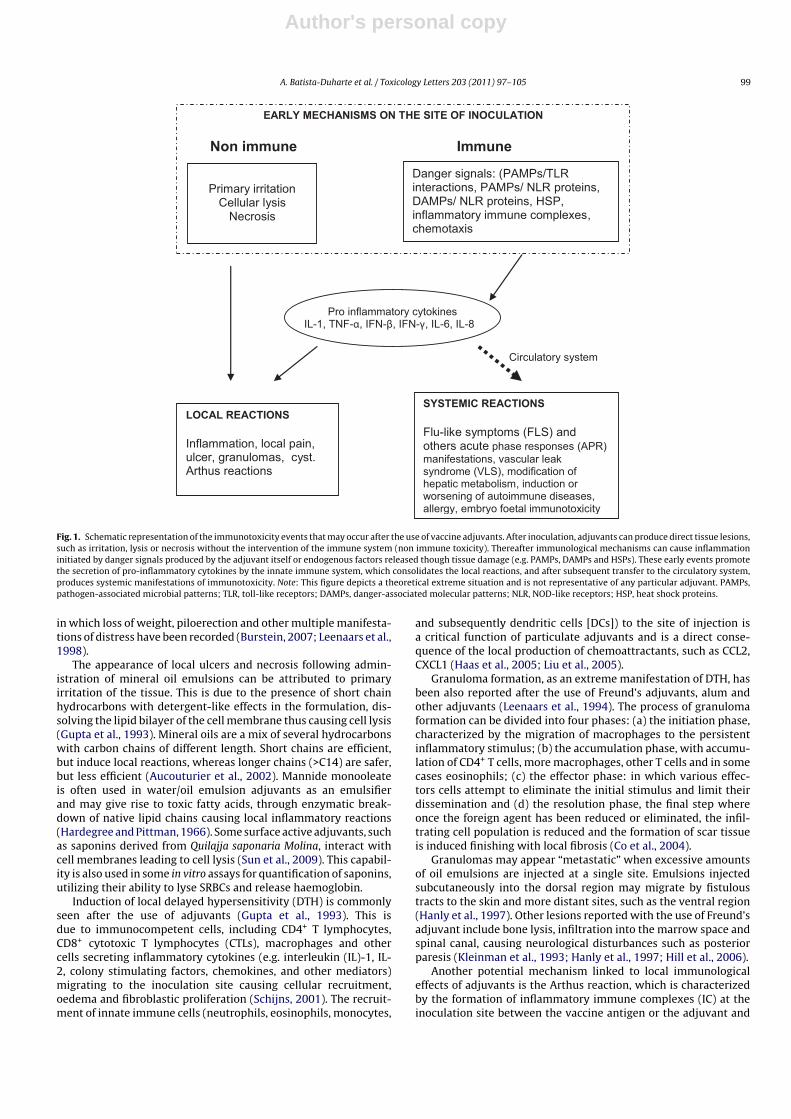
Table 1A classification of immunological adjuvants.
Modes of action Adjuvants
Antigen deliverysystems
Aluminum salts, calcium phosphate, and other gels;Montanides, AS03, MF 59 and other emulsions; PLG,liposomes, virosomes, ISCOMs, virus-like particles,cochleates, and other micro or nanoparticles
Immunopotentiators LPS; MPL and synthetic derivatives; MDP andderivatives; saponins and derivatives, includingISCOMs; CpG oligonucleotides; flagellin; cytokines;dsRNA; resiquimod, imiquimod and othersmall-molecule immunopotentiators
AS03, adjuvant system 03; PLG, polylactide co-glycolide; ISCOMs, immunostim-ulating complexes; LPS, lipopolysaccharide; MLP, monophosphoryl lipid A; MDP,muramyl dipeptide; dsRNA, double-stranded RNA.
Bearing this in mind, two main categories of adjuvant applica-tions can be distinguished (Dawson and Taylor, 1995; Degen et al.,2003):
1. Applications for which efficacy has been given priority over thesafety aspects (e.g. antibody production in animals, investigationof the immune system in rodent models).
2. Applications where safety is more important than efficacy (e.g.preventive human vaccines).
3. Effects of adjuvants
Many different types of adjuvants have been described, with awide range of structures though knowledge about the true mecha-nisms underlying adjuvanticity is still modest and largely empirical.Consequently, it is difficult to predict the exact profile of side effectswhen an adjuvant is administered as part of a vaccine formulation,therefore adjuvants have been called “the immunologist’s dirty lit-tle secret” by Charles Janeway (Janeway, 1989). In spite of this, someadvances have been achieved due to growing knowledge in basicimmunology and as a result the “secret” is slowly being unveiled(Tritto et al., 2009).
It is now accepted that adjuvants provide start signals forimmune reactivity and guide the response to an acceptable magni-tude, through regulation and facilitation of immune responses byantigen delivery (signal 1), co-stimulation (signal 2) and regulatingthe quality of immunity (signal 3) (Schijns, 2006). They are oftendivided into two main types according to their mode of action: (a)antigen delivery systems, which promote antigen uptake by anti-gen presenting cells (APCs), and (b) immunopotentiators, whichactivate the APC, mainly through receptors of the innate immunesystem (O’Hagan, 2006) (Table 1).
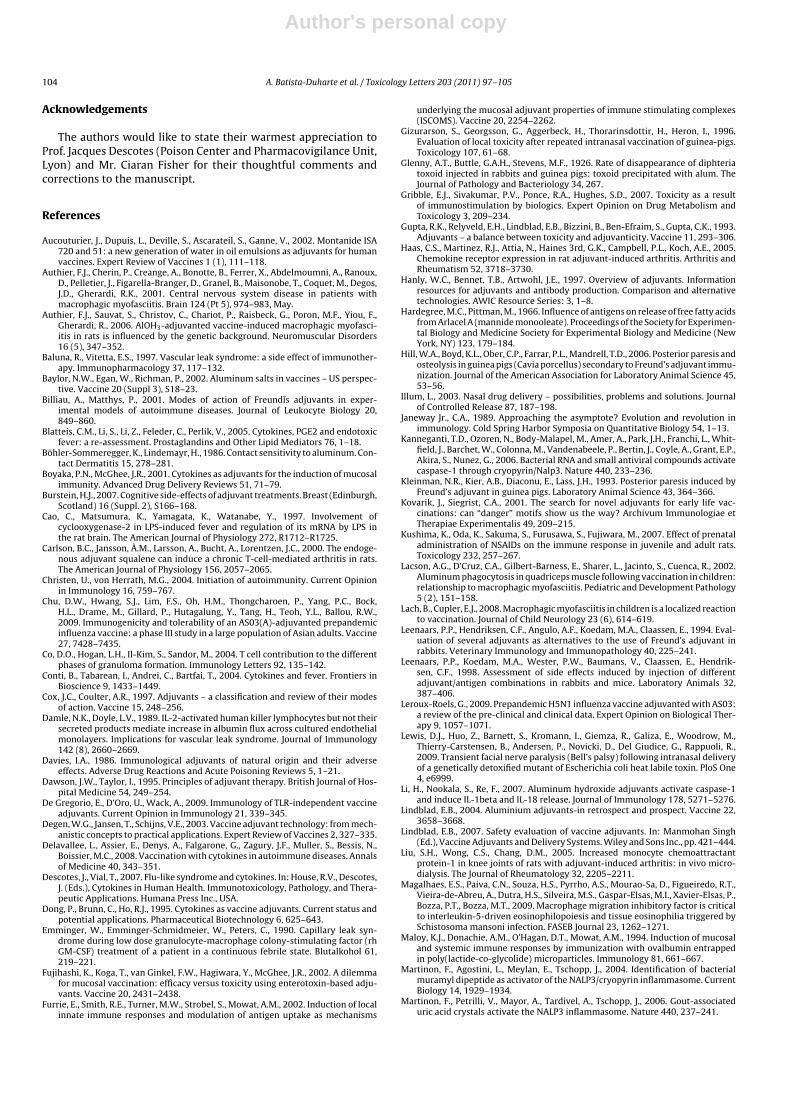
The immunological action of adjuvants can be associatedwith local or systemic side effects that involve various immuno-logical mechanisms although some initial reactions can benon-immunological in nature (Fig. 1 and Table 2): as previouslymentioned, it is a balance (often dose dependant) of judgementwhen a negligible subclinical, local reaction may develop into asignificant and clinically unacceptable adverse reaction.
3.1. Local effects
The majority of parenteral adjuvants produce adverse reactionsat the inoculation site. The most common reactions are local ten-derness and swelling, while the most severe reactions involve theformation of painful abscesses and nodules (Gupta et al., 1993).Local pain is often associated with the use of adjuvants and is aconsequence of tissue damage or the formation of a local inflamma-tory focus at the injection site. The severity of local pain can causebehavioural disturbances and has been observed in animal models
Author's personal copy
A. Batista-Duharte et al. / Toxicology Letters 203 (2011) 97–105 99
LOCAL REACTIONS
Inflammation, local pain, ulcer, granulomas, cyst.Arthus reactions
SYSTEMIC REACTIONS
Flu-like symptoms (FLS) and others acute phase responses (APR)manifestations, vascular leak syndrome (VLS), modification of hepatic metabolism, induction or worsening of autoimmune diseases, allergy, embryo foetal immunotoxicity
EARLY MECHANISMS ON THE SITE OF INOCULATION
Non immune Immune
Primary irritationCellular lysis
Necrosis
Danger signals: (PAMPs/TLR interactions, PAMPs/ NLR proteins, DAMPs/ NLR proteins, HSP, inflammatory immune complexes, chemotaxis
Pro inflammatory cytokinesIL-1, TNF- , IFN- , IFN- , IL-6, IL-8
Circulatory system
Fig. 1. Schematic representation of the immunotoxicity events that may occur after the use of vaccine adjuvants. After inoculation, adjuvants can produce direct tissue lesions,such as irritation, lysis or necrosis without the intervention of the immune system (non immune toxicity). Thereafter immunological mechanisms can cause inflammationinitiated by danger signals produced by the adjuvant itself or endogenous factors released though tissue damage (e.g. PAMPs, DAMPs and HSPs). These early events promotethe secretion of pro-inflammatory cytokines by the innate immune system, which consolidates the local reactions, and after subsequent transfer to the circulatory system,produces systemic manifestations of immunotoxicity. Note: This figure depicts a theoretical extreme situation and is not representative of any particular adjuvant. PAMPs,pathogen-associated microbial patterns; TLR, toll-like receptors; DAMPs, danger-associated molecular patterns; NLR, NOD-like receptors; HSP, heat shock proteins.
in which loss of weight, piloerection and other multiple manifesta-tions of distress have been recorded (Burstein, 2007; Leenaars et al.,1998).
The appearance of local ulcers and necrosis following admin-istration of mineral oil emulsions can be attributed to primaryirritation of the tissue. This is due to the presence of short chainhydrocarbons with detergent-like effects in the formulation, dis-solving the lipid bilayer of the cell membrane thus causing cell lysis(Gupta et al., 1993). Mineral oils are a mix of several hydrocarbonswith carbon chains of different length. Short chains are efficient,but induce local reactions, whereas longer chains (>C14) are safer,but less efficient (Aucouturier et al., 2002). Mannide monooleateis often used in water/oil emulsion adjuvants as an emulsifierand may give rise to toxic fatty acids, through enzymatic break-down of native lipid chains causing local inflammatory reactions(Hardegree and Pittman, 1966). Some surface active adjuvants, suchas saponins derived from Quilajja saponaria Molina, interact withcell membranes leading to cell lysis (Sun et al., 2009). This capabil-ity is also used in some in vitro assays for quantification of saponins,utilizing their ability to lyse SRBCs and release haemoglobin.
Induction of local delayed hypersensitivity (DTH) is commonlyseen after the use of adjuvants (Gupta et al., 1993). This isdue to immunocompetent cells, including CD4+ T lymphocytes,CD8+ cytotoxic T lymphocytes (CTLs), macrophages and othercells secreting inflammatory cytokines (e.g. interleukin (IL)-1, IL-2, colony stimulating factors, chemokines, and other mediators)migrating to the inoculation site causing cellular recruitment,oedema and fibroblastic proliferation (Schijns, 2001). The recruit-ment of innate immune cells (neutrophils, eosinophils, monocytes,
and subsequently dendritic cells [DCs]) to the site of injection isa critical function of particulate adjuvants and is a direct conse-quence of the local production of chemoattractants, such as CCL2,CXCL1 (Haas et al., 2005; Liu et al., 2005).
Granuloma formation, as an extreme manifestation of DTH, hasbeen also reported after the use of Freund’s adjuvants, alum andother adjuvants (Leenaars et al., 1994). The process of granulomaformation can be divided into four phases: (a) the initiation phase,characterized by the migration of macrophages to the persistentinflammatory stimulus; (b) the accumulation phase, with accumu-lation of CD4+ T cells, more macrophages, other T cells and in somecases eosinophils; (c) the effector phase: in which various effec-tors cells attempt to eliminate the initial stimulus and limit theirdissemination and (d) the resolution phase, the final step whereonce the foreign agent has been reduced or eliminated, the infil-trating cell population is reduced and the formation of scar tissueis induced finishing with local fibrosis (Co et al., 2004).
Granulomas may appear “metastatic” when excessive amountsof oil emulsions are injected at a single site. Emulsions injectedsubcutaneously into the dorsal region may migrate by fistuloustracts to the skin and more distant sites, such as the ventral region(Hanly et al., 1997). Other lesions reported with the use of Freund’sadjuvant include bone lysis, infiltration into the marrow space andspinal canal, causing neurological disturbances such as posteriorparesis (Kleinman et al., 1993; Hanly et al., 1997; Hill et al., 2006).
Another potential mechanism linked to local immunologicaleffects of adjuvants is the Arthus reaction, which is characterizedby the formation of inflammatory immune complexes (IC) at theinoculation site between the vaccine antigen or the adjuvant and

Author's personal copy
100 A. Batista-Duharte et al. / Toxicology Letters 203 (2011) 97–105
Table 2Local and systemic reactions induced by some selected immunoadjuvants.
Manifestation oftoxicity
Examples ofadjuvant/components
Selected references
Local reactionLysis, ulcer, necrosis Mineral oil emulsions Gupta et al. (1993),
Aucouturier et al. (2002)Saponin from Q.saponaria
Sun et al. (2009)
Granuloma FCA Leenaars et al. (1994, 1998)Aluminum hydroxide Gupta et al. (1993)
Arthus reactions Toxoid-containingvaccines
Ponvert (2009)
Macrophagicmyofasciitis
Aluminum hydroxide Lacson et al. (2002),Siegrist (2005), Authieret al. (2006), Lach andCupler (2008)
Bell palsy (intranasalroute)
LTK63 Lewis et al. (2009)
Systemic reactionsAcute phase response LPS and derivatives,
MDPGupta et al. (1993)
Vascular leaksyndrome
IL-2 Baluna and Vitetta (1997)
GM-CSF Rechner et al. (2003)Modification of hepatic
metabolismLPS Yang et al. (2008)
FCA Projean et al. (2005)Induction or worsening
of autoimmuneFCA Pearson (1956)
Squalene Carlson et al. (2000)Allergy Aluminum hydroxide Böhler-Sommeregger and
Lindemayr (1986)Embryo foetal
immunotoxicityCPG Prater et al. (2006)
pre-existing antibodies or complement components. This is a phe-nomena associated with the high antibody titre induced by theadjuvant, which can be misleadingly diagnosed as DTH (Mowatet al., 1991; Maloy et al., 1994; Furrie et al., 2002). Depositionof IC triggers Fc gamma receptor-dependent inflammation, wheremigration inhibitory factor (MIF) is released by macrophages uponrecognition of IC, leading to tissue damage (Magalhaes et al., 2009;Paiva et al., 2009).
Infiltrations of aluminum containing macrophages gatheredaround the muscular fibers, in the myofascii, were observed inoccasional deltoid muscular biopsies from patients vaccinated i.m.(Siegrist, 2005). This local reaction, described in adults and chil-dren (Lacson et al., 2002; Lach and Cupler, 2008) was characterizedby the presence of AlOOH-loaded macrophages. This phenomenonwas named MMF (macrophagic myofasciitis) and was attributedto the persistence of aluminum hydroxide for years at the site ofa previous intramuscular injection (Authier et al., 2006). Attemptshave been made to link the presence of such aluminum-containingmacrophage manifestations to various clinical conditions, such asmyalgia, muscle fatigue and, more controversially, to neurologicaldisorders with no obvious etiological relation to the vaccination(Authier et al., 2001). However, such correlations are associatedwith statistical problems. There is very high vaccination cover-age in Western countries. Hence, it is expected statistically thatpatients suffering from a wide range of etiologically unrelated dis-eases would all have been vaccinated with aluminum-containingvaccines at some point in their medical history. Another problem isthat adequate statistical control groups of non-vaccinated personsmay be hard to find within the same population (Lindblad, 2007).
In a controlled study in primates it was not possible to detectany histological changes after injection of aluminum-adjuvantedvaccine. Apart from the local inflammatory focus itself, no otherabnormal clinical signs ascribable to it were found (Verdier et al.,2005), adding further to speculation as to the mechanism of thislesion.
A study using two strains of rats showed that Lewis ratswith Th1-biased immunity had significantly smaller lesions thanSprague–Dawley rats with balanced Th1/Th2 immunity. This indi-cates that genetic determinants of cytotoxic T-cell responses couldinterfere with the clearance process, causing the persistence ofvaccine-induced MMF-lesions (Authier et al., 2006).
Apart from the adjuvants, other factors associated with localreactions include: the antigen itself, contamination of the vac-cine formulation with reactogenic chemicals or microbial products(thiomersal, formaldehyde, LPS, peptidoclycans, etc.) and insta-bility of the formulation during storage with breakdown intoreactogenic by-products. As a consequence adjuvants have no gen-eral approval, but are subject to safety evaluation as part of thecomplete antigen-containing vaccine formulation.
3.1.1. Role of the “danger signals”Based on the “danger model”, some degree of disruption to tis-
sue integrity is required for the development of optimal immuneresponses, since primary immune reactions are initiated by signalsemitted from damaged or stressed cells. These signals mimic tissuedestruction and evoke the expression of co-stimulatory moleculeson antigen-presenting cells (APCs), leads to the recruitment, acti-vation and proliferation of lymphocytes (Matzinger, 1994). Basedon these elements, the definition of an adjuvant could, in principle,be extended to a preparation capable of inducing a “danger” sig-nal yielding direct/indirect tissue damage, or mimicking bacteriaand viruses. Such signals may stimulate immune cells to producepro-inflammatory cytokines necessary for an adequate immuneresponse (Schijns, 2001), but could also result in local and systemicside effects.
3.1.1.1. Heat shock proteins (HSPs). HSPs are a family of ubiquitousintracellular molecules that function as molecular chaperones innumerous processes (e.g. protein folding and transport) and areinduced under stress conditions such as fever, radiation, infec-tions and neoplasia. In addition to maintaining cell homeostasisunder physiological and stress conditions, some heat shock pro-teins (HSPs) are potent inducers of immunity and have even beenharnessed as adjuvants in experimental vaccines targeted to can-cers and infections (Segal et al., 2006).
HSPs released from necrotic cells owing to the local effectsof several adjuvants are recognized by APCs through specificreceptors, such as TLRs, scavenger receptors (LOX-1), CD91, CD14(resulting in increased antigen display by MHC class I and IImolecules) and priming T cells. Several HSPs activate the nuclearfactor NF-kB pathway and induce the maturation of DCs and thesecretion of the proinflammatory cytokines interleukin (IL)-12,tumour necrosis factor (TNF)-�, and chemokines (Srivastava andAmato, 2001; Segal et al., 2006). The migratory and cytolytic activ-ity of natural killer cells can also be activated by HSPs, such as HSP96and HSP70 (Pilla et al., 2005; Massa et al., 2005).
3.1.1.2. Pathogen-associated molecular patterns (PAMPs). PAMPsare highly conserved structural motifs expressed by microbialpathogens, and include various bacterial cell wall componentssuch as lipopolysaccharides (LPS), peptidoglycans and lipopep-tides, as well as flagellin, bacterial DNA and viral double-strandedRNA. These structures are recognized by evolutionary conservedreceptors, homologues of the Drosophila Toll gene called Toll-likeReceptors (TLRs), expressed by cells belonging to the innate systemand playing a critical role in early innate immunity against invad-ing pathogens by sensing microorganisms (Medzhitov et al., 1997).Taking into account that several recognized adjuvant active sub-stances are obtained from microorganisms bearing PAMPs, TLRscan be considered as adjuvant receptors. Most of the PAMPs usedas vaccine adjuvants, such as CpG oligonucleotides and monophos-

Author's personal copy
A. Batista-Duharte et al. / Toxicology Letters 203 (2011) 97–105 101
phoryl lipid A (MPL) are agonists of TLRs (van Duin et al., 2006).Other components with adjuvant activity, but not used as adjuvantsin practical vaccination due to toxicity (e.g. LPS and muramyldipep-tides/MDP) have also been shown to be TLR-agonists.
3.1.1.3. Danger-associated molecular patterns (DAMPs). The NOD-like receptors (NLRs) are a family of intracellular sensors ofmicrobial motifs and other signals of cellular stress (low intra-cellular potassium concentrations, reactive oxygen species (ROS),ATP increase (via P2X7 receptor), monosodium urate crystalsand calcium pyrophosphate dihydrate deposition). These recep-tors have emerged as crucial components of innate immuneresponses during inflammation (Petrilli et al., 2007a; Martinonet al., 2006). Several NLRs (NALPs and IPAF) form a caspase-1-activating multiprotein complex, termed inflammasome, whichprocesses pro-inflammatory cytokines, such as IL-1� (Petrilli et al.,2007b) and IL-18 (Li et al., 2007). Amongst the various inflam-masomes, NALP3 is particularly qualified to sense a plethoraof different molecules used as adjuvants, including bacterialpeptidoglycan/muramyl dipeptide (Martinon et al., 2004), LPS, bac-terial RNA, poly I:C and imidazolequinoline-derived compounds(Kanneganti et al., 2006). Studies in animal models suggest that thedirect activation of NOD-like receptor protein 3 (NLRP3) may con-tribute to the adjuvanticity of particulate adjuvants, such as alum,chitosan and Quil-A (De Gregorio et al., 2009), although furtherstudies are necessary to confirm these results.
Interestingly, phenotypical and functional modifica-tions have also been observed in short-term cultures ofmacrophages exposed to aluminum hydroxide in vitro. Theyexpress the classical markers of myeloid dendritic cells (HLA-DRhigh/CD86high/CD83+/CD1a−/CD14−) and display a potentability to induce MHC-II-restricted antigen-specific memoryresponses, while retaining a macrophage morphology (Rimaniolet al., 2004).
3.2. Systemic effects
Systemic reactions following the application of immunologicaladjuvants will generally disqualify them from use in practical vacci-nation. Consequently, few observations on the systemic side effectsof vaccine adjuvants are available. Where systemic side effects areobserved, they are considered rare and with the exception of acutephase responses, are regarded as subclinical.
Toxic systemic effects after adjuvant administration are gen-erally the consequence of the hyper-activation of immunologicalmechanisms induced by the adjuvant formulation. These effects areoften mediated by the release of cytokines, such as IL-1�, TNF-�,IFN-�, IFN-�, IL-6 and others (Li et al., 2007; Sokolovska et al., 2007).Cytokines used directly as immunological adjuvants (Boyaka andMcGhee, 2001; Dong et al., 1995) or subsequently released after theapplication of various immunomodulators are not only essential inthe early phase of the immune response, but are directly or indi-rectly involved in the pathogenesis of immune-mediated disordersdescribed in humans and animals. Therefore, it is not surprising thatsuch disorders develop after the administration of pharmacologicaldoses of these cytokines (Vial and Descotes, 1995; Delavallee et al.,2008).
3.2.1. Acute phase response (APR)APR defines a transient syndrome characterized by changes in
plasma proteins and is associated with fever and other constitu-tional symptoms (leukocytosis, changes in cell/tissue metabolismand organ function) that accompany typical side effects seen in flu-like reactions, such as fatigue and anorexia (Gribble et al., 2007).Flu-like symptoms (FLS), as part of APR, are one of the main sideeffects reported to develop after hyperstimulation of the immune
system. They are mediated by several cytokines and other humoralfactors, usually appearing within hours following administrationof an immunostimulating drug or vaccine, and uneventfully recedewithin a few hours. FLS typically consist of fever, chills, fatigue,myalgia, headache and nausea. Fever is the most common findingin patients with FLS. It can be of variable magnitude, ranging froma moderate increase in body temperature (38–39 ◦C) to markedhyperpyrexia exceeding 40 ◦C, in some cases (Descotes and Vial,2007).
IL-1 released by leukocytes, and other endogenous factors(IL-1�, TNF-�, IFN-�, IFN-�, IL-6, IL-8) along with macrophageinflammatory protein-1, act as pyrogens (Conti et al., 2004).These circulating cytokines can enter the circumventricular organsthrough fenestrated capillaries where they induce the productionof prostaglandins, such as PGE2, a centrally controlled mediator offever. Synthesis of PGE2 depends on cyclooxygenase (COX) activ-ity; the induction of COX-2 in response to peripheral injection ofa fever-inducing dose of LPS was demonstrated in brain endothe-lial cells, perivascular microglia and meningeal macrophages (Caoet al., 1997; Descotes and Vial, 2007).
A role for the complement anaphylatoxins in APR has been alsosuggested. It has been shown that intravenous administration ofLPS triggers complement cascade activation within 2 min via thealternative pathway, resulting in the production of C4a, C3a and C5ain the blood (Descotes and Vial, 2007). PGE2 is produced in responseto activation via the hydrolysis of membrane-associated phospho-inositide by phosphoinositide-specific phospholipase C, activatedby the complement cascade. The anaphylatoxin C5a has been iden-tified as an important mediator in this pathway (Blatteis et al.,2005). However, these are findings from experimental immunol-ogy, as vaccines are never administered intravenously.
Some of the main targets of APR are the liver, the hematopoieticsystem, and the hypothalamic–pituitary–adrenal axis. Hepatocytesrespond to these cytokines primarily by altering gene tran-scription and increasing the production and secretion of acutephase proteins. Several hepatic proteins are elevated in serumincluding complement factors (C2, C3, C4, C5, C9, C1-inhibitor,C4-binding protein; mannose-binding lectin [MBL], Factor B), coag-ulation/fibrinolysis factors (fibrinogen, plasminogen, protein S, vonWillebrand Factor [VWF], plasminogen activator inhibitor-1 [PAI-1], tissue plasminogen activator [TPA]), C-reactive protein [CRP]and serum amyloid A [SAA]. Several of these, such as CRP and SAA,are currently being used as biomarkers of APR (Gribble et al., 2007).Although these findings have no direct relevance to vaccinationthey are never the less of interest for the understanding of thetoxicity of LPS.
3.2.2. Induction or worsening of autoimmune diseasesAlthough there are only a few examples of autoimmune effects
induced by adjuvants without any joint-specific antigen (Pearson,1956; Stasiuk et al., 1997; Billiau and Matthys, 2001), this is oneof the best examples for the potential of adverse reaction with thecombined effect of adjuvant/antigen.
Experimentally, it is rather easy to induce an autoimmune dis-ease in genetically susceptible animals via immunization witha formulation containing a strong adjuvant (e.g. FCA) and anautoantigen (Billiau and Matthys, 2001). In clinical practice how-ever, reports of possible associations between vaccines andautoimmune diseases are rare, relative to the large number of vac-cinated human subjects, and epidemiological studies have so farfailed to demonstrate any causal relationship between vaccinationand autoimmune diseases (Schattner, 2005).
In theory, a classical vaccine formulation contains the necessaryelements for a possible induction of autoimmune diseases. Namely,the antigen that may contain mimetic epitopes and the adjuvant forthe upregulation of co-stimulatory molecules and other products of

Author's personal copy
102 A. Batista-Duharte et al. / Toxicology Letters 203 (2011) 97–105
inflammation leading to the polyclonal activation of autoreactive Tcells. Furthermore, bystander activation can wake up functionallysilenced (anergic) cells with auto-aggressive potential or triggerthe expansion of low-affinity autoreactive cells that have escapednegative selection (Christen and von Herrath, 2004). On the otherhand, the role of pre-existing risk factors including genetic predis-position and environmental factors are largely accepted as beinginvolved (Vial and Descotes, 2004).
In human populations with a high level of diversity amongst HLAhaplotypes, it is not surprising to find differences in susceptibilityto developing autoimmune reactions (Theofilopoulos, 1995). It hasbeen suggested that some genetic pattern could be a predispos-ing factor in determined subjects, in which antigen presentation,influenced by certain HLA haplotypes, can lead to the autoimmunecascade after vaccination (Santoro et al., 2010).
The causative relationship between hyperstimulation of theimmune system associated to vaccination and induction of autoim-munity remains unclear. This highlights the need for more researchto understand the possible role of adjuvants and vaccines in trig-gering autoimmunity in clinical practice.
3.2.3. Modification of hepatic metabolismAdministration of immunostimulating drugs, Bacillus Calmette
Guerin (BCG) vaccine, or interferons has been shown to affectoxidative drug metabolism by the hepatic cytochrome P 450(CYP) system. For example, Renton and co-workers (Renton, 2001)reported decreased elimination of theophylline after influenzavaccination, associated with changes in the hepatic metabolismof sufficient magnitude to cause acute theophylline toxicity inhumans, despite the use of therapeutic doses. In another report,these authors also demonstrated that the release of cytokines, suchas IL-1, IL-2, IL-6, TNF, TGF-� and IFNs is involved in modulat-ing the expression of several P450 isoforms. Reversible changesin the pharmacokinetic parameters of theophylline and decreasedexpression of CYP1A, 2B1/2, and 3A subfamily have also beenreported in rats after intravenous administration of lipopolysac-charide (endotoxin) derived from Klebsiella pneumoniae (Yang et al.,2008).
Other studies have shown the rapid decrease in the total CYP450liver content of FCA-treated rats and the selective down-regulationof specific CYP isoforms through a direct reduction in mRNA levels(CYP2B, CYP2CI1, CYP3A1, and CYP2E1), protein content (CYP2B,CYP2C11, and CYP2E1) and catalytic activity (CYP2C6, CYP2C11,and CYP2E1) (Projean et al., 2005). CYP3A1 mRNA levels wereseverely decreased by FCA administration, whereas CYP3A2 mRNAand protein levels remained unchanged. These early biochemicaland metabolic modifications may have pharmacokinetic and phar-macodynamic consequences when drugs cleared by the liver areadministered in conjunction with FCA (Projean et al., 2005) andpossibly other potent immunostimulators. Shifts in hepatic CYPexpression could also lead to increased exposure to drugs or toxicmetabolites. This effect has been considered as a possible cause ofhepatic toxicities, as the variable suppression of drug-metabolizingenzymes involved in drug biotransformation and elimination dur-ing infection and inflammation could lead to a range of patterns ofhepatic damage (Renton, 2001).
3.2.4. Vascular leak syndrome (VLS)VLS is a serious side effect seen after administration of IL-2
(Vial and Descotes, 1995), IL-1 (Worth et al., 1997), granulocyte-macrophage colony-stimulating factor (GM-CSF) (Emminger et al.,1990), and other cytokines, previously proposed as vaccine adju-vants. VLS is characterized by an increase in vascular permeability,accompanied by extravasation of fluids and proteins resultingin interstitial oedema and organ failure. Manifestations of VLSinclude increased body weight, hypotension, fluid shifts, periph-
eral oedema, pleural and pericardial effusions and ascites. In severecases, VLS-related conditions may progress to pulmonary and car-diovascular failure (Baluna and Vitetta, 1997).
The pathogenesis of endothelial cell damage associated to thissyndrome is complex and poorly understood. It may involve theactivation or damage of endothelial cells and leukocytes, therelease of cytokines and inflammatory mediators (e.g., IL-1, TNF-�), components of the complement cascade, alteration in cell–cellinteractions and cell matrix adhesion, as well as alterations incytoskeleton function resulting in disturbance of vascular integrity(Baluna and Vitetta, 1997). Other studies have connected cyto-toxicity of lymphokine-activated killer (LAK) cells on vascularendothelial cells with the development of VLS (Damle and Doyle,1989) and demonstrated the involvement of perforin, Fas lig-and and CD44 through the use of gene-targeted mice (Rafi et al.,1998, 1999). Not surprisingly the observation of VLS led to thedisqualification of these cytokines from use as adjuvants in vac-cines.
3.2.5. AllergyThe question of allergy to adjuvants is controversial. The gen-
eral ability of aluminum adjuvants to stimulate the production ofIgE as part of the overall Th2 profile and increase eosinophilia iswell established although, the underlying mechanism is unknown.However in practical conditions, it has been difficult to demon-strate cases where vaccination with aluminum adjuvants has led toIgE-mediated allergy toward the vaccine antigen (Lindblad, 2004).
An adjuvant-induced increase in IgE levels against the antigenshould be considered as a potential concern regarding the devel-opment of hypersensitivity reactions.
3.2.6. Embryonic immunotoxicityAdjuvants could have immunomodulatory effects on a success-
ful embryonic gestation and may also affect the immune systemof the developing foetus. The balance of Th1/Th2 responses isimportant for a successful pregnancy where maternal responsesare being biased toward humoral immunity (Th2) and away fromcell-mediated immunity (Th1) (Raghupathy, 1997).
Recent reports have revealed that a shift toward a Th1 cytokineprofile during pregnancy may increase the risk of foetal morpho-logical defects. The injection of high doses of CpG ODN, a vaccineadjuvant inducing strong Th1 responses, to pregnant C57BL/6mice resulted in a marked increase in foetal resorption and cran-iofacial/limb defects, while lower doses had little or no effect.The histological examination of the placentas showed cellularnecrosis with mixed inflammation and calcification in the spon-giotrophoblast layer and dysregulation of labyrinthine vasculardevelopment (Prater et al., 2006).
Hence, any vaccine or adjuvant that skews immune responsestoward Th1 could, in theory, have adverse effects on the embryo-foetal development through Th1-type immunity as it appears topose a risk to successful pregnancy (Raghupathy, 1997). Cytokines,NK cells and gamma delta T cells of maternal origin are all thoughtto be involved in processes, such as foetal recognition, placentaldevelopment and regulation of gene expression during organo-genesis (Szekeres-Bartho, 2002). Hence any effect of vaccinationon these cells and cytokines may potentially affect developmentof the foetus. Currently there is little information on developmen-tal immunotoxicity available, thus highlighting the importance ofcarrying out further studies on alterations of the immune systemafter pre- or post-natal exposure to xenobiotic agents (Kushimaet al., 2007). Teratogenic effects that may result from exposureto vaccine adjuvants require a particular attention (Prater et al.,2006).

Author's personal copy
A. Batista-Duharte et al. / Toxicology Letters 203 (2011) 97–105 103
4. Impact of routes of administration on the effects ofadjuvants
The selected route of administration may, to some extent, influ-ence the profile of side effects associated with adjuvants. With thesubcutaneous route (s.c.), the vaccine inoculum is introduced into acompartment with numerous sensorial nerve cells. This inductionof a local inflammatory response may lead to local irritation, itching(pruritus), erythema and pain. In addition, transient swelling as aconsequence of the inflammatory focus, may become easily palpa-ble through the skin (Leenaars et al., 1998). With the intramuscularroute (i.m.), swelling even of similar size may be less easily palpableas it is located deeper within the tissue. In addition, the intramuscu-lar compartment is not as innervated with sensory neurons as theskin (Davies, 1986; Spickler and Roth, 2003). On the other hand,lesions in the muscular tissue following i.m. administration maybe associated with the release of creatine phosphokinase (CPK) intothe circulation, which in some cases is used as an indicator of tis-sue damage (Patel et al., 2002). Adjuvants can be also administeredvia the intraperitoneal (i.p.) route to animals, primarily in experi-mental immunology. Here the inoculum is introduced into the bodycavity and instead of forming a localized deposit, it may spread outand induce disseminated reactions, including chemical peritonitis,ascitis and the formation of fibrous adherences between variousorgans in the body cavity.
Gizurarson et al. (1996) evaluated the local toxicity of sev-eral adjuvants after the intranasal vaccination of guinea pigs. Theyfound damage to the mucosal epithelium ascribed to direct toxic-ity of a given inoculum. This can lead to contact of lymphoid cellswith the submucosal tissue or draining lymphatic vessels, causingimmunological responses in which antigen uptake is carried outby M cells (Gizurarson et al., 1996). However, a damaged mucosamay lead to the establishment of secondary opportunistic infec-tions due to breach of the mucosal barrier, an important aspectthat requires further investigation. Another concern when usingthe nasal route for immunization is the potentially direct passageof the inoculum into the brain through the olfactory pathways.Noticeably, entry into the central nervous system (CNS) throughprimary sensory olfactory neurons is an established route for sev-eral viruses however, no data is yet available for the entry ofbacteria or bacterial-derived toxins through the same route (Illum,2003). Hence, the potential for neurotoxicity must not be ignoredin the use of mucosal adjuvants in humans. The expression of GM1gangliosides on sensory olfactory neurons in the nasal tract pro-vides a pathway for entry of enterotoxins used as adjuvants intothe olfactory bulbs (e.g. CT and LT) following nasal application(Fujihashi et al., 2002). It has been reported that nasally appliedLT induced inflammatory responses in the meninges, the olfac-tory nerve and glomerular layers of the olfactory bulbs (reviewedin Fujihashi et al., 2002) also a link has been suggested betweenthis pathway and diseases, such as Alzheimer’s dementia (Weller,1998). However, to date there is not sufficient evidence to supportthe hypothesis that intranasal immunization is dangerous for thebrain.
An association has been established between facial nerveparalysis (Bell’s palsy) and the intranasal administration of aninactivated influenza virosome vaccine containing an LT adjuvantin Switzerland (Mutsch et al., 2004). Further reports have beenpublished in relation to intranasal vaccines containing geneticallydetoxified mutant of Escherichia coli LT (LTK63). The lack of reportedfacial nerve paralysis following nasal immunization in subjects notreceiving LT adjuvants, implies a causal relationship with LT andallows the suggestion that this paralysis may be due to a tran-sient interference with peripheral nerve function. Such transientinterference could be due to accumulation of LTK63 molecules, toinflammation arising from immune response to LTK63 following
ganglioside binding, retrograde neuronal transport or to other stillunknown causes (Lewis et al., 2009). This pathological condition iscurrently the focus of attention for vaccine regulatory agencies.
5. Present problems and future perspectives
The search for new, more potent and safe adjuvants representsone scientific challenge today. Over the last decades very few adju-vants have been licensed for prophylactic vaccines due to toxicproperties detected during pre-clinical or clinical studies of themany new candidates being evaluated. Researchers or companies,sometimes unfairly, refer to a “regulatory barrier” forgetting that asfar as preventive formulation is concerned, the primary obligationis safety.
Unfortunately, there are several challenges in the design ofadequate safety studies: the lack of suitable experimental modelsand standardized predictive methods, the difficulties in studyingcertain adverse effects (e.g. autoimmunity and hypersensitivityreactions) and the clinical hurdle of detecting infrequent effectsthat can develop in certain specific “at-risk” subpopulations (e.g.those carrying particular HLA phenotypes, or suffering from cer-tain chronic diseases, or exposed to drugs and other environmentalfactors).
Surveillance systems should be expected to be able to detectadverse effects in these specific situations. Currently however,these are mostly passive surveillance systems collecting reportsof events voluntarily submitted by patients who experiencedthem, caregivers, or others. Passive surveillance systems (such asVAERS) are subject to multiple limitations, including underreport-ing, reporting of temporal associations or unconfirmed diagnoses,and a lack of denominator data and unbiased comparison groups.Due to these limitations, determining causal associations betweenvaccines and adverse events from these reports is usually not pos-sible.
Although vaccines are the most successful medical inventionof the last century, it is obvious that future vaccines will requireadjuvants with predictable activity (Schijns, 2006). For these rea-sons, the regulatory agencies are very reluctant to approve noveladjuvants, consequently efforts for global harmonization towardimproved and consistent standards for nonclinical and clinicalevaluation are required to better assess the safety of preventivevaccines and predict their toxicity.
The aim of this paper is to draw attention to the fact that, to alarge extent, the very same parameters of the immune system thatare essential for the normal function of both the innate and theadaptive immune response, are also key players in the reactionsand side effects that we consider adverse in nature. It is a balanceand a matter of judgement when for example a normal subclin-ical local reaction to a vaccination turns from being a temporarycosmetic issue into a clinically unacceptable reaction because ofhyperstimulation of the immune system. We have discussed dataobtained from practical vaccination, but also from model systemsthrough immunostimulatory regimens that would be consideredcompletely unacceptable for practical vaccination. However, whentaken together these data illustrate the dilemma of immunostimu-lation and demonstrate the variety of factors with the potential toaffect the level of immunotoxicity. We believe that fellow scientistsshould take inspiration from this when they evaluate new potentialadjuvants intended for use in vaccines. Even though some progresshas been achieved in regulatory policies and guidelines, approachesto improve the safety assessment of preventive vaccines is stillevolving and should be promoted by regulatory agencies.
Conflict of interest statement
The authors declare no conflict of interest.
Author's personal copy
104 A. Batista-Duharte et al. / Toxicology Letters 203 (2011) 97–105
Acknowledgements
The authors would like to state their warmest appreciation toProf. Jacques Descotes (Poison Center and Pharmacovigilance Unit,Lyon) and Mr. Ciaran Fisher for their thoughtful comments andcorrections to the manuscript.
References
Aucouturier, J., Dupuis, L., Deville, S., Ascarateil, S., Ganne, V., 2002. Montanide ISA720 and 51: a new generation of water in oil emulsions as adjuvants for humanvaccines. Expert Review of Vaccines 1 (1), 111–118.
Authier, F.J., Cherin, P., Creange, A., Bonotte, B., Ferrer, X., Abdelmoumni, A., Ranoux,D., Pelletier, J., Figarella-Branger, D., Granel, B., Maisonobe, T., Coquet, M., Degos,J.D., Gherardi, R.K., 2001. Central nervous system disease in patients withmacrophagic myofasciitis. Brain 124 (Pt 5), 974–983, May.
Authier, F.J., Sauvat, S., Christov, C., Chariot, P., Raisbeck, G., Poron, M.F., Yiou, F.,Gherardi, R., 2006. AlOH3-adjuvanted vaccine-induced macrophagic myofasci-itis in rats is influenced by the genetic background. Neuromuscular Disorders16 (5), 347–352.
Baluna, R., Vitetta, E.S., 1997. Vascular leak syndrome: a side effect of immunother-apy. Immunopharmacology 37, 117–132.
Baylor, N.W., Egan, W., Richman, P., 2002. Aluminum salts in vaccines – US perspec-tive. Vaccine 20 (Suppl 3), S18–23.
Billiau, A., Matthys, P., 2001. Modes of action of Freundı̌s adjuvants in exper-imental models of autoimmune diseases. Journal of Leukocyte Biology 20,849–860.
Blatteis, C.M., Li, S., Li, Z., Feleder, C., Perlik, V., 2005. Cytokines, PGE2 and endotoxicfever: a re-assessment. Prostaglandins and Other Lipid Mediators 76, 1–18.
Böhler-Sommeregger, K., Lindemayr, H., 1986. Contact sensitivity to aluminum. Con-tact Dermatitis 15, 278–281.
Boyaka, P.N., McGhee, J.R., 2001. Cytokines as adjuvants for the induction of mucosalimmunity. Advanced Drug Delivery Reviews 51, 71–79.
Burstein, H.J., 2007. Cognitive side-effects of adjuvant treatments. Breast (Edinburgh,Scotland) 16 (Suppl. 2), S166–168.
Cao, C., Matsumura, K., Yamagata, K., Watanabe, Y., 1997. Involvement ofcyclooxygenase-2 in LPS-induced fever and regulation of its mRNA by LPS inthe rat brain. The American Journal of Physiology 272, R1712–R1725.
Carlson, B.C., Jansson, Å.M., Larsson, A., Bucht, A., Lorentzen, J.C., 2000. The endoge-nous adjuvant squalene can induce a chronic T-cell-mediated arthritis in rats.The American Journal of Physiology 156, 2057–2065.
Christen, U., von Herrath, M.G., 2004. Initiation of autoimmunity. Current Opinionin Immunology 16, 759–767.
Chu, D.W., Hwang, S.J., Lim, F.S., Oh, H.M., Thongcharoen, P., Yang, P.C., Bock,H.L., Drame, M., Gillard, P., Hutagalung, Y., Tang, H., Teoh, Y.L., Ballou, R.W.,2009. Immunogenicity and tolerability of an AS03(A)-adjuvanted prepandemicinfluenza vaccine: a phase III study in a large population of Asian adults. Vaccine27, 7428–7435.
Co, D.O., Hogan, L.H., Il-Kim, S., Sandor, M., 2004. T cell contribution to the differentphases of granuloma formation. Immunology Letters 92, 135–142.
Conti, B., Tabarean, I., Andrei, C., Bartfai, T., 2004. Cytokines and fever. Frontiers inBioscience 9, 1433–1449.
Cox, J.C., Coulter, A.R., 1997. Adjuvants – a classification and review of their modesof action. Vaccine 15, 248–256.
Damle, N.K., Doyle, L.V., 1989. IL-2-activated human killer lymphocytes but not theirsecreted products mediate increase in albumin flux across cultured endothelialmonolayers. Implications for vascular leak syndrome. Journal of Immunology142 (8), 2660–2669.
Davies, I.A., 1986. Immunological adjuvants of natural origin and their adverseeffects. Adverse Drug Reactions and Acute Poisoning Reviews 5, 1–21.
Dawson, J.W., Taylor, I., 1995. Principles of adjuvant therapy. British Journal of Hos-pital Medicine 54, 249–254.
De Gregorio, E., D’Oro, U., Wack, A., 2009. Immunology of TLR-independent vaccineadjuvants. Current Opinion in Immunology 21, 339–345.
Degen, W.G., Jansen, T., Schijns, V.E., 2003. Vaccine adjuvant technology: from mech-anistic concepts to practical applications. Expert Review of Vaccines 2, 327–335.
Delavallee, L., Assier, E., Denys, A., Falgarone, G., Zagury, J.F., Muller, S., Bessis, N.,Boissier, M.C., 2008. Vaccination with cytokines in autoimmune diseases. Annalsof Medicine 40, 343–351.
Descotes, J., Vial, T., 2007. Flu-like syndrome and cytokines. In: House, R.V., Descotes,J. (Eds.), Cytokines in Human Health. Immunotoxicology, Pathology, and Thera-peutic Applications. Humana Press Inc., USA.
Dong, P., Brunn, C., Ho, R.J., 1995. Cytokines as vaccine adjuvants. Current status andpotential applications. Pharmaceutical Biotechnology 6, 625–643.
Emminger, W., Emminger-Schmidmeier, W., Peters, C., 1990. Capillary leak syn-drome during low dose granulocyte-macrophage colony-stimulating factor (rhGM-CSF) treatment of a patient in a continuous febrile state. Blutalkohol 61,219–221.
Fujihashi, K., Koga, T., van Ginkel, F.W., Hagiwara, Y., McGhee, J.R., 2002. A dilemmafor mucosal vaccination: efficacy versus toxicity using enterotoxin-based adju-vants. Vaccine 20, 2431–2438.
Furrie, E., Smith, R.E., Turner, M.W., Strobel, S., Mowat, A.M., 2002. Induction of localinnate immune responses and modulation of antigen uptake as mechanisms
underlying the mucosal adjuvant properties of immune stimulating complexes(ISCOMS). Vaccine 20, 2254–2262.
Gizurarson, S., Georgsson, G., Aggerbeck, H., Thorarinsdottir, H., Heron, I., 1996.Evaluation of local toxicity after repeated intranasal vaccination of guinea-pigs.Toxicology 107, 61–68.
Glenny, A.T., Buttle, G.A.H., Stevens, M.F., 1926. Rate of disappearance of diphteriatoxoid injected in rabbits and guinea pigs: toxoid precipitated with alum. TheJournal of Pathology and Bacteriology 34, 267.
Gribble, E.J., Sivakumar, P.V., Ponce, R.A., Hughes, S.D., 2007. Toxicity as a resultof immunostimulation by biologics. Expert Opinion on Drug Metabolism andToxicology 3, 209–234.
Gupta, R.K., Relyveld, E.H., Lindblad, E.B., Bizzini, B., Ben-Efraim, S., Gupta, C.K., 1993.Adjuvants – a balance between toxicity and adjuvanticity. Vaccine 11, 293–306.
Haas, C.S., Martinez, R.J., Attia, N., Haines 3rd, G.K., Campbell, P.L., Koch, A.E., 2005.Chemokine receptor expression in rat adjuvant-induced arthritis. Arthritis andRheumatism 52, 3718–3730.
Hanly, W.C., Bennet, T.B., Artwohl, J.E., 1997. Overview of adjuvants. Informationresources for adjuvants and antibody production. Comparison and alternativetechnologies. AWIC Resource Series: 3, 1–8.
Hardegree, M.C., Pittman, M., 1966. Influence of antigens on release of free fatty acidsfrom Arlacel A (mannide monooleate). Proceedings of the Society for Experimen-tal Biology and Medicine Society for Experimental Biology and Medicine (NewYork, NY) 123, 179–184.
Hill, W.A., Boyd, K.L., Ober, C.P., Farrar, P.L., Mandrell, T.D., 2006. Posterior paresis andosteolysis in guinea pigs (Cavia porcellus) secondary to Freund’s adjuvant immu-nization. Journal of the American Association for Laboratory Animal Science 45,53–56.
Illum, L., 2003. Nasal drug delivery – possibilities, problems and solutions. Journalof Controlled Release 87, 187–198.
Janeway Jr., C.A., 1989. Approaching the asymptote? Evolution and revolution inimmunology. Cold Spring Harbor Symposia on Quantitative Biology 54, 1–13.
Kanneganti, T.D., Ozoren, N., Body-Malapel, M., Amer, A., Park, J.H., Franchi, L., Whit-field, J., Barchet, W., Colonna, M., Vandenabeele, P., Bertin, J., Coyle, A., Grant, E.P.,Akira, S., Nunez, G., 2006. Bacterial RNA and small antiviral compounds activatecaspase-1 through cryopyrin/Nalp3. Nature 440, 233–236.
Kleinman, N.R., Kier, A.B., Diaconu, E., Lass, J.H., 1993. Posterior paresis induced byFreund’s adjuvant in guinea pigs. Laboratory Animal Science 43, 364–366.
Kovarik, J., Siegrist, C.A., 2001. The search for novel adjuvants for early life vac-cinations: can “danger” motifs show us the way? Archivum Immunologiae etTherapiae Experimentalis 49, 209–215.
Kushima, K., Oda, K., Sakuma, S., Furusawa, S., Fujiwara, M., 2007. Effect of prenataladministration of NSAIDs on the immune response in juvenile and adult rats.Toxicology 232, 257–267.
Lacson, A.G., D’Cruz, C.A., Gilbert-Barness, E., Sharer, L., Jacinto, S., Cuenca, R., 2002.Aluminum phagocytosis in quadriceps muscle following vaccination in children:relationship to macrophagic myofasciitis. Pediatric and Development Pathology5 (2), 151–158.
Lach, B., Cupler, E.J., 2008. Macrophagic myofasciitis in children is a localized reactionto vaccination. Journal of Child Neurology 23 (6), 614–619.
Leenaars, P.P., Hendriksen, C.F., Angulo, A.F., Koedam, M.A., Claassen, E., 1994. Eval-uation of several adjuvants as alternatives to the use of Freund’s adjuvant inrabbits. Veterinary Immunology and Immunopathology 40, 225–241.
Leenaars, P.P., Koedam, M.A., Wester, P.W., Baumans, V., Claassen, E., Hendrik-sen, C.F., 1998. Assessment of side effects induced by injection of differentadjuvant/antigen combinations in rabbits and mice. Laboratory Animals 32,387–406.
Leroux-Roels, G., 2009. Prepandemic H5N1 influenza vaccine adjuvanted with AS03:a review of the pre-clinical and clinical data. Expert Opinion on Biological Ther-apy 9, 1057–1071.
Lewis, D.J., Huo, Z., Barnett, S., Kromann, I., Giemza, R., Galiza, E., Woodrow, M.,Thierry-Carstensen, B., Andersen, P., Novicki, D., Del Giudice, G., Rappuoli, R.,2009. Transient facial nerve paralysis (Bell’s palsy) following intranasal deliveryof a genetically detoxified mutant of Escherichia coli heat labile toxin. PloS One4, e6999.
Li, H., Nookala, S., Re, F., 2007. Aluminum hydroxide adjuvants activate caspase-1and induce IL-1beta and IL-18 release. Journal of Immunology 178, 5271–5276.
Lindblad, E.B., 2004. Aluminium adjuvants-in retrospect and prospect. Vaccine 22,3658–3668.
Lindblad, E.B., 2007. Safety evaluation of vaccine adjuvants. In: Manmohan Singh(Ed.), Vaccine Adjuvants and Delivery Systems. Wiley and Sons Inc., pp. 421–444.
Liu, S.H., Wong, C.S., Chang, D.M., 2005. Increased monocyte chemoattractantprotein-1 in knee joints of rats with adjuvant-induced arthritis: in vivo micro-dialysis. The Journal of Rheumatology 32, 2205–2211.
Magalhaes, E.S., Paiva, C.N., Souza, H.S., Pyrrho, A.S., Mourao-Sa, D., Figueiredo, R.T.,Vieira-de-Abreu, A., Dutra, H.S., Silveira, M.S., Gaspar-Elsas, M.I., Xavier-Elsas, P.,Bozza, P.T., Bozza, M.T., 2009. Macrophage migration inhibitory factor is criticalto interleukin-5-driven eosinophilopoiesis and tissue eosinophilia triggered bySchistosoma mansoni infection. FASEB Journal 23, 1262–1271.
Maloy, K.J., Donachie, A.M., O’Hagan, D.T., Mowat, A.M., 1994. Induction of mucosaland systemic immune responses by immunization with ovalbumin entrappedin poly(lactide-co-glycolide) microparticles. Immunology 81, 661–667.
Martinon, F., Agostini, L., Meylan, E., Tschopp, J., 2004. Identification of bacterialmuramyl dipeptide as activator of the NALP3/cryopyrin inflammasome. CurrentBiology 14, 1929–1934.
Martinon, F., Petrilli, V., Mayor, A., Tardivel, A., Tschopp, J., 2006. Gout-associateduric acid crystals activate the NALP3 inflammasome. Nature 440, 237–241.
Author's personal copy
A. Batista-Duharte et al. / Toxicology Letters 203 (2011) 97–105 105
Massa, C., Melani, C., Colombo, M.P., 2005. Chaperon and adjuvant activity ofhsp70: different natural killer requirement for cross-priming of chaperoned andbystander antigens. Cancer Research 65, 7942–7949.
Matzinger, P., 1994. Tolerance, danger, and the extended family. Annual Review ofImmunology 12, 991–1045.
Medzhitov, R., Preston-Hurlburt, P., Janeway Jr., C.A., 1997. A human homologue ofthe Drosophila Toll protein signals activation of adaptive immunity. Nature 388,394–397.
Mowat, A.M., Donachie, A.M., Reid, G., Jarrett, O., 1991. Immune-stimulating com-plexes containing Quil A and protein antigen prime class I MHC-restricted Tlymphocytes in vivo and are immunogenic by the oral route. Immunology 72,317–322.
Mutsch, M., Zhou, W., Rhodes, P., Bopp, M., Chen, R.T., Linder, T., Spyr, C., Steffen, R.,2004. Use of the inactivated intranasal influenza vaccine and the risk of Bell’spalsy in Switzerland. The New England Journal of Medicine 350, 896–903.
O’Hagan, D.T., 2006. Microparticles as vaccine delivery system. In: Schijns, V.E.,O’Hagan, D.T. (Eds.), Immunopotentiators in Modern Vaccines. , 1st ed. ElsevierAcademic Press, USA, pp. 123–147.
Paiva, C.N., Arras, R.H., Magalhaes, E.S., Alves, L.S., Lessa, L.P., Silva, M.H., Ejzemberg,R., Canetti, C., Bozza, M.T., 2009. Migration inhibitory factor (MIF) released bymacrophages upon recognition of immune complexes is critical to inflammationin Arthus reaction. Journal of Leukocyte Biology 85, 855–861.
Patel, G.B., Omri, A., Deschatelets, L., Sprott, G.D., 2002. Safety of archaeosome adju-vants evaluated in a mouse model. Journal of Liposome Research 12, 353–372.
Pearson, C.M., 1956. Development of arthritis, periarthritis and periostitis in ratsgiven adjuvants. Proceedings of the Society for Experimental Biology andMedicine Society for Experimental Biology and Medicine (New York, NY) 91,95–101.
Petrilli, V., Dostert, C., Muruve, D.A., Tschopp, J., 2007a. The inflammasome: a dangersensing complex triggering innate immunity. Current Opinion in Immunology19, 615–622.
Petrilli, V., Papin, S., Dostert, C., Mayor, A., Martinon, F., Tschopp, J., 2007b. Activa-tion of the NALP3 inflammasome is triggered by low intracellular potassiumconcentration. Cell Death and Differentiation 14, 1583–1589.
Pilla, L., Squarcina, P., Coppa, J., Mazzaferro, V., Huber, V., Pende, D., Maccalli, C.,Sovena, G., Mariani, L., Castelli, C., Parmiani, G., Rivoltini, L., 2005. Naturalkiller and NK-Like T-cell activation in colorectal carcinoma patients treatedwith autologous tumor-derived heat shock protein 96. Cancer Research 65,3942–3949.
Ponvert, C., 2009. Allergic and non-allergic hypersensitivity reactions to toxoid-containing vaccines. Archives de Pediatrie 16 (4), 391–395.
Prater, M.R., Johnson, V.J., Germolec, D.R., Luster, M.I., Holladay, S.D., 2006. Maternaltreatment with a high dose of CpG ODN during gestation alters fetal craniofacialand distal limb development in C57BL/6 mice. Vaccine 24, 263–271.
Projean, D., Dautrey, S., Vu, H.K., Groblewski, T., Brazier, J.L., Ducharme, J., 2005.Selective downregulation of hepatic cytochrome P450 expression and activityin a rat model of inflammatory pain. Pharmaceutical Research 22, 62–70.
Rafi, J.A.Q., Zeytun, A., Bradley, M.J., Sponenberg, D.P., Grayson, R.L., Nagarkatti, M.,Nagarkatti, P.S., 1998. Evidence for the involvement of Fas ligand and perforinin the induction of vascular leak syndrome. Journal of Immunology 161 (6),3077–3086.
Rafi, J.A.Q., Chen, D., Schmits, R., Mak, T.W., Grayson, R.L., Sponenberg, D.P.,Nagarkatti, M., Nagarkatti, P.S., 1999. Evidence for the involvement of CD44 inendothelial cell injury and induction of vascular leak syndrome by IL-2. Journalof Immunology 163 (3), 1619–1627.
Raghupathy, R., 1997. Th1-type immunity is incompatible with successful preg-nancy. Immunology Today 18, 478–482.
Rechner, I., Brito-Babapulle, F., Fielden, J., 2003. Systemic capillary leak syndromeafter granulocyte colony-stimulating factor (G-CSF). The Hematology Journal 4,54–56.
Renton, K.W., 2001. Alteration of drug biotransformation and elimination duringinfection and inflammation. Pharmacology and Therapeutics 92, 147–163.
Rimaniol, A.C., Gras, G., Verdier, F., Capel, F., Grigoriev, V.B., Porcheray, F., Sauzeat, E.,Fournier, J.G., Clayette, P., Siegrist, C.A., Dormont, D., 2004. Aluminum hydroxide
adjuvant induces macrophage differentiation towards a specialized antigen-presenting cell type. Vaccine 22 (23–24), 3127–3135.
Santoro, D., Vita, G., Vita, R., Mallamace, A., Savica, V., Bellinghieri, G., Ben-venga, S., Gangemi, S., 2010. HLA haplotype in a patient with systemiclupus erythematosus triggered by hepatitis B vaccine. Clinical Nephrology 74,150–153.
Schattner, A., 2005. Consequence or coincidence? The occurrence, pathogenesisand significance of autoimmune manifestations after viral vaccines. Vaccine 23,3876–3886.
Schijns, V.E., 2001. Induction and direction of immune responses by vaccine adju-vants. Critical Reviews in Immunology 21, 75–85.
Schijns, V.E., 2006. Unraveling “the immunologists dirty little secret”. In: Schijns,V.E., O’Hagan, D.T. (Eds.), Immunopotentiators in Modern Vaccines. , 1st ed.Elsevier Academic Press, USA, pp. 1–16.
Schwarz, T.F., Horacek, T., Knuf, M., Damman, H.G., Roman, F., Drame, M., Gillard, P.,Jilg, W., 2009. Single dose vaccination with AS03-adjuvanted H5N1 vaccines ina randomized trial induces strong and broad immune responsiveness to boostervaccination in adults. Vaccine 27, 6284–6290.
Segal, B.H., Wang, X.Y., Dennis, C.G., Youn, R., Repasky, E.A., Manjili, M.H., Subjeck,J.R., 2006. Heat shock proteins as vaccine adjuvants in infections and cancer.Drug Discovery Today 11 (11/12), 534–540.
Siegrist, C.A., 2005. Vaccine adjuvants and macrophagic myofasciitis. Archives dePediatrie 12 (1), 96–101.
Sokolovska, A., Hem, S.L., HogenEsch, H., 2007. Activation of dendritic cells andinduction of CD4(+) T cell differentiation by aluminum-containing adjuvants.Vaccine 25, 4575–4585.
Spickler, A.R., Roth, J.A., 2003. Adjuvants in veterinary vaccines: modes of actionand adverse effects. Journal of Veterinary Internal Medicine/American Collegeof Veterinary Internal Medicine 17, 273–281.
Srivastava, P.K., Amato, R.J., 2001. Heat shock proteins: the ‘Swiss Army Knife’ vac-cines against cancers and infectious agents. Vaccine 19, 2590–2597.
Stasiuk, L.M., Ghoraishian, M., Elson, C., Thompson, S.J., 1997. Pristane-inducedarthritis is CD4+ T cell-dependent. Immunology 90, 81–86.
Sun, H.X., Xie, Y., Ye, Y.P., 2009. Advances in saponin-based adjuvants. Vaccine 27,1787–1796.
Szekeres-Bartho, J., 2002. Immunological relationship between the mother and thefetus. International Reviews of Immunology 21, 471–495.
Theofilopoulos, A.N., 1995. The basis of autoimmunity: part II. Genetic predisposi-tion. Immunology Today 16, 150–159.
Tritto, E., Mosca, F., De Gregorio, E., 2009. Mechanism of action of licensed vaccineadjuvants. Vaccine 27, 3331–3334.
van Duin, D., Medzhitov, R., Shaw, A.C., 2006. Triggering TLR signaling in vaccination.Trends in Immunology 27, 49–55.
Verdier, F., Burnett, R., Michelet-Habchi, C., Moretto, P., Fievet-Groyne, F., Sauzeat,E., 2005. Aluminium assay and evaluation of the local reaction at several timepoints after intramuscular administration of aluminiun containing vaccines inthe Cynomolgus monkey. Vaccine 23 (11), 1359–1367.
Vesikari, T., Groth, N., Karvonen, A., Borkowski, A., Pellegrini, M., 2009. MF59-adjuvanted influenza vaccine (FLUAD) in children: safety and immunogenicityfollowing a second year seasonal vaccination. Vaccine 27, 6291–6295.
Vial, T., Descotes, J., 1995. Clinical toxicity of cytokines used as haemopoietic growthfactors. Drug Safety 13, 371–406.
Vial, T., Descotes, J., 2004. Autoimmune diseases and vaccinations. European Journalof Dermatology 14, 86–90.
Weller, R.O., 1998. Pathology of cerebrospinal fluid and interstitial fluid of the CNS:significance for Alzheimer disease, prion disorders and multiple sclerosis. Jour-nal of Neuropathology and Experimental Neurology 57, 885–894.
Worth, L.L, Jaffe, N., Benjamin, R.S., 1997. Phase II study of recombinant inter-leukin1alpha and etoposide in patients with relapsed osteosarcoma. ClinicalCancer Research 3, 1721–1729.
Yang, K.H., Jung, Y.S., Lee, D.Y., Lee, J.H., Kim, Y.C., Lee, M.G., 2008. Time-dependenteffects of Klebsiella pneumoniae endotoxin (KPLPS) on the pharmacokinetics oftheophylline in rats: return of the parameters in 96-hour KPLPS rats to thecontrol levels. Drug Metabolism and Disposition 36 (5), 811–815.