The kinase activity of the Ser/Thr kinase BUB1 promotes TGF-β signaling
Upload
independentCategory
view
2download
0
2001;61:5106-5115. Cancer Res Yun Dai, Chunrong Yu, Victor Singh, et al. Apoptosis in Human Leukemia Cellswith UCN-01 to Induce Mitochondrial Dysfunction andKinase (MAPK) Kinase/MAPK Cascade Interact Synergistically Pharmacological Inhibitors of the Mitogen-activated Protein
Updated version
http://cancerres.aacrjournals.org/content/61/13/5106
Access the most recent version of this article at:
Cited Articles
http://cancerres.aacrjournals.org/content/61/13/5106.full.html#ref-list-1
This article cites by 64 articles, 41 of which you can access for free at:
Citing articles
http://cancerres.aacrjournals.org/content/61/13/5106.full.html#related-urls
This article has been cited by 53 HighWire-hosted articles. Access the articles at:
E-mail alerts related to this article or journal.Sign up to receive free email-alerts
Subscriptions
Reprints and
To order reprints of this article or to subscribe to the journal, contact the AACR Publications
Permissions
To request permission to re-use all or part of this article, contact the AACR Publications
Research. on August 1, 2014. © 2001 American Association for Cancercancerres.aacrjournals.org Downloaded from
Research. on August 1, 2014. © 2001 American Association for Cancercancerres.aacrjournals.org Downloaded from

[CANCER RESEARCH 61, 5106–5115, July 1, 2001]
Pharmacological Inhibitors of the Mitogen-activated Protein Kinase (MAPK)Kinase/MAPK Cascade Interact Synergistically with UCN-01 to InduceMitochondrial Dysfunction and Apoptosis in Human Leukemia Cells1
Yun Dai, Chunrong Yu, Victor Singh, Lin Tang, Zhiliang Wang, Robert McInistry, Paul Dent, and Steven Grant2
Division of Hematology/Oncology [Y. D., C. Y., V. S., Z. W., S. G.], Departments of Pharmacology [S. G.], Biochemistry [S. G.], Microbiology [L. T., S. G.], and RadiationOncology [R. M., P. D.], Medical College of Virginia, Richmond, Virginia 23298
ABSTRACT
Interactions between the checkpoint abrogator UCN-01 and severalpharmacological inhibitors of the mitogen-activated protein kinase(MAPK) kinase (MEK)/MAPK pathway have been examined in a varietyof human leukemia cell lines. Exposure of U937 monocytic leukemia cellsto a marginally toxic concentration of UCN-01 (e.g., 150 nM) for 18 hresulted in phosphorylation/activation of p42/44 MAPK. Coadministra-tion of the MEK inhibitor PD184352 (10 mM) blocked UCN-01-inducedMAPK activation and was accompanied by marked mitochondrial dam-age (e.g.,cytochromec release and loss ofDCm), caspase activation, DNAfragmentation, and apoptosis. Similar interactions were noted in the caseof other MEK inhibitors (e.g., PD98059; U0126) as well as in multipleother leukemia cell types (e.g.,HL-60, Jurkat, CCRF-CEM, and Raji).Coadministration of PD184352 and UCN-01 resulted in reduced bindingof the cdc25C phosphatase to 14-3-3 proteins, enhanced dephosphoryl-ation/activation of p34cdc2, and diminished phosphorylation of cyclicAMP-responsive element binding protein. The ability of UCN-01, whencombined with PD184352, to antagonize cdc25C/14-3-3 protein binding,promote dephosphorylation of p34cdc2, and potentiate apoptosis was mim-icked by the ataxia telangectasia mutation inhibitor caffeine. In contrast,cotreatment of cells with UCN-01 and PD184352 did not substantiallyincrease c-Jun-NH2-terminal kinase activation nor did it alter expressionof Bcl-2, Bcl-xL, Bax, or X-inhibitor of apoptosis. However, coexposure ofU937 cells to UCN-01 and PD184352 induced a marked increase in p38MAPK activation. Moreover, SB203580, which inhibits multiple kinasesincluding p38 MAPK, partially antagonized cell death. Lastly, althoughUCN-01 6 PD184352 did not induce p21CIP1, stable expression of ap21CIP1 antisense construct significantly increased susceptibility to thisdrug combination. Together, these findings indicate that exposure ofleukemic cells to UCN-01 leads to activation of the MAPK cascade andthat interruption of this process by MEK inhibition triggers perturbationsin several signaling and cell cycle regulatory pathways that culminate inmitochondrial injury, caspase activation, and apoptosis. They also raisethe possibility that disrupting multiple signaling pathways, e.g.,by com-bining UCN-01 with MEK inhibitors, may represent a novel antileukemicstrategy.
INTRODUCTION
UCN-01 (7-hydroxystaurosporine) is a derivative of staurosporinethat was originally developed as an inhibitor of PKC3 (1). However,UCN-01 has since been shown to inhibit other kinases, including
Chk1, which is responsible for phosphorylation, binding to 14-3-3proteins, and subsequent degradation of the cdc25c phosphatase (2).Degradation of cdc25c results in phosphorylation and inactivation ofCDKs such as CDK1 (p34cdc2), which are critically involved in cellcycle arrest after DNA damage and other insults (3). In this way,UCN-01 acts as a checkpoint abrogator, an action that may accountfor its ability to enhance the lethal actions of various cytotoxic agents,including cisplatin (4), mitomycin C (5), camptothecin (6), fludara-bine (7), gemcitabine (8), and 1-b-D-arabinofuranosylcytosine (9, 10),among others. When administered alone, UCN-01 induces arrest inG2M or G0G1, depending upon cell type, or, alternatively, the p53 orpRb status of the cell (11, 12). UCN-01 is also a potent inducer ofapoptosis, particularly in hematopoietic cells, a phenomenon thatappears to be more closely related to dephosphorylation of CDKs thanto inhibition of PKC (13).
Phase I and pharmacokinetic studies of UCN-01 have been initiatedin humans and have shown that this compound exhibits a very longplasma half-life, presumably a consequence of extensive binding toa1
acidic glycoprotein (14). Nevertheless, free plasma levels of UCN-01capable of inhibiting Chk 1 and abrogating checkpoint control eventsappear to be achievable (15, 16). In a preliminary study (16), combi-nation of UCN-01 with established cytotoxic agents was associatedwith evidence of clinical activity in a patient with advanced non-Hodgkin’s lymphoma, raising the possibility that UCN-01 may en-hance thein vivo activity of conventional chemotherapeutic drugs.
Despite the intense interest in UCN-01 as an antineoplastic agent,the mechanism(s) by which it induces cell death remain(s) incom-pletely understood. Recently, considerable attention has focused onthe role of signal transduction pathways in the regulation of cellsurvival, particularly those related to three parallel MAPK modules.Of these, the SAPK/JNK and p38 kinase are primarily induced byenvironmental insults (e.g.,DNA damage or osmotic stress) and aregenerally associated with pro-apoptotic actions (17, 18). In contrast,p42/44 MAPKs (ERKs) are induced by mitogenic or differentiation-related stimuli and are most frequently (although not invariably)associated with pro-survival activity (19, 20). In fact, there is evidencethat the relative outputs of the JNK and p42/44 MAPK cascadesdetermine whether a cell lives or dies in response to a noxiousstimulus (e.g.,growth factor deprivation; Ref. 21). p42/44 MAPK liesdownstream of a signaling pathway consisting of PKC, Raf-1, andMEK1 (22). Investigation of the functional role of p42/44 MAPK incell death decisions, as well as other biological processes, has beengreatly facilitated by the development of pharmacological MEK in-hibitors, including PD98059 (23), U0126 (24), and SL327 (25). Re-cently, Seybolt-Leopoldet al. (26) described a novel MEK inhibitor,PD184352, which is able to block MAPK activation and to inhibit thein vivo growth of colon tumor cells in mice. Aside from their intrinsicantitumor activity, MEK inhibitors may also have a role as potentia-tors of established chemotherapeutic drug action (27).
The relationship between UCN-01 actions and activity of the
Received 1/10/01; accepted 4/20/01.The costs of publication of this article were defrayed in part by the payment of page
charges. This article must therefore be hereby markedadvertisementin accordance with18 U.S.C. Section 1734 solely to indicate this fact.
1 Supported by Awards CA 63753, CA 77141, and DK 52825 from the NIH, and byAwards 6630-01 from the Leukemia and Lymphoma Society of America and BC980148from the Department of Defense.
2 To whom requests for reprints should be addressed, at Division of Hematology/Oncology, Medical College of Virginia, MCV Station Box 230, Richmond, VA 23298.Phone: (804) 828-5211; Fax: (804) 828-8079; E-mail: [email protected].
3 The abbreviations used are: PKC, protein kinase C; CDK, cyclin-dependentkinase; MAPK, mitogen-activated protein kinase; MEK, MAPK kinase; SAPK, stress-activated protein kinase; JNK, c-Jun NH2-terminal kinase; ERK, extracellular regu-lated kinase; TUNEL, terminal deoxynucleotidyl transferase-mediated nick endlabeling; DiOC6, 3,3-dihexyloxacarbocynine; BrdUrd, bromodeoxyuridine; CREB,cyclic AMP-responsive element binding protein; PARP, poly(ADP-ribose) poly-
merase; RIPA, radioimmunoprecipitation assay; CHX, cycloheximide; GFX, bisindo-lylmaleimide; PMA, phorbol 12-myristate 13-acetate.
5106
Research. on August 1, 2014. © 2001 American Association for Cancercancerres.aacrjournals.org Downloaded from
MAPK pathway is poorly understood. Given the fact that UCN-01 canfunction as a PKC inhibitor (1) and that it has been shown to mimicsome of the actions of the PKC down-regulator bryostatin 1 as well asthe kinase inhibitor staurosporine (25), the possibility that UCN-01might block the downstream PKC targets MEK1/2 and MAPK ap-peared plausible. To address this issue, we have examined the apo-ptotic actions of UCN-01 in relation to its effects on the MEK/MAPKcascade. Contrary to expectations, exposure of multiple myeloid andlymphoid cell lines to submicromolar concentrations of UCN-01potentiated, rather than reduced, MAPK phosphorylation/activation.Moreover, interference with this process by several pharmacologicalMEK inhibitors, including PD98059, U0126, and PD184352, resultedin a highly synergistic enhancement of mitochondrial damage, caspaseactivation, and apoptosis in these cells. Together, these findingssuggest that exposure of human leukemia cells to UCN-01 elicits acytoprotective MAPK response and raise the possibility that combin-ing this agent with pharmacological MEK inhibitors may effectivelylower the apoptotic threshold.
MATERIALS AND METHODS
Cells. U937, HL-60, Jurkat, CCRF-CEM, and Raji cells are human histi-ocytic lymphoma, acute promyelocytic leukemia, acute T-cell leukemia, acutelymphoblastic leukemia, and Burkitt lymphoma cell lines, respectively. All ofthe cells were derived by the American Type Culture Collection and main-tained in RPMI 1640 medium containing 10% FBS, 200 units/ml penicillin,200 mg/ml streptomycin, minimal essential vitamins, sodium pyruvate, andglutamine, as reported previously (28). U937/p21AS and U937/pREP4 cellswere obtained by stable transfection of cells with plasmids containing anti-sense-oriented p21 cDNA or an empty vector (pREP4), and clones wereselected with hygromycin (29).
Drugs and Reagents.Selective MEK inhibitors (PD98059 and UO126),selective PKC inhibitors (GF 109203X or GFX I and safingol), and specificinhibitors of p38 MAPK (SB203580) were supplied by Calbiochem (SanDiego, CA) as powder. The MEK inhibitor PD184352 was kindly provided byDr. Judith Sebolt-Leopold (Warner Lambert/Parke-Davis Co., Ann Arbor, MI).Materials were dissolved in sterile DMSO and stored frozen under light-protected conditions at220°C. UCN-01 was kindly provided by Dr. EdwardSausville (Developmental Therapeutics Program/Cancer Treatment and Eval-uation Program (CTEP), National Cancer Institute). It was dissolved in DMSOat a stock concentration of 1 mM, stored at220°C, and subsequently dilutedwith serum-free RPMI medium before use. Caffeine (Alexis Co., San Diego,CA) was dissolved in chloroform and stored at220°C. In all of the experi-ments, the final concentration of DMSO or chloroform did not exceed 0.1%.Caspase inhibitor (Z-VAD-fmk) and caspase 8 inhibitor (Z-IETD-fmk) werepurchased from Enzyme System Products (Livermore, CA), dissolved inDMSO, and stored at 4°C. Cycloheximide was purchased from Sigma Chem-ical Co. (St. Louis, MO), stored frozen in DMSO, and diluted in RPMI 1640medium before use.
Experimental Format. All of the experiments were performed using log-arithmically growing cells (3–53 105 cells/ml). Cell suspensions were placedin sterile 25 cm2 T-flasks (Corning, Corning, NY) and incubated with MEK orPKC inhibitors for 30 min at 37°C. At the end of this period, UCN-01 (or insome cases, caffeine) was added to the suspension, and the flasks were placedin 37°C/5% CO2 incubator at various intervals, generally 18 h. In some studies,the p38 MAP kinase inhibitor SB203580 was added concurrently with MEKinhibitors. After drug treatment, cells were harvested and subjected to furtheranalysis as described below.
Analysis of Apoptosis. The extent of apoptosis was evaluated by assess-ment of Wright-Giemsa-stained preparation under light microscopy and scor-ing the number of cells exhibiting classic morphological features of apoptosis.For each condition, 5 to 10 randomly selected fields/condition were evaluated,encompassing at least 500 cells (28). To confirm the results of morphologicalanalysis, in some cases cells were also evaluated by TUNEL staining (30) andassessment of oligonucleosomal DNA fragmentation of total DNA. DNAfragmentation was analyzed by 1.8% agarose gel electrophoresis as describedpreviously (31). For TUNEL staining, cytocentrifuge preparations were ob-
tained and fixed with 4% formaldehyde. The slides were treated with aceticacid/ethanol (1:2), stained with terminal transferase reaction mixture contain-ing 13 terminal transferase reaction buffer, 0.25 units/ml terminal transferase,2.5 mM CoCl2, and 2 pmol fluorescein-12-dUTP (Boehringer Mannheim,Indianapolis, IN), and visualized using fluorescence microscopy.
Analysis of Mitochondrial Membrane Potential (DCm). Cells (23 105)were incubated with 40 nM DiOC6 (Molecular Probes Inc., Eugene, OR) inPBS at 37°C for 20 min and then analyzed by flow cytometry as describedpreviously (29). The percentage of cells exhibiting a low level of DiOC6
uptake, which reflects loss of mitochondrial membrane potential, was deter-mined using a Becton Dickinson FACScan (Becton Dickinson, San Jose, CA).
Cell Cycle Analysis and S-phase Content.Cells (23 106) were pelletedat 4°C, resuspended, fixed at 4°C with 67% ethanol overnight, and treated onice with a propidium iodide solution containing 3.8 mM Na citrate, 0.5 mg/mlRNase A (Sigma Chemical Co.), and 0.01 mg/ml propidium iodide (SigmaChemical Co.) for 3 h. Cell cycle analysis was performed by flow cytometryusing Verity Winlist software (Topsham, ME).
Incorporation of BrdUrd was monitored to evaluate S-phase content. Foreach condition, 23 106 cells (cell density5 5 3 105/ml) were incubated with10mM BrdUrd for 30 min at 37°C. After washing twice with 1% BSA/PBS, thecells were resuspended in 70% ethanol and fixed for 30 min on ice. TheBrdUrd-labeled cells were denatured and nuclei released by incubation with 2N HCl/0.5% Triton X-100 for 30 min at room temperature. After centrifuga-tion, the pellet was resuspended in 0.1M Na2B4O4 (pH 8.5) to neutralize theacid. Cells (13 106)/100ml in 0.5% Tween 20/1% BSA/PBS were incubatedwith FITC-conjugated anti-BrdUrd (1:10; mouse monoclonal; DAKO, Carpin-teria, CA) for 30 min at 4°C. After washing once with 0.5% Tween 20/1%BSA/PBS, the cells were resuspended in PBS containing 5mg/ml propidiumiodide and analyzed by flow cytometry. The percentage of S-phase cells wasdetermined by measuring BrdUrd FITC-positive part in a dot plot of FL-3 (redfluorescence) against FL-1 (green fluorescence).
Immunoblot and Immunoprecipitation Analysis. Whole-cell pelletswere lysed by sonication in 13 sample buffer [62.5 mM Tris base (pH6.8), 2%SDS, 50 mM DTT, 10% glycerol, 0.1% bromphenol blue, and 5mg/ml eachchymostatin, leupeptin, aprotinin, pepstatin, and soybean trypsin inhibitor] andboiled for 5 min. For analysis of phospho-proteins, 1 mM each Na vanadate andNa PPi was added to the sample buffer. Protein samples were collected fromthe supernatant after centrifugation of the samples at 12,8003 g for 5 min, andprotein was quantified using Coomassie Protein Assay Reagent (Pierce, Rock-ford, IL). Equal amounts of protein (30mg) were separated by SDS-PAGE andelectrotransferred onto a nitrocellulose membrane. For blotting phospho-proteins, no SDS was included in the transfer buffer. The blots were blockedwith 5% milk in PBS-Tween 20 (0.1%) at room temperature for 1 h and probedwith the appropriate dilution of primary antibody overnight at 4°C. The blotswere washed twice in PBS-Tween 20 for 15 min and then incubated with a1:2000 dilution of horseradish peroxidase-conjugated secondary antibody(Kirkegaard & Perry, Gaithersburg, MD) in 5% milk/PBS-Tween 20 at roomtemperature for 1 h. After washing twice in PBS-Tween 20 for 15 min, theproteins were visualized by Western Blot Chemiluminescence Reagent (NENLife Science Products, Boston, MA). For analysis of phospho-proteins, Tris-buffered saline was used instead of PBS throughout. Where indicated, the blotswere reprobed with antibodies against actin (Signal Transduction Laboratories)or tubulin (Calbiochem) to ensure equal loading and transfer of proteins. Thefollowing antibodies were used as primary antibodies: phospho-p44/42 MAPK(Thr202/Tyr204) antibody (1:1000; rabbit polyclonal; NEB, Beverly, MA);p44/42 MAPK antibody (1:1000; rabbit polyclonal; NEB); phospho-p38MAPK (Thr180/Tyr182) antibody (1:1000; rabbit polyclonal; NEB); phospho-SAPK/JNK (Thr183/Tyr185) antibody (1:1000; rabbit polyclonal; Cell Sig-naling Technology, Beverly, MA); SAPK/JNK antibody (1:1000; rabbit poly-clonal; Cell Signaling Technology); anti-phospho-CREB (1:1000; rabbitpolyclonal; Upstate Biotechnology, Lake Placid, NY); phospho-cdc2 (Tyr15)antibody (1:1000; rabbit polyclonal; Cell Signaling Technology); anti-p21Cip/WAF1 (1:500; mouse monoclonal; Transduction Laboratories, Lexington,KY); anti-p27kip1 (1:500; mouse monoclonal; PharMingen, San Diego, CA);MAP kinase phosphatase-1 (M-18; 1:200; rabbit polyclonal; Santa Cruz Bio-technology Inc., Santa Cruz, CA); MAP kinase phosphatase-3 (C-20; 1:100;goat polyclonal; Santa Cruz Biotechnology Inc.); antihuman Bcl-2 oncoprotein(1:2000; mouse monoclonal; DAKO, Carpinteria, CA); Bax (N-20; 1:2000;rabbit polyclonal; Santa Cruz Biotechnology Inc.); Bcl-xS/L (S-18; 1:500;
5107
INDUCTION OF APOPTOSIS BY UCN-01 AND MEK INHIBITORS
Research. on August 1, 2014. © 2001 American Association for Cancercancerres.aacrjournals.org Downloaded from
rabbit polyclonal; Santa Cruz Biotechnology Inc.); antihuman/mouse XIAP(1:500; rabbit polyclonal; R&D System, Minneapolis, MN); anti-caspase-3(1:1000; rabbit polyclonal; PharMingen); cleaved-caspase-3 (Mr 17,000) anti-body (1:1000; rabbit polyclonal; Cell Signaling Technology); anti-caspase-9(1:1000; rabbit polyclonal; PharMingen); anti-PARP (1:2500; mouse mono-clonal; Calbiochem); and cleaved PARP (Mr 89,000) antibody (1:1000; rabbitpolyclonal; Cell Signaling Technology).
Immunoprecipitation was performed to determine the extent of cdc25Cactivation (32). Briefly, 23 107 cells were lysed in RIPA buffer (1% NP40,0.5% Na deoxycholate, 1 mM phenylmethylsulfonyl fluoride, 1 mM Na vana-date, 5mg/ml chymostatin, leupeptin, aprotinin, pepstatin, and soybean trypsininhibitor, and 0.1% SDS in PBS) by syringing approximately 20 times with a23-gauge needle. Protein samples were centrifuged at 12,8003 g for 30 minand quantified. Two-hundredmg of protein/condition were incubated undercontinuous shaking with 1mg of anti-cdc25C (mouse monoclonal; PharMin-gen) overnight at 4°C. Twentyml/condition of Dynabeads (goat antimouseIgG; Dynal, Oslo, Norway) were added and incubated for an additional 4 h.After washing three times with RIPA buffer, the bead-bound protein waseluted by vortexing and boiling in 20ml of 1 3 sample buffer. The sampleswere separated by 12% SDS-PAGE and subjected to immunoblot analysis asdescribed above. Anti-14-3-3b (rabbit polyclonal; Santa Cruz BiotechnologyInc.) was used as primary antibody at a dilution of 1:200.
Analysis of Cytosolic Cytochromec. Cells (23 106) were washed in PBSand lysed by incubating for 30 s in lysis buffer (75 mM NaCl, 8 mM Na2HPO4,1 mM NaH2PO4, 1 mM EDTA, and 350mg/ml digitonin). The lysates werecentrifuged at 12,0003 g for 1 min, and the supernatant was collected andadded to an equal volume of 23 sample buffer. The protein samples werequantified, separated by 15% SDS-PAGE, and subjected to immunoblot anal-ysis as described above. Anticytochromec (mouse monoclonal; PharMingen)was used as primary antibody at a dilution of 1:500.
Cdk1/cdc2 Kinase Assay.Cdk1/cdc2 Kinase Assay Kit (Upstate Biotech-nology) was used to determine the activity of cdk1/cdc2 kinase according tothe manufacturer’s instructions. Briefly, 23 107 cells were lysed in RIPAbuffer by sonication. Protein samples were centrifuged at 12,8003 g for 30min and quantified. Fiftymg of protein/condition were incubated with 400mg/ml histone H1, 2mCi of [g-32P]ATP, and 1:5 inhibitor cocktail in assaydilution buffer (total volume, 50ml) at 30°C for 20 min. A 25-ml aliquot ofreaction mixture was transferred onto P81 paper. After washing three timeswith 0.75% phosphoric acid and once with acetone, cpm of [g-32P] incorpo-rated into histone H1 was monitored using TRI-CARB 2100TR Liquid Scin-tillation Analyzer (Packard Instrument Co., Downers Grove, IL). In somecases, 10ml of 2 3 sample buffer was added to 10ml of the reaction mixtureand boiled for 5 min. [g-32P]histone H1 was separated by 12% SDS-PAGE andvisualized by exposure of the dried gels to X-ray film (KODAK) at280°Cfor 1 h.
Clonogenic Assay and Cell Proliferation Assays.Colony formation afterdrug treatment was evaluated using a soft agar cloning assay as describedpreviously (33). Briefly, cells were washed three times with serum-free RPMImedium. Subsequently, 500 cells/well were mixed with RPMI medium con-taining 20% FBS and 0.3% agar and plated on 12-well plates (three wells/condition). The plates were then transferred to a 37°C/5% CO2, fully humid-ified incubator. After 10 days of incubation, colonies, consisting of groups of.50 cells, were scored using an Olympus Model CK inverted microscope, andcolony formation for each condition was calculated in relation to valuesobtained for untreated control cells. For cell viability assays, CellTiter 96AQueous One Solution (Promega, Madison, WI) was used according to themanufacturer’s instructions, and the absorbance at 490 nm was recorded usinga 96-well plate reader (Molecular Devices, Sunnyvale, CA).
Normal Peripheral Blood Mononuclear Cells. Peripheral blood was ob-tained with informed consent from normal volunteers, diluted 1:3 with RPMI1640 medium, and layered over a cushion of 10 ml of Ficoll-Hypaque (specificgravity, 1.077; Sigma Chemical Co.) in sterile 50-ml plastic centrifuge tubes.These studies have been approved by the Human Investigations Committee ofVirginia Commonwealth University. After centrifugation for 40 min at400 3 g at room temperature, the interface layer, consisting of mononuclearcells, was extracted with a sterile Pasteur pipette and diluted in fresh RPMImedium. The cells were washed32 in medium and resuspended in RPMI 1640medium containing 10% FCS in 25-cm2 tissue culture flasks at a cell densityof 106 cells/ml. Various concentrations of UCN-016 PD 184352 were added
to the flasks, after which they were placed in the incubator for 24 h. At the endof this period, cytospin preparations were obtained and stained with Wright-Giemsa, and the cells were scored under light microscopy for the typicalmorphological features of apoptosis.
Statistical Analysis. For morphological assessment of apoptotic cells, cellcycle analysis, S-phase content, cdk1/cdc2 kinase assay, analysis ofDCm, andclonogenic and cell proliferation assays, experiments were repeated at leastthree times. Values represent the means6 SD for at least three separateexperiments performed in triplicate. The significance of differences betweenexperimental variables was determined using the Studentt test.
RESULTS
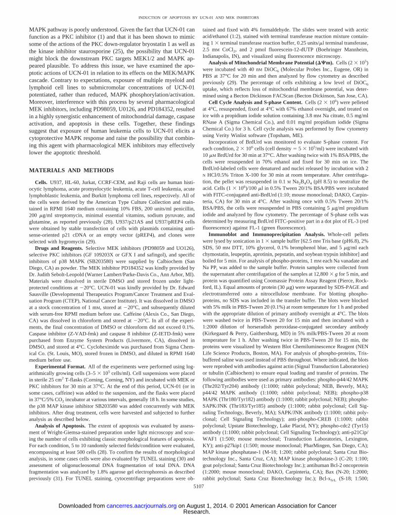
The effects of combined exposure of human monocytic leukemiacells (U937) to UCN-01 and the MEK inhibitor PD184352 were firstexamined in relation to MAPK activation and apoptosis (Fig. 1).Unexpectedly, incubation with UCN-01 (150 nM) induced phospho-rylation (activation) of MAPK by 2 h, and this effect persisted overthe ensuing 18 h (Fig. 1A). Coincubation of U937 cells withPD184532 (10mM) attenuated induction of phospho-MAPK at 2 h,and inhibition of MAPK activation was essentially complete after18 h. To determine what impact this phenomenon had on cell fate, theextent of apoptosis was monitored in cells exposed to each agentindividually and in combination. Whereas exposure to PD184352 or
Fig. 1. A, logarithmically growing U937 cells were incubated for the designatedintervals in the presence of 150 nM UCN-016 10 mM PD184352, after which cells werelysed, and proteins were separated by SDS-PAGE and probed with antibodies directedagainst phospho-ERK, as described in “Materials and Methods.” Blots were subsequentlystripped and reprobed with antibodies directed against total ERK. Two additional studiesyielded equivalent results.B, cells were treated with PD184352 (PD) and/or UCN-01(UCN; 61 mM CHX) as above for 18 h, after which Wright Giemsa-stained cytospinpreparations were evaluated by light microscopy, and the percentage of cells exhibitingclassic apoptotic features was determined by examining 5–10 randomly selected fieldsencompassing$500 cells. Values represent the means6 SD for three separate experi-ments performed in triplicate.C, cells were treated as inA, and the proteins were separatedby SDS-PAGE and probed with antibodies directed against caspase-3, caspase-9, orPARP. CF, cleavage fragment. Alternatively, cytosolic fractions were obtained as de-scribed in “Materials and Methods,” and expression of cytochromec was monitored asabove. Eachlanewas loaded with 30mg of protein. Blots were stripped and reprobed withantibodies to actin or tubulin to ensure equal loading and transfer.D, cells were treatedwith UCN-016 PD184352 (61 mM cycloheximide) as above, after which the percentageof cells exhibiting reduced mitochondrial membrane potential (DCm) was determined bymonitoring DiOC6 uptake as described in “Materials and Methods.” Results represent themeans6 SD for three separate experiments performed in triplicate.
5108
INDUCTION OF APOPTOSIS BY UCN-01 AND MEK INHIBITORS
Research. on August 1, 2014. © 2001 American Association for Cancercancerres.aacrjournals.org Downloaded from
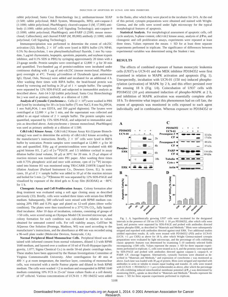
150 nM UCN-01 alone was minimally toxic to these cells (,10%apoptosis in each case), combined treatment resulted in a dramaticincrease in cell death (i.e.,;60%; Fig. 1B). Furthermore, this effectwas not attenuated by coadministration of the protein synthesis inhib-itor CHX (1 mM). Consistent with these findings, combined treatmentwith UCN-01 and PD184352, but not individual exposure, inducedmarked cleavage of procaspases-3 and -9, PARP degradation, andcytochromec release into the cytoplasmic S-100 fraction (Fig. 1C).Cotreatment of cells with UCN-01 and PD184352 also resulted in amarked increase in the number of cells exhibiting loss of the mito-chondrial membrane potential (e.g.,Dcm; Fig. 1D), an action that wasalso not attenuated by CHX. TUNEL assays confirmed that a smallnumber of cells exposed to UCN-01 or PD184352 alone for 18 h (Fig.2, B and C) displayed DNA breaks containing overhanging 39-OHends, whereas coexposure resulted in a high percentage of TUNEL-positive cells. Similarly, agarose gel electrophoresis demonstrated amarked increase in oligonucleosomal DNA fragmentation in cellsexposed to both agents (Fig. 2;bottom panel). Together, these find-ings indicate that coadministration of the MEK inhibitor PD184352blocks MAPK activation and dramatically increases apoptosis inU937 cells exposed to a marginally toxic concentration of UCN-01.
To determine whether these findings could be extended to otherknown MEK inhibitors, U937 cells were incubated for 24 h with 200nM UCN-01 either alone or in combination with PD98059 (50mM), anaminoflavone that was among the earliest of the MEK inhibitors to be
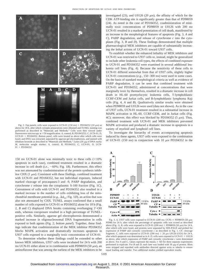
investigated (23), and U0126 (20mM), the affinity of which for theCDK ATP-binding site is significantly greater than that of PD98059(24). As noted in the case of PD184352, coadministration of mini-mally toxic concentrations of PD98059 or U0126 with 200 nM
UCN-01 resulted in a marked potentiation of cell death, manifested byan increase in the morphological features of apoptosis (Fig. 3,A andC), PARP degradation, and release of cytochromec into the cyto-plasm (Fig. 3,B and D). These findings demonstrated that multiplepharmacological MEK inhibitors are capable of substantially increas-ing the lethal actions of UCN-01 toward U937 cells.
To establish whether the enhanced lethality of MEK inhibitors andUCN-01 was restricted to U937 cells or, instead, might be generalizedto include other leukemia cell types, the effects of combined exposureto UCN-01 and PD184352 were examined in several additional leu-kemia cell lines (Fig. 4). Because the sensitivity of these cells toUCN-01 differed somewhat from that of U937 cells, slightly higherUCN-01 concentrations (e.g.,150–300 nM) were used in some cases.On the basis of standard morphological criteria as well as evidence ofPARP degradation, it can be seen that combined treatment withUCN-01 and PD184352, administered at concentrations that weremarginally toxic by themselves, resulted in a dramatic increase in celldeath in HL-60 promyelocytic leukemia cells, T-lymphoblasticCCRF-CEM and Jurkat cells, and B-lymphoblastic lymphoma Rajicells (Fig. 4,A and B). Qualitatively similar results were obtainedwhen PD98059 and U0126 were used (data not shown). As in the caseof U937 cells, UCN-01 treatment resulted in a substantial increase inMAPK activation in HL-60, CCRF-CEM, and in Jurkat cells (Fig.4C); moreover, this effect was blocked by PD184352 (5mM). Thus,combined treatment with UCN-01 and MEK inhibitors preventedMAPK activation and produced a dramatic increase in apoptosis in avariety of myeloid and lymphoid cell lines.
To investigate the hierarchy of events accompanying apoptosisinduced by these agents, U937 cells were exposed to the combinationof UCN-01 (150 nM) in conjunction with 10mM PD184352 in the
Fig. 2.Top panels, cells were exposed to UCN-01 (150 nM) 6 PD184352 (10mM) asabove for 18 h, after which cytospin preparations were obtained and TUNEL staining wasperformed as described in “Materials and Methods.” Cells were then viewed underfluorescence microscopy at350 magnification.A, control;B, PD1843252;C, UCN-01;D,UCN-01 1 PD184352.Bottom panel, cells were treated as above after which cells werelysed, and DNA was extracted, separated by agarose gel electrophoresis, and stained withethidium bromide as described in “Materials and Methods.”Lanes(20 mg of DNA each):M, molecular weight marker;A, control; B, PD184352; C, UCN-01; D, UCN-01 1 PD184352.
Fig. 3. A, U937 cells were exposed to UCN-01 (200 nM; UCN) 6 PD98059 (50mM;PD98) for 24 h, after which the percentage of apoptotic cells was scored as describedabove (Fig. 1).B, cells were exposed to UCN-01 (200 nM) 6 PD98059 (50mM) for 24 h,after which cells were lysed, and proteins were separated by SDS-PAGE and probed forexpression of PARP and cytosolic cytochromec as described in Fig. 1.CF, cleavagefragment.C, cells were exposed to UCN-01 (200 nM) 6 20 mM U0126 (UO1) for 24 h,after which apoptosis was determined as inA. D, cells were exposed to UCN-016 U0126as above, after which expression of PARP and cytosolic cytochromec were determinedas above. ForA andC, values represent the means6 SD for three separate experimentsperformed in triplicate. ForB andD, eachlane was loaded with 30mg of protein. Blotswere stripped and reprobed with antibodies directed against actin or tubulin to ensureequal loading and transfer. Two additional studies yielded equivalent results.
5109
INDUCTION OF APOPTOSIS BY UCN-01 AND MEK INHIBITORS
Research. on August 1, 2014. © 2001 American Association for Cancercancerres.aacrjournals.org Downloaded from
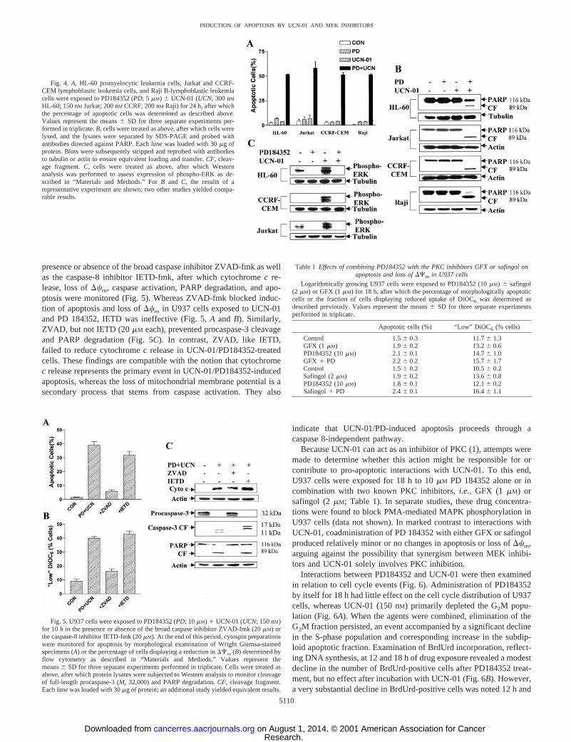
presence or absence of the broad caspase inhibitor ZVAD-fmk as wellas the caspase-8 inhibitor IETD-fmk, after which cytochromec re-lease, loss ofDcm, caspase activation, PARP degradation, and apo-ptosis were monitored (Fig. 5). Whereas ZVAD-fmk blocked induc-tion of apoptosis and loss ofDcm in U937 cells exposed to UCN-01and PD 184352, IETD was ineffective (Fig. 5,A and B). Similarly,ZVAD, but not IETD (20mM each), prevented procaspase-3 cleavageand PARP degradation (Fig. 5C). In contrast, ZVAD, like IETD,failed to reduce cytochromec release in UCN-01/PD184352-treatedcells. These findings are compatible with the notion that cytochromec release represents the primary event in UCN-01/PD184352-inducedapoptosis, whereas the loss of mitochondrial membrane potential is asecondary process that stems from caspase activation. They also
indicate that UCN-01/PD-induced apoptosis proceeds through acaspase 8-independent pathway.
Because UCN-01 can act as an inhibitor of PKC (1), attempts weremade to determine whether this action might be responsible for orcontribute to pro-apoptotic interactions with UCN-01. To this end,U937 cells were exposed for 18 h to 10mM PD 184352 alone or incombination with two known PKC inhibitors,i.e., GFX (1 mM) orsafingol (2mM; Table 1). In separate studies, these drug concentra-tions were found to block PMA-mediated MAPK phosphorylation inU937 cells (data not shown). In marked contrast to interactions withUCN-01, coadministration of PD 184352 with either GFX or safingolproduced relatively minor or no changes in apoptosis or loss ofDcm,arguing against the possibility that synergism between MEK inhibi-tors and UCN-01 solely involves PKC inhibition.
Interactions between PD184352 and UCN-01 were then examinedin relation to cell cycle events (Fig. 6). Administration of PD184352by itself for 18 h had little effect on the cell cycle distribution of U937cells, whereas UCN-01 (150 nM) primarily depleted the G2M popu-lation (Fig. 6A). When the agents were combined, elimination of theG2M fraction persisted, an event accompanied by a significant declinein the S-phase population and corresponding increase in the subdip-loid apoptotic fraction. Examination of BrdUrd incorporation, reflect-ing DNA synthesis, at 12 and 18 h of drug exposure revealed a modestdecline in the number of BrdUrd-positive cells after PD184352 treat-ment, but no effect after incubation with UCN-01 (Fig. 6B). However,a very substantial decline in BrdUrd-positive cells was noted 12 h and
Fig. 4. A, HL-60 promyelocytic leukemia cells, Jurkat and CCRF-CEM lymphoblastic leukemia cells, and Raji B-lymphoblastic leukemiacells were exposed to PD184352 (PD; 5mM) 6 UCN-01 (UCN; 300 nMHL-60; 150 nM Jurkat; 200 nM CCRF; 200 nM Raji) for 24 h, after whichthe percentage of apoptotic cells was determined as described above.Values represent the means6 SD for three separate experiments per-formed in triplicate.B, cells were treated as above, after which cells werelysed, and the lysates were separated by SDS-PAGE and probed withantibodies directed against PARP. Eachlanewas loaded with 30mg ofprotein. Blots were subsequently stripped and reprobed with antibodiesto tubulin or actin to ensure equivalent loading and transfer.CF, cleav-age fragment.C, cells were treated as above, after which Westernanalysis was performed to assess expression of phospho-ERK as de-scribed in “Materials and Methods.” ForB and C, the results of arepresentative experiment are shown; two other studies yielded compa-rable results.
Fig. 5. U937 cells were exposed to PD184352 (PD; 10mM) 1 UCN-01 (UCN; 150 nM)for 10 h in the presence or absence of the broad caspase inhibitor ZVAD-fmk (20mM) orthe caspase-8 inhibitor IETD-fmk (20mM). At the end of this period, cytospin preparationswere monitored for apoptosis by morphological examination of Wright Giemsa-stainedspecimens (A) or the percentage of cells displaying a reduction inDCm (B) determined byflow cytometry as described in “Materials and Methods.” Values represent themeans6 SD for three separate experiments performed in triplicate. Cells were treated asabove, after which protein lysates were subjected to Western analysis to monitor cleavageof full-length procaspase-3 (Mr 32,000) and PARP degradation.CF, cleavage fragment.Eachlanewas loaded with 30mg of protein; an additional study yielded equivalent results.
Table 1 Effects of combining PD184352 with the PKC inhibitors GFX or safingol onapoptosis and loss ofDCm in U937 cells
Logarithmically growing U937 cells were exposed to PD184352 (10mM) 6 safingol(2 mM) or GFX (1mM) for 18 h, after which the percentage of morphologically apoptoticcells or the fraction of cells displaying reduced uptake of DiOC6 was determined asdescribed previously. Values represent the means6 SD for three separate experimentsperformed in triplicate.
Apoptotic cells (%) “Low” DiOC6 (% cells)
Control 1.56 0.3 11.76 1.3GFX (1 mM) 1.96 0.2 13.26 0.6PD184352 (10mM) 2.16 0.1 14.76 1.0GFX 1 PD 2.26 0.2 15.76 1.7Control 1.56 0.2 10.56 0.2Safingol (2mM) 1.96 0.2 13.66 0.8PD184352 (10mM) 1.86 0.1 12.16 0.2Safingol1 PD 2.46 0.1 16.46 1.1
5110
INDUCTION OF APOPTOSIS BY UCN-01 AND MEK INHIBITORS
Research. on August 1, 2014. © 2001 American Association for Cancercancerres.aacrjournals.org Downloaded from
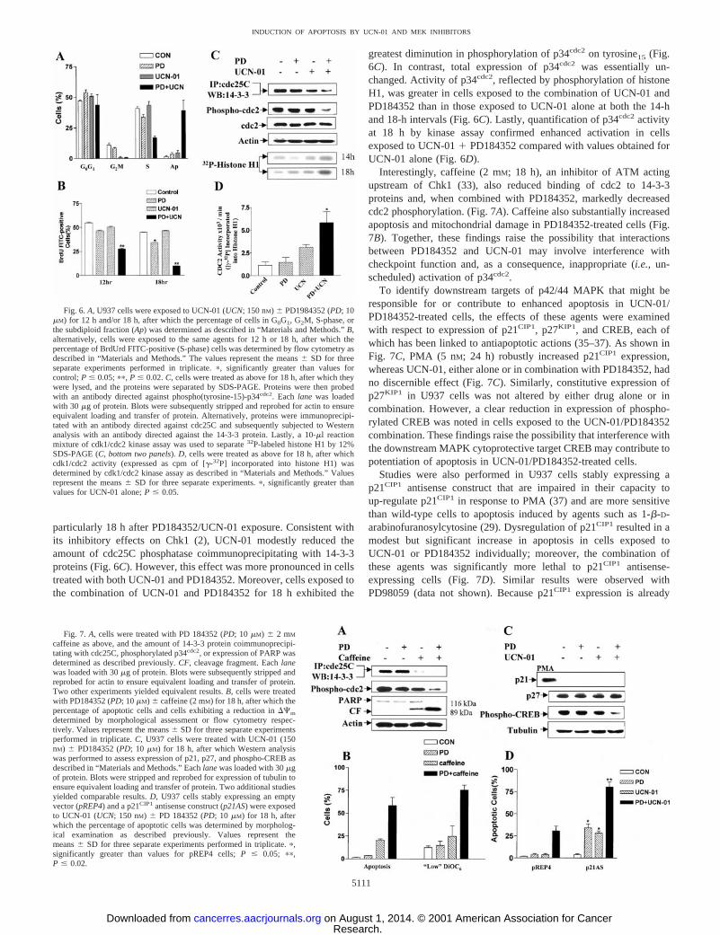
particularly 18 h after PD184352/UCN-01 exposure. Consistent withits inhibitory effects on Chk1 (2), UCN-01 modestly reduced theamount of cdc25C phosphatase coimmunoprecipitating with 14-3-3proteins (Fig. 6C). However, this effect was more pronounced in cellstreated with both UCN-01 and PD184352. Moreover, cells exposed tothe combination of UCN-01 and PD184352 for 18 h exhibited the
greatest diminution in phosphorylation of p34cdc2 on tyrosine15 (Fig.6C). In contrast, total expression of p34cdc2 was essentially un-changed. Activity of p34cdc2, reflected by phosphorylation of histoneH1, was greater in cells exposed to the combination of UCN-01 andPD184352 than in those exposed to UCN-01 alone at both the 14-hand 18-h intervals (Fig. 6C). Lastly, quantification of p34cdc2activityat 18 h by kinase assay confirmed enhanced activation in cellsexposed to UCN-011 PD184352 compared with values obtained forUCN-01 alone (Fig. 6D).
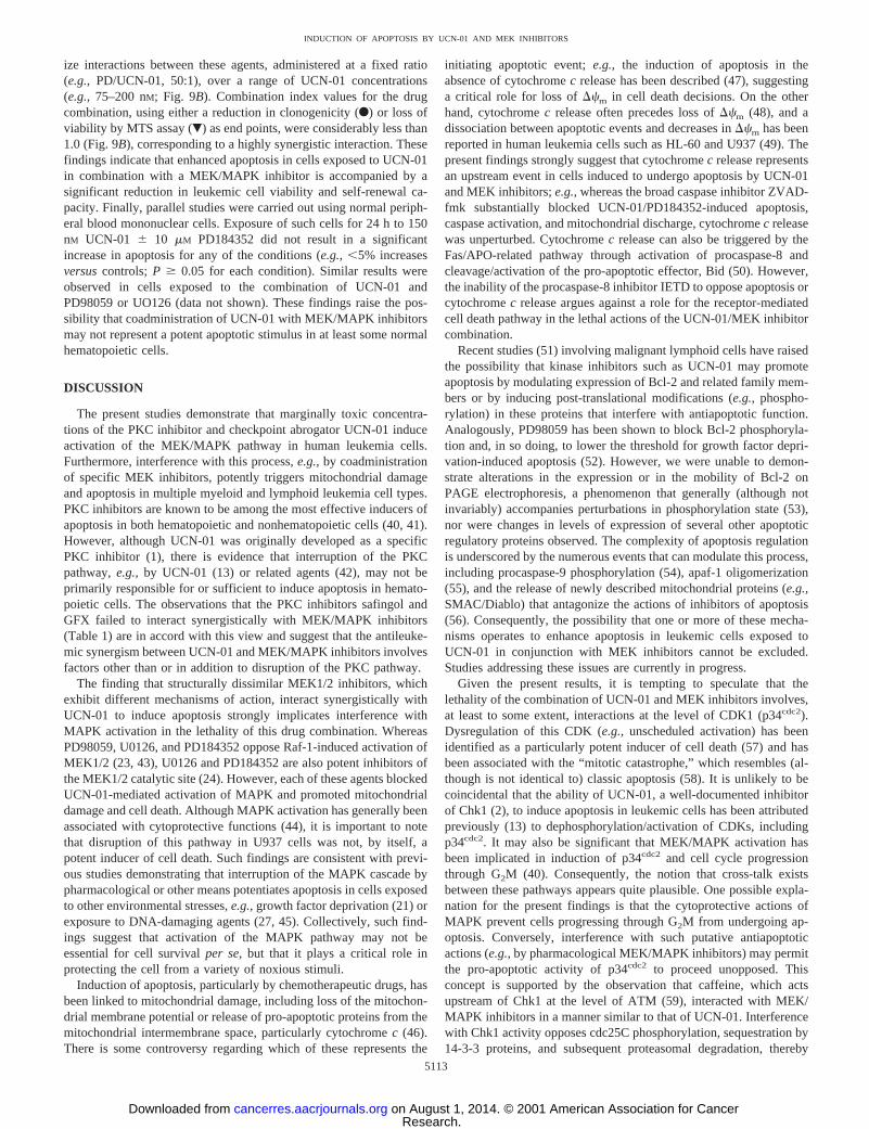
Interestingly, caffeine (2 mM; 18 h), an inhibitor of ATM actingupstream of Chk1 (33), also reduced binding of cdc2 to 14-3-3proteins and, when combined with PD184352, markedly decreasedcdc2 phosphorylation. (Fig. 7A). Caffeine also substantially increasedapoptosis and mitochondrial damage in PD184352-treated cells (Fig.7B). Together, these findings raise the possibility that interactionsbetween PD184352 and UCN-01 may involve interference withcheckpoint function and, as a consequence, inappropriate (i.e., un-scheduled) activation of p34cdc2.
To identify downstream targets of p42/44 MAPK that might beresponsible for or contribute to enhanced apoptosis in UCN-01/PD184352-treated cells, the effects of these agents were examinedwith respect to expression of p21CIP1, p27KIP1, and CREB, each ofwhich has been linked to antiapoptotic actions (35–37). As shown inFig. 7C, PMA (5 nM; 24 h) robustly increased p21CIP1 expression,whereas UCN-01, either alone or in combination with PD184352, hadno discernible effect (Fig. 7C). Similarly, constitutive expression ofp27KIP1 in U937 cells was not altered by either drug alone or incombination. However, a clear reduction in expression of phospho-rylated CREB was noted in cells exposed to the UCN-01/PD184352combination. These findings raise the possibility that interference withthe downstream MAPK cytoprotective target CREB may contribute topotentiation of apoptosis in UCN-01/PD184352-treated cells.
Studies were also performed in U937 cells stably expressing ap21CIP1 antisense construct that are impaired in their capacity toup-regulate p21CIP1 in response to PMA (37) and are more sensitivethan wild-type cells to apoptosis induced by agents such as 1-b-D-arabinofuranosylcytosine (29). Dysregulation of p21CIP1 resulted in amodest but significant increase in apoptosis in cells exposed toUCN-01 or PD184352 individually; moreover, the combination ofthese agents was significantly more lethal to p21CIP1 antisense-expressing cells (Fig. 7D). Similar results were observed withPD98059 (data not shown). Because p21CIP1 expression is already
Fig. 6.A, U937 cells were exposed to UCN-01 (UCN; 150 nM) 6 PD1984352 (PD; 10mM) for 12 h and/or 18 h, after which the percentage of cells in G0G1, G2M, S-phase, orthe subdiploid fraction (Ap) was determined as described in “Materials and Methods.”B,alternatively, cells were exposed to the same agents for 12 h or 18 h, after which thepercentage of BrdUrd FITC-positive (S-phase) cells was determined by flow cytometry asdescribed in “Materials and Methods.” The values represent the means6 SD for threeseparate experiments performed in triplicate.p, significantly greater than values forcontrol;P # 0.05;pp, P # 0.02.C, cells were treated as above for 18 h, after which theywere lysed, and the proteins were separated by SDS-PAGE. Proteins were then probedwith an antibody directed against phospho(tyrosine-15)-p34cdc2. Each lane was loadedwith 30 mg of protein. Blots were subsequently stripped and reprobed for actin to ensureequivalent loading and transfer of protein. Alternatively, proteins were immunoprecipi-tated with an antibody directed against cdc25C and subsequently subjected to Westernanalysis with an antibody directed against the 14-3-3 protein. Lastly, a 10-ml reactionmixture of cdk1/cdc2 kinase assay was used to separate32P-labeled histone H1 by 12%SDS-PAGE (C,bottom two panels).D, cells were treated as above for 18 h, after whichcdk1/cdc2 activity (expressed as cpm of [g-32P] incorporated into histone H1) wasdetermined by cdk1/cdc2 kinase assay as described in “Materials and Methods.” Valuesrepresent the means6 SD for three separate experiments.p, significantly greater thanvalues for UCN-01 alone;P # 0.05.
Fig. 7. A, cells were treated with PD 184352 (PD; 10mM) 6 2 mM
caffeine as above, and the amount of 14-3-3 protein coimmunoprecipi-tating with cdc25C, phosphorylated p34cdc2, or expression of PARP wasdetermined as described previously.CF, cleavage fragment. Eachlanewas loaded with 30mg of protein. Blots were subsequently stripped andreprobed for actin to ensure equivalent loading and transfer of protein.Two other experiments yielded equivalent results.B, cells were treatedwith PD184352 (PD; 10mM) 6 caffeine (2 mM) for 18 h, after which thepercentage of apoptotic cells and cells exhibiting a reduction inDCm
determined by morphological assessment or flow cytometry respec-tively. Values represent the means6 SD for three separate experimentsperformed in triplicate.C, U937 cells were treated with UCN-01 (150nM) 6 PD184352 (PD; 10mM) for 18 h, after which Western analysiswas performed to assess expression of p21, p27, and phospho-CREB asdescribed in “Materials and Methods.” Eachlanewas loaded with 30mgof protein. Blots were stripped and reprobed for expression of tubulin toensure equivalent loading and transfer of protein. Two additional studiesyielded comparable results.D, U937 cells stably expressing an emptyvector (pREP4) and a p21CIP1antisense construct (p21AS) were exposedto UCN-01 (UCN; 150 nM) 6 PD 184352 (PD; 10mM) for 18 h, afterwhich the percentage of apoptotic cells was determined by morpholog-ical examination as described previously. Values represent themeans6 SD for three separate experiments performed in triplicate.p,significantly greater than values for pREP4 cells;P # 0.05; pp,P # 0.02.
5111
INDUCTION OF APOPTOSIS BY UCN-01 AND MEK INHIBITORS
Research. on August 1, 2014. © 2001 American Association for Cancercancerres.aacrjournals.org Downloaded from

dysregulated in the antisense line, these and the preceding findings(Fig. 7C) argue against the possibility that potentiation of UCN-01-related apoptosis by MEK/MAPK inhibitors involves impaired induc-tion of the downstream MAPK target p21CIP1.
To assess the influence of MEK inhibitors and UCN-01 on otherMAPK pathways, the effects of these agents were examined in rela-tion to JNK and p38 phosphorylation (Fig. 8). In contrast to theincrease in expression of phospho-MAPK, UCN-01, either alone or incombination with PD184352, did not noticeably induce JNK phos-phorylation in U937 cells (Fig. 8A). Similar results were obtained withPD98059 (data not shown). In separate studies (38) involving U937cell transfectants, stable expression of a dominant-negative c-Juntransactivation domain-deficient mutant (TAM67) did not attenuatePD184352/UCN-01-mediated apoptosis (data not shown). Interest-ingly, coadministration of UCN-01 and PD184352, but not individualdrug exposure, resulted in a marked increase in expression of phos-pho-p38 MAPK. However, coadministration of the p38 MAPK inhib-itor SB203580 (10mM) only partially attenuated apoptosis and mito-chondrial injury in PD184352/UCN-01-treated cells (Fig. 8B). Lastly,combined drug exposure exerted did not increase expression of theMKP1 and MKP3 phosphatases (Fig. 7A). Together, these findingsindicate that potentiation of UCN-01-related apoptosis by MEK in-hibitors is accompanied by a marked increase in p38 MAPK but notJNK phosphorylation.
To determine whether coadministration UCN-01 and MEK inhib-itors modified the expression of apoptotic regulatory proteins, levelsof Bcl-2, Bcl-xL, Bax, and XIAP were monitored by Western analysis(Fig. 8C). Coadministration of UCN-01 and PD184352 did not result
in a significant change in expression of Bcl-2, Bcl-xL, Bax, or XIAP.Similar results were observed in cells exposed to the combination ofUCN-01 and PD98059 (data not shown). In addition, separation ofproteins on a 15-cm, 12% SDS-PAGE gel, which permitted visual-ization of a slowly migrating, putatively phosphorylated Bcl-2 species(Fig. 7C,second panel from top), revealed no significant change afterexposure of cells to PD184352 in combination with UCN-01. Theseobservations argue against the possibility that potentiation of UCN-01-induced apoptosis by MEK inhibitors stemmed from increasedexpression of Bax or diminished expression of the antiapoptoticproteins Bcl-2, Bcl-xL, or XIAP.
Finally, the impact of combined treatment of U937 cells withUCN-01 and PD184352 was examined in relation to effects on clo-nogenic survival (Fig. 9). UCN-01 (150 nM; 18 h) by itself had a verymodest effect on colony formation, whereas PD184352 (10mM; 18 h)administered alone reduced clonogenic survival by;40%. However,combined treatment with both agents resulted in a substantial reduc-tion in clonogenicity (e.g.,to ;10% of control values; Fig. 9A).Furthermore, median dose effect analysis (39) was used to character-
Fig. 8.A, U937 cells were exposed to UCN-01 (UCN; 150 nM) 6 PD184352 (PD; 10mM) for 18 h, after which cells were lysed, and the lysates were separated by SDS-PAGEand probed with antibodies directed against phospho-p38 MAPK, phospho-JNK, totalJNK, MKP1, and MKP3.B, cells were exposed to PD184352 and UCN-01 as above for18 h in the presence or absence of the p38 MAPK inhibitor SB203580 (10mM), afterwhich the percentage of cells exhibiting the morphological features of apoptosis orreduction in DCm, reflected by a diminished uptake of DiOC6, was determined asdescribed previously. Values represent the means6 SD for three separate experimentsperformed in triplicate.pp, significantly less than values for UCN1 PD withoutSB203580;P # 0.02.C, cells were treated as above, after which levels of expression ofBcl-2, Bax, Bcl-xL, or XIAP were determined by Western analysis as described in“Materials and Methods.” Alternatively, proteins were separated by running a 15-cm 12%SDS-PAGE gel, which permitted detection of a putatively phosphorylated, slowly mi-grating Bcl-2 species (designated phospho-Bcl-2). ForA andC, eachlanewas loaded with30 mg of protein. Blots were stripped and reprobed for actin to ensure equivalent loadingand transfer of protein. In each case, two additional studies yielded comparable results.
Fig. 9.A, cells were exposed to PD184352 (5mM) 6 UCN-01 (100 nM) for 18 h, afterwhich cells were washed free of drug and plated in soft agar as described in the text. After12 days of incubation, colonies, consisting of groups of$50 cells, were scored, andcolony formation for each condition was expressed relative to untreated control cells.Values represent the means6 SD for three separate experiments.B, U937 cells wereexposed to a range of PD184352 (e.g.,3.75–10mM) and UCN-01 (e.g.,75–200 nM)concentrations alone and in combination at fixed ratio (e.g.,50:1) for 18 h. At the end ofthis period, colony formation was determined for each condition as above. Alternatively,cell viability was determined using the cell titer 96 reagent as described in “Materials andMethods.” In each case, the fraction affected values were determined by comparing resultswith those of untreated controls, and median dose-effect analysis was used to characterizethe nature of the interaction between UCN-01 and PD184352 using a commerciallyavailable program (CalcuSyn; Biosoft).F, values obtained for clonogenic assays;�,values obtained for viability assays. Combination index values less than 1.0 denote asynergistic interaction. Two additional studies yielded equivalent results.C, normalperipheral blood mononuclear cells were exposed to 150 nM UCN-016 10mM PD184352for 18 h, after which the percentage of apoptotic cells was determined by morphologicalexamination as described previously. Values represent the means6 SD for triplicatedetermination; a second independent study yielded equivalent results.
5112
INDUCTION OF APOPTOSIS BY UCN-01 AND MEK INHIBITORS
Research. on August 1, 2014. © 2001 American Association for Cancercancerres.aacrjournals.org Downloaded from
ize interactions between these agents, administered at a fixed ratio(e.g., PD/UCN-01, 50:1), over a range of UCN-01 concentrations(e.g., 75–200 nM; Fig. 9B). Combination index values for the drugcombination, using either a reduction in clonogenicity (F) or loss ofviability by MTS assay (�) as end points, were considerably less than1.0 (Fig. 9B), corresponding to a highly synergistic interaction. Thesefindings indicate that enhanced apoptosis in cells exposed to UCN-01in combination with a MEK/MAPK inhibitor is accompanied by asignificant reduction in leukemic cell viability and self-renewal ca-pacity. Finally, parallel studies were carried out using normal periph-eral blood mononuclear cells. Exposure of such cells for 24 h to 150nM UCN-01 6 10 mM PD184352 did not result in a significantincrease in apoptosis for any of the conditions (e.g.,,5% increasesversuscontrols;P $ 0.05 for each condition). Similar results wereobserved in cells exposed to the combination of UCN-01 andPD98059 or UO126 (data not shown). These findings raise the pos-sibility that coadministration of UCN-01 with MEK/MAPK inhibitorsmay not represent a potent apoptotic stimulus in at least some normalhematopoietic cells.
DISCUSSION
The present studies demonstrate that marginally toxic concentra-tions of the PKC inhibitor and checkpoint abrogator UCN-01 induceactivation of the MEK/MAPK pathway in human leukemia cells.Furthermore, interference with this process,e.g.,by coadministrationof specific MEK inhibitors, potently triggers mitochondrial damageand apoptosis in multiple myeloid and lymphoid leukemia cell types.PKC inhibitors are known to be among the most effective inducers ofapoptosis in both hematopoietic and nonhematopoietic cells (40, 41).However, although UCN-01 was originally developed as a specificPKC inhibitor (1), there is evidence that interruption of the PKCpathway,e.g., by UCN-01 (13) or related agents (42), may not beprimarily responsible for or sufficient to induce apoptosis in hemato-poietic cells. The observations that the PKC inhibitors safingol andGFX failed to interact synergistically with MEK/MAPK inhibitors(Table 1) are in accord with this view and suggest that the antileuke-mic synergism between UCN-01 and MEK/MAPK inhibitors involvesfactors other than or in addition to disruption of the PKC pathway.
The finding that structurally dissimilar MEK1/2 inhibitors, whichexhibit different mechanisms of action, interact synergistically withUCN-01 to induce apoptosis strongly implicates interference withMAPK activation in the lethality of this drug combination. WhereasPD98059, U0126, and PD184352 oppose Raf-1-induced activation ofMEK1/2 (23, 43), U0126 and PD184352 are also potent inhibitors ofthe MEK1/2 catalytic site (24). However, each of these agents blockedUCN-01-mediated activation of MAPK and promoted mitochondrialdamage and cell death. Although MAPK activation has generally beenassociated with cytoprotective functions (44), it is important to notethat disruption of this pathway in U937 cells was not, by itself, apotent inducer of cell death. Such findings are consistent with previ-ous studies demonstrating that interruption of the MAPK cascade bypharmacological or other means potentiates apoptosis in cells exposedto other environmental stresses,e.g.,growth factor deprivation (21) orexposure to DNA-damaging agents (27, 45). Collectively, such find-ings suggest that activation of the MAPK pathway may not beessential for cell survivalper se, but that it plays a critical role inprotecting the cell from a variety of noxious stimuli.
Induction of apoptosis, particularly by chemotherapeutic drugs, hasbeen linked to mitochondrial damage, including loss of the mitochon-drial membrane potential or release of pro-apoptotic proteins from themitochondrial intermembrane space, particularly cytochromec (46).There is some controversy regarding which of these represents the
initiating apoptotic event;e.g., the induction of apoptosis in theabsence of cytochromec release has been described (47), suggestinga critical role for loss ofDcm in cell death decisions. On the otherhand, cytochromec release often precedes loss ofDcm (48), and adissociation between apoptotic events and decreases inDcm has beenreported in human leukemia cells such as HL-60 and U937 (49). Thepresent findings strongly suggest that cytochromec release representsan upstream event in cells induced to undergo apoptosis by UCN-01and MEK inhibitors;e.g.,whereas the broad caspase inhibitor ZVAD-fmk substantially blocked UCN-01/PD184352-induced apoptosis,caspase activation, and mitochondrial discharge, cytochromec releasewas unperturbed. Cytochromec release can also be triggered by theFas/APO-related pathway through activation of procaspase-8 andcleavage/activation of the pro-apoptotic effector, Bid (50). However,the inability of the procaspase-8 inhibitor IETD to oppose apoptosis orcytochromec release argues against a role for the receptor-mediatedcell death pathway in the lethal actions of the UCN-01/MEK inhibitorcombination.
Recent studies (51) involving malignant lymphoid cells have raisedthe possibility that kinase inhibitors such as UCN-01 may promoteapoptosis by modulating expression of Bcl-2 and related family mem-bers or by inducing post-translational modifications (e.g., phospho-rylation) in these proteins that interfere with antiapoptotic function.Analogously, PD98059 has been shown to block Bcl-2 phosphoryla-tion and, in so doing, to lower the threshold for growth factor depri-vation-induced apoptosis (52). However, we were unable to demon-strate alterations in the expression or in the mobility of Bcl-2 onPAGE electrophoresis, a phenomenon that generally (although notinvariably) accompanies perturbations in phosphorylation state (53),nor were changes in levels of expression of several other apoptoticregulatory proteins observed. The complexity of apoptosis regulationis underscored by the numerous events that can modulate this process,including procaspase-9 phosphorylation (54), apaf-1 oligomerization(55), and the release of newly described mitochondrial proteins (e.g.,SMAC/Diablo) that antagonize the actions of inhibitors of apoptosis(56). Consequently, the possibility that one or more of these mecha-nisms operates to enhance apoptosis in leukemic cells exposed toUCN-01 in conjunction with MEK inhibitors cannot be excluded.Studies addressing these issues are currently in progress.
Given the present results, it is tempting to speculate that thelethality of the combination of UCN-01 and MEK inhibitors involves,at least to some extent, interactions at the level of CDK1 (p34cdc2).Dysregulation of this CDK (e.g.,unscheduled activation) has beenidentified as a particularly potent inducer of cell death (57) and hasbeen associated with the “mitotic catastrophe,” which resembles (al-though is not identical to) classic apoptosis (58). It is unlikely to becoincidental that the ability of UCN-01, a well-documented inhibitorof Chk1 (2), to induce apoptosis in leukemic cells has been attributedpreviously (13) to dephosphorylation/activation of CDKs, includingp34cdc2. It may also be significant that MEK/MAPK activation hasbeen implicated in induction of p34cdc2 and cell cycle progressionthrough G2M (40). Consequently, the notion that cross-talk existsbetween these pathways appears quite plausible. One possible expla-nation for the present findings is that the cytoprotective actions ofMAPK prevent cells progressing through G2M from undergoing ap-optosis. Conversely, interference with such putative antiapoptoticactions (e.g.,by pharmacological MEK/MAPK inhibitors) may permitthe pro-apoptotic activity of p34cdc2 to proceed unopposed. Thisconcept is supported by the observation that caffeine, which actsupstream of Chk1 at the level of ATM (59), interacted with MEK/MAPK inhibitors in a manner similar to that of UCN-01. Interferencewith Chk1 activity opposes cdc25C phosphorylation, sequestration by14-3-3 proteins, and subsequent proteasomal degradation, thereby
5113
INDUCTION OF APOPTOSIS BY UCN-01 AND MEK INHIBITORS
Research. on August 1, 2014. © 2001 American Association for Cancercancerres.aacrjournals.org Downloaded from

allowing this phosphatase to dephosphorylate and activate p34cdc2(3).Thus, in the present studies, coadministration of caffeine, like UCN-01, with PD184352 resulted in reduced binding of the cdc25C phos-phatase to 14-3-3 proteins, dephosphorylation of p34cdc2, and amarked increase in lethality. Collectively, these findings suggest thatthe combination of p34cdc2 activation with disruption of the MAPKcascade represents a potent apoptotic stimulus, at least in the case ofmalignant hematopoietic cells.
The possibility that other downstream MAPK effectors contributeto this phenomenon cannot be ruled out, particularly in view of thereduced phosphorylation of CREB noted in UCN-01/PD184352-treated cells. CREB has been identified recently (36, 60) as a cyto-protective target of the Raf3MAPK3Rsk cascade, and it seemsplausible that interference with phosphorylation/activation of thistranscription factor (e.g.,by PD184352) contributed to the observedpotentiation of apoptosis. In addition, cross-talk between cytoprotec-tive and stress-related MAPK modules has been described (61), and itis possible that such interactions might contribute to the lethality ofthe UCN-01/PD184352 combination. In fact, the observations thatinhibition of UCN-01-induced MAPK activation by PD184352 wasassociated with a reciprocal increase in p38 MAPK induction and thatcoadministration of the p38 MAPK inhibitor SB203580 partiallyprotected cells from apoptosis induced by UCN-01/PD184352 raisethe possibility that the p38 MAPK cascade could be involved, at leastto some extent, in potentiation of cell death by this drug combination.However, given recent evidence (62) that SB203580 inhibits kinasesother than p38 MAPK and the finding that protection from apoptosisby SB203580 was incomplete, it seems highly likely that other factorsare involved in the lethal effects of this drug combination. Lastly, thepossibility that MEK inhibitors specifically act by interfering withinduction of p21CIP1, a known MAPK downstream target (63), ap-pears remote, given the findings that: (a) UCN-01 failed to inducep21CIP1; and (b) p21CIP1 antisense-expressing cells, which alreadyexhibit dysregulation of this CDKI, displayed enhanced susceptibilityto the UCN-01/PD184352 combination. Nevertheless, these findingsremain compatible with a cytoprotective role for basal p21CIP1 ex-pression, a phenomenon that has been described previously (64).
In summary, the present studies demonstrate that the kinase inhib-itor and checkpoint antagonist UCN-01 unexpectedly activatesMAPK in human leukemia cells and that interference with this proc-ess by multiple pharmacological MEK/MAPK inhibitors leads to amarked potentiation of mitochondrial injury (e.g.,cytochromec re-lease), caspase activation, and apoptosis. Moreover, these eventsoccur in a variety of myeloid and lymphoid leukemia cell types,indicating that this phenomenon is not lineage-specific. Finally, en-hanced apoptosis in these cells is associated with perturbations inseveral signaling and cell cycle regulatory pathways, including de-phosphorylation of p34cdc2 and CREB, as well as activation of p38MAPK. Significantly, such observations raise the possibility thatinterruption of multiple signaling pathways (e.g.,by pharmacologicalkinase inhibitors) may provide a particularly potent apoptotic stimu-lus, at least in malignant hematopoietic cells. Aside from providinginsights into factors that regulate the lethal actions of UCN-01, thesefindings have potential therapeutic implications;e.g., in humans,levels of free UCN-01 achievable in the plasma and potentiallyavailable to tumor cells are limited by extensive binding of this agentto a1 acidic glycoprotein (14). It is conceivable that MEK inhibitors,particularly those which, like PD184352, are activein vivo (26), couldpotentiate the antileukemic activity of pharmacologically relevantconcentrations of UCN-01. In this regard, it would be of interest todetermine whether synergistic interactions between UCN-01 andMEK/MAPK inhibitors could be extended to other hematological and
nonhematological tumor types. Accordingly, studies addressing thisquestion are currently underway.
REFERENCES
1. Mizuno, K., Noda, K., Ueda, Y., Hanaki, H., Saido, T. C., Ikuta, T., Kuroki, T.,Tamaoki, T., Hirai, S., and Osada, S. UCN-01, an anti-tumor drug, is a selectiveinhibitor of the conventional PKC subfamily. FEBS Lett.,359: 259–261, 1995.
2. Graves, P. R., Yu, L., Schwarz, J. K., Gales, J., Sausville, E. A., O’Connor, P. M., andPiwnica-Worms, H. The Chk1 protein kinase and the Cdc25C regulatory pathwaysare targets of the anticancer agent UCN-01. J. Biol. Chem.,275: 5600–5605, 2000.
3. Peng, C-Y., Gravesa, P. R., Thoma, R. S., Wu, Z., Shaw, A. S., and Piwnica-Worms,H. Mitotic and G2 checkpoint control: regulation of 14-3-3 protein binding byphosphorylation of Cdc25C on serine-216. Science (Wash. DC),277: 1501–1505,1997.
4. Bunch, R. T., and Eastman, A. Enhancement ofcis-platinum-induced cytotoxicity by7-hydroxystaurosporine, a new G2 checkpoint inhibitor. Clin. Cancer Res.,2: 791–797, 1996.
5. Akinaga, S., Nomura, K., Gomi, K., and Okabe, M. Enhancement of antitumoractivity of mitomycin C in vitro and in vivo by UCN-01, a selective inhibitor ofprotein kinase C. Cancer Chemother. Pharmacol.,32: 183–189, 1993.
6. Shao, R-G., Cao, C-X., Shimizu, T., O’Connor, P. M., Kohn, K. W., and Pommier,Y. Abrogation of an S-phase checkpoint and potentiation of camptothecin cytotox-icity by 7-hydroxystaurosporine (UCN-01) in human cancer cell lines, possiblyinfluenced by p53 function. Cancer Res.,57: 4029–4035, 1997.
7. Harvey, S., Decker, R., Dai, Y., Schaefer, G., Tang, L., Kramer, L., Dent, P., andGrant, S. Interactions between 2-fluoroadenine 9-b-D-arabinofuranoside and the ki-nase inhibitor UCN-01 in human leukemia and lymphoma cells. Clin. Cancer Res.,7:320–330, 2001.
8. Shi, Z., Azuma, A., Sampath, D., Li, Y. X., Huang, P., and Plunkett, W. S-Phasearrest by nucleoside analogues and abrogation of survival without cell cycle progres-sion by 7-hydroxystaurosporine. Cancer Res.,61: 1065–1072, 2001.
9. Tang, L., Boise, L. H., Dent, P., and Grant, S. Potentiation of 1-b-D-arabinofurano-sylcytosine-mediated mitochondrial damage and apoptosis in human leukemia cells(U937) overexpressing bcl-2 by the kinase inhibitor 7-hydroxystaurosporine (UCN-01). Biochem. Pharmacol.,60: 1445–1456, 2000.
10. Wang, S., Vrana, J. A., Bartimole, T. M., Freemerman, A. J., Jarvis, W. D., Kramer,L. B., Krystal, G., Dent, P., and Grant, S. Agents that down-regulate or inhibit proteinkinase C circumvent resistance to 1-b-D-arabinofuranosylcytosine-induced apoptosisin human leukemia cells that overexpress Bcl-2. Mol. Pharmacol.,52: 1000–1009,1997.
11. Akinaga, S., Nomura, K., Gomi, K., and Okabe, M. Effect of UCN-01, a selectiveinhibitor of protein kinase C, on the cell-cycle distribution of human epidermoidcarcinoma, A431 cells. Cancer Chemother. Pharmacol.,33: 273–280, 1994.
12. Chen, X., Lowe, M., and Keyomarsi, K. UCN-01-mediated G1 arrest in normal butnot tumor breast cells is pRb-dependent and p53-independent. Oncogene,18: 5691–5702, 1999.
13. Wang, Q., Worland, P. J., Clark, J. L., Carlson, B. A., and Sausville, E. A. Apoptosisin 7-hydroxystaurosporine-treated T lymphoblasts correlates with activation of cy-clin-dependent kinases 1 and 2. Cell Growth Differ.,6: 927–936, 1995.
14. Fuse, E., Tanii, H., Takai, K., Asanome, K., Kurata, N., Kobayashi, H., Kuwabara, T.,Kobayashi, S., and Sugiyama, Y. Altered pharmacokinetics of a novel anticancerdrug, UCN-01, caused by specific high affinity binding toa1-acid glycoprotein inhumans. Cancer Res.,59: 1054–1060, 1999.
15. Kurata, N., Kuwabara, T., Tanii, H., Fuse, E., Akiyama, T., Akinaga, S., Kobayashi,H., Ymaguchi, K., and Kobayashi, S. Pharmacokinetics and pharmacodynamics of anovel protein kinase inhibitor UCN-01. Cancer Chemother. Pharmacol.,44: 12–18,1999.
16. Wilson, W. H., Sorbara, L., Figg, W. D., Mont, E. K., Sausville, E., Warren, K. E.,Balis, F. M., Bauer, K., Raffeld, M., Senderowicz, A. M., and Monks, A. Modulationof clinical drug resistance in a B-cell lymphoma patient by the protein kinase inhibitor7-hydroxystaurosporine: presentation of a novel therapeutic paradigm. Clin. CancerRes.,6: 415–421, 2000.
17. Leppa, S., and Bohmann, D. Diverse functions of JNK signaling and c-Jun in stressresponse and apoptosis. Oncogene,18: 6158–6162, 1999.
18. Verheij, M., Bose, R., Lin, X. H., Yao, B., Jarvis, W. D., Grant, S., Birrer, M. J.,Szabo, E., Zon, L. I., Kyriakis, J. M., Haimovitz-Friedman, A., Fuks, Z., andKolesnick, R. Requirement for ceramide-initiated SAPK/JNK signaling in stress-induced apoptosis. Nature (Lond.),380: 75–79, 1996.
19. Segar, R., and Krebs, E. G. The MAPK signaling cascade. FASEB J.,9: 726–735,1995.
20. Cross, T. G., Scheel-Toellner, D., Henriquez, N. V., Deacon, E., Salmon, M., andLord, J. M. Serine/threonine protein kinases and apoptosis. Exp. Cell Res.,256:34–41, 2000.
21. Xia, Z., Dickens, M., Raingeaud, J., Davis, R., and Greenberg, M. E. Opposing effectsof ERK and JNK-p38 MAP kinases on apoptosis. Science (Wash. DC),270: 1326–1331, 1995.
22. Tibbles, L. A., and Woodgett, J. R. The stress-activated protein kinase pathways. Cell.Mol. Life Sci., 55: 1230–1254, 1999.
23. Dudley, D. T., Pang, L., Decker, S. J., Bridges, A. J., and Saltiel, A. R. A syntheticinhibitor of the mitogen-activated protein kinase cascade. Proc. Natl. Acad. Sci. USA,92: 7686–7689, 1995.
24. Favata, M. F., Horiuchi, K. Y., Manos, E. J., Daulerio, A. J., Stradley, D. A., Feeser,W. S., Van Dyk, D. E., Pitts, W. J., Earl, R. A., Hobbs, F., Copeland, R. A., Magolda,
5114
INDUCTION OF APOPTOSIS BY UCN-01 AND MEK INHIBITORS
Research. on August 1, 2014. © 2001 American Association for Cancercancerres.aacrjournals.org Downloaded from
R. L., Scherle, P. A., and Trzaskos, J. M. Identification of a novel inhibitor ofmitogen-activated protein kinase kinase. J. Biol. Chem.,273: 18623–18632, 1998.
25. Davis, S., Vanhoutte, P., Pages, C., Caboche, J., and Laroche, S. The MAPK/ERKcascade targets both Elk-1 and cAMP response element-binding protein to controllong-term potentiation-dependent gene expression in the dentate gyrusin vivo. J. Neu-rosci.,20: 4563–4572, 2000.
26. Sebolt-Leopold, J. S., Dudley, D. T., Herrera, R., Van Becelaere, K., Wiland, A.,Gowan, R. C., Tecle, H., Barrett, S. D., Bridges, A., Przybranowski, S., Leopold,W. R., and Saltiel, A. R. Blockade of the MAP kinase pathway suppresses growth ofcolon tumorsin vivo. Nat. Med.,5: 810–816, 1999.
27. Jarvis, W. D., Fornari, F. A., Jr., Tombes, R. M., Erukulla, R. K., Bittman, R.,Schwartz, G. K., Dent, P., and Grant, S. Evidence for involvement of mitogen-activated protein kinase, rather than stress-activated protein kinase, in potentiation of1-b-D-arabinofuranosylcytosine-induced apoptosis by interruption of protein kinase Csignaling. Mol. Pharmacol.,54: 844–856, 1998.
28. Vrana, J. A., and Grant, S. Synergistic induction of apoptosis in human leukemia cells(U937) exposed to bryostatin 1 and the proteasome inhibitor lactacystin involvesdysregulation of the PKC/MAPK cascade. Blood,97: 2107–2114, 2001.
29. Wang, Z., Van Tuyle, G., Conrad, D., Fisher, P. B., Dent, P., and Grant, S.Dysregulation of the cyclin-dependent kinase inhibitor p21WAF1/CIP1/MDA6 in-creases the susceptibility of human leukemia cells (U937) to 1-b-D-arabinofurano-sylcytosine-mediated mitochondrial dysfunction and apoptosis. Cancer Res.,59:1259–1267, 1999.
30. Gorczyca, W., Gong, J., and Darzynkiewicz, Z. Detection of DNA strand breaks inindividual apoptotic cells by thein situ terminal deoxynucleotidyl transferase and nicktranslation assays. Cancer Res.,53: 1945–1951, 1993.
31. Jarvis, W. D., Povirk, L. F., Turner, A. J., Traylor, R. S., Gewirtz, D. A., Pettit, G. R.,and Grant, S. Effects of bryostatin 1 and other pharmacological activators of proteinkinase C on 1-[b-D-arabinofuranosyl]cytosine-induced apoptosis in HL-60 humanpromyelocytic leukemia cells. Biochem. Pharmacol.,47: 839–852, 1994.
32. Peng, C. Y., Graves, P. R., Ogg, S., Thoma, R. S., Byrnes, M. J., Wu, Z., Stephenson,M. T., and Piwnica-Worms, H. C-TAK1 protein kinase phosphorylates humanCdc25C on serine 216 and promotes 1-3-3 binding. Cell Growth Differ.,9: 197–208,1998.
33. Blasina, A., Price, B. D., Turenne, G. A., and McGowan, C. H. Caffeine inhibits thecheckpoint kinase ATM. Curr. Biol.,9: 1135–1138, 1999.
34. Suzuki, A., Tsutomi, Y., Yamamoto, N., Shibutani, T., and Akahane, K. Mitochon-drial regulation of cell death: mitochondria are essential for procaspase 3-p21 com-plex formation to resist Fas-mediated cell death. Mol. Cell. Biol.,19: 3842–3847,1999.
35. St. Croix, B., Florenes, V. A., Rak, J. W., Flanagan, M., Bhattacharya, N.,Slingerland, J. M., and Kerbel, R. S. Impact of the cyclin-dependent kinaseinhibitor p27Kip1 on resistance of tumor cells to anticancer agents. Nat. Med.,2:1204 –1210, 1996.
36. Bonni, A., Brunet, A., West, A. E., Datta, S. R., Takasu, M. A., and Greenberg, M. E.Cell survival promoted by the Ras-MAPK signaling pathway by transcription-depen-dent and -independent mechanisms. Science (Wash. DC),286: 1358–1362, 1999.
37. Wang, Z., Su, Z. Z., Fisher, P. B., Wang, S., VanTuyle, G., and Grant, S. Evidenceof a functional role for the cyclin-dependent kinase inhibitor p21(WAF1/CIP1/MDA6) in the reciprocal regulation of PKC activator-induced apoptosis and differ-entiation in human myelomonocytic leukemia cells. Exp. Cell Res.,244: 105–116,1998.
38. Freemerman, A. J., Turner, A. J., Birrer, M. J., Szabo, E., Valerie, K., and Grant, S.Role of c-jun in human myeloid leukemia cell apoptosis induced by pharmacologicalinhibitors of protein kinase C. Mol. Pharmacol.,49: 788–795, 1996.
39. Chou, T-C., and Talalay, P. Quantitative analysis of dose-effect relationships: thecombined effects of multiple drugs or enzyme inhibitors. Adv. Enzyme Regul.,22:27–55, 1984.
40. Bertrand, R., Solary, E., O’Connor, P., Kohn, K. W., and Pommier, Y. Induction ofa common pathway of apoptosis by staurosporine. Exp. Cell Res.,211: 314–321,1994.
41. Jarvis, W. D., Turner, A. J., Povirk, L. F., Traylor, R. S., and Grant, S. Induction ofapoptotic DNA fragmentation and cell death in HL-60 human promyelocytic leuke-mia cells by pharmacological inhibitors of protein kinase C. Cancer Res.,54:1707–1714, 1994.
42. Harkin, S. T., Cohen, G. M., and Gescher, A. Modulation of apoptosis in ratthymocytes by analogs of staurosporine: lack of direct association with inhibition ofprotein kinase C. Mol. Pharmacol.,54: 663–670, 1998.
43. Davies, S. P., Reddy, H., Caivano, M., and Cohen, P. Specificity and mechanism ofaction of some commonly used protein kinase inhibitors. Biochem. J.,351: 95–105,2000.
44. Tsuneoka, M., and Mekada, E. Ras/MEK signaling suppresses Myc-dependent ap-optosis in cells transformed by c-myc and activated ras. Oncogene,19: 115–123,2000.
45. Persons, D. L., Yazlovitskaya, E. M., and Pelling, J. C. Effect of extracellularsignal-regulated kinase on p53 accumulation in response to cisplatin. J. Biol. Chem.,275: 35778–35785, 2000.
46. Yang, J., Liu, X., Bhalla, K., Kim, C. N., Ibrado, A. M., Cai, J., Peng, T. I., Jones,D. P., and Wang, X. Prevention of apoptosis by Bcl-2: release of cytochrome c frommitochondria blocked. Science (Wash. DC),275: 1129–1132, 1997.
47. Chauhan, D., Pandey, P., Ogata, A., Teoh, G., Krett, N., Halgren, R., Rosen, S., Kufe,D., Kharbanda, S., and Anderson, K. Cytochrome c-dependent and -independentinduction of apoptosis in multiple myeloma cells. J. Biol. Chem.,272: 29995–29997,1997.
48. Bossy-Wetzel, E., Newmeyer, D. D., and Green, D. R. Mitochondrial cytochrome crelease in apoptosis occurs upstream of DEVD-specific caspase activation and inde-pendently of mitochondrial transmembrane depolarization. EMBO J.,17: 37–49,1998.
49. Finucane, D. M., Waterhouse, N. J., Amarante-Mendes, G. P., Cotter, T. G., andGreen, D. R. Collapse of the inner mitochondrial transmembrane potential is notrequired for apoptosis of HL60 cells. Exp. Cell Res.,251: 166–174, 1999.
50. Sun, X. M., MacFarlane, M., Zhuang, J., Wolf, B. B., Green, D. R., and Cohen, G. M.Distinct caspase cascades are initiated in receptor-mediated and chemical-inducedapoptosis. J. Biol. Chem.,274: 5053–5060, 1999.
51. Kitada, S., Zapata, J. M., Andreeff, M., and Reed, J. C. Protein kinase inhibitorsflavopiridol and 7-hydroxy-staurosporine down-regulate antiapoptosis proteins inB-cell chronic lymphocytic leukemia. Blood,96: 393–397, 2000.
52. Deng, X., Ruvolo, P., Carr, B., and May, W. S., Jr. Survival function of ERK1/2 asIL-3-activated, staurosporine-resistant Bcl2 kinases. Proc. Natl. Acad. Sci. USA,97:1578–1583, 2000.
53. Haldar, S., Jena, N., and Croce, C. M. Inactivation of Bcl-2 by phosphorylation. Proc.Natl. Acad. Sci. USA,92: 4507–4511, 1995.
54. Cardone, M. H., Roy, N., Stennicke, H. R., Salvesen, G. S., Franke, T. F., Stanbridge,E., Frisch, S., and Reed, J. C. Regulation of cell death protease caspase-9 byphosphorylation. Science (Wash. DC),282: 1318–1321, 1998.
55. Cain, K., Bratton, S. B., Langlais, C., Walker, G., Brown, D. G., Sun, X. M., andCohen, G. M. Apaf-1 oligomerizes into biologically active approximately 700-kDaand inactive approximately 1.4-MDa apoptosome complexes. J. Biol. Chem.,275:6067–6070, 2000.
56. Chai, J., Du, C., Wu, J. W., Kyin, S., Wang, X., and Shi, Y. Structural andbiochemical basis of apoptotic activation by Smac/DIABLO. Nature (Lond.),406:855–862, 2000.
57. Shimizu, T., O’Connor, P. M., Kohn, K. W., and Pommier, Y. Unscheduled activationof cyclin B1/Cdc2 kinase in human promyelocytic leukemia cell line HL60 cellsundergoing apoptosis induced by DNA damage. Cancer Res.,55: 228–231, 1995.
58. King, K. L., and Cidlowski, J. A. Cell cycle and apoptosis: common pathways to lifeand death. J. Cell. Biochem.,58: 175–180, 1995.
59. Zhou, B. B., Chaturvedi, P., Spring, K., Scott, S. P., Johanson, R. A., Mishra, R.,Mattern, M. R., Winkler, J. D., and Khanna, K. K. Caffeine abolishes the mammalianG(2)/M DNA damage checkpoint by inhibiting ataxia-telangiectasia-mutated kinaseactivity. J. Biol. Chem.,275: 10342–10348, 2000.
60. Riccio, A., Ahn, S., Davenport, C. M., Blendy, J. A., and Ginty, D. D. Mediation bya CREB family transcription factor of NGF-dependent survival of sympatheticneurons. Science (Wash. DC),286: 2358–2361, 1999.
61. Ganiatsas, S., Kwee, L., Fujiwara, Y., Perkins, A., Ikeda, T., Labow, M. A., and Zon,L. I. SEK1 deficiency reveals mitogen-activated protein kinase cascade crossregula-tion and leads to abnormal hepatogenesis. Proc. Natl. Acad. Sci. USA,95: 6881–6886, 1998.
62. Lali, F. V., Hunt, A. E., Turner, S. J., and Foxwell, B. M. The pyridinyl imidazoleinhibitor SB203580 blocks phosphoinositide-dependent protein kinase activity, pro-tein kinase B phosphorylation, and retinoblastoma hyperphosphorylation in interleu-kin-2-stimulated T cells independently of p38 mitogen-activated protein kinase.J. Biol. Chem.,275: 7395–7402, 2000.
63. Pumiglia, K. M., and Decker, S. J. Cell cycle arrest mediated by the MEK/mitogen-activated protein kinase pathway. Proc. Natl. Acad. Sci. USA,94: 448–452, 1997.
64. Park, J-S., Carter, S., Reardon, D. B., Schmidt-Ullrich, R., Dent, P., and Fisher, P. B.Roles for basal and stimulated p21Cip-1/WAF1/MDA6 expression and mitogen-activatedkinase signaling in radiation-induced cell cycle checkpoint control in carcinoma cells.Mol. Biol. Cell, 10: 4231–4246, 1999.
5115
INDUCTION OF APOPTOSIS BY UCN-01 AND MEK INHIBITORS
Research. on August 1, 2014. © 2001 American Association for Cancercancerres.aacrjournals.org Downloaded from
Copyright © 2022 FDOKUMEN