Multiple Signals Converge on a Differentiation MAPK Pathway
15
Multiple Signals Converge on a Differentiation MAPK Pathway Colin A. Chavel, Heather M. Dionne ¤a , Barbara Birkaya, Jyoti Joshi ¤b , Paul J. Cullen* Department of Biological Sciences, State University of New York at Buffalo, Buffalo, New York, United States of America Abstract An important emerging question in the area of signal transduction is how information from different pathways becomes integrated into a highly coordinated response. In budding yeast, multiple pathways regulate filamentous growth, a complex differentiation response that occurs under specific environmental conditions. To identify new aspects of filamentous growth regulation, we used a novel screening approach (called secretion profiling) that measures release of the extracellular domain of Msb2p, the signaling mucin which functions at the head of the filamentous growth (FG) MAPK pathway. Secretion profiling of complementary genomic collections showed that many of the pathways that regulate filamentous growth (RAS, RIM101, OPI1, and RTG) were also required for FG pathway activation. This regulation sensitized the FG pathway to multiple stimuli and synchronized it to the global signaling network. Several of the regulators were required for MSB2 expression, which identifies the MSB2 promoter as a target ‘‘hub’’ where multiple signals converge. Accessibility to the MSB2 promoter was further regulated by the histone deacetylase (HDAC) Rpd3p(L), which positively regulated FG pathway activity and filamentous growth. Our findings provide the first glimpse of a global regulatory hierarchy among the pathways that control filamentous growth. Systems-level integration of signaling circuitry is likely to coordinate other regulatory networks that control complex behaviors. Citation: Chavel CA, Dionne HM, Birkaya B, Joshi J, Cullen PJ (2010) Multiple Signals Converge on a Differentiation MAPK Pathway. PLoS Genet 6(3): e1000883. doi:10.1371/journal.pgen.1000883 Editor: Michael Snyder, Stanford University School of Medicine, United States of America Received October 5, 2009; Accepted February 14, 2010; Published March 19, 2010 Copyright: ß 2010 Chavel et al. This is an open-access article distributed under the terms of the Creative Commons Attribution License, which permits unrestricted use, distribution, and reproduction in any medium, provided the original author and source are credited. Funding: This work was supported by grants from NIH (1R03DE018425-01, http://www.nidcr.nih.gov/), American Cancer Society (TBE-114083, http://www.cancer. org/docroot/home/index.asp), and American Heart Association (0535393T, http://www.americanheart.org/). The funders had no role in study design, data collection and analysis, decision to publish, or preparation of the manuscript. Competing Interests: The authors have declared that no competing interests exist. * E-mail: [email protected] ¤a Current address: Janelia Farm Research Campus, Howard Hughes Medical Institute, Ashburn, Virginia, United States of America ¤b Current address: Department of Biochemistry and Molecular Biology, University of Medicine and Dentistry, Newark, New Jersey, United States of America Introduction Signal transduction pathways regulate the response to extracel- lular stimuli. Complex behaviors frequently require the action of multiple pathways that act in concert to reprogram cell fate. In metazoan development for example, a highly regulated network of interactions between evolutionarily conserved pathways like Notch and EGFR coordinates every facet of cell growth and differenti- ation [1]. An important question therefore is to understand how different pathway activities are coordinated during complex behaviors. Addressing this question is increasingly problematic because signaling pathways operate in vast interconnected web- like information networks [2]. Miscommunication between pathways is an underlying cause of diseases such as cancer [3], and therefore it is both critically important and extremely challenging to precisely define the regulatory connections among signaling pathways. The budding yeast Saccharomyces cerevisiae undergoes a variety of different responses to extracellular stimuli as a result of the function of evolutionarily conserved signal transduction pathways. In response to nutrient limitation, yeast undergoes filamentous growth [4,5,6], a cellular differentiation response in which changes in polarity, cell-cycle progression, and gene expression induce the formation of branched chains of interconnected and elongated filaments. The filamentous cell type is widely regarded as a model for differentiation [7,8,9,10], and in pathogens like Candida albicans, filamentous growth is a critical aspect of virulence [11,12,13]. A number of different pathways are required for filamentous growth (Figure 1A, left panel). These include a MAPK pathway commonly referred to as the FG pathway (Figure 1A, right panel [14,15,16]), the RAS pathway [4,17], the target of rapamycin or TOR pathway [18], the RIM101 pathway [19,20,21], the retrograde pathway (RTG [7]), the inositol regulatory transcription factor Opi1p [22], and a global glucose control protein kinase Snf1p [23,24,25]. It is not clear whether these different pathways function together or independently to regulate filamentous growth. This question is compounded by the fact that several hundred other proteins have been implicated in the filamentation response [7,26,27]. To identify new aspects of filamentous growth regulation, we developed a screening approach to identify regulators of the MAPK pathway that controls filamentous growth. The FG pathway is regulated by the signaling mucin Msb2p [28], a cell- surface glycoprotein [29] that mediates signaling through the RHO guanine nucleotide triphosphatase (GTPase) Cdc42p [30]. Msb2p is processed in its extracellular domain by the aspartyl protease Yps1p, and release of the extracellular domain is required for FG pathway activation [31]. By measuring release of the extracellular domain of Msb2p in complementary genomic collections, we identified new regulators of the FG pathway. Unexpectedly, many PLoS Genetics | www.plosgenetics.org 1 March 2010 | Volume 6 | Issue 3 | e1000883
-
Upload
independent -
Category
Documents
-
view
0 -
download
0
Transcript of Multiple Signals Converge on a Differentiation MAPK Pathway
Multiple Signals Converge on a Differentiation MAPKPathwayColin A. Chavel, Heather M. Dionne¤a, Barbara Birkaya, Jyoti Joshi¤b, Paul J. Cullen*
Department of Biological Sciences, State University of New York at Buffalo, Buffalo, New York, United States of America
Abstract
An important emerging question in the area of signal transduction is how information from different pathways becomesintegrated into a highly coordinated response. In budding yeast, multiple pathways regulate filamentous growth, a complexdifferentiation response that occurs under specific environmental conditions. To identify new aspects of filamentous growthregulation, we used a novel screening approach (called secretion profiling) that measures release of the extracellulardomain of Msb2p, the signaling mucin which functions at the head of the filamentous growth (FG) MAPK pathway.Secretion profiling of complementary genomic collections showed that many of the pathways that regulate filamentousgrowth (RAS, RIM101, OPI1, and RTG) were also required for FG pathway activation. This regulation sensitized the FGpathway to multiple stimuli and synchronized it to the global signaling network. Several of the regulators were required forMSB2 expression, which identifies the MSB2 promoter as a target ‘‘hub’’ where multiple signals converge. Accessibility to theMSB2 promoter was further regulated by the histone deacetylase (HDAC) Rpd3p(L), which positively regulated FG pathwayactivity and filamentous growth. Our findings provide the first glimpse of a global regulatory hierarchy among the pathwaysthat control filamentous growth. Systems-level integration of signaling circuitry is likely to coordinate other regulatorynetworks that control complex behaviors.
Citation: Chavel CA, Dionne HM, Birkaya B, Joshi J, Cullen PJ (2010) Multiple Signals Converge on a Differentiation MAPK Pathway. PLoS Genet 6(3): e1000883.doi:10.1371/journal.pgen.1000883
Editor: Michael Snyder, Stanford University School of Medicine, United States of America
Received October 5, 2009; Accepted February 14, 2010; Published March 19, 2010
Copyright: � 2010 Chavel et al. This is an open-access article distributed under the terms of the Creative Commons Attribution License, which permitsunrestricted use, distribution, and reproduction in any medium, provided the original author and source are credited.
Funding: This work was supported by grants from NIH (1R03DE018425-01, http://www.nidcr.nih.gov/), American Cancer Society (TBE-114083, http://www.cancer.org/docroot/home/index.asp), and American Heart Association (0535393T, http://www.americanheart.org/). The funders had no role in study design, datacollection and analysis, decision to publish, or preparation of the manuscript.
Competing Interests: The authors have declared that no competing interests exist.
* E-mail: [email protected]
¤a Current address: Janelia Farm Research Campus, Howard Hughes Medical Institute, Ashburn, Virginia, United States of America¤b Current address: Department of Biochemistry and Molecular Biology, University of Medicine and Dentistry, Newark, New Jersey, United States of America
Introduction
Signal transduction pathways regulate the response to extracel-
lular stimuli. Complex behaviors frequently require the action of
multiple pathways that act in concert to reprogram cell fate. In
metazoan development for example, a highly regulated network of
interactions between evolutionarily conserved pathways like Notch
and EGFR coordinates every facet of cell growth and differenti-
ation [1]. An important question therefore is to understand how
different pathway activities are coordinated during complex
behaviors. Addressing this question is increasingly problematic
because signaling pathways operate in vast interconnected web-
like information networks [2]. Miscommunication between
pathways is an underlying cause of diseases such as cancer [3],
and therefore it is both critically important and extremely
challenging to precisely define the regulatory connections among
signaling pathways.
The budding yeast Saccharomyces cerevisiae undergoes a variety of
different responses to extracellular stimuli as a result of the
function of evolutionarily conserved signal transduction pathways.
In response to nutrient limitation, yeast undergoes filamentous
growth [4,5,6], a cellular differentiation response in which changes
in polarity, cell-cycle progression, and gene expression induce the
formation of branched chains of interconnected and elongated
filaments. The filamentous cell type is widely regarded as a model
for differentiation [7,8,9,10], and in pathogens like Candida albicans,
filamentous growth is a critical aspect of virulence [11,12,13].
A number of different pathways are required for filamentous
growth (Figure 1A, left panel). These include a MAPK pathway
commonly referred to as the FG pathway (Figure 1A, right panel
[14,15,16]), the RAS pathway [4,17], the target of rapamycin or
TOR pathway [18], the RIM101 pathway [19,20,21], the
retrograde pathway (RTG [7]), the inositol regulatory transcription
factor Opi1p [22], and a global glucose control protein kinase
Snf1p [23,24,25]. It is not clear whether these different pathways
function together or independently to regulate filamentous growth.
This question is compounded by the fact that several hundred
other proteins have been implicated in the filamentation response
[7,26,27].
To identify new aspects of filamentous growth regulation, we
developed a screening approach to identify regulators of the
MAPK pathway that controls filamentous growth. The FG
pathway is regulated by the signaling mucin Msb2p [28], a cell-
surface glycoprotein [29] that mediates signaling through the RHO
guanine nucleotide triphosphatase (GTPase) Cdc42p [30]. Msb2p
is processed in its extracellular domain by the aspartyl protease
Yps1p, and release of the extracellular domain is required for FG
pathway activation [31]. By measuring release of the extracellular
domain of Msb2p in complementary genomic collections, we
identified new regulators of the FG pathway. Unexpectedly, many
PLoS Genetics | www.plosgenetics.org 1 March 2010 | Volume 6 | Issue 3 | e1000883

of the major filamentation regulatory pathways (RAS, RIM101,
OPI1, and RTG) were found to be required for MAPK activation.
Our study indicates that the pathways that control filamentous
growth are connected in co-regulatory circuits, which brings to
light a systems-level coordination of this differentiation response.
Results
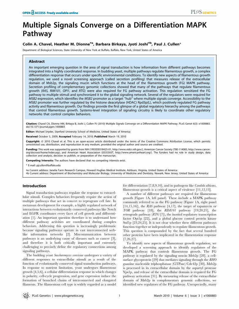
Secretion Profiling as an Approach to Identify FGPathway Regulatory Proteins
To identify new regulators of the FG pathway, secretion of the
extracellular domain of Msb2p [31] was examined using a high-
throughput screening (HTS) approach in complementary genomic
collections. Similar approaches have identified regulators of
protein trafficking by mis-sorting and secretion of carboxypepti-
dase Y [32,33,34]. An ordered collection of 4,845 mutants deleted
for nonessential open reading frames (ORFs, [35]) was trans-
formed with a plasmid carrying a functional epitope-tagged MSB2-
HA fusion gene, and transformants were screened by colony
immunoblot to identify mutants with altered Msb2p-HA secretion
(Figure 1B). Computational methods were used to quantitate,
normalize, and compare secretion between mutants, which
allowed ranking by the level of secreted Msb2p-HA (Table S3).
As a result, 67 mutants were identified that showed reduced
secretion of Msb2p-HA (Figure 1C, yellow), and 58 mutants were
identified that showed elevated secretion (Figure 1C, green).
The secretion of Msb2p was also examined using an
overexpression collection of 5,411 ORFs under the control of the
inducible GAL1 promoter [36]. This collection allows examination
of essential genes and can be assessed in the S1278b background,
in which filamentous growth occurs in an Msb2p- and MAPK
pathway-dependent manner [37]. Approximately 390 genes were
identified that influenced the secretion of Msb2p-HA when
overexpressed (Table S4). The two screens identified few common
genes (Figure 1D, 1.5% overlap), which is not entirely surprising
given that gene overexpression does not necessarily induce the
same (or opposite) phenotype as gene deletion [38] and because
the two backgrounds exhibit different degrees of filamentous
growth [37]. Significant overlap was observed at the level of gene
process/function (Figure 1E, 89% overlap), which resulted in
classification of genes into different functional categories (Figure
S1; Tables S3 and S4). Introduction of an MSB2-lacZ reporter
showed a high correlation between genes that affect Msb2p
secretion and MSB2 expression (Figure 1F; compare 1d to MZ).
Because MSB2 is itself a target of the FG pathway [28], many of
the genes identified likely influence the activity of the FG
pathway.
In total, 505 genes were identified that influenced Msb2p
secretion, which might represent an underestimate due to the
stringent statistical cutoff employed. This unexpectedly large
collection suggests that Msb2p is subject to extensive regulation,
although presumably many of these genes exert their effects
indirectly. To enrich for genes that specifically regulate the FG
pathway, secondary tests were performed. In one test, the
secretion profile of Msb2p was compared to the secretion profile
of two other mucins, the signaling mucin Hkr1p [39,40] and
transmembrane mucin Flo11p [41,42]. Almost half the genes were
common to multiple mucins (44%, Figure 1G) and may function
in the general maturation of large secreted glycoproteins. In a
second test, the secretion profile of Msb2p was compared to a
genomic screen for genes that when overexpressed influence the
expression of a FG pathway-dependent reporter (Figure 1H).
These tests eliminated general regulators of mucin maturation/
trafficking and enriched for potential MAPK regulatory proteins
(,72 candidate genes).
Several mutants were identified that were expected to
influence Msb2p secretion. Mutants lacking FG pathway
components (see Figure 1A), which are required for MSB2
expression in a positive feedback loop [28], showed a defect in
Msb2p-HA secretion (ste20D, ste50D, ste11D, and ste7D; Table
S3). The mutant lacking the aspartyl protease Yps1p, which
processes Msb2p and is required for release of the extracellular
domain [31], was also identified (yps1D; Table S3). A subset of
the genes that influence Msb2p secretion but not its expression
might function through regulating expression of the YPS1 gene,
which is highly regulated [43]. The cell-cycle regulatory
transcription factors Swi4p and Swi6p [44,45,46,47,48], were
also found to regulate Msb2p secretion (Table S3B and S7). The
swi4 and swi6 mutants had different phenotypes in Msb2p-HA
secretion (Table S3), which suggests that the Swi4p and Swi6p
proteins may play different roles in regulating cell-cycle
dependent expression of the MSB2 gene [49]. Some mating
pathway-specific genes (STE5) were also identified, which may
have an as yet unappreciated role in communication between
the mating and FG pathways, which share a number of
components [50].
As a proof-of-principle test, we disrupted fourteen genes that
came out of the deletion screen in the S1278b background and
tested for defects in Msb2p-HA secretion and FG pathway
signaling. The test showed a .70% recovery rate based on
phenotype and identified a novel connection between the tRNA
modification complex Elongator and MSB2 expression, establish-
ing this protein complex as a novel regulator of the MAPK
pathway [51]. Therefore, secretion profiling is a valid approach to
identify established and potentially novel regulators of the FG
pathway.
Multiple Signaling Pathways Regulate the FG PathwayTo identify new genes that regulate the FG pathway, ,50
candidate genes were disrupted in wild-type strains of the S1278b
background, and the resulting mutants were tested for effects on
FG pathway activity. To distinguish between mutants that
influence filamentous growth from those that have a specific effect
on FG pathway activity, a transcriptional reporter (FUS1) was used
that in S1278b strains lacking an intact mating pathway (ste4) is
dependent upon Msb2p and other FG pathway components
including the transcription factor Ste12p (Figure 2A and 2B [28]).
A number of potential MAPK regulatory proteins were identified
by this approach, many of which have been implicated in
regulating filamentous growth through their functions in other
pathways.
Author Summary
Signal integration is an essential feature of informationflow through signal transduction pathways. The mecha-nisms by which signals from multiple pathways becomeintegrated into a coordinated response remain unclear. Weshow that multiple pathways that regulate filamentousgrowth converge on a differentiation-dependent MAPKpathway. Our findings indicate that more extensivecommunication occurs between signaling pathways thatcontrol the filamentation response than has previouslybeen appreciated. We suggest that global communicationhierarchies regulate information flow in other systems,particularly higher eukaryotes where multiple pathwaystypically function simultaneously to modulate a complexresponse.
Signal Convergence on a MAPK Pathway
PLoS Genetics | www.plosgenetics.org 2 March 2010 | Volume 6 | Issue 3 | e1000883
Signal Convergence on a MAPK Pathway
PLoS Genetics | www.plosgenetics.org 3 March 2010 | Volume 6 | Issue 3 | e1000883
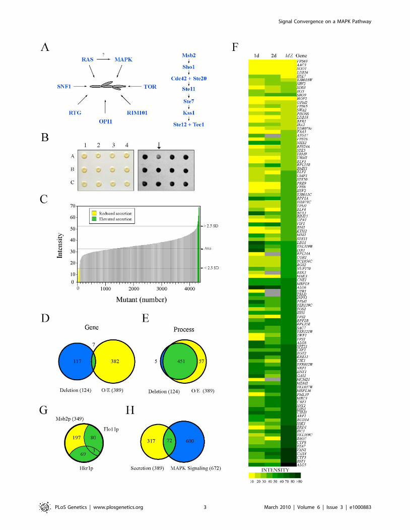
Mitochondrial retrograde signaling (the RTG network), which is
responsible for mitochondrial communication with the nucleus,
was required for FG pathway signaling. The RTG network
responds to the integrity of the mitochondria and nutrient state,
specifically the availability of certain amino acids. The proteins
Rtg1p, Rtg2p, and Rtg3p are the main targets of the pathway, and
these factors initially induce the expression of genes involved in the
TCA cycle [52]. Rtg1p, Rtg2p, and Rtg3p were required for FG
pathway signaling (Figure 2A). These factors are under the control
of Mks1p, which inhibits Rtg3p translocation to the nucleus,
preventing expression of target genes [53]. Mks1p is subsequently
under the control of Rtg2p, which will bind and sequester Mks1p
Figure 1. Secretion profiling of Msb2p. (A) Filamentous growth regulation in yeast. At left, different regulatory proteins and pathways that havebeen implicated in filamentous growth. It is unclear whether RAS regulates the FG pathway, as shown by the question mark. At right, the FG MAPKpathway. Cell surface proteins Msb2p and Sho1p connect to the Rho GTPase Cdc42p and effector p21 activated kinase Ste20p. The MAPKKK Ste11p,MAPKK Ste7p, and MAPK Kss1p regulate the activity of the transcription factors Ste12p and Tec1p. (B) Colonies from the MATa deletion collectioncontaining pMSB2-HA were pinned to nitrocellulose filters on SD-URA medium and incubated for 48h (left panel). Filters were rinsed in a stream ofwater and probed with anti-HA antibodies to detect shed Msb2p-HA (right panel). The mutant at position A2 (arrow) was identified as defective forMsb2p-HA secretion. (C) The secretion profile of Msb2p-HA in the haploid (MATa) ordered deletion collection. Y-axis, normalized spot intensity(Intensity) as a measure of secreted Msb2p-HA. X-axis, deletion mutants ranked by normalized spot intensity. Yellow, mutants that showed a decrease(2.5 standard deviations below average, - 2.5 SD) in Msb2p-HA secretion. Green, mutants that showed elevated Msb2p-HA secretion (2.5 SD aboveaverage). (D) Overlap between genes identified in the deletion screen (blue circle at left, 124 genes) and the overexpression (O/E) screen (yellow circleat right, 389 genes). Seven common genes were identified (see Table S4C for details). (E) Overlap based on genes that share a common process/function. (F) Heat map showing normalized spot intensity (at 1d and 2d intervals) as a readout of Msb2p secretion compared to expression levels ofan MSB2-lacZ (MZ) reporter. Yellow, reduced secretion; green, elevated secretion; grey, N/D. See Table S5 for details. (G) Comparative mucin secretionprofiling. Genes that influence Msb2p-HA secretion when overexpressed (,389 genes) were examined in strains containing Hkr1p-HA (PC2740) andFlo11p-HA (PC2043). Yellow, Msb2p-specific genes; blue, genes common to multiple mucins (Table S8). (H) Comparison between the secretion profileof Msb2p and regulators of a MAPK pathway growth reporter (FUS1-HIS3, [134]) whose expression is dependent upon the transcription factor Ste12pand elements of the STE pathway [135,136,137].doi:10.1371/journal.pgen.1000883.g001
Figure 2. The role of filamentation control proteins on FG pathway activity. (A) FUS1-lacZ expression was monitored in the indicated strainsin mid-log phase grown in YEPD medium. The experiment was performed in duplicate, and error bars represent standard deviation betweenexperiments. In synthetic medium, the opi1 mutant showed a .3-fold decrease in FUS1-lacZ expression compared to wild type (data not shown). (B)Comparison between FUS1-HIS3 expression and invasive growth for a subset of mutants shown in (A). Equal concentrations of cells were spottedonto YEPD medium or synthetic medium containing glucose (SD) and all amino acids (AA), amino acids except histidine (-HIS), or amino acids excepthistidine and containing the competitive inhibitor 4-amino-1,2,4-triazole. After two days, the plates were photographed. The YEPD plate wasphotographed, washed in a stream of water, and photographed again (Washed). Growth on SD-HIS is indicative of MAPK pathway activity. Growth onSD-HIS+AT is indicative of hyperactivity. Although the tor1 mutant induces hyperinvasive growth it does not influence MAPK activity.doi:10.1371/journal.pgen.1000883.g002
Signal Convergence on a MAPK Pathway
PLoS Genetics | www.plosgenetics.org 4 March 2010 | Volume 6 | Issue 3 | e1000883
and prevent its interaction with Rtg3p [54]. Consistent with its
inhibitory role in the RTG pathway, Mks1p had an inhibitory role
on FG pathway activity (Figure 2A and 2B). Rtg2p, in turn, is
negatively regulated by the Tor1p complex via Lst8p [55]. Several
mitochondrial components, including ribosomal subunits and
enzymes of the TCA cycle, showed varied expression and signaling
defects in the screen (Tables S3 and S4; Figure S7A). Deletion of
Tor1p did not have the same effect as Mks1p (Figure 2A and 2B),
but it has been shown that RTG can function independently of
Tor1p inputs [56]. Consistent with a connection to RTG, the
activity of the FG pathway was sensitive to certain amino acids
such as glutamate (Figure S2).
Components of the Rim101p pathway [19], including the
transcriptional repressor Rim101p and Dfg16p, which is required
for processing and activation of Rim101p [20,21], were required
for FG pathway activity (Figure 2A). The Rim101p pathway is
required for pH-dependent invasive growth, and the activity of the
FG pathway was slightly sensitive to pH levels (Figure S2). Other
regulators of the FG pathway included components of the RAS
pathway (see below) and the inositol regulatory transcription factor
Opi1p (Figure 2A [57,58]), which has recently been tied to
filamentous growth regulation [22].
The discovery that many filamentous growth regulatory
pathways impinge on the FG pathway suggests a systems-level
coordination between the pathways that regulate filamentous
growth. We directly tested other filamentation regulatory proteins.
Filamentous growth is regulated by the global glucose-regulatory
protein Snfl1p [23,24,25]. Snf1p was not required for FG pathway
activity (Figure 2A). The negative regulators Fkh1p/Fkh2p [59]
and Nrg1p/Nrg2p [23,60], which function to inhibit invasive
growth, did not influence FG pathway activity (Figure 2A).
Therefore, many but not all inputs into filamentous growth
regulation also regulate the FG pathway. This finding may begin
to account for the large number of genes identified by secretion
profiling that impinge on the FG pathway.
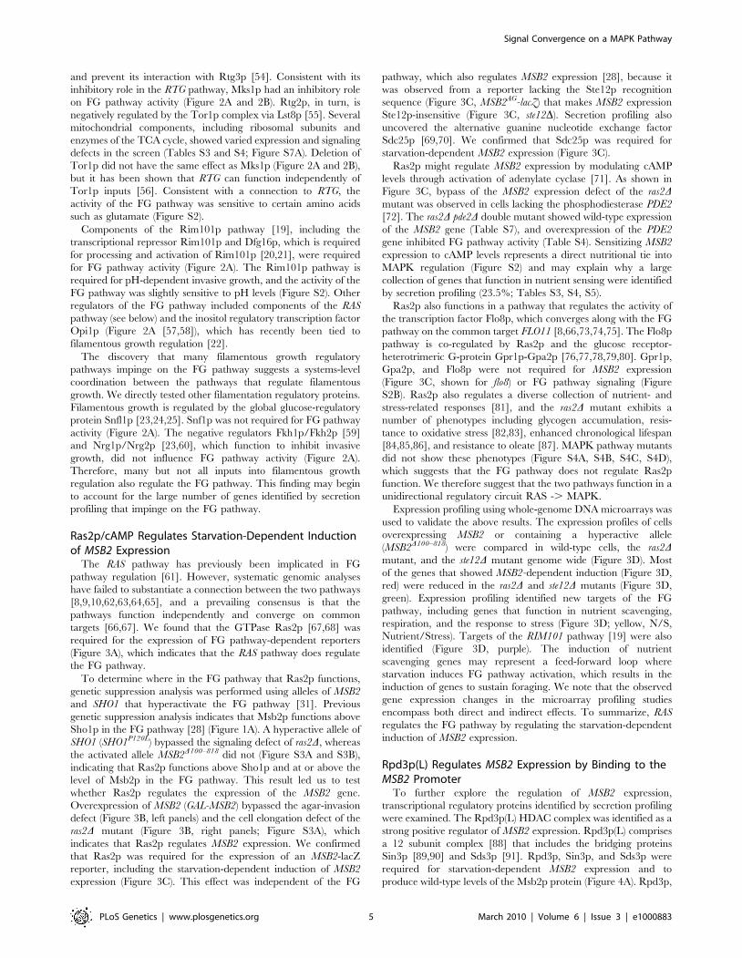
Ras2p/cAMP Regulates Starvation-Dependent Inductionof MSB2 Expression
The RAS pathway has previously been implicated in FG
pathway regulation [61]. However, systematic genomic analyses
have failed to substantiate a connection between the two pathways
[8,9,10,62,63,64,65], and a prevailing consensus is that the
pathways function independently and converge on common
targets [66,67]. We found that the GTPase Ras2p [67,68] was
required for the expression of FG pathway-dependent reporters
(Figure 3A), which indicates that the RAS pathway does regulate
the FG pathway.
To determine where in the FG pathway that Ras2p functions,
genetic suppression analysis was performed using alleles of MSB2
and SHO1 that hyperactivate the FG pathway [31]. Previous
genetic suppression analysis indicates that Msb2p functions above
Sho1p in the FG pathway [28] (Figure 1A). A hyperactive allele of
SHO1 (SHO1P120L) bypassed the signaling defect of ras2D, whereas
the activated allele MSB2D100–818 did not (Figure S3A and S3B),
indicating that Ras2p functions above Sho1p and at or above the
level of Msb2p in the FG pathway. This result led us to test
whether Ras2p regulates the expression of the MSB2 gene.
Overexpression of MSB2 (GAL-MSB2) bypassed the agar-invasion
defect (Figure 3B, left panels) and the cell elongation defect of the
ras2D mutant (Figure 3B, right panels; Figure S3A), which
indicates that Ras2p regulates MSB2 expression. We confirmed
that Ras2p was required for the expression of an MSB2-lacZ
reporter, including the starvation-dependent induction of MSB2
expression (Figure 3C). This effect was independent of the FG
pathway, which also regulates MSB2 expression [28], because it
was observed from a reporter lacking the Ste12p recognition
sequence (Figure 3C, MSB2AG-lacZ) that makes MSB2 expression
Ste12p-insensitive (Figure 3C, ste12D). Secretion profiling also
uncovered the alternative guanine nucleotide exchange factor
Sdc25p [69,70]. We confirmed that Sdc25p was required for
starvation-dependent MSB2 expression (Figure 3C).
Ras2p might regulate MSB2 expression by modulating cAMP
levels through activation of adenylate cyclase [71]. As shown in
Figure 3C, bypass of the MSB2 expression defect of the ras2Dmutant was observed in cells lacking the phosphodiesterase PDE2
[72]. The ras2D pde2D double mutant showed wild-type expression
of the MSB2 gene (Table S7), and overexpression of the PDE2
gene inhibited FG pathway activity (Table S4). Sensitizing MSB2
expression to cAMP levels represents a direct nutritional tie into
MAPK regulation (Figure S2) and may explain why a large
collection of genes that function in nutrient sensing were identified
by secretion profiling (23.5%; Tables S3, S4, S5).
Ras2p also functions in a pathway that regulates the activity of
the transcription factor Flo8p, which converges along with the FG
pathway on the common target FLO11 [8,66,73,74,75]. The Flo8p
pathway is co-regulated by Ras2p and the glucose receptor-
heterotrimeric G-protein Gpr1p-Gpa2p [76,77,78,79,80]. Gpr1p,
Gpa2p, and Flo8p were not required for MSB2 expression
(Figure 3C, shown for flo8) or FG pathway signaling (Figure
S2B). Ras2p also regulates a diverse collection of nutrient- and
stress-related responses [81], and the ras2D mutant exhibits a
number of phenotypes including glycogen accumulation, resis-
tance to oxidative stress [82,83], enhanced chronological lifespan
[84,85,86], and resistance to oleate [87]. MAPK pathway mutants
did not show these phenotypes (Figure S4A, S4B, S4C, S4D),
which suggests that the FG pathway does not regulate Ras2p
function. We therefore suggest that the two pathways function in a
unidirectional regulatory circuit RAS -. MAPK.
Expression profiling using whole-genome DNA microarrays was
used to validate the above results. The expression profiles of cells
overexpressing MSB2 or containing a hyperactive allele
(MSB2D100–818) were compared in wild-type cells, the ras2Dmutant, and the ste12D mutant genome wide (Figure 3D). Most
of the genes that showed MSB2-dependent induction (Figure 3D,
red) were reduced in the ras2D and ste12D mutants (Figure 3D,
green). Expression profiling identified new targets of the FG
pathway, including genes that function in nutrient scavenging,
respiration, and the response to stress (Figure 3D; yellow, N/S,
Nutrient/Stress). Targets of the RIM101 pathway [19] were also
identified (Figure 3D, purple). The induction of nutrient
scavenging genes may represent a feed-forward loop where
starvation induces FG pathway activation, which results in the
induction of genes to sustain foraging. We note that the observed
gene expression changes in the microarray profiling studies
encompass both direct and indirect effects. To summarize, RAS
regulates the FG pathway by regulating the starvation-dependent
induction of MSB2 expression.
Rpd3p(L) Regulates MSB2 Expression by Binding to theMSB2 Promoter
To further explore the regulation of MSB2 expression,
transcriptional regulatory proteins identified by secretion profiling
were examined. The Rpd3p(L) HDAC complex was identified as a
strong positive regulator of MSB2 expression. Rpd3p(L) comprises
a 12 subunit complex [88] that includes the bridging proteins
Sin3p [89,90] and Sds3p [91]. Rpd3p, Sin3p, and Sds3p were
required for starvation-dependent MSB2 expression and to
produce wild-type levels of the Msb2p protein (Figure 4A). Rpd3p,
Signal Convergence on a MAPK Pathway
PLoS Genetics | www.plosgenetics.org 5 March 2010 | Volume 6 | Issue 3 | e1000883

Signal Convergence on a MAPK Pathway
PLoS Genetics | www.plosgenetics.org 6 March 2010 | Volume 6 | Issue 3 | e1000883
Sin3p, and Sds3p were required for the induction of FG pathway
reporters (Table S7) and invasive growth (Figure S6A).
Rpd3p functions in distinct large and small complexes with
different cellular functions. Rpd3p(S) recognizes methylated
histone H3 subunits and functions to repress spurious transcription
initiation from cryptic start sites in open reading frames [92].
Rpd3p(S) contains unique proteins Rco1p and Eaf3p. In contrast,
Rpd3p(L) is enriched for different proteins including Rxt2p [92],
which is required for Rpd3p(L) function [93]. Immunoblot
analysis (Figure 4A), transcriptional reporters (Figure 4B), and
invasive growth assays (Figure S6A), showed that Rxt2p was
required for MSB2 expression and FG pathway activation. In
contrast, Rpd3p(L) was not required for pheromone response
pathway activation (Figure S5), which shares components with the
FG pathway but induces different target genes [5,94,95] and
which does not require Msb2p function [28].
Histone deacetylases typically repress gene expression by
causing compaction of chromatin into structures inaccessible to
transcription factors. With respect to MSB2 expression however,
Rpd3p(L) had a positive role. We tested whether Rpd3p(L)
negatively regulated an inhibitor of MSB2 expression, such as the
transcription factor Dig1p [96,97,98] but were unable to find
evidence to support this possibility (Figure S6B). Rpd3p(L)
functions as a positive regulator of some genes, including targets
of the high osmolarity glycerol response (HOG) pathway by direct
association with the promoters of HOG pathway targets [99], and
in the regulation of some mating-specific targets [100]. Therefore,
Rpd3p(L) might positively regulate MSB2 expression by associat-
ing with the MSB2 promoter. Chromatin immunoprecipitation
(ChIP) analysis identified Rpd3p(L) at the MSB2 promoter
(Figure 4C, Sin3p-HA). In the sin3D mutant, less Ste12p was
found at the MSB2 promoter (Figure 4C and 4D, compare wild
type to sin3D). Msb2p and the FG pathway also control STE12
expression. Ste12p-HA protein levels were reduced in the sin3Dmutant (Figure 4E), as a result of a decrease in STE12 expression
determined by q-PCR (data not shown). Therefore, Rpd3p(L)
positively regulates the FG pathway by promoting MSB2
expression. Rpd3p(L) may also indirectly promote FG pathway
activity by the regulation of other transcription factors that
influence MSB2 expression.
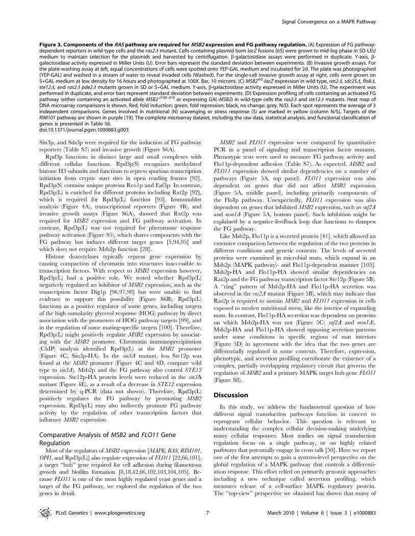
Comparative Analysis of MSB2 and FLO11 GeneRegulation
Most of the regulators of MSB2 expression [MAPK, RAS, RIM101,
OPI1, and Rpd3p(L)] also regulate expression of FLO11 [22,66,101],
a target ‘‘hub’’ gene required for cell adhesion during filamentous
growth and biofilm formation [8,18,42,66,102,103,104,105]. Be-
cause FLO11 is one of the most highly regulated yeast genes and a
target of the FG pathway, we explored the regulation of the two
genes in detail.
MSB2 and FLO11 expression were compared by quantitative
PCR in a panel of signaling and transcription factor mutants.
Phenotypic tests were used to measure FG pathway activity and
Flo11p-dependent adhesion (Table S7). As expected, MSB2 and
FLO11 expression showed similar dependencies on a number of
pathways (Figure 5A, top panel). FLO11 expression was also
dependent on genes that did not affect MSB2 expression
(Figure 5A, middle panel), including primarily components of
the Flo8p pathway. Unexpectedly, FLO11 expression was also
dependent on genes that inhibited MSB2 expression, such as snf2Dand msn1D (Figure 5A, bottom panel). Such inhibition might be
explained by a negative-feedback loop that functions to dampen
the FG pathway.
Like Msb2p, Flo11p is a secreted protein [41], which allowed an
extensive comparison between the regulation of the two proteins in
different conditions and genetic contexts. The levels of secreted
proteins were examined in microbial mats, which expand in an
Msb2p (MAPK pathway)- and Flo11p-dependent manner [105].
Msb2p-HA and Flo11p-HA showed similar dependencies on
Ras2p and the FG pathway transcription factor Ste12p (Figure 5B).
A ‘‘ring’’ pattern of Msb2p-HA and Flo11p-HA secretion was
observed in the ras2D mutant (Figure 5B), which may indicate that
Ras2p is required to sustain MSB2 and FLO11 expression in cells
exposed to modest nutritional stress, like the interior of expanding
mats. In contrast, Flo11p-HA secretion was dependent on proteins
on which Msb2p-HA was not (Figure 5C; snf2D and msn1D).
Msb2p-HA and Flo11p-HA showed opposing secretion patterns
under some conditions in specific regions of mat interiors
(Figure 5D) in agreement with the idea that the two genes are
differentially regulated in some contexts. Therefore, expression,
phenotypic, and secretion profiling corroborate the existence of a
complex, partially overlapping regulatory circuit that governs the
regulation of MSB2 and a primary MAPK target hub gene FLO11
(Figure S8).
Discussion
In this study, we address the fundamental question of how
different signal transduction pathways function in concert to
reprogram cellular behavior. This question is relevant to
understanding the complex cellular decision-making underlying
many cellular responses. Most studies on signal transduction
regulation focus on a single pathway, or on highly related
pathways that potentially engage in cross talk [50]. Here we report
one of the first attempts to gain a systems-level perspective on the
global regulation of a MAPK pathway that controls a differenti-
ation response. This effort relied on primarily genomic approaches
including a new technique called secretion profiling, which
measures release of a cell-surface MAPK regulatory protein.
The ‘‘top-view’’ perspective we obtained has shown that many of
Figure 3. Components of the RAS pathway are required for MSB2 expression and FG pathway regulation. (A) Expression of FG pathway-dependent reporters in wild-type cells and the ras2D mutant. Cells containing plasmid born lacZ fusions [65] were grown to mid-log phase in SD-LEUmedium to maintain selection for the plasmids and harvested by centrifugation. b-galactosidase assays were performed in duplicate. Y-axis, b-galactosidase activity expressed in Miller Units (U). Error bars represent the standard deviation between experiments. (B) Invasive growth assays. Forthe plate-washing assay at left, equal concentrations of cells were spotted onto YEP-GAL medium and incubated for 2d. The plate was photographed(YEP-GAL) and washed in a stream of water to reveal invaded cells (Washed). For the single-cell invasive growth assay at right, cells were grown onS+GAL medium at low density for 16 hours and photographed at 100X. Bar, 10 microns. (C) MSB2AG-lacZ expression in wild type, ras2D, sdc25D, flo8D,ste12D, and ras2D pde2D mutants grown in SD or S+GAL medium. Y-axis, b-galactosidase activity expressed in Miller Units (U). The experiment wasperformed in duplicate, and error bars represent standard deviation between experiments. (D) Expression profiling of cells containing an activated FGpathway (either containing an activated allele MSB2D100–818 or expressing GAL-MSB2) in wild-type cells the ras2D and ste12D mutants. Heat map ofDNA microarray comparisons is shown. Red, fold induction; green, fold repression; black, no change; grey, N/D. Each spot represents the average of 3independent comparisons. Genes involved in nutritional (N) scavenging or stress response (S) are marked in yellow (column N/S). Targets of theRIM101 pathway are shown in purple [19]. The complete microarray dataset, including the raw data, statistical analysis, and functional classification ofgenes is presented in Table S6.doi:10.1371/journal.pgen.1000883.g003
Signal Convergence on a MAPK Pathway
PLoS Genetics | www.plosgenetics.org 7 March 2010 | Volume 6 | Issue 3 | e1000883

the major regulatory proteins that control filamentous growth also
control MAPK signaling. This finding is challenging to accept
given that many of the pathways are currently viewed as separate
entities. Nonetheless, this view is consistent with an emerging
systems-level appreciation of pathway regulation in complex
situations – cell differentiation, stem cell research, and cancer -
where pathway interconnectedness drives the rationale for drug
development and new therapeutic endeavors.
New Connections between Signaling NetworksWe specifically show that four major regulators of filamentous
growth are also required for FG pathway signaling (Figure 5E).
The connections between the RIM101, OPI1, and RTG pathways
to FG pathway regulation are entirely novel. We further show that
the RAS pathway and several other pathways [MAPK, Swi4p,
Rpd3p(L)] converge on the promoter of the key upstream
regulator (MSB2) of the FG pathway. Like other signal integration
mechanisms [8,66], the MSB2 promoter is a target ‘‘hub’’ where
multiple pathways converge. This regulatory point makes sense
from the perspective that Msb2p levels dictate FG pathway activity
[28]. Given that the FG pathway exhibits multimodality in
regulation of pathway outputs [39], its precise modulation is
critical to induce an appropriate response.
The regulation of the FG pathway differs from that of a related
MAPK pathway, the pheromone response pathway, where
secreted peptide pheromones provide the single major input into
activation [5,106,107]. The pheromone response pathway is not
regulated by the RAS, RIM, or RTG pathways and does not appear
to be influenced by the Rpd3p(L) HDAC, although some target
mating genes do require Rpd3p(L) for expression [100]. Both the
FG and mating pathways are regulated by positive feedback
[28,65,108,109], presumably to provide signal amplification.
Therefore, the FG pathway is distinguished in that it is highly
sensitized to multiple external inputs. Because our screening
approach was highly biased to identify regulators that function at
the ‘‘top’’ of the pathway (at Msb2p), it is likely that the FG
pathway may be subject to additional regulation not identified by
this approach.
The networks that regulate filamentation signaling pathways
appear in some cases to be redundant. For example, RAS regulates
FLO11 expression through Flo8p and the FG pathway (Figure 5E).
RIM101 similarly controls FLO11 expression through Nrg1p/
Nrg2p and FG regulation (Figure 5E). However, any one of the
major filametation regulatory pathways when absent appears fully
defective for the response. Such parallel processing of signaling
networks might allow for ‘‘fine-tuning’’ of the differentiation
response, or alternatively to synchronize all of the pathways to the
Figure 4. Rpd3p(L) promotes MSB2 gene expression at the MSB2promoter. (A) Immunoblot of Msb2p-HA in wild-type cells and therxt2D, sin3D, rpd3D, and sds3D mutants. The arrow refers to Msb2p-HA.The asterisk refers to a background band. (B) The activity oftranscriptional (lacZ) reporters of the FG pathway in wild-type cellsand the rxt2D mutant. b-galactosidase assays were performed induplicate, and error bars represent standard deviation betweenreplicates. (C) ChIP analysis of the MSB2 promoter in wild-type cells orthe sin3D mutant containing Ste12p-HA or Sin3p-HA fusion proteins. I,input; No Ab, no antibody control; aHA, anti-HA antibody. (D)Quantitation of the ChIP data. Graph shows relative levels of bindingat the MSB2 promoter relative to an intragenic region and to the ACT1gene as a control based on quantitative PCR analysis. DDCt, thresholdcycle; Bars correspond to the IP/Input ratio. (E) Immunoblot of Ste12p-HA in wild-type cells and the sin3D mutant. Ste12p-HA wasimmunoprecipitated to concentrate the protein. Ctl refers to abackground band that served as a loading control.doi:10.1371/journal.pgen.1000883.g004
Signal Convergence on a MAPK Pathway
PLoS Genetics | www.plosgenetics.org 8 March 2010 | Volume 6 | Issue 3 | e1000883
global regulatory circuit. Such synchronization may coordinate
different aspects of filamentous growth (cell-cell adhesion, cell
cycle regulation, reorganization of polarity) into a cohesive
response.
RAS/cAMP Sensitizes MAPK Activity to Cellular NutrientStatus
Filamentous growth occurs within a narrow nutritional range
[25], between high nutrient levels that support vegetative growth
and limiting nutrients that force entry into stationary phase. The
finding that Ras2p controls the overall levels of MSB2 expression
extends the initial connection between RAS and the FG pathway
[61] in several important ways. First, the results explain how
Ras2p connects to the FG pathway above Cdc42p. Second, the
data provide a link between nutrition and FG pathway signaling at
the level of cellular cAMP levels. Third, Ras2p does not appear to
function as a component of the FG pathway. This conclusion is
based on the conditional requirement for Ras2p in MSB2
regulation, the suppression of the ras2D signaling defect by loss
of PDE2, and the placement of Ras2p above MSB2 in the FG
pathway. Our results fit with the general idea that RAS controls a
broad response to cellular stress that encompasses FG pathway
regulation. Ras2p has recently been shown to regulate the MAPK
pathway that controls sporulation [110], a diploid-specific
starvation response. Ras2p may therefore function as a general
regulator of MAPK signaling in response to nutritional stress.
HDAC Regulation of MAPK SignalingExamples by which MAPK pathways control target gene
expression by recruitment of chromatin remodeling proteins is
relatively common and include Rpd3p(L) regulation of HOG
[99,111,112] and mating pathway [100] target genes. However,
Figure 5. Comparative expression and secretion profiling of the MSB2 and FLO11 genes. (A) Quantitative PCR analysis of MSB2 and FLO11expression in the indicated mutants, all of which were created in the S1278b background (Table S1). Heat map shows fold change in geneexpression. Red, fold induced; green, fold repressed. See Table S7 for details. (B) Comparison of Msb2p-HA and Flo11p-HA secretion in microbial mats.Mats expressing Msb2p-HA (wild type, PC999; ras2D, PC2689; ste12D, PC2691) or Flo11p-HA (wild type, PC2043; ras2D, PC2693; ste12D, PC2695) werespotted onto YEPD medium (0.3% agar) on nitrocellulose filters and were allowed to expand at for 2d, 4d, 8d, and 10d. Cells were washed off thefilters, which were probed with antibodies against the HA epitope. Each experiment was performed in duplicate and a representative image is shown.For some panels, the intensity of the secreted protein was below the threshold for visibility in the exposure shown. N/D, not determined. Matperimeters were at secretion boundaries. (C) Secretion of Msb2p-HA and Flo11p-HA in the snf2D and msn1D mutants grown as mats for 8d. Theexperiment was performed as described in (B). (D) Comparison between Msb2p-HA and Flo11p-HA secretion on mats grown on YEP-GAL media for8d. A light and dark exposure is shown. (E) Model for integration between pathways that control filamentous growth. The RAS, RIM101, and RTGpathways regulate the MAPK pathway. The TOR and SNF1 pathways were not found to influence MAPK activity. The RAS [66] and RIM101 [19]pathways likely regulate filamentous growth at multiple levels.doi:10.1371/journal.pgen.1000883.g005
Signal Convergence on a MAPK Pathway
PLoS Genetics | www.plosgenetics.org 9 March 2010 | Volume 6 | Issue 3 | e1000883

the regulation of MAPK activity through chromatin remodeling
proteins provides a hierarchical mechanism for global cellular
reprogramming. Regulation of FG pathway activity by Rpd3p(L)
may contribute to the establishment of a differentiated state. Once
activated, FG pathway activity may be sustained by Rpd3p(L) to
reinforce accessibility to the MSB2 promoter and formation of the
filamentous cell type. Although the connection between Rpd3p(L)
and nutrition is not entirely clear, Rpd3p(L) preferentially localizes
to highly expressed genes, such as those required for anabolic
processes [113]. Therefore, Rpd3p(L) may coordinate overall
growth rate with the persistence of MAPK activity. Precedent for
Rpd3p(L) HDACs in regulating developmental transitions comes
from Drosophila DRpd3, which together with the Chameau HAT
function as opposing cofactors of JNK/AP-1-dependent transcrip-
tion during metamorphosis [114]. HDAC regulation of MAPK
activity may therefore represent a general feature of MAPK
regulation.
In conclusion, we have identified an unprecedented degree of
regulation of a differentiation-dependent MAPK pathway by
multiple regulatory proteins and pathways. Our findings open up
new avenues for exploring the relationships between pathways and
the extent of their cross-regulation with the ultimate goal of
understanding all functionally relevant pathway interactions in a
comprehensive manner.
Materials and Methods
Strains, Plasmids, and Microbiological TechniquesYeast strains are described in Table S1. Plasmids are described
in Table S2. Yeast and bacterial strains were manipulated by
standard methods [115,116]. PCR-based methods were used to
generate gene disruptions, GAL1 promoter fusions [117,118], and
insertion of epitope fusions [119], using auxotrophic and antibiotic
resistant markers [120]. Integrations were confirmed by PCR
Southern analysis and DNA sequencing. Plasmids pMSB2-GFP
and pMSB2-HA have been described [31], as have plasmids
pMSB2-lacZ and pMSB2AG-lacZ [39]. Plasmids containing FG
pathway targets KSS1, SVS1, PGU1, and YLR042C fused to the lacZ
gene were provided by C. Boone [65]. pFLO8 was provided by G.
Fink [37]. Plasmid pIL30-URA3 containing FgTy-lacZ was
provided by B. Errede [121], and pFRE-lacZ was provided by
H. Madhani [16]. The positions of the hemagglutinin (HA)
epitope fusions were 500 amino acid residues for the Msb2p
protein [31], at 1015 residues for the Flo11p protein [41], and at
1000 residues for the Hkr1p protein [39].
The single cell invasive growth assay [25] and the plate-washing
assay [14] were performed to assess filamentous growth. Budding
pattern was based on established methodology [122], and
confirmed for some experiments by visual inspection of connected
cells [25]. Halo assays were performed as described [123].
Microbial mat assays were performed as described [105] by
growing cells on low-agar (0.3%) YEPD medium. Oleate medium
was derived from standard synthetic medium lacking amino acids
that was supplemented with 0.1% yeast extract, 0.5% potassium
phosphate pH 6.0, and oleic acid (Toyko Kasei Kogyo Co. TCI)
at a final concentration of 0.125% (w/v) solubilized in 0.5%
Tween-20. Antimycin A, from Streptomyces sp. (Sigma-Aldrich,
St. Louis, MO) was used at 3 mg/ml. Oligomycin (Sigma-Aldrich,
St. Louis, MO) was used at 3 mg/ml, and rapamycin (Sigma-
Aldrich) was added at 20 ng/ml. b-galactosidase assays were
performed as previously described [124]. For some experiments,
b-galactosidase assays were performed in 96-well format by
growing cells containing the MSB2-lacZ reporter to saturation in
synthetic medium lacking uracil (SD–URA) to maintain selection
for the plasmid in 96-well plates at 30uC. Inductions were
performed in duplicate, and the average of at least two
independent experiments is reported. All experiments were carried
out at 30uC unless otherwise indicated.
Secretion Profiling of Msb2pThe MATa haploid deletion collection [35] was transformed
with a plasmid carrying a functional hemagglutinin (HA)-tagged
MSB2 gene (pMSB2-HA; [31]) using a high-throughput microtiter
plate transformation protocol [125]. The deletion collection was
manipulated with the BioMek 2000 automated workstation
(Beckman-Coulter, Fullerton CA). For some experiments, a 96-
fixed pinning tool (V & P Scientific, 23 VP 408) and plate
replication tool (V & P Scientific, VP 381) were used. Sterilization
was performed by sequential washes in 5% bleach, distilled water,
70% ethanol, and 95% ethanol. Ethanol (5 ml of 95%) was added
to each transformation mix (200 ml) to increase transformation
efficiency [126]. Transformants were harvested by centrifugation
in 96-well plates, resuspended in 30 ml of water, and transferred to
synthetic medium containing 2% glucose and lacking uracil (SD-
URA) medium in Omnitrays (VWR International Inc. Bridgeport
NJ). Transformants were pinned to SD-URA for 48 h. Colonies
were transferred to 96-well plates containing 100 ml of water and
pinned to SD-URA medium overlaid with a nitrocellulose filter
(0.4 mm; HAHY08550 Millipore) and incubated for 48 h at 30uC.
Filters were rinsed in distilled water to remove cells and probed by
immunoblot analysis. Cross-contamination was estimated at 0.8%
based on growth in blank positions; ,93% of the collection (4554
mutants) was examined.
For the overexpression screen, a collection of ,5,500
overexpression plasmids [36] was examined in a wild-type
S1278b strain containing a functional MSB2-HA gene integrated
at the MSB2 locus under the control of its endogenous promoter
(PC999). Plasmids were purified from Escherichia coli stocks by
alkaline lysis DNA preparation in 96-well format. Plasmid DNA
was transformed into PC999 using the high-throughput transfor-
mation protocol described above. Transformants were selected on
SD-URA and screened by pinning to nitrocellulose filters on
synthetic medium containing 2% galactose and lacking uracil
(S+GAL-URA) to induce gene overexpression. Colonies were
incubated for 2 days at 30uC. Filters were washed in a stream of
water and probed by immunoblot analysis as described above.
Candidate genes/deletions that were initially identified were
confirmed by retesting. Approximately ,35% of the deletion
strains and 37% of the overexpression plasmids failed retesting and
were considered false positives (Tables S3B and S4B). Compar-
ative secretion profiling of Msb2p-HA, Hkr1p-HA, and Flo11p-
HA was performed by transforming overexpression plasmids
identified in the Msb2p-HA screen into strains that contain
Hkr1p-HA (PC2740) and Flo11p-HA (PC2043). Transformants
were pinned onto S-GAL-URA on nitrocellulose filters. Colonies
were incubated for 48h, at which time filters were rinsed and
evaluated by colony immunoblot analysis. The complete deletion
collection transformed with pMSB2-HA (Table S3) and overex-
pression collection in strain PC999 (Table S4) were frozen in
aliquots at 280uC and are available upon request.
Computational AnalysisTo compare levels of secreted protein between samples, spot
intensity was measured and normalized to colony size. Small
colonies initially scored as undersecretors and large colonies
(particularly at plate corners) scored as hypersecretors were
eliminated or flagged for retesting. As an additional test, standard
immunoblots were performed from cells grown in liquid culture.
Signal Convergence on a MAPK Pathway
PLoS Genetics | www.plosgenetics.org 10 March 2010 | Volume 6 | Issue 3 | e1000883
Supernatants (S) and cell pellets (P) were separated by centrifu-
gation. In most cases (.80%), differential secretion by colony
immunoblot was reflected by an altered S/P ratio by standard
immunoblot analysis. The ImageJ MicroArray Profile.jar algo-
rithm (http://www.optinav.com/download/MicroArray_Profile.
jar) created by Dr. Bob Dougherty and Dr. Wayne Rasband,
commonly used for Microarray analysis, was used as a plugin for
the ImageJ program. Image intensity was determined in 96-panel
format by inverting the image after background subtraction. Plate-
to-plate variation was normalized by dividing the average intensity
of all spots with the average intensity of each plate, and this factor
was applied to the intensity of each spot. Heat maps for expression
and secretion profiling were generated as described [127].
Classification of genes based on process/function was determined
using GO ontology terms in publicly available databases including
the Saccharomyces genome database (http://www.yeastgenome.
org/). Analysis of human mucin genes was facilitated by the
Human Protein Reference Database (http://www.hprd.org/).
DNA Microarray AnalysisDNA microarray analysis was performed as described
[128,129]. Wild type (PC538), GAL-MSB2 (PC1083), GAL-MSB2
ste12D (PC1079), GAL-MSB2 ras2D (PC2949), MSB2D100–818
(PC1516), MSB2D100–818 ste12D (PC1811), and MSB2D100–818
ras2D (PC2364) strains were grown in YEP-GAL for 6 h, at which
point cells were examined by microscopy for the characteristic
filamentation response. RNA was prepared by hot acid phenol and
passage over an RNeasy column (Qiagen). Microarray construc-
tion, target labeling, and hybridization protocols were as described
[130]. Sample comparisons were independently replicated at least
3 times from separate inductions. Fluoro-reverse experiments were
used to identify sequence-specific dye biases. Arrays were scanned
using a GenePix 4000 scanner (Axon Instruments). Image analysis
was performed using GenePix Pro 3.0. Array features (i.e., spots)
having low signal intensities or signals compromised by artifacts
were removed from further analysis. Background subtracted Cy5/
Cy3 ratios were log2 transformed and a Loess normalization
strategy (f = 0.67) was applied for each array using S-Plus
(MathSoft, Cambridge, MA). Each feature where the |log2
(ratio)|$0.8, the corresponding gene was considered differentially
expressed.
MicroscopyDifferential-interference-contrast (DIC) and fluorescence mi-
croscopy using the FITC filter set were performed using an
Axioplan 2 fluorescent microscope (Zeiss) with a PLAN-APOC-
HROMAT 100X/1.4 (oil) objective (N.A. 0.17). Digital images
were obtained with the Axiocam MRm camera (Zeiss). Axiovision
4.4 software (Zeiss) was used for image acquisition and analysis.
For Msb2p-GFP localization, cells were grown to saturation in
selective medium to maintain plasmids harboring MSB2-GFP
fusions. Cells were harvested by centrifugation and resuspended in
YEPD medium for 4.5 h. Cells were harvested, washed three
times in water, and visualized at 100X.
Immunoblot AnalysisImmunoblots were performed as described [31]. To compare
protein levels between strains, cells were grown to saturation in
YEPD medium and subcultured into YEPD or YEP-GAL medium
for 8 h. Culture volumes were adjusted to account for differences
in cell number and harvested by centrifugation. Supernatant
volumes were similarly adjusted. Cells were disrupted by addition
of 200 ml lysis buffer (8 M Urea, 5 % SDS, 40 mM Tris-HCl
pH 6.8, 0.1 M EDTA, 0.4 mg/ml Bromophenol blue and 1 % b-
mercaptoethanol) and glass beads followed by vortexing for 5
minutes at the highest setting and boiling 5 min. Supernatants
were examined by boiling in 1.5 volumes of lysis buffer for 5 min.
For some experiments, cells were lysed in spheroplast buffer (1.2M
sorbitol, 50 mM potassium phosphate pH 7.4, 1 mM MgCl,
250 ug/ml of zymolyase), and protein concentration was deter-
mined by Bradford assays (Bio-Rad, Hercules CA). Equal
concentrations of protein were loaded into each lane. For some
experiments, blots were stripped and re-probed using anti-actin
monoclonal antibodies (Chemicon; Billerica, MA). Monoclonal
antibodies against the HA epitope were used (12CA5). Proteins
were separated by SDS-PAGE on 10% or 10%–20% gradient gels
(Bio-Rad, Hercules CA) and transferred to nitrocellulose mem-
branes (protran BA85, VWR International Inc. Bridgeport NJ).
Membranes were incubated in blocking buffer (5% nonfat dry
milk, 10 mM Tris-HCl pH 8, 150 mM NaCl and 0.05% Tween
20) for 1 hr at 25uC. Nitrocellulose membranes were incubated for
18 hr at 4uC in blocking buffer containing primary antibodies.
ECL Plus immunoblotting reagents were used to detect secondary
antibodies (Amersham Biosciences, Piscataway NJ). Immunoblots
of proteins secreted from mats were performed by growing cells on
nitrocellulose filters on low-agar (0.3%) YEPD medium.
Chromatin Immunoprecipitation (ChIP) AnalysisChIP assays were performed as described [131]. Strains
PC3021 (STE12-HA), PC3353 (sin3D, STE12-HA) and PC3579
(SIN3-HA) were grown in YEPD medium for 8 h. Cross-linking
was performed with 1% formaldehyde for 15 min at 25uC. Cells
were collected by centrifugation and washed twice in PBS buffer.
Cells were resuspended in ChIP lysis buffer (Upstate, Billerica,
CA), and lysed by Fast Prep 24 (MP) for one cycle at 6.5 for 45 sec.
After puncturing the bottom of Fast Prep tubes with a 22 gauge
needle, lysates were collected by centrifugation. DNA was sheared
by sonication on a Branson Digital Sonifier at setting 20%
amplitude, 15 pulses for 20 sec, 55 sec. rest. Pull downs were
performed using a ChIP assay kit (Upstate) with anti-HA
antibodies with 10% input sample set aside for a control. qPCR
was used to determine relative amount of immunoprecipitated
specific DNA loci in IP, Input, and Mock (no antibody) samples.
The housekeeping ACT1 gene was used to normalize quantifica-
tion in qPCR reactions. Data are expressed as IP/Input where
DDCt = (Ct IP_MSB2-Ct IP_ACT1)-(Ct Input_MSB2-Ct Input_
ACT1). Primers used were MSB2 promoter forward 59-CGA-
TAGCTGATAGACTGTGGAGTCG-39 and reverse 59- CTG-
GCAACGCCCGACGTGTCTAGCC-39, MSB2 intergenic re-
gion forward 59- TGACCAAACTTCGACTGCTGG-39 and
reverse 59- AGCTGCTGATGCAGTGGTAA-39; and ACT1
forward 59- GGCTTCTTTGACTACCTTCCAACA-39 and
reverse 59- GATGGACCACTTTCGTCGTATTC-39. ChIP pull
downs were also visualized by gel electrophoresis.
mRNA Level Determination Using Quantitative PCRTotal RNA was isolated from 25 ml cultures grown in YEP
GAL for 8 h using hot acid phenol extraction. cDNA synthesis was
carried out using 1 mg RNA and the iScript cDNA Synthesis Kit
(Bio-Rad; Hercules CA) according to the manufacturer’s instruc-
tions. One tenth of the synthesized cDNA was used as the template
for real-time PCR. 25 ul real time PCR reactions were performed
on the BioRad MyiQ Cycler with iQ SYBR Green Supermix (Bio-
Rad). RT qPCR was performed using the following amplification
cycles: initial denaturation for 8 min at 95uC, followed by
356cycle 2 (denaturation for 15 sec at 95uC and annealing for
1 min at 60uC). Melt curve data collection was enabled by
decreasing the set point temperature after cycle 2 by 0.5uC. The
Signal Convergence on a MAPK Pathway
PLoS Genetics | www.plosgenetics.org 11 March 2010 | Volume 6 | Issue 3 | e1000883
specificity of amplicons was confirmed by generating the melt
curve profile of all amplified products. Gene expression was
quantified as described [132]. Primers were based on a previous
report [133] and were FLO11 forward 59- GTTCAACCAGTC-
CAAGCGAAA-39 and reverse 59- GTAGTTACAGGTGTGG-
TAGGTGAAGTG-39and those described for ChIP assays above.
All reactions were performed in duplicate and average values are
reported.
Supporting Information
Figure S1 Genes that regulate Msb2p-HA secretion comprise a
number of different functional categories. Pie charts of functional
categories enriched in the genomic screens. Far left, functional
categories that contributed to Msb2p-HA secretion; middle panel,
functional categories that were inhibitory for Msb2p-HA secretion;
far right, the overall functional classification of genes in the yeast
genome for reference. Functional classification of yeast genes was
facilitated by SGD (http://www.yeastgenome.org/).
Found at: doi:10.1371/journal.pgen.1000883.s001 (6.08 MB TIF)
Figure S2 MSB2 expression is influenced by a different
extracellular stimuli. The expression of an MSB2-lacZ fusion was
examined under the conditions described. Cells were grown to
mid-log phase in YEPD medium (,6h) or medium lacking glucose
(YEP), containing a poor carbon source (YEP-GAL), or to
saturation in a poor carbon source (YEP-GAL 16h). MSB2-lacZ
expression was also compared in medium supplemented with the
amino acid glutamate and medium at pH 3, pH 7, and pH 8.
Cells were harvested, and b-galactosidase assays were performed
in independent replicates. The error bars represent standard
deviation between experiments.
Found at: doi:10.1371/journal.pgen.1000883.s002 (2.70 MB TIF)
Figure S3 Genetic suppression analysis of RAS pathway
components based on the activity of the FUS1-HIS3 reporter.
(A) The morphology of wild-type cells and cells containing the
activated allele MSB2D100–818 or overexpressing MSB2 (GAL-
MSB2) in combination with ras2D and ste12D mutations. Images
were taken by DIC at 100X. Bar, 20 microns. (B) Equal
concentrations of cells of the indicated genotypes were spotted
onto synthetic medium supplemented with glucose (SD) or
galactose (S+GAL) that contained all amino acids (+AA), or that
lacked uracil (-URA), and/or lacking histidine (-HIS). Plates were
incubated at 30uC and spots were photographed. Growth on
medium is indicative of the activity of the FG pathway. In the top
panel, SHO1P120L and SHO1S22oF bypass the signaling defect of the
ras2D mutant suggesting that Sho1p functions below Ras2p, and in
line with the idea that Ras2p controls MSB2 expression. In the
middle panel, overexpression of MSB2 bypasses the signaling
defect of the ras2D mutant to a greater degree than the activated
allele MSB2D100–818. In the bottom panel, Flo8p pathway mutants
are not required to activate the FG pathway reporter.
Found at: doi:10.1371/journal.pgen.1000883.s003 (3.55 MB TIF)
Figure S4 The FG pathway does not regulate RAS pathway
outputs. (A) The ras2D mutant accumulates glycogen, whereas FG
pathway mutants do not. Equal concentrations of cells of the
indicated genotypes were spotted onto YEPD medium for 4d at
30uC. Plates were exposed to iodine vapor for ,1 min and
photographed. (B) Chronological survival of strains lacking Ras2p
or FG MAPK components. Longevity was evaluated in YEPD
medium over a 21-day time course. At the indicated days, a
sample of the culture was removed and examined for the number
of viable colonies by serial dilution on YEPD medium. (C)
Sensitivity of RAS and MAPK components to oxidative stress.
Equal concentrations of cells were spotted onto YEPD and YEP-
GAL media containing the indicated volume of hydrogen
peroxide. Plates were incubated for 2d at 30uC and photographed.
(D) The ras2 mutant is resistant to oleate in comparison to wild-
type cells and FG pathway mutants. Equal concentrations of cells
were spotted onto media containing oleate and examined after 7d
at 30uC.
Found at: doi:10.1371/journal.pgen.1000883.s004 (2.72 MB TIF)
Figure S5 Genetic analysis of the Sin3p-Rpd3p complex in
regulating MAPK signaling. (A) Halo assays. Wild-type cells and
the indicated mutants were spread onto YEPD medium and 1 ml
of 1uM a-factor was applied to plates. (B) Genetic suppression
analysis of the rxt2D mutant in combination with other mutants in
the filamentous growth pathway. (C) Cell morphologies of the
ras2D and rxt2D mutants in combination with the activated allele
MSB2D100–818. Bar, 10 microns.
Found at: doi:10.1371/journal.pgen.1000883.s005 (4.03 MB TIF)
Figure S6 The Dig1p protein does not function through
Rpd3(L). (A) Plate-washing assay showing the agar-invasion
defects of Sin3p-Rpd3p complex mutants. (B) Immunoblot
analysis of Msb2p-HA levels in wild-type cells and the dig1Drxt2D and dig1Drxt2D double mutant.
Found at: doi:10.1371/journal.pgen.1000883.s006 (9.03 MB TIF)
Figure S7 The relationship between filamentous growth regu-
lation and cellular respiration. (A) Secretion profiling identifies
nutritional regulatory and enzymatic genes and genes that
function in respiration or mitochondrial functions. (B) The role
of the mitochondria on filamentous growth. Antimycin and
oligomycin, but not rapamycin induce filamentation. Petite
mutants are capable of undergoing filamentous growth. Cells
were grown on semi-solid agar medium for 24 h and assessed by
microscopy for filamentous growth. Bar, 10 microns. (C) A
functional mitochondria is required for mat expansion. Mats were
incubated on YEPD medium and assessed over time for
expansion. On the final day, the plate was washed to reveal
invaded cells. (D) Expression of MSB2-lacZ in cells exposed to
mitochondrial inhibitors antimycin or oligomycin or in peitie
mutants.
Found at: doi:10.1371/journal.pgen.1000883.s007 (11.14 MB
TIF)
Figure S8 Model for the combinatorial regulation of the MSB2
and FLO11 promoters. Nutritional information is conveyed to the
regulation of MSB2 expression through Ras2p/cAMP and
Rpd3(L). Rpd3(L) is required MSB2 expression by association
with the MSB2 promoter (blue line). Ras2p contributes to MAPK
regulation by several mechanisms (red lines). Ras2p/cAMP is
required to activate MSB2 expression through an unknown
mechanism (denoted by question mark). In contrast, Ras2p-
cAMP-PKA-Flo8p is required for FLO11 expression through the
Flo8p transcription factor. Msb2p regulates FLO11 expression by
MAPK signaling (green arrows). Msb2p further regulates its own
expression through autofeedback by the MAPK pathway (green
arrows). The dotted green line represents the feed-forward loop,
by MAPK induction of genes that function in nutritional
scavenging. FLO11-specific regulators are shown in yellow (Snf1p,
TOR).
Found at: doi:10.1371/journal.pgen.1000883.s008 (4.19 MB TIF)
Table S1 Yeast strains.
Found at: doi:10.1371/journal.pgen.1000883.s009 (0.22 MB
DOC)
Table S2 Plasmids used in this study.
Signal Convergence on a MAPK Pathway
PLoS Genetics | www.plosgenetics.org 12 March 2010 | Volume 6 | Issue 3 | e1000883

Found at: doi:10.1371/journal.pgen.1000883.s010 (0.08 MB
DOC)
Table S3 Msb2p-HA secretion analysis in the MATa collection
of deletion mutants.
Found at: doi:10.1371/journal.pgen.1000883.s011 (0.71 MB
XLS)
Table S4 Genes that when overexpressed influence the secretion
of Msb2p-HA.
Found at: doi:10.1371/journal.pgen.1000883.s012 (1.33 MB
XLS)
Table S5 Secondary screens to evaluate Msb2p-HA secretion.
Found at: doi:10.1371/journal.pgen.1000883.s013 (0.06 MB
XLS)
Table S6 Expression profiling of cells containing an activated
allele of MSB2 or overexpressing the MSB2 gene in combination
with ras2 and ste12.
Found at: doi:10.1371/journal.pgen.1000883.s014 (8.71 MB
XLS)
Table S7 Comparison of MSB2 and FLO11 expression levels,
invasive growth, and MAPK signaling in mutants identified by
secretion profiling.
Found at: doi:10.1371/journal.pgen.1000883.s015 (0.03 MB
XLS)
Table S8 Comparative secretion profiling between yeast mucins.
Found at: doi:10.1371/journal.pgen.1000883.s016 (0.16 MB
XLS)
Acknowledgments
We thank Drs. S. Emr (Cornell University, Ithaca, NY), P. Novick (Yale
University, New Haven CT), B. Errede (University of North Carolina,
Chapel Hill, NC), C. Boone (University of Toronto, Ontario CA), G. Fink
(MIT, Cambridge MA), H. Madhani (University of California San
Francisco, CA), and J. Pringle (Stanford University, Palo Alto, CA) for
providing strains and plasmids. Bob Dougherty provided advice on the
ImageJ MicroArray Profile.jar algorithm. Jeffrey Delrow, Ryan Basom,
and Jimiane Ashe (Fred Hutchinson Cancer Research Center, Seattle,
WA) performed microarray hybridizations and analysis. Thanks also to
Kevin Struhl (Harvard University, Boston MA), Ron Berezney (SUNY-
Buffalo), and Stephen Free (SUNY-Buffalo) for comments related to the
manuscript. Thanks to Sheela Karunanithi for experimental and editorial
contributions.
Author Contributions
Conceived and designed the experiments: CAC HMD PJC. Performed the
experiments: CAC HMD BB JJ. Analyzed the data: HMD JJ PJC. Wrote
the paper: CAC PJC.
References
1. Doroquez DB, Rebay I (2006) Signal integration during development:
mechanisms of EGFR and Notch pathway function and cross-talk. Crit Rev
Biochem Mol Biol 41: 339–385.
2. Hurlbut GD, Kankel MW, Lake RJ, Artavanis-Tsakonas S (2007) Crossing
paths with Notch in the hyper-network. Curr Opin Cell Biol 19: 166–175.
3. Wagner EF, Nebreda AR (2009) Signal integration by JNK and p38 MAPK
pathways in cancer development. Nat Rev Cancer 9: 537–549.
4. Gimeno CJ, Ljungdahl PO, Styles CA, Fink GR (1992) Unipolar cell divisions
in the yeast S. cerevisiae lead to filamentous growth: regulation by starvation
and RAS. Cell 68: 1077–1090.
5. Schwartz MA, Madhani HD (2004) Principles of map kinase signaling
specificity in Saccharomyces cerevisiae. Annu Rev Genet 38: 725–748.
6. Verstrepen KJ, Klis FM (2006) Flocculation, adhesion and biofilm formation in
yeasts. Mol Microbiol 60: 5–15.
7. Jin R, Dobry CJ, McCown PJ, Kumar A (2008) Large-scale analysis of yeast
filamentous growth by systematic gene disruption and overexpression. Mol Biol
Cell 19: 284–296.
8. Borneman AR, Leigh-Bell JA, Yu H, Bertone P, Gerstein M, et al. (2006)Target hub proteins serve as master regulators of development in yeast. Genes
Dev 20: 435–448.
9. Prinz S, Avila-Campillo I, Aldridge C, Srinivasan A, Dimitrov K, et al. (2004)
Control of yeast filamentous-form growth by modules in an integrated
molecular network. Genome Res 14: 380–390.
10. Madhani HD, Galitski T, Lander ES, Fink GR (1999) Effectors of a
developmental mitogen-activated protein kinase cascade revealed by expression
signatures of signaling mutants. Proc Natl Acad Sci U S A 96: 12530–12535.
11. Lo HJ, Kohler JR, DiDomenico B, Loebenberg D, Cacciapuoti A, et al. (1997)
Nonfilamentous C. albicans mutants are avirulent. Cell 90: 939–949.
12. Whiteway M, Bachewich C (2007) Morphogenesis in Candida albicans. Annu
Rev Microbiol 61: 529–553.
13. Nobile CJ, Mitchell AP (2006) Genetics and genomics of Candida albicans
biofilm formation. Cell Microbiol 8: 1382–1391.
14. Roberts RL, Fink GR (1994) Elements of a single MAP kinase cascade in
Saccharomyces cerevisiae mediate two developmental programs in the same
cell type: mating and invasive growth. Genes Dev 8: 2974–2985.
15. Madhani HD, Fink GR (1997) Combinatorial control required for thespecificity of yeast MAPK signaling. Science 275: 1314–1317.
16. Madhani HD, Styles CA, Fink GR (1997) MAP kinases with distinct inhibitory
functions impart signaling specificity during yeast differentiation. Cell 91:
673–684.
17. Mosch HU, Kubler E, Krappmann S, Fink GR, Braus GH (1999) Crosstalk
between the Ras2p-controlled mitogen-activated protein kinase and cAMP
pathways during invasive growth of Saccharomyces cerevisiae. Mol Biol Cell
10: 1325–1335.
18. Vinod PK, Sengupta N, Bhat PJ, Venkatesh KV (2008) Integration of global
signaling pathways, cAMP-PKA, MAPK and TOR in the regulation of
FLO11. PLoS ONE 3: e1663. doi:10.1371/journal.pone.0001663.
19. Lamb TM, Mitchell AP (2003) The transcription factor Rim101p governs
ion tolerance and cell differentiation by direct repression of the regulatory
genes NRG1 and SMP1 in Saccharomyces cerevisiae. Mol Cell Biol 23:
677–686.
20. Barwell KJ, Boysen JH, Xu W, Mitchell AP (2005) Relationship of DFG16 to
the Rim101p pH response pathway in Saccharomyces cerevisiae and Candida
albicans. Eukaryot Cell 4: 890–899.
21. Rothfels K, Tanny JC, Molnar E, Friesen H, Commisso C, et al. (2005)
Components of the ESCRT pathway, DFG16, and YGR122w are required for
Rim101 to act as a corepressor with Nrg1 at the negative regulatory element of
the DIT1 gene of Saccharomyces cerevisiae. Mol Cell Biol 25: 6772–6788.
22. Reynolds TB (2006) The Opi1p transcription factor affects expression of
FLO11, mat formation, and invasive growth in Saccharomyces cerevisiae.
Eukaryot Cell 5: 1266–1275.
23. Kuchin S, Vyas VK, Carlson M (2002) Snf1 protein kinase and the repressors
Nrg1 and Nrg2 regulate FLO11, haploid invasive growth, and diploid
pseudohyphal differentiation. Mol Cell Biol 22: 3994–4000.
24. Kuchin S, Vyas VK, Carlson M (2003) Role of the yeast Snf1 protein kinase in
invasive growth. Biochem Soc Trans 31: 175–177.
25. Cullen PJ, Sprague GF, Jr. (2000) Glucose depletion causes haploid invasive
growth in yeast. Proc Natl Acad Sci U S A 97: 13619–13624.
26. Palecek SP, Parikh AS, Kron SJ (2000) Genetic analysis reveals that FLO11
upregulation and cell polarization independently regulate invasive growth in
Saccharomyces cerevisiae. Genetics 156: 1005–1023.
27. Mosch HU, Fink GR (1997) Dissection of filamentous growth by transposon
mutagenesis in Saccharomyces cerevisiae. Genetics 145: 671–684.
28. Cullen PJ, Sabbagh W, Jr., Graham E, Irick MM, van Olden EK, et al. (2004)
A signaling mucin at the head of the Cdc42- and MAPK-dependent
filamentous growth pathway in yeast. Genes Dev 18: 1695–1708.
29. Singh PK, Hollingsworth MA (2006) Cell surface-associated mucins in signal
transduction. Trends Cell Biol 16: 467–476.
30. Park HO, Bi E (2007) Central roles of small GTPases in the development of cell
polarity in yeast and beyond. Microbiol Mol Biol Rev 71: 48–96.
31. Vadaie N, Dionne H, Akajagbor DS, Nickerson SR, Krysan DJ, et al. (2008)
Cleavage of the signaling mucin Msb2 by the aspartyl protease Yps1 is required
for MAPK activation in yeast. J Cell Biol 181: 1073–1081.
32. Roberts CJ, Raymond CK, Yamashiro CT, Stevens TH (1991) Methods for
studying the yeast vacuole. Methods Enzymol 194: 644–661.
33. Schluter C, Lam KK, Brumm J, Wu BW, Saunders M, et al. (2008) Global
Analysis of Yeast Endosomal Transport Identifies the Vps55/68 Sorting
Complex. Mol Biol Cell 19: 1282–1294.
34. Bonangelino CJ, Chavez EM, Bonifacino JS (2002) Genomic screen for
vacuolar protein sorting genes in Saccharomyces cerevisiae. Mol Biol Cell 13:
2486–2501.
35. Winzeler EA, Shoemaker DD, Astromoff A, Liang H, Anderson K, et al. (1999)
Functional characterization of the S. cerevisiae genome by gene deletion and
parallel analysis. Science 285: 901–906.
Signal Convergence on a MAPK Pathway
PLoS Genetics | www.plosgenetics.org 13 March 2010 | Volume 6 | Issue 3 | e1000883
36. Gelperin DM, White MA, Wilkinson ML, Kon Y, Kung LA, et al. (2005)
Biochemical and genetic analysis of the yeast proteome with a movable ORF
collection. Genes Dev 19: 2816–2826.
37. Liu H, Styles CA, Fink GR (1996) Saccharomyces cerevisiae S288C has a
mutation in FLO8, a gene required for filamentous growth. Genetics 144:
967–978.
38. Niu W, Li Z, Zhan W, Iyer VR, Marcotte EM (2008) Mechanisms of cell cycle
control revealed by a systematic and quantitative overexpression screen in S.
cerevisiae. PLoS Genet 4: e1000120. doi:10.1371/journal.pgen.1000120.
39. Pitoniak A, Birkaya B, Dionne HS, Vadiae N, Cullen PJ (2009) The Signaling
Mucins Msb2 and Hkr1 Differentially Regulate the Filamentation MAPK
Pathway and Contribute to a Multimodal Response. Mol Biol Cell.
40. Tatebayashi K, Tanaka K, Yang HY, Yamamoto K, Matsushita Y, et al.
(2007) Transmembrane mucins Hkr1 and Msb2 are putative osmosensors in
the SHO1 branch of yeast HOG pathway. Embo J 26: 3521–3533.
41. Karunanithi SR, Vadaie N, Birkaya B, Dionne HM, Joshi J, et al.
(SUBMITTED) Regulation and Functional Basis of Mucin Shedding in a
Unicellular Eukaryote.
42. Guo B, Styles CA, Feng Q, Fink GR (2000) A Saccharomyces gene family
involved in invasive growth, cell-cell adhesion, and mating. Proc Natl Acad
Sci U S A 97: 12158–12163.
43. Krysan DJ, Ting EL, Abeijon C, Kroos L, Fuller RS (2005) Yapsins are a
family of aspartyl proteases required for cell wall integrity in Saccharomyces
cerevisiae. Eukaryot Cell 4: 1364–1374.
44. Andrews BJ, Herskowitz I (1989) The yeast SWI4 protein contains a motif
present in developmental regulators and is part of a complex involved in cell-
cycle-dependent transcription. Nature 342: 830–833.
45. Breeden L, Mikesell GE (1991) Cell cycle-specific expression of the SWI4
transcription factor is required for the cell cycle regulation of HO transcription.
Genes Dev 5: 1183–1190.
46. Nasmyth K, Dirick L (1991) The role of SWI4 and SWI6 in the activity of G1
cyclins in yeast. Cell 66: 995–1013.
47. Ogas J, Andrews BJ, Herskowitz I (1991) Transcriptional activation of CLN1,
CLN2, and a putative new G1 cyclin (HCS26) by SWI4, a positive regulator of
G1-specific transcription. Cell 66: 1015–1026.
48. Baetz K, Andrews B (1999) Regulation of cell cycle transcription factor Swi4
through auto-inhibition of DNA binding. Mol Cell Biol 19: 6729–6741.
49. Bean JM, Siggia ED, Cross FR (2005) High functional overlap between MluI
cell-cycle box binding factor and Swi4/6 cell-cycle box binding factor in the
G1/S transcriptional program in Saccharomyces cerevisiae. Genetics 171:
49–61.
50. Bardwell L (2006) Mechanisms of MAPK signalling specificity. Biochem Soc
Trans 34: 837–841.
51. Abdullah U, Cullen PJ (2009) The tRNA modification complex elongator
regulates the Cdc42-dependent mitogen-activated protein kinase pathway that
controls filamentous growth in yeast. Eukaryot Cell 8: 1362–1372.
52. Jia Y, Rothermel B, Thornton J, Butow RA (1997) A basic helix-loop-helix-
leucine zipper transcription complex in yeast functions in a signaling pathway
from mitochondria to the nucleus. Mol Cell Biol 17: 1110–1117.
53. Dilova I, Aronova S, Chen JC, Powers T (2004) Tor signaling and nutrient-
based signals converge on Mks1p phosphorylation to regulate expression of
Rtg1.Rtg3p-dependent target genes. J Biol Chem 279: 46527–46535.
54. Ferreira Junior JR, Spirek M, Liu Z, Butow RA (2005) Interaction between
Rtg2p and Mks1p in the regulation of the RTG pathway of Saccharomyces
cerevisiae. Gene 354: 2–8.
55. Liu Z, Sekito T, Epstein CB, Butow RA (2001) RTG-dependent mitochondria
to nucleus signaling is negatively regulated by the seven WD-repeat protein
Lst8p. Embo J 20: 7209–7219.
56. Giannattasio S, Liu Z, Thornton J, Butow RA (2005) Retrograde response to
mitochondrial dysfunction is separable from TOR1/2 regulation of retrograde
gene expression. J Biol Chem 280: 42528–42535.
57. Klig LS, Homann MJ, Carman GM, Henry SA (1985) Coordinate regulation
of phospholipid biosynthesis in Saccharomyces cerevisiae: pleiotropically
constitutive opi1 mutant. J Bacteriol 162: 1135–1141.
58. White MJ, Hirsch JP, Henry SA (1991) The OPI1 gene of Saccharomyces
cerevisiae, a negative regulator of phospholipid biosynthesis, encodes a protein
containing polyglutamine tracts and a leucine zipper. J Biol Chem 266:
863–872.
59. Voth WP, Yu Y, Takahata S, Kretschmann KL, Lieb JD, et al. (2007)
Forkhead proteins control the outcome of transcription factor binding by
antiactivation. Embo J 26: 4324–4334.
60. Kim TS, Lee SB, Kang HS (2004) Glucose repression of STA1 expression is
mediated by the Nrg1 and Sfl1 repressors and the Srb8-11 complex. Mol Cell
Biol 24: 7695–7706.
61. Mosch HU, Roberts RL, Fink GR (1996) Ras2 signals via the Cdc42/Ste20/
mitogen-activated protein kinase module to induce filamentous growth in
Saccharomyces cerevisiae. Proc Natl Acad Sci U S A 93: 5352–5356.
62. Carter GW, Rupp S, Fink GR, Galitski T (2006) Disentangling information
flow in the Ras-cAMP signaling network. Genome Res 16: 520–526.
63. Robertson LS, Causton HC, Young RA, Fink GR (2000) The yeast A kinases
differentially regulate iron uptake and respiratory function. Proc Natl Acad
Sci U S A 97: 5984–5988.
64. Zaman S, Lippman SI, Schneper L, Slonim N, Broach JR (2009) Glucoseregulates transcription in yeast through a network of signaling pathways. Mol
Syst Biol 5: 245.
65. Roberts CJ, Nelson B, Marton MJ, Stoughton R, Meyer MR, et al. (2000)Signaling and circuitry of multiple MAPK pathways revealed by a matrix of
global gene expression profiles. Science 287: 873–880.
66. Rupp S, Summers E, Lo HJ, Madhani H, Fink G (1999) MAP kinase andcAMP filamentation signaling pathways converge on the unusually large
promoter of the yeast FLO11 gene. Embo J 18: 1257–1269.
67. Zaman S, Lippman SI, Zhao X, Broach JR (2008) How SaccharomycesResponds to Nutrients. Annu Rev Genet.
68. Santangelo GM (2006) Glucose signaling in Saccharomyces cerevisiae.
Microbiol Mol Biol Rev 70: 253–282.
69. Boy-Marcotte E, Ikonomi P, Jacquet M (1996) SDC25, a dispensable Ras
guanine nucleotide exchange factor of Saccharomyces cerevisiae differs from
CDC25 by its regulation. Mol Biol Cell 7: 529–539.
70. Poullet P, Crechet JB, Bernardi A, Parmeggiani A (1995) Properties of the
catalytic domain of sdc25p, a yeast GDP/GTP exchange factor of Ras
proteins. Complexation with wild-type Ras2p, [S24N]Ras2p and [R80D,N81D]Ras2p. Eur J Biochem 227: 537–544.
71. Toda T, Uno I, Ishikawa T, Powers S, Kataoka T, et al. (1985) In yeast, RASproteins are controlling elements of adenylate cyclase. Cell 40: 27–36.
72. Sass P, Field J, Nikawa J, Toda T, Wigler M (1986) Cloning and
characterization of the high-affinity cAMP phosphodiesterase of Saccharomy-ces cerevisiae. Proc Natl Acad Sci U S A 83: 9303–9307.
73. Pan X, Heitman J (1999) Cyclic AMP-dependent protein kinase regulates
pseudohyphal differentiation in Saccharomyces cerevisiae. Mol Cell Biol 19:4874–4887.
74. van Dyk D, Pretorius IS, Bauer FF (2005) Mss11p is a central element of the
regulatory network that controls FLO11 expression and invasive growth inSaccharomyces cerevisiae. Genetics 169: 91–106.
75. Robertson LS, Fink GR (1998) The three yeast A kinases have specific signaling
functions in pseudohyphal growth. Proc Natl Acad Sci U S A 95:13783–13787.
76. Lorenz MC, Pan X, Harashima T, Cardenas ME, Xue Y, et al. (2000) The G
protein-coupled receptor gpr1 is a nutrient sensor that regulates pseudohyphaldifferentiation in Saccharomyces cerevisiae. Genetics 154: 609–622.
77. Lemaire K, Van de Velde S, Van Dijck P, Thevelein JM (2004) Glucose and
Sucrose Act as Agonist and Mannose as Antagonist Ligands of the G Protein-Coupled Receptor Gpr1 in the Yeast Saccharomyces cerevisiae. Mol Cell 16:
293–299.
78. Harashima T, Anderson S, Yates JR, 3rd, Heitman J (2006) The kelch proteins
Gpb1 and Gpb2 inhibit Ras activity via association with the yeast RasGAP
neurofibromin homologs Ira1 and Ira2. Mol Cell 22: 819–830.
79. Peeters T, Louwet W, Gelade R, Nauwelaers D, Thevelein JM, et al. (2006)
Kelch-repeat proteins interacting with the Galpha protein Gpa2 bypass
adenylate cyclase for direct regulation of protein kinase A in yeast. Proc NatlAcad Sci U S A 103: 13034–13039.
80. Harashima T, Heitman J (2002) The Galpha protein Gpa2 controls yeast
differentiation by interacting with kelch repeat proteins that mimic Gbetasubunits. Mol Cell 10: 163–173.
81. Broach JR (1991) RAS genes in Saccharomyces cerevisiae: signal transductionin search of a pathway. Trends Genet 7: 28–33.
82. Charizanis C, Juhnke H, Krems B, Entian KD (1999) The oxidative stress
response mediated via Pos9/Skn7 is negatively regulated by the Ras/PKApathway in Saccharomyces cerevisiae. Mol Gen Genet 261: 740–752.
83. Hasan R, Leroy C, Isnard AD, Labarre J, Boy-Marcotte E, et al. (2002) The
control of the yeast H2O2 response by the Msn2/4 transcription factors. MolMicrobiol 45: 233–241.
84. Fabrizio P, Liou LL, Moy VN, Diaspro A, Valentine JS, et al. (2003) SOD2
functions downstream of Sch9 to extend longevity in yeast. Genetics 163:35–46.
85. Longo VD, Ellerby LM, Bredesen DE, Valentine JS, Gralla EB (1997) Human
Bcl-2 reverses survival defects in yeast lacking superoxide dismutase and delaysdeath of wild-type yeast. J Cell Biol 137: 1581–1588.
86. Sinclair D, Mills K, Guarente L (1998) Aging in Saccharomyces cerevisiae.
Annu Rev Microbiol 52: 533–560.
87. Igual JC, Navarro B (1996) Respiration and low cAMP-dependent protein
kinase activity are required for high-level expression of the peroxisomal thiolase
gene in Saccharomyces cerevisiae. Mol Gen Genet 252: 446–455.
88. Rundlett SE, Carmen AA, Kobayashi R, Bavykin S, Turner BM, et al. (1996)
HDA1 and RPD3 are members of distinct yeast histone deacetylase complexes
that regulate silencing and transcription. Proc Natl Acad Sci U S A 93:14503–14508.
89. Kadosh D, Struhl K (1997) Repression by Ume6 involves recruitment of acomplex containing Sin3 corepressor and Rpd3 histone deacetylase to target
promoters. Cell 89: 365–371.
90. Kasten MM, Dorland S, Stillman DJ (1997) A large protein complexcontaining the yeast Sin3p and Rpd3p transcriptional regulators. Mol Cell
Biol 17: 4852–4858.
91. Lechner T, Carrozza MJ, Yu Y, Grant PA, Eberharter A, et al. (2000) Sds3(suppressor of defective silencing 3) is an integral component of the yeast
Sin3[middle dot]Rpd3 histone deacetylase complex and is required for histone
deacetylase activity. J Biol Chem 275: 40961–40966.
Signal Convergence on a MAPK Pathway
PLoS Genetics | www.plosgenetics.org 14 March 2010 | Volume 6 | Issue 3 | e1000883
92. Carrozza MJ, Li B, Florens L, Suganuma T, Swanson SK, et al. (2005) Histone
H3 methylation by Set2 directs deacetylation of coding regions by Rpd3S tosuppress spurious intragenic transcription. Cell 123: 581–592.
93. Colina AR, Young D (2005) Raf60, a novel component of the Rpd3 histone
deacetylase complex required for Rpd3 activity in Saccharomyces cerevisiae.J Biol Chem 280: 42552–42556.
94. Qi M, Elion EA (2005) MAP kinase pathways. J Cell Sci 118: 3569–3572.95. Murphy LO, Blenis J (2006) MAPK signal specificity: the right place at the
right time. Trends Biochem Sci 31: 268–275.
96. Cook JG, Bardwell L, Kron SJ, Thorner J (1996) Two novel targets of the MAPkinase Kss1 are negative regulators of invasive growth in the yeast
Saccharomyces cerevisiae. Genes Dev 10: 2831–2848.97. Olson KA, Nelson C, Tai G, Hung W, Yong C, et al. (2000) Two regulators of
Ste12p inhibit pheromone-responsive transcription by separate mechanisms.Mol Cell Biol 20: 4199–4209.
98. Chou S, Lane S, Liu H (2006) Regulation of mating and filamentation genes by
two distinct Ste12 complexes in Saccharomyces cerevisiae. Mol Cell Biol 26:4794–4805.
99. De Nadal E, Zapater M, Alepuz PM, Sumoy L, Mas G, et al. (2004) TheMAPK Hog1 recruits Rpd3 histone deacetylase to activate osmoresponsive
genes. Nature 427: 370–374.
100. Vidal M, Strich R, Esposito RE, Gaber RF (1991) RPD1 (SIN3/UME4) isrequired for maximal activation and repression of diverse yeast genes. Mol Cell
Biol 11: 6306–6316.101. Barrales RR, Jimenez J, Ibeas JI (2008) Identification of novel activation
mechanisms for FLO11 regulation in Saccharomyces cerevisiae. Genetics 178:145–156.
102. Lo WS, Dranginis AM (1996) FLO11, a yeast gene related to the STA genes,
encodes a novel cell surface flocculin. J Bacteriol 178: 7144–7151.103. Lambrechts MG, Bauer FF, Marmur J, Pretorius IS (1996) Muc1, a mucin-like
protein that is regulated by Mss10, is critical for pseudohyphal differentiation inyeast. Proc Natl Acad Sci U S A 93: 8419–8424.
104. Lo WS, Dranginis AM (1998) The cell surface flocculin Flo11 is required for
pseudohyphae formation and invasion by Saccharomyces cerevisiae. Mol BiolCell 9: 161–171.
105. Reynolds TB, Fink GR (2001) Bakers’ yeast, a model for fungal biofilmformation. Science 291: 878–881.
106. Bardwell L (2004) A walk-through of the yeast mating pheromone responsepathway. Peptides 25: 1465–1476.
107. Elion EA (2000) Pheromone response, mating and cell biology. Curr Opin
Microbiol 3: 573–581.108. Nakayama N, Miyajima A, Arai K (1987) Common signal transduction system
shared by STE2 and STE3 in haploid cells of Saccharomyces cerevisiae:autocrine cell-cycle arrest results from forced expression of STE2. Embo J 6:
249–254.
109. Bender A, Sprague GF, Jr. (1986) Yeast peptide pheromones, a-factor andalpha-factor, activate a common response mechanism in their target cells. Cell
47: 929–937.110. McDonald CM, Wagner M, Dunham MJ, Shin ME, Ahmed NT, et al. (2009)
The Ras/cAMP pathway and the CDK-like kinase Ime2 regulate the MAPKSmk1 and spore morphogenesis in Saccharomyces cerevisiae. Genetics 181:
511–523.
111. Mas G, de Nadal E, Dechant R, de la Concepcion ML, Logie C, et al. (2009)Recruitment of a chromatin remodelling complex by the Hog1 MAP kinase to
stress genes. Embo J 28: 326–336.112. Dunn KL, Espino PS, Drobic B, He S, Davie JR (2005) The Ras-MAPK signal
transduction pathway, cancer and chromatin remodeling. Biochem Cell Biol
83: 1–14.113. Kurdistani SK, Robyr D, Tavazoie S, Grunstein M (2002) Genome-wide
binding map of the histone deacetylase Rpd3 in yeast. Nat Genet 31: 248–254.114. Miotto B, Sagnier T, Berenger H, Bohmann D, Pradel J, et al. (2006) Chameau
HAT and DRpd3 HDAC function as antagonistic cofactors of JNK/AP-1-
dependent transcription during Drosophila metamorphosis. Genes Dev 20:101–112.
115. Sambrook J, Fritsch EF, Maniatis T (1989) Molecular cloning: a laboratory
manual. Cold Spring Harbor Laboratory Press, Cold Spring Harbor, NY.116. Rose MD, Winston F, Hieter P (1990) Methods in yeast genetics. Cold Spring
Harbor Laboratory Press, Cold Spring Harbor, NY.
117. Baudin A, Ozier-Kalogeropoulos O, Denouel A, Lacroute F, Cullin C (1993) Asimple and efficient method for direct gene deletion in Saccharomyces
cerevisiae. Nucleic Acids Res 21: 3329–3330.118. Longtine MS, McKenzie A, 3rd, Demarini DJ, Shah NG, Wach A, et al. (1998)
Additional modules for versatile and economical PCR-based gene deletion and
modification in Saccharomyces cerevisiae. Yeast 14: 953–961.119. Schneider BL, Seufert W, Steiner B, Yang QH, Futcher AB (1995) Use of
polymerase chain reaction epitope tagging for protein tagging in Saccharomy-ces cerevisiae. Yeast 11: 1265–1274.
120. Goldstein AL, McCusker JH (1999) Three new dominant drug resistancecassettes for gene disruption in Saccharomyces cerevisiae. Yeast 15:
1541–1553.
121. Laloux I, Jacobs E, Dubois E (1994) Involvement of SRE element of Ty1transposon in TEC1-dependent transcriptional activation. Nucleic Acids Res
22: 999–1005.122. Chant J, Pringle JR (1995) Patterns of bud-site selection in the yeast
Saccharomyces cerevisiae. J Cell Biol 129: 751–765.
123. Jenness DD, Goldman BS, Hartwell LH (1987) Saccharomyces cerevisiaemutants unresponsive to alpha-factor pheromone: alpha-factor binding and
extragenic suppression. Mol Cell Biol 7: 1311–1319.124. Cullen PJ, Schultz J, Horecka J, Stevenson BJ, Jigami Y, et al. (2000) Defects in
protein glycosylation cause SHO1-dependent activation of a STE12 signalingpathway in yeast. Genetics 155: 1005–1018.
125. Gietz RD, Schiestl RH (2007) Microtiter plate transformation using the LiAc/
SS carrier DNA/PEG method. Nat Protoc 2: 5–8.126. Gietz RD, Woods RA (2002) Transformation of yeast by lithium acetate/
single-stranded carrier DNA/polyethylene glycol method. Methods Enzymol350: 87–96.
127. Eisen MB, Spellman PT, Brown PO, Botstein D (1998) Cluster analysis and
display of genome-wide expression patterns. Proc Natl Acad Sci U S A 95:14863–14868.
128. DeRisi JL, Iyer VR, Brown PO (1997) Exploring the metabolic and geneticcontrol of gene expression on a genomic scale. Science 278: 680–686.
129. Lashkari DA, DeRisi JL, McCusker JH, Namath AF, Gentile C, et al. (1997)Yeast microarrays for genome wide parallel genetic and gene expression
analysis. Proc Natl Acad Sci U S A 94: 13057–13062.
130. Fazzio TG, Kooperberg C, Goldmark JP, Neal C, Basom R, et al. (2001)Widespread collaboration of Isw2 and Sin3-Rpd3 chromatin remodeling
complexes in transcriptional repression. Mol Cell Biol 21: 6450–6460.131. Yu MC, Lamming DW, Eskin JA, Sinclair DA, Silver PA (2006) The role of
protein arginine methylation in the formation of silent chromatin. Genes Dev
20: 3249–3254.132. Pfaffl MW (2001) A new mathematical model for relative quantification in real-
time RT-PCR. Nucleic Acids Res 29: e45.133. Voynov V, Verstrepen KJ, Jansen A, Runner VM, Buratowski S, et al. (2006)
Genes with internal repeats require the THO complex for transcription. ProcNatl Acad Sci U S A 103: 14423–14428.
134. McCaffrey G, Clay FJ, Kelsay K, Sprague GF, Jr. (1987) Identification and
regulation of a gene required for cell fusion during mating of the yeastSaccharomyces cerevisiae. Mol Cell Biol 7: 2680–2690.
135. O’Rourke SM, Herskowitz I (2004) Unique and redundant roles for HOGMAPK pathway components as revealed by whole-genome expression analysis.
Mol Biol Cell 15: 532–542.
136. Rivers DM, Sprague GF, Jr. (2003) Autocrine activation of the pheromoneresponse pathway in matalpha2- cells is attenuated by SST2- and ASG7-
dependent mechanisms. Mol Genet Genomics 270: 225–233.137. Hagen DC, McCaffrey G, Sprague GF, Jr. (1991) Pheromone response
elements are necessary and sufficient for basal and pheromone-induced
transcription of the FUS1 gene of Saccharomyces cerevisiae. Mol Cell Biol 11:2952–2961.
Signal Convergence on a MAPK Pathway
PLoS Genetics | www.plosgenetics.org 15 March 2010 | Volume 6 | Issue 3 | e1000883


![[- 200 [ PROVIDING MODULATED COMMUNICATION SIGNALS ]](https://static.fdokumen.com/doc/165x107/6328adc85c2c3bbfa804c60f/-200-providing-modulated-communication-signals-.jpg)