Identifying regulators of pheromone signalling in the fission yeast Schizosaccharomyces pombe
Upload
independentCategory
view
0download
0
(272), ra26. [DOI: 10.1126/scisignal.2003312] 6Science SignalingAlejandro Colman-Lerner (23 April 2013) Rodrigo Baltanás, Alan Bush, Alicia Couto, Lucía Durrieu, Stefan Hohmann andby Activating the HOG MAPK Pathway
Pheromone-Induced Morphogenesis Improves Osmoadaptation Capacity`
This information is current as of 24 April 2013. The following resources related to this article are available online at http://stke.sciencemag.org.
Article Tools http://stke.sciencemag.org/cgi/content/full/sigtrans;6/272/ra26
Visit the online version of this article to access the personalization and article tools:
MaterialsSupplemental
http://stke.sciencemag.org/cgi/content/full/sigtrans;6/272/ra26/DC1 "Supplementary Materials"
Related Content http://stke.sciencemag.org/cgi/content/abstract/sigtrans;3/144/ra75
's sites:ScienceThe editors suggest related resources on
References http://stke.sciencemag.org/cgi/content/full/sigtrans;6/272/ra26#otherarticles
This article cites 53 articles, 23 of which can be accessed for free:
Glossary http://stke.sciencemag.org/glossary/
Look up definitions for abbreviations and terms found in this article:
Permissions http://www.sciencemag.org/about/permissions.dtl
Obtain information about reproducing this article:
the American Association for the Advancement of Science; all rights reserved. byAssociation for the Advancement of Science, 1200 New York Avenue, NW, Washington, DC 20005. Copyright 2008
(ISSN 1937-9145) is published weekly, except the last week in December, by the AmericanScience Signaling
on April 24, 2013
stke.sciencemag.org
Dow
nloaded from

R E S E A R C H A R T I C L E
C E L L B I O L O G Y
Pheromone-Induced MorphogenesisImproves Osmoadaptation Capacity byActivating the HOG MAPK PathwayRodrigo Baltanás,1 Alan Bush,1 Alicia Couto,2 Lucía Durrieu,1
Stefan Hohmann,3 Alejandro Colman-Lerner1*
Dow
nloaded from
Environmental and internal conditions expose cells to a multiplicity of stimuli whose consequences aredifficult to predict. We investigate the response to mating pheromone of yeast cells adapted to highosmolarity. Events downstream of pheromone binding involve two mitogen-activated protein kinase(MAPK) cascades: the pheromone response (PR) and the cell wall integrity (CWI) response. Although the PRMAPK pathway shares components with a third MAPK pathway, the high osmolarity (HOG) response, eachone is normally only activated by its cognate stimulus, a phenomenon called insulation. We found that incells adapted to high osmolarity, PR activated the HOG pathway in a pheromone- and osmolarity-dependentmanner. Activation of HOG by the PR was not due to loss of insulation, but rather a response to a reductionin internal osmolarity, which resulted from an increase in glycerol release caused by the PR. By analyzingsingle-cell time courses, we found that stimulation of HOG occurred in discrete bursts that coincided withthe “shmooing” morphogenetic process. Activation required the polarisome, the CWI MAPK Slt2, and theaquaglyceroporin Fps1. HOG activation resulted in high glycerol turnover, which improved adaptability torapid changes in osmolarity. Our work shows how a differentiation signal can recruit a second, unrelatedsensory pathway to fine-tune yeast response in a complex environment.
stke.
on April 24, 2013
sciencemag.org
INTRODUCTION
Signal transduction systems have been traditionally studied in single-inputconditions. However, natural environments often present multiple stimulithat simultaneously activate several regulatory systems. The responses elic-ited by these systems might be contradictory, for instance, when cells aresimultaneously exposed to growth-promoting and growth-arresting stimuli.Little is known about how cells integrate such information to make adapt-ive decisions.
In haploid Saccharomyces cerevisiae, two of the four mitogen-activatedprotein kinase (MAPK)–based systems (Fig. 1A), the high osmolarity glyc-erol (HOG) (1) and the cell wall integrity (CWI) (2), maintain homeostasisagainst environmental and developmental changes. The other two MAPKsystems, the mating pheromone response (PR) (3) and the filamentationsystem (4), control developmental transitions in response to mating phero-mone and certain nutritional metabolites, respectively.
Pheromones secreted by haploid yeast cells of opposite mating type(a and a factors, secreted by MATa and MATa cells) trigger a cell fate de-cision that leads to mating, which involves a G protein (heterotrimeric guaninenucleotide–binding protein)–coupled receptor and a scaffolded MAPK cas-cade that activates the extracellular signal–regulated kinase (ERK) 1/2–likeMAPKs Fus3 and Kss1 (3) (Fig. 1A, left). The pathway is activated inMATa cells when a factor binds to the receptor Ste2, ultimately culminat-ing in the activation of the MAPKs Fus3 and Kss1. Activated Fus3 andKss1 regulate cytoplasmic and nuclear targets, resulting in the induction of
1Instituto de Fisiología, Biología Molecular y Neurociencias, Consejo Nacionalde Investigaciones Científicas y Técnicas y Departamento de Fisiología, BiologíaMolecular y Celular, Facultad de Ciencias Exactas y Naturales, Universidad deBuenos Aires, Buenos Aires C1428EHA, Argentina. 2CIHIDECAR-Departamentode Química Orgánica, Facultad de Ciencias Exactas y Naturales, Universidad deBuenos Aires, Buenos Aires C1428EHA, Argentina. 3Department of Cell and Mo-lecular Biology, University of Gothenburg, Gothenburg 413 90, Sweden.*Corresponding author. E-mail: [email protected]
w
about 100 genes (5). Fus3 and Kss1 stimulate actin polarization toward thepheromone source, leading to a cell-shaping process called “shmooing.”This type of polarized cellular growth requires the assembly of a multi-protein complex called polarisome at the site of polarization. Deletion ofsome of its components, such as Spa2 and Pea2, does not affect the per-formance of the MAPK cascade or polarization itself (6) but prevents theformation of the characteristic narrow, pointy shmoos exhibited by wild-type cells. Shmooing stresses the cell wall, leading to activation of the CWIMAPK cascade (7–9) (Fig. 1A). The CWI response mediates the turnoverof cell wall material (2) and is activated by many stimuli, including hypo-osmotic shock, cell wall disruptors, heat shock, and pheromone treatment.Mucin-like proteins connecting the plasma membrane and cell wall serve assensors and indirectly activate protein kinase C in yeast, resulting in stim-ulation of the Bck1, Mkk1 and Mkk2, Slt2 MAPK cascade and modula-tion of transcription by Slt2. Activation of Slt2 by pheromone requires thepolarisome component Spa2, which acts as scaffold for Mkk1 and Slt2.Deletion of the SLT2 gene causes cell lysis during shmooing (7, 8).
An increase in external osmolarity causes loss of turgor pressure andcell volume, triggering a homeostatic response leading to accumulation ofglycerol, which acts as the compensating osmolyte and to which the plasmamembrane is only slightly permeable (10). The response also includes atemporary cell cycle arrest, changes in enzyme and transporter activities,and stimulation of gene expression (1), responses that are mediated by theHOG system. The two signaling branches, Sln1 and Sho1, converge on theactivation of the MAPK kinase Pbs2, which phosphorylates the p38-likeMAPK Hog1 (11). Activation of the Sho1 branch by the mucin-like sensorsMsb2 and Hrk1 causes the recruitment of Cdc42 to the membrane anchorOpy2, leading to activation of Ste20, which activates Ste11. Subsequently,Sho1 and the Opy2-Ste50 complex recruit Pbs2, enabling Ste11 to phos-phorylate Pbs2 (12). The Sln1 branch transduces the signal through aphosphorelay signaling module, Sln1-Ypd1-Ssk1. In the absence of hyper-osmotic stress, Sln1 is active, maintaining Ssk1 in its phosphorylated form.
ww.SCIENCESIGNALING.org 23 April 2013 Vol 6 Issue 272 ra26 1

R E S E A R C H A R T I C L E
stkeD
ownloaded from
After a hyperosmotic shock, Sln1 activity decreases, leading to dephos-phorylation of Ssk1. Unphosphorylated Ssk1 activates the MAPK kinasekinases Ssk2 and Ssk22 (13), which phosphorylate Pbs2. PhosphorylatedHog1 translocates to the nucleus, where it associates with transcriptionfactors such as Hot1 (14) and participates in the induction of various genes(15), including those encoding enzymes and transporters required for glyc-erol accumulation (1).
Osmotic shock also triggers HOG-independent responses, such asrapid closure of the aquaglyceroporin Fps1 (16). Glycerol efflux throughFps1 occurs continuously in cells growing in low-osmolarity medium, butstops after osmotic shock and remains low after cells have adapted. Whenadapted cells are transferred into a low-osmolarity environment, Fps1 opens,resulting in glycerol efflux and alleviating excessive pressure (16). Propercontrol of Fps1 activity seems to require two proteins, Rgc1 and Ask10,without which defects in Fps1 opening result in excessive accumulation ofglycerol, leading to cell wall stress (17).
Despite their similar core architecture consisting of two scaffoldedMAPK cascades, the PR and HOG display substantially different dynamicresponses to constant stimulation. Exposure to a constant high pheromoneconcentration results in sustained gene induction and prolonged cell cyclearrest (5, 18). In contrast, a hyperosmotic shock causes a transient HOGactivation followed by a slower deactivation phase as cells adapt (1). Afteradaptation, HOG is thought to return to its pre-shock state (11, 19, 20).This “perfect adaptation” implies that cells maintain a higher intracellularglycerol concentration (21) without the need for further HOG activity. Al-though the Sho1 branch of HOG shares components with PR (Fig. 1A),activation of each pathway does not cause activation of the other (22–24).
w
PR is insulated from cross-activation by high-osmolarity through an un-known, cytoplasmic mechanism that requires Hog1 (23, 25).
Here, we examined the activity of HOG and its insulation from PRafter adaptation to high osmolarity. We found that contrary to a previousreport (19), HOG activity persists after adaptation in a dose-dependentmanner. Unexpectedly, in osmoadapted cells, PR activates HOG. This ac-tivation is not due to loss of insulation but to glycerol release caused byshmooing. HOG activation results in a state of high glycerol turnover thatimproves adaptability to rapid changes in osmolarity during mating. Ourresults illustrate the interplay between three MAPK pathways to mediate acell shape change in the presence of external stress.
RESULTS
HOG remains active after adaptation to ahyperosmotic shockTo study the interaction between the PR and the HOG systems, we con-structed haploidMATa strains that contained three fluorescent protein–basedtranscriptional reporters: PPRM1-mCherry, which is induced by pheromone(26), PSTL1-YFP (yellow fluorescent protein), which is induced byHOG (15),and PBMH2-CFP (cyan fluorescent protein), which is constitutively expressedand insensitive to either pathway (27) (Fig. 1A). To accurately control externalpheromone concentration,we usedDbar1 strains,which lack the extracellularprotease Bar1. We applied pheromone or high osmolarity (NaCl, sorbitol,glycerol, or equal amounts of the last two compounds) and measured YFP,CFP, and mCherry fluorescence by microscopy-based cytometry (28, 29).
on April 24, 2013
.sciencemag.org
A B
C
Ste2
Fig. 1. The pheromone, HOG, and CWI pathways inS. cerevisiae. (A) Sche- type (WT) (LD3342, Dbar1 PPRM1-mCherry PSTL1-YFP PBMH2-CFP) cells with
matic representation of the threeMAPK pathways relevant for this study. Ac-tivation of the PR pathway induces the PPRM1-mCherry transcriptionalreporter (left). When phosphorylated and activated Hog1 translocates tothe nucleus, it associates with transcription factors such as Hot1 to drivethe expression of the PSTL1-YFP transcriptional reporter (middle). Right:CWIMAPKpathway. (B andC) Single-stimulus behavior of the transcription-al reporters used in this study. We stimulated exponentially growing wild-a factor (B) or the indicatedconcentrationsofNaCl (C), andcollectedsamplesinto cycloheximide at the indicated times for imaging and fluorescent proteinquantification. Each input activatedonly one reporter.AdditionofNaCl causedan increase in YFP fluorescence, followedby adecline to a lower steady state.Data represent theaverage totalmCherry (B) or YFP (C) fluorescence intensityper cell. Error bars represent SEM (n ~ 700 cells for each data point). Plotsshow a representative experiment of three biological replicates.
ww.SCIENCESIGNALING.org 23 April 2013 Vol 6 Issue 272 ra26 2

R E S E A R C H A R T I C L E
As expected, each input activated only one reporter (Fig. 1, B and C)(5, 15). Pheromone caused a steady, roughly linear accumulation ofmCherry. Considering that degradation of these fluorescent proteins isnegligible (27), and that cells are arrested by pheromone, the linear in-crease indicates that the expression of this reporter is constant, and suggeststhat the system is in steady state (27). High osmolarity caused a fast in-crease in YFP fluorescence followed by a slower decline. In cells adaptedafter overnight growth in high-osmolarity medium, the steady-state HOG
w
transcriptional output did not return to pre-shock values, but instead, theoutput remained higher, in direct proportion to the external osmolarity(Fig. 2A). Expression of the constitutive PBMH2-CFP reporter was not sig-nificantly affected by external osmolarity (fig. S1A). The positive correla-tion between external osmolarity and steady-state activity of HOG suggeststhat Hog1 is required to maintain an increased internal concentration ofglycerol during growth in high external osmolarities. At the same time,the chemical gradient across the membrane resulting from the increased
on April 24, 2013
stke.sciencemag.org
Dow
nloaded from
SC
26 mmSC + Sorbitol 1 M
Ratio to SC: 0.72
Ratio to SC: 0.65
Ratio to SC: 1
SC + Mannitol 1 M
SC + Glycerol 1 M
A
C D E
B
Fig. 2. Steady-state activity ofHOGafter adaptation is osmolarity-dependent.(A) HOG transcriptional output. WT (LD3342) cells were adapted overnight to
localization. WT yeast (ySP69) expressing Hog1-Venus and Hta2-CFP werecultured as in (A), and Z-stacks were acquired with a confocal microscope.
SC (synthetic complete) medium supplemented with sorbitol, glycerol, or bothosmoticaas indicated.Data represent theaverage total YFP fluorescence inten-sity per cell normalized to that in SCmedium. Inset:HOGadaptive response. I1,initial external osmolarity; I2, final external osmolarity; Ops, HOG output pre-shock; Opeak, HOGoutput atmaximum response; Oss, HOGoutput at steadystate post-shock. Nomenclature was according to Ma et al. (58). Oss − Opsincreaseswith the external osmolarity (P<1×10⁻⁴). Error bars represent SEM.n~700 cells for eachdata point. (B) HOGphosphorylation. LD3342 cellswerecultured as in (A), and protein extracts were analyzed byWestern blot. Repre-sentative Western blot with antibodies against the indicated proteins (left).Quantification of Hog1 phosphorylation under the indicated conditions (right).Data represent the indicated ratios, normalized to valuesat 0addedosM(n=3experiments).Hog1pp/GAPDH (glyceraldehyde-3- phosphatedehydrogenase)and Hog1 total/GAPDH increase with external osmolarity (P = 0.016 and P =0.03, respectively). Hog1pp/Hog1 total between 0.5 and 1 osM increases sig-nificantly with respect to 0 osM (a =0.05, Tukey’smultiple contrasts). (C) HOG
Montage of cells with in-focus nucleus (left). Quantification of nuclear and cy-toplasmicHog1-Venus fluorescence intensityat the indicatedosmolarity (right).Nuclear Hog1 increases with external osmolarity (76 ± 4%/osM, P< 1 × 10⁻⁴).Cytoplasmic Hog1 slightly increaseswith osmolarity (20 ± 6%/osM P= 0.014).Seealso figs. S2andS3.Data representmeans±SEM(n=3experiments). (D)PR transcriptional output. LD3342 cells were cultured as in (A) and treatedwith1 mM a factor. At the indicated times, samples were collected into cyclohexi-mide for imaging and fluorescent protein quantification. Data represent themean totalmCherry fluorescence intensity per cell ±SEMofone representativeexperiment (n = 3). n ~ 700 cells for each data point. The transcription rate ofmCherry decreasedwith external osmolarity (P< 1× 10⁻⁴ for this experiment).(E) PR inducedcell cycle arrest. Images correspond to halo assays performedwith LD3342 yeast on SC plates with the indicated added osmoticum. Thedoubling time of yeast in these differentmediawas similar: 86 ± 1min (SCwithno additions), 93 ± 1min (SC + 1M sorbitol), 93 ± 3 (SC + 1M glycerol), and91 ± 4 min (SC + 1 Mmannitol). See also figs. S1 to S3.
ww.SCIENCESIGNALING.org 23 April 2013 Vol 6 Issue 272 ra26 3

R E S E A R C H A R T I C L E
internal glycerol concentration drives glycerol out of cells, which can becompensated by a higher synthesis rate. To test whether HOG was neces-sary to maintain a high internal glycerol concentration, we determined if itsactivity depends on the strength of the glycerol gradient. We comparedreporter expression in cells grown in the presence of 1 M sorbitol or with0.5 M sorbitol and 0.5 M glycerol. These two media present a similarosmotic challenge (30). However, in the second solution, yeast experiencea lower glycerol gradient and expression of the HOG reporter was lowerthan that in 1 M sorbitol, adopting a value intermediate between 1 M glyc-erol and 1 M sorbitol (Fig. 2A). Hence, expression of the HOG reporter insteady-state cultures depends on the glycerol gradient rather than on theabsolute external osmolarity, supporting the notion that HOG activity inthese cultures is directed at maintaining internal glycerol concentrations.
To confirm that HOG was active after osmoadaptation, we measuredphosphorylated Hog1, total Hog1, and Hog1 subcellular localization incells cultured overnight at different osmotic strength. Phosphorylated Hog1increased proportionally with external osmolarity (Fig. 2B). This increasewas due in part to an increase in total Hog1, especially at osmotic strengths
on April 24, 2013
stke.sciencemag.org
Dow
nloaded from
higher than 1 osM. In cells adapted to 1 Msorbitol, phosphorylation of Hog1 was sim-ilar to that observed after 5 min of osmo-shock with 0.1 M NaCl (fig. S1B). Also, theamount of nuclear Hog1 in adapted cells in-creased with osmolarity (Fig. 2C and fig.S2) and exhibited large cell-to-cell variability(fig. S3). The fluorescence of cytoplasmicHog1-Venus increased to a lesser extent thanthat of nuclear Hog1. Together, and in con-trast to the perfect adaptation concept (19),these findings suggest that HOG remainsactive after osmoadaptation.
To examine the effect of osmoadaptationon the activity of the pheromone pathway,we measured pheromone-dependent geneexpression in cells grown at different osmoticstrengths and stimulated with mating phero-mone. Increased osmolarity resulted in de-creased pheromone-dependent transcription(Fig. 2D) (for example, about 65% inmediumwith 1.6 M sorbitol compared to unstressedcells). Pheromone-induced cell cycle arrestwas monitored by a halo assay, in whichthe size of the “halo” indicates the area ofgrowth inhibition and the sensitivity of thecells to a factor. Cells spread on plates with1 M sorbitol or mannitol formed smallerhalos than those spread on medium withoutadded osmoticum (about 72 and 65%shorterdiameter, respectively), whereas cells spreadon plates with 1 M glycerol formed halos ofnormal size (Fig. 2E). Thus, when cells needto maintain the HOG system in an activestate, the PR is partly inhibited. This is con-sistent with reports that show that hyper-osmolarity dampens the mating response(24, 31).
PR activates HOGWhereas steady-state HOG activity inhibitedthe pheromone pathway, pheromone further
w
stimulated the HOG system. When cells preadapted to sorbitol were stim-ulated with pheromone, the expression of the HOG transcriptional reporterPSTL1-YFP began to increase after about 50 min (Fig. 3A) and was delayedcompared to the PR reporter (fig. S4A). Consistent with the increase intranscriptional activity, the amount of dually phosphorylated MAPK Hog1(Fig. 3B) and that of nuclear Hog1 (Fig. 3C) also increased. HOG reportergene expression under these conditions was dependent on Hog1 because itsactivity was negligible in pheromone-stimulated, high osmolarity–adaptedDhog1 cells (fig. S5).
To demonstrate the involvement of the PRMAPK cascade in pheromone-dependent HOG activation, the experiment was repeated in Dfus3 cells.Induction of the HOG reporter, as well as the increase in Hog1 dual phos-phorylation, was diminished (figs. S6A and S14 and table S1). Althoughthe PR and the Sho1 branch of the HOG system share components (Fig. 1),deletion of the Sln1 branch components SSK1 or SSK2, but not of SHO1,blocked reporter gene induction (Fig. 3A) as well as phosphorylation ofHog1 in response to pheromone (fig. S14 and table S1). Hence, only theSln1 branch mediates pheromone-dependent activation of HOG (Fig. 3D).
0 50 100 150 200 250 300
5
10
15
2.4
(Hog1pp/Hog1 total)
(Hog1 total/GAPDH)
A B
C D
Fig. 3. Mating pheromone activates HOG. (A) HOG transcriptional output. WT (LD3342), Dsho1 (RB3704),
Dssk1 (RB3382a), and Dssk2 (RB3642) cells adapted to sorbitol were stimulated with a factor and collectedinto cycloheximide at the indicated time points for imaging. Data represent the mean total YFP fluores-cence intensity ± SEM per cell of one experiment (n = 3 experiments) (n ~ 700 cells for each data point).YFP fluorescence significantly increased 50min after pheromone treatment forWT (P< 1× 10⁻⁴) and Dsho1(P < 1 × 10⁻⁴), but not for Dssk1 (P = 0.31) or Dssk2 (P = 0.07). (B) HOG phosphorylation. RepresentativeWestern blot with protein extracts from LD3342 cells cultured as in (A) with antibodies against the indicatedproteins (left). Quantification of Hog1 phosphorylation under the indicated conditions (right). Data representthe means ± SEM of the indicated ratios, normalized to values at time 0 (n = 3). Hog1-pp to total Hog1increased after pheromone treatment (P < 1 × 10⁻⁴). (C) HOG localization. WT yeast (ySP69) expressingHog1-VenusandHta2-CFPwere culturedas in (A) and imaged in a confocalmicroscope.Montageshowingcellswith in-focusnucleus (left).QuantificationofnuclearHog1-Venus fluorescence localizationat the indicatedosmolarity (right). Data represent the mean ratios ± SEM of nuclear YFP to CFP signal (n = 3 experiments).Nuclear YFP-to-CFP ratio increased 50 min after pheromone treatment (P < 1 × 10⁻⁴). (D) Model of pheromone-dependent activation of HOG through stimulation of the Sln1 branch. See also figs. S4 to S7.ww.SCIENCESIGNALING.org 23 April 2013 Vol 6 Issue 272 ra26 4

R E S E A R C H A R T I C L E
Dow
nloa
We next determined whether activation of HOG by PR takes placeduring mating. We mixedMATa reporter strains withMATa cells, both pre-adapted to growth in high osmolarity. As with synthetic pheromone, the HOGreporter PSTL1-YFP in theMATa strain showed increased activity (fig. S7).Together, these results suggest that during mating in high-osmolarity environ-ments, the PR, through the Sln1 branch, activates HOG.
PR activates HOG in burstsIn response to pheromone, average HOG reporter activity increased at aconstant rate similar to that of the PR reporter (Fig. 3A and fig. S4).However, at single-cell level, the two reporters behaved differently. Whereascells induced the PR reporter steadily over time (Fig. 4A), the HOG reporterwas regulated in a seemingly stochastic manner (Fig. 4B). In addition, theactivity of these two reporters was not substantially correlated (fig. S8).
To understand the mechanism of HOG activation, we identified cellswith similar temporal expression patterns with an adapted hierarchicalclustering approach (32). Individual cells were clustered in rows accordingto a similar PSTL1-YFP expression pattern over time (fig. S9A), which re-vealed that reporter induction was not random and occurred in transientbursts that typically lasted around 60 min. Cells exhibited bursts early orrelatively late after pheromone stimulation. In general, there was a contin-uous temporal variation, from early to late bursts (Fig. 4C). Moreover,
w
some cells exhibited more than one burst, separated by a variable interval(figs. S9B and S10).
PR activation of HOG coincides with shmooingInspection of individual time courses revealed that bursts in HOG activityusually occurred around the time a mating projection formed (shmooing)(Fig. 5A and figs. S10 and S11). To analyze the overall population behavior,we calculated the time interval between the moment the shmoo was firstevident and the peak in HOG reporter expression for each cell. We founda large distribution of intervals, with a mode of about 50 min (Fig. 5B). Tofurther support the idea that HOG activation occurs after shmooing, wecorrelated the onset of this shape change with the entry of Hog1 to the nu-cleus during PR and showed that pheromone-induced Hog1 translocationtends to occur after shmoos have formed (Fig. 5C). This sequence ofevents, during which shmooing formation precedes HOG activation, sug-gested that the former might be necessary for the latter.
A requirement for shmooing in pheromone-dependent HOG activationwould explain why cells do not activate HOG simultaneously, becausecells reach the G1 cell cycle stage, when shmooing can occur, at differenttimes after pheromone addition. The timing of entry into G1 correlatedwith the beginning of shmooing and the onset of HOG reporter expression(fig. S11). The dependence of HOG activation on shmooing would also
ww.SCIENCESIGNALING.org
on April 24, 2013
stke.sciencemag.org
ded from
explain why some cells exhibit more thanone activation burst: Cells continuously ex-posed to pheromone develop new matingprojections periodically. Although all burstsof HOG activity coincided with the forma-tion of a mating projection, not all matingprojections were accompanied by a burstin HOG activity (figs. S10 and S11).
We explored the possibility of a causallink between shmooing and HOG activationby testing if PR activation of HOG occurredexclusively in conditions that promote propershmooing. The polarisome is a multiproteincomplex required for this type of polarizedcellular growth. Dspa2 and Dpea2 mutantslacking two polarisome components (6) re-sponded normally to pheromone and grewin a polarized manner but did not form thenarrow, pointy shmoos characteristic ofwild-type cells (Fig. 6, A and B). Additionof pheromone to either of these mutants re-sulted in reduced HOG activation. We fur-ther investigated the dose dependence ofPR activation of HOG by exploiting the factthat the concentration of pheromone requiredfor shmooing is larger than that required forfull transcriptional induction of pheromone-inducible genes (33, 34). Therefore, if HOGactivation required shmooing, the inductionof the HOG reporter (PSTL1-YFP) shouldalso require more pheromone for activationthan thePRreporter (PPRM1-mCherry). Indeed,although maximum PR reporter expressionrequired 10 nM pheromone, the HOG report-er requiredmore than 30 nM. In addition, thedose-response curves of HOG reporter andof the fraction of cells exhibiting pointyshmoos were not significantly different
A C
B
Fig. 4. Pheromone-dependent activation of HOG occurs in unsynchronized bursts. WT (LD3342) cells
were adapted to sorbitol, attached to a glass-bottom 96-well plate, and followed under a microscopeafter pheromone stimulation. (A and B) PR (A) or HOG (B) transcriptional output in single cells. Eachtrace corresponds to mCherry (PPRM1-mCherry; A) or YFP (PSTL1-YFP; B) fluorescence intensity overtime, normalized to the maximum and minimum values for each cell. (C) Heat map representation ofsingle-cell HOG transcriptional output time-course profiles. Cells were ordered (bottom up) on thebasis of the time at which they showed their first burst of HOG activity, and those cells with equal bursttime of first burst were sorted on the basis of their second burst. DYFP/Dt, time derivative of the YFPfluorescence density. See also figs. S9 to S11.23 April 2013 Vol 6 Issue 272 ra26 5

R E S E A R C H A R T I C L E
on April 24, 2013
stke.sciencemag.org
Dow
nloaded from
(Fig. 6B; for our shape classification criteria, see figs. S12 and S13).Together, the data indicate that pheromone-stimulated activation of HOGrequires shmooing.
Activation of HOG by PR requires the CWI MAPK Slt2The polarisome assists in activating the CWI MAPK cascade through thescaffold Spa2, which interacts and recruits to the shmoo tip Slt2 and itsMAPK kinase Mkk1 (35). This process results in transient activation ofSlt2 (Fig. 1) during shmooing (8). Because phosphorylation of Slt2 afterpheromone addition occurred at around the same time as Hog1 phospho-rylation (Fig. 3B and fig. S4), we hypothesized that activated Slt2 could bethe signal that connected PR to the activation of HOG during shmooing.When Dslt2 cells were adapted to high osmolarity and treated with pher-omone, they formed normal shmoos but failed to activate HOG (Fig. 6A),indicating that morphogenesis is not sufficient to activate HOG and thatSlt2 plays a critical role in this process. It also suggested that failure toactivate Slt2 is the reason for the lack of activation of HOG in Dspa2 cells.Consistent with this notion, phosphorylation of Slt2 was reduced in thepolarisomemutantsDspa2 andDpea2 as compared towild-type (fig. S14).
PR activates HOG by blocking a homeostaticnegative feedbackThe intracellular glycerol concentration is determined by the rates of syn-thesis, degradation, and efflux. Glycerol efflux occurs through the aquagly-ceroporin Fps1, which has been implicated in cell fusion during mating(36). We speculated that pheromone might cause a rapid increase in glycerolefflux, thereby leading to a burst in HOG activity (Fig. 7A).
Consistent with this idea, the extracellular glycerol concentration percell increased to a greater extent in supernatants of cells treated with pher-omone than in those of control cells (Fig. 7B). To determine whether therelease of glycerol was necessary for HOG activation, we reduced the
w
chemical gradient of glycerol by using glycerol instead of sorbitol as theosmoticum. In this condition, pheromone treatment failed to activate HOG(Fig. 7C). Moreover, in cells with diminished plasma membrane perme-ability for glycerol [deletion mutants for FPS1 or either RGC1 or ASK10,which encode proteins required for Fps1 opening (17)], pheromone did notactivate HOG (Fig. 7D). We further surmised that in cells adapted to osmo-larities higher than 1 M sorbitol, because they need to accumulate moreglycerol, pheromone treatment would lead to a larger outward flux of glyc-erol. Under these conditions, cells would compensate for glycerol loss bymore strongly activating HOG. Indeed, PR-mediated activation of HOGincreased with increasing concentrations of extracellular sorbitol (Fig. 7E).These experiments indicate that pheromone-stimulated glycerol loss throughFps1 is necessary for the activation of HOG.
We also monitored phosphorylation of Hog1 byWestern blot in mutantswith reduced reporter activity. Strains that showed reduced activation of theHOG transcriptional reporter in response to pheromone (Drgc1, Dask10,Drgc1Dask10, Dfps1, Dfus3, Dssk1, Dssk2, Dslt2, Dpea2, and Dspa2) alsoshowed less increase in phosphorylation of Hog1 (fig. S14 and table S1).In contrast, these strains displayed a normal ability to activate HOG in re-sponse to a hyperosmotic shock (fig. S16), indicating that these proteins arespecifically involved in PR activation of HOG. Finally, we tested whetherthese strains failed to activate HOG due to a defect in the PR system itself.However, expression of the PR reporter PPRM1-mCherry was similar in allmutants to wild-type, except for Dfus3, which lacks the main MAPK of thePR (figs. S6 and S14).
Pheromone activation of HOG facilitates acuteresponse to high osmolarity shockPheromone-mediated activation of glycerol efflux appears to lead to a newsteady state in which cells maintain their intracellular concentration ofglycerol by compensating for glycerol loss with increased rate of produc-
A B CC
Fig. 5. Pheromone-dependent activation of HOG correlates with shmooing. (A) Timecourse (left) of a cell with three bursts of HOG activity, selected from the time coursesin Fig. 4 (see also figs. S10 and S11). Plots show HOG and PR transcriptional output,overlaid with shmooing times (light purple L-shaped arrows mark shmooing periods,starting at the time each mating projection is first apparent). Photomontage (right) showsthe bright-field time-lapse images used to determine the timing of shmooing. Numbersmark shmoos, and arrows highlight the position of the nascent mating projection in each
cell. Accumulation of the HOG reporter is slightly delayed (about 30 to 40 min) from the time of shmooing because of the slow (40 min) maturation ofYFP (28). Data represent the fluorescence intensity normalized to the maximum and minimum values. (B) Distribution of times between the first frameat which a shmoo was detectable and the time of the peak expression (highest DYFP/Dt) of the subsequent HOG burst. (C) Heat map representationof single-cell Hog1-Venus nuclear localization time-course profiles from Fig. 3C. Cells were ordered (bottom up) on the basis of the time they showtheir first shmoo (black dots). Data represent nuclear YFP over CFP signal at each time point, which is significantly higher after than before shmooformation (P < 1 × 10⁻⁴).ww.SCIENCESIGNALING.org 23 April 2013 Vol 6 Issue 272 ra26 6

R E S E A R C H A R T I C L E
on April 24, 2013
stke.sciencemag.org
Dow
nloaded from
tion (Fig. 8A). Such regulation provides for faster turnover, enablingsystems to adapt faster to changing signals (Fig. 8B) (37). Thus, we hy-pothesized that pheromone-treated osmoadapted cells might respond fasterto further hyperosmotic stress. To test this idea, we shocked yeast adapted tosorbitol and pretreated with pheromone with NaCl (Fig. 8C) (28, 29) andmonitored their change in volume over time. Indeed, pheromone-treatedcells recovered twice as fast as untreated cells (Fig. 8D, left), although cellsin both populations lost the same fraction of their initial volume after theNaCl shock. In contrast, Drgc1 cells, which cannot induce HOG in re-sponse to pheromone (Fig. 7D), recovered their original volume at thesame speed independently of pheromone treatment and comparably to un-treated wild-type cells (Fig. 8D). Similarly, control cells grown withoutosmoticum recovered from a 0.65MNaCl shock only slightly faster whenpretreated with pheromone (fig. S17). Together, these results indicate thatpheromone treatment results in an enhanced ability of cells to respond toosmotic shock.
DISCUSSION
We report a link between the pheromone and the high osmolarity response:In cells adapted to high osmolarity, pheromone leads to HOG activation.
w
We demonstrate that this effect is not due to direct crosstalk but ratherto pheromone-induced drop in turgor pressure, which in turn is causedby glycerol release, probably mediated by the MAPK Slt2. Consequent-ly, a discrete morphogenetic event links all four yeast MAP kinases:Fus3 and Kss1 as part of the pheromone pathway as well as periodicactivation of Slt2 (8) and Hog1 (this work) by the morphogeneticchanges induced by pheromone. Throughout the process, all MAPKsmaintain proper responsiveness to their canonical stimulus.
A previous study did not detect the link between PR and HOG (24).The discrepancy might be due to differences in experimental design, suchas culture density. We have observed that PR activation of HOG is sen-sitive to this variable. In addition, we monitored HOG output at latertime points and at higher external osmolarity. In one of the experimentsreported by Patterson et al. (24), HOG output seems to be higher in thecostimulated sample, suggesting that HOG activation by PR actually occursin this experiment.
The PR and HOG systems have been reported to inhibit each other (38),but we and others (24) have not been able to reproduce those results. Incontrast, we detected a mild dampening of the PR pathway by high osmo-larity, consistent with another report (31). This diminished PR output maybe due to Hog1-mediated inhibition of Ste50 (31).
WT
WT
WT
Strain
Strain
A
0
300
0
300
0
300
Time(min)
0
300
0
300
Time(min)
B
Fig. 6. Pheromone-dependent HOG activation requires an intact polarisome and the CWIMAPK Slt2. (A) WT (LD3342 or RB3406), Dpea2 (RB3862), Dspa2 (RB3865), and Dslt2(RB3376a) cells were cultured as in Fig. 3, stimulated with pheromone, and collectedfor imaging in cycloheximide. Data represent the average HOG reporter expression ± SEM.
YFP (top) or CFP (bottom) fluorescence intensity per cell over time. HOG reporter expression is reduced in Dpea2 (P < 1 × 10⁻⁴), Dspa2 (P = 0.0097), andDslt2 (P < 1 × 10⁻⁴) as compared to the correspondent WT strains. Images of each strain at time 0 and 300 min after pheromone stimulation are alsoshown. Note the typical “peanut” morphology of the polarisome mutants. (B) WT (LD3342) cells were cultured as in Fig. 3, stimulated with the in-dicated pheromone concentrations, and collected for imaging in cycloheximide. Data represent the average YFP (circles) or mCherry (diamonds)expression ± SEM, normalized to maximum value (left y axis), or the percentage ± SD of cells with the different mating projection morphologycategories (from 1 to 4), aggregated as indicated (right y axis). The effective concentration of half maximal response (EC50) for the HOG output differssignificantly from the PR output (P < 1 × 10⁻⁴), but not from the fraction of “sharp shmoos” (P = 0.64). Sample bright-field images are shown below the plot.Numbers on topof selectedcells illustrate themorphologyclassification used (category 5corresponds todividingcells). Note that sharp shmoos (category 1)form only above 10 nM. Results from a representative experiment are shown (n > 3). n ~ 700 cells for each data point. See also figs. S12 and S13.
ww.SCIENCESIGNALING.org 23 April 2013 Vol 6 Issue 272 ra26 7

R E S E A R C H A R T I C L E
on April 24, 2013
stke.sciencemag.org
Dow
nloaded from
Increased glycerol release may have several functionsWe report here that pheromone stimulation of osmoadapted cells leads to asteady state with higher glycerol turnover. Such cells are able to adjustfaster to changes in external osmolarity: Faced with a new osmotic shock,pheromone-treated cells recovered twice as fast than untreated cells. Suchan advantage may justify the energy invested in seemingly futile cycles ofsynthesis and degradation or loss.
w
In general, the increased glycerol turnover should help cells to controlturgor faster and more precisely through the opening and closing of Fps1.Fast and precise turgor control maybe important specifically during mating.In support of this view, inability to control the osmotic balance inDfps1 cellsprevents mating at the prezygote step before cell wall degradation (36).
There could be additional roles for the increased release of glycerolinduced by pheromone. For example, altered medium viscosity caused
0 50 100 150 200 250 3000
500
1000
1500
0 50 100 150 200 2500
2
4
6
A B
C D E
Fig. 7. Pheromone activates HOG by stimulating glycerol loss. (A) Schematic (LD3342) cells were cultured overnight in sorbitol (squares) or glycerol
model of HOG activation by pheromone in cells growing in a high-osmolarityenvironment. Addition of pheromone (or the presence of a mating partner)activates the MAPK Fus3, leading to the mating projection formation(shmooing). The machinery involved in shmooing (the polarisome) and cellwall stress activate the CWI pathway and its MAPK Slt2. Activated Fus3together with Slt2 increases glycerol loss. Intracellular glycerol is requiredto maintain normal turgor pressure (Pt). The pheromone-induced loss ofglycerol (E!) results in low turgor pressure and in activation of HOG, leadingto a compensatory increase in the glycerol synthesis rate (S!) accomplishedby HOG activity. (B to E) Testing four predictions of the model in (A). (B)Extracellular accumulation of glycerol. WT (LD3342) cells were culturedovernight in sorbitol and collected at the indicated times from cultures stimu-lated (squares) or not (diamonds) with pheromone to measure the amount ofglycerol in the supernatant. Data represent the average concentration ofglycerol in the supernatant divided by the number of cells at each time point(see fig. S15). Bars represent SD (n = 3 independent experiments). Glycerolproduction is significantly increased in pheromone-treated cells (P = 0.0028).(C) Replacement of sorbitol by glycerol as extracellular osmoticum. WT(triangles) and then stimulated with pheromone; samples were collectedinto cycloheximide at the indicated times to measure reporter expressionby microscopy. The HOG reporter was induced in the sorbitol-adaptedcells (P < 1 × 10⁻⁴), but not in the glycerol-adapted ones (P = 0.39). (D)Pheromone-mediated HOG activation requires the aquaglyceroporin Fps1.WT (LD3342), Dfps1 (RB3396a), Drgc1 (RB3710), Dask10 (RB3717), andDrgc1 Dask10 (RB3722) cells were cultured overnight with 1 M sorbitol, stim-ulated with pheromone, and collected into cycloheximide to measure re-porter gene expression by microscopy. All mutants had significantly lowerHOG reporter transcription than WT cells (P < 1 × 10⁻⁴). (E) Pheromone-mediated HOG activation increases with increased external osmolarity.WT (LD3342) cells were cultured overnight with the indicated concentra-tions of sorbitol, stimulated with pheromone, and collected into cycloheximideto measure reporter gene expression by microscopy. YFP transcription rateincreased with external osmolarity (P < 1 × 10⁻⁴). (C to E) Data represent theaverage YFP fluorescence intensity per cell ± SEM of one representative ex-periment. n = 3 experiments in all panels. n ~ 700 cells for each data point.P values were calculated for the shown data.
ww.SCIENCESIGNALING.org 23 April 2013 Vol 6 Issue 272 ra26 8

R E S E A R C H A R T I C L E
on April 24, 2013
stke.sciencemag.org
Dow
nloaded from
by glycerol might affect the shape of the pheromone gradient. Also, therole of glycerol efflux in redox regulation may be relevant: Cells exposedto high pheromone concentrations produce increased amounts of reactiveoxygen species (ROS) (39). High glycerol turnover provides an alternativepath to mitochondrial reoxidation of NADH (reduced form of nicotinamideadenine dinucleotide), thus helping to reduce mitochondrial ROS produc-tion during mating. Consistent with this notion, cells with a deficient CWIpathway, which cannot activate HOG and thus are unable to increase therate of glycerol turnover, produce increased ROS concentrations (39).
A cascade of MAPK cascades controls glycerolturnover rateMutations that prevent shmooing or that disrupt the CWI pathway greatlyreduce pheromone-mediated HOG activation in osmoadapted cells. Be-cause CWI is activated by shmooing, a straightforward interpretation isthat HOG activation by PR is caused by the PR-stimulated CWI. This leadsto the hypothesis that CWI might stimulate Fps1-mediated glycerol release,which causes HOG activation to compensate turgor loss. Because both CWI
w
and Fps1 are required for adaptation to hypo-osmotic shock, it appearsreasonable to propose that CWI controls Fps1-mediated glycerol release,although there is no experimental evidence to support it. Deletion of SLT2and FPS1 causes synthetic lethality, which has been interpreted as geneticevidence that these two gene products act in parallel (36, 40). On the otherhand, slt2mutants display a weaker cell wall, which, in combination with adefect in glycerol release, explains the observed hyper-fragility of the slt2fps1 double mutant, without excluding a direct regulation of Fps1 by Slt2.Here, we report a condition in which the CWI pathway is required to acti-vate HOG. Cell wall stress has been previously reported to activate the CWIpathway in a HOG-dependent manner through an osmolarity-independentmechanism that uses the Sho1 branch of HOG (41).
The HOG pathway remains active after osmoadaptationOur work highlights a role for HOG activity during growth under highosmolarity. The glycerol production machinery has been postulated to have“memory” (19): HOG sets glycerol production to a desired amount duringthe acute response, which is later maintained without further HOG activity.In a continuously dividing cell population, this does not seem to be thecase. Our findings suggest that although HOG activity peaks and declinesafter an osmolarity increase, HOG activity is sustained at an osmolarity-dependent amount to maintain appropriate glycerol accumulation. Thus,the dynamics of HOG does not display perfect adaptation. The need fora sustained osmolarity-dependent activity of HOG appears logical. Whencell volume increases during growth, glycerol-producing enzymes wouldbecome diluted unless their synthesis matches the rate of volume change.It seems reasonable that HOG activity is continually required to maintain ahigh capacity for glycerol production with enhanced steady-state abun-dance of the enzymes involved (42, 43).
Not every shmoo results in a burst of HOG activityAlthough PR-mediated HOG activation always coincides with mating pro-jection formation, not all projections, even those of the same cell, lead toinduction of the HOG transcriptional reporter (Fig. 5A and figs. S10 andS11). This behavior could be due to stochastic transitions in the state ofthe chromatin at the loci inducible by Hog1 activity (44). Such stochasticeffects are more evident when cells are exposed to mild osmostress, andindeed, the extent of Hog1 phosphorylation achieved after pheromonetreatment is similar to a mild osmoshock (100 mM NaCl) (fig. S1B).Hence, HOG activity might burst in every cell during every shmoo, butthis activity might not always be translated into enhanced gene expression,our experimental output. As an alternative explanation, glycerol might bereleased only during some of the shmooing events. We have previouslyshown that the activity of the PR system displays large cell-to-cell variation(27). Given our findings that activation of HOG requires activation of twoMAPK cascades, it is conceivable that cell-to-cell differences in the activ-ity of components necessary for HOG activation result in some cells beingunable to release glycerol.
Definition of pathways depends on contextA signaling pathway is commonly defined by a specific stimulus resultingin a distinct output. This experimental definition may not hold for manynatural conditions. For instance, activation of the HOG pathway is not anoutput triggered by pheromone under low-osmolarity conditions. How-ever, low osmolarity might not necessarily be a normal condition foryeast. Yeast thrives in environments with high sugar concentrations thatconstitute a high-osmolarity condition, such as grape juice, which has anosmolarity around 1 osM (45), similar to the experimental conditions usedin this study. Consequently, in such a condition, activation of the HOGpathway appears to be a normal output for the stimulus pheromone. Thus,
A B
C
D
Fig. 8. Pheromone-stimulated cells display improved osmoadaptation.
(A) Cells maintain an internal glycerol concentration, depending on the ex-ternal osmolarity. In the absence of pheromone, glycerol concentration hasa given turnover rate. Pheromone increases the rate of glycerol loss, accel-erating its turnover rate. (B) The effect of turnover rate on the dynamics of aputative system in which molecule X is synthesized at a rate b and lost/degraded at a rate a, following the equation dX/dt = b − aX. The plot showsthe dynamics of this system starting at X = 0 for three different values of band a (0.1, 0.05, and 0.025 in units of 1/time). The faster the turnover rate, thefaster the system reaches steady state. (C) Cells cultured overnight in sorbi-tol were stimulated with pheromone, then NaCl was added, and cell volumewas monitored by microscopy. (D) WT (LD3342) (left) or Drgc1 (RB3710)(right) cells. Data represent the average volume ± SEM, normalized to time0. The horizontal dotted line marks 50% volume recovery. The vertical dottedline marks the time at which this recovery was achieved. WT cells treatedwith pheromone recover faster than untreated cells (P = 0.0083), but Drgc1do not (P = 0.88). See also fig. S16. n = 3 experiments.ww.SCIENCESIGNALING.org 23 April 2013 Vol 6 Issue 272 ra26 9

R E S E A R C H A R T I C L E
Dow
nloaded fr
in “real life,” classically defined pathways might operate in a far moreflexible manner, using different connectivity depending on context.
Integration of signals in eukaryotic cells expandspathway’s functionsAs with yeast PR and HOG systems, which respond to different types ofsignals (developmental and stress), many apparently independent signalingpathways in eukaryotic cells might have evolved mechanisms of interac-tion. These mechanisms might only become apparent in the appropriatesignaling context. The consequence of the activation of one system byanother is to bring about different functional properties to the overall be-havior. In the case presented here, the HOG system enables volume controlduring mating response. This type of interaction is likely to be common inmulticellular eukaryotes, in both normal and pathological conditions. Forexample, growth factors and morphogens drive cellular and tissue differen-tiation, often involving cell shape changes, as well as changes in cell-celland cell-matrix interactions. In turn, these mechanical stresses activate path-ways that shape the response to the growth signal (46, 47). Similarly, mis-regulation of this interplay between mechano- and growth factor–initiatedsignaling might enhance cancer progression (48, 49). Such multiple signalsmodulate one another, resulting in system-level behaviors that are hard toguess a priori. Understanding the nature and consequences of these inter-actions might help design new strategies that block (or enhance) the abilityof one pathway to recruit another.
on April 24, 2013
stke.sciencemag.org
om
MATERIALS AND METHODS
Genetic and molecular biological methodsNucleic acid and yeast manipulations were performed as described (50, 51).
Yeast strainsStrains are detailed in table S2. ACL379 was used as the parental strain,which is a Dbar1 strain derived from YAS245-5C (can1::HO-CAN1ho::HO-ADE2 ura3 ade2 leu2 trp1 his3), which in turn is a W303-1adescendant (27).
LD3342 was constructed in three steps. First, we transformed ACL379with plasmid pRS406-PSTL1-YFP, which bears the HOG-inducible reporterSTL1. The plasmid was linearized with Afl II within the STL1 promoterand transformed into yeast, and transformants were selected for uracilprototrophy. To make ACL3341, we replaced the open reading frame ofPRM1, a pheromone-inducible gene, with mCherry. Replacement wasachieved by homologous recombination using a polymerase chain reaction(PCR) product containing mCherry followed by a hygromycin resistancecassette, flanked by 40 nucleotides (nt) of homology with the borders ofPRM1 coding sequence. In a third step, we obtained LD3342 by trans-forming ACL3341 with plasmid pBMH2-CFP-404 linearized within theBMH2 promoter using Nde I, and selecting transformants for uracil andtryptophan prototrophy. The resulting strain expressed CFP constitutivelyand maintained inducibility of YFP and mCherry.
PlasmidspRS404-PSTL1-CFP and pRS406-PSTL1-YFP were constructed as follows.As backbone, we used pTC414-CFP and pTC416-YFP (27), which arederivatives of pRS414 and pRS416 (52) and contain the CEN-ARS (cen-tromere and autonomously replicating sequence) region flanked by AatII sites. pTC414-CFP and pTC416-YFP contain the GAL1 promotercloned between Acc 65I and Eco RI driving the expression of CFP orYFP, respectively. pTC414-CFP and pTC416-YFP were cut with Acc 65Iand Eco RI, and the backbone was gel-purified from the GAL1 promoter.
w
In parallel, we PCR-amplified the STL1 promoter region (−950 to −1 withrespect to the START codon) using primers 5′ STL1p (ctcggaattaaccctcac-taaagggaacaaaagctgggtaccCAGTCTGATATTACGAGCGAC) and 3′STL1p (cagtgaaaagttcttctcctttactcatggatccgaattcGGTCTAAAACTTTCTA-TGTTCTA) and yeast genomic DNA as template. These primers contain a40-nt tail (denoted by the lowercase letters) homologous to the ends of thepurified backbone of pTC414-CFP and pTC416-YFP. The PCR productwith the STL1 promoter was cotransformed with either of the cut backbonesinto the ACL379 yeast strain (27), and colonies were selected on SC-Trp (SCmedium lacking tryptophan) or SC-Ura plates. Plasmidswere recovered fromcolonies that expressed CFP or YFP in 1 M NaCl SC medium. CEN-ARSfrom these plasmids were removed by digestion with Aat II and religated tocreate pRS404-PSTL1-CFP and pRS406-PSTL1-YFP. pRS404-PSTL1-CFP orpRS406-PSTL1-YFP was used to integrate the STL1 promoter driving CFPor YFP, at the TRP1 and URA3 locus, respectively. pPBMH2-CFP-TRP1was used to integrate the PBMH2-CFP construct at the BMH2 locus (27).fps1D::HIS3-long was used to delete FPS1 (42).
Single-cell microscopy methodsCultures were maintained in exponential growth for at least 15 hours.We typically inoculated two or three cultures with different amount ofcells to ensure that after overnight growth at 30°C with agitation, atleast one of the cultures was at an absorbance (A600 nm) between 0.05and 0.1. This low absorbance minimizes cellular autofluorescence, espe-cially in the vacuole (27, 28, 53). In addition, we have observed that PRactivation of the HOG reporter is reduced in cultures at absorbanceshigher than 0.3.
For samples taken at different times from a liquid culture, cycloheximidewas added to samples at a final concentration of 100 mg/ml, and cells wereincubated for at least 3 hours before imaging, which stops further transla-tion but allows for full maturation of the reporter fluorescent proteins YFP,CFP, and mCherry produced at each given time point (27).
For movies, cultures were sonicated to disperse clumps, and 100 ml ofcell suspension (~106 cells) was applied to 96-well glass-bottom platesthat had been precoated with concanavalin A type V (Sigma-Aldrich;10 mg per well). After allowing yeast to settle and attach for 10 min, un-bound cells were washed off. At this point, we manually selected three ormore image fields per well, acquired time 0 images, replaced the mediumwith fresh medium containing a factor (Sigma-Aldrich), and started time-lapse imaging.
For imaging, a 60× PlanApo objective [numerical aperture (NA), 1.4]was used under oil immersion in an Olympus IX81 inverted microscopeequipped with a mercury lamp, a motorized XYZ stage, and a CoolSnapHQ2cooled charge-coupled device camera (Photometrix) in a 30°C room. Weautomatically and repeatedly imaged multiple wells over time using MetaMorph7.5 software (Universal Imaging Corporation). MetaMorph was also usedto control the built-in motorized XYZ motor and cube changer.
To avoid nonspecific binding of a factor to plastic, a factor was pre-pared in BSM (Q-Bio gene, MP Biomedicals) medium containing casein(20 mg/ml) as a blocking reagent (from the DIG Nucleic Acid DetectionKit, Roche Diagnostics Corporation). Blocking nonspecific adsorptionenabled reproducibility when performing experiments with low doses ofa factor.
For each time and field, we acquired a bright-field image and mCherry,YFP, and CFP fluorescence images using filter sets 41004, 31044v2, and41028 from Chroma Technologies Corp.
To extract quantitative information, we processed images with VCell-IDas previously described (28, 29). Data extracted from VCell-ID were fur-ther analyzed using R (http://www.r-project.org) and the package Rcell(http://cran.r-project.org/web/packages/Rcell/index.html).
ww.SCIENCESIGNALING.org 23 April 2013 Vol 6 Issue 272 ra26 10

R E S E A R C H A R T I C L E
on April 24, 2013
stke.sciencemag.org
Dow
nloaded from
To calculate the transcriptional output of each pathway per cell (de-fined as the total corrected fluorescence signal in a cell from the corre-sponding reporter gene), the background signal measured outside thecells was subtracted from the integrated total fluorescence for a given cell(the sum of the fluorescence values of all the pixels that VCell-ID asso-ciated with that cell).
To quantify the nuclear localization of Hog1, we acquired images in anOlympus IX81 inverted microscope coupled to a spectral confocal moduleFV1000 (Olympus) with a 60× 1.35 NA oil immersion objective. Excita-tion and filters were as follows: for Hta2-CFP, excitation 458 nm, emission470 to 510 nm; for Hog1-Quadruple-Venus, excitation 515 nm, emission530 to 630 nm. Different channels were acquired sequentially in Kalmanline filtering mode. The pinhole diameter was 300 mm. We acquired six Zslices with a 0.21-mm step to obtain maximum fluorescence correspondingto the focal plane of the nucleus. Images were processed in VCell-ID, andthe fluorescent nuclear tag was specified as the CFP channel (29). Weused the variables f.nucl4.y (quantifies the fluorescence of a disc with aradius of 5 pixels) and a.nucl4.y for the nuclear fluorescence and nucleararea, respectively. We used f.tot.y minus f.nucl6.y and a.tot minus a.nucl6to estimate the fluorescence and the area of the cytoplasm, respectively.f.tot.y corresponds to the yellow fluorescence of all pixels inside the cell.f.nucl6.y accounts for all the nuclear fluorescence in all cells analyzed(fig. S2). Because the equatorial planes of cells were at slightly differentZ positions, resulting in different cells having their maximum fluores-cence intensity in different Z slices, and because nuclei are not necessarilylocated at the equatorial plane of each cell, we used Rcell’s function “ap-pend.in.focus” to find the nucleus focal plane for each cell. This plane isdefined as the one that maximizes f.nucl1.c.
To analyze a large number of cells for statistical analysis, we sampledseven different fields. Because the time courses were long, we started themovies with a low number of cells in each field to avoid cells dividing andgrowing too close to each other. After VCell-ID processing, we used Rcellto filter cells that were recognized with incorrect boundaries and cells thatwere not recognized in most of the time points of the movies. We preferredusing YFP density variable because it is more robust to cell boundary ar-tifacts. After filtering, 200 cells were analyzed.
Hierarchical clustering of the cells was performed with the software“Gene cluster 3.0” (http://bonsai.hgc.jp/~mdehoon/software/cluster/).Cells were clustered with the following variable for each cell:
DðYFP densityÞDðtimeÞ
which corresponds to the change in the abundance of the HOG reporterfrom time i and time i − 1.
Several similarity metrics and clustering methods were tested withthe conclusion that the “centered correlation” metric clustered using the“average linkage” algorithm worked best to separate the different groupof cells.
To determine the HOG bursts in single-cell time courses, we calculatedthe time of YFP fluorescence increase (time of burst) by applying a localpolynomial regression fitting (loess) (54) to the time-dependent YFP fluo-rescence signal of each cell, calculating the time derivative of the smoothedtraces, and selecting the local maxima of these derivatives as the time for aburst. The time of shmoo formation was manually annotated for a randomsubset of cells on the basis of the transmission images.
To quantify mating projection morphology, we assigned shape catego-ries as shown in fig. S12. A “shmoo” was defined as those cells present-ing a sharp mating projection with concave curvatures in its base (category1). The fractions of shmoos and the other morphologies were quantified
ww
with the ImageJ plug-in “cell counter.” We provide the images with thelabels used in the quantitation (fig. S13).
We measured volume recovery as previously described (55). Briefly, weacquired a Z-stack of eight slices of 0.4 µm covering 2 µm on either side ofthe focused image. Images were acquired for 10 min to quantify the pre-shock volumes of the pheromone-stimulated or control cells. Cells werethen stimulated by hyperosmotic shock, which resulted in a 30-s gap inthe time lapse. We then acquired images every minute for 100 to 120 min.For each Z stack, we selected the picture in focus as follows. The “Kurtosiscoefficient” of the distribution of pixel intensities of bright-field imagescalculated by ImageJ (http://rsbweb.nih.gov/ij/) shows a maximum at the sliceof the bright-field picture that is in focus. In addition, the SD of this pixeldistribution presents a minimum value in the picture that is in focus. Thus,we created a new variable in R called “kurtosis.focus” that is 0 (zero) for theimage in focus (maximum kurtosis) and increases for images of slices abovefocus (for example, 1, 2, 3, and so on) and decreases for images below focus(for example, −1, −2, −3, and so on). The defocused images 1.2 mmbelow thefocal plane were processed using VCell-ID. We used the VCell-ID variablesphere.vol to quantify cell volume.
Protein methodsSamples were prepared with a modified protocol of Yu et al. (53). For eachtime point, 5.0 ml of exponentially growing cells (A600 nm ≤ 0.3) weremixed with 412.5 ml of 10 M NaOH and 50 ml of 100% 2-mercaptoethanol(premixed immediately before the experiment). Samples were incubatedat room temperature for at least 5 min, then 750 ml of 100% trichloro-acetic acid (Sigma-Aldrich) was added. The mixture was incubated on icefor at least 10 min. The samples were sedimented at 4500g for 30 min at 4°Cthe supernatant was aspirated, and 700 ml of ice-cold acetone solution[70% (v/v) acetone in 100mM tris-HCl, pH 6.8] was added, and the sampleswere incubated for 5 min on ice. The pellet was sonicated for 20 s at 40%amplitude (SonicDismembrator, Fisher Scientific), transferred to amicrofugetube and sedimented at >16,000g for 2 min, aspirated, and air-dried.
For protein quantitation, samples were prepared by resuspending thepellets in 40 ml of resuspension buffer [100 mM tris (pH 8.8), 3% SDS]at 65°C for 30 min and in a disruptor genie (Scientific Industries) for 2 min,followed by centrifugation for 10 min at >16,000g (at 4°C) to clear cellulardebris before quantitation. Protein concentration was quantified with theBCA Protein Assay Kit (Pierce catalog no. 23225).
After quantitation, protein solutions were mixed with an equal volumeof SDS–polyacrylamide gel electrophoresis loading buffer [10 mM tris(pH 6.8), 0.002% bromophenol blue, 100 mM dithiothreitol, 2% SDS,50% glycerol], boiled for 5 min, and loaded onto the electrophoresis gel.
Protein samples were separated with 12% tris-HCl/glycine polyacrylamidegels (Bio-Rad), then transferred onto a 0.2-mm Immobilon-FL polyvinylidenedifluoride membrane (IPFL00010, Millipore). Membranes were blockedfor at least 1 hour at room temperature in tris-buffered saline [TBS; 10 mMtris (pH 7.4), 150 mM NaCl] plus 1/10th volume of Western Blocking Re-agent (WBR) (Roche). Phosphorylated MAPKs Fus3, Kss1, and Mpk1/Slt2 were detected with rabbit anti–phospho-p44/p42 at 1:1000 dilution(Cell Signaling Technology, catalog no. 9101 L). Hog1 was detected withmouse anti–phospho-p38 at 1:1000 dilution (Cell Signaling Technology,catalog no. 9216 L), which detects only dually phosphorylated MAPK, inprimary antibody binding buffer (TBS, 1/10th volume WBR, and 0.05%Tween). Total Hog1 was detected with anti-Hog1 (yC-20 SC-6815) at1:5000 dilution (Santa Cruz Biotechnology Inc.). GAPDHwas detected witha 1:10,000 dilution of mouse anti-GAPDH antibody (Sigma, catalog no.A9521-1VL). Blots were washed in TBS with 0.05% Tween and probedfor 1 hour at room temperature with 800 nm donkey anti-rabbit [926-32213 IRDye 800CW Donkey Anti-Rabbit IgG (H+L), Li-Cor Biosciences],
w.SCIENCESIGNALING.org 23 April 2013 Vol 6 Issue 272 ra26 11

R E S E A R C H A R T I C L E
on April 24, 2013
stke.sciencemag.org
Dow
nloaded from
800 nm donkey anti-mouse [926-32212 IRDye 800CW Donkey Anti-Mouse IgG (H+L), Li-Cor Biosciences], and 680 nm donkey anti-goat[926-68074 IRDye 680RD Donkey Anti-Goat IgG (H+L)] fluorophore-conjugated secondary antibodies (Invitrogen), diluted 1:15,000 in sec-ondary antibody binding buffer (TBS, 1/20th volume WBR, 0.05% Tween).Blots were washed in TBS and then scanned on a Li-Cor infrared imagingsystem (Li-Cor Biosciences). Band intensities were quantified with OdysseyImageStudio software (Li-Cor Biosciences) using median top/bottombackground correction.
For some experiments, we used peroxidase-labeled secondary antibodies(1:10,000, Bio-Rad), incubated for 1 hour at room temperature in TBS with1/20th volume ofWBRand 0.05%Tween.Afterwashing three times (10mineach) in TBS at room temperature, blots were developed with the PierceSuperSignal ECLWestern Blotting Detection Reagents (catalog no. 34075)with a luminescence image analyzer (LAS-1000, FujiFilm).
Glycerol measurementsYeast cells were grown overnight in SC medium with 1 M sorbitol at 30°Cstarting with an absorbance such that on the following day, the absorbancewas 0.2. The culture was filtered (cellulose ester filtering discs of 0.45-µmpore size and 25-mm diameter; Millipore, catalog no. HAWP02500) in a“1225 manifold” (Millipore, catalog no. XX2702550) and resuspended infresh medium. This step is necessary to remove glycerol produced beforethe experiment. Cells were divided into two cultures of 30 ml. One of thesecultures was treated with 5 mM a factor. As a reference value for glycerolrelease, cells were subjected to a hypo-osmotic shock to induce release ofglycerol (16) by resuspending 6.5 ml of the filtered untreated culture in SCmedium without sorbitol at 0, 100, 200, and 300 min of the time-courseexperiment. The sampleswere evaporated in a Speed-Vac for 30min to removeethanol, dialysis-filtered with AmicomUltra <3 kD (UFC500396,Millipore)to remove high–molecular weight compounds, and subjected to high-performance liquid chromatography for glycerol quantification.
In the same experiments, we collected samples at 0, 50, 100, 150,200, 250, and 300 min; added cycloheximide to a final concentrationof 100 mg/ml; measured absorbance; and imaged cells to quantify cellvolume and reporter expression.
The concentration of glycerol was measured by high-pH anion exchangechromatography with pulse amperometric detection in an ICS-3000 chro-matographic system (Dionex), as previously described (56). We used aCarboPac MA1 column (4 × 250 mm, Dionex) and a CarboPac MA1guard column (4 × 50 mm, Dionex). An isocratic program with 200 mMNaOH was used at a flow rate of 0.5 ml/min and a loop of 20 ml. The stan-dard curve was measured between 0 and 4000 ng of glycerol. Samples werediluted as necessary to obtain data in this range.
The following mathematical transformation was used to calculate theamount of glycerol produced per cell.
GlycerolðpmolÞCell
����tx
¼
½Glycerol�txðng=mlÞODtx
� OD ¼ 1
3� 107cells=ml
� �����Vrel¼1
� Vreltx �106 ml1 ml
� �ð1Þ
where Vreltx is the relative volume of cells at a certain time (tx) relativeto the volume of cells at time 0. In fig. S15, changes in cell number andcell volume over time are shown for these experiments.
Osmolarity determinationThe osmolarity of all media was measured with a Vapro 5520 osmometer(Wescor Inc.).
w
Halo assaysHalo assays were performed as previously described (50, 51). Briefly, 106
cells were plated on agar plates with different media: SC or SC supple-mented with either sorbitol, mannitol, or glycerol at several concentrations.After plating the cells, a sterile disc of Whatman paper (5-mm diameter)was placed in the middle of the petri dish, and 3 ml of 1 mM a factor wasapplied.
Statistical methodsSeveral statistical methods were used throughout this work. For the analysisof fluorescence transcriptional reporters (PSTL1-YFP, PPRM1-mCherry) andlocalization reporters (Hog1-Venus) and for the analysis of Western blotsdata, linear mixed-effects models were fitted to the data using the nlmepackage for R (57). When analyzing the dependence of a response varia-ble with a continuous covariate (namely, time or osmolality), a polynomialmodel of adequate order was used. As examples, a cubic term was neededto fit the data of Fig. 2A, and linear terms on the variables were required tofit the data of Fig. 2C.
Random effects were used to explain systematic differences amongrepetitions of an experiment. For example, if three repetitions of a Westernblot were done in three independent gels, a random “gel effect” was usedto account for gel-to-gel variations (that might derive from small differencesin incubation times, antibody concentrations, etc.). This procedure reducesthe unexplained variability and therefore increases the power of the analysis(57). Tukey’s multiple comparisons were implemented with the multcomppackage for R when required. The quality of the fits was inspected visuallyby comparing the predicted values with the measured data and by lookingat the distribution of the residuals. Models that account for heteroscedasticdata were used when appropriate.
The dose-response curves in Fig. 6B were independently fitted by non-linear least squares to Hill functions, and the EC50 values were comparedon the basis of the estimated values and SEs.
To test whether there was a significant correlation between the time ofshmoo formation and the increase in nuclear Hog1-Venus (Fig. 5C), a per-mutation analysis was done. The integral of the nuclear Hog1-Venus toHta2-CFP ratio after the time of shmoo was used as a statistic (regionto the right of the dotted line in Fig. 5C). The distribution of the statisticunder the null hypothesis was calculated by permutations of the time ofshmoo formation between cells. Only 1 of the 10,000 permutations resultedin a value greater than the one observed. Similar results are obtained withother statistics.
For the volume recovery experiments (Fig. 8D and fig. S17), the vol-ume traces for each cell were smoothed with a loess regression (54), andthe ET25 (effective time it takes each cell to recover 25% of the volumelost after the shock) were calculated. A mixed-effects model was fitted tothe distribution of ET25.
SUPPLEMENTARY MATERIALSwww.sciencesignaling.org/cgi/content/full/6/272/ra26/DC1Fig. S1. Behavior of the constitutive control reporter in cells adapted to high external os-molarity and comparison of Hog1 phosphorylation in cells acutely shocked and cellsadapted to high osmotic strength media.Fig. S2. Quantification criteria for Hog1 subcellular localization.Fig. S3. Hog1-Venus nuclear localization at different external osmolarities in single cells.Fig. S4. PR outputs during HOG activation induced by a factor.Fig. S5. Dhog1 cells do not induce PSTL1-YFP in response to pheromone.Fig. S6. Fus3 is necessary for the pheromone-induced activation of HOG.Fig. S7. HOG transcriptional output is induced during mating in high external osmolarity.Fig. S8. Large cell-to-cell variability in the pheromone-dependent activation of HOG.Fig. S9. Alternative ordering of cells on the basis of HOG transcriptional bursts and thetiming of shmoo formation.
ww.SCIENCESIGNALING.org 23 April 2013 Vol 6 Issue 272 ra26 12

R E S E A R C H A R T I C L E
Fig. S10. Correlation between shmooing and HOG output during pheromone-dependentHOG activation.Fig. S11. The effect of the cell cycle position on the timing of HOG activation by pheromone.Fig. S12. Mating projection classification and their distribution in response to differentpheromone concentrations.Fig. S13. Examples of mating projection classification at different pheromone concentrations.Fig. S14. Quantification of MAPK phosphorylation in wild-type and mutant strains in responseto pheromone.Fig. S15. Additional data from the extracellular glycerol accumulation experiments.Fig. S16. HOG reporter activation in wild-type and mutant strains in response to an acutehyperosmotic shock.Fig. S17. Additional volume recovery experiments.Table S1. Statistical analysis of phosphorylation of MAPKs related to fig. S14.Table S2. Yeast strains.
on April 24, 2013
stke.sciencemag.org
Dow
nloaded from
REFERENCES AND NOTES1. S. Hohmann, Osmotic stress signaling and osmoadaptation in yeasts. Microbiol. Mol.
Biol. Rev. 66, 300–372 (2002).2. D. E. Levin, Regulation of cell wall biogenesis in Saccharomyces cerevisiae: The cell
wall integrity signaling pathway. Genetics 189, 1145–1175 (2011).3. H. G. Dohlman, J. W. Thorner, Regulation of G protein-initiated signal transduction in
yeast: Paradigms and principles. Annu. Rev. Biochem. 70, 703–754 (2001).4. H. D. Madhani, G. R. Fink, The control of filamentous differentiation and virulence in
fungi. Trends Cell Biol. 8, 348–353 (1998).5. C. J. Roberts, B. Nelson, M. J. Marton, R. Stoughton, M. R. Meyer, H. A. Bennett,
Y. D. He, H. Dai, W. L. Walker, T. R. Hughes, M. Tyers, C. Boone, S. H. Friend, Signalingand circuitry of multiple MAPK pathways revealed by a matrix of global gene expressionprofiles. Science 287, 873–880 (2000).
6. J. Chenevert, N. Valtz, I. Herskowitz, Identification of genes required for normalpheromone-induced cell polarization in Saccharomyces cerevisiae. Genetics 136,1287–1296 (1994).
7. B. Errede, R. M. Cade, B. M. Yashar, Y. Kamada, D. E. Levin, K. Irie, K. Matsumoto,Dynamics and organization of MAP kinase signal pathways. Mol. Reprod. Dev. 42,477–485 (1995).
8. B. M. Buehrer, B. Errede, Coordination of the mating and cell integrity mitogen-activated protein kinase pathways in Saccharomyces cerevisiae. Mol. Cell. Biol.17, 6517–6525 (1997).
9. P. Zarzov, C. Mazzoni, C. Mann, The SLT2(MPK1) MAP kinase is activated duringperiods of polarized cell growth in yeast. EMBO J. 15, 83–91 (1996).
10. F. C. Sutherland, F. Lages, C. Lucas, K. Luyten, J. Albertyn, S. Hohmann, B. A. Prior,S. G. Kilian, Characteristics of Fps1-dependent and -independent glycerol transport inSaccharomyces cerevisiae. J. Bacteriol. 179, 7790–7795 (1997).
11. E. de Nadal, G. Ammerer, F. Posas, Controlling gene expression in response tostress. Nat. Rev. Genet. 12, 833–845 (2011).
12. K. Yamamoto, K. Tatebayashi, K. Tanaka, H. Saito, Dynamic control of yeast MAPkinase network by induced association and dissociation between the Ste50 scaffoldand the Opy2 membrane anchor. Mol. Cell 40, 87–98 (2010).
13. F. Posas, H. Saito, Activation of the yeast SSK2 MAP kinase kinase kinase by theSSK1 two-component response regulator. EMBO J. 17, 1385–1394 (1998).
14. P. Ferrigno, F. Posas, D. Koepp, H. Saito, P. A. Silver, Regulated nucleo/cytoplasmicexchange of HOG1 MAPK requires the importin b homologs NMD5 and XPO1. EMBOJ. 17, 5606–5614 (1998).
15. S. M. O’Rourke, I. Herskowitz, Unique and redundant roles for HOG MAPK pathwaycomponents as revealed by whole-genome expression analysis. Mol. Biol. Cell 15,532–542 (2004).
16. K. Luyten, J. Albertyn, W. F. Skibbe, B. A. Prior, J. Ramos, J. M. Thevelein, S. Hohmann,Fps1, a yeast member of the MIP family of channel proteins, is a facilitator for glyceroluptake and efflux and is inactive under osmotic stress. EMBO J. 14, 1360–1371(1995).
17. S. E. Beese, T. Negishi, D. E. Levin, Identification of positive regulators of the yeastfps1 glycerol channel. PLoS Genet. 5, e1000738 (2009).
18. S. A. Moore, a-Factor inhibition of the rate of cell passage through the “start” step ofcell division in Saccharomyces cerevisiae yeast: Estimation of the division delay pera-factor receptor complex. Exp. Cell Res. 171, 411–425 (1987).
19. D. Muzzey, C. A. Gómez-Uribe, J. T. Mettetal, A. van Oudenaarden, A systems-levelanalysis of perfect adaptation in yeast osmoregulation. Cell 138, 160–171 (2009).
20. A. Miermont, J. Uhlendorf, M. McClean, P. Hersen, The dynamical systemsproperties of the HOG signaling cascade. J. Signal Transduct. 2011, 930940(2011).
21. R. Oliveira, C. Lucas, Expression studies of GUP1 and GUP2, genes involved in glyc-erol active transport in Saccharomyces cerevisiae, using semi-quantitative RT-PCR.Curr. Genet. 46, 140–146 (2004).
ww
22. F. Posas, H. Saito, Osmotic activation of the HOG MAPK pathway via Ste11p MAPKKK:Scaffold role of Pbs2p MAPKK. Science 276, 1702–1705 (1997).
23. S. M. O’Rourke, I. Herskowitz, The Hog1 MAPK prevents cross talk between the HOGand pheromone response MAPK pathways in Saccharomyces cerevisiae. GenesDev. 12, 2874–2886 (1998).
24. J. C. Patterson, E. S. Klimenko, J. Thorner, Single-cell analysis reveals that insulationmaintains signaling specificity between two yeast MAPK pathways with commoncomponents. Sci. Signal. 3, ra75 (2010).
25. P. J. Westfall, J. C. Patterson, R. E. Chen, J. Thorner, Stress resistance and signalfidelity independent of nuclear MAPK function. Proc. Natl. Acad. Sci. U.S.A. 105,12212–12217 (2008).
26. M. G. Heiman, P. Walter, Prm1p, a pheromone-regulated multispanning membraneprotein, facilitates plasma membrane fusion during yeast mating. J. Cell Biol. 151,719–730 (2000).
27. A. Colman-Lerner, A. Gordon, E. Serra, T. Chin, O. Resnekov, D. Endy, C. G. Pesce,R. Brent, Regulated cell-to-cell variation in a cell-fate decision system. Nature 437,699–706 (2005).
28. A. Gordon, A. Colman-Lerner, T. E. Chin, K. R. Benjamin, R. C. Yu, R. Brent, Single-cellquantification of molecules and rates using open-source microscope-based cytometry.Nat. Methods 4, 175–181 (2007).
29. A. Chernomoretz, A. Bush, R. Yu, A. Gordon, A. Colman-Lerner, Using Cell-ID 1.4with R for microscope-based cytometry. Curr. Protoc. Mol. Biol. Chapter 14, Unit14.18 (2008).
30. S. Karlgren, N. Pettersson, B. Nordlander, J. C. Mathai, J. L. Brodsky, M. L. Zeidel,R. M. Bill, S. Hohmann, Conditional osmotic stress in yeast: A system to studytransport through aquaglyceroporins and osmostress signaling. J. Biol. Chem.280, 7186–7193 (2005).
31. M. J. Nagiec, H. G. Dohlman, Checkpoints in a yeast differentiation pathwaycoordinate signaling during hyperosmotic stress. PLoS Genet. 8, e1002437 (2012).
32. M. B. Eisen, P. T. Spellman, P. O. Brown, D. Botstein, Cluster analysis and display ofgenome-wide expression patterns. Proc. Natl. Acad. Sci. U.S.A. 95, 14863–14868 (1998).
33. S. A. Moore, Comparison of dose-response curves for a factor-induced cell divisionarrest, agglutination, and projection formation of yeast cells. Implication for themechanism of a factor action. J. Biol. Chem. 258, 13849–13856 (1983).
34. R. Dorer, P. M. Pryciak, L. H. Hartwell, Saccharomyces cerevisiae cells execute adefault pathway to select a mate in the absence of pheromone gradients. J. Cell Biol.131, 845–861 (1995).
35. F. van Drogen, M. Peter, Spa2p functions as a scaffold-like protein to recruit the Mpk1pMAP kinase module to sites of polarized growth. Curr. Biol. 12, 1698–1703 (2002).
36. J. Philips, I. Herskowitz, Osmotic balance regulates cell fusion during mating inSaccharomyces cerevisiae. J. Cell Biol. 138, 961–974 (1997).
37. U. Alon, An Introduction to Systems Biology: Design Principles of Biological Circuits(Chapman & Hall, Boca Raton, FL, 2006).
38. M. N. McClean, A. Mody, J. R. Broach, S. Ramanathan, Cross-talk and decisionmaking in MAP kinase pathways. Nat. Genet. 39, 409–414 (2007).
39. N. N. Zhang, D. D. Dudgeon, S. Paliwal, A. Levchenko, E. Grote, K. W. Cunningham,Multiple signaling pathways regulate yeast cell death during the response to matingpheromones. Mol. Biol. Cell 17, 3409–3422 (2006).
40. M. J. Tamas, K. Luyten, F. C. Sutherland, A. Hernandez, J. Albertyn, H. Valadi, H. Li,B. A. Prior, S. G. Kilian, J. Ramos, L. Gustafsson, J. M. Thevelein, S. Hohmann, Fps1pcontrols the accumulation and release of the compatible solute glycerol in yeast osmo-regulation. Mol. Microbiol. 31, 1087–1104 (1999).
41. J. M. Rodriguez-Peña, R. Garcia, C. Nombela, J. Arroyo, The high-osmolarity glycerol(HOG) and cell wall integrity (CWI) signalling pathways interplay: A yeast dialoguebetween MAPK routes. Yeast 27, 495–502 (2010).
42. M. Rep, J. Albertyn, J. M. Thevelein, B. A. Prior, S. Hohmann, Different signallingpathways contribute to the control of GPD1 gene expression by osmotic stress inSaccharomyces cerevisiae. Microbiology 145 (Pt. 3), 715–727 (1999).
43. E. Klipp, B. Nordlander, R. Krüger, P. Gennemark, S. Hohmann, Integrative model ofthe response of yeast to osmotic shock. Nat. Biotechnol. 23, 975–982 (2005).
44. S. Pelet, F. Rudolf, M. Nadal-Ribelles, E. de Nadal, F. Posas, M. Peter, Transientactivation of the HOG MAPK pathway regulates bimodal gene expression. Science332, 732–735 (2011).
45. B. E. Wendland, G. S. Arbus, Oral fluid therapy: Sodium and potassium content andosmolality of some commercial “clear” soups, juices and beverages. Can. Med. Assoc. J.121, 564–566, 568, 571 (1979).
46. C. H. Streuli, N. Akhtar, Signal co-operation between integrins and other receptorsystems. Biochem. J. 418, 491–506 (2009).
47. H. M. Bill, B. Knudsen, S. L. Moores, S. K. Muthuswamy, V. R. Rao, J. S. Brugge,C. K. Miranti, Epidermal growth factor receptor-dependent regulation of integrin-mediated signaling and cell cycle entry in epithelial cells. Mol. Cell. Biol. 24, 8586–8599(2004).
48. D. T. Butcher, T. Alliston, V. M. Weaver, A tense situation: Forcing tumour progres-sion. Nat. Rev. Cancer 9, 108–122 (2009).
w.SCIENCESIGNALING.org 23 April 2013 Vol 6 Issue 272 ra26 13

R E S E A R C H A R T I C L E
Dow
nloa
49. R. Xu, A. Boudreau, M. J. Bissell, Tissue architecture and function: Dynamic reciprocityvia extra- and intra-cellular matrices. Cancer Metastasis Rev. 28, 167–176 (2009).
50. F. M. Ausubel, R. Brent, R. E. Kingston, D. D. Moore, J. G. Seidman, J. A. Smith,K. Struhl, in Current Protocols in Molecular Biology, F. M. Ausubel, R. Brent, R. E. Kingston,D. D. Moore, J. G. Seidman, J. A. Smith, K. Struhl, Eds. (John Wiley and Sons Inc.,New York, 1987–2006).
51. C. Guthrie, G. R. Fink, in Methods in Enzymology: Guide to Yeast Genetics andMolecular Biology, C. Guthrie, G. R. Fink, Eds. (Academic Press, San Diego, CA,1991).
52. R. S. Sikorski, P. Hieter, A system of shuttle vectors and yeast host strains designedfor efficient manipulation of DNA in Saccharomyces cerevisiae. Genetics 122, 19–27(1989).
53. R. C. Yu, C. G. Pesce, A. Colman-Lerner, L. Lok, D. Pincus, E. Serra, M. Holl, K. Benjamin,A. Gordon, R. Brent, Negative feedback that improves information transmission in yeastsignalling. Nature 456, 755–761 (2008).
54. W. S. Cleveland, E. Grosse, W. M. Shyu, in Statistical Models in S, J. M. Chambers,T. J. Hastie, Eds. (Wadsworth and Brooks/Cole, Pacific Grove, CA,1992).
55. J. Schaber, R. Baltanas, A. Bush, E. Klipp, A. Colman-Lerner, Modelling reveals novelroles of two parallel signalling pathways and homeostatic feedbacks in yeast. Mol.Syst. Biol. 8, 622 (2012).
56. F. Tarocco, R. E. Lecuona, A. S. Couto, J. A. Arcas, Optimization of erythritol and glycerolaccumulation in conidia of Beauveria bassiana by solid-state fermentation, using re-sponse surface methodology. Appl. Microbiol. Biotechnol. 68, 481–488 (2005).
57. J. C. Pinheiro, D. M. Bates, Mixed-Effects Models in S and S-PLUS (Springer Verlag,New York, 2000).
58. W. Ma, A. Trusina, H. El-Samad, W. A. Lim, C. Tang, Defining network topologies thatcan achieve biochemical adaptation. Cell 138, 760–773 (2009).
w
Acknowledgments:We are grateful to G. Pesce for constant feedback and advice. Wethank S. Pelet (Matthias Peter lab), M. Tamás, G. Pesce, R. Yu, and E. Serra (Molec-ular Sciences Institute) for providing strains and plasmids and D. Levin for sharingunpublished information on Rgc1 and Ask10. We also thank S. Regot (Francesc Posaslab), R. Salcedo, E. Petelenz, D. Ahmadpour (Stefan Hohmann lab), C. Waltermann(Edda Klipp lab), and A. Kornblihtt for helpful discussions. Funding: Work in the lab-oratory of S.H. was supported by the Swedish Research Council and the EuropeanCommission (UNICELLSYS, contract 201142). Work in the laboratory of A.C.-L. wassupported by grants PICT2005-33624, PICT-2007-847, and PICT2010-2248 from theArgentine Agency of Research and Technology (ANPCyT) and grant 1R01GM097479-01,subaward 0000713502, from the National Institute of General Medical Sciences, NIH.Author contributions: R.B. performed most of the experiments and constructed most of thestrains used in the study. A.C. measured glycerol content of samples. R.B. and A.B. performedHog1-Venus nuclear localization and volume recovery experiments. A.B. performed all the sta-tistical analysis of the data. L.D. made strain LD3342. R.B. processed all the data of the paper.R.B., A.B., and A.C.-L. analyzed the results. R.B. and A.C.-L. wrote the paper. S.H. contributedto framing the research questions, interpretation of the data, and editing of the manuscript.Competing interests: The authors declare that they have no competing interests.
Submitted 15 June 2012Accepted 13 March 2013Final Publication 23 April 201310.1126/scisignal.2003312Citation: R. Baltanás, A. Bush, A. Couto, L. Durrieu, S. Hohmann, A. Colman-Lerner,Pheromone-induced morphogenesis improves osmoadaptation capacity by activatingthe HOG MAPK pathway. Sci. Signal. 6, ra26 (2013).
de
ww.SCIENCESIGNALING.org 23 April 2013 Vol 6 Issue 272 ra26 14
on April 24, 2013
stke.sciencemag.org
d from

www.sciencesignaling.org/cgi/content/full/6/272/ra26/DC1
Supplementary Materials for
Pheromone-Induced Morphogenesis Improves Osmoadaptation Capacity by Activating the HOG MAPK Pathway
Rodrigo Baltanás, Alan Bush, Alicia Couto, Lucía Durrieu, Stefan Hohmann,
Alejandro Colman-Lerner*
*Corresponding author. E-mail: [email protected]
Published 23 April 2013, Sci. Signal. 6, ra26 (2013) DOI: 10.1126/scisignal.2003312
The PDF file includes:
Fig. S1. Behavior of the constitutive control reporter in cells adapted to high external osmolarity and comparison of Hog1 phosphorylation in cells acutely shocked and cells adapted to high osmotic strength media. Fig. S2. Quantification criteria for Hog1 subcellular localization. Fig. S3. Hog1-Venus nuclear localization at different external osmolarities in single cells. Fig. S4. PR outputs during HOG activation induced by α factor. Fig. S5. ∆hog1 cells do not induce PSTL1-YFP in response to pheromone. Fig. S6. Fus3 is necessary for the pheromone-induced activation of HOG. Fig. S7. HOG transcriptional output is induced during mating in high external osmolarity. Fig. S8. Large cell-to-cell variability in the pheromone-dependent activation of HOG. Fig. S9. Alternative ordering of cells on the basis of HOG transcriptional bursts and the timing of shmoo formation. Fig. S10. Correlation between shmooing and HOG output during pheromone-dependent HOG activation. Fig. S11. The effect of the cell cycle position on the timing of HOG activation by pheromone. Fig. S12. Mating projection classification and their distribution in response to different pheromone concentrations. Fig. S13. Examples of mating projection classification at different pheromone concentrations. Fig. S14. Quantification of MAPK phosphorylation in wild-type and mutant strains in response to pheromone. Fig. S15. Additional data from the extracellular glycerol accumulation experiments.

Fig. S16. HOG reporter activation in wild-type and mutant strains in response to an acute hyperosmotic shock. Fig. S17. Additional volume recovery experiments. Table S1. Statistical analysis of phosphorylation of MAPKs related to fig. S14. Table S2. Yeast strains.

Figure S1. Behavior of the constitutive control reporter in cells adapted to high external osmolarity and
comparison of Hog1 phosphorylation in cells acutely shocked and cells adapted to high osmotic
strength media.
Data are from the same cells as in Figure 2A. A. Wild-type (LD3342) cells were grown in SC medium
supplemented with: (i) increasing concentrations of sorbitol, (ii) SC + 1 M glycerol or (iii) SC + (0.5 M
glycerol + 0.5 M sorbitol). Data corresponds to fold increase over the value measured in SC medium of the
average fluorescence intensity of the PBMH2-CFP reporter per cell. There is no significant increase of CFP
with osmolarity (p = 0.48). Error bars represent the standard error of the mean (n ~ 700 cells for each data
point). B. Steady state Hog1-pp for different added osmoticum. Same WB as in Figure 2B. Shared letters
between bars indicate non-significant differences (α = 0.05 Tukey’s multiple comparison test).

Figure S2. Quantification criteria for Hog1 subcellular localization.
Wild-type (ySP69) cells were grown in SC plus the indicated concentrations of sorbitol for at least 16 hours
to ensure complete adaptation to high osmolarity (final OD ≤ 0.1). Images of cells and data from the plot
are from the same experiment as in Figure 2C and S3. A. Exponentially growing ySP69 cells (expressing
Hta2-CFP and Hog1-Venus) were cultured in SC (top) or SC + 1 M sorbitol (bottom). Right images show YFP
channel and left images show the corresponding merged image to highlight the location of the nucleus.
Zoomed-in image shows one cell with a white rectangle on top of the nucleus, corresponding to the VCell-
ID variables used in the plot shown in (B). B. VCell-ID variables used for quantification. Diagram shows
variables f.nucl4.y, f.nucl6.y and f.tot.y of indifferent regions of the cell zoomed-in in (A). Variables f.nucl4
and 6 correspond to the sum of the intensity in a disc of radius 5, and 7 pixels centered in the region of
maximum intensity in the CFP channel. f.tot corresponds to the fluorescence of all pixells inside the cell.
f.nucl6.y includes the fluorescence inside the total area of the nucleus in all cells analyzed. Therefore, in
Figure 2C we calculated the cytoplasmatic fluorescence intensity as (f.tot.y/a.tot) - (f.nucl6.y/a.nucl6) and
Hog1-Venus nuclear intensity as f.nucl4/a.nucl4.

Figure S3. Hog1-Venus nuclear localization at different external osmolarities in single cells. Data
corresponds to the same cells as Figure 2C. ySP69 cells (wild-type) expressing Hog1-Venus and Hta2-CFP
(nucleus tag) as their sole source of Hog1 and Hta2 were grown overnight in SC medium supplemented
with increasing concentrations of sorbitol. A to C. Histograms showing cell to cell variability in nuclear
Hog1-venus in cells adapted to the indicated osmolarities. Plot shows data amalgamated from three
biological replicates.

Figure S4. PR outputs during HOG activation induced by αααα factor.
Wild-type (LD3342) cells were grown in SC + 1 M sorbitol medium for at least 16 h to ensure complete
adaptation to high osmolarity (A. OD ≤ 0.1, B-D. OD ≤ 0.25). On the following day cells were stimulated
with 1 μM α factor. (A) HOG and PR transcriptional outputs. Data corresponds to the average ± SEM of
YFP and mCherry fluorescence per cell, from a representative experiment. (N = 3). (B to D) Quantification
of phosphorylation of Slt2, Kss1 or Fus3 as a function of time using data amalgamated from three
biological replicates of the Western blots in Fig. 3.

Figure S5. ΔΔΔΔhog1 cells do not induce PSTL1-YFP in response to pheromone. Wild-type or Δhog1 (RB3801)
cells were grown in SC medium supplemented with 0.5 M sorbitol for at least 16 hours. Several dilutions
were started to obtain cells in exponential phase (OD ≤ 0.1) after overnight growth. The following day
cells were stimulated with 1 μM α factor and samples were collected into cycloheximide for imaging. Data
corresponds to average PSTL1-YFP fluorescence intensity per cell. Error bars represent the standard error of
the mean. YFP expression rate is lower in Δhog1 as compared to WT cells (p < e-4). (n > 1000 cells). Plot
shows data amalgamated from three biological replicates.

Figure S6. Fus3 is necessary for the pheromone-induced activation of HOG.
Wild-type (LD3342) or Δfus3 (RB3712) cells were grown in SC + 1 M sorbitol medium for at least 16 hours
to ensure that cells were fully adapted to high osmolarity (OD ≤ 0.1). On the following day cells were
stimulated with 1 μM α factor. (A) HOG transcriptional output in wild-type is higher than in Δfus3 cells (p
< e-4). B. Pheromone response transcriptional output in wild-type is higher than in Δfus3 cells (p < e-4).
Data corresponds to the average total YFP or mCherry fluorescence per cell. Error bars represent the
standard error of the mean. (n ~ 700 cells). Plots show one representative experiment out of three
biological replicates.

Figure S7. HOG transcriptional output is induced during mating in high external osmolarity.
Strains were adapted to SC media supplemented with 1 M sorbitol for at least 16 hours to ensure
complete adaptation. A. Left: Scheme indicates the fluorescent reporters present in the strains used in the
mating mix . Right: Images were taken at different times after mixing. The images show the expression of
the HOG reporter (PSTL1-YFP) over time. Center: Brightfield image with the CFP and mCherry channels
superimposed. Cyan cells are MATa and red cells are MATα (MATa cells have not had time to induce the
pheromone controlled PPRM1-mCherry reporter). B. Left. Schema of the mating mix: Wild-type W303 MATa
(LD3342) x wild-type W303b MATα. Right. After 240 minutes of mating, cells were imaged for zygotes. In
the example shown, the cells express the PSTL1-YFP and PPRM1-mCherry reporters (both in the MATa cell) as
well as the constitutive reporter PBMH2-CFP.

Figure S8. Low correlation between the PR and HOG reporters.
Wild type (LD3342) cells were grown in SC + 1 M sorbitol (A) or SC (B) medium for at least 16 hours to
ensure complete adaptation to high osmolarity (OD ≤ 0.1). Subsequently cells were stimulated with 1 μM
α factor. A. Scatter plot showing HOG compared to PR transcriptional outputs. Each data point
corresponds to the signal of an individual cell at a given time after stimulation with α factor. At 240 min all
cells activated PR dependent transcription (all circles are on the high side of the x axis) but many cells do
not show HOG activation (some circles are low and some are high in the y axis). B. Activation of HOG by
pheromone requires high osmolarity. Same experiment as in (A), but including data obtained at 240 min
from cells grown in SC medium without sorbitol or in SC + 1 M sorbitol. In SC medium there is no
activation of the HOG reporter. In SC + 1 M sorbitol medium, activation of the HOG reporter showed large
cell to cell variation. The correlation between PSTL1-YFP and PPRM1-mCherry (ρ = 0.61 ± 0.05) was
significantly lower than the correlation between PPRM1-mCherry and PBMH2-CFP (ρ = 0.92 ± 0.01), for all
pheromone doses and times tested (p < 0.001). C. Montage showing images of cells with the
transcriptional reporters expressed to various degrees. Images correspond to the cells shown in (B).
Circles highlight one cell from region 1 and one cell from region 2 in (B). The image was colored as follows:
mCherry in purple, YFP in yellow, and CFP in cyan.

Figure S9. Alternative ordering of cells on the basis of HOG transcriptional bursts and the timing of
shmoo formation.
A. Same experiment as in Figure 4. Hierarchical clustering of single cell HOG transcriptional output
profiles. Cells were clustered based on the pattern of expression of the HOG reporter (PSTL1-YFP) over
time, using the Pearson correlation to assess distance, and average linkage to build the tree. For
clustering, we used “ΔYFP/Δt”, the time derivative of the YFP fluorescence density. Rows correspond to
cells and columns to time interval. Data is displayed as a clustered heat-map of cells. B. Non-clustering-
based ordering of the cells. Wild-type (LD3342) cells were grown overnight in SC plus 1 M sorbitol and
then stimulated with 1 μM α factor and imaged over time as explained in Figure 4. Time course data for
every cell was analyzed to find the time at which a HOG burst took place as explained in Figure S10. The
detected times of burst are marked with a dot. We centered cells on time of their first burst and then
sorted them (bottom-up) based on the time they show their second burst. Approximately half of the cells
do not exhibit a clear second burst.

Figure S10. Correlation between shmooing and HOG output during pheromone-dependent HOG
activation.
Additional examples from the experiment shown in Figure 4. Wild-type (LD3342) cells were grown
overnight in SC plus 1 M sorbitol and then stimulated with 1 μM α factor and imaged over time as
explained in the legend to Figure 4. Single cell transcriptional output after α factor stimulation as a
function of time at high external osmolarity. Six representative cells highlighting the heterogeneity of
responses are shown. Some cells activate HOG in the first shmoo, whereas others in the second or third,
or in more than one shmoo. Plots show HOG and PR transcriptional output, overlaid with shmooing times
(light purple L shaped arrows mark shmooing periods, starting at the time each mating projection is first
apparent). Photomontage shows the brightfield time-lapse images used to determine the timing of
shmooing. Numbers mark shmoos and arrows highlight the position of the nascent mating projection in
each cell. Accumulation of the HOG reporter is slightly delayed (about 30 to 40 minutes) from the time of
shmooing due to the slow maturation of YFP, which is about 40 minutes (28). Data corresponds to the
fluorescence intensity normalized to the maximum and minimum values for each cell. Below each plot
time lapse images obtained in the YFP channel (HOG reporter) are shown.


Figure S11. The effect of the cell cycle position on the timing of HOG activation by pheromone.
Same experiment as in Figures 4 and 5A to B. Wild-type (LD3342) cells were grown overnight in SC plus 1
M sorbitol, then stimulated with 1 μ M α factor and imaged over time. Six individual cells are shown:
Three that activated HOG early in the time course (top) and three that activated it later (bottom). Left.
Brightfield (top), PR output (center) and HOG output (bottom) images (ucid corresponds to the unique
identifier given by our image analysis software VCell-ID). Blue: beginning of shmooing. Green: first frame
in which a given cell is clearly in the G1 phase of the cell cycle. Red: Pheromone transcriptional output,
highlighting the time frame of its onset (cross) and the increasing accumulation of mCherry over the time
course. Gray: HOG transcriptional output highlighting transcriptional bursts (crosses). Right. Plots showing
quantification of HOG and PR transcriptional outputs, overlaid with shmooing times (light purple L shaped
arrows mark shmooing periods, starting at the time each mating projection is first apparent). A vertical
dotted line was overlaid at the time of the first HOG burst on the top cell to help compare the timing of
activation in different cells.

Figure S12. Mating projection classification and their distribution in response to different pheromone
concentrations.
Same experiment as in Figure 6B. Wild-type (LD3342) cells were grown in SC + 1 M sorbitol medium for at
least 16 hours to ensure complete adaptation to high osmolarity (OD ≤ 0.1). On the following day cells
were treated with the indicated pheromone concentrations. After 4 hours, samples were collected for
imaging in cycloheximide. Left. Data corresponds to the average YFP (circles) or mCherry (diamonds)
normalized to maximum value (left Y axis); or the percentage of cells with the different mating projection
morphology categories 1 to 5 (right Y axis). Right. The image corresponds to cells after 180 minutes of α
factor stimulation showing representative cells of each category. Cells showing a sharp mating projection
with concave curvature at the projection base were considered sharp shmoos, cells with an elongated
shape were considered peanuts. We considered cells with asymmetric shape and cell diameter narrowing
in the middle as peanut I, and cells more symmetric and without a narrowing in the middle were classified
as peanut II. The less elongated cells were classified as growing peanuts. Budding cells were classified as
dividing cells. We quantified two fields for every pheromone concentration. Error bars represent standard
error of the mean for transcription and SD for morphology. Plots show a representative experiment out of
three biological replicates.
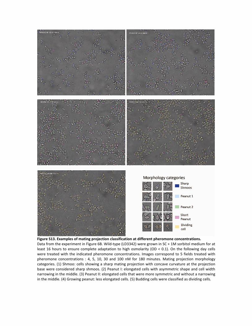
Figure S13. Examples of mating projection classification at different pheromone concentrations.
Data from the experiment in Figure 6B. Wild-type (LD3342) were grown in SC + 1M sorbitol medium for at
least 16 hours to ensure complete adaptation to high osmolarity (OD < 0.1). On the following day cells
were treated with the indicated pheromone concentrations. Images correspond to 5 fields treated with
pheromone concentrations : 4, 5, 10, 30 and 100 nM for 180 minutes. Mating projection morphology
categories. (1) Shmoo: cells showing a sharp mating projection with concave curvature at the projection
base were considered sharp shmoos. (2) Peanut I: elongated cells with asymmetric shape and cell width
narrowing in the middle. (3) Peanut II: elongated cells that were more symmetric and without a narrowing
in the middle. (4) Growing peanut: less elongated cells. (5) Budding cells were classified as dividing cells.


Figure S14. Quantification of MAPKs phosphorylation in wild-type and mutant strains in response to
pheromone.
Wild-type (LD3342) or mutant cells were grown in SC + 1 M sorbitol medium (or 1 M glycerol) for at least
16 hours to assure complete adaptation to high osmolarity (OD ≤ 0.2). On the following day cells were
stimulated with 1 μM α factor. A. Western blot against phosphorylated Hog1, Fus3, Kss1, Slt2 and total
Hog1. B. HOG phosphorylation quantification. Hog1 dual phosphorylation at times 0 and 260 min after α
factor stimulation in all mutants. C to E. Pheromone response and CWI phosphorylation outputs. Dual
phosphorylation of Fus3, Kss1 and Slt2 at times 0 and 260 min after α factor stimulation in all mutants. F.
HOG transcriptional output. Average PSTL1-YFP fluorescence per cell. Error bars represent standard error of
the mean. G. Pheromone response transcriptional output. Average PPRM1-mCherry fluorescence per cell.
Error bars represent standard error of the mean. Strains used: Wild type (LD3342), Δsho1 (RB3704), Δssk1
(RB3382a), Δssk2 (RB3642) and Δfus3 (RB3713), Δfps1 (RB3396a), Δrgc1 (RB3710), Δask10 (RB3717) and
Δrgc1 Δask10 (RB3722), Δpea2 (RB3865), Δspa2 (RB3862) and Δ slt2 (RB3376a). A to G. Samples from
the same experiments. Data amalgamated from three biological replicates.

Figure S15. Additional data from the extracellular glycerol accumulation experiments. Data is from the
experiments in Figure 7B. Wild-type cells were grown in SC + 1 M sorbitol medium for at least 16 hours to
ensure complete adaptation to high osmolarity (OD ≤ 0.2). Plots show, A. optical density (OD 600nm), B.
number of cells and C. average of cell volume normalized to the average volume at time 0 of the cultures
treated (squares) or not (diamonds) with 1 μM pheromone as a function of time. Error bars represent SD
of the average of three independent experiments. All the experiments were started at the same optical
density.

Figure S16. HOG reporter activation in wild-type and mutant strains in response to an acute
hyperosmotic shock.
Cells were grown in SC medium for at least 16 hours. Several dilutions were started to obtain cells in
exponential phase (OD ≤ 0.1) after overnight growth before exposing suitable cultures to an acute 0.4 M
NaCl hyperosmotic shock. A to D. HOG transcriptional output. A. HOG pathway deletion mutants and
Δfus3. Strains: Wild-type (LD3342), Δsho1 (RB3704), Δssk1 (RB3382a), Δssk2 (RB3642) and Δfus3
(RB3713). (B) Glycerol homeostasis deletion mutants. Strains: Wild-type (LD3342), Δfps1 (RB3396a),
Δrgc1 (RB3710), Δask10 (RB3717) and Δrgc1 Δask10 (RB3722). C and D. CWI pathway mutants. Strains:
Wild-type (LD3342 or RB3406), Δpea2 (RB3865), Δspa2 (RB3862) and Δslt2 (RB3376a). E. BMH2
transcriptional output measured in the same cells of experiments a to d as control. Points in A to E
represent average fluorescence of each transcriptional reporter per cell. Error bars represent standard
error of the mean. Plots show a representative experiment out of three biological replicates.

Figure S17. Additional volume recovery experiments.
Wild-type (LD3342) cells were grown in SC medium for at least 16 hours (cells were in exponential phase
OD ≤ 0.1), exposed to the indicated hyperosmotic shock and imaged over time to measure volume as
detailed in supplementary methods. Data corresponds to the average normalized volume. Error bars
represent standard error of the mean. Plots show a representative experiment of three biological
replicates. A. Acute hyperosmotic shocks. 0.2 M NaCl, 0.4 M NaCl and 0.8 M NaCl. B. Acute hyperosmotic
shock in cells growing in SC medium with or without α factor pretreatment. Cells pretreated or not with 1
μM α factor for 4 h and then shocked with 0.65 M NaCl. In contrast to the results in Fig. 8D, cells growing
in SC and not adapted to high osmolarity conditions recover only slightly faster if treated with pheromone
(0.88 ± 0.07 of the non-treated cells recovery time, p = 0.027). (N = 3).

Table S1. Statistical analysis of phosphorylation of MAPKs related to fig. S14.
Hog1pp/Hog1 Time=0 Hog1pp/Hog1
Time=260 min
Hog1pp/Hog1 absolute increase in
260 min
Hog1pp/Hog1 fold increase
in 260 min
Strain Mean SE
p-value
vs WT Mean SE p-value
vs WT
Mean
increase SE
p-value vs
zero
increase
fold
increase SE
p-value
vs WT
WT 0.108 7.6E-03 NA 0.218 0.008 NA
0.110 0.009 6.7E-16 2.039 0.091 NA
Δrgc1 0.129 1.9E-02 8.8E-02 0.189 0.019 1.7E-02 0.061 0.013 3.3E-05 1.518 0.217
4.3E-04
Δask10 0.150 1.9E-02 8.7E-04 0.195 0.019 5.4E-02 0.045 0.013 1.4E-03 1.348 0.217 1.6E-05
Δrgc1Δask10 0.130 1.9E-02 7.2E-02 0.128 0.019 6.5E-10 -0.002 0.013
8.8E-01 1.035 0.217
6.2E-08
Δfps1 0.140 1.9E-02 8.6E-03 0.120 0.019 5.9E-11 -0.021 0.013 1.2E-01 0.906 0.217 7.9E-09
Δfus3 0.138 1.9E-02 1.6E-02 0.174
0.019 4.3E-04 0.036 0.013 9.0E-03 1.342 0.217 1.5E-05
Δssk1 0.074 1.9E-02 6.3E-03 0.069 0.019 4.8E-17 -0.006 0.013 6.8E-01
0.862 0.217 4.0E-09
Δsho1 0.164 1.9E-02 1.9E-05 0.202 0.019 1.8E-01 0.038 0.013 6.0E-03 1.191 0.217 9.1E-07
Δssk2 0.090 1.9E-02
1.4E-01 0.129 0.019
1.0E-09 0.039 0.013 4.7E-03 1.388 0.217 3.5E-05
Δslt2 0.071 1.9E-02 3.0E-03 0.069 0.019 4.7E-17 -0.002 0.013 8.5E-01 0.933
0.217 1.2E-08
Δpea2 0.166 1.9E-02 1.1E-05 0.225 0.019 5.5E-01 0.059 0.013 4.4E-05 1.304 0.217 7.2E-06
Δspa2 0.155 1.9E-02 2.2E-04 0.220 0.019 8.6E-01
0.065 0.013 1.0E-05 1.372 0.217 2.5E-05
Slt2pp/Hog1 Time=0 Slt2pp/Hog1
Time=260 min
Slt2pp/Hog1 absolute increase in
260 min
Slt2pp/Hog1 fold increase in
260 min
Strain Mean SE p-value
vs WT Mean SE
p-value
vs WT
Mean
increase
SE p-value vs
zero
increase
fold
increase SE
p-value
vs WT
WT 0.612 3.2E-02 NA 2.368 0.095 NA 1.756 0.099 6.1E-23 3.939 0.174 NA
Δrgc1 0.649
8.7E-02 5.1E-01 2.313
0.256 7.3E-01 1.664 0.138 3.2E-16 3.549 0.453 1.8E-01
Δask10 0.672 8.8E-02 2.9E-01 2.004 0.244 1.9E-02 1.332 0.125 2.4E-14
2.972 0.442 1.6E-03
Δrgc1Δask10 0.732
9.1E-02 4.9E-02 2.603 0.267 1.8E-01 1.872 0.153 1.6E-16 3.567 0.453 2.0E-01
Δfps1 0.739 9.1E-02 3.9E-02 2.662 0.269 9.9E-02 1.923 0.155 1.1E-16
3.585 0.454 2.2E-01
Δfus3 0.626 8.6E-02 7.9E-01 1.480 0.225 1.4E-08 0.854 0.100 3.3E-11 2.351 0.428 2.8E-06
Δssk1 0.436 7.7E-02
3.2E-04 1.542 0.227 1.1E-07 1.106 0.099 4.4E-15 3.549 0.452 1.8E-01
Δsho1 0.663 8.8E-02 3.6E-01 2.227 0.253 3.8E-01 1.564 0.135 1.2E-15 3.382 0.449 5.5E-02
Δssk2 0.456 7.8E-02 1.5E-03 2.167 0.250 2.0E-01 1.711 0.128 6.6E-18 4.731 0.473 1.5E-02

Δslt2 0.518 8.1E-02 6.4E-02 0.398 0.194 4.6E-25 -0.120 0.048 1.6E-02 0.831 0.386 7.7E-13
Δpea2 0.735 9.1E-02 4.3E-02 0.873 0.206
4.4E-18 0.138 0.077
8.1E-02 1.209 0.398 3.0E-11
Δspa2 0.725 9.1E-02 6.2E-02 0.828 0.204 8.7E-19 0.103 0.075 1.7E-01 1.179 0.397 2.3E-11
Kss1pp/Hog1 Time=0 Kss1pp/Hog1
Time=260 min
Kss1pp/Hog1 absolute increase in
260 min
Kss1pp/Hog1 fold increase in
260 min
Strain Mean SE p-value
vs WT Mean SE
p-value
vs WT
Mean
increase SE
p-value vs
zero
increase
fold
increase SE
p-value
vs WT
WT 0.233 3.3E-02 NA 0.993 0.033 NA
0.760 0.040 2.6E-24 4.297 0.205 NA
Δrgc1 0.224 8.3E-02 8.7E-01 1.060 0.083 1.9E-01 0.836 0.056 9.6E-20 4.916 0.527
6.7E-02
Δask10 0.277 8.3E-02 3.8E-01 0.981 0.083 8.2E-01 0.704 0.056 7.8E-17 3.686 0.527 7.1E-02
Δrgc1Δask10 0.232 8.3E-02 9.9E-01 1.099 0.083 3.9E-02 0.868 0.056
2.1E-20 4.959 0.527
5.2E-02
Δfps1 0.231 8.3E-02 9.8E-01 1.116 0.083 1.8E-02 0.884 0.056 9.7E-21 4.967 0.527 4.9E-02
Δfus3 0.626 8.3E-02 3.9E-10 3.091
0.083 7.0E-40 2.465 0.056 6.3E-41 5.016 0.527 3.6E-02
Δssk1 0.360 8.3E-02 1.5E-02 1.317 0.083 5.1E-08 0.957 0.056 3.7E-22
3.595 0.527 4.0E-02
Δsho1 0.218 8.3E-02 7.7E-01 1.036 0.083 3.9E-01 0.818 0.056 2.3E-19 4.644 0.527 2.9E-01
Δssk2 0.352 8.3E-02
2.2E-02 1.356 0.083
3.3E-09 1.003 0.056 4.9E-23 3.763 0.527 1.1E-01
Δslt2 0.313 8.3E-02 1.2E-01 1.271 0.083 1.3E-06 0.958 0.056 3.5E-22 3.948
0.527 2.9E-01
Δpea2 0.255 8.3E-02 6.5E-01 0.921 0.083 1.6E-01 0.666 0.056 6.1E-16 3.516 0.527 2.4E-02
Δspa2 0.265 8.3E-02 5.3E-01 0.860 0.083 1.1E-02
0.595 0.056 3.3E-14 3.165 0.527 1.9E-03
Fus3pp/Hog1 Time=0 Fus3pp/Hog1
Time=260 min
Fus3pp/Hog1 absolute increase in
260 min
Fus3pp/Hog1 fold increase in
260 min
Strain Mean SE p-value
vs WT Mean SE
p-value
vs WT
Mean
increase
SE p-value vs
zero
increase
fold
increase SE
p-value
vs WT
WT 0.393 3.1E-02 NA 4.744 0.347 NA 4.351 0.348 7.3E-17 12.634 1.202 NA
Δrgc1 0.350
8.0E-02 3.9E-01 5.126
0.979 5.5E-01 4.775 0.530 5.7E-12 15.088 3.729 3.4E-01
Δask10 0.338 7.9E-02 2.6E-01 4.574 0.933 7.7E-01 4.235 0.475 7.6E-12
13.597 3.450 6.7E-01
Δrgc1Δask10 0.352
8.0E-02 4.1E-01 5.333 0.996 3.7E-01 4.981 0.551 5.1E-12 15.078 3.727 3.4E-01
Δfps1 0.290 7.5E-02 2.4E-02 4.794 0.951 9.3E-01 4.504 0.496 4.6E-12
16.610 4.039 1.7E-01
Δfus3 0.110 6.3E-02 2.6E-11 0.141 0.694 7.8E-18 0.031 0.020 1.3E-01 1.274 2.405 3.4E-09
Δssk1 0.429 8.7E-02
5.2E-01 6.683 1.114 1.5E-02 6.255 0.686 4.0E-12 16.369 3.988 1.9E-01
Δsho1 0.434 8.7E-02 4.7E-01 4.949 0.964 7.4E-01 4.515 0.513 1.2E-11 11.736 3.141 6.5E-01
Δssk2 0.455 8.9E-02 2.9E-01 7.376 1.177 2.6E-03 6.921 0.756 3.4E-12 16.515 4.019 1.8E-01

Δslt2 0.743 1.2E-01 1.7E-04 8.167 1.249 4.1E-04 7.424 0.837 9.1E-12 11.390 3.088 5.2E-01
Δpea2 0.571 1.0E-01 1.3E-02 5.013 0.969
6.7E-01 4.442 0.521
3.0E-11 8.829 2.759 2.3E-02
Δspa2 0.408 8.5E-02 7.8E-01 5.449 1.006 2.9E-01 5.041 0.563 6.9E-12 13.452 3.424 7.2E-01
The mean and SE reported correspond to the fixed effects of the linear mixed-effects model used to
analyze this data (see statistical methods for details).

Table S2. Yeast strains.
Strain Relevant genotype Reference
ACL3341 MATa bar1Δ prm1::PPRM1-mCherry::hygB stl1::PSTL1-YFP::URA3 This study
LD3342 MATa bar1Δ prm1::PPRM1-mCherry::hygB stl1::PSTL1-YFP::URA3
bmh2::PBMH2-CFP::TRP1
This study
RB3376a MATa mpk1Δ::kanMX, bar1Δ prm1::PPRM1-mCherry::hygB stl1::PSTL1-
YFP::URA3 bmh2::PBMH2-CFP::TRP1
This study
RB3704 MATa sho1Δ::kanMX, bar1Δ prm1::PPRM1-mCherry::hygB stl1::PSTL1-
YFP::URA3 bmh2::PBMH2-CFP::TRP1
This study
RB3382a MATa ssk1Δ::kanMX, bar1 prm1::PPRM1-mCherry::hygB stl1::PSTL1-
YFP::URA3 bmh2::PBMH2-CFP::TRP1
This study
RB3642 MATa ssk2Δ::HisMX, bar1Δ prm1::PPRM1-mCherry::hygB stl1::PSTL1-
YFP::URA3 bmh2::PBMH2-CFP::TRP1
This study
RB3713 MATa fus3Δ::KanMX, bar1Δ prm1::PPRM1-mCherry::hygB stl1::PSTL1-
YFP::URA3 bmh2::PBMH2-CFP::TRP1
This study
RB3710 MATa rgc1Δ::KanMX, bar1Δ prm1::PPRM1-mCherry::hygB stl1::PSTL1-
YFP::URA3 bmh2::PBMH2-CFP::TRP1
This study
RB3717 MATa rgc2Δ::KanMX, bar1Δ prm1::PPRM1-mCherry::hygB stl1::PSTL1-
YFP::URA3 bmh2::PBMH2-CFP::TRP1
This study
RB3722 MATa rgc1Δ::KanMX rgc2Δ::HisMX bar1Δ prm1::PPRM1-
mCherry::hygB stl1::PSTL1-YFP::URA3 bmh2::PBMH2-CFP::TRP1
This study
RB3396a MATa fps1Δ::His bar1Δ prm1::PPRM1-mCherry::hygB stl1::PSTL1-
YFP::URA3 bmh2::PBMH2-CFP::TRP1
This study
RB3406 MATa bar1Δ stl1::PSTL1-CFP::TRP1 This study
RB3862 MATa spa2Δ::KanMX bar1Δ stl1::PSTL1-CFP::TRP1 This study
RB3865 MATa pea2Δ::KanMX, bar1Δ prm1::PPRM1-mCherry::hygB stl1::PSTL1-
YFP::URA3 bmh2::PBMH2-CFP::TRP1
This study
RB3801 MATa hog1Δ::KanMX, bar1Δ prm1::PPRM1-mCherry::hygB stl1::PSTL1-
YFP::URA3 bmh2::PBMH2-CFP::TRP1
This study

ySP69 MATa HTA2::HTA2-CFP-no marker HOG1-Venus::TRP1 leu2-3,112
trp1-1 can1-100 ura3-1 ade2-1 his3-11,15
(46)
yGP1281 BY4742 s288c MATα Pact1-RFP -URA3 in lyp1
G. Pesce,
Molecular Sciences
Institute, CA
Copyright © 2022 FDOKUMEN