Physicochemical characterization and biological activity of synthetic TLR4 agonist formulations
8
ORIGINAL PAPER Physico-chemical characterization and biological response of Labeo rohita-derived hydroxyapatite scaffold S. Mondal • A. Mondal • N. Mandal • B. Mondal • S. S. Mukhopadhyay • A. Dey • S. Singh Received: 27 August 2013 / Accepted: 10 November 2013 / Published online: 28 November 2013 Ó Springer-Verlag Berlin Heidelberg 2013 Abstract The chemically treated Labeo rohita scale is used for synthesizing hydroxyapatite (HAp) biomaterials. Thermogravimetric and differential thermal analyses of fish scale materials reveal the different phase changes with temperature and find out the suitable calcination tempera- tures. The composition and structures of wet ball-milled calcined HAp powders are characterized by Fourier trans- form infrared spectroscopy, X-ray diffraction, field emis- sion scanning electron microscopy, transmission electron microscopy, energy dispersive X-ray analysis (EDX). The EDX as well as chemical analysis of fish scale-derived apatite materials confirms that the Ca/P ratio is 1.71. The compressive stress, hardness and porosity have been eval- uated on sintered HAp biomaterials. The cell attachment on HAp surfaces, cytotoxicity evaluation and MTT assay, which are carried out in RAW macrophage-like cell line media demonstrate good biocompatibility. The histological analysis also supports the bioaffinity of processed HAp biomaterials in Wistar rat model for investigating the contact reaction and stability at the artificial or natural prosthesis interface. Keywords Labeo rohita scale Á Hydroxyapatite Á Biocompatibility Á Cytotoxicity Á In vivo Introduction Fish as a diet has several nutritional and therapeutic ben- efits for health. The most important constituents of fish as food are protein, fat, carbohydrates, sodium, potassium, calcium, magnesium, vitamin B6 and vitamin B12, which help in maintaining good health as well as the central nervous system of a body [1]. Apart from its nutritional values, fish waste like scale, fin and bone is an important source of hydroxyapatite (HAp) and collagen biomaterials. Each scale consists of two distinct regions: an external (osseous) layer and an internal fibrillary plate. In the upper external layer, collagen fibers are randomly arranged and embedded in a proteoglycan matrix. The collagen fibers are produced within the fibrillary layer by scleroblasts located at the base of the scales. Mineralization of the scales occurs continuously throughout the life of the organism. The external layer is initially mineralized with matrix vesicles and calcium phosphate-based materials are deposited bio- logically on the layer matrix. HAp is the most stable cal- cium phosphate salt at normal temperatures with a pH between 4 and 12 [2]. It is an important material of great interest in protein chromatography applications, waste water treatment processes and suitable scaffold materials [3, 4]. HAp is also considered as a model compound to study biomineralization phenomena [5, 6]. There is an intensive attempt for the development of well-defined HAp crystals toward physicochemical behavior in vitro and S. Mondal Á N. Mandal Á B. Mondal (&) Centre for Advanced Materials Processing, CSIR-Central Mechanical Engineering Research Institute, Mahatma Gandhi Avenue, Durgapur 713 209, India e-mail: [email protected]; [email protected] A. Mondal Department of Biotechnology, Heritage Institute of Technology, Kolkata, India S. S. Mukhopadhyay Á A. Dey Department of Biotechnology, National Institute of Technology, Durgapur 713 209, India S. Singh CSIR-Centre for Cellular and Molecular Biology, Hyderabad, India 123 Bioprocess Biosyst Eng (2014) 37:1233–1240 DOI 10.1007/s00449-013-1095-z
-
Upload
washington -
Category
Documents
-
view
1 -
download
0
Transcript of Physicochemical characterization and biological activity of synthetic TLR4 agonist formulations

ORIGINAL PAPER
Physico-chemical characterization and biological responseof Labeo rohita-derived hydroxyapatite scaffold
S. Mondal • A. Mondal • N. Mandal •
B. Mondal • S. S. Mukhopadhyay • A. Dey •
S. Singh
Received: 27 August 2013 / Accepted: 10 November 2013 / Published online: 28 November 2013
� Springer-Verlag Berlin Heidelberg 2013
Abstract The chemically treated Labeo rohita scale is
used for synthesizing hydroxyapatite (HAp) biomaterials.
Thermogravimetric and differential thermal analyses of
fish scale materials reveal the different phase changes with
temperature and find out the suitable calcination tempera-
tures. The composition and structures of wet ball-milled
calcined HAp powders are characterized by Fourier trans-
form infrared spectroscopy, X-ray diffraction, field emis-
sion scanning electron microscopy, transmission electron
microscopy, energy dispersive X-ray analysis (EDX). The
EDX as well as chemical analysis of fish scale-derived
apatite materials confirms that the Ca/P ratio is 1.71. The
compressive stress, hardness and porosity have been eval-
uated on sintered HAp biomaterials. The cell attachment on
HAp surfaces, cytotoxicity evaluation and MTT assay,
which are carried out in RAW macrophage-like cell line
media demonstrate good biocompatibility. The histological
analysis also supports the bioaffinity of processed HAp
biomaterials in Wistar rat model for investigating the
contact reaction and stability at the artificial or natural
prosthesis interface.
Keywords Labeo rohita scale � Hydroxyapatite �Biocompatibility � Cytotoxicity � In vivo
Introduction
Fish as a diet has several nutritional and therapeutic ben-
efits for health. The most important constituents of fish as
food are protein, fat, carbohydrates, sodium, potassium,
calcium, magnesium, vitamin B6 and vitamin B12, which
help in maintaining good health as well as the central
nervous system of a body [1]. Apart from its nutritional
values, fish waste like scale, fin and bone is an important
source of hydroxyapatite (HAp) and collagen biomaterials.
Each scale consists of two distinct regions: an external
(osseous) layer and an internal fibrillary plate. In the upper
external layer, collagen fibers are randomly arranged and
embedded in a proteoglycan matrix. The collagen fibers are
produced within the fibrillary layer by scleroblasts located
at the base of the scales. Mineralization of the scales occurs
continuously throughout the life of the organism. The
external layer is initially mineralized with matrix vesicles
and calcium phosphate-based materials are deposited bio-
logically on the layer matrix. HAp is the most stable cal-
cium phosphate salt at normal temperatures with a pH
between 4 and 12 [2]. It is an important material of great
interest in protein chromatography applications, waste
water treatment processes and suitable scaffold materials
[3, 4]. HAp is also considered as a model compound to
study biomineralization phenomena [5, 6]. There is an
intensive attempt for the development of well-defined HAp
crystals toward physicochemical behavior in vitro and
S. Mondal � N. Mandal � B. Mondal (&)
Centre for Advanced Materials Processing, CSIR-Central
Mechanical Engineering Research Institute, Mahatma Gandhi
Avenue, Durgapur 713 209, India
e-mail: [email protected]; [email protected]
A. Mondal
Department of Biotechnology, Heritage Institute of Technology,
Kolkata, India
S. S. Mukhopadhyay � A. Dey
Department of Biotechnology, National Institute of Technology,
Durgapur 713 209, India
S. Singh
CSIR-Centre for Cellular and Molecular Biology,
Hyderabad, India
123
Bioprocess Biosyst Eng (2014) 37:1233–1240
DOI 10.1007/s00449-013-1095-z

in vivo studies for reconstructive bone replacement and
other medical applications [7, 8]. In literature, several
methods of preparation of HAp crystals from bio-wastes
have been reported, including solid-state reactions, crystal
growth under hydrothermal reaction, layer hydrolysis of
other calcium phosphate salts, sol–gel crystallization [9–
15]. Due to low production cost and worldwide availabil-
ity, the researchers [16] are concentrated on the use of fish
scale bio-waste for the processing of HAp crystal [17].
Many processing technologies have been employed to
obtain porous ceramics for bone tissue engineering from
different bio-wastes. In tissue repair application, the mac-
ropores and highly interconnected networks are required
for the growth of surrounding host tissues. The porosity of
filler scaffold is about 30–33 % which is also similar to the
results reported by Gross et al. [18]. The optimal scaffold
design and fabrication techniques must be able to create
porous structures adequate for attaining the desired
mechanical function and mass transport properties [18, 19].
In this paper, an attempt has been made to synthesize HAp
powder from Labeo rohita scale. The powder is charac-
terized through thermogravimetric analysis, X-ray diffrac-
tion (XRD) and Fourier transform infrared spectroscopy
(FTIR), etc. The mechanical properties of the sintered HAp
have been evaluated for development of porous scaffold.
The cell attachment on HAp surfaces, cytotoxicity evalu-
ation and MTT assay on RAW macrophage cell line media
are carried out to demonstrate good biocompatibility.
Methods and materials
Synthesis and characterization of hydroxyapatite
powder from fish scale
Labeo rohita scales are used as raw material for HAp pro-
cessing. Scales are initially washed thoroughly with water to
discard impurities and then deproteinized by solvent system.
The cleaned de-protienated scale is calcined at 700–800 �C
to synthesis HAp ceramics after differential thermal analysis
of scale from room temperature to 1,200 �C by NETZSCH
Jupiter STA 491 at a heating rate of 10 �C/min. XRD Ana-
lysis of HAp powder is performed at the scanning range of
2h = 20�–80� with CuKa target by Shimadzu, XRD-6000
instrument. The crystallographic phases of HAp with dif-
ferent temperatures are recorded and confirmed the HAp
using standard JCPDS files. FTIR analysis is carried out at
the scanning range of 4,000–400 cm-1 (Shimadzu IR Pres-
tige—21, Japan) to determine functional groups present in
HAp powder. FESEM and EDX (Model No: SUPRA 40,
CARLZEISSSMT, Oxford) analyses reveal the composi-
tional and morphological structure of green as well as sin-
tered powder of HAp. Quantitative energy dispersive X-ray
is also performed to confirm the Ca/P ratio of the sample
apart from chemical method. Ultrasonic instruments are
employed for dispersing and homogeneously distribute the
particles into liquids to prepare samples for TEM analysis
(TEM, Jeol Gem Microscope). ‘‘Image J’’ software is used to
evaluate the typical images of HAp particles.
Preparation of fillers and scaffold
The calcined powder is wet ball-milled for 48 h in plane-
tary mill. The particle size of dried milled powder is
measured in Malvern particle size analyzer. The average
size of the particle is *0.5 lm. Triton–X surfactant is
added drop wise to the dried ball-milled powders to make a
paste for injection molding. The sample is prepared as
small rod-shaped fillers with 2 mm diameter and 4 mm
long. The fillers are then dried at 80 �C for several hours.
Finally, the dried samples are sintered at 1,200 �C for 2 h.
Alternatively, hydroxyapatite samples are mixed well with
requisite amount of starch porogen and compacted with
unidirectional pressure at 200–250 MPa to make 12 mm
dia. The sample was sintered at 1,200 �C for crystallo-
graphic phase determination as well as physico-mechanical
property evaluation. The process flow of fish scale-derived
HAp scaffold development and in vivo bio-implantation is
shown in Fig. 1.
Results and discussion
Differential thermogravimetric analysis
The DTA/TG analysis reveals the phase transformation and
determines the weight loss with respect to temperatures.
Two major and one minor mass losses are distinctly
observed as depicted in Fig. 2. The first major weight loss
is observed in the range of 100–400 �C, on the heating
process from room temperature to 1,200 �C. This mass loss
is reported as near about 4.56 % of the sample taken for
experimentation. The first mass loss process may be due to
the removal of adsorbed water from the surface and inter-
particle spaces [17]. Other exothermic peaks at *407 and
709 �C correspond to weight losses about 0.86 and 0.17 %,
respectively. The weight loss at the range of 400–500 �C is
due to the combustion of organic materials. However, there
is a minor weight loss observed on heating up beyond 710�to 1,200 �C, which indicates better thermal stability of the
sample in this range.
X-ray diffraction analysis
The crystalline phase analysis of the HAp powder from fish
scale is carried out by XRD studies. The XRD of HAp
1234 Bioprocess Biosyst Eng (2014) 37:1233–1240
123

indicates the visible crystalline nature of typical apatite
crystal structures. It is also noticed that crystallographic
behavior of HAp resembles to that of XRD pattern of
biological apatite [20] which is shown in Fig. 3. However,
the pattern of the fish scale-derived HAp powder peaks
corresponds to the JCPDS card No. 72-1243 of pure HAp.
The highest intensity peak of synthesized powder is in the
plane of 211 and it resembles to the pure HAp crystal XRD
characteristics. The two minor b-TCP peaks are observed
at 31� and 34� after sintering at 1,200 �C.
Fourier transform infrared spectroscopy analysis
FTIR spectroscopy is employed to characterize the differ-
ent functional groups of HAp [Ca10 (PO4)6(OH)2]. FTIR
spectrum of HAp powder calcined at 700 �C is recorded in
the range of 4,000–400 cm-1 as shown in Fig. 4. The
characteristics of FTIR peaks in the wave range
570–632 cm-1 resemble asymmetric bending vibration of
P–O band of HAp materials, whereas the peak at
960–965 cm-1 is due to symmetric stretching vibration of
P–O band of PO43- ion. The spectral data at the wave
number values of 876, 1,412 and 1,451 cm-1 suggest the
presence of carbonate ion [21] in the prepared HAp
materials calcined at 700 �C. The strong peaks at 1,053 and
1,095 cm-1 are ascribed to asymmetric stretching mode of
vibration of P–O bands of PO4 tetrahedra [22, 23]. The
characteristic frequencies derived from PO43- modes are
better resolved with increasing temperature. The existence
of O–H and C–O bands disappears beyond calcinations
temperature of the powder (1,200 �C). But the structural
O–H peaks at 1,655 and 3,370 cm-1 are predominant with
increasing temperature, whereas the existence of C–O
bands is reduced significantly. Therefore, sintered HAp
Fig. 1 Pictorial representation of a fish Labeo rohita, b chemically
treated dried fish scale, c scanning electron microscopy of treated fish
scale, d synthesized HAp powder, e fabrication of small fillers by
injection press molding, f sintered fillers, g SEM images of porous
fillers, h schematic representation of fillers in rat model
Fig. 2 TG/DT analysis of HAp powder
Fig. 3 XRD analysis of HAp powder
Bioprocess Biosyst Eng (2014) 37:1233–1240 1235
123

ceramics synthesized from fish scale beyond 1,200 �C is
established.
FE-SEM and EDX analyses
The FE-SEM micrograph of fish scale-derived HAp dried
powder and fillers scaffolds is depicted in Fig. 5. The
morphology of calcined wet ball-milled dried HAp powder
shows soft agglomerated ultrafine particles. The biocom-
patible powder consists of a uniform porous structure upon
compacting at 200–250 MPa with porogen (starch granules
Merck). The compacted HAp-sintering surface shows dis-
tinct grain boundary with uniform porous structure. EDX
analysis of the synthesized HAp powder shows that the
ratio of Ca/P is 1.71 as depicted in Fig. 6.
Chemical analysis of Ca/P ratio in fish scale HAp
To perform the chemical analysis, a weighed quantity of
hydroxyapatite powder is dissolved in dilute (1:1) nitric
acid (HNO3). Calcium content is determined from an ali-
quot part of the solution by reverse complexometric titra-
tion, i.e., by titration of the excess of complexone III
(disodium salt of ethylenediamine tetra-acetic acid) with
the use of a solution of nickel(II) chloride in ammonia.
Murexide (ammonium purpurate) is used as the indicator
for titration.
Phosphorus was determined colorimetrically as the
phosphovanadomolybdate complex with a UV–VIS Shi-
madzu Model spectrophotometer using a wavelength of
420 nm. Phosphorus content is determined by the photo-
metric method on the basis of the formation of yellow
phosphomolybdic acid and its subsequent reduction to a
blue complex compound in a hydrochloric acid solution of
thiocarbamide in the presence of copper (II) sulfate. The
error of the analysis is 0.5 % Ca and 0.1 % P [24]. The
experiment is repeated thrice to ascertain the Ca/P ratio.
The ratio of Ca/P is in the range of 1.70–1.73, whereas the
EDX experiment shows 1.71 as depicted in Fig. 6.
Fig. 4 FTIR analysis of HAp powder
Fig. 5 SEM images of porous HAp scaffold and HAp particles
synthesized from fish scale
Fig. 6 EDX analysis of HAp particles
1236 Bioprocess Biosyst Eng (2014) 37:1233–1240
123
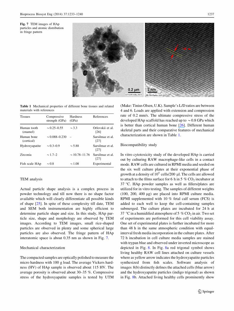
TEM analysis
Actual particle shape analysis is a complex process in
powder technology and till now there is no shape factor
available which will clearly differentiate all possible kinds
of shape [25]. In spite of these complexity till date, TEM
and SEM both instrumentation are highly efficient to
determine particle shape and size. In this study, HAp par-
ticle size, shape and morphology are observed by TEM
images. According to TEM images, small rice-shaped
particles are observed in plenty and some spherical large
particles are also observed. The fringe pattern of HAp
interatomic space is about 0.35 nm as shown in Fig. 7.
Mechanical characterization
The compacted samples are optically polished to measure the
micro hardness with 100 g load. The average Vickers hard-
ness (HV) of HAp sample is observed about 115 HV. The
average porosity is observed about 30–35 %. Compressive
stress of the hydroxyapatite samples is tested by UTM
(Make: Tinius Olsen, U.K). Sample’s L/D ratios are between
4 and 6. Loads are applied with extension and compression
rate of 0.2 mm/s. The ultimate compressive stress of the
developed HAp scaffold has reached up to *0.8 GPa which
is better than cortical human bone [26]. Different human
skeletal parts and their comparative features of mechanical
characterization are shown in Table 1.
Biocompatibility study
In vitro cytotoxicity study of the developed HAp is carried
out by culturing RAW macrophage-like cells in a contact
mode. RAW cells are cultured in RPMI media and seeded on
the six well culture plates at their exponential phase of
growth at a density of 105 cells/200 ll. The cells are allowed
to attach to the films surface for 6 h in 5 % CO2 incubator at
37 �C. HAp powder samples as well as fillers/plates are
utilized for in vitro testing. The samples of different weights
(100, 200, 400 lg) are placed into RPMI culture media.
RPMI supplemented with 10 % fetal calf serum (FCS) is
added to each well to keep the cell-containing samples
submerged. The culture plates are incubated for 24 h at
37 �C in a humidified atmosphere of 5 % CO2 in air. Two set
of experiments are performed for this cell viability assay.
One set of experimental plates is further incubated for more
than 48 h in the same atmospheric condition with equal-
interval fresh media incorporation in the culture plates. After
72 h incubation in cell culture media samples are stained
with trypan blue and observed under inverted microscope as
depicted in Fig. 8. In Fig. 8a red trigonal symbol shows
living healthy RAW cell lines attached on culture vessels
where as yellow arrow indicates the hydroxyapatite particles
synthesized from fish scales. Software analysis of
images 8(b) distinctly defines the attached cells (blue arrow)
and the hydroxyapatite particles (indigo trigonal) as shown
in Fig. 8b. Attached living healthy cells prominently show
Fig. 7 TEM images of HAp
particles and atomic distribution
in fringe pattern
Table 1 Mechanical properties of different bone tissues and related
materials with references
Tissues Compressive
strength (GPa)
Hardness
(GPa)
References
Human tooth
(enamel)
*0.25–0.55 *3.3 Orlovskii et al.
[26]
Human bone
(cortical)
*0.088–0.230 – Sarsilmaz et al.
[27]
Hydroxyapatite *0.3–0.9 *5.88 Sarsilmaz et al.
[27]
Zirconia *1.7–2 *10.78–11.76 Sarsilmaz et al.
[27]
Fish scale HAp *0.8 *1.08 Experimental
Bioprocess Biosyst Eng (2014) 37:1233–1240 1237
123

their protoplasm in Fig. 8b. The experimental result reveals
the non-immunogenic effect of hydroxyapatite particles
toward cell lines (Table 2).
On the other hand, second set of experimental plate is
utilized for MTT assay. After 24 h incubation, MTT
(10 ll) is added to each well at a strength of 10 % (v/v) and
incubated for further 4 h at 37 �C. Subsequently, the media
containing MTT is removed, and 100 ll of DMSO
(dimethylsulfoxide) is added to dissolve the formazan
crystals. The plates are agitated for 5 min and read at
550 nm on a scanning multi-well spectrophotometer plate
reader (Biorad, USA). t test is performed for statistical
significance analysis and a P value of\0.05 is determined
to represent a significant difference. The percentage of cell
viable is expressed as
Absorbance of treated cells
Absorbance of controlled cells� 100 %:
Cell attachment study
HAp blocks are used for in vitro cell attachment studies.
HAp blocks are sterilized and then RAW cell lines are used
in RPMI cell culture media for this study. After 72 h
incubation samples cell culture media is decanted and
washed thoroughly with phosphate buffer solution (PBS).
After washing 2 % paraformaldehyde solution is utilized
for cellular fixation. Figure 8c shows SEM image attached
cells on to the HAp surface. This study reveals that cells
can be attached on the HAp surface which would be uti-
lized as scaffold material for tissue engineering applica-
tion. Software analysis of Fig. 8c shows the exact attached
location of cell lines on hydroxyapatite surface as shown in
Fig. 8d. This experimental result shows the bioaffinity of
synthesized hydroxyapatite particles toward cell line.
Histological analysis
In the present pilot study, histological analysis [27, 28] is
performed in Wistar rats. Wistar rats are easy to handle and
their femur is brittle but not fragile during drilling. Skel-
etally mature 3-months-old female Wistar rats of body
weight 150–200 g from the animal house are used for the
experiment. The animals are maintained at standard envi-
ronmental conditions (temperature 22–25 �C, humidity
40–70 % with 12:12 dark/light photoperiod) approved by
the Committee for the Purpose of Control and Supervision
of Experiment on Animals (CPCSEA) whereas all the
experimental protocols are approved by IAEC, CCMB.
Animals were anesthetized with ketamine and xylazine
with a dose of 40 mg and 5 mg/kg body weight, respec-
tively. Lateral and median aspect of the femur is cleaned.
An incision is made to cut open the skin on the dorsal
aspect above the femur. A careful incision is then made to
Fig. 8 Cytotoxicity evaluation
in RAW cell line media and
attachment of cells on HAp
surface
Table 2 Results of MTT assay: percentage of cell viable
HAp concentration (lgm/ml) Viable cell (%)
100 131
200 125
400 128
1238 Bioprocess Biosyst Eng (2014) 37:1233–1240
123

cut open the biceps femoris muscles to expose femur bone.
A Pediatric bone driller is used to make a hole of 5 mm in
the dorsal surface of femur without touching the bone
marrow thus preventing any hemorrhage. The drilled femur
of control animal is left unfilled, whereas in experimental
group the HAp rods are fixed to fill up the gaps. The
muscle layer is stitched with simple interrupted suture
using absorbable materials and skin is closed using non-
absorbable suture materials. Povidone, iodine ointment is
applied externally for 3 days. At the end of the experiment
after 3 months, the animals are sacrificed by overdose of
CO2. Detail necropsy is done and femur bone is collected
and fixed in 10 % formalin [29]. Fixed bone is decalcified
with EDTA before embedding in paraffin wax. Fixed and
paraffin embedded bones are cut at 5 lm thickness, stained
with Hematoxylin and Eosin following standard procedure
and examined under light microscope as shown in Fig. 9a.
The response of tissues and the implanted material is
assessed. Trauma regions are being recovered by healing
new cells as cell infiltration on materials is observed.
Deeply stained region may conclude the presence of pre-
osteoblast cells because this type of cells consists of mono
nucleus with prominent nucleoli. In some regions, new cell
linings are appeared which may be the result of osteo-
conduction as shown in blue arrow in Fig. 9b.
Conclusion
• Labeo rohita scale-derived HAp is almost similar to the
structure of pure HAp.
• The porous HAp scaffold with *30 % porosity shows
considerable improvement in compressive strength
*0.8 GPa which may be suitable for nutrient and
biological fluid transportation for in vivo system.
• Cytotoxicity analysis of HAp particles and MTT assay
indicates no cytotoxic effects on macrophage-like
RAW cell line media.
• The histological analysis concludes cell infiltration and
integration in HAp fillers which is a good result of
bioactivity.
• Cellular attachment on HAp surface reveals that
hydroxyapatite microsphere is able to support cell lines
to adhere and proliferate.
Acknowledgments The authors would like to express their grati-
tude to Director, CSIR-CMERI for his kind permission to publish this
paper. The authors are thankful to Dr. Syamal Roy, Head of the
department of Immunology and infectious diseases at IICB Kolkata
for their kind support for cell culture, toxicity studies. The authors are
also indebted to CSIR-Centre for Cellular and Molecular Biology
(CSIR-CCMB), Hyderabad for histological and in vivo studies. The
financial support from CSIR is highly acknowledged.
References
1. Kawarazuka N (2010) The contribution of fish intake, aquacul-
ture, and small-scale fisheries to improving nutrition: a literature
review. The World Fish Center Working Paper No. 2106. The
World Fish Center, Malaysia
2. Koutsopoulos S (2002) Synthesis and characterization of
hydroxyapatite crystals: a review study on the analytical methods.
J Biomed Mater Res 62:600–612
3. Zhang J, Zhang W, Bao T, Chen Z (2013) Mussel-inspired
polydopamine-assisted hydroxyapatite as the stationary phase
for capillary electrochromatography. Analyst. doi:10.1039/c3an
01668d
4. Intapong S, Raksudjarit A (2013) Treatment of agricultural
wastewater using porous ceramics composite of hydroxyapatite
and silica. Adv Mater Res 622–623:915–918
5. Kim MH, Himeno T, Kawashita M, Kokubo T, Nakamura T
(2004) The mechanism of biomineralization of bone-like apatite
on synthetic hydroxyapatite: an in vitro assessment. J R Soc
Interface 1:17–22
Fig. 9 Histological analysis of
femur bone of Wistar rat with
HAp prosthetic fillers. a and
b shows Bone formation on
hydroxyapatite implant (arrow)
shows lining cells. Healing is on
progress in 3 months sample.
Some integration of cells are
seen, cell infiltration on
materials are also observed
(star). Pre-osteoblast cells
appear which could be identified
by mono nuclear cells wih
prominent nulceoli and deeply
stained cytoplasm (rhombus).
Each bar (thick line) represents
50 lm
Bioprocess Biosyst Eng (2014) 37:1233–1240 1239
123

6. Zhang HM, Wu B (2011) Biomineralization of the hydroxyapa-
tite with 3D-structure for enamel reconstruction. Adv Mater Res
391–392:633–637
7. Koutsopoulos S, Demakopoulos J, Argiriou X, Dalas E, Klouras
N, Spanos N (1995) Inhibition of hydroxyapatite formation by
zirconocenes. Langmuir 11:1831–1834
8. Ozawa M, Suzuki S (2002) Microstructural development of
natural hydroxyapatite originated from fish-bone waste through
heat treatment. J Am Ceram Soc 85:1315–1317
9. Dorozhkin Sergey V (2010) Bioceramics of calcium orthophos-
phates. Biomaterials 31(2010):1465–1485
10. Venkatesan J, Ji Qian Z, Ryu B, Vinay Thomas N, Kim SK
(2011) A comparative study of thermal calcination and an alka-
line hydrolysis method in the isolation of hydroxyapatite from
Thunnus obesus bone. Biomed Mater 6(3):035003
11. Bardhan R, Mahata S, Mondal B (2011) Processing of natural
resourced hydroxyapatite from egg shell waste by wet precipita-
tion method. Adv Appl Ceram Struct Funct Bioceram 110:80–86
12. Liao CJ, Lin FH, Chen KS, Sun JS (1999) Thermal decomposi-
tion and reconstitution of hydroxyapatite in air atmosphere.
Biomaterials 20:1807–1813
13. Yamasaki N, Kai T, Nishioka M, Yanagisawa K, Ioku K (1990)
Porous hydroxyapatite ceramics prepared by hydrothermal hot
pressing. J Mater Sci 9:1150–1151
14. Cheng PT (1987) Formation of octacalcium phosphate and sub-
sequent transformation to hydroxyapatite at low supersaturation:
a model for cartilage calcification. Calcif Tissue Int 40:339–343
15. Piccirillo C, Silva MF, Pullar RC, Braga da Cruz I, Jorge R,
Pintado MME, Castro PML (2013) Extraction and characterisa-
tion of apatite- and tricalcium phosphate-based materials from
cod fish bones. Mater Sci Eng C 33(1):103–110
16. Boutinguiza M, Pou J, Comesana R, Lusquinos F, de Carlos A,
Leon B (2012) Biological hydroxyapatite obtained from fish
bones. Mater Sci Eng C 32(3):478–486
17. Mondal S, Mahata S, Kundu S, Mondal B (2010) Processing of
natural resourced hydroxyapatite ceramics from fish scale. Adv
Appl Ceram Struct Funct Bioceram 109:234–239
18. Gross KA, Rodrıguez-Lorenzo LM (2004) Biodegradable com-
posite scaffolds with an interconnected spherical network for
bone tissue engineering. Biomaterials 25(20):4955–4962
19. Miranda P, Saiz E, Gryn K, Tomsia AP (2006) Sintering and
robocasting of b-tricalcium phosphate scaffolds for orthopaedic
applications. Acta Biomater 2(4):457–466
20. Bose S, Roy M, Bandyopadhyay A (2012) Recent advances in
bone tissue engineering scaffolds. Trends Biotechnol 30(10):
546–554
21. Liu Q, Huang S, Matinlinna JP, Chen Z, Pan H (2013) Insight
into biological apatite: physiochemical properties and preparation
approaches. Biomed Res Int. doi:10.1155/2013/929748 (Article
ID 929748)
22. Panda RN, Hsieh MF, Chung RJ, Chin TS (2003) FTIR, XRD,
SEM and solid state NMR investigation of carbonated hydroxy-
apatite nano particles synthesized by hydroxide gel technique.
J Phys Chem solids 64:193–199
23. Fathi MH, Hanifi A, Mortazavi V (2008) Preparation and bio-
activity evaluation of bone-like hydroxyapatite nanopowder.
J Mater Process Technol 202(1–3):536–542
24. Garbuz VV, Dubok VA, Kravchenko LF, Kurochkin VD,
Ul’yanchich NV, Kornilova VI (1998) Analysis of the chemical
composition of a bioceramic based on hydroxyapatite and tri cal-
cium phosphate. Powder Metall Metal Ceram 37(3–4):193–195
25. Pourghahrsamani P, Forssberg E (2005) Review of applied par-
ticle shape descriptors and produced particle shapes in grinding
environments: part II: particle shape. Miner Process Extr Metall
Rev 26:145–166
26. Orlovskii VP, Komlev VS, Barinov SM (2002) Hydroxyapa-
tite and hydroxyapatite-based ceramics. Inorg Mater 38(10):
973–984
27. Sarsilmazb F, Orhanb N, Unsaldia E, Durmusa AS, Colakogluc N
(2007) A polyethylene- high proportion hydroxyapatite implant
and its investigation in vivo. Acta Bioeng Biomech 9(2):9–16
28. Flautre B, Anselme K, Delecourt C, Lu J, Hardouin P, Descamps
M (1999) Histological aspects in bone regeneration of an asso-
ciation with porous hydroxyapatite and bone marrow cells.
J Mater Sci Mater Med 10:811–814
29. Sujatha R, Isnik S, Guha A, Mahesh Kumar J, Sinha A, Singh S
(2012) Evaluation of nano-biphasic calcium phosphate ceramics
for bone tissue engineering applications: in vitro and preliminary
in vivo studies. J Biomater Appl 27:565–575
1240 Bioprocess Biosyst Eng (2014) 37:1233–1240
123