Effects of monoolein-based cubosome formulations on lipid ...
12
Toxicology Research PAPER Cite this: Toxicol. Res., 2015, 4, 1025 Received 16th March 2015, Accepted 23rd April 2015 DOI: 10.1039/c5tx00078e www.rsc.org/toxicology Effects of monoolein-based cubosome formulations on lipid droplets and mitochondria of HeLa cells Angela Maria Falchi,* a Antonella Rosa, a Angela Atzeri, a Alessandra Incani, a Sandrina Lampis, b Valeria Meli, b Claudia Caltagirone b and Sergio Murgia* b Despite the remarkable development of nanoparticles for different purposes, relatively little is known about their interaction with biological systems and individual cells. Here the effects of two monoolein- based cubosome formulations stabilized by Pluronic F108 and F127 were investigated against HeLa cells. Microscopy analysis on living cells loaded with organelle-specific fluorescent probes was performed to assess the formation of cytoplasmic lipid droplets after nanoparticle treatment. Mitochondrial membrane potential and mitochondrial ROS generation were also investigated in relation to the capability of the accumulated lipids to affect mitochondrial functions. Values of the main cellular unsaturated fatty acids were also measured to assess cell lipid profile modulation. Results from this study show that the uptake of both cubosome formulations induced modification of the cell lipid profile, lipid droplet accumulation, mitochondrial hyperpolarization and mitochondrial ROS generation. These results shed some light on the influence exerted by monoolein-based cubosome formulations on subcellular organelles and their possible adverse effects on cell functions. Introduction Monoolein (MO, glycerol monooleate) self-assembles in water, forming a number of nanostructures, including two bicontinu- ous cubic phases characterized by a different space group: the Ia3d, and the Pn3m. 1 These amazing liquid crystalline nano- structures are composed of a curved bilayer whose three- dimensional folding originates two disconnected, continuous water channels. 2,3 They were ubiquitously found in Nature, and several studies were dedicated to shed some light on their role in the cell machinery. 4–6 Since MO-based bicontinuous cubic phases are nanostructures that can suitably host hydro- phobic molecules with biological relevance, they were exten- sively investigated in the past for their possible applications in the pharmaceutical field. 7–9 Remarkably, cubic phases can be formulated as nanoparticle dispersions, known as cubosomes, typically stabilized in water using amphiphilic polymers having long polyethylene oxide (PEO) chains, such as Pluronics or polysorbate 80. 10–12 These liquid crystalline nanoparticles are often proposed to be suitable drug delivery nanocarriers and, recently, their possible application as theranostic nano- medicine was discussed in several papers. 13–15 However, little information about in vitro cellular interactions and toxicity of monoolein-based cubosomes has been reported so far, and the lack of shared protocols precludes straightforward com- parisons between these investigations. For example, focusing on monoolein-based formulations exposed to adherent cells, to the best of our knowledge only six papers (two from our group, not including the present) have discussed the cytotoxi- city of pristine cubosomes (i.e., not decorated with targeting agents or loaded with drugs/imaging agents). 13,16–20 In all these cases, experiments were performed using different cell lines, at different incubation times, and with formulations having different compositions (basically, they were prepared using a different monoolein/Pluronic ratio, sometimes using different types of Pluronic). It is worth noting that several papers have reported on the haemolytic properties of monoo- lein-based cubosome formulations tested on mouse blood, 18,21 indicating 20 μg mL −1 of monoolein as the maximum tolerable concentration. 20 However, another recent investigation con- ducted on human and porcine plasma has disputed these results, attributing to the same formulation a very low haemo- lytic activity. 22 We have previously shown no toxic effects of monoolein-based nanoparticles (liposomes and cubosomes) at short times against HeLa cells and 3T3 fibroblasts 16,23 based on an 80% viability threshold. a Department of Biomedical Sciences, University of Cagliari, 09042 Monserrato, CA, Italy. E-mail: [email protected]; Fax: +39 070 6754003; Tel: +39 070 6754055 b Department of Chemical and Geological Sciences, University of Cagliari, 09042 Monserrato, CA, Italy. E-mail: [email protected]; Fax: +39 070 6754003; Tel: +39 070 6754388 This journal is © The Royal Society of Chemistry 2015 Toxicol. Res. , 2015, 4, 1025–1036 | 1025 Downloaded from https://academic.oup.com/toxres/article/4/4/1025/5573508 by guest on 13 February 2022
-
Upload
khangminh22 -
Category
Documents
-
view
1 -
download
0
Transcript of Effects of monoolein-based cubosome formulations on lipid ...

Toxicology Research
PAPER
Cite this: Toxicol. Res., 2015, 4, 1025
Received 16th March 2015,Accepted 23rd April 2015
DOI: 10.1039/c5tx00078e
www.rsc.org/toxicology
Effects of monoolein-based cubosomeformulations on lipid droplets and mitochondriaof HeLa cells
Angela Maria Falchi,*a Antonella Rosa,a Angela Atzeri,a Alessandra Incani,a
Sandrina Lampis,b Valeria Meli,b Claudia Caltagironeb and Sergio Murgia*b
Despite the remarkable development of nanoparticles for different purposes, relatively little is known
about their interaction with biological systems and individual cells. Here the effects of two monoolein-
based cubosome formulations stabilized by Pluronic F108 and F127 were investigated against HeLa cells.
Microscopy analysis on living cells loaded with organelle-specific fluorescent probes was performed to
assess the formation of cytoplasmic lipid droplets after nanoparticle treatment. Mitochondrial membrane
potential and mitochondrial ROS generation were also investigated in relation to the capability of the
accumulated lipids to affect mitochondrial functions. Values of the main cellular unsaturated fatty acids
were also measured to assess cell lipid profile modulation. Results from this study show that the uptake of
both cubosome formulations induced modification of the cell lipid profile, lipid droplet accumulation,
mitochondrial hyperpolarization and mitochondrial ROS generation. These results shed some light on the
influence exerted by monoolein-based cubosome formulations on subcellular organelles and their
possible adverse effects on cell functions.
Introduction
Monoolein (MO, glycerol monooleate) self-assembles in water,forming a number of nanostructures, including two bicontinu-ous cubic phases characterized by a different space group: theIa3d, and the Pn3m.1 These amazing liquid crystalline nano-structures are composed of a curved bilayer whose three-dimensional folding originates two disconnected, continuouswater channels.2,3 They were ubiquitously found in Nature,and several studies were dedicated to shed some light on theirrole in the cell machinery.4–6 Since MO-based bicontinuouscubic phases are nanostructures that can suitably host hydro-phobic molecules with biological relevance, they were exten-sively investigated in the past for their possible applications inthe pharmaceutical field.7–9 Remarkably, cubic phases can beformulated as nanoparticle dispersions, known as cubosomes,typically stabilized in water using amphiphilic polymershaving long polyethylene oxide (PEO) chains, such as Pluronicsor polysorbate 80.10–12 These liquid crystalline nanoparticlesare often proposed to be suitable drug delivery nanocarriers
and, recently, their possible application as theranostic nano-medicine was discussed in several papers.13–15 However, littleinformation about in vitro cellular interactions and toxicity ofmonoolein-based cubosomes has been reported so far, andthe lack of shared protocols precludes straightforward com-parisons between these investigations. For example, focusingon monoolein-based formulations exposed to adherent cells,to the best of our knowledge only six papers (two from ourgroup, not including the present) have discussed the cytotoxi-city of pristine cubosomes (i.e., not decorated with targetingagents or loaded with drugs/imaging agents).13,16–20 In allthese cases, experiments were performed using different celllines, at different incubation times, and with formulationshaving different compositions (basically, they were preparedusing a different monoolein/Pluronic ratio, sometimes usingdifferent types of Pluronic). It is worth noting that severalpapers have reported on the haemolytic properties of monoo-lein-based cubosome formulations tested on mouse blood,18,21
indicating 20 μg mL−1 of monoolein as the maximum tolerableconcentration.20 However, another recent investigation con-ducted on human and porcine plasma has disputed theseresults, attributing to the same formulation a very low haemo-lytic activity.22 We have previously shown no toxic effects ofmonoolein-based nanoparticles (liposomes and cubosomes) atshort times against HeLa cells and 3T3 fibroblasts16,23 basedon an 80% viability threshold.
aDepartment of Biomedical Sciences, University of Cagliari, 09042 Monserrato, CA,
Italy. E-mail: [email protected]; Fax: +39 070 6754003; Tel: +39 070 6754055bDepartment of Chemical and Geological Sciences, University of Cagliari, 09042
Monserrato, CA, Italy. E-mail: [email protected]; Fax: +39 070 6754003;
Tel: +39 070 6754388
This journal is © The Royal Society of Chemistry 2015 Toxicol. Res., 2015, 4, 1025–1036 | 1025
Dow
nloaded from https://academ
ic.oup.com/toxres/article/4/4/1025/5573508 by guest on 13 February 2022
All cell types have been found to readily take up the lipidspresent in culture media and sequester them into lipid dro-plets (LDs). LDs are cytoplasmic organelles that consist of ahydrophobic core of neutral lipids (such as triglycerides andcholesterol esters) encased by a phospholipid monolayer har-boring a set of enzymes and regulatory proteins that catalyzethe highly metabolically controlled synthesis and mobilizationof fat stores.24 LDs, found in almost all cells under physiologi-cal or pathological conditions, are heterogeneous not only insize but also in the lipid content and the composition of theirprotein coat.25 The most widely accepted model for LD for-mation hypothesizes that they arise in the endoplasmic reticu-lum (ER) through the accumulation of neutral lipid at specificsites between the leaflets of the phospholipid bilayer (ERbudding model).24 Dynamical interactions between LDs andorganelles other than ER, including mitochondria and peroxi-somes, have been suggested to facilitate the exchange of pro-teins and lipids in cells, while interactions between the ERand mitochondria are crucial for processes such as lipid syn-thesis, storage and transport as well as mitochondrial func-tions (calcium homeostasis and apoptosis).26,27
In the present study, we used organelle-specific dyes toexplore the changes occurring in lipid droplets and mitochon-dria of living HeLa cells when exposed to treatment withmonoolein-based cubosomes stabilized by Pluronic F108 orPluronic F127. Analyses were carried out to examine lipiddroplet content, mitochondria membrane potential, reactiveoxygen species (ROS), and cellular lipid profile at 4 and 24 hafter nanoparticle treatment. Our results demonstrate that theuptake of both cubosome formulations induces a modificationin the cellular lipid profile, accumulation of lipid dropletsassociated with a significant dysfunction of mitochondria,
including mitochondrial hyperpolarization and mitochondrialROS generation.
Results and discussionCubosome characterization
Colloidal dispersions of a bicontinuous cubic phase (cubo-somes) were prepared by dispersing via sonication meltedmonoolein (MO) either in a Pluronic F108 (PF108) or in aPluronic F127 (PF127) aqueous solution. These nanoparticleformulations were then characterized for particle size andmorphology, as well as for their inner structure. The cryo-TEMimages of MO-based PF127 and PF108 stabilized cubosomes(MO/PF127 and MO/PF108) are shown in Fig. 1A and B. Theseimages show various nanoparticles revealing the regular alter-nation of bright spots (water channels) and dark matrix (lipidbilayer) classically observed in the presence of a cubic arrange-ment of the interface, along with the vesicular material thattypically co-exists with the cubosomes in these kinds of formu-lations.16 On the basis of repeated observations, the samplesunder investigation via cryo-TEM were found to be a dispersionof cubosomes having diameter in the range of 100–200 nm, instriking agreement with DLS analysis (Table 1). SAXS definitelyconfirmed the inner cubic structure of both cubosome formu-lations. As reported in Table 1, the diffraction patterns showedthe Bragg peaks of the bicontinuous cubic double diamondand primitive phases (with space groups Pn3m and Im3m,respectively, see also Fig. 1C and D). It deserves noticing thatthe simultaneous presence of these two phases has alreadybeen described for cubosome formulations having compo-sition similar to those investigated here,28 and their occur-
Fig. 1 Cryo-TEM images of MO-based cubosomes stabilized by PF127 (A) and PF108 (B). I(q) vs. q data obtained by SAXS of the cubosome formu-lation stabilized by PF127 (C) and PF108 (D). The Miller indices are reported on top of the corresponding Bragg peaks along with the indication of thespace group.
Paper Toxicology Research
1026 | Toxicol. Res., 2015, 4, 1025–1036 This journal is © The Royal Society of Chemistry 2015
Dow
nloaded from https://academ
ic.oup.com/toxres/article/4/4/1025/5573508 by guest on 13 February 2022
rence is explained with the non-uniform partition of the Pluro-nic. Indeed, during the dispersion process the dispersant pre-ferentially localizes at the cubosome surface, originatingPluronic-depleted regions characterized by double diamondsymmetry.29
Accumulation of lipids in the lipid droplets
Several studies have shown LD accumulation after treatmentwith free fatty acids30 or associated with oxidative stressinduced by nanoparticle treatment.31,32 Indeed, after theinternalization in the cells, unsaturated fatty acids such asoleic acid (OA) can be either esterified and stored as neutrallipids inside LDs or become part of the cellular membranesor, after activation to fatty acyl-CoAs, oxidized in mitochondriafor energy production.30 Here, to investigate LD accumulation,HeLa cells were treated with cubosome formulations (5 µL in1 mL of medium) at 4 and 24 h and, after nanoparticle wash-out, co-loaded with Nile Red (NR) and Hoechst probes, to stainLDs and the nucleus, respectively.
In untreated control cells the number of small LDs,detected as punctuated green fluorescent cytoplasmic struc-tures, was very different from cell to cell. Upon short exposureto cubosomes, the number of lipid droplets dispersed through-out the cytoplasm and their size increased in a remarkableway and droplets appeared larger and more numerous inall cells (Fig. 2A), suggesting that LD accumulation resultsfrom the synthesis of neutral lipids from nanoparticle-derivedmonoolein.
When compared to untreated control cells, an increase inthe IOD (Integrated Optical Density) per cell of 7.9 (p < 0.001)-and 3.1 (p < 0.001)-fold was estimated for, respectively,MO/PF127 and MO/PF108, indicating a statistically signifi-cantly increased production of LDs (Fig. 2B). Subsequently,HeLa cells were grown in the presence of MO dissolved inDMSO and Pluronic aqueous solutions (administered at thesame concentrations as MO and Pluronics present in the cubo-some formulations) and OA (18:1 n-9) (used as the positivecontrol at the non-cytotoxic concentration of 100 µM inDMSO). At short times, MO and OA treatments caused 1.3 (p <0.05)- and 2.8 (p < 0.001)-fold increase, respectively, in IOD percell in comparison with untreated cells, whereas Pluronic solu-tions showed the same IOD per cell as the control (Fig. 2B). Atlonger exposure time (24 h), differently from MO- and OA-treated cells where the IOD did not change, the IOD of
MO/PF127 and MO/PF108-treated cells increased by 25.6 (p <0.001)- and 31.5-fold (p < 0.001), respectively. Because of thehuge cubosome uptake, cells appeared to be full of lipidsinside the cytoplasm with many large droplets aggregated inseveral clusters resembling grapes (Fig. 2A and B).
While the LD formation induced by long-chain unsaturatedfatty acids such as OA is a well-known phenomenon, datareported here proved that LD formation also occurs upon MOand MO-based cubosome treatment. Compared with MO-treated cells, statistically significantly increased production oflipid droplets was detected after cubosome exposure, thussuggesting a strong internalization ability of both MO-basedcubosome formulations. Since the amount of MO adminis-tered was exactly the same, the differences could be ascribedto different uptake mechanisms. Indeed, while nanoparticlescan be transported into the cells via endocytosis, the fatty aciduptake across the lipid bilayer of the plasma membrane wasproposed to proceed by diffusion or through membrane-associated proteins (acting as fatty acid transporters).33,34
Remarkably, statistically significant differences (p < 0.001)between the two cubosome formulations were observed in theIOD only at short time treatments.
Modification of the lipid cell profile
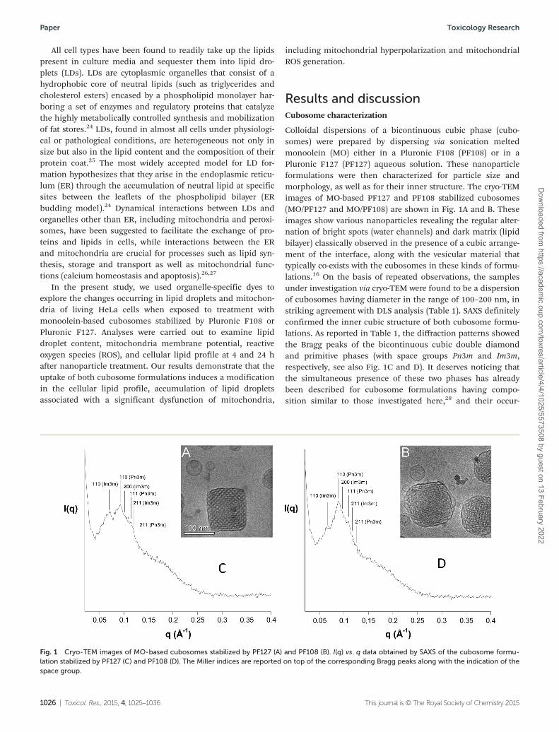
The same MO/PF108 and MO/PF127 cubosome formulationsand MO, OA, and Pluronic solutions previously described werethen tested to assess their effects on the lipid composition ofHeLa cells. After 4 and 24 h of incubation, the cell lipid frac-tion was extracted and the variations in the levels of unsatu-rated fatty acids and cholesterol were analyzed with respect tountreated control cells. The unsaturated fatty acid composition(expressed as µg per plate) and the chromatographic profile ofHeLa control cells obtained by HPLC are shown in Fig. 3. Thecell content of the most abundant unsaturated fatty acids wasdetected as follows: 78.19, 19.87, 6.38, 4.89, 3.11, and 3.05 µgper plate for 18:1 isomers (OA and 18:1 n-7), 16:1 n-7, arachi-donic acid (20:4 n-6), docosahexaenoic acid (DHA, 22:6 n-3),eicosapentaenoic acid (EPA 20:5 n-3), and 18:2 n-6, respect-ively; minor amounts were measured for 18:3 n-3, 20:3, and22:4 n-6. The incubation of HeLa cells with PF108 or PF127solution did not induce changes in unsaturated fatty acidlevels, with treated cells showing a profile similar to that ofcontrol cells at both incubation times (data at 4 h are shown inFig. 3A).
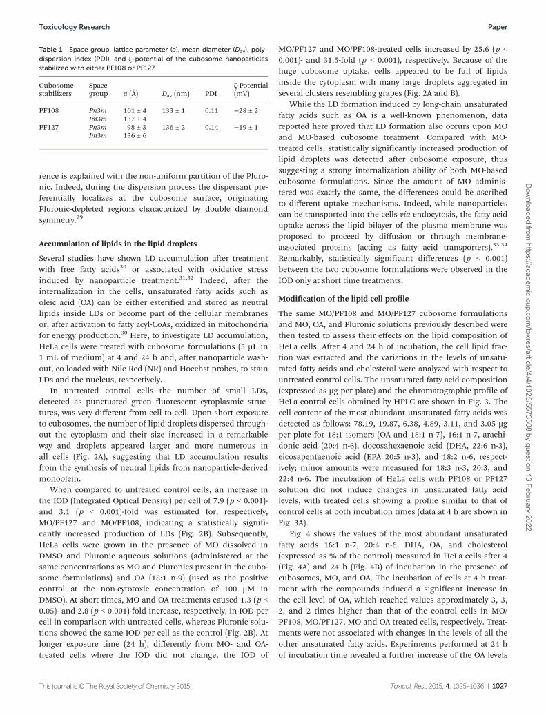
Fig. 4 shows the values of the most abundant unsaturatedfatty acids 16:1 n-7, 20:4 n-6, DHA, OA, and cholesterol(expressed as % of the control) measured in HeLa cells after 4(Fig. 4A) and 24 h (Fig. 4B) of incubation in the presence ofcubosomes, MO, and OA. The incubation of cells at 4 h treat-ment with the compounds induced a significant increase inthe cell level of OA, which reached values approximately 3, 3,2, and 2 times higher than that of the control cells in MO/PF108, MO/PF127, MO and OA treated cells, respectively. Treat-ments were not associated with changes in the levels of all theother unsaturated fatty acids. Experiments performed at 24 hof incubation time revealed a further increase of the OA levels
Table 1 Space group, lattice parameter (a), mean diameter (Dav), poly-dispersion index (PDI), and ζ-potential of the cubosome nanoparticlesstabilized with either PF108 or PF127
Cubosomestabilizers
Spacegroup a (Å) Dav (nm) PDI
ζ-Potential(mV)
PF108 Pn3m 101 ± 4 133 ± 1 0.11 −28 ± 2Im3m 137 ± 4
PF127 Pn3m 98 ± 3 136 ± 2 0.14 −19 ± 1Im3m 136 ± 6
Toxicology Research Paper
This journal is © The Royal Society of Chemistry 2015 Toxicol. Res., 2015, 4, 1025–1036 | 1027
Dow
nloaded from https://academ
ic.oup.com/toxres/article/4/4/1025/5573508 by guest on 13 February 2022

(5 times higher than the control) in cells treated with cubo-some formulations, while levels of all the other unsaturatedfatty acids remained substantially unaltered with respect to thecontrol. Remarkably, the levels of OA recorded in MO and OAtreated cells were very similar to those observed at 4 h-treatedHeLa cells. This result parallels the finding discussed in theprevious paragraph concerning the lipid droplet accumulationand, similarly, can be reconciled taking into account thedifferent uptake mechanisms. Using HPLC, the total chole-sterol level was measured in control cells as the mean contentof 72.52 ± 0.55 µg per plate. Fig. 4 also shows the values ofcholesterol measured in the control and 4 and 24 h-treated
HeLa cells. The different treatments did not seem to affect thelevel of cholesterol.
Mitochondrial membrane potential
Mitochondria play a central role in normal cell functions suchas energy supply and in cell death by inducing apoptosis.Changes in the mitochondrial membrane potential (mitoMP)represent one of the adaptation processes to a variety ofenvironmental factors that regulate the increased energydemands, cell growth and differentiation, or cellular stress. Inmitochondria, mitoMP is produced by the electron transportchain as it pumps H+ ions into the membrane space,
Fig. 2 Lipid droplet accumulation induced in HeLa cells by cubosome treatment. (A) Representative composite color images of HeLa cells exposedto the monoolein (MO), MO/PF127 and MO/PF108 cubosomes at 4 and 24 h of incubation time. Membranes (red) and lipid droplets (green) werestained with Nile Red (colocalization in yellow). Scale bar = 20 µm. (B) Results of the lipid droplet formation at 4 and 24 h of incubation time. Oleicacid (OA) was used as the positive control at the non-cytotoxic concentration of 100 µM. Data are expressed as mean ± SD from four independentexperiments. Statistically significant differences are indicated by * (p < 0.05), *** (p < 0.001) by t-test vs. untreated-control cells.
Paper Toxicology Research
1028 | Toxicol. Res., 2015, 4, 1025–1036 This journal is © The Royal Society of Chemistry 2015
Dow
nloaded from https://academ
ic.oup.com/toxres/article/4/4/1025/5573508 by guest on 13 February 2022
thus creating an electrochemical proton gradient which drivesATP synthesis. Variations of mitoMP are usually measuredin living cells with the cationic fluorescent dye TMRM,
which accumulates in the mitochondrial matrix driven by thevoltage gradient across the inner mitochondrial membrane.Using the TMRM probe, control cells showed morphologically
Fig. 3 Fatty acid composition and chromatographic profile of HeLa cells. Values of the unsaturated fatty acids (16:1 n-7, 18:1, 18.2 n-6, 18:3 n-3,20:3, 20:4 n-6, 20:5 n-3, 22:4 n-6, 22:6 n-3, expressed as µg per plate) measured in control cells and after 4 h of incubation in the presence ofPF127 and PF108 solutions (A) and the corresponding HPLC chromatographic profile of the control HeLa cells (B). Data are expressed as mean ± SDfrom four independent experiments (involving duplicate analyses for each sample).
Fig. 4 Lipid profile modulation in cubosome-treated cells. The values of the main unsaturated fatty acids, 16:1 n-7, 18:1 (OA), 20:4 n-6, 22:6 n-3,and cholesterol (expressed as % of the control) measured in HeLa control cells and after 4 (A) and 24 h (B) of incubation in the presence of MO/PF127 and MO/PF108 cubosomes, MO, and OA (used as the positive control). Data are expressed as mean ± SD from four independent experiments(involving duplicate analyses for each sample). Statistically significant differences, using one-way analysis of variation (one-way ANOVA) and theBonferroni post test (GraphPad INSTAT software, San Diego, CA), are indicated by ** (p < 0.01), *** (p < 0.001) vs. untreated-control cells.
Toxicology Research Paper
This journal is © The Royal Society of Chemistry 2015 Toxicol. Res., 2015, 4, 1025–1036 | 1029
Dow
nloaded from https://academ
ic.oup.com/toxres/article/4/4/1025/5573508 by guest on 13 February 2022
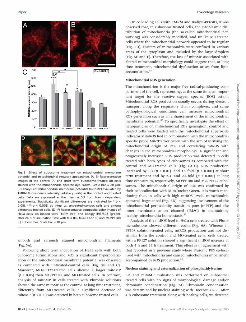
smooth and variously stained mitochondrial filaments(Fig. 5A).
Following short term incubation of HeLa cells with bothcubosome formulations and MO, a significant hyperpolariz-ation of the mitochondrial membrane potential was observedas compared with untreated-control cells (Fig. 5B and C).Moreover, MO/PF127-treated cells showed a larger mitoMP(p < 0.05) than MO/PF108- and MO-treated cells. In contrast,analysis of mitoMP in cells treated with Pluronic solutionsshowed the same mitoMP as the control. At long time treatment,differently from MO-treated cells, a significant decrease ofmitoMP (p < 0.05) was detected in both cubosome-treated cells.
On co-loading cells with TMRM and Bodipy 493/503, it wasobserved that, in cubosome-treated cells, the cytoplasmic dis-tribution of mitochondria (the so-called mitochondrial net-working) was considerably modified, and unlike MO-treatedcells where the mitochondrial network appeared to be regular(Fig. 5D), clusters of mitochondria were confined in variousareas of the cytoplasm and excluded by the large droplets(Fig. 5E and F). Therefore, the loss of mitoMP associated withaltered mitochondrial morphology could suggest that, at longtime treatment, mitochondrial dysfunction arises from lipidaccumulation.35
Mitochondrial ROS generation
The mitochondrion is the major free radical-producing com-partment of the cell, representing, at the same time, an impor-tant target for the reactive oxygen species (ROS) action.Mitochondrial ROS production usually occurs during electrontransport along the respiratory chain complexes, and samepathophysiological conditions can increase mitochondrialROS generation such as an enhancement of the mitochondrialmembrane potential.36 To specifically investigate the effect ofnanoparticles on mitochondrial ROS generation, control andtreated cells were loaded with the mitochondrial superoxideindicator MitoROS Red in combination with the mitochondria-specific probe MitoTracker Green with the aim of verifying themitochondrial origin of ROS and correlating mtROS withchanges in the mitochondrial morphology. A significant andprogressively increased ROS production was detected in cellstreated with both types of cubosomes as compared with thecontrol and MO-treated cells (Fig. 6A–C). ROS productionincreased by 1.3 (p < 0.01)- and 1.9-fold (p < 0.001) at shortterm treatment and by 2.3- and 2.4-fold (p < 0.001) at longtime exposure to, respectively, MO/PF108 and MO/PF127 cubo-somes. The mitochondrial origin of ROS was confirmed bytheir co-localization with MitoTracker Green. It is worth men-tioning that, in cells with high mtROS levels, mitochondriaappeared fragmented (Fig. 6D), suggesting involvement of themitochondrial permeability transition pore (mPTP) and theinner membrane anion channel (IMAC) in maintaininghealthy mitochondria homeostasis.37
Analysis of the mtROS level in HeLa cells treated with Pluro-nic solutions showed different results (Fig. 6A). Whereas inPF108 solution-treated cells, mtROS production was not dis-similar from the control and MO-treated cells, cells treatedwith a PF127 solution showed a significant mtROS increase atboth 4 h and 24 h treatment. This effect is in agreement withthat reported in a previous study where Pluronic P85 co-loca-lized with mitochondria and caused mitochondria impairmentaccompanied by ROS production.38
Nuclear staining and externalization of phosphatidylserine
LD and mitoMP evaluation was performed on cubosome-treated cells with no signs of morphological damage and/orchromatin condensation (Fig. 7A). Chromatin condensationwas determined by nuclear staining with Hoechst 33258. After4 h cubosome treatment along with healthy cells, we detected
Fig. 5 Effect of cubosome treatment on mitochondrial membranepotential and mitochondrial network appearance. (A, B) Representativeimages of the control (A) and short-term cubosome-treated (B) cellsstained with the mitochondria-specific dye TMRM. Scale bar = 20 µm.(C) Analysis of mitochondrial membrane potential (mitoMP) evaluated byTMRM fluorescence intensity (arbitrary units) in the control and treatedcells. Data are expressed as the mean ± SD from four independentexperiments. Statistically significant differences are indicated by *(p <0.05), ***(p < 0.001) by t-test vs. untreated-control cells and amongdifferently treated cells. (D–F) Representative composite color images ofHeLa cells, co-loaded with TMRM (red) and Bodipy 493/503 (green),after 24 h of incubation time with MO (D), MO/PF127 (E) and MO/PF108(F) cubosomes. Scale bar = 10 µm.
Paper Toxicology Research
1030 | Toxicol. Res., 2015, 4, 1025–1036 This journal is © The Royal Society of Chemistry 2015
Dow
nloaded from https://academ
ic.oup.com/toxres/article/4/4/1025/5573508 by guest on 13 February 2022
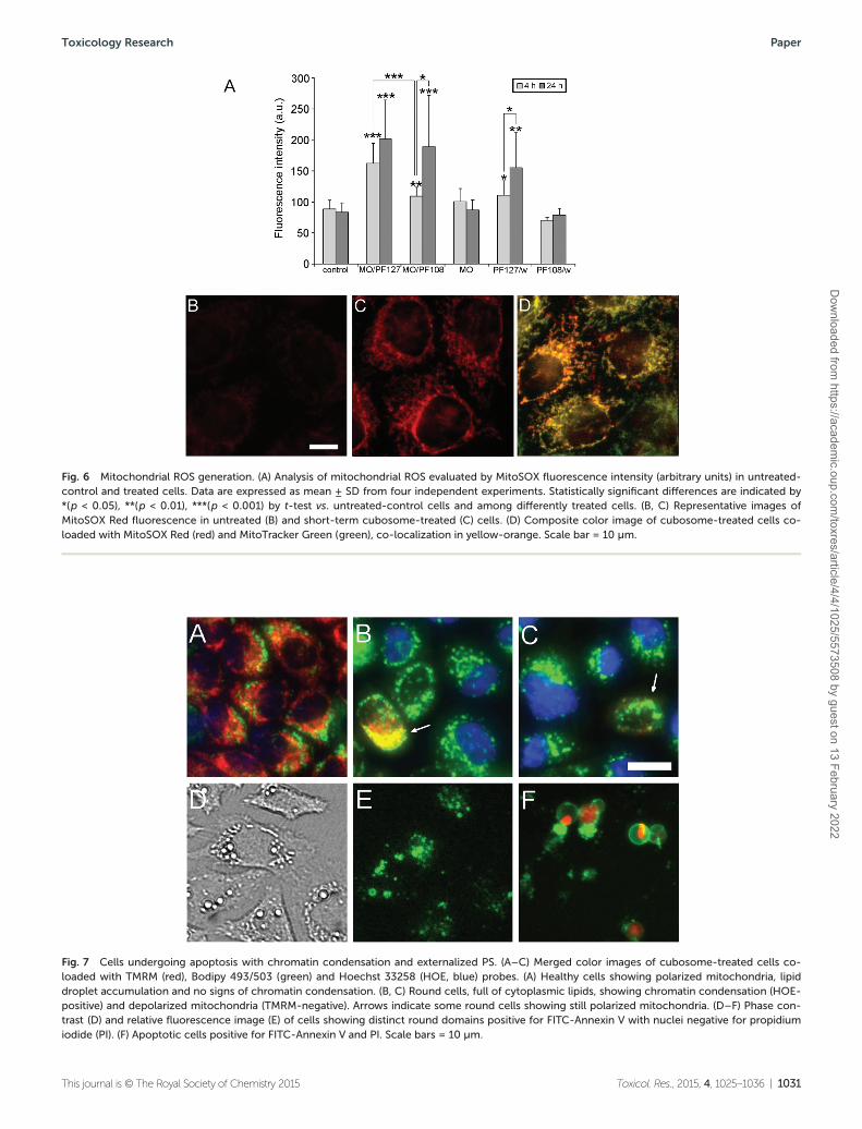
Fig. 6 Mitochondrial ROS generation. (A) Analysis of mitochondrial ROS evaluated by MitoSOX fluorescence intensity (arbitrary units) in untreated-control and treated cells. Data are expressed as mean ± SD from four independent experiments. Statistically significant differences are indicated by*(p < 0.05), **(p < 0.01), ***(p < 0.001) by t-test vs. untreated-control cells and among differently treated cells. (B, C) Representative images ofMitoSOX Red fluorescence in untreated (B) and short-term cubosome-treated (C) cells. (D) Composite color image of cubosome-treated cells co-loaded with MitoSOX Red (red) and MitoTracker Green (green), co-localization in yellow-orange. Scale bar = 10 µm.
Fig. 7 Cells undergoing apoptosis with chromatin condensation and externalized PS. (A–C) Merged color images of cubosome-treated cells co-loaded with TMRM (red), Bodipy 493/503 (green) and Hoechst 33258 (HOE, blue) probes. (A) Healthy cells showing polarized mitochondria, lipiddroplet accumulation and no signs of chromatin condensation. (B, C) Round cells, full of cytoplasmic lipids, showing chromatin condensation (HOE-positive) and depolarized mitochondria (TMRM-negative). Arrows indicate some round cells showing still polarized mitochondria. (D–F) Phase con-trast (D) and relative fluorescence image (E) of cells showing distinct round domains positive for FITC-Annexin V with nuclei negative for propidiumiodide (PI). (F) Apoptotic cells positive for FITC-Annexin V and PI. Scale bars = 10 µm.
Toxicology Research Paper
This journal is © The Royal Society of Chemistry 2015 Toxicol. Res., 2015, 4, 1025–1036 | 1031
Dow
nloaded from https://academ
ic.oup.com/toxres/article/4/4/1025/5573508 by guest on 13 February 2022
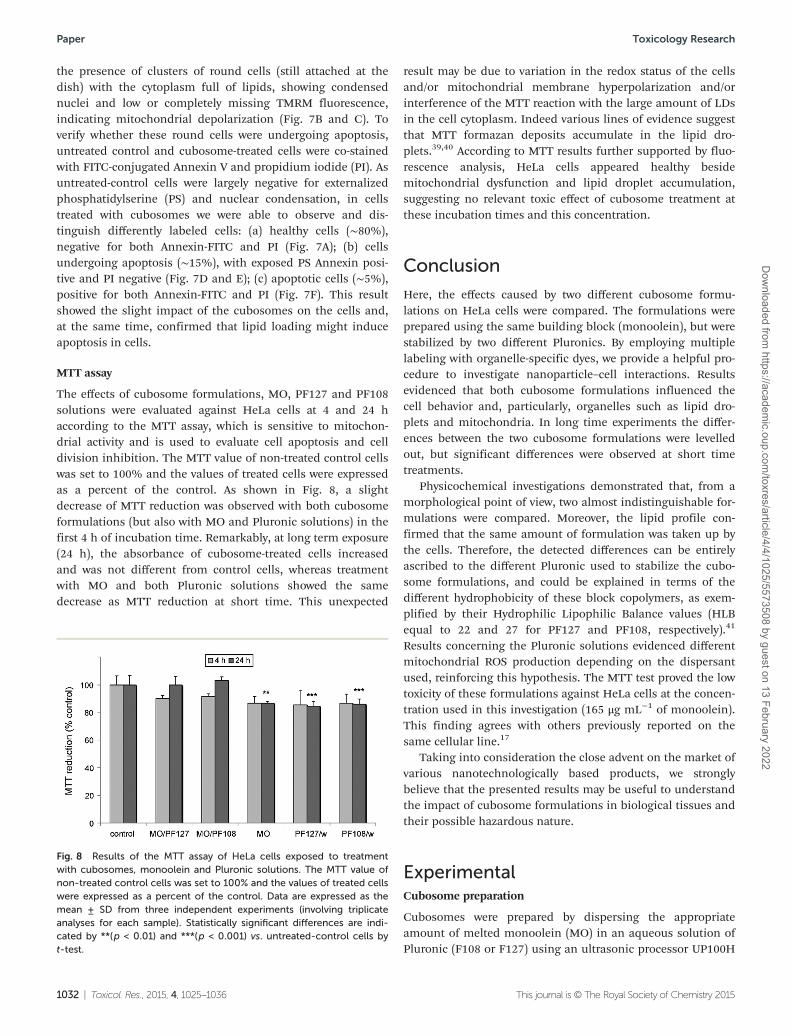
the presence of clusters of round cells (still attached at thedish) with the cytoplasm full of lipids, showing condensednuclei and low or completely missing TMRM fluorescence,indicating mitochondrial depolarization (Fig. 7B and C). Toverify whether these round cells were undergoing apoptosis,untreated control and cubosome-treated cells were co-stainedwith FITC-conjugated Annexin V and propidium iodide (PI). Asuntreated-control cells were largely negative for externalizedphosphatidylserine (PS) and nuclear condensation, in cellstreated with cubosomes we were able to observe and dis-tinguish differently labeled cells: (a) healthy cells (∼80%),negative for both Annexin-FITC and PI (Fig. 7A); (b) cellsundergoing apoptosis (∼15%), with exposed PS Annexin posi-tive and PI negative (Fig. 7D and E); (c) apoptotic cells (∼5%),positive for both Annexin-FITC and PI (Fig. 7F). This resultshowed the slight impact of the cubosomes on the cells and,at the same time, confirmed that lipid loading might induceapoptosis in cells.
MTT assay
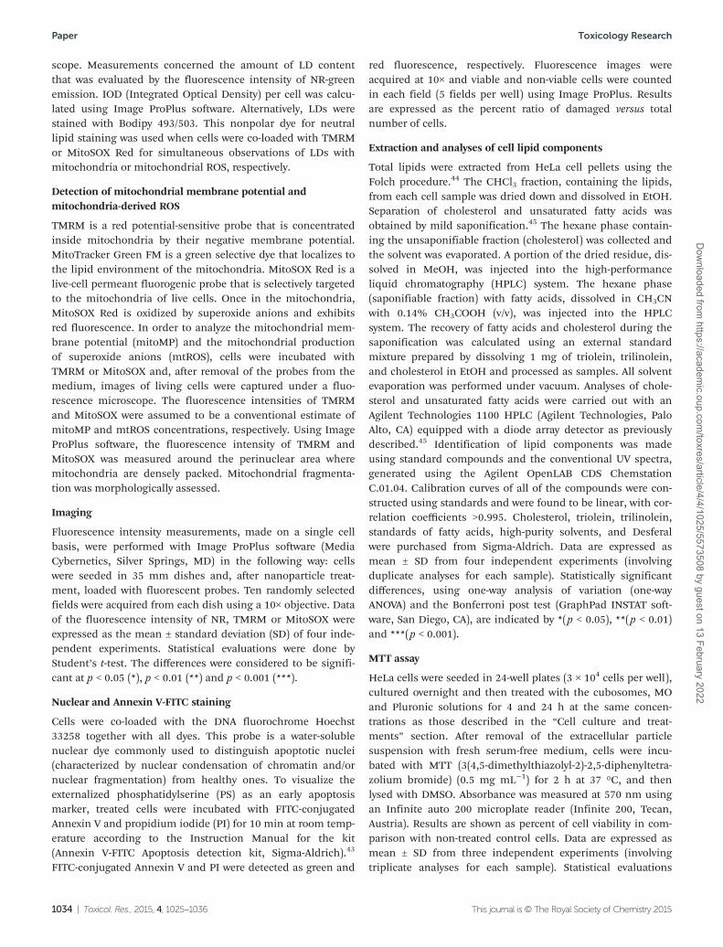
The effects of cubosome formulations, MO, PF127 and PF108solutions were evaluated against HeLa cells at 4 and 24 haccording to the MTT assay, which is sensitive to mitochon-drial activity and is used to evaluate cell apoptosis and celldivision inhibition. The MTT value of non-treated control cellswas set to 100% and the values of treated cells were expressedas a percent of the control. As shown in Fig. 8, a slightdecrease of MTT reduction was observed with both cubosomeformulations (but also with MO and Pluronic solutions) in thefirst 4 h of incubation time. Remarkably, at long term exposure(24 h), the absorbance of cubosome-treated cells increasedand was not different from control cells, whereas treatmentwith MO and both Pluronic solutions showed the samedecrease as MTT reduction at short time. This unexpected
result may be due to variation in the redox status of the cellsand/or mitochondrial membrane hyperpolarization and/orinterference of the MTT reaction with the large amount of LDsin the cell cytoplasm. Indeed various lines of evidence suggestthat MTT formazan deposits accumulate in the lipid dro-plets.39,40 According to MTT results further supported by fluo-rescence analysis, HeLa cells appeared healthy besidemitochondrial dysfunction and lipid droplet accumulation,suggesting no relevant toxic effect of cubosome treatment atthese incubation times and this concentration.
Conclusion
Here, the effects caused by two different cubosome formu-lations on HeLa cells were compared. The formulations wereprepared using the same building block (monoolein), but werestabilized by two different Pluronics. By employing multiplelabeling with organelle-specific dyes, we provide a helpful pro-cedure to investigate nanoparticle–cell interactions. Resultsevidenced that both cubosome formulations influenced thecell behavior and, particularly, organelles such as lipid dro-plets and mitochondria. In long time experiments the differ-ences between the two cubosome formulations were levelledout, but significant differences were observed at short timetreatments.
Physicochemical investigations demonstrated that, from amorphological point of view, two almost indistinguishable for-mulations were compared. Moreover, the lipid profile con-firmed that the same amount of formulation was taken up bythe cells. Therefore, the detected differences can be entirelyascribed to the different Pluronic used to stabilize the cubo-some formulations, and could be explained in terms of thedifferent hydrophobicity of these block copolymers, as exem-plified by their Hydrophilic Lipophilic Balance values (HLBequal to 22 and 27 for PF127 and PF108, respectively).41
Results concerning the Pluronic solutions evidenced differentmitochondrial ROS production depending on the dispersantused, reinforcing this hypothesis. The MTT test proved the lowtoxicity of these formulations against HeLa cells at the concen-tration used in this investigation (165 μg mL−1 of monoolein).This finding agrees with others previously reported on thesame cellular line.17
Taking into consideration the close advent on the market ofvarious nanotechnologically based products, we stronglybelieve that the presented results may be useful to understandthe impact of cubosome formulations in biological tissues andtheir possible hazardous nature.
ExperimentalCubosome preparation
Cubosomes were prepared by dispersing the appropriateamount of melted monoolein (MO) in an aqueous solution ofPluronic (F108 or F127) using an ultrasonic processor UP100H
Fig. 8 Results of the MTT assay of HeLa cells exposed to treatmentwith cubosomes, monoolein and Pluronic solutions. The MTT value ofnon-treated control cells was set to 100% and the values of treated cellswere expressed as a percent of the control. Data are expressed as themean ± SD from three independent experiments (involving triplicateanalyses for each sample). Statistically significant differences are indi-cated by **(p < 0.01) and ***(p < 0.001) vs. untreated-control cells byt-test.
Paper Toxicology Research
1032 | Toxicol. Res., 2015, 4, 1025–1036 This journal is © The Royal Society of Chemistry 2015
Dow
nloaded from https://academ
ic.oup.com/toxres/article/4/4/1025/5573508 by guest on 13 February 2022

by Dr Hielscher, cycle 0.9, amplitude 90%, for 10 minutes. Thesample volume was 4 mL with 96.4 wt% of water, 3.3 wt% ofMO and 0.3 wt% of Pluronic (F108 or F127). MO (RYLO MG 19PHARMA, glycerol monooleate, 98.1 wt%) was kindly providedby Danisco A/S, DK-7200, Grinsted, Denmark. Pluronic F108(PEO132–PPO50–PEO132) and Pluronic F127 (PEO100–PPO65–
PEO100) were purchased from Sigma-Aldrich. Distilled waterpassed through a Milli-Q water purification system (Millipore)was used to prepare the samples.
Cryogenic-transmission electron microscopy (cryo-TEM)
Vitrified specimens were prepared in a controlled environmentvitrification system (CEVS) at 25 °C and 100% relative humid-ity. A drop of the sample was placed on a perforated carbonfilm-coated copper grid, blotted with filter paper, and plungedinto liquid ethane at its freezing point. The vitrified specimenswere transferred to a 626 Gatan cryo-holder, and observed at120 kV acceleration voltage in a FEI Tecnai T12 G2 trans-mission electron microscope at about −175 °C in the low-doseimaging mode to minimize electron-beam radiation-damage.Images were digitally recorded with a Gatan US1000 high-resolution CCD camera.
Small-angle X-ray scattering (SAXS) experiments
Small-angle X-ray scattering was recorded with an S3-MICROSWAXS camera system (HECUS X-ray Systems, Graz, Austria).Cu Kα radiation of wavelength 1.542 Å was provided by a GeniXX-ray generator, operating at 50 kV and 1 mA. A 1D-PSD-50 Msystem (HECUS X-ray Systems, Graz, Austria) containing 1024channels of width 54.0 µm was used for the detection of scat-tered X-rays in the small-angle region. The working q-range(Å−1) was 0.003 ≤ q ≤ 0.6, where q = 4π sin(θ)λ−1 is the scatter-ing wave vector. Thin-walled 2 mm glass capillaries were used.The lattice parameter a of the cubic phase was determinedusing the relation a = d(h2 + k2 + l2)1/2 from linear fits of theplots of 1/d versus (h2 + k2 + l2)1/2, where d = 2π/q and h, k, andl are the Miller indices.
Dynamic light scattering (DLS)
Particle size and ζ-potential determinations of the nano-particles were performed with a ZetaSizer Nano ZS (MalvernInstruments, Malvern, UK) at a temperature of 25 ± 0.1 °C.Samples were backscattered by a 4 mW He–Ne laser (operatingat a wavelength of 633 nm) at an angle of 173°. Dilutedsamples (1 : 50) were housed in disposable polystyrene cuvettesof 1 cm optical path length with water as the solvent. At least 2independent samples were taken, each of which was measured3–5 times. The width of the DLS hydrodynamic diameter distri-bution is indicated by PDI (polydispersion index).
Cell culture and treatments
The human cervical carcinoma cell line HeLa (ATCC collec-tion), chosen as model cancer cells, was grown in phenol red-free Dulbecco’s modified Eagle’s medium (DMEM, Invitrogen,USA) with high glucose, supplemented with 10% (v/v) fetalbovine serum, penicillin (100 U mL−1) and streptomycin (100
µg mL−1) (Invitrogen) in a 5% CO2 incubator at 37 °C. Cellswere seeded in 35 mm dishes and experiments were carriedout two days after seeding when cells had reached 90% con-fluency. MO-based cubosomes were added to the cells at a con-centration of 1 : 200 (5 µL in 1 mL of fresh medium) andincubated at 37 °C for 4 and 24 h. Treatments were also per-formed with oleic acid (OA) in DMSO (100 µM), MO in DMSO(463 µM) and Pluronic F108 and F127 aqueous solutions (205µM and 238 µM, respectively) equimolar respectively with thelipid and surfactant present in the cubosome formulations.OA and MO stock solutions in DMSO were 1000-fold concen-trated to not exceed the 0.1% concentration of vehicle in themedium. The same concentration of vehicle was added tocontrol cells when required by experimental design. For livecell imaging, fresh serum-free medium was used to remove theextracellular particle suspension. Then, cells were loaded withfluorescent probes and, after the incubation time, the latterwere washed before the imaging session.
Fluorescence microscopy
Cells were stained with the following probes: 300 nM Nile Red(NR) (9-diethylamino-5H-benzo[a]phenoxazine-5-one) for15 min; 25 nM tetramethylrhodamine methyl ester perchlorate(TMRM) for 30 min; 5 µM MitoSox Red for 10 min; 100 nMMitoTracker Green FM (MitoTracker) for 30 min; 38 µM Bodipy493/503 for 30 min; 650 nM Hoechst 33258 for 30 min. NR wasfrom Fluka (Buchs, SG, Switzerland); TMRM, MitoTrackerGreen, MitoSox Red and Bodipy 493/503 were from MolecularProbes (Eugene, OR, USA); Hoechst from Sigma-Aldrich(St. Louis, MO, USA). Microscopy observations were madeusing a Zeiss (Axioskop) upright fluorescence microscope(Zeiss, Oberkochen, Germany) equipped with 10×, 20× and40×/0.75 NA water immersion objectives and a HBO 50 WL-2 mercury lamp (Osram, Berlin, Germany). Twelve-bit-deepimages were acquired with a monochrome cooled CCD camera(QICAM, Qimaging, Canada) with variable exposure. For theobservation of TMRM and MitoSOX, filters were: ex 546 ±6 nm, em 620 ± 60 nm. For MitoTracker Green, Bodipy 493/503and Annexin V-FITC, filters were: ex 470 ± 20 nm, em 535 ±40 nm. For Hoechst 33258, filters were: ex 360 ± 20 nm, em460 ± 25 nm. For Nile Red, filters were: ex 470 ± 20 nm, em535 ± 40 nm for nonpolar lipids; ex 546 ± 6 nm, em620 ± 60 nm for total lipids.
Lipid droplet quantification
Fluorescent-based detection of the lipid droplet number,volume and cellular distribution is usually achieved in livecells with NR and Bodipy 493/503. NR is an ideal probe for thedetection of lipids, as it exhibits high affinity, specificity andsensitivity to the degree of hydrophobicity of lipids. The latterfeature results in a shift of the emission spectrum from red togreen in the presence of polar and non-polar lipids respect-ively.42 For this reason, on staining living cells with NR, thecytoplasmic membranes are stained in red whereas neutrallipids of LDs are stained in green. Fluorescence images of NR-stained cells were acquired at 10× under a fluorescence micro-
Toxicology Research Paper
This journal is © The Royal Society of Chemistry 2015 Toxicol. Res., 2015, 4, 1025–1036 | 1033
Dow
nloaded from https://academ
ic.oup.com/toxres/article/4/4/1025/5573508 by guest on 13 February 2022
scope. Measurements concerned the amount of LD contentthat was evaluated by the fluorescence intensity of NR-greenemission. IOD (Integrated Optical Density) per cell was calcu-lated using Image ProPlus software. Alternatively, LDs werestained with Bodipy 493/503. This nonpolar dye for neutrallipid staining was used when cells were co-loaded with TMRMor MitoSOX Red for simultaneous observations of LDs withmitochondria or mitochondrial ROS, respectively.
Detection of mitochondrial membrane potential andmitochondria-derived ROS
TMRM is a red potential-sensitive probe that is concentratedinside mitochondria by their negative membrane potential.MitoTracker Green FM is a green selective dye that localizes tothe lipid environment of the mitochondria. MitoSOX Red is alive-cell permeant fluorogenic probe that is selectively targetedto the mitochondria of live cells. Once in the mitochondria,MitoSOX Red is oxidized by superoxide anions and exhibitsred fluorescence. In order to analyze the mitochondrial mem-brane potential (mitoMP) and the mitochondrial productionof superoxide anions (mtROS), cells were incubated withTMRM or MitoSOX and, after removal of the probes from themedium, images of living cells were captured under a fluo-rescence microscope. The fluorescence intensities of TMRMand MitoSOX were assumed to be a conventional estimate ofmitoMP and mtROS concentrations, respectively. Using ImageProPlus software, the fluorescence intensity of TMRM andMitoSOX was measured around the perinuclear area wheremitochondria are densely packed. Mitochondrial fragmenta-tion was morphologically assessed.
Imaging
Fluorescence intensity measurements, made on a single cellbasis, were performed with Image ProPlus software (MediaCybernetics, Silver Springs, MD) in the following way: cellswere seeded in 35 mm dishes and, after nanoparticle treat-ment, loaded with fluorescent probes. Ten randomly selectedfields were acquired from each dish using a 10× objective. Dataof the fluorescence intensity of NR, TMRM or MitoSOX wereexpressed as the mean ± standard deviation (SD) of four inde-pendent experiments. Statistical evaluations were done byStudent’s t-test. The differences were considered to be signifi-cant at p < 0.05 (*), p < 0.01 (**) and p < 0.001 (***).
Nuclear and Annexin V-FITC staining
Cells were co-loaded with the DNA fluorochrome Hoechst33258 together with all dyes. This probe is a water-solublenuclear dye commonly used to distinguish apoptotic nuclei(characterized by nuclear condensation of chromatin and/ornuclear fragmentation) from healthy ones. To visualize theexternalized phosphatidylserine (PS) as an early apoptosismarker, treated cells were incubated with FITC-conjugatedAnnexin V and propidium iodide (PI) for 10 min at room temp-erature according to the Instruction Manual for the kit(Annexin V-FITC Apoptosis detection kit, Sigma-Aldrich).43
FITC-conjugated Annexin V and PI were detected as green and
red fluorescence, respectively. Fluorescence images wereacquired at 10× and viable and non-viable cells were countedin each field (5 fields per well) using Image ProPlus. Resultsare expressed as the percent ratio of damaged versus totalnumber of cells.
Extraction and analyses of cell lipid components
Total lipids were extracted from HeLa cell pellets using theFolch procedure.44 The CHCl3 fraction, containing the lipids,from each cell sample was dried down and dissolved in EtOH.Separation of cholesterol and unsaturated fatty acids wasobtained by mild saponification.45 The hexane phase contain-ing the unsaponifiable fraction (cholesterol) was collected andthe solvent was evaporated. A portion of the dried residue, dis-solved in MeOH, was injected into the high-performanceliquid chromatography (HPLC) system. The hexane phase(saponifiable fraction) with fatty acids, dissolved in CH3CNwith 0.14% CH3COOH (v/v), was injected into the HPLCsystem. The recovery of fatty acids and cholesterol during thesaponification was calculated using an external standardmixture prepared by dissolving 1 mg of triolein, trilinolein,and cholesterol in EtOH and processed as samples. All solventevaporation was performed under vacuum. Analyses of chole-sterol and unsaturated fatty acids were carried out with anAgilent Technologies 1100 HPLC (Agilent Technologies, PaloAlto, CA) equipped with a diode array detector as previouslydescribed.45 Identification of lipid components was madeusing standard compounds and the conventional UV spectra,generated using the Agilent OpenLAB CDS ChemstationC.01.04. Calibration curves of all of the compounds were con-structed using standards and were found to be linear, with cor-relation coefficients >0.995. Cholesterol, triolein, trilinolein,standards of fatty acids, high-purity solvents, and Desferalwere purchased from Sigma-Aldrich. Data are expressed asmean ± SD from four independent experiments (involvingduplicate analyses for each sample). Statistically significantdifferences, using one-way analysis of variation (one-wayANOVA) and the Bonferroni post test (GraphPad INSTAT soft-ware, San Diego, CA), are indicated by *(p < 0.05), **(p < 0.01)and ***(p < 0.001).
MTT assay
HeLa cells were seeded in 24-well plates (3 × 10⁴ cells per well),cultured overnight and then treated with the cubosomes, MOand Pluronic solutions for 4 and 24 h at the same concen-trations as those described in the “Cell culture and treat-ments” section. After removal of the extracellular particlesuspension with fresh serum-free medium, cells were incu-bated with MTT (3(4,5-dimethylthiazolyl-2)-2,5-diphenyltetra-zolium bromide) (0.5 mg mL−1) for 2 h at 37 °C, and thenlysed with DMSO. Absorbance was measured at 570 nm usingan Infinite auto 200 microplate reader (Infinite 200, Tecan,Austria). Results are shown as percent of cell viability in com-parison with non-treated control cells. Data are expressed asmean ± SD from three independent experiments (involvingtriplicate analyses for each sample). Statistical evaluations
Paper Toxicology Research
1034 | Toxicol. Res., 2015, 4, 1025–1036 This journal is © The Royal Society of Chemistry 2015
Dow
nloaded from https://academ
ic.oup.com/toxres/article/4/4/1025/5573508 by guest on 13 February 2022
were done by Student’s t-test. The differences were consideredto be significant at p < 0.05 (*), p < 0.01 (**) and p < 0.001(***).
Conflict of interest
The authors declare that they have no conflict of interest.
Funding sources
This work was supported by grants from the RegioneAutonoma della Sardegna (CRP-59699).
Acknowledgements
Y. Talmon and J. Schmidt are kindly thanked for the cryo-TEMimages of cubosomes. Sardegna Ricerche Scientific Park (Pula,CA, Italy) is acknowledged for free access to facilities of theNanobiotechnology Laboratory.
References
1 S. T. Hyde, J. Phys. Chem., 1989, 93, 1458.2 K. Larsson, J. Phys. Chem., 1989, 93, 7304.3 S. Hyde, S. Andersson, K. Larsson, Z. Blum, T. Landh,
S. Lidin and B. W. Ninham, The Language of Shape, Elsevier,Amsterdam, 1997.
4 Z. A. Almsherqi, S. D. Kohlwein and Y. Deng, J. Cell Biol.,2006, 173, 839.
5 Z. A. Almsherqi, T. Landh, S. D. Kohlwein and Y. Deng, Int.Rev. Cell Mol. Biol., 2009, 274, 275.
6 S. Murgia, S. Lampis, P. Zucca, E. Sanjust andM. Monduzzi, J. Am. Chem. Soc., 2010, 132, 16176.
7 J. Shah, Y. Sadhale and D. M. Chilukuri, Adv. Drug DeliveryRev., 2001, 47, 229.
8 F. Caboi, G. S. Amico, P. Pitzalis, M. Monduzzi, T. Nylanderand K. Larsson, Chem. Phys. Lipids, 2001, 109, 47.
9 S. Murgia, F. Caboi and M. Monduzzi, Chem. Phys. Lipids,2001, 110, 11.
10 T. Landh, J. Phys. Chem., 1994, 98, 8453.11 X. Mulet, B. J. Boyd and C. J. Drummond, J. Colloid Inter-
face Sci., 2013, 393, 1.12 F. Tiberg, M. Johnsson, J. Barauskas and A. Norlin,
J. Nanosci. Nanotechnol., 2006, 6, 3017.13 B. W. Muir, D. P. Acharya, D. F. Kennedy, X. Mulet,
R. A. Evans, S. M. Pereira, K. L. Wark, B. J. Boyd,T.-H. Nguyen, T. M. Hinton, L. J. Waddington, N. Kirby,D. K. Wright, H. X. Wang, G. F. Egan and B. A. Moffat, Bio-materials, 2012, 33, 2723.
14 C. Caltagirone, A. M. Falchi, S. Lampis, V. Lippolis, V. Meli,M. Monduzzi, L. Prodi, J. Schmidt, M. Sgarzi, Y. Talmon,R. Bizzarri and S. Murgia, Langmuir, 2014, 3, 6228.
15 S. Murgia, S. Bonacchi, A. M. Falchi, S. Lampis, V. Lippolis,V. Meli, M. Monduzzi, L. Prodi, J. Schmidt, Y. Talmon andC. Caltagirone, Langmuir, 2013, 29, 6673.
16 S. Murgia, A. M. Falchi, M. Mano, S. Lampis, R. Angius,A. M. Carnerup, J. Schmidt, G. Diaz, M. Giacca, Y. Talmonand M. Monduzzi, J. Phys. Chem. B, 2010, 114, 3518.
17 S. Deshpande, E. Venugopal, S. Ramagiri, J. R. Bellare,G. Kumaraswamy and N. Singh, ACS Appl. Mater. Interfaces,2014, 6, 17126.
18 T. M. Hinton, F. Grusche, D. Acharya, R. Shukla, V. Bansal,L. J. Waddington, P. Monaghan and B. W. Muir, Toxicol.Res., 2014, 3, 11.
19 S. Murgia, A. M. Falchi, V. Meli, K. Schillén, V. Lippolis,M. Monduzzi, A. Rosa, J. Schmidt, Y. Talmon, R. Bizzarriand C. Caltagirone, Colloids Surf., B, 2015, 129, 87.
20 N. Tran, X. Mulet, A. M. Hawley, T. M. Hinton, S. T. Mudie,B. W. Muir, E. C. Giakoumatos, L. J. Waddington,N. M. Kirby and C. J. Drummond, RSC Adv., 2015, 5, 26785.
21 J. Barauskas, C. Cervin, M. Jankunec, M. Špandyreva,K. Ribokaitė, F. Tiberg and M. Johnsson, Int. J. Pharm.,2010, 391, 284.
22 J. C. Bode, J. Kuntsche, S. S. Funari and H. Bunjes,Int. J. Pharm., 2013, 448, 87.
23 M. Carboni, A. M. Falchi, S. Lampis, C. Sinico,M. L. Manca, J. Schmidt, Y. Talmon, S. Murgia andM. Monduzzi, Adv. Healthcare Mater., 2013, 2, 692.
24 S. O. Olofsson, P. Bostrom, L. Andersson, M. Rutberg,J. Perman and J. Boren, Biochim. Biophys. Acta, 2009, 1791,448.
25 M. Digel, R. Ehehalt and J. Fullekrug, FEBS Lett., 2010, 584,2168.
26 M. Beller, K. Thiel, P. J. Thul and H. Jackle, FEBS Lett.,2010, 584, 2176.
27 J. E. Vance, Biochim. Biophys. Acta, 2014, 1841, 595.28 M. Nakano, A. Sugita, H. Matsuoka and T. Handa,
Langmuir, 2001, 17, 3917.29 M. Nakano, T. Teshigawara, A. Sugita, W. Leesajakul,
A. Taniguchi, T. Kamo, H. Matsuoka and T. Handa,Langmuir, 2002, 18, 9283.
30 Y. Fujimoto, J. Onoduka, K. J. Homma, S. Yamaguchi,M. Mori, Y. Higashi, M. Makita, T. Kinoshita, J. Noda,H. Itabe and T. Takanoa, Biol. Pharm. Bull., 2006, 29, 2174.
31 A. Khatchadourian and D. Maysinger, Mol. Pharm., 2009, 6,1125.
32 D. Maysinger, Org. Biomol. Chem., 2007, 5, 2335.33 H. Hillaireau and P. Couvreur, Cell. Mol. Life Sci., 2009, 66,
2873.34 X. Su and A. A. Abumrad, Trends Endocrinol. Metab., 2009,
20, 72.35 C. L. Gao, C. Zhu, Y. P. Zhao, X. H. Chen, C. B. Ji,
C. M. Zhang, J. G. Zhu, Z. K. Xia, M. L. Tong and X. R. Guo,Mol. Cell. Endocrinol., 2010, 320, 25.
36 L. Formentini, M. Sanchez-Arago, L. Sanchez-Cenizo andJ. M. Cuezva, Mol. Cell, 2012, 45, 731.
37 D. B. Zorov, M. Juhaszova and S. J. Sollot, Physiol. Rev.,2014, 94, 909.
Toxicology Research Paper
This journal is © The Royal Society of Chemistry 2015 Toxicol. Res., 2015, 4, 1025–1036 | 1035
Dow
nloaded from https://academ
ic.oup.com/toxres/article/4/4/1025/5573508 by guest on 13 February 2022

38 D. Y. Alakhova, N. Y. Rapoport, E. V. Batrakova,A. A. Timoshin, S. Li, D. Nicholls, V. Y. Alakhov andA. V. Kabanov, J. Controlled Release, 2010, 142, 89.
39 G. Diaz, M. Melis, A. Musinu, M. Pliludu, M. Piras andA. M. Falchi, Eur. J. Histochem., 2007, 51, 213.
40 J. C. Stockert, A. Blazquez-Castro, M. Canete andR. W. Horobin, Acta Histochem., 2012, 114, 785.
41 M. Y. Kozlov, N. S. Melik-Nubarov, E. V. Batrakova andA. V. Kabanov, Macromolecules, 2000, 33, 3305.
42 P. Greenspan, E. P. Mayer and S. D. Fowler, J. Cell Biol.,1985, 100, 965.
43 S. Y. Hwang, S. H. Cho, B. H. Lee, Y. J. Song and E. K. Lee,Apoptosis, 2011, 16, 1068.
44 J. Folch, M. Lees and G. H. Sloane Stanley, J. Biol. Chem.,1957, 226, 497.
45 A. Rosa, A. Rescigno, A. Piras, A. Atzeri, P. Scano,S. Porcedda, P. Zucca and M. A. Dessi, Food Chem. Toxicol.,2012, 50, 3799.
Paper Toxicology Research
1036 | Toxicol. Res., 2015, 4, 1025–1036 This journal is © The Royal Society of Chemistry 2015
Dow
nloaded from https://academ
ic.oup.com/toxres/article/4/4/1025/5573508 by guest on 13 February 2022