Lipid-drug interaction: A structural analysis of pindolol effects on model membranes
10
B~ochimica el Bio#hysica Acre, t 107(1992) 165-174 165 ~, 1992EIsevieiSciencePublishers B.V. All rights reserved0005.2736/92/$05.00 BBAMEM75653 Lipid-drug interaction: a structural analysis of pindolol effects on model membranes Adriana Colotto ~,1, Paolo Mariani ~, Maria Grazia Ponzi Bossi a, Franco Rustichelli a Gianni Aibertini b and Lia Q. Amaral ~ "Istituto di Scienze Fisiche, Facolt~ di Medic#:~, Atzcona (Italy), b Dipartimcnto di Scienze dci Materiali e della Terra, Uni~'ersitf* di Ancona, Ancona (Italy) mid " h~stituto de Fisica. Unil'crsidade de Sad Peele, S~o Patdo (Brazil) (Received 19 Novembcl 1991) Key words: Lipid-drug interaction:X-raydiffraction; CalorimetP/;Pindolol:Model membrane: Swelling The ternary system constituted hy dislearnylphosphatidyleholine, pindolol (a vasodilator drug) and water has been investigated by using X-ray diffraction and calorimetric techniques. The structural modifications induced by the drug have been determined and a possible interaction model has been derived. In particular, the pindolol content-temperature dependent phase diagram shows the occurrence of two new phases: the first is an intcrdigitated gel, and the second is a lamellar structure presenting an unusual mixed disordered-ordered conformation of the hydrocarbon chains (L~). The camp:~rativ¢ analysis of electron density profiles relative to the L,,~ phase, reveals significant modifications in the paraffinie region of the lipid layer. In agreement with thermodynamic results, the structural data suggest lhat the drug induces a stiffening and a tightening of the hydrocarbon chains. Moreover° the hydrophilic properties of the membrane (particularly il~ P~, and L,~ phases) present an co;dent dependence with the drug concentration. 1. Introduction The pharmacokinetic behaviour is an important de- terminant in the therapeutic utility of a drug. In fact, to a great degree, both the de,qirable actions and undesir- able side effects of drugs are governed by their concen- tration and persistence in the various body subcom- partments. As the lipidic plasma membrane consists in an inevitable part of the route from the site of adminis- tration to the plasma compartment, there is an evident interest in getting as much i)dormation as possible on the interaction between drugs and membranes [1]. Moreover, it is believed that the interaction with the lipid constituents of membranes, resulting in an alter- ation of the fluidity, may be an impoitant mode of action of some drugs [2-4]. On the other hand, (~ue to the unique morphology and unusual transport proper~.ies of phospholipid bilay- ers, the use of liposomes as carriers for drugs and other biologically active substances has been largely explored. In I~articular, the capability of liposomes to t Present address: Institut fdr R{intgenfeinstrukturforschung, Steyr- ergasse, 17/VI, E.~I0 Graz, Austria. Correspondence: P. Mariani, Istitmo di Sclenze Fisiche, Facolth di Medicine,via Ranieri Monte d'Ago. 60131 Ancona. Italy. simulate many of those properties of the living cell determined at the membrane level, increases still more the interest in this field [5]. In this context, the study of the alterations induced by drugs in model membranes, composed by a single lipid and water, has turned to be a common procedure. In this work we report the results of a combined X-ray diffraction and calorimetric analysis of a model membrane system constituted of 1,2-distearoyl-3-phos- phatidylcholine (DSPC) and water, when pindolol (a vasodilator drug widely used in heart treatments) is present. The ternary system has boon studied firstly in fully hydrated conditions, for various drug concentra- tiol~s, Significant changes in both thermodynamical and structural parameters wore observed: in par)ieul~r, the pindelol induces the appearance of an interdigitated 'gel' phase and a high temperature lamellar phase, which is unusually characterized by ...~ixed conforma- tion of the lipid hydrocarbon chains. In order to fully characterize such structures, some representative t)in- dolol concentrations have been considered, and the samples investigated as a function of water content, 2. Materials and Methods 2.1. Sample preparation 1,2.Distearoyl-3-phosphatidyleholine (DSPC) was ~urchased fl'otn Sigma (St, Louis, Me, USA) and was
Transcript of Lipid-drug interaction: A structural analysis of pindolol effects on model membranes

B~ochimica el Bio#hysica Acre, t 107 (1992) 165-174 165 ~, 1992 EIseviei Science Publishers B.V. All rights reserved 0005.2736/92/$05.00
BBAMEM 75653
Lipid-drug interaction: a structural analysis of pindolol effects on m o d e l m e m b r a n e s
A d r i a n a C o l o t t o ~,1, P a o l o M a r i a n i ~, M a r i a G r a z i a Ponz i Bos s i a, F r a n c o R u s t i c h e l l i a G i a n n i A i b e r t i n i b a n d L i a Q. A m a r a l ~
"Istituto di Scienze Fisiche, Facolt~ di Medic#:~, Atzcona (Italy), b Dipartimcnto di Scienze dci Materiali e della Terra, Uni~'ersitf* di Ancona, Ancona (Italy) mid " h~stituto de Fisica. Unil'crsidade de Sad Peele, S~o Patdo (Brazil)
(Received 19 Novembcl 1991)
Key words: Lipid-drug interaction: X-ray diffraction; CalorimetP/; Pindolol: Model membrane: Swelling
The ternary system constituted hy dislearnylphosphatidyleholine, pindolol (a vasodilator drug) and water has been investigated by using X-ray diffraction and calorimetric techniques. The structural modifications induced by the drug have been determined and a possible interaction model has been derived. In particular, the pindolol content-temperature dependent phase diagram shows the occurrence of two new phases: the first is an intcrdigitated gel, and the second is a lamellar structure presenting an unusual mixed disordered-ordered conformation of the hydrocarbon chains (L~). The camp:~rativ¢ analysis of electron density profiles relative to the L,,~ phase, reveals significant modifications in the paraffinie region of the lipid layer. In agreement with thermodynamic results, the structural data suggest lhat the drug induces a stiffening and a tightening of the hydrocarbon chains. Moreover° the hydrophilic properties of the membrane (particularly il~ P~, and L,~ phases) present an co;dent dependence with the drug concentration.
1. Introduction
The pharmacokinetic behaviour is an important de- terminant in the therapeutic utility of a drug. In fact, to a great degree, both the de,qirable actions and undesir- able side effects of drugs are governed by their concen- tration and persistence in the various body subcom- partments. As the lipidic plasma membrane consists in an inevitable part of the route from the site of adminis- tration to the plasma compartment, there is an evident interest in getting as much i)dormation as possible on the interaction between drugs and membranes [1]. Moreover, it is believed that the interaction with the lipid constituents of membranes, resulting in an alter- ation of the fluidity, may be an impoitant mode of action of some drugs [2-4].
On the other hand, (~ue to the unique morphology and unusual transport proper~.ies of phospholipid bilay- ers, the use of liposomes as carriers for drugs and other biologically active substances has been largely explored. In I~articular, the capability of liposomes to
t Present address: Institut fdr R{intgenfeinstrukturforschung, Steyr- ergasse, 17/VI, E.~I0 Graz, Austria.
Correspondence: P. Mariani, Istitmo di Sclenze Fisiche, Facolth di Medicine, via Ranieri Monte d'Ago. 60131 Ancona. Italy.
simulate many of those properties of the living cell determined at the membrane level, increases still more the interest in this field [5]. In this context, the study of the alterations induced by drugs in model membranes, composed by a single lipid and water, has turned to be a common procedure.
In this work we report the results of a combined X-ray diffraction and calorimetric analysis of a model membrane system constituted of 1,2-distearoyl-3-phos- phatidylcholine (DSPC) and water, when pindolol (a vasodilator drug widely used in heart treatments) is present. The ternary system has boon studied firstly in fully hydrated conditions, for various drug concentra- tiol~s, Significant changes in both thermodynamical and structural parameters wore observed: in par)ieul~r, the pindelol induces the appearance of an interdigitated 'gel' phase and a high temperature lamellar phase, which is unusually characterized by ...~ixed conforma- tion of the lipid hydrocarbon chains. In order to fully characterize such structures, some representative t)in- dolol concentrations have been considered, and the samples investigated as a function of water content,
2. Materials and Methods
2.1. Sample preparation 1,2.Distearoyl-3-phosphatidyleholine (DSPC) was
~urchased fl'otn Sigma (St, Louis, Me , USA) and was

166
H
| OCHa ICHO"It tChCH ( CH 3) z
OH Fig. I. The pindolol molecule.
used wi thout fu r the r purif icat ion. Pindolcl (99% puri ty (Fig. 1) was provided by the Pharmaco loyy Insti tute o f the Universi ty o f Ancona , Italy. The melt ing of pure pindolol occurs at 172 ° C.
P rope r quant i t ies o f d rug were a d d e d to the phos- phol ipid acr~)~ding to the de, ired mola r ra t io R be- tween pindolol and DSPC, rang ing f rom (I to 1. The mixtures were dissolved in chloroform, dr ied in nitro- gen s t ream a n d lyophilized. Bidistilled wa te r was then a d d e d in the des i red weight concent ra t ions .
Excess water samples were p r e p a r e d with a wa te r to D S P C weight ra t io of 3. These aqueous mixtures were incuba ted for abou t 4 h at 65 ° C, and vortexcd several t imes for abou t 1 min. The wate r compc.sition of the phase has been es t imated by gravimetr ie analysis (see Table I). Not fidly hydra ted samples were p r e p a r e d by add ing wa te r at the desired we!~b, c , , ,w, ,nt~) i , , . ,~ ~.-.a . , , ;k,~;u,g the system to hydra te at r o o m t empe ra tu r e fl)r several days.
Z Z C a l o r i m e t ~ Calor imet r ic measu remen t s were pe r fo rmed using a
Perk in-Elmel calor imeter , model DSC-2C with re la ted da ta processor . A 2.5 C ° / m i n scan ra te was used both in the hea t ing and cooling cycles. A lumin ium conta in- ers o f 20 #1 were used. The ca lo r imete r was s t anda rd - ized and ca l ibra ted using pure di:;tilled wa te r and n-oc- t adecane [6]. Trans i t ion t empe ra tu r e were taken as the point of intersect ion be tween the t angen t to the transi- t ion peak at its s teepest point a n d the approx imate ex t rapo la ted baseline. Transiti , m entha lp ies were de- t e rmined f rom the peak a rea a n d a rc refer r ing to the amoun t of lipid in the DSC pan, which has been e.,;timated by gravimetr ic analysis cons ider ing the initial sample composi t ion.
2.3. X-ray di f fract ion t .ow-angle X-ray diffract ion exper iments were per-
fo rmed using a convent ional X-ray g e n e r a t o r ( C u - K a radiat ion) . A Guin ie r camera ope ra t ing in vacuum with a bent qua r t z crystal m o n o c h r o m a t o r (A = 1.54 A) and a cylindrican film casset te with d i amete r of 123 mm was used. The samples were located be tween two mica windows in a c i rcular hole and were cont inuously ro- t~d.cd dur ing exposure to avoid or ien ta t ion effects. The
t empera tu re was cont ro l led within + IC ° by using a c i rculat ion thermosta t . The dif f ract ion pa t t e rns were recorded on a s tack of four Kodak DAF-392 films. Densi tometr ic t races o i the films were ob ta ined on a Enra f -Nonius Deist model I1 mic rodens i tomete r and the relative intensit ies o f ref lect ions were ca lcula ted as r epor t ed in Ref. 7. Uni t cell d imens ions were calcu- la ted f rom Bragg ' s equa t ion .
High-angle X-ray dif f ract ion pa t t e rns were ob ta ined by using a powder d i f f rac tomcte r o a a Rigaku Denki RV300 ro ta t ing anode genera tor . The sample holders and t empe ra tu r e control were the same descr ibed above for low-angle X-ray dif f ract ion exper iments .
TABLE 1
Chcmh'al tvmq)osition and stnwtural data: fi~l!), hydrated samph, s
All dala refer to DSPC/water/pindolol mixtures in condition of maximum hydratkm, i.e.. m; cl,~se as p~ssible tt~ the fully hydrati,~n ~.' and C,.,,,t are the weight ;lad volu:T.t~ coneentralions of water, respeclively. Volume concentrations have been calculated according to Ref. 13 using specific wflumes of I).958 em3/g for DSPC [7] and of I cm3/g for both water and piudolol, d is the lamellar repeat distance, which corrcsptmdr, It) Ihe Iolal thickness of lipid and water layers, while dti" is lhe thickness of the lipid layer only. calculated using dho = d'(I - C~.~,,0 [13]. 0 is the angle of tilt of the paraffinic chains with respect to the normal to the lamellae Illl.
T = 25~C, L,, phase: ° o
R C £',.,,,,t d ( A ; ,:,,p ' A ) 0 (°)
II 0.30 11.31D 67.9 46.9 41}.11 O.I) l 0.30 0.308 69.4 48.0 38.6 0.115 0.30 0.3110 74.2 51.5 32.9
T = 25°C, intermediate blphasic region: R C (_',.,,,,. dL,,. (A) dg,:t (.~) II. I 0.30 11.3119 67,9 56.0 11.2 0.311 0.308 0h.11 54.2
T = 25°C, gel phase:
R ( ' C,.,,at d (/~) dtii, (/~)
0.4 0.35 11.331) 54.9 36.8 11.5 11.35 11.327 54.2 36.5 0.5 a 0.30 0.277 52.0 37.6
T = 52°C, P,, phase: R ( ' C, ,,... d (.~.) d,,, (A) 0 f ') I) II.33 11.339 7.1.11 48.9 37.2 0.1 0.35 11.353 77.4 51).0 35,4 0.5 t, 11.37 0.345 77.8 511.9 33,9
T = 65°C, L, and L,q) phases: R C C,.~,,, d (.~) It,., (A)
() 0.311 0.3111 611.-: 41.7 {1.05 0.31 11.319 61.11 41.5 11. I 11.35 0.359 65.5 42.11 0.2 11.35 q.359 65,8 42.2 (I.5 0.35 0.358 68.5 44.11 0.8 ~ 11.40 0.411S 77.4 45.8
a The dala correspond to a uoi fully hydrated sample. i, The sample temperature was 54 ~ C. " Pindoh)l crystals were detected in this sample.

3. Study of pindol01 effects on fully hydrated DSPC samples
3. I. Differential scannb~g calorimetry Calorimetric measurements were performed as a
function of the pindolol content on fully hydrated DSPC samples. The investigated temperature range was from 15°C to 70°C. As expected, the calorimetric curves relative to the reference sample tR = 0) show two ch,aracteristic endothermic peaks, corresponding to the so-called pre- and main-transitions [8]. Within the investigated pindolol concentration range, two en- dothermie peaks were still observed, indicating the persistence of the two phase transitions: however, their thermodynamical characteristics appear to change as a function of the drug content. In particular, the lower frame of Fig. 2 shows that both the transition tempera- tures slightly increase for drug eonceutratiens higher than about 0.2. The maximum shift, about 5°C for the pre- and 30C for the main-t~ansition, is observed for R = 0.8. More evident effects concern the transition eothalpies (see the upper frame of Fig. 2), In particu- lar, the pre-transition enthalpy significantly reduces
I', i i' ++0 . . . . .
12 1.2
~ 0.8
.~ 4 0.4
['",~ I ...... , ,g l 0.2 0.6 1
PindoIol Molar Ratio R
p.., [-,-*-r.. . . . . . . . . . . . .
[ [11 .... 113n'5,t IOn I
' ~ - " I P"~" +,~,~,t,or, I
0.2 0.6 Plndolol Molar Ratio R
Fig. 2. Calorimetric data observed as a funelion of pindolol molar ratit~ (R= molvi,d/molt~m, c) in fully I~ydrated DSPC/watcr mix- lures. Lower part: transition temperature. Upper Dart: lransilion unlhalpy {the scale in lhe left side is relative to the so-called main
transition, and lira one in Ihe righl refers to lhe pre-lransition).
167
when the drug content exceeds tile R •0.1 value, while, on the contrary, the eothalpy associated to the second phase transition increases as a function of drug concentration, approaching a plateau, but softly de- creases when R overtakes 0.5. It must be noticed, however, that enthalpy calculations for molar ratio higher than 0.5 are affected by very large errors, as samples contain not-negligible amountu of pindolol crystals (see also below),
Some indications arise from such results. At one hand, the presence of two phase transitions, which seem to maintain the same meaning of those observed in t~c pure DSPC/water system, indicates that low concentrations of pintlolol do not alter the character- istic phase sequence. On tilt: other side, the behaviour observed on the transition temperatures seems to indi- cate that pindolol does not show any fluidising effect on the membrane, lr~deed, the small increase of the melting chain transition temperature and the effect on the entha!py obsewed at higher drug concentrations may indicate that pindolol has actually a rigidifying effect.
3.2. X-ray diffraction As well known, three different phases, namely the
Lt~', Ptr and L,, phas,.'s, are observed in the tempera- ture dependent pha:,e diagram of a folly hydrated DSPC/water system [9-12]. Such phases show a lamel- lar structure and are, respectively, characterized by a distorted pseudo-hexagonal, hexagonal and liquid-like arrangement of the hydrocarbon chains inside the layer [I 132]. Moreover, in the Pt+' phase, the lipid bilayers ~re distorted by a periodic ripple so that the structure presents a two-dimensional lattice. As asual in lipid crystallography, we refer in tile following to two dis. tinct regions of the X-ray scattering spectra, consider- ing separately the low-angle pattern (20 diffraction angles ranging, from about 10' to 10 ° ) from the high- angle scattering (centered around a 20 angle of about 20°). The first region gives information about the long-range organization of the structure elements and specJies the crystalline lattice and symmetry. The sec- ond region is related to the short range molecular organization and specifies the conformation of the hydrocarbon chains [11,13].
Concerning the present X-ray diffraction experi- ments, two peculiar points must be remarked. First, for drug molar ratios higher than about 0.6, and at all temperatures investigated, several very narrow peaks were detected in X-ray diffraction profiles. Consider- ing their spacings, these peaks appear related to the presence of pindolol crystals, indicating that the drug is not fully disperse in lipids. Therefore, in this condition, the pindolol sample concentration does Ilot correspond to the pindolol 'phase' concentration, so that a de. tailed crystallographic analysis appears meaningless.

168
i , i J i ,
E
18 20 22 2 t" SCATTERING ANGLE ( 2 O )
I:ig. 3. High-angle X-ray diffraction profiles observed at 25°C for different pindolol molar ratio tR = molpi.a/mOlDsPc) in fully hy- drated DSPC/water mixtures. A: /~ = 0; B: R = 0.01: C: R = 0.05: D: R = 0.1; E: R = 0.4. Note that the profiles show different seaneri,'ig intensities, mainly due to the not perfect crystallinity of the samples.
other lipid-drug systems, the vanishi,g of the hydrocar- bon chain tilt could explain this behaviour [14,15].
in the temperature range between the two phase transitions, the hydrocarbon chains of a fully hydrated DSPC sample still show the /3' conformation [11]. However, as the chain packing is hexagonal, the diffraction profile presents only one symmetrical re- flection [11], which, at 52°C, appears centered at about 4.2 ,&. When pindolol i~ added, this pattern is modi- fied. In particular, at concentration higher than R = 0.4, a shoulder appears in the high-angle side of the peak (at about 4.1 ,&). Noticeable is the fact that its intensity presents a strong dependence on the drug concentration attd temperature, being more and more visible as far as the pindolol content increases and disappearing when the temperature rises. This be- haviour seems to indicate a coexistence of the /3' conformation with an untilted chain arrangement.
The main transition corresponds to the melting of the chains, which assume a liquid-like a conformation [l 1]: in the case of a fully hydrated DSPC sample, the X-ray diffraction profile shows a s!ngle broad reflec- tion, centered at 65°C at about 4.5 A (see in Fig. 4 the profile A). The addition of pindolol in molar ratio higher than about 0.1, determines the appearance of a sharp reflection, centered at about 4.2 ~, (profiles B and C in Fig. 4). The intensity of that sharp reflection appears strongly dependent on the drug concentration.
3.2.2. Low-angle scattering analysis. The low-angle diffraction pattern observed below the pro-transition temperature for a fully hydcated DSPC sample is char-
Second, long times appeared necessary to readl 'equi- librium' conditions during sample preparation, as the phospholipids hydration in presence of high quantity of pindolol often resulted n~t homogenenous. Moreover, la,:ge hysteresis were detected during heating or cool- ing cycles, and in particular in correspondence ~ith phase transitions, so that repeatable behaviours were obtained only with some difficulty.
3.2. l. High-angle scattedng analysis. The hydrocar- bon chains of a fully hydrated DSPC sample show at 25°C the so-called /~' conformation: the chains a~e tilted with respect to the polar/al~olar interface, stiff, fully elongated and packed in a quasi-hexagonal bidi- mcn~i~na! lattice [11]. 1he corresponding high-angle X-ray diffraction profile is character;.sed by a sharp peak at 4.25 ,~, and by a diffuse band centred at about 4.0 ,~ (Fig. 3, profile A). The addition of pindo[ol determines ~ignifieant changes, as reported in Fig. 3. In particular, the sharp peak at 4.25 ,~ decreases in height and flattens, while the broad band becomes sharper (see for example the profiles B and D); when R over- takes 0.4, only a sharp peak, centered at about 4.1 A, still remains (see the profile E). As already observed in
c
ta r, .\~
t 6 20 2/~
SCATTERING ANGLE (20)
Fig. 4. High-angle X-ray diffraction profiie:~ observed at 65°C for different pindolol molar ratio (R= molp,~=/molospc) in ful:y hy-
drated DSPC/~atcr mixtures. A: R = 0; B: R = 0,2; C: R = 0.5.

acterised by several sharp peaks, the spacings of which clearly indicate a one-dimensional lamellar lattice (see Table I1). According to the previously discussed high-
TABLE II
F, xperimental data: huh:ring o f reflections ohst,rred i,~ fidl~' hydrated samples at 250C
S,,~,, is the reciprocal spacing of observed reflections, and S,, k. is the value calculated considering a one-diraenskmal lamellar lat',ice i.e., using SL.nk, = h / d . where d is the lattice parameter and h is the Miller index of the reflection. The underlined S values correspond to reflections not observed in tile X-ray p'mern, Other notations as in the text and Table I.
R = I1.11 Lp, phase, d = 67.9
S,,t,, (111 -~ ,~ - I ) h S,.,,t,. ( 10- 3 .~ - I )
14.8 I 14.7 29.6 2 29.5 44. I 3 44.2 58.9 4 58.9
5 73.6 . . . .
R = (1.01 Lp. phase, d = 69.4/~
S,,t,, (10 -~ .,~ - ( ) h S~,1~ (111 "~A -w~
14.4 1 14.4 28.6 2 28,8 43.11 3 43.2 57.6 4 57.6
5 72.1
R - q.{~5 L#, phase, d = 74.2 ..
S,,t, ,( l i ) '~,~ -z) h S,.,~.tl i)-~A, ~ l )
13.5 I 13.5 26.9 2 27.11 40.7 3 4fl.4 53.6 4 53.9
5 67.4
R = 0.1 Ll,, phase, d = 67.9 A Gel phase, d = 56.0/~
S,,t,,(10 3,~.1) I; .~,,t,.(l(i-~.~, t) It S,.,,k.(ll t ~/~ i1
14.h I 14.7 - 18.0 - I 17.9 2~:.6 2 29.5 - 35.4 - 2 35.7 44.2 3 44.2
3 53.6 59.2 4 58.9
4 71.4
R = 11.2 I,p, pllase, d = 66.11/~ Gel phase, d = 54.2/~
So1,~ (11)-. %°\ i) h 5~,,l~.(it I ~ I) it S¢.I~(IO .~.~ i)
15.2 I 15,2 - 18.2 - I 18.5 311.2 2 30.3 _
3 7 . 4 - 2 36.9 3 45._~5 -
3 55.4 60.6 4 60.6 -
4 73.8
TABLE II (continued)
169
R = 11.4 Gel phase, d = 54.9
S.1,,(111-3~, i) h S,.,a,.(l11 3~- - , )
18,3 i IS,2 36.6 2 36A 54.3 3 54.6
4 7.'.,~
R = 0.5 Gel phase, d = 54.2/~
S,, t, (111 3~ I) h Sc,k.(111-3,~ i )
IS.S I 18.5 36.7 2 36,9 55.7 3 55.4
4 73.__~
angle profile, this phase is identified as L v, [I 1,12]. The addit ion of pindolol at molar ratios lower than 0. I does not change this picture: however, the unit cell dimen- sion as well as the lipid leafleat thickness (calculated as reported in Table I) appear to increase, confirming the possible occurrence of a gradual transition tnwards the /3 (until tcd) chain conformation. In the intermediate range of the pindolol concentration, and in particular for R between 0.1 and 0.4, it was necessary to repeat the experiments several times, as the diffraction pro- files showed the presence of some unindexable reflec- tions. A careful analysis of data indicates finally that two different lameUar phases coexist [n this concentra- t ion range, one having structural parameters compati- ble with the Lp, phase and the other being eharac- teriscd by a suprising small laycr thickness (see Tables i and li).
When the drug molar ratio is equal to 0.5, the lamcllar phase with relatively small unit cell is indeed observed pure (see Tables I and II). The layer periodic- ity is of about 52 /~ , and appears not-compatible with Ih¢ occurrence of a Lt~ phase. In fact, the above discussed high-angle diffraction pat tern seems to ind!- ~'-~.',: a.n umil ted ar rangement of hydrocarbon chains: :;owever, the expected thickness of two apposing DSPC molecules with fully extended unti l ted chains is about 5 8 / ~ [I I], i.e., already larger that the layer periodicity ohscrvcd. We conclude that this phase is actually an intcrdigitate "gel', namely a structure in which the phospholipid hydrocarbon chains, which are stiff; fully ekmgatcd and perpendicular to the lamcllar planes, arc interdigitatcd. The difference between the ex- pected thickness of a lipid bilayer made of DSPC molecules with fully extended untiRcd chains (58 /~,) and the lipid bilayer as determined from the sample concentrat ion (about 37 .~, see Table I), is 21 ]k, i.e., assuming 1.25 ,~ per C H , group, of about 17 C H , groups. This corresponds to an interpenetrat ion of hydrocarbon chains up to the first C H , groups of the
170
lipid of the apposing monolayer. It is interesting to note that similar gel phases were previously detected in many phospholipid/water systems containing drugs (see for example Rcf. 14) or surface-active molecules [16].
Above the pre-transition temperature, the well known PtJ' lamellar phase is expected [11,12]. Once more, the addition of small quantities of pindolol seems not to modify dramatically the structure, even if the lamellar thickness gradually increases (at 52°C from 74.11 ,A to 77.4 .A when R changes t~'om 0 to 0.1). At the same temperature, when the drug molar ratio is larger than 0.3, an extcnted two-))hase region is observed. According, to the high-angle profiles, it possible to identify the two phases as the previously discussed interdigitated gel, which coexists with the Pa. phase. However, and in agreement with the high-angle obser- vations, the pure P.~, phase can be still obtained stowly increasing the temperature. Nevertheless, the corre- sponding layer thickness appears unusually large (at 55°C, the R = 0.5 sample shows a repeat distance of about 77.8 ~). The structural data, as reported in Table I, indicate that the presence of pindolol de- creases the lipid chain tilt but also modify the hy- drophilic properties of the bilayer, as )he water phase composition increases.
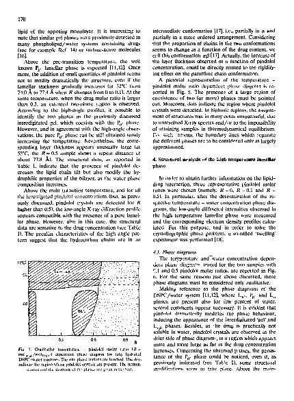
Above the main transition temperature, and for all the ii)ve:;'..;galCd pi;Id:31cl cenccntr;~tions (but, as prcvi- ously discussed, pindoh)l crystals are detected for R high,.'r than 0.5), the low-nolle X-ray diffraction profile appear~ compatible whh the presence of a pure lamel- lar phase. However, also in this case. the structural data arc sensitive to the drug concentration (see Table 1). The pcculim characteristics of the high angle pat. tern suggest that the hydrocarbon chains are in an
" . C . . . : . " :. :: . : : . ' , . . 2 .
0.3 0,6 0,9 R
Fie. 5. Oualitalive temperature - phtdoh)l molar ratio (R= mt)ll.md/nll))ll:.,i,(.) dependent phase diagnlm fl)r fully hydrated DSPC./water mixtures. The one-phase legions are halched. The dl)|~, indicate the rc[.,io~ w]lert? pindt)h')l cl3'.stltls are present. The nonllzn-
¢1,~tu¢¢ ,lfid the: Mi-ug|ur(: ~;i lh : phi~:,~;s arc ~.iven in th~ 1,:~.
intermediate conformation [17], i.e., partially in ot and paltially in a mo~e ordered arrangement. Considering that ).he proportion of chains in the two conformations seems to change as a function of the drug content, we cell this conformation a/3 [17]. Actually, the increase of the layer thickness observed as a function of pindolol concentration, could be directly related to the rigidify- ing effect on the paraffinic chain conformation.
A pictorial representation of the temperature - pindolol molar ratio dcpcndcr.t ph::s,: diagran b, ,'e- ported in Fig. 5. The presence of a large re,;ion of coexistence of two (or more) phases must be I:,ointcd out. Moreover, dots indicate the regkm where pindolol crystals were detected, in biphasie regions, the assigne- merit of structures was in many c~:ses unsuccessful, due to unresolved X-ray spectra and /o r to the impossibility of attaining samples in thermod~namical equilibrium. For siach rc::~ons, the boundary lines which ::.eparate the different phases are to be considered only as largely approximated.
4. Structural atralysis af th~ high tempezature lamellar phase
!p order to obtain further information on the lipid- drug interaction, three representative pindolol molar ratios were chosen (namely, R =0 , R =° 0.1 and R = 0.5). In particular, after thc determit~ation of the re- spective temperature - water concentrallon phase dia- grams, the low-angle diffracted intensities observed in the high temperature lamellar phase were measured and the corresponding electron density profiles calcu- lated. For this purpose, and in order to solve the crystallographic phase problem, a so-callcd "swelling" experiment was pcrformcd [18].
• 1.1. Phase a&grams The temperature and%vater concentration depen-
dem. phust: r.]iaL;r,','~,:. !ra~ed for the two samples with ~).1 and 0.5 pindolol molar ratios, are reported in Fig, 0. ['or the same reasons just above discussed, these phase diagrams must be considered only oualitativc.
Making, reference to the phase diagrams of the DSPC/water system [11,12], where L~., i~, and L,, phases are present also for few percent of water, several comments appear neccssary. It is evident that pind-h,I dr'~m:ltieally modifies the phase behaviour, inducing the appcarancc of the i~lterdigitatcd 'gel' and L,,t) phases. Besides, as 'he dru~ ~:s practically not soluble in waler, pindolol crystals arc observed in the drier side of phase diagram,, m a region which appears more and more large as far as the drug concentratkm increases. Concerning the observed phases, the peJ'sis- tancc of the Pl~' phase could be notice.d, even if, as previously intlicated (see Table 1), some structural i:~odific:-;!ions seem to take p~JCe. Above the main-

C,XAT
Tt°C)
. : , . . . . . . .
171
4.2. Swell ing experiments The intensity analysis was made within the regions
of phase diagrams where the continuous transition from L , to the L,,a phase seems to occur.
X-ray diffraction pat terns give a set of integral in- tensifies from which the module of the structure fac- [¢,i~") Cd.;~ bc ~ I I~* ~,a,cu,,.ed. In the simple case of one-di- a.ca~ionaI pha;cs, the equation which gives the struc- ture factors F ( h ) reduces to:
F2(h) = I h) /m( I )
where h is ~he Miller index of the reflection, i ( h ) is its observed intensity and re(h) the multiplicity [20]. in order to solve the crystallographic 'phase problem', i.e., to determine the signs of the structure factors, it is t radit ional in lipid crystallography to perform swelling
TABLE Ill
Cht,lt;i('t:! ('on;po.~'itiotl atÁ(I strlEtura! rl(tta: su't'Uing experirtletll.~
The intensities of any experiment are normalised such that Yl(h)= 1.1). The experimental error in measuring diffracted intensities is estimated to be :h0.0!2. The all* , are measured from the electron dca~;b' ,T,a,~:i (see text). Other notations as ill the text and Table l.
R~a • 0.11) 0.17 0.26 0,30
0.2 0 . ~ C ~w-:,r ('~,*~.t 0.I0 0.18 0.27 0.31 d (~) 53.11 55.7 59.~ 60.4
Fig. 6. Ouamalive temperature - water eoncentratinn dependent phase diagrams for pindt)lol/DSPC mixtures having pindolol uMar dlip (~) 47.5 45,9 43,8 41.7 ralios ( R = mol~i,d/mtll I~l.v, ~ equal to 0,1 (upper diagram) and 11.5 dl~* n (/~) - - .~0.4 flower diagram). The one-phase regions are hatched. "1 he dots ind;- (:;tle the region where the It-bases coexist with bulk waler. The nonlenchaure and Ihe structure of tile phases arc given in the Iexl.
transition temperature , both phase diagrams show the L,,tj pha~e. As discussed above, this phase presents a clear sensitivity to the pindolol content. Considering the structural and chemical data reported in Table 111, it appears that pindolol induces a larger hydrophilicity of samples, as they saturate at higher water concentra- tion (about 41)% w/' ,v in',;tcad ef 30% [I I,',9], see Table liD. h is also interest ing to note that the thickness of the lipid leaflet decreases as a function of the water conteat. However, such a 'compression' effect appears balanced by the presence of pindolol, which acts stiff- ening the hydrocarbon chain organization: therefore, at the slime water concentratiun, th, z sample containing the higher quanti ty of drug shows the larger lipid layer thickness.
As a final note, we remark that in the R = 0 . 5 sample, at about 50°C and for water concenbat ions ranging from about 15% to 20% (w/w) , a coexi,stencc of a one-dimensional lamellar phase (L or P) and of a not fully eharacterised more ordered phase Q (cubic?) was det,~cted.
(O35)
60,7
I( I ) 0.609 0,563 11.489 11.482 1(2) 0.(130 11.112 0.236 0.254 •(3) 0.h)11 0.185 I).2111 0.~.03 1(41 '.).25~) o . t40 o.1165 0,5~,~ I(5) 0 0.112
R = a . I C 0.08 I).15 0.2~ 0.35 (0,75) C'~..~,. 0.08 0,16 0,22 0.36 d (A) 51,8 56,2 57.3 1"15.6 65.8 dli p (A t 47.5 47.4 44.9 42,0
_.~_C,:P_! . . . . - _ _ _ 42.9
i11, 0.~19 0.~0~ 0.7,~ o.475 1(2) 11.1211 0.1.~ 0.07: 0.302 113) 11.1132 0 ~ , 5 11.1126 0.,1~3 -
•(4) 0,029 11,167 (),184 11,097 •(5) 0 0 0 O.(H3
R = 0.5 C 0.2o 0.'25 I).31) 0.35 (11.75) Cv.w. ~ 0.21 0.26 0.31 0..t6 d (A) 62.8 64.2 67.1 68.5 68.9 dji . (/~) ~,9.9 47.7 ar~4 44,() ,I,*,. (At 45.2
I( I ) 0.720 I).559 0A68 0,370 112) ~).079 11.185 0.331 I).3811 1(3) 0.055 0,tJ6f) fl,082 0.(|fi9 - •(4) 11.146 11.1117 0.()77 11.124 - i l ;) 1) (U)28 0,042 0.057 -

172
experiments [18,21,22]. In fact, the addition of water to an unsaturated lamellar system enl-~rges the dimension ~ 6()o of the unit cell, as the amount of water between the lipid layers increases. If the projected electron density g ,4oo of the bilayer remains constant with swelling, the proper ~ ~ oo scaled intensifies, measured varying the water content, are expected to sample the squared Fourier transform ~ o of the structure. Then, the signs of each structure factor may be deduced observing the behaviour of the ~ ;oo continuous transform close to the zeros of iutc,sities [18,21]. Actually, inside a s~:rlliag series, the structure may be modified, but if cha1:ges are small and continu- ous, the structure factor signs can still be obtaincd with reasonable certainty [22].
In the present ease, three series of swelling experi- ments were performed at 65°C for different pindolol contents, namely R=O, R=O.! and Re0 .5 . The ex- perimental data a~c listed it, Tabel Ill. The plot of the structure factors as a fu~qction of the reciprocal spacing S t,~-=~ is reported in Fig. 7. In particular, the ampli- tudes were normalised according to [23]:
Y: l t h ) = d / dmi"
where d is the observed lamellar repeat distance deter- mined in each experimen,t and dr.i. is the correspond- ing value observed for the sample of the series contain- ing the. minimum Quantity of water From the c:lrve~ in Fig. 7, the position of zeros can be easily inferred. It could be noticed that the smooth curves do not exactly ~ ooo correspond to the Fourier transform of the same con- ~" stant structure. This indicates that the bilayer is chang- ~ 400 ing its structure with swelling: as already proposed [21], '.g ?oo the increased disorder of the lipid moh;cules at higher water contents could explain such a behaviour. o
Once determined the signs of the st~ructure factors, ~, 2oo the electron density distributions were finally evalu- ated, and electron density maps traced (for data nor- malization * see also Ref. 22). A comparative analysis of the results obtained in the three series is presented in Fig. 8, where electron d~:nsity profileg relative to samples in the more hydrated condition {i.e., close to the fully hy~lration) are- reported. The more e-vident effect due to the presence of pindclol is the deepening of the electron density profile in the paraffinic chain r,~g;on; the posit~on of the low eiectron-densc terminal
* In c()n~qucnce of the data normalization, the elc::tron dcnsily distribution (,3p(r). al~) called tile map) i~i a normalized dimel). sionless expressiun of the fluctu;)tion of the electron densily p(r):
A p t r ) = [p(, ' ) - ( p ) J / [ p : ( r ) - (/,>~]'/=
being p ( r ) Ire l-'imrier transform of lhe sol oI l;b.~crwd MlaCInI,~ factors and <p) the average value of p(r) over the volume. See Ref. 22 -,:A ~ef,:rence~: therein.
.,, ............ I
o o ~ o . ~ , / , IO~C I | )FO( d l B [ )~ I . I fR ] ,% (
~oo I ~ .~ ~ i 400 •
oo'°°°tl 1 , , . . . . . . . . ~ l l l , i . . . . .
o o? 0.06 o.1 I~c(:ipro(:al 5l)~(:','a(] 5 (A I)
!;
o
002 C) O() (~ : I~(!clprm,~l ~|)d(.fflq ~, (A I)
Fig. 7. Plot of the scaled structure factors observed in DSPC/waler/ pindolol mixtures at 65°C and at different degrees of swelling, as a fimction of the reciprocal spacings (see Table Ill). From top to tiouom, the pindolol molar ratios (R = m~d pi~d/tool l)sl,c ) are: 0, 0, I
and 0.5.
methyl groups in fact appears more and more visible, eotffirming the proposed tightening of the hydrocarbon chain conformation. Moreover, it could he st,cssed that almost all drug effect is already present at R = 0.1, as not much difference seems to occur between the el:ctron density profiles of R = 0.1 and R = 0,5 sam- pies.
From the electron density maps, other information can be obtained, as it is possible to directly measure the dimension of tLhc lipid and of the paraffinic layers

i
A
R=O
-20 0 *20
Fig. 8. Electron density profiles, obtained by Fomka trans[orm of normalised structure factors (see Re£ 22). relative to the lamellar phase observed at maximum hydration at 65°C in DSPC/water/ pindolol mixtures. From top to bottom, the pindolol molar ratios (R=molpind/mOlDspc) are: 0.5, 0.l and 0. The origin is at the center of the hydrocarbon region. The dashed area represents the
uncertainties in the electron density curves.
[22]. These data are reported in Table Ill: it could be noticed that the agreemem between them and the corresponding values calculated from the chemical pa- rameters is very good. in particular, the increase of the thickness of the lipid layer observed as a function of pindoloi concentration r, ppears directly confirmed.
S. C o n c l u s i o n s
S~veral conclu.~-;on~ can be derived from the above reports. First of all, the whole results show that pin- dolol determines structural modifications mainly at the paraffinic level of the model membrane, inducing a stiffening and tightening of the hydrocarbon chain ar- rangement, in the Lo, and P#, phase.s, this appears as a decrease of the hydrocarbon chain tilt, while, at the higher drug content, a gel phase is induced: in this phase the chains are forced to completely inturdigitat~. Above the DSPC chain melting temperature, the pin- dolol effect persists, so that the chains assume a mixed 'disordered-ordered' conformation: a dear deepening of the electron density profile in correspondence of the paraffinic region is then observed in the calculated electron density maps. Noticeable is the fact tl~at the drug modifies also the 'hydrophilic' properties ,of the mcmbrane: in both PO' and La~ phases, the ~indoloi increases the water content between the lipid layers.
173
The unusual phases induced by the pindolol appear more common then previously thought, in particular, gel phases have been observed in hydrated bilayers of dipalmitoylphosphatidylcholine at high concentratiou of glycerol or ethylene glycol [24] as well .as of dipalmi- toylphosphatidylglycero! .-flc.r the addition of choline, acetylcholiae and polymixine B [25,26]. More recentiy, it has been shown that several surface active small molecules (for example, drugs as tctraeaine [!6], ehlor- promazine [16], azelaic acid [14], propanolol [5,27] and so on) but also uncharged compounds (as benz~,l alco- hol or phenylethanol [16]) can induce interdigitated phases. On the contrary, high temperature structural studies are relatively rare, so that the observation of a L,~ phase appears quite unusual.
On the basis of these considerations, a model rela- tive to the lipid-pindolol interaction could be tenta- tivety proposed. According to Mclntosh et al. [16], it appears that all the molecules (either charged or neu- tral) just above considered, show amphiphil/e proper. ties, and tend to locate at the lipid/water interface. However, it has been observed that, to produce inter- digitation, the added molecule must present some size constraints [5,i6]; in particuiar, if tire tuolecul~ leaves vacant places in the paraffiaic regien of the lipid bilayer, the chains can interpenetrate. Considering this picture, it is plausible that pindolol, anchoring at the interface, causes voids b¢tween lipid chains and then induces interdigitation. However, the effects observed in the Lo, and P~, phases as well as the induction at high temperature of the L,0 phase, seem to indicate that the pindolol nonpolar moiety extends rather deeply into the bilayer interior, so that the hydrocarbor, chains must adopt ~ different conformation. At low tempera- tore, the los.,; of the required chemical homogeneity of the hydrocarbon moiety determil~es the observed grad- ual transition towards the untUted chain conformation. At high temperature, in order to satisfy the van der Waals interactions, the chains assume a mixed 'dis- ordered-ordered' conforma.tion. This behaviour could be related to the more general stabilizing effect ob- served in such kind of drugs.
Aclmuwledgements
it is a pleasure to thank Mr. Enzo Mosca for dra:v ing the figures. A.C. acknowledges the !CYI'P (Trleste, italy) for the financial support during her stay in Italy. This work was partially supported by the CNPq (Brazil) and CNR (Itaiy) exchange programs.
References
I Filders, FJ.T. (1981) in Liposomes: from Physical Structure to Therapeutic Applica~[r, ns (Dingle, J,'T, and Gordon, J,L,, eds,}, pp, 465-485, Elsevier/North Holland Biomedical PNss, New York.

174
2 Cao, A., Hantz-Brachet, E., Azize. B., Taillandier. E. and Pcrret. G. 11991)Chem. Phys. Lip. 58. 225-232.
3 Hansch, C. and Clayton, J. (1973)J. Pharm. Sei. 62. 1-21. 4 Zimmer, (.i. 119x4) m [~i~membra,':¢~, VL, I. 12 (Kate:., M. and
Manson, L.A., eds.h pp. 169-201. Plenum Press. New York. 5 Harish. M.P. and Ryman. B.E. (1981) in Liposome~: fr,,~r~ Ph,,,!:i-
eal Struelure to Therapeutic Applications (Dingle. J.T. and Got- den. J.L.. eds.), pp. 409-441, Elscvi~r/North-HolP'!~',! B!~'.'O!~*'! Press. New York.
6 Schaefer, A.A. (1955)J. Am. Chem. Soc. 77, 21)17-202-~. 7 Tardieu. A. (1972)Thesis, Universit~ Paris-Sud. 8 Stumpel, J... Eibl. H. and Nicksch, A. (1983) Biochim. Biophys.
Acta 727. 246-254. 9 Bhmrock. A.E. and Wilkins, M.H.F. (1%9) Nature 223, 9[16-911.
IO Levine, Y.K. 11973) [ad'ogr. Surf. Sci. 3, 279-352. I l "fardh;n. A.. Luzzali. V. and Rt.man. F.C. (1973) J. Mo,. Biol. 75.
711-733. 12 Janiak, MJ., Small, D.M. and Shipley. (].G. (1976) Biochemistry
15, 4575-4580. 13 Luzzati, V. (1908) in Bioh~gical Membrane i (Chap, an. D., ed.).
pp. 71-123..,~eademic Press. New York. 14 Bossi G.. Dubin,. B.. l~.:~ri~ni. P.. Poazi Bo.,,si, M.G.. Rustichelli,
F. ant' Phadke. R.S. (199()1 II Nuovo Cimemo I2D. 1293-,308.
15 Auger. M., Jarrel. H.C., Smith, I,C.P., Siminoviteh. D.J., Mautseh, - H. and Wong, P.T.T. (1988) Biochemistry 27, 6086-6093.
16 Melnlosh. T.J.. MeDanlel. R.V. and Simon. S.A. tieS3) Biochim. Biophys. Aeta 731. 109-114,
17 Ranck, J.L., Matcu. L., Sadler, D.M.. Tardieu, A., Gulik-Krzy- wicki, T. and Luzzat,, V. (1074)J. Mol. Biol. 85. 249-277,
18 Laggner. P. (1988)Top. Cunt;nt Chem I.;5. ]7--' "~)2. 19 Bourges, M., Small, D.M. and Derv,chia~:. O.G, (1967) Biochim.
Biophys. Aura ,37, 157-167, 21) International Table:, for X-Ray Crystallography 11952) V,~ls. 1
and 2, The Kynoch Press, Birmingha,"a,, 21 Franks. N.P. 11976).I. Mol. Biol. 100. 343-358. 22 Mariani. P.. Culotte, A. and AIbertini. G. (t990) Chem. Phys.
Lipids 55, 283-294. 23 Blaurock. A.E. (1971),I. Mul. Biol. 56, 35-52. 24 McDaniel, R.V.. M¢Intosh. TJ . and Simon. S,A. (It)82) Biochim.
Biophys. Acta 731.97-11)8. 25 Ranck, J,L. and T(:'.~; ",.:'. J.F. 11982) FEBS Lett. 143, 171-i74, 26 Ranck, J.L. and T(~canne, J.F. t1982) FEBS Left. 143. 175-178. 27 Alberfini. G.. Donati, C., Phadke, R.S., Ponzi Bossi, M.G. and
Rustichelli, F. 0990) Chem, Phys. Lipids 55, 331-337.


![Fluorescence properties of a potential antitumoral benzothieno[3,2-b]pyrrole in solution and lipid membranes](https://static.fdokumen.com/doc/165x107/63440ec5df19c083b1076b23/fluorescence-properties-of-a-potential-antitumoral-benzothieno32-bpyrrole-in.jpg)