Alterations in Lipid Levels of Mitochondrial Membranes Induced by Amyloid-B: A Protective role of...
14
Hindawi Publishing Corporation International Journal of Alzheimer’s Disease Volume 2012, Article ID 459806, 14 pages doi:10.1155/2012/459806 Research Article Alterations in Lipid Levels of Mitochondrial Membranes Induced by Amyloid-β: A Protective Role of Melatonin Sergio A. Rosales-Corral, 1, 2 Gabriela Lopez-Armas, 2 Jose Cruz-Ramos, 2 Valery G. Melnikov, 3 Dun-Xian Tan, 1 Lucien C. Manchester, 1 Ruben Munoz, 4 and Russel J. Reiter 1 1 Department of Cellular and Structural Biology, University of Texas Health Science Center at San Antonio, 7703 Floyd Curl Dr., San Antonio, TX 78229, USA 2 Divisi´ on Neurociencias, Centro de investigaci´ on Biom´ edica de Occidente, Instituto Mexicano del Seguro Social, Sierra Mojada 800 Col. Independencia, 44340 Guadalajara, JAL, Mexico 3 University Center for Biomedical Research Center, University of Colima, Colima, COL, Mexico 4 Departamento de Qu´ ımica, Centro de Ciencias Exactas e Ingenieria (CUCEI), Universidad de Guadalajara, Blvd. Marcelino Garc´ ıa Barragan 1421, 44430 Guadalajara, JAL, Mexico Correspondence should be addressed to Sergio A. Rosales-Corral, [email protected] Received 19 December 2011; Accepted 9 February 2012 Academic Editor: K. S. Jagannatha Rao Copyright © 2012 Sergio A. Rosales-Corral et al. This is an open access article distributed under the Creative Commons Attribution License, which permits unrestricted use, distribution, and reproduction in any medium, provided the original work is properly cited. Alzheimer pathogenesis involves mitochondrial dysfunction, which is closely related to amyloid-β (Aβ) generation, abnormal tau phosphorylation, oxidative stress, and apoptosis. Alterations in membranal components, including cholesterol and fatty acids, their characteristics, disposition, and distribution along the membranes, have been studied as evidence of cell membrane alterations in AD brain. The majority of these studies have been focused on the cytoplasmic membrane; meanwhile the mitochondrial membranes have been less explored. In this work, we studied lipids and mitochondrial membranes in vivo, following intracerebral injection of fibrillar amyloid-β (Aβ). The purpose was to determine how Aβ may be responsible for beginning of a vicious cycle where oxidative stress and alterations in cholesterol, lipids and fatty acids, feed back on each other to cause mitochondrial dysfunction. We observed changes in mitochondrial membrane lipids, and fatty acids, following intracerebral injection of fibrillar Aβ in aged Wistar rats. Melatonin, a well-known antioxidant and neuroimmunomodulator indoleamine, reversed some of these alterations and protected mitochondrial membranes from obvious damage. Additionally, melatonin increased the levels of linolenic and n-3 eicosapentaenoic acid, in the same site where amyloid β was injected, favoring an endogenous anti-inflammatory pathway. 1. Introduction We hypothesized that due to its amphipathic nature [1], its physicochemical functions [2] and aided by the induced oxidative stress [3, 4], Aβ paves its own pathway from the extracellular space to the mitochondria where it disrupts membrane fluidity and causes energetic dysfunction. This mechanism of membrane permeabilization induced by Aβ and its own internalization might be the major cause of mitochondrial dysfunction. Following injection of Aβ 1−42 into the hippocampus of healthy Wistar 6-month-old rats, we reported deposits of this peptide fragment forming plaques in the extracellular space. Thereafter Aβ was observed in cytoplasmic membranes, especially those of axons, where it accumulated in one or two poles of the axons giving the appearance of onion bulbs, as observed in demyelinating pathologies. Demyelination is also a feature of Alzheimer’s disease (AD) [5, 6]. It is noteworthy that the animals used in the experiments had no other condition or genetic predisposition to form plaques or other AD features. Aβ peptide then appeared in the cytoplasm, and finally in mitochondrial membranes, where its presence was associated with mitochondrial dysfunction. The question remaining is what alterations Aβ produce in the
Transcript of Alterations in Lipid Levels of Mitochondrial Membranes Induced by Amyloid-B: A Protective role of...

Hindawi Publishing CorporationInternational Journal of Alzheimer’s DiseaseVolume 2012, Article ID 459806, 14 pagesdoi:10.1155/2012/459806
Research Article
Alterations in Lipid Levels of Mitochondrial Membranes Inducedby Amyloid-β: A Protective Role of Melatonin
Sergio A. Rosales-Corral,1, 2 Gabriela Lopez-Armas,2 Jose Cruz-Ramos,2 Valery G. Melnikov,3
Dun-Xian Tan,1 Lucien C. Manchester,1 Ruben Munoz,4 and Russel J. Reiter1
1 Department of Cellular and Structural Biology, University of Texas Health Science Center at San Antonio,7703 Floyd Curl Dr., San Antonio, TX 78229, USA
2 Division Neurociencias, Centro de investigacion Biomedica de Occidente, Instituto Mexicano del Seguro Social,Sierra Mojada 800 Col. Independencia, 44340 Guadalajara, JAL, Mexico
3 University Center for Biomedical Research Center, University of Colima, Colima, COL, Mexico4 Departamento de Quımica, Centro de Ciencias Exactas e Ingenieria (CUCEI), Universidad de Guadalajara,Blvd. Marcelino Garcıa Barragan 1421, 44430 Guadalajara, JAL, Mexico
Correspondence should be addressed to Sergio A. Rosales-Corral, [email protected]
Received 19 December 2011; Accepted 9 February 2012
Academic Editor: K. S. Jagannatha Rao
Copyright © 2012 Sergio A. Rosales-Corral et al. This is an open access article distributed under the Creative CommonsAttribution License, which permits unrestricted use, distribution, and reproduction in any medium, provided the original work isproperly cited.
Alzheimer pathogenesis involves mitochondrial dysfunction, which is closely related to amyloid-β (Aβ) generation, abnormal tauphosphorylation, oxidative stress, and apoptosis. Alterations in membranal components, including cholesterol and fatty acids, theircharacteristics, disposition, and distribution along the membranes, have been studied as evidence of cell membrane alterationsin AD brain. The majority of these studies have been focused on the cytoplasmic membrane; meanwhile the mitochondrialmembranes have been less explored. In this work, we studied lipids and mitochondrial membranes in vivo, following intracerebralinjection of fibrillar amyloid-β (Aβ). The purpose was to determine how Aβ may be responsible for beginning of a viciouscycle where oxidative stress and alterations in cholesterol, lipids and fatty acids, feed back on each other to cause mitochondrialdysfunction. We observed changes in mitochondrial membrane lipids, and fatty acids, following intracerebral injection of fibrillarAβ in aged Wistar rats. Melatonin, a well-known antioxidant and neuroimmunomodulator indoleamine, reversed some ofthese alterations and protected mitochondrial membranes from obvious damage. Additionally, melatonin increased the levels oflinolenic and n-3 eicosapentaenoic acid, in the same site where amyloid β was injected, favoring an endogenous anti-inflammatorypathway.
1. Introduction
We hypothesized that due to its amphipathic nature [1],its physicochemical functions [2] and aided by the inducedoxidative stress [3, 4], Aβ paves its own pathway from theextracellular space to the mitochondria where it disruptsmembrane fluidity and causes energetic dysfunction. Thismechanism of membrane permeabilization induced by Aβand its own internalization might be the major cause ofmitochondrial dysfunction.
Following injection of Aβ1−42 into the hippocampus ofhealthy Wistar 6-month-old rats, we reported deposits of this
peptide fragment forming plaques in the extracellular space.Thereafter Aβ was observed in cytoplasmic membranes,especially those of axons, where it accumulated in one ortwo poles of the axons giving the appearance of onion bulbs,as observed in demyelinating pathologies. Demyelinationis also a feature of Alzheimer’s disease (AD) [5, 6]. It isnoteworthy that the animals used in the experiments had noother condition or genetic predisposition to form plaquesor other AD features. Aβ peptide then appeared in thecytoplasm, and finally in mitochondrial membranes, whereits presence was associated with mitochondrial dysfunction.The question remaining is what alterations Aβ produce in the

2 International Journal of Alzheimer’s Disease
lipid composition of mitochondrial membranes, particularlyin those fatty acids and phospholipids previously related tothe Aβ pathogeny. Moreover, if Aβ-induced oxidative stressplays a key role in damaging lipids in cell membranes, whatis the potential role of the antioxidant melatonin when thisprocess affects mitochondrial membranes?
Lipid and fatty acid changes have been studied primarilyin plasma membranes or in synaptosomes prepared from ADpostmortem brains; those changes have been associated withaging, Aβ deposits, dementia, and even with mild cognitiveimpairment. Other results have been obtained using purifiedsynaptosomal plasma membranes from transgenic mousebrain. To our knowledge, no studies have focused on mito-chondrial membranes in response to in vivo extracellulardeposits of fibrillar Aβ. This approach has relevance to theaforementioned hypothesis and our previous results.
The lipid sensing and lipid-regulated proteolysis is a well-characterized phenomenon. This involves the processing ofthe sterol regulatory element binding proteins (SREBPs),as the best known example [7]. Likewise, saturated fattyacids are related both to amyloidogenesis and to tauhyperphosphorylation. Palmitic acid (PA) is related to theβ-site amyloid precursor protein (APP) cleaving enzyme(BACE1) upregulation and amyloidogenic processing of APPin primary rat cortical neurons [8]. Also, cortical neuronsgrowing in conditioned media from astrocytes treated withpalmitic acid and the unsaturated oleic acid expressedhyperphosphorylation of the tau protein. This was laterfound to be an oxidative-related phenomenon since theaddition of the antioxidant N-acetyl cysteine reduced hyper-phosphorylation of tau [9]. Changes or reaccommodationsof fatty acids within the lipidic structures are continuous, andthis may represent oxidative stress-induced remodeling [10].However, an AD-specific or an Aβ-induced specific changedoes not exist. Nonetheless, it is also known that unsaturatedfatty acids, specifically oleic and linoleic acids, may stimulatepresenilin-1 levels and γ-secretase activity, which participatein the generation of Aβ [11].
The relationship between oxidative stress, peroxidationof membrane lipids, and neurodegeneration has been pri-marily explored in postmortem studies, where a significantdecline in polyunsaturated fatty acids (PUFAs), especiallyarachidonic and docosahexenoic acids, has been found [12].These changes are directly related to the augmentation in 4-hydroxynonenal, a toxic by-product of the peroxidation ofmembrane PUFAs, especially arachidonic acid (AA) [13]. Infact, Aβ may induce the phospholipase A2, responsible forAA release from cytoplasmic membranes [14]. The observeddrop in docosahexaenoic acid (DHA), on the other hand,is correlated with the dietary intake of DHA which mayreduce the amyloid burden [15] by stimulating the non-amyloidogenic processing of APP [16]. Fatty acids modulatethe production and activity of a variety of neurotransmittersand the alterations of fatty acids in the diet of rodents havebeen demonstrated to result in changes in the ability of theanimals to learn or retain new information [17, 18]. Theratio between unsaturated and saturated FA (U/S) expressesthe degree of unsaturation being linked to less membranefluidity as an adaptative phenomenon. Changes in the ratio
of these fatty acids have been thought to be involved in avariety of diseases including cancer, diabetes, and neurologicdiseases. The overproduction of Aβ leads to a decline in Δ-9 desaturase activity with an alteration in membrane fattyacids This results in altered membrane mobility leading to adecline in neurotransmitter activity and a decreased releaseof acetylcholine [19].
It has been demonstrated that Aβ peptides interact withanionic lipids which leads to a significant alteration in theproperties of the bilayer itself. Phosphate groups in anioniclipids and aliphatic aminoacids (Val-Val-Ile-Ala) at the C-terminal end of Aβ mediate that interaction while oxidativestress induces a significant rise in anionic phosphatidylserine(Ptd-Ser). Membrane disruption induced by Aβ-peptide ismediated through perturbations of the lipid order caused byinteraction of peptides with head groups and/or formationof micelles [20]. Reciprocally, when incubated with Ptd-Ser, Aβ undergoes transformation from a random coil toa β-structure [21]. Furthermore, there seems to be a cellselective neurotoxicity due to Aβ determined by surface Ptd-Ser, apart from the levels of ATP [22]; this also relates toour hypothesis. Thus, the capacity of cells to bind Aβ seemsto be associated with cells with expressed measurable Ptd-Ser on the membrane surface, and this feature is correlatedwith apoptotic signaling. It should be noted that Ptd-Ser isnormally found on the inner face of the surface membraneof healthy cells. In the early stage of apoptosis, however, orunder specific stimulation, such as Ca2+ influx, a hallmark inthe mitochondrial pathogenic mechanisms possibly involvedin AD [23, 24], Ptd-Ser can translocate to the outer leafletsof the plasma membrane and be exposed to the extracellularspace. Thus, Ptd-Ser becomes a surface receptor site for Aβbinding, in such a manner that annexin or apolipoproteinE2, proteins with the ability to interact with Ptd-Ser, mayprotect neurons from Aβ neurotoxicity [25]. Since Ptd-Seris an anionic phospholipid, it may produce an acidic localenvironment, which is optimal for aggregation of the Aβpeptide [26].
Phosphatidylcholine (PtdChol) is a major constituent ofcell membranes, commonly found in the outer leaflet, and itis a particular target for Aβ. For example, evaluated in zwitte-rionic bilayers based on PtdChol membranes, Aβ associateswith lipid heads, and when fused into a zwitterionic planarbilayer, it is rapidly transformed from helical- to β-structureand exhibits a channel-like behavior [27]. In this manner, Aβdisturbs intracellular calcium homeostasis because it renderslipid bilayers permeable to ions.
Changes in packing and orientation of phospholipidsin membranes is a phenomenon promoted by cholesterol,which modulates the membrane binding of amyloidogenicproteins [28, 29]. It has been documented that cholesterolincreases the binding of Aβ to lipid membranes [30,31]. The concentration of cholesterol in the membraneshas been related to the extent and depth of insertionof Aβ into the membrane [32]. However, a computa-tional study using PtdChol and PtdChol/cholesterol bilay-ers, which mimic the cholesterol depleted and enrichedlipid domains of neuronal membranes, revealed a pro-tective role of cholesterol in preventing both Aβ-induced

International Journal of Alzheimer’s Disease 3
membrane disruption and membrane surface-induced β-sheet formation [33]. Nonetheless, by electron paramagneticresonance spectroscopy, it has been demonstrated how,driven by hydrophobic interactions, Aβ is inserted intobilayers, between the outer part of the hydrophobic core andthe external hydrophilic layer. This causes displacement ofcholesterol towards the more external part of the membranewhere the crowding of cholesterol in turn causes membranerigidity in this region of the bilayer [34]. This membranerigidity has been demonstrated in mitochondria obtainedfrom postmortem AD brain [35]; importantly, alterationsin mitochondrial membrane fluidity are primarily relatedto lipid peroxidation [36], which, again, emphasizes theimportance of oxidative stress.
Since oxidative stress is a major event in AD progressionand is especially related to membrane dysfunction, mito-chondrial failure, and apoptosis, the antioxidant melatoninhas proven useful in delaying the progression of damagein AD [24]. We have demonstrated in in vivo experimentsafter the injection of Aβ directly into the hippocampus thatorally administered melatonin is more effective in reducingoxidative stress than are vitamins C and E [4]. The effectof melatonin has been shown to be especially protectivefor PUFAs during nonenzymatic lipid peroxidation [37], asobserved in transgenic mouse model of AD [38]. Melatoninalso may preserve arachidonic and docosapentaenoic acids asobserved during ascorbate-Fe++ peroxidation in rat testicularmicrosomes and mitochondria [39].
The current work is based in those fatty acids or lipidspreviously reported to be involved in Aβ-lipid interactionsand the protective effect of melatonin on Aβ-induced mem-brane disruption; this latter process is mediated throughperturbations of the lipid order caused by an interactionof peptides with head groups and/or formation of micelles[20]. Our results correspond exclusively at the region of thehippocampus where the Aβ was injected.
2. Materials and Methods
2.1. Animals and Experimental Design. Surgical and animalcare procedures were performed with strict adherence to theguide for the Care and Use of Laboratory Animals (NationalInstitutes of Health, publication number 86–23, Bethesda,MD, USA). All protocols and procedures were approvedby the institution’s Animal Care and Use Committee. MaleWistar rats (250–280 grams; 3-month-old) were housed inpairs in a colony room on a 12:12 dark/light cycle with lightsoff at 20:00 h; food and water were provided ad libitum. Therats were divided (n = 5) into the following groups: (1) PBS-injected rats, (2) fibrillar Aβ1−42-injected rats (fAβ), and (3)H2O2 (200 μM) intracerebrally injected rats. Two additionalgroups, fAβ+Mel and H2O2+Mel, were included. In this case,the fAβ or H2O2-intracerebrally injected animals receivedantioxidant treatment with melatonin (Sigma, St. Louis, MO,USA) dissolved in the drinking water to yield an estimateddaily dose of 20 mg/kg/day. IPBS was used as control insteadof Aβ peptides since even nontoxic Aβ derivatives, such as thescrambled Aβ usually employed as control in in vivo models,
may themselves produce free radicals [40, 41]. H2O2 waschosen as a positive control because of its close relationshipwith Aβ pathogeny [42]. H2O2 is considered its principalmediator [43] and secondary messenger of death signals[44]. Additionally, H2O2 accumulates in mitochondria longbefore the appearance of Aβ plaques in the extracellular spaceas evaluated in Tg2576 mice [45].
2.2. Brain Coordinates for Hippocampal Injections. Hip-pocampal injections of Aβ1−42 (2 μL at a final concentrationof 1 mM) were performed as previously described [4, 46, 47].Lyophilized synthetic Aβ1−42 (Sigma, St. Louis, MO, USA)peptide was solubilized (10−4 M) in filtered PBS; it wasthen allowed to incubate with continuous agitation (Teflonstir bar at 800 rpm) at 23◦C for 36 h in order to formfibrillar aggregates. Rats, anaesthetized with chloral hydrate(350 mg/kg, i.p.), were placed in a stereotaxic instrumentfor intracerebral injection over a 5 min period (coordinates:anterior-posterior = −3.8 mm, medial-lateral = 2.0 mm,dorsal-ventral = 2.6 mm from bregma; this corresponds tothe CA1 region as determined by the atlas of Paxinos andWatson [48] as a guide) using 5 μL Hamilton microsyringecoupled to a 30-gauge needle through flexible tubing. Theneedle was left in place for 5 min after the injection. Thesame coordinates were used for experiments with H2O2.36 hours after the injections, rats were deeply anesthetizedand transcardially perfused with 200 mL of PBS. Thoseanimals used for immunohistochemical procedures wereadditionally perfused with 4% paraformaldehyde. The ratswere sacrificed by decapitation and the brain was removedimmediately, placed in cold PBS, and a piece of tissue (164–180 mg), including the lesioned area, was taken with a punch(diameter 10 mm), at the base of the needle tract. This pieceincluded the hippocampus and adjacent cortical areas.
2.3. Immunoelectron Microscopy. For immunoelectronmicroscopy, the hippocampal tissue samples were fixed in4% paraformaldehyde for 24 hours and immersed in 2.3 Msucrose solution for 24 hours. Thereafter, small blocks werecut and postfixed in osmium tetroxide (2% in PB 0.2 M)for 45 minutes and then embedded for 48 hours in Embed812 (Electron microscopy Sciences). Ultrathin sections of70–90 nm were cut with an ultramicrotome (Reichert Om3)and mounted on nickel grids and then incubated for 2 hoursin 5% BSA and 0.1% fish gelatin. For the immunolabelingexperiment, the mounted sections were then incubatedfor 24 hours at 4◦C with the primary polyclonal antibodyAnti-βA (Anti-βA42, from Santa Cruz) at a dilution 1 : 1000,then washed four times with PBS 0.1 M and 0.1% tween-20,and further incubated for 3 hours at room temperaturewith a 6 nM gold-conjugated secondary goat anti-rabbitantibody (Jackson Immunoresearch Laboratories) at adilution of 1 : 500. After four washes with PBS, sections werecounterstained with uranyl acetate (2%) for 15 minutes andlead citrate for 5 minutes and examined in a Zeiss EM 906transmission electron microscope (Oberkochen, Germany).

4 International Journal of Alzheimer’s Disease
2.4. In Vivo Analysis of Mitochondrial Free Radicals. Analysisof mitochondrial free radical generation-Mitotracker redCM-H2XRos (Molecular Probes), a rosamine derivative usedto detect mitochondrial free radicals, was diluted in DMSOto form a 1 mM stock solution. 100 μL of that solution wasdiluted in 5 mL of physiological saline and stored sterile at4◦C as a working solution. Applied at a dose of 0.030 μg/kg,CM-H2XRos did not affect the functional properties of mito-chondria after loading, since neither the respiratory outputnor cell viability was significantly changed, as evaluated ina separate study (data not shown). Two hours following theintraperitoneal injection of CM-H2XRos, animals were per-fused transcardially with PBS followed by 4% paraformalde-hyde. The brain was immediately removed and immersedin the fixative for 8–10 h. Following a brief washing in PBS,brain slices were cut into 25–30 μm thick sections, includingthe area of interest, with the vibratome and incubated free-floating in Mito Tracker Green (Molecular Probes, Ex/Em490/516 nm), which selectively stains mitochondria both inlive cells and in cells that have been fixed. Then sectionswere mounted on adhesive (Vecta Bond) coated glass slides,with a DNA dye, 4′,6 diamidino-2-phenylindole (DAPI),containing mounting medium (Vectashield, Vector Labo-ratories) in order to evaluate mitochondrial mass in cellswith nuclear counterstaining in blue (Ex/Em 359/461 nm).The mitochondrial free radicals were analyzed by monitoringthe oxidized fluorescence product (Ex/Em 554/576 nm) ofCM-H2XRos under a fluorescence microscope. Integratedoptical density (IOD), number of mitochondria, and itsmitochondrial area were determined by using image analysissoftware (Image-Pro Plus v5.0). Results are presented here asa CMH2XRos/MitGreen IODs quotient.
2.5. Mitochondrial Isolation. For mitochondrial isolation,briefly, brain tissue was minced and placed in prechilledDounce homogenizer with SHE buffer (0.25 M sucrose,5 mM HEPES and 1 mM EGTA,. PH 7.4), followed bycentrifugation at 2,500 rpm for 10 min, 4◦C, and recentrifu-gation of the supernatant (8,500 rpm, 10 min), to obtain acrude mitochondrial pellet. Following a 10 min incubationin ice, the pellet was resuspended again in SHE plus deli-pidized bovine serum albumin (Sigma Chemical Company).Albumin was eliminated by centrifugating this suspension ofmitochondria at 9,500 rpm for 10 min. The protein contentin the mitochondrial fraction was determined by Lowry’smethod [49].
2.6. Fluidity Changes of Mitochondrial Membranes. 1,3 dipy-renylpropane (DPP) incorporation into membranes to formintramolecular excimers depends mainly on medium micro-viscosity and temperature of determination (24). Membranefluidity is determined by estimating the excimer to monomerfluorescence intensity ratio (Ie/Im) of this fluorescent probe,a quotient that reflects lateral mobility of membranephospholipids (25). Briefly, mitochondria were resuspendedin Tris-HCl buffer (50 mM, pH 8) and then fragmentedby sonication for 15 seconds before being separated bycentrifugation at 13,000 rpm. The mitochondrial membrane
pellet was resuspended and proteins were measured byLowry’s method. 0.1 mg of mitochondrial protein was mixedin a spectrofluorometric cell containing Tris-HCl (20 mM,pH 7.5). DPP solution in ethanol of spectroscopic gradewas diluted (0.02 mg/mL) and mixed with membranesgiven a molar ratio of fluorescent probe to membranephospholipids of 1 : 1400; these mixtures were incubated indarkness for 4 hours at room temperature. Fluorescence ofDPP incorporated into membranes was measured at 24◦Con a Perkin Elmer fluorescence spectrometer, LS50B. Thefluorophore was excited at 329 nm and the monomer andexcimer fluorescence intensities were read at 379 and 480 nm,respectively.
2.7. Chromatographic Analysis of Fatty Acids. Fatty acidsfrom membranes were extracted with chloroform:methanol(2 : 1 vol/vol) and analyzed by gas-liquid chromatography.Briefly, C17:0 heptadecanoic acid, as internal standard, wasadded to 1 mg of mitochondrial protein and the mixtureof methanol and chloroform, both dissolved in BHT, wasadded. After centrifugation, the chloroform phase wasextracted and a second extraction was done by addinganhydrous sodium sulphate. The extract was evaporatedunder nitrogen, reacted with a mix of methanol, sulfuricacid, and toluene at 90◦C for 2 hours, and then redissolved inhexane and a 5% saline solution. Following the extraction ofthe organic phase, the hexane was evaporated under nitrogento obtain derivatized fatty acids to be placed into the injectorof a Carlo Erba gas chromatograph with flame ionizationdetection; the temperature of the injector was 250◦C and theoven temperature was maintained at 196◦C, using helium asa carrier gas at 1.4 kg/cm2.
2.8. Chromatographic Anaylsis of Phospholipids. Phospho-lipids were extracted with a methanol/chloroform solutionmixed in a 2 : 1 ratio, dried in a SpeedVac, and thenredissolved in chloroform. Following a second extractionadding anhydrous sodium sulphate, the solution was filtratedand then evaporated. Samples were analyzed by high-pressure liquid chromatography. Results are provided inrelative percentage from the correspondent areas in thechromatogram.
2.9. Chromatographic Anaylsis of Cholesterol. Membranelipids were extracted with chloroform:methanol (2 : 1 vol/vol) and analyzed by gas-liquid chromatography. Briefly,10 μg of stigmasterol, as internal standard, was added to 1 mgof mitochondrial protein and the mixture of methanol andchloroform, both dissolved in BHT, was added. Extractedlipids react for 1 hour at 60◦C with a mix of hexamethyld-isilazane, trimethyl fluorosilane, and dry pyridine to convertfree cholesterol and stigmasterol in their correspondingtrimethyl esters. The mixture was evaporated with nitrogenand then redissolved in hexane to be injected into the CarloErba gas chromatograph with flame ionization detection;the temperature of the injector was set to 275◦C, thetemperature of the detector was 260◦C, and that of the oven

International Journal of Alzheimer’s Disease 5
was maintained at 275◦C. Helium was used as a carrier gas at1.5 kg/cm2.
2.10. Statistical Analysis. All data are shown as means ± SEof triplicate experiments. Statistical analysis of the data formultiple comparisons was performed by two-way ANOVAfollowed by Student’s tests. For a single comparison, the sig-nificance of any differences between means was determinedby unpaired t-tests. The criterion for significance was P <0.05 in all statistical evaluations.
3. Results
3.1. Aβ at the Brain Enters the Neurons and EventuallyPresents in Mitochondrial Membranes. 12, 24, 36 and 48hours following the intracerebral injection of fibrillar Aβ,deposits of Aβ forming aggregates were reactive to Aβpolyclonal antibody and revealed by immunohistochemistry.Congophilic amyloid deposits remained visible up to 21days following the intracerebral injection (data not shown).Tissue sections of 50 μm, obtained with a vibratome, wereused for immunoelectron microscopy and the Aβ positiveimmunoreactions were observed in mitochondria along themembranes and deep in the cristae. The presence of Aβdeposits in mitochondria was accompanied by a significantlost of their architecture, characterized by swelling, brokencristae, lost of membrane integrity, and vacuole formation(Figure 1).
3.2. The Lost of the Cytoarchitecture Was Related to FreeRadical Overproduction. CM-H2XROS is a reduced, nonfluorescent X-rosamine derivative, which is sequestered bymitochondria where it is retained and oxidized. Under oxi-dation, CM-H2XROS emits fluorescence as a consequenceof the number of free radicals produced by mitochondria.This reagent is normally used in in vitro experiments afteradding it to cells in culture. To demonstrate the effectsof Aβ in vivo, we have introduced a variant by injectingCM-H2XROS intraperitoneally 15 minutes before tissuecollections (as explained). Once the tissue was obtained,sections of the lesioned area were immediately cut in avibratome and stained with Mito Tracker Green (MitGreen),which is essentially nonfluorescent in aqueous solutions,only becoming fluorescent once it accumulates within thelipid environment of the mitochondrion. Thus, the CM-H2XROS/MitGreen IOD quotient identifies the quantity offree radicals by the mitochondria present on each field of themicroscope. We found a significant overproduction of freeradicals both in Aβ- and in H2O2-treated brains (P < 0.05)as compared to PBS-injected brains. Brains of animals whohad received melatonin showed significantly lower levels offree radicals (P < 0.05) (Figure 2).
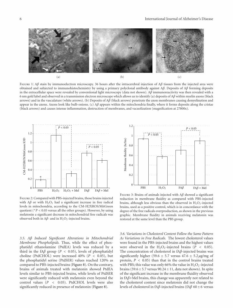
3.3. Membrane Fluidity Was Inversely Correlated to FreeRadical Overproduction. The highest value in membranefluidity was observed in PBS-injected brains, which showedthe lowest amount of free radicals as well (Figure 3). The
highest overproduction of free radicals, according to the CM-H2XROS/MitGreen IOD quotient, was observed in H2O2-injected brains. Interestingly, even though the productionof free radicals in brains injected with fAβ was significantlyhigher than the quantity of free radicals observed in thePBS group, the difference between membrane fluidity andfree radical overproduction was less obvious, as comparedwith the positive control group of H2O2. Brains of animalstreated with melatonin had significantly reduced levels offree radicals and the difference between these and membranefluidity was again obvious (Figure 3).
3.4. The Unsaturated/Saturated Ratio, Significantly Affected byAβ, Is Restored by Melatonin. Aβ increased palmitic (16 : 0)and estearic (18 : 0) saturated fatty acids, 39 and 37% (P <0.05), correspondingly. Additionally, Aβ reduced linoleicacid (18 : 2) at less than 35% the observed value in thePBS-injected brains (P < 0.05) and decreased linolenicacid (18 : 3) value 80% below the observed value in thePBS-injected brains. Additionally, Aβ significantly increasedthe polyunsaturated arachidonic acid (20 : 4) (P < 0.05,from 29.4 ± 2 to 51 ± 2.5). Thus, the elevated increase insaturated plus the severe decrement in linoleic and linolenicacids (Figure 4) was mostly responsible for an alteration inthe ratio of unsaturated to saturated fatty acids in membranephospholipids which is critical to normal cellular function.
The lower unsaturated to saturated ratio was observedin fAβ-injected brains to be even lower than the positivecontrol group of H2O2, fAβ and H2O2 being the groups ofstudy where the overproduction of free radicals was signif-icant (Figure 2). With the use of melatonin, the U/S ratioreturned significantly closer to control values (Figure 5),which reflected melatonin’s role on each particular fatty acid(Figure 4).
However, the aforementioned showed that a drasticreduction in linolenic acid, a precursor of arachidonic acid(20 : 4, n6), and in linoleic acid, a precursor of docoso-hexanoic acid (22 : 6, n3), was reflected in the Aβ-inducedlevels of AA and DHA levels (Figure 6). In the Aβ-injectedbrains, arachidonic acid rose 20% in relation to the PBS-injected control values (P < 0.05), while DHA levels showedapproximately 30% increase value (P < 0.05). This similarincrement in both variables allows that the relationshipbetween these relative values or DHA/AA ratio remainsstable in Aβ-injected brains as compared to PBS-injectedbrains (Figure 6).
Except for the palmitic acid and the arachidonic acid, therest of the Aβ-altered fatty acids tend to return to their basallevels, similar to PBS levels, when the animals were treatedwith melatonin (fAβ+Mel group), as shown in Figures 4–6. Another exception was the N3, 20 : 5, polyunsaturatedeicosapentaenoic fatty acid (EPA) which seemed to beunaffected by Aβ (Figure 7). However, the EPA-to-AA ratioshowed a significant reduction (P < 0.05). Interestingly,in Aβ-injected brains from melatonin-treated animals, EPAvalues were 60% and 43% higher than those observed in PBS-and in Aβ-injected brains. As a result, the EPA to AA ratiowas restored (Figure 7).
6 International Journal of Alzheimer’s Disease
(a) (b) (c)
Figure 1: Aβ stain by immunoelectron microscopy. 36 hours after the intracerebral injection of Aβ tissues from the injected area wereobtained and subjected to immunohistochemistry by using a primary polyclonal antibody against Aβ. Deposits of Aβ forming depositsin the extracellular space were revealed by conventional light microscopy (data not shown). Aβ immunoreactivity was then revealed with a6 nm gold label and observed in a transmission electron microscope which allows us to identify (a) deposits of Aβ within myelin axons (blackarrows) and in the vasculature (white arrows). (b) Deposits of Aβ (black arrows) penetrate the axon membranes causing demyelination andappear in the axons. Axons look like bulb onions. (c) Aβ appears within the mitochondria finally, where it forms deposits along the cristae(black arrows) and causes intense inflammation, destruction of membranes, and vacuolization (magnification at 27800x).
CM
-H2X
RO
S/M
itG
reen
IO
D
PBS H2O2 H2O2 + Mel FAβ FAβ + Mel
P < 0.05
P < 0.05
0
0.2
0.4
0.6
0.8
1
1.2
∗
Figure 2: Compared with PBS-injected brains, those brains injectedwith Aβ or with H2O2 had a significant increase in free radicallevels in mitochondria, according to the CM-H2XROS/MitGreenquotient (∗P < 0.05 versus all the other groups). However, by usingmelatonin a significant decrease in mitochondrial free radicals wasobserved both in Aβ- and in H2O2-injected brains.
3.5. Aβ Induced Significant Alterations in MitochondrialMembrane Phospholipids. Thus, while the effect of phos-phatidyl ethanolamine (PtdEA) levels was reduced by athird in the fAβ group (P < 0.05), levels of phosphatidylcholine (PtdCHOL) were increased 40% (P < 0.05), butthe phosphatidyl serine (PtdSER) values reached 120% ascompared to PBS-injected brains (Figure 8). On the contrary,brains of animals treated with melatonin showed PtdEAlevels similar to PBS-injected brains, while levels of PtdSERwere significantly reduced with melatonin even beyond thecontrol values (P < 0.05). PtdCHOL levels were alsosignificantly reduced in presence of melatonin (Figure 8).
0
0.2
0.4
0.6
0.8
1
1.2M
embr
ane
flu
idit
y (I
e/Im
)
PBS H2O2 FAβ
P < 0.05
P < 0.05
FAβ + Mel
Figure 3: Brains of animals injected with Aβ showed a significantreduction in membrane fluidity as compared with PBS-injectedbrains, although less obvious than the observed in H2O2-injectedbrains, used as a positive control, which is in concordance with thedegree of the free radicals overproduction, as shown in the previousgraphic. Membrane fluidity in animals receiving melatonin wasrestored at the same level than the PBS group.
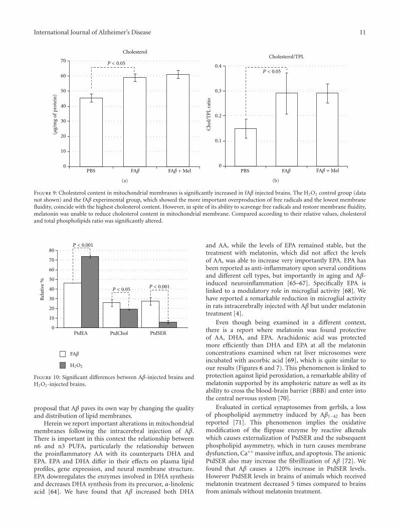
3.6. Variations in Cholesterol Content Follow the Same PatternAs Variations in Free Radicals. The lowest cholesterol valueswere found in the PBS-injected brains and the highest valueswere observed in the H2O2-injected brains (P < 0.05).The concentration of cholesterol in fAβ-injected brains wassignificantly higher (59.6 ± 5.7 versus 47.6 ± 5.2μg/mg ofprotein, P < 0.05) than that in the control brains treatedwith PBS; this value was only 66% the value in H2O2-injectedbrains (59.6±5.7 versus 90.24±11, data not shown). In spiteof the significant increase in the membrane fluidity observedin fAβ+Mel brains, this change was apparently not related tothe cholesterol content since melatonin did not change thelevels of cholesterol in fAβ-injected brains (fAβ 60±6 versus

International Journal of Alzheimer’s Disease 7
0
20
40
60
80
100
120
140 P < 0.05
PBS FAβ
Palmitic acid 16 : 0
FAβ + Mel
Rel
ativ
e %
(a)
0
20
40
60
80
100
120
140 P < 0.05 P < 0.05
PBS FAβ
Estearic acid 18 : 0
FAβ + Mel
Rel
ativ
e %
(b)
0
1
2
3
4
5
6
7
8
9 P < 0.05
P < 0.05
PBS FAβ
Linoleic acid 18 : 2
FAβ + Mel
Rel
ativ
e %
(c)
0
2
4
6
8
10
12
14
16
18P < 0.05
P < 0.05
PBS FAβ FAβ + Mel
Linolenic acid 18 : 3
Rel
ativ
e %
(d)
Figure 4: Aβ and H2O2 (not shown) had similar and highly significant effects on saturated fatty acids particularly on palmitic and estearicacids whose percentages were increased 39 and 37% correspondingly. Linoleic acid was reduced to a third from the control, while linolenicacid was reduced to less than a quart from the control value, as shown. These important effects of Aβ on specific saturated and unsaturatedfatty acids affected the unsaturated/saturated (U/S) balance.
fAβ+Mel 61±7) (Figure 9). The cholesterol-to-phospholipidratio, on the other side, which reflects a loss of phospholipidand membrane rigidity, was significantly altered by Aβ, withthe inability of melatonin to return it to the level found inPBS-injected brains (Figure 9).
4. Discussion
Lipid and fatty acid changes have been studied in plasmamembranes, especially in both postmortem AD brain andtransgenic mice. These changes have been associated withaging, Aβ deposits, dementia, and even mild cognitiveimpairment. Other experiments have been carried out invitro by using purified synaptosomal plasma or mito-chondrial membranes. To our knowledge, no studies havefocused on mitochondrial membranes in response to in vivo
extracellular deposits of fibrillar Aβ. In spite of its knownability to induce oxidative stress, alterations in lipid contentof mitochondrial membranes induced by extracellular Aβdiffered from those induced by H2O2.
Mitochondrial dysfunction has been related to oxidativestress in neurodegeneration as both cause and effect. At thesame time, there is increasing evidence for membrane lipid,fatty acid, and cholesterol interactions with Aβ. These inter-actions have significant consequences in the pathogenesis ofAlzheimer’s disease. Our original aim was to demonstratethat extracellular deposits of Aβ, in vivo, would be able toinduce mitochondrial failure as well as changes in mitochon-drial lipid composition as a consequence of its ability toinduce oxidative stress. H2O2 was chosen as positive controlsince endogenous hydrogen peroxide has been indicated as asecondary messenger mediating the intracellular effects of Aβextracellular deposits. Furthermore, a relationship between
8 International Journal of Alzheimer’s Disease
1.2
1.15
1.1
1.05
1
P < 0.05
P < 0.05
PBS FAβ FAβ + Mel
Unsaturated/saturated ratio
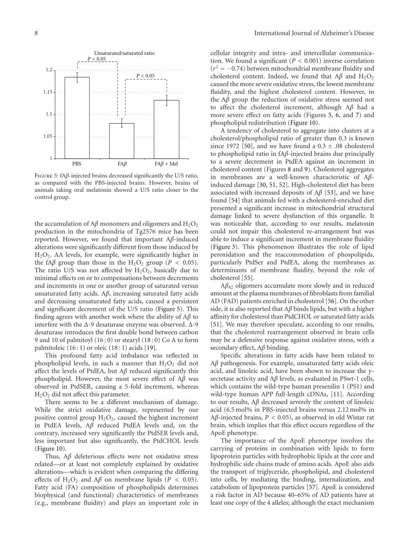
Figure 5: fAβ-injected brains decreased significantly the U/S ratio,as compared with the PBS-injected brains. However, brains ofanimals taking oral melatonin showed a U/S ratio closer to thecontrol group.
the accumulation of Aβ monomers and oligomers and H2O2
production in the mitochondria of Tg2576 mice has beenreported. However, we found that important Aβ-inducedalterations were significantly different from those induced byH2O2. AA levels, for example, were significantly higher inthe fAβ group than those in the H2O2 group (P < 0.05).The ratio U/S was not affected by H2O2, basically due tominimal effects on or to compensations between decrementsand increments in one or another group of saturated versusunsaturated fatty acids. Aβ, increasing saturated fatty acidsand decreasing unsaturated fatty acids, caused a persistentand significant decrement of the U/S ratio (Figure 5). Thisfinding agrees with another work where the ability of Aβ tointerfere with the Δ-9 desaturase enzyme was observed. Δ-9desaturase introduces the first double bond between carbon9 and 10 of palmitoyl (16 : 0) or stearyl (18 : 0) Co A to formpalmitoleic (16 : 1) or oleic (18 : 1) acids [19].
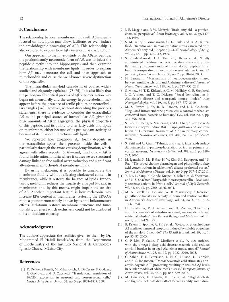
This profound fatty acid imbalance was reflected inphospholipid levels, in such a manner that H2O2 did notaffect the levels of PtdEA, but Aβ reduced significantly thisphospholipid. However, the most severe effect of Aβ wasobserved in PtdSER, causing a 5-fold increment, whereasH2O2 did not affect this parameter.
There seems to be a different mechanism of damage.While the strict oxidative damage, represented by ourpositive control group H2O2, caused the highest incrementin PtdEA levels, Aβ reduced PtdEA levels and, on thecontrary, increased very significantly the PtdSER levels and,less important but also significantly, the PtdCHOL levels(Figure 10).
Thus, Aβ deleterious effects were not oxidative stressrelated—or at least not completely explained by oxidativealterations—which is evident when comparing the differingeffects of H2O2 and Aβ on membrane lipids (P < 0.05).Fatty acid (FA) composition of phospholipids determinesbiophysical (and functional) characteristics of membranes(e.g., membrane fluidity) and plays an important role in
cellular integrity and intra- and intercellular communica-tion. We found a significant (P < 0.001) inverse correlation(r2 = −0.74) between mitochondrial membrane fluidity andcholesterol content. Indeed, we found that Aβ and H2O2
caused the more severe oxidative stress, the lowest membranefluidity, and the highest cholesterol content. However, inthe Aβ group the reduction of oxidative stress seemed notto affect the cholesterol increment, although Aβ had amore severe effect on fatty acids (Figures 5, 6, and 7) andphospholipid redistribution (Figure 10).
A tendency of cholesterol to aggregate into clusters at acholesterol/phospholipid ratio of greater than 0.3 is knownsince 1972 [50], and we have found a 0.3 ± .08 cholesterolto phospholipid ratio in fAβ-injected brains due principallyto a severe decrement in PtdEA against an increment incholesterol content (Figures 8 and 9). Cholesterol aggregatesin membranes are a well-known characteristic of Aβ-induced damage [30, 51, 52]. High-cholesterol diet has beenassociated with increased deposits of Aβ [53], and we havefound [54] that animals fed with a cholesterol-enriched dietpresented a significant increase in mitochondrial structuraldamage linked to severe dysfunction of this organelle. Itwas noticeable that, according to our results, melatonincould not impair this cholesterol re-arrangement but wasable to induce a significant increment in membrane fluidity(Figure 3). This phenomenon illustrates the role of lipidperoxidation and the reaccommodation of phospolipids,particularly PtdSer and PtdEA, along the membranes asdeterminants of membrane fluidity, beyond the role ofcholesterol [55].
Aβ42 oligomers accumulate more slowly and in reducedamount at the plasma membranes of fibroblasts from familialAD (FAD) patients enriched in cholesterol [56]. On the otherside, it is also reported that Aβ binds lipids, but with a higheraffinity for cholesterol than PtdCHOL or saturated fatty acids[51]. We may therefore speculate, according to our results,that the cholesterol rearrangement observed in brain cellsmay be a defensive response against oxidative stress, with asecondary effect, Aβ binding.
Specific alterations in fatty acids have been related toAβ pathogenesis. For example, unsaturated fatty acids oleicacid, and linoleic acid, have been shown to increase the γ-secretase activity and Aβ levels, as evaluated in PSwt-1 cells,which contains the wild-type human presenilin 1 (PS1) andwild-type human APP full-length cDNAs, [11]. Accordingto our results, Aβ decreased severely the content of linoleicacid (6.5 mol% in PBS-injected brains versus 2.12 mol% inAβ-injected brains, P < 0.05), as observed in old Wistar ratbrain, which implies that this effect occurs regardless of theApoE phenotype.
The importance of the ApoE phenotype involves thecarrying of proteins in combination with lipids to formlipoprotein particles with hydrophobic lipids at the core andhydrophilic side chains made of amino acids. ApoE also aidsthe transport of triglyceride, phospholipid, and cholesterolinto cells, by mediating the binding, internalization, andcatabolism of lipoprotein particles [57]. ApoE is considereda risk factor in AD because 40–65% of AD patients have atleast one copy of the 4 alleles; although the exact mechanism

International Journal of Alzheimer’s Disease 9
0
10
20
30
40
50
60 P < 0.05
PBS FAβ
Rel
ativ
e %
Arachidonic acid 20 : 4
FAβ + Mel
(a)
0
10
20
30
40
50
60
70
80 P < 0.05 P < 0.05
PBS FAβ
Rel
ativ
e %
DHA 22 : 6
FAβ + Mel
(b)
0
0.2
0.4
0.6
0.8
1
1.2
1.4
1.6 P < 0.05
PBS FAβ FAβ + Mel
Rel
ativ
e %
DHA/AA
(c)
Figure 6: Aβ and H2O2 (not shown) produced important increases in both n6 and n3 PUFA, which reflects the previous described changesin free fatty acids. A similar increase in DHA and AA allowed the DHA/AA ratio to remain stable, when compared with the PBS group. It isobvious that melatonin reduces the DHA/AA ratio, particularly at the expense of a decrease in DHA levels.
of this feature remains to be fully determined, an interactionwith amyloid insoluble protein aggregates or with APP seemsto be involved [58, 59]. How ApoE controls brain lipids andhow this regulation may impact the clearance of Aβ or theprogression of damage are less clear [60]. In postmortembrain samples, no significant difference in lipids or fatty acidswas found between AD patients classified as homozygousfor ApoE4 and those classified as heterozygous or having noApoE4 [10, 61]. Thus, ApoE genotype on fatty acids and lipidcomposition and/or its distribution in brain cell membranesseem to have no significance and would not bias our results.Additionally, by comparing the association of human, rat,
and rabbit ApoE with Aβ, a similar lack of affinity for Aβbetween rat ApoE and human ApoE4 has been reported [62].Thus, rat ApoE, the same as the AD-related ApoE4, does notform complex with Aβ.
Another fatty acid whose relationship with Aβ pathogenyhas been widely studied is AA. This N6 PUFA is an agonist ofproinflammatory pathways, which additionally as has beenreported increase the levels of Aβ [63]. It is known also thatAβ oligomers trigger neuronal apoptosis by early activationof a cPLA2-dependent pathway leading to production ofAA [14]. We found that the AA precursor linoleic acid wasreduced while AA was increased by Aβ, which supports the

10 International Journal of Alzheimer’s Disease
0
5
10
15
20
25P < 0.05
PBS FAβ
EPA 20 : 5
FAβ + Mel
Rel
ativ
e %
(a)
P < 0.05
P < 0.05
PBS FAβ0
0.1
0.2
0.3
0.4
0.5
0.6
EPA/AA
FAβ + Mel
Rel
ativ
e %
(b)
Figure 7: EPA was not to significantly responsive to Aβ. However, in the presence of melatonin and contrary to the results with the othermajor n3 PUFA, DHA, the relative percentage of EPA rose significantly, which impacted the EPA/AA ratio, as shown.
0
10
20
30
40
50
60
70
80
PBS FAβ
P < 0.05 P < 0.05
PtdEA
FAβ + Mel
Rel
ativ
e %
(a)
0
5
10
15
20
25
30
35
PBS FAβ
P < 0.05P < 0.05
PtdChol
FAβ + Mel
Rel
ativ
e %
(b)
0
5
10
15
20
25
30
35
PBS FAβ
P < 0.05
P < 0.05
PtdSER
FAβ + Mel
Rel
ativ
e %
(c)
Figure 8: Aβ decreased significantly the PtdEA levels and increased the levels of PtdCHOL and PtdSER, the latter with a 5-fold increment.Results are expressed in relative percentage ± standard error.
International Journal of Alzheimer’s Disease 11
0
10
20
30
40
50
60
70
PBS FAβ
P < 0.05
(μg/
mg
of p
rote
in)
Cholesterol
FAβ + Mel
(a)
P < 0.05
0
0.1
0.2
0.3
0.4
PBS FAβ
Cholesterol/TPL
Ch
ol/T
PL
rati
o
FAβ + Mel
(b)
Figure 9: Cholesterol content in mitochondrial membranes is significantly increased in fAβ injected brains. The H2O2 control group (datanot shown) and the fAβ experimental group, which showed the more important overproduction of free radicals and the lowest membranefluidity, coincide with the highest cholesterol content. However, in spite of its ability to scavenge free radicals and restore membrane fluidity,melatonin was unable to reduce cholesterol content in mitochondrial membrane. Compared according to their relative values, cholesteroland total phospholipids ratio was significantly altered.
0
10
20
30
40
50
60
70
80
P < 0.05
P < 0.001
P < 0.001
Rel
ativ
e %
PtdEA PtdSER
H2O2
FAβ
PtdChol
Figure 10: Significant differences between Aβ-injected brains andH2O2-injected brains.
proposal that Aβ paves its own way by changing the qualityand distribution of lipid membranes.
Herein we report important alterations in mitochondrialmembranes following the intracerebral injection of Aβ.There is important in this context the relationship betweenn6 and n3 PUFA, particularly the relationship betweenthe proinflammatory AA with its counterparts DHA andEPA. EPA and DHA differ in their effects on plasma lipidprofiles, gene expression, and neural membrane structure.EPA downregulates the enzymes involved in DHA synthesisand decreases DHA synthesis from its precursor, α-linolenicacid [64]. We have found that Aβ increased both DHA
and AA, while the levels of EPA remained stable, but thetreatment with melatonin, which did not affect the levelsof AA, was able to increase very importantly EPA. EPA hasbeen reported as anti-inflammatory upon several conditionsand different cell types, but importantly in aging and Aβ-induced neuroinflammation [65–67]. Specifically EPA islinked to a modulatory role in microglial activity [68]. Wehave reported a remarkable reduction in microglial activityin rats intracerebrally injected with Aβ but under melatonintreatment [4].
Even though being examined in a different context,there is a report where melatonin was found protectiveof AA, DHA, and EPA. Arachidonic acid was protectedmore efficiently than DHA and EPA at all the melatoninconcentrations examined when rat liver microsomes wereincubated with ascorbic acid [69], which is quite similar toour results (Figures 6 and 7). This phenomenon is linked toprotection against lipid peroxidation, a remarkable ability ofmelatonin supported by its amphoteric nature as well as itsability to cross the blood-brain barrier (BBB) and enter intothe central nervous system [70].
Evaluated in cortical synaptosomes from gerbils, a lossof phospholipid asymmetry induced by Aβ1−42 has beenreported [71]. This phenomenon implies the oxidativemodification of the flippase enzyme by reactive alkenalswhich causes externalization of PtdSER and the subsequentphospholipid asymmetry, which in turn causes membranedysfunction, Ca++ massive influx, and apoptosis. The anionicPtdSER also may increase the fibrillization of Aβ [72]. Wefound that Aβ causes a 120% increase in PtdSER levels.However PtdSER levels in brains of animals which receivedmelatonin treatment decreased 5 times compared to brainsfrom animals without melatonin treatment.
12 International Journal of Alzheimer’s Disease
5. Conclusions
The relationship between membrane lipids with Aβ is usuallyfocused on how lipids may allow, facilitate, or even inducethe amyloidogenic processing of APP. This relationship isalso explored to explain how Aβ causes cellular dysfunction.
Our approach to the in vivo study of the Aβ1−42 peptide,the predominantly neurotoxic form of Aβ, was to inject thepeptide directly into the hippocampus and then examinethe relationship with membrane lipids, in order to explainhow Aβ may penetrate the cell and then approach tomitochondria and cause the well-known severe dysfunctionof this organelle.
The intracellular amyloid cascade is, of course, widelystudied and elegantly explained [73–75]. It is also likely thatthe pathogenically critical process of Aβ oligomerization maybegin intraneuronally and the energy hypometabolism mayappear before the presence of senile plaques or neurofibril-lary tangles [76]. However, without discarding the previousstatements, there is evidence to consider the extracellularAβ as the principal source of intracellular Aβ, given thehuge amounts of Aβ in aggregates, the physical propertiesof this peptide, and its ability to alter fatty acids and lipidson membranes, either because of its pro-oxidant activity orbecause of its physical interactions with lipids.
We reported how exogenous Aβ forms deposits inthe extracellular space, then presents inside the cells—particularly through the axons causing demyelination, whichagrees with other reports [5, 6]—and, finally, how Aβ isfound inside mitochondria where it causes severe structuraldamage linked to free radical overproduction and significantalterations in mitochondrial membrane lipids.
By using melatonin, it is possible to ameliorate themembrane fluidity without affecting cholesterol content inmembranes, while it restores the balance of lipids. Impor-tantly, melatonin reduces the negatively charged PtdSER inmembranes and, by this means, might impair the toxicityof Aβ. Another important feature is how melatonin mayincrease EPA content in membranes, restoring the EPA/AAratio, a phenomenon widely known by its anti-inflammatoryeffects. Melatonin restores membrane structure and func-tionality, an effect which exclusively could not be attributedto its antioxidant capacity.
Acknowledgment
The authors appreciate the facilities given to them by Dr.Mohammed El Hafidi Bentlakder, from the Departmentof Biochemistry of the Instituto Nacional de CardiologıaIgnacio Chavez, Mexico City.
References
[1] D. De Pietri Tonelli, M. Mihailovich, A. Di Cesare, F. Codazzi,F. Grohovaz, and D. Zacchetti, “Translational regulation ofBACE-1 expression in neuronal and non-neuronal cells,”Nucleic Acids Research, vol. 32, no. 5, pp. 1808–1817, 2004.
[2] J. E. Maggio and P. W. Mantyh, “Brain amyloid—a physico-chemical perspective,” Brain Pathology, vol. 6, no. 2, pp. 147–162, 1996.
[3] S. M. Yatin, S. Varadarajan, C. D. Link, and D. A. Butter-field, “In vitro and in vivo oxidative stress associated withAlzheimer’s amyloid β-peptide (1–42),” Neurobiology of Aging,vol. 20, no. 3, pp. 325–342, 1999.
[4] S. Rosales-Corral, D. X. Tan, R. J. Reiter et al., “Orallyadministered melatonin reduces oxidative stress and proin-flammatory cytokines induced by amyloid-β peptide in ratbrain: a comparative, in vivo study versus vitamin C and E,”Journal of Pineal Research, vol. 35, no. 2, pp. 80–84, 2003.
[5] H. Lassmann, “Mechanisms of neurodegeneration sharedbetween multiple sclerosis and Alzheimer’s disease,” Journal ofNeural Transmission, vol. 118, no. 5, pp. 747–752, 2011.
[6] S. Mitew, M. T. K. Kirkcaldie, G. M. Halliday, C. E. Shepherd,J. C. Vickers, and T. C. Dickson, “Focal demyelination inAlzheimer’s disease and transgenic mouse models,” ActaNeuropathologica, vol. 119, no. 5, pp. 567–577, 2010.
[7] M. S. Brown, J. Ye, R. B. Rawson, and J. L. Goldstein,“Regulated intramembrane proteolysis: a control mechanismconserved from bacteria to humans,” Cell, vol. 100, no. 4, pp.391–398, 2000.
[8] S. Patil, L. Sheng, A. Masserang, and C. Chan, “Palmitic acid-treated astrocytes induce BACE1 upregulation and accumu-lation of C-terminal fragment of APP in primary corticalneurons,” Neuroscience Letters, vol. 406, no. 1-2, pp. 55–59,2006.
[9] S. Patil and C. Chan, “Palmitic and stearic fatty acids induceAlzheimer-like hyperphosphorylation of tau in primary ratcortical neurons,” Neuroscience Letters, vol. 384, no. 3, pp. 288–293, 2005.
[10] M. Igarashi, K. Ma, F. Gao, H. W. Kim, S. I. Rapoport, and J. S.Rao, “Disturbed choline plasmalogen and phospholipid fattyacid concentrations in Alzheimer’s disease prefrontal cortex,”Journal of Alzheimer’s Disease, vol. 24, no. 3, pp. 507–517, 2011.
[11] Y. Liu, L. Yang, K. Conde-Knape, D. Beher, M. S. Shearman,and N. S. Shachter, “Fatty acids increase presenilin-1 levels andγ-secretase activity in PSwt-1 cells,” Journal of Lipid Research,vol. 45, no. 12, pp. 2368–2376, 2004.
[12] M. A. Lovell, C. Xie, and W. R. Markesbery, “Decreasedglutathione transferase activity in brain and ventricular fluidin Alzheimer’s disease,” Neurology, vol. 51, no. 6, pp. 1562–1566, 1998.
[13] H. Esterbauer, R. J. Schaur, and H. Zollner, “Chemistryand Biochemistry of 4-hydroxynonenal, malonaldehyde andrelated aldehydes,” Free Radical Biology and Medicine, vol. 11,no. 1, pp. 81–128, 1991.
[14] B. Kriem, I. Sponne, A. Fifre et al., “Cytosolic phospholipaseA2 mediates neuronal apoptosis induced by soluble oligomersof the amyloid-β peptide,” The FASEB Journal, vol. 19, no. 1,pp. 85–87, 2005.
[15] G. P. Lim, F. Calon, T. Morihara et al., “A diet enrichedwith the omega-3 fatty acid docosahexaenoic acid reducesamyloid burden in an aged Alzheimer mouse model,” Journalof Neuroscience, vol. 25, no. 12, pp. 3032–3040, 2005.
[16] C. Sahlin, F. E. Pettersson, L. N. G. Nilsson, L. Lannfelt,and A. S. Johansson, “Docosahexaenoic acid stimulates non-amyloidogenic APP processing resulting in reduced Aβ levelsin cellular models of Alzheimer’s disease,” European Journal ofNeuroscience, vol. 26, no. 4, pp. 882–889, 2007.
[17] M. Umezawa, K. Kogishi, H. Tojo et al., “High-linoleateand high-α-linolenate diets affect learning ability and natural
International Journal of Alzheimer’s Disease 13
behavior in SAMR1 mice,” Journal of Nutrition, vol. 129, no. 2,pp. 431–437, 1999.
[18] N. G. Bazan, “Synaptic signaling by lipids in the life and deathof neurons,” Molecular Neurobiology, vol. 31, no. 1–3, pp. 219–230, 2005.
[19] J. E. Morley, S. A. Farr, V. B. Kumar, and W. A. Banks,“Alzheimer’s disease through the eye of a mouse: acceptancelecture for the 2001 Gayle A. Olson and Richard D. Olsonprize,” Peptides, vol. 23, no. 3, pp. 589–599, 2002.
[20] J. McLaurin and A. Chakrabartty, “Characterization of theinteractions of Alzheimer β-amyloid peptides with phospho-lipid membranes,” European Journal of Biochemistry, vol. 245,no. 2, pp. 355–363, 1997.
[21] E. Terzi, G. Holzemann, and J. Seelig, “Self-association ofβ-amyloid peptide (1–40) in solution and binding to lipidmembranes,” Journal of Molecular Biology, vol. 252, no. 5, pp.633–642, 1995.
[22] O. Simakova and N. J. Arispe, “The cell-selective neurotoxicityof the Alzheimer’s Aβ peptide is determined by surface phos-phatidylserine and cytosolic ATP levels. Membrane binding isrequired for Aβ toxicity,” Journal of Neuroscience, vol. 27, no.50, pp. 13719–13729, 2007.
[23] M. B. Hampton, D. M. Vanags, M. I. Porn-Ares, and S.Orrenius, “Involvement of extracellular calcium in phos-phatidylserine exposure during apoptosis,” FEBS Letters, vol.399, no. 3, pp. 277–282, 1996.
[24] S. A. Rosales-Corral, D. Acuna-Castroviejo, A. Coto-Monteset al., “Alzheimer’s disease: pathological mechanisms and thebeneficial role of melatonin,” Journal of Pineal Research, vol.52, no. 2, pp. 167–202, 2012.
[25] G. Lee, H. B. Pollard, and N. Arispe, “Annexin 5 andapolipoprotein E2 protect against Alzheimer’s amyloid-β-peptide cytotoxicity by competitive inhibition at a commonphosphatidylserine interaction site,” Peptides, vol. 23, no. 7,pp. 1249–1263, 2002.
[26] Z. Lai, W. Colon, and J. W. Kelly, “The acid-mediateddenaturation pathway of transthyretin yields a conformationalintermediate that can self-assemble into amyloid,” Biochem-istry, vol. 35, no. 20, pp. 6470–6482, 1996.
[27] M. R. R. de Planque, V. Raussens, S. A. Contera et al., “Beta-Sheet structured beta-amyloid(1–40) perturbs phosphatidyl-choline model membranes,” Journal of Molecular Biology, vol.368, no. 4, pp. 982–997, 2007.
[28] G. P. Eckert, C. Kirsch, S. Leutz, W. G. Wood, and W. E. Muller,“Cholesterol modulates amyloid beta-peptide’s membraneinteractions,” Pharmacopsychiatry, vol. 36, supplement 2, pp.S136–S143, 2003.
[29] W. Gibson Wood, G. P. Eckert, U. Igbavboa, and W. E.Muller, “Amyloid beta-protein interactions with membranesand cholesterol: causes or casualties of Alzheimer’s disease,”Biochim Biophys Acta, vol. 1610, no. 2, pp. 281–290, 2003.
[30] A. Kakio, S. I. Nishimoto, K. Yanagisawa, Y. Kozutsumi,and K. Matsuzaki, “Cholesterol-dependent formation of GM1ganglioside-bound amyloid beta-protein, an endogenous seedfor Alzheimer amyloid,” The Journal of Biological Chemistry,vol. 276, no. 27, pp. 24985–24990, 2001.
[31] S. Subasinghe, S. Unabia, C. J. Barrow, S. S. Mok, M. I. Aguilar,and D. H. Small, “Cholesterol is necessary both for the toxiceffect of Aβ peptides on vascular smooth muscle cells andfor Aβ binding to vascular smooth muscle cell membranes,”Journal of Neurochemistry, vol. 84, no. 3, pp. 471–479, 2003.
[32] A. Buchsteiner, T. Hauss, S. Dante, and N. A. Dencher,“Alzheimer’s disease amyloid-beta peptide analogue alters the
ps-dynamics of phospholipid membranes,” Biochim BiophysActa, vol. 1798, no. 10, pp. 1969–1976, 2010.
[33] L. Qiu, C. Buie, A. Reay, M. W. Vaughn, and K. H. Cheng,“Molecular dynamics simulations reveal the protective role ofcholesterol in beta-amyloid protein-induced membrane dis-ruptions in neuronal membrane mimics,” Journal of PhysicalChemistry B, vol. 115, no. 32, pp. 9795–9812, 2011.
[34] G. D’Errico, G. Vitiello, O. Ortona, A. Tedeschi, A. Ramunno,and A. M. D’Ursi, “Interaction between Alzheimer’s Abeta(25-35) peptide and phospholipid bilayers: the role of cholesterol,”Biochim Biophys Acta, vol. 1778, no. 12, pp. 2710–2716, 2008.
[35] P. Mecocci, A. Cherubini, M. F. Beal et al., “Altered mitochon-drial membrane fluidity in AD brain,” Neuroscience Letters,vol. 207, no. 2, pp. 129–132, 1996.
[36] J. J. Chen and B. P. Yu, “Alterations in mitochondrial mem-brane fluidity by lipid peroxidation products,” Free RadicalBiology and Medicine, vol. 17, no. 5, pp. 411–418, 1994.
[37] P. Leaden, J. Barrionuevo, and A. Catala, “The protection oflong chain polyunsaturated fatty acids by melatonin duringnonenzymatic lipid peroxidation of rat liver microsomes,”Journal of Pineal Research, vol. 32, no. 3, pp. 129–134, 2002.
[38] Z. Feng, C. Qin, Y. Chang, and J. T. Zhang, “Early melatoninsupplementation alleviates oxidative stress in a transgenicmouse model of Alzheimer’s disease,” Free Radical Biology andMedicine, vol. 40, no. 1, pp. 101–109, 2006.
[39] M. Gavazza and A. Catala, “Melatonin preserves arachidonicand docosapentaenoic acids during ascorbate-Fe2+ peroxida-tion of rat testis microsomes and mitochondria,” InternationalJournal of Biochemistry and Cell Biology, vol. 35, no. 3, pp. 359–366, 2003.
[40] K. Hensley, J. M. Carney, M. P. Mattson et al., “A model for β-amyloid aggregation and neurotoxicity based on free radicalgeneration by the peptide: relevance to Alzheimer disease,”Proceedings of the National Academy of Sciences of the UnitedStates of America, vol. 91, no. 8, pp. 3270–3274, 1994.
[41] J. J. Liang, C. L. Gu, M. L. Kacher, and C. S. Foote, “Chemistryof singlet oxygen. 45. Mechanism of the photooxidation ofsulfides,” Journal of the American Chemical Society, vol. 105,no. 14, pp. 4717–4721, 1983.
[42] N. G. N. Milton, “Role of hydrogen peroxide in the aetiologyof Alzheimer’s disease: implications for treatment,” Drugs andAging, vol. 21, no. 2, pp. 81–100, 2004.
[43] C. Behl, J. B. Davis, R. Lesley, and D. Schubert, “Hydrogenperoxide mediates amyloid β protein toxicity,” Cell, vol. 77, no.6, pp. 817–827, 1994.
[44] C. Shi, F. Wu, and J. Xu, “H2O2 and PAF mediate Aβ1–42-induced Ca2+ dyshomeostasis that is blocked by EGb761,”Neurochemistry International, vol. 56, no. 8, pp. 893–905, 2010.
[45] M. Manczak, T. S. Anekonda, E. Henson, B. S. Park, J.Quinn, and P. H. Reddy, “Mitochondria are a direct site ofAβ accumulation in Alzheimer’s disease neurons: implicationsfor free radical generation and oxidative damage in diseaseprogression,” Human Molecular Genetics, vol. 15, no. 9, pp.1437–1449, 2006.
[46] D. T. Weldon, S. D. Rogers, J. R. Ghilardi et al., “Fibrillarβ-amyloid induces microglial phagocytosis, expression ofinducible nitric oxide synthase, and loss of a select populationof neurons in the rat CNS in vivo,” Journal of Neuroscience, vol.18, no. 6, pp. 2161–2173, 1998.
[47] K. Ishii, F. Muelhauser, U. Liebl et al., “Subacute NOgeneration induced by Alzheimer’s β-amyloid in the livingbrain: reversal by inhibition of the inducible NO synthase,”The FASEB Journal, vol. 14, no. 11, pp. 1485–1489, 2000.

14 International Journal of Alzheimer’s Disease
[48] G. Paxinos, The Rat Nervous System, Academic Press, Sydney,Australia, 1984.
[49] O. H. Lowry, N. J. Rosebrough, A. L. Farr, and R. J. Randall,“Protein measurement with the Folin phenol reagent,” TheJournal of Biological Chemistry, vol. 193, no. 1, pp. 265–275,1951.
[50] D. M. Engelman and J. E. Rothman, “The planar organizationof lecithin-cholesterol bilayers,” The Journal of BiologicalChemistry, vol. 247, no. 11, pp. 3694–3697, 1972.
[51] N. A. Avdulov, S. V. Chochina, U. Igbavboa, C. S. Warden,A. V. Vassiliev, and W. G. Wood, “Lipid binding to amyloidβ-peptide aggregates: preferential binding of cholesterol ascompared with phosphatidylcholine and fatty acids,” Journalof Neurochemistry, vol. 69, no. 4, pp. 1746–1752, 1997.
[52] A. Kakio, S. Nishimoto, Y. Kozutsumi, and K. Matsuzaki,“Formation of a membrane-active form of amyloid beta-protein in raft-like model membranes,” Biochemical andBiophysical Research Communications, vol. 303, no. 2, pp. 514–518, 2003.
[53] L. Ginsberg, J. H. Xuereb, and N. L. Gershfeld, “Membraneinstability, plasmalogen content, and Alzheimer’s disease,”Journal of Neurochemistry, vol. 70, no. 6, pp. 2533–2538, 1998.
[54] S. A. Rosales-Corral, D. Acuna-Castroviejo, D.-X. Tan etal., “Alzheimer’s disease: pathological mechanisms and thebeneficial role of melatonin,” Journal Pineal Research, vol. 52,no. 2, pp. 167–202, 2012.
[55] M. Shinitzky and Y. Barenholz, “Fluidity parameters of lipidregions determined by fluorescence polarization,” Biochimicaet Biophysica Acta, vol. 515, no. 4, pp. 367–394, 1978.
[56] A. Pensalfini, M. Zampagni, G. Liguri et al., “Membranecholesterol enrichment prevents Aβ-induced oxidative stressin Alzheimer’s fibroblasts,” Neurobiology of Aging, vol. 32, no.2, pp. 210–222, 2011.
[57] J. E. Eichner, S. T. Dunn, G. Perveen, D. M. Thompson, K. E.Stewart, and B. C. Stroehla, “Apolipoprotein E polymorphismand cardiovascular disease: a HuGE review,” American Journalof Epidemiology, vol. 155, no. 6, pp. 487–495, 2002.
[58] M. J. Sadowski, J. Pankiewicz, H. Scholtzova et al., “Blockingthe apolipoprotein E/amyloid-β interaction as a potentialtherapeutic approach for Alzheimer’s disease,” Proceedingsof the National Academy of Sciences of the United States ofAmerica, vol. 103, no. 49, pp. 18787–18792, 2006.
[59] S. Haß, F. Fresser, S. Kochl, K. Beyreuther, G. Utermann, andG. Baier, “Physical interaction of ApoE with amyloid precursorprotein independent of the amyloid Aβ region in vitro,” TheJournal of Biological Chemistry, vol. 273, no. 22, pp. 13892–13897, 1998.
[60] S. Arold, P. Sullivan, T. Bilousova et al., “Apolipoprotein E leveland cholesterol are associated with reduced synaptic amyloidbeta in Alzheimer’s disease and apoE TR mouse cortex,” ActaNeuropathologica, vol. 123, no. 1, pp. 39–52, 2012.
[61] T. Fraser, H. Tayler, and S. Love, “Fatty acid composition offrontal, temporal and parietal neocortex in the normal humanbrain and in Alzheimer’s disease,” Neurochemical Research, vol.35, no. 3, pp. 503–513, 2010.
[62] M. J. LaDu, J. R. Lukens, C. A. Reardon, and G. S. Getz,“Association of human, rat, and rabbit apolipoprotein E withbeta-amyloid,” Journal of Neuroscience Research, vol. 49, no. 1,pp. 9–18, 1997.
[63] Z. Amtul, M. Uhrig, and K. Beyreuther, “Additive effects offatty acid mixtures on the levels and ratio of amyloid β40/42peptides differ from the effects of individual fatty acids,”Journal of Neuroscience Research, vol. 89, no. 11, pp. 1795–1801, 2011.
[64] B. Langelier, J. M. Alessandri, M. H. Perruchot, P. Guesnet,and M. Lavialle, “Changes of the transcriptional and fattyacid profiles in response to n-3 fatty acids in SH-SY5Yneuroblastoma cells,” Lipids, vol. 40, no. 7, Article ID L9735,pp. 719–728, 2005.
[65] T. Babcock, W. S. Helton, and N. J. Espat, “Eicosapentaenoicacid (EPA): an antiinflammatory ω-3 fat with potential clinicalapplications,” Nutrition, vol. 16, no. 11-12, pp. 1116–1118,2000.
[66] A. M. Lynch, D. J. Loane, A. M. Minogue et al., “Eicos-apentaenoic acid confers neuroprotection in the amyloid-βchallenged aged hippocampus,” Neurobiology of Aging, vol. 28,no. 6, pp. 845–855, 2007.
[67] A. M. Minogue, A. M. Lynch, D. J. Loane, C. E. Herron, andM. A. Lynch, “Modulation of amyloid-β-induced and age-associated changes in rat hippocampus by eicosapentaenoicacid,” Journal of Neurochemistry, vol. 103, no. 3, pp. 914–926,2007.
[68] A. Bernardo, G. Levi, and L. Minghetti, “Role of theperoxisome proliferator-activated receptor-γ (PPAR-γ) andits natural ligand 15-deoxy-Δ(12,14)-prostaglandin J2 inthe regulation of microglial functions,” European Journal ofNeuroscience, vol. 12, no. 7, pp. 2215–2223, 2000.
[69] P. L. Tang, M. F. Xu, and Z. M. Qian, “Differential behaviour ofcell membranes towards iron-induced oxidative damage andthe effects of melatonin,” Biological Signals, vol. 6, no. 4–6, pp.291–300, 1997.
[70] R. J. Reiter, J. Cabrera, R. M. Sainz, J. C. Mayo, L. C. Manch-ester, and D. X. Tan, “Melatonin as a pharmacological agentagainst neuronal loss in experimental models of Huntington’sdisease, Alzheimer’s disease and Parkinsonism,” Annals of theNew York Academy of Sciences, vol. 890, pp. 471–485, 1999.
[71] H. M. Abdul and D. A. Butterfield, “Protection againstamyloid beta-peptide (1–42)-induced loss of phospholipidasymmetry in synaptosomal membranes by tricyclodecan-9-xanthogenate (D609) and ferulic acid ethyl ester: implicationsfor Alzheimer’s disease,” Biochimica et Biophysica Acta, vol.1741, no. 1-2, pp. 140–148, 2005.
[72] A. Chauhan, I. Ray, and V. P. S. Chauhan, “Interaction ofamyloid beta-protein with anionic phospholipids: possibleinvolvement of Lys28 and C-terminus aliphatic amino acids,”Neurochemical Research, vol. 25, no. 3, pp. 423–429, 2000.
[73] A. J. Yang, D. Chandswangbhuvana, T. Shu, A. Henschen, andC. G. Glabe, “Intracellular accumulation of insoluble, newlysynthesized Aβn-42 in amyloid precursor protein-transfectedcells that have been treated with Aβ1-42,” The Journal ofBiological Chemistry, vol. 274, no. 29, pp. 20650–20656, 1999.
[74] C. A. Hansson Petersen, N. Alikhani, H. Behbahani et al.,“The amyloid β-peptide is imported into mitochondria viathe TOM import machinery and localized to mitochondrialcristae,” Proceedings of the National Academy of Sciences of theUnited States of America, vol. 105, no. 35, pp. 13145–13150,2008.
[75] R. H. Swerdlow and S. M. Khan, “The Alzheimer’s diseasemitochondrial cascade hypothesis: an update,” ExperimentalNeurology, vol. 218, no. 2, pp. 308–315, 2009.
[76] H. Atamna and W. H. Frey, “Mechanisms of mitochondrialdysfunction and energy deficiency in Alzheimer’s disease,”Mitochondrion, vol. 7, no. 5, pp. 297–310, 2007.