Extrachromosomal replication of shuttle vectors in Dictyostelium discoideum
Upload
independentCategory
view
1download
0
eScholarship provides open access, scholarly publishingservices to the University of California and delivers a dynamicresearch platform to scholars worldwide.
University of California
Peer Reviewed
Title:Alzheimer disease macrophages shuttle amyloid-beta from neurons to vessels, contributing toamyloid angiopathy
Author:Zaghi, Justin; Goldenson, Ben; Inayathullah, Mohammed; Lossinsky, Albert S.; Masoumi, Ava;Avagyan, Hripsime; et al.
Publication Date:2009
Publication Info:Postprints, UC Los Angeles
Permalink:http://escholarship.org/uc/item/88z6c8d9
DOI:10.1007/s00401-008-0481-0
Abstract:Neuronal accumulation of oligomeric amyloid-# (##) is considered the proximal cause of neuronaldemise in Alzheimer disease (AD) patients. Blood-borne macrophages might reduce A# stress toneurons by immigration into the brain and phagocytosis of ##. We tested migration and exportacross a blood-brain barrier model, and phagocytosis and clearance of ## by AD and normalsubjects’ macrophages. Both AD and normal macrophages were inhibited in ## export across theblood-brain barrier due to adherence of A#-engorged macrophages to the endothelial layer. Incomparison to normal subjects’ macrophages, AD macrophages ingested and cleared less ##, andunderwent apoptosis upon exposure to soluble, protofibrillar, or fibrillar ##. Confocal microscopy ofstained AD brain sections revealed oligomeric A# in neurons and apoptotic macrophages, whichsurrounded and infiltrated congophilic microvessels, and fibrillar A# in plaques and microvesselwalls. After incubation with AD brain sections, normal subjects’ monocytes intruded into neuronsand uploaded oligomeric A#. In conclusion, in patients with AD, macrophages appear to shuttleA# from neurons to vessels where their apoptosis may release fibrillar A#, contributing to cerebralamyloid angiopathy.

Acta Neuropathol (2009) 117:111–124DOI 10.1007/s00401-008-0481-0
123
ORIGINAL PAPER
Alzheimer disease macrophages shuttle amyloid-beta from neurons to vessels, contributing to amyloid angiopathy
Justin Zaghi · Ben Goldenson · Mohammed Inayathullah · Albert S. Lossinsky · Ava Masoumi · Hripsime Avagyan · Michelle Mahanian · Michael Bernas · Martin Weinand · Mark J. Rosenthal · Araceli Espinosa-JeVrey · Jean de Vellis · David B. Teplow · Milan Fiala
Received: 26 September 2008 / Revised: 5 December 2008 / Accepted: 30 December 2008 / Published online: 13 January 2009© The Author(s) 2009. This article is published with open access at Springerlink.com
Abstract Neuronal accumulation of oligomeric amyloid-�(��) is considered the proximal cause of neuronal demisein Alzheimer disease (AD) patients. Blood-borne macro-phages might reduce A� stress to neurons by immigrationinto the brain and phagocytosis of ��. We tested migrationand export across a blood-brain barrier model, and phago-cytosis and clearance of �� by AD and normal subjects’macrophages. Both AD and normal macrophages wereinhibited in �� export across the blood-brain barrier due toadherence of A�-engorged macrophages to the endotheliallayer. In comparison to normal subjects’ macrophages, ADmacrophages ingested and cleared less ��, and underwentapoptosis upon exposure to soluble, protoWbrillar, or Wbrillar��. Confocal microscopy of stained AD brain sections
revealed oligomeric A� in neurons and apoptotic macro-phages, which surrounded and inWltrated congophilicmicrovessels, and Wbrillar A� in plaques and microvesselwalls. After incubation with AD brain sections, normal sub-jects’ monocytes intruded into neurons and uploaded oligo-meric A�. In conclusion, in patients with AD, macrophagesappear to shuttle A� from neurons to vessels where theirapoptosis may release Wbrillar A�, contributing to cerebralamyloid angiopathy.
Keywords Macrophages · Amyloid-beta · Apoptosis · Alzheimer · Congophillic · Angiopathy
Introduction
The amyloid hypothesis proposes that amyloid-� (A�)accumulation in the brain is the cause of Alzheimer disease
J. Zaghi and B. Goldenson contributed equally.
Electronic supplementary material The online version of this article (doi:10.1007/s00401-008-0481-0) contains supplementary material, which is available to authorized users.
J. Zaghi · B. Goldenson · A. Masoumi · H. Avagyan · M. Mahanian · M. Fiala (&)Department of Orthopaedic Surgery, UCLA-Orthopaedic Hospital Research Center, 615 Charles E. Young Drive South, Room 410, Los Angeles, CA 90095-7358, USAe-mail: [email protected]
M. Inayathullah · D. B. TeplowDepartment of Neurology, David GeVen School of Medicine, University of California, Los Angeles, CA 90095, USA
M. Inayathullah · D. B. TeplowMolecular Biology Institute, University of California, Los Angeles, CA 90095, USA
M. Inayathullah · D. B. TeplowBrain Research Institute, University of California, Los Angeles, CA 90095, USA
A. S. LossinskyNew Jersey Neuroscience Institute, JFK Medical Center, Edison, NJ 08818, USA
M. Bernas · M. WeinandDepartment of Surgery, University of Arizona, Tucson, AZ, USA
M. J. RosenthalDepartment of Medicine, Greater LA VA Medical Center and UCLA School of Medicine, Los Angeles, CA 90095, USA
A. Espinosa-JeVrey · J. de VellisDepartment of Neurobiology, David GeVen School of Medicine, Los Angeles, CA 90095, USA

112 Acta Neuropathol (2009) 117:111–124
123
[20]. A� is potentially generated from A� precursor protein(APP) wherever APP and the �- and �-secretases are located,such as the endoplasmic reticulum and Golgi apparatus, butmost is produced at the plasma surface or in the secretorypathway [25]. A� generation may also occur during autopha-gic turnover of APP-rich organelles [32]. Increasing molecu-lar weight assemblies of A� accumulate both extra- andintra-neuronally in Alzheimer disease (AD) brain [25]; ofthese assemblies, at least the intraneuronal oligomeric A� haspathological consequences [43]. A� is cleared physiologi-cally across the blood-brain barrier by low-density lipopro-tein receptor-related protein-1 [35]. Despite thisphysiological clearance, A� is found to accumulate in neu-rons and extracellular deposits since the Wrst year of life. Thepathological consequences in AD patients are intraneuronalaccumulation of oligomeric A� in multivesicular bodies [18]and neuronal death. Surprisingly, the total neuronal load ofA� is not predictive of neuroWbrillary degeneration [47].
Brain amyloidosis of AD patients is considered to berelated to insuYcient clearance rather than over-expressionof A� [42]. Promising strategies for immune clearance ofA� include A� vaccination [48], intravenous gamma-glob-ulin [39], humanized anti-A� antibody (Bapineuzumab®),and transcriptional modulation of macrophages, such as byuse of curcuminoids [15]. Studies of AD brain tissues [11]and, recently, of APP transgenic mice brain tissues [28, 30,37] suggest that blood-derived macrophages and microglia,rather than resident brain microglia, have a key role in theclearance of A�.
Previous studies have identiWed basic immune mecha-nisms necessary for clearance of A� by macrophages. A�-induced adhesion molecules and chemokines CCL3 (MIP-1 �),CCL4 (MIP-1�), and CCL2 [9, 17, 38] act on macro-phages, microglia, and astrocytes, promoting monocytemigration, diVerentiation [9], and survival [19]. In mousemacrophages and microglia, the class B scavenger receptortype 2 (CD36) is crucial for induction by A� of chemo-kines, cytokines, and reactive oxygen species [6, 34], andexposure to A� initiates the signaling cascade that linksCD36 scavenger receptor to the actin cytoskeleton anddiverse processes such as cellular migration, adhesion, andphagocytosis [41]. Fucoidan inhibits both class A and Bscavenger receptor interactions [21], but, in our experience,does not inhibit A� phagocytosis by human macrophages(Fiala et al. unpublished data). Recent studies suggest a rolefor Toll-like receptors (TLR’s) in A� clearance: TLR’s aretranscriptionally downregulated in AD patients’ monocytes[15], and TLR-2 signaling enhances A� uptake by microglia[4]. Importantly, mononuclear cells of most patients diVerfrom those of normal subjects by transcriptional downregu-lation of �-1,4-mannosyl-glycoprotein 4-�-N-acetylglucos-aminyltransferase (MGAT-3), an enzyme important inphagocytosis of A� [15].
Immunohistochemical studies brought new insights intothe immune clearance of A� by macrophages in humanbrain. AD brain contains 11 times as many cyclooxygenase(COX-2)-positive macrophages as age-matched controlbrain [11], and a subset of these macrophages is induciblenitric oxide (NO) synthase (iNOS)-positive [10]. Monocyteimmigration might be orchestrated by neuronal and microg-lial chemokine RANTES and cytokine interleukin-1� (IL-1�) [13, 31]. In CCR2-deWcient APP transgenic mice,clearance of A� is depressed [7]. Despite the permeation ofAD brain by macrophages, clearance of neuritic plaques israndomly incomplete, suggesting functional heterogeneityof AD macrophages [13].
AD is a human disease with speciWc immune and bio-chemical defects [15], which have not been engineered inAPP transgenic mice. To clarify the pathophysiology of A�clearance in AD, we examined A� clearance using humanbrain tissues and human macrophages, and observed thatthe AD innate immune system is defective in proper dis-posal of A� in the brain.
Materials and methods
Antibodies and reagents
We stained macrophages using mouse anti-human CD68(Dako, Carpinteria, CA) and goat anti-human CD68 (SantaCruz Biotech, Santa Cruz, CA, USA). Neurons werestained with mouse anti-human neuronal nuclei (NeuN,Chemicon, Temecula, CA, USA); mouse anti-humanmicrotubule associated protein 2 (MAP2, Sigma, St Louis,MO, USA); and rabbit anti-human neuron speciWc enolase(Immunostar, Hudson, WI, USA). To visualize A� in braintissue and in macrophages, we utilized rabbit anti-A� 1-42(COOH-terminal epitope) (Millipore, Billerica, MA, USA);mouse biotinylated anti-A� 1-42 (COOH-terminal epitope)(Signet); rabbit anti-oligomer A11 (Biosource, Carlsbad,CA, USA), which recognizes A�-42 and A�-40 pre-Wbrillaroligomers [22]; and rabbit anti-Wbrillar OC, which stainsA� Wbrils, as well as �-synuclein Wbrils and islet amyloidpolypeptide Wbrils [23]. To stain apoptotic markers, weused anti-caspase-6, -7, and -8 antibodies, which wereraised in rabbits using catalytic subunits of the relevantautoprocessed recombinant caspases as immunogens(Burnham Institute, La Jolla, CA, USA) [24]. Secondaryantibodies were anti-mouse, anti-rabbit, and anti-goat IgG’sconjugated to Alexa Fluor 488, 555, and 647 (Invitrogen,Carlsbad, CA, USA). The reagents were monocyte chemo-tactic protein-1 (MCP-1) (PeproTech, Rocky Hill, NJ,USA); Xuorescein isothiocyanate (FITC)-conjugated A�(Anaspec, San Jose, CA, USA); Wbrillar FITC-A� and pro-toWbrillar A� prepared by M. Inayathullah; and 14C-labeled

Acta Neuropathol (2009) 117:111–124 113
123
A� from C. Glabe, UCI. A� was used at 2 �g/mL in mostexperiments.
Patients and controls
A total of ten patients [mean age 76.9 § 5.8 years, meanMini-Mental State Exam (MMSE) score of 21.7 § 5.1]with a diagnosis of probable AD established by theNational Institute of Neurological and CommunicationDisorders and Stroke/Alzheimer’s Disease and RelatedDisorders Association criteria [29] were recruited into thestudy since 2004 through the University of California, LosAngeles (UCLA), Alzheimer’s Disease Research Centerunder a UCLA Institutional Review Board-approved proto-col. In addition, eight aged-matched control subjects (meanage 77.5 + 6.0 years) and three young control subjects(ages 20, 21, 44) were recruited from UCLA personnel andfamilies of patients.
Isolation of PBMC’s
Peripheral blood mononuclear cells (PBMC’s) were iso-lated from venous blood of the AD patients and controlsubjects by Ficoll–Hypaque gradient centrifugation as pre-viously described [13]. Monocytes were puriWed usingRosetteSep Monocyte Enrichment Cocktail (StemCellTechnologies, Vancouver, BC, Canada) from PBMC’s ofnormal subjects (“normal monocytes” or “normal macro-phages”) and AD patients (“AD monocytes” or AD macro-phages”).
Preparation of Wbrils and protoWbrils of A� (1-42)
A� Wbrils were prepared by dissolving 1 mg of A� (1-42)peptide in 100 �L of 10 mM NaOH. The solution wasdiluted to a volume of 0.5 mL with milliQ water followedby addition of 0.5 mL of 10 mM (2£) phosphate buVer (pH7.4). The resulting solution was then centrifuged at16,000£g for 10 min. One-half milliliter of the resultingsupernatant was transferred into a new tube and incubatedat 37°C for 7 days. The Wbrils were pelleted out by centrifu-gation for 10 min and the supernatant was transferred toanother tube for protoWbril isolation. The Wbril pellet waswashed thrice with MilliQ water and the resulting pelletthereafter resuspended in MilliQ water after each wash.
ProtoWbrils were isolated from the supernatant by Wlter-ing through a Centricon Wlter (molecular weight cutoV of35 kDa) to remove any small oligomers and monomers andcollecting the Wltrate. The pellet was washed three timeswith MilliQ and then reconstituted and diluted with MilliQwater. A small aliquot of each sample was analyzed byamino acid analysis to determine the protein concentration.The samples were characterized by size exclusion chroma-
tography and electron microscopy (Fig. 2). Fibrillar andprotoWbrillar A� also was prepared in smaller amountsfrom FITC-A�.
Monocyte migration across a human blood-brain barrier model
A human blood-brain barrier model (BBB) model with pri-mary human brain microvascular endothelial cells(BMVEC’s) was constructed in a 24-well plate as describedpreviously [8, 12]. In the model, 50,000 BMVEC’s in pas-sages 4–8 coated either the upper surface (“regular model”)or the lower surface of a porous membrane insert (Collabo-rative Biomedical Products, Bedford, MA, USA) (“reversemodel”) which rests above a well. Both the well and themembrane insert contained RPMI medium with 10% fetalbovine serum or 10% autologous serum.
In migration experiments, 250,000 monocytes from twocontrol subjects (ages 74 and 78) and two AD patients (ages80 and 84 with MMSE scores 20 and 23, respectively) wereallowed to migrate across the BBB for 17 h. The number oftransmigrated cells was determined by triplicate cell count-ing in eight sections of a hemocytometer chamber. Theinserts were washed gently with a buVer containing 0.1 Msodium cacodylate plus 0.2 M sucrose X2, then Wxed with3% glutaraldehyde in 0.1 M sodium cacodylate buVer for1 h at 25°C [26]. The cells were stored in this buVer at 4°C,post-Wxed with 1% osmium tetroxide at 4°C, dehydrated inan ethanol series and embedded in plastic [27]. One-micronsections were stained with toluidine blue and examined bybright Weld and transmission electron microscopy.
A� phagocytosis and apoptosis by macrophages
Macrophages of four controls (ages 74, 74, 81, 90) and fourAD patients (ages 70, 77, 82, 86 with MMSE scores 15, 27,19, 27, respectively) were prepared in 8-well chamberslides as described [13]. The cultures were incubated withWbrillar or protoWbrillar A� for 3 days. Macrophage apop-tosis was determined using the FLICA VAD-FMK poly-caspases assay kit (Immunohistochemistry Technologies,Bloomington, MN, USA). This assay utilizes a membranepermeant, sulforhodamine B (SR)-labeled inhibitor targetedto all active caspases to covalently label apoptotic cells.Macrophage cultures were incubated with the FLICA apop-tosis detection probe for 1 h at 37°C and then washed toremove any non-covalently bound probe from non-apopto-tic cells. Cells were examined with an Olympus Bmax Xuo-rescence microscope with 100£ objective. Fluorescencedensity was determined by Image-Pro Plus 4.1 (MediaCybernetics, Silver Spring, MD). We examined the middlestrip of each well for 6–9 consecutive Welds with macro-phages, analyzing the integrated optical density (IOD) of

114 Acta Neuropathol (2009) 117:111–124
123
A� (green) and FLICA (red) per macrophage in threeexperiments for each group.
In a set of related experiments, control and AD macro-phages were incubated with soluble FITC-A� for 1 h, or 3,5, or 7, and apoptosis was detected using the FLICA assay.In some experiments, macrophages were incubated withsoluble unconjugated A� for 1 h, washed twice, then incu-bated for 2, 4, or 6 days. Apoptosis was determined by theFLICA assay, and A� Wbrils and oligomers were detectedby the indirect technique using OC and A11 antibodies,respectively.
ELISA assay of A� clearance from monocytes
PuriWed monocytes (300,000 per sample) were incubatedwith soluble A� (2 �g/mL) for 2 h, washed four times, re-incubated for the indicated number of days, and the amountof intracellular A� remaining at each time point was deter-mined (0, 1, 2, 3, 5, and 7 days). To measure the amount ofintracellular A�, the cells were harvested into an ELISAlysis buVer (Invitrogen), supplemented with a proteaseinhibitor cocktail (Sigma) and assayed using the A� 1-42ELISA kit (Invitrogen) by spectrophotometry. Monocytesfrom four control subjects and three AD patients weretested.
Brain tissues
Frozen sections from the frontal lobe of two control sub-jects with no neuropathology (ages 23 and 37) and four ADpatients were provided by the UCLA Brain Bank. The ADbrain sections were from: (1) a 62-year-old patient withBinswanger encephalopathy and scattered senile plaques inthe entorhinal cortex and hippocampus; (2) a 64-year-oldBraak stage VI patient; (3) a 69-year-old Braak stage VIpatient; (4) an 82-year-old Braak stage VI patient withLewy body disease.
Co-incubation of PBMC’s with brain tissues
Frozen sections of post-mortem brain tissues of four ADpatients and two controls were co-incubated with 500,000PBMC’s (from seven normal donors, ages 20, 21, 72, 74,74, 79, and 85 years old) for 1 to 6 days in Dulbecco’sMinimum Essential Medium (DMEM) with 10% fetal calfserum, washed with PBS, Wxed with 4% paraformaldehyde,and processed for immunoXuorescence.
In some experiments, PBMC’s were Wrst pre-labeledwith Qtracker 525, which distributes green Qdot nano-crystals in cytoplasmic vesicles, or CellTracker CMFDA(Invitrogen, Carlsbad, CA, USA), which undergoes an esterasereaction to produce a green Xuorescent product in the cyto-plasm. After isolation of PBMC’s from blood, cells were
incubated with Qtracker or CellTracker according to themanufacturer’s recommendations, pelleted and washedtwice with DMEM, and then incubated with tissues asabove.
ImmunoXuorescence and confocal microscopy of brain tissues
Fixed tissues were permeabilized with 1% Triton X-100and blocked with 1% bovine serum albumin (BSA) at37°C for 30 min each. Brain sections were then incubatedwith various primary antibodies for 48 h at 4°C, washedwith PBS, and incubated with appropriate secondary anti-bodies labeled with Alexa 488, Alexa 555, and Alexa 647Xuorophores for 1 h at 37°C. A mixture consisting of0.2% Triton X-100 and 1% BSA was used as the diluentof both primary and secondary antibodies. As controlstaining, the sections were stained by secondary antibod-ies without primary antibodies. The preparations wereexamined using a Zeiss 510 Meta multiphoton confocalmicroscope or a Bio-Rad Laboratories MRC-1024 Eslaser scanning confocal system attached to a Nikon E800Xuorescent microscope.
Statistical analysis
The data on monocyte migration and 14C-�� transport wereanalyzed by t test and Mann–Whitney and Kruskall–Wallistests.
Results
A� uptake by monocytes inhibits monocyte emigration and A� export across BBB
In the human BBB model, we compared the rate of migra-tion by normal monocytes in both directions using either aregular model (blood chamber above apical side ofBMVEC’s) or a reverse model (brain chamber above ablu-minal side of BMVEC’s). The rate was greater in the direc-tion from brain to blood (36.4 § 0.7%) compared to thatfrom blood to brain (13.6 § 2%). In the course of inXam-mation, chemokines stimulate and lipoxins inhibit leuko-cyte migration; to conWrm the value of this model inreplicating in vivo mononuclear cell migration, we usedeither the chemokine MCP-1 (CCL2) alone or both MCP-1and lipoxin A4 together in the lower chamber. MCP-1(50 ng/mL) increased monocyte migration from0.4 § 0.8% to 6.5 § 0.2%, and lipoxin A4 decreased thismigration in a dose-responsive fashion from 6.5% withMCP-1 alone to 3.1 § 0.1% [0.1 nM (lipoxin A4, plus50 ng/mL MCP-1)], 1.6 § 0.1% (1 nM), and 0.8 § 0.1%

Acta Neuropathol (2009) 117:111–124 115
123
(10 nM). We then investigated A� export using the reversemodel with the chemokine MCP-1 in the lower chamber.As expected, the emigration of monocytes was highest(77,250 § 9,032) in the presence of MCP-1 in the lowerchamber and lowest without MCP-1 (5,375 § 411). When14C-A� (10,000 DPM or 1 �g/mL) was placed in the brainchamber, monocyte emigration was reduced by 62% (from77,250 § 9,032 to 29,600 § 8,988) (Table 1). Furthermore,maximal 14C-A� export across the blood brain barrier wasfound in the absence of monocytes (mean § SEM,1,960 § 198 CPM). The presence of monocytes in theupper chamber diminished export by 13% (to 1,712 § 94CPM), and monocytes in the upper chamber with MCP-1 inthe lower chamber reduced the export by 26%(1,456 § 168 CPM, P < 0.05) (Table 1). In two subsequentexperiments with two diVerent control/AD subject pairs,both AD and normal monocytes were inhibited in migrationby A�-loading (SI Fig. 1).
To visualize monocyte migration, the BBB models fromthe wells with A� [either Wbrillar (Fig. 1a) or soluble(Fig. 1b)] or without A� (Fig. 1c, d) were Wxed, sectioned,
stained using toluidine blue, and examined by light andtransmission electron microscopy. Large macrophages withvacuoles suggestive of uploaded A� were adherent to thinendothelial cells (Fig. 1a, b). In the wells without A�,monocytes freely migrated across the BBB model and, theendothelial cells displayed tall cytoplasm and abundantmitochondria (Fig. 1c, d). Thus macrophages loaded withA� adhere to endothelial cells and are inhibited in emigra-tion across the BBB.
Normal monocytes bind and clear more A� in comparison to AD monocytes
When tested in bulk by A� ELISA, normal monocytesuploaded more A� and cleared it more rapidly than ADmonocytes (Fig. 2). After the initial 2 h incubation, controlmonocytes bound an average of 256 pg/mL A�, comparedto 112 pg/mL by AD monocytes. After thorough washingand re-incubation for 24 h, control monocytes had clearedon average 54% of the uploaded A� while AD monocyteshad degraded only 28%.
Table 1 A� uptake by monocytes in the brain chamber inhibits monocyte migration and A� export into the blood chamber
A total of 250,000 monocytes and/or 10,000 CPM of 14C-A� were placed into the upper (brain) chamber of a blood-brain barrier model with orwithout MCP-1 in the lower (blood) chamber. Monocytes and 14C-A� were measured in the lower chamber after 24 h
Treatment Brain Chamber Blood Chamber
Monocytes 14C-A� (CPM) MCP-1 (nM) Monocytes 14C-A� (CPM)
A 250,000 – 20 77,250 § 9,032 –
B – 10,000 – – 1,960 § 198
C 250,000 10,000 – 13,750 § 3,774 1,712 § 94
D 250,000 10,000 20 29,600 § 8,998 1,456 § 168
E 250,000 – – 5,375 § 411 –
Fig. 1 MM’s with A� adhere to brain endothelial cells, whereas MM’s without A� transmigrate. Monocytes, which were exposed to A� (2 �g/mL) [Wbrillar (a) and soluble (b)] in the upper chamber and attracted for migra-tion by MCP-1 (50 ng/mL) in the lower chamber, adhered to brain endothelial cells, which appeared Xat. Monocytes, which were not exposed to A�, migrat-ed freely across endothelial cells (c) (light microscopy, £100) and the endothelial cells exposed to migrating monocytes became tall and disorganized with a monocyte migrating to the ablu-minal side (d) (transmission electron microscopy, £22,000)

116 Acta Neuropathol (2009) 117:111–124
123
Fibrillar, protoWbrillar, and soluble A� induce apoptosis of AD macrophages
After 3-day exposure to Wbrillar A� (Fig. 3a), AD macro-phages displayed large Wbrils and showed a robust apopto-tic signal, whereas in normal macrophages A� was found insmall fragments and the macrophages showed a low apop-totic signal (Fig. 4a). Similarly, after 3-day exposure to pro-toWbrillar A� (Fig. 3b), AD macrophages displayed arobust apoptotic signal, whereas normal macrophages hadalmost no apoptotic signal although they showed a highuptake of protoWbrils (Fig. 4b).
After only 1-h exposure to soluble FITC-A�, an apopto-tic FLICA signal became visible in AD, but not control,macrophages; by 5 days AD macrophages were seen to bedisrupted (Fig. 5a). In a follow-up experiment AD macro-phages which were incubated with soluble A� for 1 h,washed, and re-incubated for 4 or 6 days demonstrated astrong apoptotic signal and lysed and heterogeneously
contained and released oligomeric and Wbrillar A� (Fig. 5b).In comparison, control macrophages showed a weak FLICAsignal, intracellular A� decreased and the cells remainedintact.
�� explore macrophage clearance of A� from AD brain,we Wrst examined the distribution of A� assemblies in ADbrain sections.
Soluble and oligomeric A� are present in neurons and macrophages, and Wbrillar A� in plaques and congophilic microvessels
In AD brain tissues, A� was detected in neurons in a patchybut distinct fashion using immunoXuorescence with anti-A� 1-42 (COOH terminal) (Fig. 6a) or anti-oligomer anti-bodies (Fig. 6b). Fibrillar A� was detected in neuriticplaques and brain vessels using the OC antibody; CD68-positive cells inWltrated plaques and vessels (Fig. 6c). Clus-ters of macrophages, which were loaded with oligomericA� (Fig. 6d), abutted and inWltrated the wall of brainvessels and displayed activated caspases: initiator caspase-6,and eVector caspases -7 and -8, indicating their apoptosis(Fig. 6e, f, g). The brain vessels displayed Wbrillar (Fig. 6c)but not oligomeric A� (Fig. 6d). Unstained AD tissues, aswell as tissues incubated with secondary but not primaryantibodies, were inspected by Xuorescence microscopy andtheir weak Xuorescent signal due to autoXuorescence waseasily distinguished from the speciWc signal in preparationsstained with both primary and secondary antibodies. Inaddition, control brain tissues did not disclose many inWl-trating macrophages.
Monocytes upload soluble and oligomeric A� in neurons
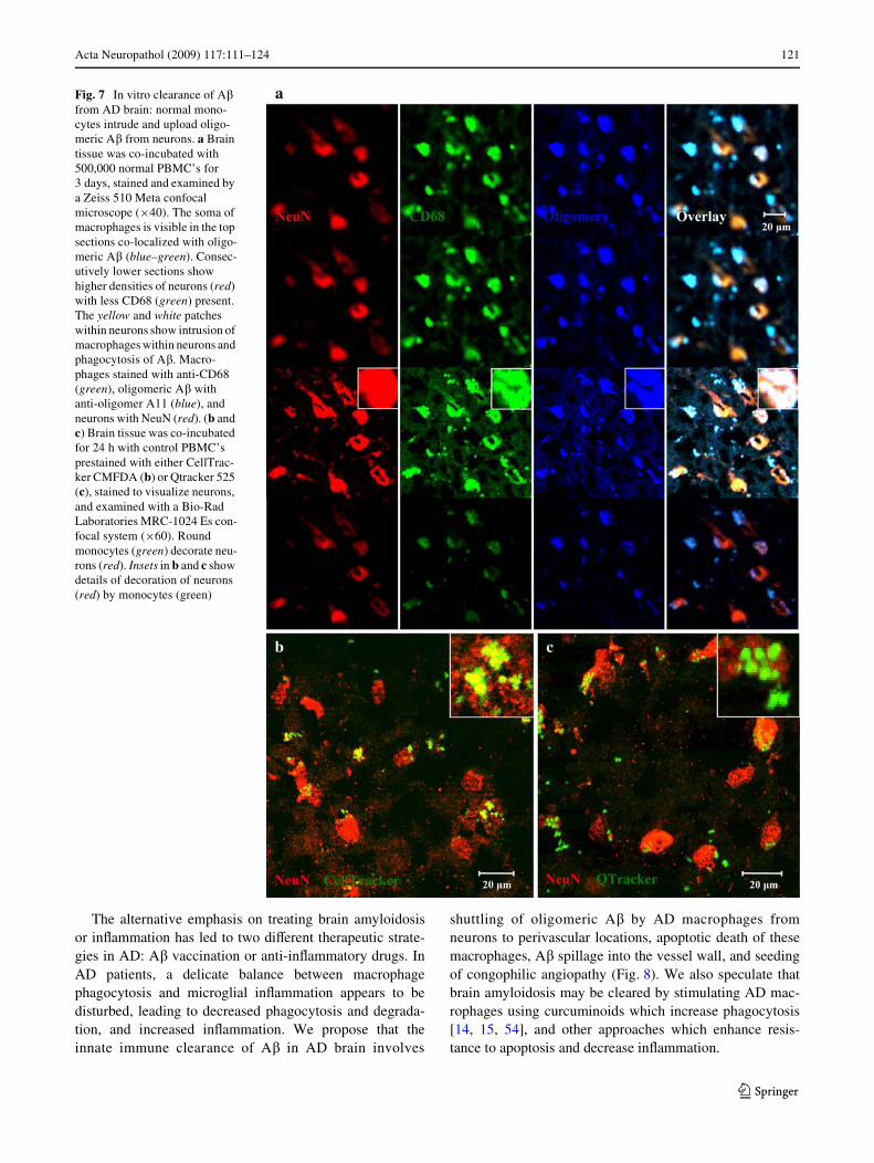
To experimentally investigate the brain clearance of A�,we co-incubated AD brain sections with control PBMC’sfor 3 days. Examination of these tissues by confocalmicroscopy showed monocytes (green) colocalizing in apatchy fashion with neurons (red) and uploading oligo-meric �� (blue). The soma of the monocytes was found inhigher z-sections compared to the soma of the neurons sug-gesting intrusion of these monocytes into neurons fromabove. In midsections, neurons, monocytes, and A� allcolocalized (Fig. 7a). In addition, after 24 h incubationwith AD brain sections, CellTracker and Qtracker pre-labeled monocytes were seen to co-localize with neurons,conWrming intrusion of exogenous monocytes into neurons(Fig. 6b). We did not notice obvious diVerences in clear-ance of A� between the monocytes of younger and elderlycontrol subjects but did not analyze these diVerences. Wealso stained AD brain tissues co-incubated without exoge-nous monocytes but did not Wnd CD68 positive monocytesintruding into neurons.
Fig. 2 Uptake and removal of A� by normal monocytes are greatercompared to AD monocytes. Monocytes were exposed to A� (2 �g/mL)for 2 h, washed, and incubated for the indicated number of days whenA� was measured by the ELISA assay. Control monocytes (a) showedhigher uptake of A� after 2-h exposure than AD monocytes (b) andsimilarly showed more rapid removal of A� in comparison to ADmonocytes
Controls
0
50
100
150
200
250
300
350
400
0 1 2 3 4 5 6 7Day
Am
ylo
id B
eta
(pg
/ml) Control 1
Control 2
Control 3
Control 4
Patients
0
50
100
150
200
250
300
350
400
0 1 2 3 4 5 6 7
Day
Am
ylo
id B
eta
(pg
/ml) Patient 1
Patient 2
Patient 3

Acta Neuropathol (2009) 117:111–124 117
123
Discussion
In transgenic animal models of AD, A�-induced pathologyseems to be upstream of tau and is considered the primarymechanism [33]. We have attempted to glean the mecha-nisms responsible for clearance of A� from human brain bycomparing the results of morphologic and experimentalinvestigations. Although emigration of macrophages acrossthe BBB has been infrequently investigated, macrophageimmigration is well known in HIV-1 encephalitis and isbecoming accepted in AD [11]. Our results in the modelBBB exclude the emigration of A�-engorged macrophagesas a mechanism of A� clearance since these adhered to theendothelium and were inhibited in emigration. Thus thefate of A� in aging brain seems to depend upon its handlingand degradation inside the BBB.
Our observations suggest that oligomeric A� in perivas-cular macrophages may contribute to the Wbrillar A� incongophilic vessels. AD macrophages are defective inuptake of A� (Fig. 3), yet are particularly susceptible toapoptosis from all assembly states of A� (Figs. 4, 5), con-trasting with the ability of control macrophages to phagocy-tize A� [15] and to resist apoptosis (Figs. 4, 5). Thephagocytic propensity of AD macrophages is compoundedby their low clearance of A� (Fig. 2). These deWcits mightbe related to downregulation of transcription of certaingenes, including MGAT-3 and Toll-like receptors [15].
In AD brain, macrophages are apoptotic through activa-tion of caspases -6, -7, and -8 (Fig. 6e, f, g), and they abutand inWltrate congophilic microvessels from the outside
[11] (Fig. 6d), in agreement with the assembly of �� Wbrilsoutside of the basal lamina [52]. Immunohistochemicalstudies of cerebral amyloid angiopathy (CAA) revealed itsassociation with a signiWcant increase and activation ofmacrophages in leptomeningeal and cortical vessels—implicating a central role for macrophages in amyloidangiopathy [53]. Furthermore, the BBB in AD brain isimpaired [11] by immigrating macrophages (Fig. 1d), thusallowing leakage of plasma proteins into the brain and furtheraggravation of angiopathy. Taken together, AD macrophagesloaded with oligomeric A� seem to suVer apoptosis, disin-tegrate and release oligomeric and Wbrillar A� into the wallof congophilic vessels. Still, it is important to recognizethat although phagocytosis of �� is generally greater incontrol monocytes compared to AD monocytes, this is amultifactorial and heterogeneous eVect, and monocytes ofsome aged control subjects occasionally demonstrate inferiorinnate immunity [15] and phagocytosis of A� (SI Fig. 1).
Proposed mechanisms for the pathogenesis of CAA haveincluded production of A� by myocytes in vessel walls [51],derivation of A� from blood or cerebrospinal Xuid, and, morerecently, deposition of A� from interstitial Xuid being drainedfrom the central nervous system [49]. In APP-transgenicmice, amyloid angiopathy developed speciWcally in areas ofthe brain that over-produce A�, suggesting that brain, asopposed to blood, is the major source of A� [44]. In mice,tracers were injected intracerebrally and found to be distrib-uted, similarly to A�, within the basement membranes ofintracerebral capillaries and arteries, and in leptomeningealarteries [3]. Interestingly, within 24 h of injection the tracers
Fig. 3 Fibrillar and protoWbril-lar A� (1-42). Five microlitre of samples were spotted on a glow-discharged, carbon-coated Formvar grid and incubated for 5 min, washed with distilled water, Wxed with 2.5% glutaral-dehyde, stained with a 1% (w/v) aqueous uranyl acetate solution, and examined using a JEOL transmission electron microscope

118 Acta Neuropathol (2009) 117:111–124
123
were located in perivascular macrophages and these cellsremained in brain microvessels, experiencing limited migra-tion. Thus both drainage of the interstitial Xuid and shuttlingof A� by macrophages seem to lead to entrapment of A� inperivascular macrophages.
The results of the experimental co-culture of normal sub-jects’ monocytes with AD brain tissues show intrusion bythese monocyte/macrophages into neurons and uploading ofoligomeric A�, suggesting a protective neuronal mechanismby the normal innate immune system. However, AD mono-cyte/macrophages seem to be defective in such protection ofneurons [15]. Although we have not investigated this mech-anism by electron microscopy of AD brain tissues, we haveobserved that macrophages and monocytes exposed to A� invitro develop proliWc microvilli and larger dendrites, whichcould reach into neurons and upload A� [15].
In animal models of amyloidosis, microglia were Wrstnoted for their proinXammatory role [1]. Microglia of APOE4transgenic mice produced more nitric oxide compared to
APOE3 transgenic mice [5]. Subsequently, microglia werefound to provide clearance of A� and were assisted in clear-ing A� deposits by antibodies [2] and by complement opsoni-zation [16, 45, 46]. Clearance of A� was recognized toinclude two phases: the Wrst mediated by antibodies and thesecond by microglia and antibodies [50]. However, in tripletransgenic APP-thymidine kinase mice, bone marrow-derivedrather than resident microglia restricted amyloid deposits asshown by ganciclovir destruction of microglia [36].
However, human brain disorders involve both microglialactivation and macrophage recruitment [40]. In AD brain,the initial inXammatory signal is induced by microglia [1]and neuronal chemokine RANTES. These stimuli attractlarge macrophages (clearly not resembling ramiWedmicroglia) to migrate across brain microvessels and invadeneuritic plaques in AD brain [11]. Enzyme studies mitigateagainst the role of microglia in A� clearance since, incomparison to macrophages, degradation of A� by microg-lia is poor due to defective lysosomal enzymes [28].
Fig. 4 Fibrillar and protoWbril-lar A� induce apoptosis of AD macrophages. After overnight exposure to Wbrillar (a) or proto-Wbrillar A� (2 �g/mL) (b), nor-mal macrophages displayed a low apoptotic FLICA signal (red) but a high uptake of proto-Wbrillar �� (green), whereas AD macrophages showed a robust FLICA signal and a high uptake of large Wbrils (yellow or white due to spectral bleed-through of FITC) but low uptake of protoW-brils. FLICA signal (red), proto-Wbrillar or Wbrillar A� (green) (duplicate images, Xuorescence microscopy, £100). Charts show the mean IOD of FLICA and A� with error bars indicat-ing standard error of mean
Normal
AD
a
b
Normal
AD
10 µm
10 µm
FLICA signal per macrophage
Aβ per macrophage
Aβ per macrophage
FLICA signal per macrophage
0
2000
4000
6000
8000
10000
12000
14000
16000
Ave
rag
e IO
D p
er M
acro
ph
age Control
Subjects
ADPatients
0
1000
2000
3000
4000
5000
6000
7000
8000
Ave
rag
e IO
D p
er M
acro
ph
age Control
Subjects
ADPatients
0
20000
40000
60000
80000
100000
Ave
rag
e IO
D p
er M
acro
ph
age
ControlSubjectsADPatients
0
2000
4000
6000
8000
10000
12000
14000
Ave
rag
e IO
D p
er M
acro
ph
age
ControlSubjectsADPatients

Acta Neuropathol (2009) 117:111–124 119
123
Fig. 5 Soluble A� induces apoptosis of AD macrophages and release of oligomeric and Wbrillar A�. a After incubation with FITC A� (2 �g/mL), AD macrophages displayed a signiW-cantly stronger apoptotic FLICA signal (red) compared to control macrophages. AD macrophages heterogeneously lysed by 5 days (Xuorescence microscopy, £40). b AD macrophages were incu-bated with soluble A� (2 �g/mL) for 1 h, washed with PBS, incu-bated for 4 or 6 days, and stained with FLICA (red) and anti-olig-omeric or anti-Wbrillar antibod-ies (green). After 4-day incubation, A� oligomers and Wbrils are visible within the apoptotic macrophages, and by 6 days the cells appear dis-rupted. A� oligomers diVused from the lysed macrophages whereas the denser A� Wbrils remained stuck in place on the glass slides (Xuorescence microscopy, £100)
Normal Macrophages AD Macrophages
1 Hour
3 Days
5 Days
Oligomeric Aβ
4 Days
6 Days
AD Macrophages
20 µm
10 µm
Fibrillar Aβ
a
b

120 Acta Neuropathol (2009) 117:111–124
123
Fig. 6 A� assemblies in AD brain: oligomeric and soluble A� is found in neurons and peri-vascular apoptotic macrophages, and Wbrillar A� in neuritic plaques and vessels. AD brain tissues show monomeric (a) and oligomeric A� (b) in neurons in a patchy fashion, Wbrillar A� in plaques and microvessel walls (c), and oligomeric �� in peri-vascular macrophages (d). Peri-vascular macrophages display activated caspases -6, -7, and -8 (e, f, and g). Red soluble A�, oligomeric A�, Wbrillar A�, and activated caspases. Green neu-rons and macrophages. Blue nu-clei. (a-c, e: £40 objective with Zeiss 510 Meta confocal micro-scope; d: £60 objective with Bio-Rad Laboratories MRC-1024 Es confocal system; f-g: £40 objective with Olympus Bmax Xuorescence microscope)
Acta Neuropathol (2009) 117:111–124 121
123
The alternative emphasis on treating brain amyloidosisor inXammation has led to two diVerent therapeutic strate-gies in AD: A� vaccination or anti-inXammatory drugs. InAD patients, a delicate balance between macrophagephagocytosis and microglial inXammation appears to bedisturbed, leading to decreased phagocytosis and degrada-tion, and increased inXammation. We propose that theinnate immune clearance of A� in AD brain involves
shuttling of oligomeric A� by AD macrophages fromneurons to perivascular locations, apoptotic death of thesemacrophages, A� spillage into the vessel wall, and seedingof congophilic angiopathy (Fig. 8). We also speculate thatbrain amyloidosis may be cleared by stimulating AD mac-rophages using curcuminoids which increase phagocytosis[14, 15, 54], and other approaches which enhance resis-tance to apoptosis and decrease inXammation.
Fig. 7 In vitro clearance of A� from AD brain: normal mono-cytes intrude and upload oligo-meric A� from neurons. a Brain tissue was co-incubated with 500,000 normal PBMC’s for 3 days, stained and examined by a Zeiss 510 Meta confocal microscope (£40). The soma of macrophages is visible in the top sections co-localized with oligo-meric A� (blue–green). Consec-utively lower sections show higher densities of neurons (red) with less CD68 (green) present. The yellow and white patches within neurons show intrusion of macrophages within neurons and phagocytosis of A�. Macro-phages stained with anti-CD68 (green), oligomeric A� with anti-oligomer A11 (blue), and neurons with NeuN (red). (b and c) Brain tissue was co-incubated for 24 h with control PBMC’s prestained with either CellTrac-ker CMFDA (b) or Qtracker 525 (c), stained to visualize neurons, and examined with a Bio-Rad Laboratories MRC-1024 Es con-focal system (£60). Round monocytes (green) decorate neu-rons (red). Insets in b and c show details of decoration of neurons (red) by monocytes (green)

122 Acta Neuropathol (2009) 117:111–124
123
Acknowledgments We thank J. Sayre for statistical analysis,S. Krajewski (Burnham Institute) for antibodies to activated caspases,B. Carter for nursing assistance with collection of blood specimensfrom patients and control subjects, B. Magrys for cutting sections ofthe blood-brain barrier model, C. Glabe for providing 14C-�� and A11and OC antibodies, and C. Serhan for lipoxin A4. The UCLA BrainBank provided the brain tissues for immunostaining.
ConXict of interest statement This work was supported by theAlzheimer’s Association, MPBio Ltd (MF) and NIH grant AG027818(DBT).
Open Access This article is distributed under the terms of the Crea-tive Commons Attribution Noncommercial License which permits anynoncommercial use, distribution, and reproduction in any medium,provided the original author(s) and source are credited.
References
1. Akiyama H, Barger S, Barnum S, Bradt B, Bauer J, Cole GM,Cooper NR, Eikelenboom P, Emmerling M, Fiebich BL, FinchCE, Frautschy S, GriYn WS, Hampel H, Hull M, Landreth G, LueL, Mrak R, Mackenzie IR, McGeer PL, O’Banion MK, Pachter J,Pasinetti G, Plata-Salaman C, Rogers J, Rydel R, Shen Y, Streit W,
Strohmeyer R, Tooyoma I, Van Muiswinkel FL, Veerhuis R,Walker D, Webster S, Wegrzyniak B, Wenk G, Wyss-Coray T(2000) InXammation and Alzheimer’s disease. Neurobiol Aging21:383–421. doi:10.1016/S0197-4580(00)00124-X
2. Bard F, Cannon C, Barbour R, Burke RL, Games D, Grajeda H,Guido T, Hu K, Huang J, Johnson-Wood K, Khan K, KholodenkoD, Lee M, Lieberburg I, Motter R, Nguyen M, Soriano F, VasquezN, Weiss K, Welch B, Seubert P, Schenk D, Yednock T (2000)Peripherally administered antibodies against amyloid beta-peptideenter the central nervous system and reduce pathology in amouse model of Alzheimer disease. Nat Med 6:916–919.doi:10.1038/78682
3. Carare RO, Bernardes-Silva M, Newman TA, Page AM, NicollJA, Perry VH, Weller RO (2008) Solutes, but not cells, drain fromthe brain parenchyma along basement membranes of capillariesand arteries: signiWcance for cerebral amyloid angiopathy andneuroimmunology. Neuropathol Appl Neurobiol 34:131–144.doi:10.1111/j.1365-2990.2007.00926.x
4. Chen K, Iribarren P, Hu J, Chen J, Gong W, Cho EH, Lockett S,Dunlop NM, Wang JM (2006) Activation of Toll-like receptor 2on microglia promotes cell uptake of Alzheimer disease-associ-ated amyloid beta peptide. J Biol Chem 281:3651–3659.doi:10.1074/jbc.M508125200
5. Colton CA, Brown CM, Cook D, Needham LK, Xu Q, Czapiga M,Saunders AM, Schmechel DE, Rasheed K, Vitek MP (2002)APOE and the regulation of microglial nitric oxide production: a
Fig. 8 Congophilic angiopathy immunopathogenesis. A� is pro-duced in neurons by cleavage of the amyloid-precursor protein (APP) by the �- and �-secretases, releasing A�, which is re-uptaken into neurons and becomes oligo-meric. Blood-borne monocytes (1) are attracted to neurons by chemokines produced by neurons (e.g. RANTES), intrude into neurons and neuritic plaques (2), and upload oligo-meric A� from neurons and Wbrillar A� in neuritic plaques. However, AD monocyte/macro-phages (3) are heterogeneously deWcient in A� clearance (up-take and degradation), and have deWcient MGAT-III transcrip-tion and increased activation of caspases leading to their apopto-sis. Some AD macrophages loaded with A� migrate to ves-sels (4) but are unable emigrate due to their engorgement with A� and consequently undergo apoptosis. Simultaneously, solu-ble and oligomeric A� have undergone peptide assembly into Wbrillar A�, which is discharged into the vessel wall (5). Further-more, congophilic angiopathy induces oxidative stress to neurons, and inXammatory cyto-kines produced by microglia potentiate neuronal damage
Neuritic Plaque
NormalVessel
AmyloidAngiopathy
Apoptoticmacrophage
Healthymacrophage
Proto-fibrillar A
Oligomeric A
Fibrillar A
APP
Basement Membrane
SmoothMuscle Cell
EndothelialCell
CCL5(RANTES)
Neuron with A
MigratingMonocyte
InflammationTNF- , IL-6
AD macrophages:
Activated Caspases –6, -7, -8 MGAT-3 RNA
4
1
2
3
5

Acta Neuropathol (2009) 117:111–124 123
123
link between genetic risk and oxidative stress. Neurobiol Aging23:777–785. doi:10.1016/S0197-4580(02)00016-7
6. El Khoury JB, Moore KJ, Means TK, Leung J, Terada K, Toft M,Freeman MW, Luster AD (2003) CD36 mediates the innate hostresponse to beta-amyloid. J Exp Med 197:1657–1666.doi:10.1084/jem.20021546
7. El Khoury J, Toft M, Hickman SE, Means TK, Terada K, Geula C,Luster AD (2007) Ccr2 deWciency impairs microglial accumula-tion and accelerates progression of Alzheimer-like disease. NatMed 13:432–438. doi:10.1038/nm1555
8. Fiala M, Looney DJ, Stins M, Way DD, Zhang L, Gan X, Chiapp-elli F, Schweitzer ES, Shapshak P, Weinand M, Graves MC, WitteM, Kim KS (1997) TNF-alpha opens a paracellular route for HIV-1 invasion across the blood-brain barrier. Mol Med 3:553–564
9. Fiala M, Zhang L, Gan X, Sherry B, Taub D, Graves MC, HamaS, Way D, Weinand M, Witte M, Lorton D, Kuo YM, Roher AE(1998) Amyloid-beta induces chemokine secretion and monocytemigration across a human blood–brain barrier model. Mol Med4:480–489
10. Fiala M, Liu NQ, Reddy S, Graves M (2001) Macrophages inWl-trate the brain and express COX-2 and iNOS in Alzheimer’s dis-ease and AIDS. Alzheimers Rep 4:1–7
11. Fiala M, Liu QN, Sayre J, Pop V, Brahmandam V, Graves MC,Vinters HV (2002) Cyclooxygenase-2-positive macrophages inWl-trate the Alzheimer’s disease brain and damage the blood-brainbarrier. Eur J Clin Invest 32:360–371. doi:10.1046/j.1365-2362.2002.00994.x
12. Fiala M, Eshleman AJ, Cashman J, Lin J, Losssinsky AS, SuarezV, Yang W, Zhang J, Popik W, Singer EJ, Chiappelli F, Carro E,Weinand M, Witte M, Arthos J (2005) Cocaine increases humanimmunodeWciency virus type 1 neuroinvasion through remodelingbrain microvascular endothelial cells. J Neurovirol 11:1–11.doi:10.1080/13550280590952835
13. Fiala M, Lin J, Ringman J, Kermani-Arab V, Tsao G, Patel A,Lossinsky AS, Graves MC, Gustavson A, Sayre J, Sofroni E,Suarez T, Chiappelli F, Bernard G (2005) IneVective phagocytosisof amyloid-beta by macrophages of Alzheimer’s disease patients.J Alzheimers Dis 7:221–232 discussion 255–262
14. Fiala M, Cribbs DH, Rosenthal M, Bernard G (2007) Phagocytosisof amyloid-beta and inXammation: two faces of innate immunityin Alzheimer’s disease. J Alzheimers Dis 11:457–463
15. Fiala M, Liu PT, Espinosa-JeVrey A, Rosenthal MJ, Bernard G,Ringman JM, Sayre J, Zhang L, Zaghi J, Dejbakhsh S, Chiang B,Hui J, Mahanian M, Baghaee A, Hong P, Cashman J (2007) Innateimmunity and transcription of MGAT-III and Toll-like receptorsin Alzheimer’s disease patients are improved by bisdemethoxycur-cumin. Proc Natl Acad Sci USA 104:12849–12854. doi:10.1073/pnas.0701267104
16. Frautschy SA, Yang F, Irrizarry M, Hyman B, Saido TC, Hsiao K,Cole GM (1998) Microglial response to amyloid plaques in APPswtransgenic mice. Am J Pathol 152:307–317 see comments
17. Giri R, Shen Y, Stins M, Du Yan S, Schmidt AM, Stern D, KimKS, Zlokovic B, Kalra VK (2000) Beta-amyloid-induced migra-tion of monocytes across human brain endothelial cells involvesRAGE and PECAM-1. Am J Physiol Cell Physiol 279:C1772–C1781
18. Gouras GK, Almeida CG, Takahashi RH (2005) IntraneuronalAbeta accumulation and origin of plaques in Alzheimer’s disease.Neurobiol Aging 26:1235–1244. doi:10.1016/j.neurobiolaging.2005.05.022
19. Hamilton JA, Whitty G, White AR, Jobling MF, Thompson A,Barrow CJ, Cappai R, Beyreuther K, Masters CL (2002)Alzheimer’s disease amyloid beta and prion protein amyloido-genic peptides promote macrophage survival, DNA synthesis andenhanced proliferative response to CSF-1 (M-CSF). Brain Res940:49–54. doi:10.1016/S0006-8993(02)02589-1
20. Hardy J, Selkoe DJ (2002) The amyloid hypothesis of Alzheimer’sdisease: progress and problems on the road to therapeutics. Science297:353–356. doi:10.1126/science.1072994
21. Husemann J, Loike JD, Anankov R, Febbraio M, Silverstein SC(2002) Scavenger receptors in neurobiology and neuropathology:their role on microglia and other cells of the nervous system. Glia40:195–205. doi:10.1002/glia.10148
22. Kayed R, Head E, Thompson JL, McIntire TM, Milton SC,Cotman CW, Glabe CG (2003) Common structure of solubleamyloid oligomers implies common mechanism of pathogenesis.Science 300:486–489. doi:10.1126/science.1079469
23. Kayed R, Head E, Sarsoza F, Saing T, Cotman CW, Necula M,Margol L, Wu J, Breydo L, Thompson JL, Rasool S, Gurlo T,Butler P, Glabe CG (2007) Fibril speciWc, conformation dependentantibodies recognize a generic epitope common to amyloid Wbrilsand Wbrillar oligomers that is absent in preWbrillar oligomers. MolNeurodegener 2:18. doi:10.1186/1750-1326-2-18
24. Krajewska M, Rosenthal RE, Mikolajczyk J, Stennicke HR,Wiesenthal T, Mai J, Naito M, Salvesen GS, Reed JC, Fiskum G,Krajewski S (2004) Early processing of Bid and caspase-6, -8, -10,-14 in the canine brain during cardiac arrest and resuscitation. ExpNeurol 189:261–279. doi:10.1016/j.expneurol.2004.05.020
25. LaFerla FM, Green KN, Oddo S (2007) Intracellular amyloid-betain Alzheimer’s disease. Nat Rev Neurosci 8:499–509. doi:10.1038/nrn2168
26. Liu NQ, Lossinsky AS, Popik W, Li X, Gujuluva C, KriedermanB, Roberts J, Pushkarsky T, Bukrinsky M, Witte M, Weinand M,Fiala M (2002) Human immunodeWciency virus type 1 entersbrain microvascular endothelia by macropinocytosis dependenton lipid rafts and the mitogen-activated protein kinase signalingpathway. J Virol 76:6689–6700. doi:10.1128/JVI.76.13.6689-6700.2002
27. Lossinsky AS, Shivers RR (2003) Studies of cerebral endotheliumby scanning and high-voltage electron microscopy. In: Nag S (ed)The blood-brain barrier: biology and research protocols, vol 89.Humana Press, Totawa, pp 67–82
28. Majumdar A, Chung H, Dolios G, Wang R, Asamoah N, Lobel P,MaxWeld FR (2008) Degradation of Wbrillar forms of Alzheimer’samyloid beta-peptide by macrophages. Neurobiol Aging 29:707–715. doi:10.1016/j.neurobiolaging.2006.12.001
29. McKhann G, Drachman D, Folstein M, Katzman R, Price D, Stad-lan EM (1984) Clinical diagnosis of Alzheimer’s disease: report ofthe NINCDS-ADRDA Work Group under the auspices of Depart-ment of Health and Human Services Task Force on Alzheimer’sDisease. Neurology 34:939–944
30. Mildner A, Schmidt H, Nitsche M, Merkler D, Hanisch UK, MackM, Heikenwalder M, Bruck W, Priller J, Prinz M (2007) Microgliain the adult brain arise from Ly-6C(hi)CCR2(+) monocytes onlyunder deWned host conditions. Nat Neurosci 10:1544–1553.doi:10.1038/nn2015
31. Mrak RE, GriYn WS (2005) Potential inXammatory biomarkers inAlzheimer’s disease. J Alzheimers Dis 8:369–375
32. Nixon RA (2007) Autophagy, amyloidogenesis and Alzheimerdisease. J Cell Sci 120:4081–4091. doi:10.1242/jcs.019265
33. Oddo S, Caccamo A, Tran L, Lambert MP, Glabe CG, Klein WL,LaFerla FM (2006) Temporal proWle of amyloid-beta (Abeta)oligomerization in an in vivo model of Alzheimer disease. A linkbetween Abeta and tau pathology. J Biol Chem 281:1599–1604.doi:10.1074/jbc.M507892200
34. Paresce DM, Ghosh RN, MaxWeld FR (1996) Microglial cellsinternalize aggregates of the Alzheimer’s disease amyloid beta-protein via a scavenger receptor. Neuron 17:553–565. doi:10.1016/S0896-6273(00)80187-7
35. Sagare A, Deane R, Bell RD, Johnson B, Hamm K, Pendu R,Marky A, Lenting PJ, Wu Z, Zarcone T, Goate A, Mayo K, Perl-mutter D, Coma M, Zhong Z, Zlokovic BV (2007) Clearance of

124 Acta Neuropathol (2009) 117:111–124
123
amyloid-beta by circulating lipoprotein receptors. Nat Med13:1029–1031. doi:10.1038/nm1635
36. Simard AR, Rivest S (2006) Neuroprotective properties of theinnate immune system and bone marrow stem cells in Alzheimer’sdisease. Mol Psychiatry 11:327–335. doi:10.1038/sj.mp.4001809
37. Simard AR, Soulet D, Gowing G, Julien JP, Rivest S (2006) Bonemarrow-derived microglia play a critical role in restricting senileplaque formation in Alzheimer’s disease. Neuron 49:489–502.doi:10.1016/j.neuron.2006.01.022
38. Smits HA, Rijsmus A, van Loon JH, Wat JW, Verhoef J, BovenLA, Nottet HS (2002) Amyloid-beta-induced chemokine produc-tion in primary human macrophages and astrocytes. J Neuroimmu-nol 127:160–168. doi:10.1016/S0165-5728(02)00112-1
39. Solomon B (2007) Intravenous immunoglobulin and Alzheimer’sdisease immunotherapy. Curr Opin Mol Ther 9:79–85
40. Stoll G, Jander S (1999) The role of microglia and macrophages inthe pathophysiology of the CNS. Prog Neurobiol 58:233–247.doi:10.1016/S0301-0082(98)00083-5
41. Stuart LM, Bell SA, Stewart CR, Silver JM, Richard J, Goss JL,Tseng AA, Zhang A, El Khoury JB, Moore KJ (2007) CD36 sig-nals to the actin cytoskeleton and regulates microglial migration viaa p130Cas complex. J Biol Chem 282:27392–27401. doi:10.1074/jbc.M702887200
42. Tanzi RE, Moir RD, Wagner SL (2004) Clearance of Alzheimer’sAbeta peptide: the many roads to perdition. Neuron 43:605–608
43. Tseng BP, Green KN, Chan JL, Blurton-Jones M, LaFerla FM(2008) Abeta inhibits the proteasome and enhances amyloid andtau accumulation. Neurobiol Aging 29:1607–1618. doi:10.1016/j.neurobiolaging.2007.04.014
44. Van Dorpe J, Smeijers L, Dewachter I, Nuyens D, Spittaels K, VanDen Haute C, Mercken M, Moechars D, Laenen I, Kuiperi C,Bruynseels K, Tesseur I, Loos R, Vanderstichele H, Checler F,Sciot R, Van Leuven F (2000) Prominent cerebral amyloidangiopathy in transgenic mice overexpressing the London mutantof human APP in neurons. Am J Pathol 157:1283–1298
45. Webster SD, Yang AJ, Margol L, Garzon-Rodriguez W, GlabeCG, Tenner AJ (2000) Complement component C1q modulatesthe phagocytosis of Abeta by microglia. Exp Neurol 161:127–138.doi:10.1006/exnr.1999.7260
46. Webster SD, Galvan MD, Ferran E, Garzon-Rodriguez W, GlabeCG, Tenner AJ (2001) Antibody-mediated phagocytosis of theamyloid beta-peptide in microglia is diVerentially modulated byC1q. J Immunol 166:7496–7503
47. Wegiel J, Kuchna I, Nowicki K, Frackowiak J, Mazur-Kolecka B,Imaki H, Mehta PD, Silverman WP, Reisberg B, Deleon M,Wisniewski T, Pirttilla T, Frey H, Lehtimaki T, Kivimaki T,Visser FE, Kamphorst W, Potempska A, Bolton D, Currie JR,Miller DL (2007) Intraneuronal Abeta immunoreactivity is not a pre-dictor of brain amyloidosis-beta or neuroWbrillary degeneration.Acta Neuropathol 113:389–402. doi:10.1007/s00401-006-0191-4
48. Weiner HL, Frenkel D (2006) Immunology and immunotherapyof Alzheimer’s disease. Nat Rev Immunol 6:404–416. doi:10.1038/nri1843
49. Weller RO, Nicoll JA (2003) Cerebral amyloid angiopathy:pathogenesis and eVects on the ageing and Alzheimer brain.Neurol Res 25:611–616. doi:10.1179/016164103101202057
50. Wilcock DM, DiCarlo G, Henderson D, Jackson J, Clarke K, UgenKE, Gordon MN, Morgan D (2003) Intracranially administeredanti-Abeta antibodies reduce beta-amyloid deposition by mecha-nisms both independent of and associated with microglial activa-tion. J Neurosci 23:3745–3751
51. Wisniewski HM, Wegiel J (1994) Beta-amyloid formation bymyocytes of leptomeningeal vessels. Acta Neuropathol 87:233–241. doi:10.1007/BF00296738
52. Wisniewski HM, Wegiel J, Wang KC, Lach B (1992) Ultrastruc-tural studies of the cells forming amyloid in the cortical vessel wallin Alzheimer’s disease. Acta Neuropathol 84:117–127.doi:10.1007/BF00311383
53. Yamada M, Itoh Y, Shintaku M, Kawamura J, Jensson O,Thorsteinsson L, Suematsu N, Matsushita M, Otomo E (1996)Immune reactions associated with cerebral amyloid angiopathy.Stroke 27:1155–1162
54. Zhang L, Fiala M, Cashman J, Sayre J, Espinosa-JeVrey A,Mahanian M, Zaghi J, Badmaev V, Graves M, Bernard G, RosenthalM (2006) Curcuminoids enhance amyloid-beta uptake by mac-rophages of Alzheimer’s disease patients. J Alzheimers Dis10:1–7
Copyright © 2022 FDOKUMEN