
Interactions of the antiviral and antiparkinson agent amantadine with lipid membranes and human...
8
Interactions of the antiviral and antiparkinson agent amantadine with lipid membranes and human erythrocytes Mario Suwalsky a, ⁎, Malgorzata Jemiola-Rzeminska b,c , Mariella Altamirano d , Fernando Villena d , Nathan Dukes e , Kazimierz Strzalka b,c a Faculty of Chemical Sciences, University of Concepción, Concepción, Chile b Faculty of Biochemistry, Biophysics and Biotechnology, Jagiellonian University, Krakow, Poland c Malopolska Centre of Biotechnology, Jagiellonian University, Krakow, Poland d Faculty of Biological Sciences, University of Concepción, Concepción, Chile e Faculty of Medicine, P. Catholic University of Chile, Santiago, Chile HIGHLIGHTS • Interactions of amantadine with human erythrocytes and lipid bilayers were assessed. • Amantadine changed the morphology of erythrocytes inducing formation of echinocytes. • This change of shape indicates that it was inserted in the outer leaflet of the erythrocyte membrane. • Amantadine interacted with a class of phospholipid present in the outer leaf- let of the erythrocyte membrane. GRAPHICAL ABSTRACT abstract article info Article history: Received 27 March 2015 Received in revised form 1 April 2015 Accepted 1 April 2015 Available online 9 April 2015 Keywords: Amantadine Erythrocyte membrane Phospholipid bilayer Aimed to better understand the molecular mechanisms of its interactions with cell membranes, human erythro- cyte and molecular models of the red cell membrane were utilized. The latter consisted of bilayers of dimyristoylphosphatidylcholine (DMPC) and dimyristoylphosphatidylethanolamine (DMPE), representative of phospholipid classes located in the outer and inner monolayers of the human erythrocyte membrane, respective- ly. The capacity of amantadine to perturb the bilayer structures of DMPC and DMPE was evaluated by X-ray dif- fraction, fluorescence spectroscopy and differential scanning calorimetry (DSC). In an attempt to further elucidate its effects on cell membranes, the present work also examined amantadine influence on the morphol- ogy of intact human erythrocytes by means of scanning electron microscopy (SEM). Results indicated that aman- tadine induced morphological changes to human erythrocytes and interacted in a concentration-dependent manner with DMPC bilayers in contrast to DMPE that was hardly affected by the presence of the drug. © 2015 Elsevier B.V. All rights reserved. Biophysical Chemistry 202 (2015) 13–20 Abbreviations: RBCS, red blood cell suspension; SEM, scanning electron microscopy; DMPC, dimyristoylphosphatidylcholine; DMPE, dimyristoylphosphatidylcholine; DSC, differential scanning calorimetry; MLV, multilamellar vesicles. ⁎ Corresponding author. E-mail address: [email protected] (M. Suwalsky). http://dx.doi.org/10.1016/j.bpc.2015.04.002 0301-4622/© 2015 Elsevier B.V. All rights reserved. Contents lists available at ScienceDirect Biophysical Chemistry journal homepage: http://www.elsevier.com/locate/biophyschem
-
Upload
jagiellonian -
Category
Documents
-
view
1 -
download
0
Transcript of Interactions of the antiviral and antiparkinson agent amantadine with lipid membranes and human...

Biophysical Chemistry 202 (2015) 13–20
Contents lists available at ScienceDirect
Biophysical Chemistry
j ourna l homepage: ht tp : / /www.e lsev ie r .com/ locate /b iophyschem
Interactions of the antiviral and antiparkinson agent amantadine withlipid membranes and human erythrocytes
Mario Suwalsky a,⁎, Malgorzata Jemiola-Rzeminska b,c, Mariella Altamirano d, Fernando Villena d,Nathan Dukes e, Kazimierz Strzalka b,c
a Faculty of Chemical Sciences, University of Concepción, Concepción, Chileb Faculty of Biochemistry, Biophysics and Biotechnology, Jagiellonian University, Krakow, Polandc Malopolska Centre of Biotechnology, Jagiellonian University, Krakow, Polandd Faculty of Biological Sciences, University of Concepción, Concepción, Chilee Faculty of Medicine, P. Catholic University of Chile, Santiago, Chile
H I G H L I G H T S G R A P H I C A L A B S T R A C T
• Interactions of amantadine with humanerythrocytes and lipid bilayers wereassessed.
• Amantadine changed the morphologyof erythrocytes inducing formation ofechinocytes.
• This change of shape indicates that itwas inserted in the outer leaflet of theerythrocyte membrane.
• Amantadine interacted with a class ofphospholipid present in the outer leaf-let of the erythrocyte membrane.
Abbreviations:RBCS, red blood cell suspension; SEM, sscanning calorimetry;MLV,multilamellar vesicles.⁎ Corresponding author.
E-mail address: [email protected] (M. Suwalsky).
http://dx.doi.org/10.1016/j.bpc.2015.04.0020301-4622/© 2015 Elsevier B.V. All rights reserved.
a b s t r a c t
a r t i c l e i n f oArticle history:Received 27 March 2015Received in revised form 1 April 2015Accepted 1 April 2015Available online 9 April 2015
Keywords:AmantadineErythrocyte membranePhospholipid bilayer
Aimed to better understand themolecular mechanisms of its interactions with cell membranes, human erythro-cyte and molecular models of the red cell membrane were utilized. The latter consisted of bilayers ofdimyristoylphosphatidylcholine (DMPC) and dimyristoylphosphatidylethanolamine (DMPE), representative ofphospholipid classes located in the outer and innermonolayers of the human erythrocytemembrane, respective-ly. The capacity of amantadine to perturb the bilayer structures of DMPC and DMPE was evaluated by X-ray dif-fraction, fluorescence spectroscopy and differential scanning calorimetry (DSC). In an attempt to furtherelucidate its effects on cell membranes, the present work also examined amantadine influence on the morphol-ogy of intact human erythrocytes bymeans of scanning electronmicroscopy (SEM). Results indicated that aman-tadine induced morphological changes to human erythrocytes and interacted in a concentration-dependentmanner with DMPC bilayers in contrast to DMPE that was hardly affected by the presence of the drug.
© 2015 Elsevier B.V. All rights reserved.
canning electronmicroscopy; DMPC, dimyristoylphosphatidylcholine; DMPE, dimyristoylphosphatidylcholine; DSC, differential

Fig. 1. Schematic formula of amantadine hydrochloride.
14 M. Suwalsky et al. / Biophysical Chemistry 202 (2015) 13–20
1. Introduction
Amantadine (1-aminoadamantane, Fig. 1), a glutaminergic re-ceptor antagonist, is a stable quasi-spherical cyclic primary aminewhich is therapeutically used for both influenza and Parkinson's dis-ease [1–5]. This compound inhibits viral replication by blocking thechannel activity of the M2 proton channel that is critical in the viruslife cycle [6]. It has been suggested that the mechanism of M2 chan-nel inhibition would be due to amantadine interaction with themembrane side of the channel [7]. Association between amantadineand cellular membrane was first reported by Epand et al. [8] whodemonstrated that the drug inhibited membrane fusion. Studies per-formed by neutron and X-ray diffraction on dioleoylphosphatidylcholine(DOPC) showed that the hydrophobic cyclic region of amantadine hydro-chloride located between the phosphate and ester linkages of DOPCwiththepolar+NH3 groupprotruding into thewater space [1]. A solutionNMRstudy on dimyristoylphosphatidylcholine (DMPC) agreed in that amanta-dine localized near the negatively charged phosphate group and the hy-drocarbon chain of DMPC [7]. A more recent work on solid-state NMRandmolecular dynamics simulations reported that the long axis of aman-tadine located parallel to DMPC bilayer normal and its amino group wasoriented toward the lipid head group [9].
Studies on the effects of amantadine onhuman erythrocytes are veryscarce. It has been reported that side effects of amantadine treatmentinclude anemia which could result from suicidal erythrocyte death oreryptosis, which accelerates the clearance of circulating erythrocytes[4]. Typical eryptosis is characterized by cell shrinkage and cell mem-brane scrambling. However, amantadine does not decrease cell volume;on the contrary, it produces cell swelling, effect that could not be satis-factorily explained [4]. Amantadine has also been shown to affect eryth-rocyte shape, inducing stomatocytosis [10]. With the aim to betterunderstand themolecularmechanisms of the interaction of amantadinewith cell membranes we have utilized human erythrocytes and molec-ularmodels of itsmembrane. Human erythrocyteswere chosen becauseof their only single membrane and no internal organelles constitute anideal cell system for studying interactions of chemical compoundswith cell membranes [11]. On the other hand, although less specializedthanmany other cell membranes they carry on enough functions in com-mon with them such as active and passive transport, and the productionof ionic and electric gradients to be considered representative of the plas-mamembrane in general. Themolecularmodels of the erythrocytemem-brane consisted in bilayers of dimyristoylphosphatidylcholine (DMPC)and dimyristoylphosphatidylethanolamine (DMPE), representative ofphospholipid classes located in the outer and inner monolayers of cellmembranes, particularly of the human erythrocyte, respectively [12,13].The capacity of amantadine to interact with the bilayer structures ofDMPC andDMPEwas evaluated byX-ray diffraction anddifferential scan-ning calorimetry (DSC), andofDMPCbyfluorescence spectroscopy; intacthuman erythrocytes were observed by scanning electron microscopy
(SEM). These systems and techniques have been used in our laboratoriesto determine the interaction with and the membrane-perturbing effectsof other therapeutic compounds, including antiarrhythmic drugs [14–17].
2. Materials and methods
2.1. X-ray diffraction studies of DMPC and DMPE multilayers
The capacity of amantadine to interact with DMPC and DMPEmulti-layers was evaluated by X-ray diffraction. Synthetic DMPC (lot 140PC-24, MW 677.9) and DMPE (lot 140PE-54, MW 635.9) from AvantiPolar Lipids (AL, USA), and amantadine hydrochloride (MW 394.5)from Sigma-Aldrich (Milwaukee, USA) were used without further purifi-cation. About 2 mg of each phospholipid was introduced into Eppendorftubes which were then filled with 200 l of (a) distilled water (control),and (b) aqueous solutions of amantadine in a range of concentrations(50 μM–100 μM for DMPC and 1 mM–10 mM for DMPE experiments).The specimens were shaken, incubated for 30 min at 30 °C and 60 °Cwith DMPC and DMPE, respectively and centrifuged for 10 min at2400 rpm. Samples were then transferred into 1.5 mm diameter specialglass capillaries (Glas-Technik&Konstruktion, Berlin, Germany) andX-ray diffracted utilizing Ni-filtered CuKα radiation from a BrukerKristalloflex 760 (Karlsruhe, Germany) X-ray system. Specimen-to-film distances were 8 and 14 cm, standardized by sprinkling calcitepowder on the capillary surface. The relative reflection intensitiesand interplanar spacings were obtained from an MBraun PSD-50 Mlinear position-sensitive detector system (Garching, Germany) andASA software; no correction factors were applied. Data analyseswere performed by means of Origin 3.0 software (Origin Lab Corp.,USA). The experiments were performed at 19 °C and 1 °C, which isbelow the main phase transition temperature of both DMPC andDMPE. Higher temperatures would have induced transitions ontofluid phases making the detection of structural changes harder.Each experiment was performed in triplicate.
2.2. Differential scanning calorimetry (DSC) studies on DMPC and DMPEliposomes
Appropriate amounts of DMPC or DMPE dissolved in chloroformwere gently evaporated to dryness under a stream of gaseous nitrogenuntil a thin film on thewall of the glass test tubewas formed. To removethe remnants of moisture, the samples were subsequently exposed tovacuum for 1 h and then dry lipid films were suspended in distilledwater. Amantadine was added in the concentration range of 0.10 to10 mM for DMPC and 0.10 to 1.0 mM for DMPE. The multilamellar lipo-somes (MLV) were prepared by vortexing the samples at the tempera-ture above gel-to-liquid crystalline phase transition of the pure lipid(about 30 °C for DMPC and 60 °C for DMPE). DSC experimentswere per-formed using a NANODSC Series III Systemwith Platinum Capillary Cell(TA Instruments, USA). The calorimeter was equipped with the originaldata acquisition and analysis software. In order to avoid bubble forma-tion during heating mode the samples were degassed prior to beingloaded by pulling a vacuum of 0.3–0.5 atm on the solution for a periodof 10–15 min. Then the sample cell was filled with about 400 μl ofMLV suspension and an equal volume of buffer was used as a reference.The cellswere sealed and thermally equilibrated for about 10min belowstarting temperature of the run. All measurements were made on sam-ples under 3-bar pressure. The data were collected in the range of0–40 °C (DMPC) and 30–70 °C (DMPE) at the scan rate 1 °C min−1
both for heating and cooling. Scans of buffer as a sample and a referencewere also performed to collect the apparatus baseline. In order to checkthe reproducibility, each sample was prepared and recorded at leastthree times. Each data set was analyzed for thermodynamic parameterswith the software package supplied by TA Instruments.

Fig. 2.Microdensitograms from X-ray diffraction patterns of DMPC (A) and DMPE (B) inwater and aqueous solutions of amantadine hydrochloride; (SA) small-angle and (WA)wide-angle reflections.
15M. Suwalsky et al. / Biophysical Chemistry 202 (2015) 13–20
2.3. Fluorescence measurements of large unilamellar vesicles (LUV) and ofisolated unsealed human erythrocyte membranes (IUM)
The influence of amantadine on the physical properties of DMPC LUVand IUM was examined by fluorescence spectroscopy using DPH andlaurdan (Molecular Probe, Eugene, OR, USA) fluorescent probes. DPHiswidely used as a probe for the hydrophobic regions of the phospholip-id bilayers because of its favorable spectral properties. Their steady-state fluorescence anisotropy measurements were used to investigatethe structural properties of DMPC LUV and IUMas it provides ameasureof the rotational diffusion of the fluorophor restricted within a certainregion such as a cone due to the lipid acyl chain packing order. Laurdan,an amphiphilic probe, has high excitation sensitivity and emission spec-tra to the physical state of membranes. With the fluorescent moietywithin a shallow position in the bilayer, laurdan provides informationabout the polarity and/or molecular dynamics at the level of the phos-pholipid glycerol backbone. The quantification of the laurdan fluores-cence spectral shift was effected by means of the general polarization(GP) concept [18]. DMPC LUV suspended in water were prepared byextrusion of frozen and thawed multilamellar liposome suspensions(final lipid concentration 0.4 mM) through two stacked polycarbonatefilters of 400 nm pore size (Nucleopore, Corning Costar Corp., MA,USA) under nitrogen pressure at 10 °C above the lipid phase transitiontemperature. Erythrocytes were separated from heparinized venousblood samples obtained from normal casual donors by centrifugationand washing procedures. IUM were prepared by lysis, according toDodge et al. [19]. DPH and laurdan were incorporated into LUV andIUM by addition of 2 μl/ml aliquots of 0.5 mM solutions of the probein dimethylformamide and ethanol, respectively, in order to obtainfinal analytical concentrations of 1 × 10⁻³ mM, and incubated them at37 °C for 45 min. Fluorescence spectra and anisotropy measurementswere performed in a phase shift and modulation K2 steady-state andtime resolved spectrofluorometer (ISS, Inc., Champaign, IL, USA) inter-faced to computer. Software from ISS was used for both data collectionand analysis. LUV suspension measurements were carried out at 18 °Cand 37 °C, and IUM measurements were made at 37 °C using 10 mmpath-length square quartz cuvettes. Sample temperaturewas controlledby an external bath circulator (Cole-Parmer, Chicago, IL, USA) andmon-itored before and after each measurement using an Omega digital ther-mometer (Omega Engineering Inc., Stanford, CT, USA). Anisotropymeasurements were made in the L configuration using Glan Thompsonprism polarizers (I.S.S., Inc.) in both exciting and emitting beams. Theemission was measured by means of a WG-420 Schott high-pass filter(Schott WG-420, Mainz, Germany) with negligible fluorescence. DPHfluorescence anisotropy (r) was calculated according to the definition:r = (I∥ − I⊥) / (I∥ + 2I⊥), where I∥ and I⊥ are the corresponding paralleland perpendicular emission fluorescence intensities with respect to thevertically polarized excitation light [20]. Laurdan fluorescence spectralshifts were quantitatively evaluated using the GP concept (see above)which is defined by the expression GP = (Ib − Ir) / (Ib + Ir), where Iband Ir are the emission intensities at the blue and red edges of the emis-sion spectrum, respectively. These intensities have been measured atthe emission wavelengths of 440 and 490 nm, which correspond tothe emission maxima of laurdan in both gel and liquid crystallinephases, respectively [21]. Amantadine was incorporated into LUV andIUM suspensions by addition of adequate (10 mM) aliquots of concen-trated solutions in order to obtain the different concentrations used inthis work. Samples thus prepared were then incubated at 37 °C, for ca.15 min and measured at 18 °C and 37 °C; at 18 °C because the X-rayexperiments were performed at about this temperature, and at 37 °Cbecause that is the normal temperature at which erythrocytes circulatein humans. Blank subtraction was performed in all measurementsusing unlabeled samples without probes. Data presented in Fig. 6represent mean values and standard error of ten measurements intwo independent samples. Unpaired Student's t-test was used forstatistical calculations.
2.4. Scanning electron microscopy (SEM) studies on human erythrocytes
One blood drop from a human healthy donor not receiving anypharmacological treatment was obtained by puncturing a previouslydisinfected ear and received in an Eppendorff tube containing 100 μlof heparin (5000 UI/ml) in 900 μl of phosphate buffer saline (PBS),pH 7.4. Red blood cells were centrifuged (1000 rpm × 10 min), washedthree times in PBS and bovine serum albumin (BSA) and then distribut-ed in several Eppendorf tubes that were centrifuged; the supernatantswere replaced by 100 μl of amantadine in a range of concentrations,and then incubated at 37 °C for 1 h, period in line with the larger effectsinduced by compounds on red cell shape [22,23]. Controls were cells re-suspended PBS without amantadine. Samples were incubated at 37 °Cfor 1 h and centrifuged at 1000 rpm for 10min, the supernatant was re-placed by 1000 μl of 2.5% glutaraldehyde and left to rest for 24 h at 5 °C.Samples were washed three times in distilled water and centrifuged(1000 rpm×10min.); about 20 μl of each samplewas placed on silicon-ized Al glass covered stubs, air-dried at room temperature, gold coatedfor 3 min at 13.3 Pa in a sputter device (Edwards S 150, Sussex,England), and examined in a scanning electron microscope (JEOL JSM-6380LV, Japan).
3. Results
3.1. X-ray diffraction studies of DMPC and DMPE multilayers
Fig. 2A exhibits results obtained by incubatingDMPCwithwater andamantadine. As expected, water altered the structure of DMPC as its bi-layer repeat (phospholipid bilayer width plus the layer of water) in-creased from about 55 Å in its dry crystalline form to 64.5 Å whenimmersed in water, and its small-angle reflections, which correspondto DMPC polar terminal groups, were reduced to only the first two or-ders of the bilayer width. On the other hand, only one strong reflectionof 4.2 Å showed up in the wide-angle region which corresponds to theaverage distance between fully extended acyl chains organized withrotational disorder in hexagonal packing [24]. These results were indic-ative of the less ordered state reached by DMPC bilayers. Fig. 2A dis-closes that after being exposed to 60 μM amantadine concentrationthere was a considerable weakening of the small- and wide-angle re-flection intensities (indicated as SA and WA in the figure, respectively)

16 M. Suwalsky et al. / Biophysical Chemistry 202 (2015) 13–20
which with 100 μM concentration practically disappeared. From theseresults it can be concluded that amantadine produced a significantstructural perturbation of DMPC bilayers. Fig. 2B shows the results ofthe X-ray diffraction analysis of DMPE bilayers incubated with waterand amantadine. As reported elsewhere, water did not significantly af-fect the bilayer structure of DMPE [24]. Fig. 2B shows that increasingconcentrations of amantadine did not cause any significant effect onDMPE reflection intensities, all of which still remained practically un-changed even with 1 mM amantadine.
Fig. 3. Representative DSC curves obtained for multilamellar DMPC (A) and DMPE(B) liposomes containing amantadine hydrochloride at various concentrations. Scanswere obtained at a heating rate of 1 °C min−1.
3.2. Differential scanning calorimetry (DSC) studies on DMPC and DMPEliposomes
Fully hydrated DMPC and DMPE bilayers in the absence of anyadditives, showed a well-defined and known thermal behavior. In thetemperature range of 0–30 °C DMPC liposomes exhibited a strong andsharp main transition at 23.83 °C, with an enthalpy change (ΔH) of21.46 kJ mol−1 which arises from the conversion of the rippled gelphase (Pβ′) to the lamellar liquid-crystal (Lα) phase. Here, the transitiontemperature corresponds to the transition peak at the maximal peakheat and the transition enthalpy is equal to the integrated area underthe peak divided by the lipid concentration. The transition was revers-ible and hysteresis of about 0.77 °C was observed between the heatingand cooling processes ascribed to the formation of an intermediatemetastable phase that slowly interconverts to the Lα phase [25]. Theshape of the peak was roughly symmetrical, with only a slight skewingtoward lower temperatures. At 14.71 °C, a pretransition arising from theconversion of a lamellar gel phase (Lβ) to a rippled gel phase wasobserved, with ΔH of 2.93 kJ mol−1. DMPE liposomes exhibited asingle sharp transition at 50.60 °C; with an enthalpy change of28.15 kJ mol−1in the 30–70 °C thermal range. This transition, designat-ed as transformation of the gel phase (Lβ) to the Lα phase, was highly re-producible, strong and sharp, with a nearly symmetrical profile. Thethermodynamic data of the pure DMPC and DMPE are consistent withprevious reports [26–28].
In Fig. 3A, a set of the representative high-sensitivity DSC heatingthermograms is depicted that were obtained for pure DMPCmultibilayervesicles and binary mixtures of DMPC and amantadine at 0.1 to 10 mMcontent. Upon the addition of the drug, thermotropic phase behavior ofDMPC changed and as a result it was observed a gradual diminishing ofthe main phase transition peak and its shift to the lower temperatures.Moreover, amantadine affected the pretransition of DMPC bilayer; how-ever, itwas still retained at as high drug concentration as 10mM. Illustrat-ed in Fig. 3B, heating profiles recorded for DMPE bilayers showedconsiderably smaller ability of amantadine to distort the phase transitionof the phospholipid molecules bearing ethanolamine moiety. The effec-tiveness in perturbations of DMPE thermotropic phase transition exertedby amantadine was further analyzed in terms of thermodynamic param-eters. Tables 1 and 2 present values of temperature, enthalpy and entropyfor DMPC and DMPE systems, respectively, determined on the basis ofheating and cooling scans. In Fig. 4 the values of the main-transition(Tm) and pretransition (Tp) temperatures are plotted as a function ofthe amantadine concentration. As a general feature, both in heating andcooling processes in the presence of amantadine a non-linear decreaseof Tm and Tp was observed. Additionally, from Fig. 4A it could beseen that for DMPC bilayers pretransition was abolished duringcooling at drug concentrations higher than 0.5 mM. While the pres-ence of amantadine in DMPC bilayer at the concentration range upto 10 mM resulted in gradual decrease of Tm, Fig. 4B shows that Tmof DMPE membrane decreases up to 0.5 mM and remains constantthereafter, especially during cooling. Lowering of DMPCmain phase tran-sition and DMPE transition in concentration-dependent manner is pre-sented in Fig. 5. When present at 1 mM, amantadine caused a decline oftransition peak height by 38% and 29% in case of DMPC and DMPEbilayers, respectively.
3.3. Fluorescence measurements of large unilamellar vesicles (LUV) and ofisolated unsealed human erythrocyte membranes (IUM)
The concentration-dependent effects of amantadine on DMPC LUVand IUM were explored at two different depths of their bilayers: at thedeep hydrophobic core determined by DPH steady-state fluorescenceanisotropy (r), and at the hydrophilic/hydrophobic level, estimatedfrom laurdan fluorescence spectral shift through the GP parameter.These effects were studied at 18 °C and 37 °C on DMPC LUV andat 37 °C on IUM. The steady state fluorescence anisotropy of DPH as afunction of the additive in DMPC LUV is depicted in Fig. 6A. A verysharp decrease was observed in the bilayer gel state (18 °C) at10 μM amantadine, result that can be interpreted as an acute struc-tural disordering of DMPC acyl chains. However, the opposite effectwas produced at 37 °C when the bilayer was in a fluid liquid crystal-line phase. As shown in Fig. 6B, 10 μM amantadine sharply increasedlaurdan GP at 18 °C, effect that can be interpreted as an acute struc-tural ordering of the polar head group region of DMPC bilayer; how-ever, at 37 °C once again the opposite effect was induced. Fromthese results it can be concluded that amantadine interacted withbothDMPC regions producing opposite effects: in its gel phase (18 °C) or-dering its polar head groups and disordering the hydrophobic acyl chainswhereas in the liquid crystalline phase (37 °C) ordered the polar groupsand disordered the acyl chains. Fig. 6C presents the results observed

Table 1Thermodynamic parameters of the pretransition and main phase transition of pure, fully hydrated DMPC multilamellar liposomes and DMPC/amantadine mixtures determined fromheating and cooling scans collected at a heating (cooling) rate of 1 °C min−1. The accuracy for the main phase transition temperature and enthalpy was ±0.01 °C and ±0.8 kJ/mol,respectively.
Compound Pretransition heating Main transition heating Pretransition cooling Main transition cooling
Conc [mM] ΔH [kJ/mol] ΔS [J/molK] Tp [°C] ΔH [kJ/mol] ΔS [J/molK] Tm [°C] ΔH [kJ/molK] ΔS [J/molK] Tp [°C] ΔH [kJ/mol] ΔS [J/molK] Tm [°C]
DMPC1.0 2.93 1.02 14.71 21.46 7.23 23.83 1.30 0.46 9.04 20.34 6.87 23.06
+ amantadine0.1 2.76 0.96 14.64 21.25 7.11 23.83 1.29 0.46 9.00 17.45 5.89 23.060.5 2.75 0.96 13.30 20.71 6.97 23.73 1.10 0.42 8.56 18.50 6.25 22.931.0 2.17 0.76 12.96 18.93 6.28 23.66 – – – 16.07 5.43 22.825.0 1.85 0.65 11.59 21.41 7.22 23.28 – – – 18.45 6.24 22.4110.0 1.53 0.54 9.94 25.87 8.74 22.94 – – – 22.01 7.46 22.03
17M. Suwalsky et al. / Biophysical Chemistry 202 (2015) 13–20
from the interaction of amantadinewith erythrocytemembranes at 37 °C.A significant decreasewas observed in laurdan GP in the drug 10–100 μMrange concentration; on the other hand, 10 μM amantadine produced asharp increase in DPH anisotropy and then a sharp decrease; these resultsimply that amantadine mostly perturbed the hydrophobic core of theerythrocyte membrane bilayer. It is worthwhile to be mentioned thatmost of these effects were producedwith the lowest assayed amantadineconcentrations (10 μM).
3.4. Scanning electron microscopy (SEM) studies of human erythrocytes
The effects of the interaction of amantadine with human erythrocyteswere evaluated in vitro by SEM. The resulting micrographs (Fig. 7) showthat the drug induced notorious changes in the morphology of the redblood cells. The normal resting shape of the human red blood cell is aflat biconcave disc (discocyte) ~8 μm diameter which can be observedin Fig. 7A, corresponding to the erythrocytes incubated with PBS 1×(pH 7.4) (control). On the other hand, morphological analysis of theresults revealed that amantadine changed the normal shape of the redblood cells in a dose-dependent manner. Fig. 7B (50 μM) clearly showsthat discocytes underwent a partial transformation into echinocytes(erythrocytes with crenated shapes), and 100 μM amantadine (Fig. 7C)somewhat increased the number of echinocytes.
4. Discussion
Interactions with biological membranes are of utmost importancefor drug's pharmacokinetic (molecular pathway to specific receptorsite) and pharmacodynamic (specific interactions with high affinity re-ceptor sites) actions. It is not only dependent on their partitioning intolipid bilayer and drug permeability but also their therapeutic targetare often anchored in membranes. Moreover, it is believed that numer-ous cardiovascular and other diseases are related to modifications ofmembrane lipid composition and structure. Our studies here representan attempt to analyze the molecular basis of membrane structuralperturbations induced by amantadine using the human erythrocyte
Table 2Thermodynamic parameters of the phase transition of pure, fully hydrated DMPEmultilamellar liposomes and DMPE/amantadine mixtures determined from heating andcooling scans collected at a heating (cooling) rate of 1 °Cmin−1. The accuracy for themainphase transition temperature and enthalpy was ±0.01 °C and ±0.8 kJ/mol, respectively.
Compound Heating Cooling
Conc[mM]
ΔH[kJ/mol]
ΔS[J/molK]
Tm[ºC]
ΔH[kJ/mol]
ΔS[J/molK]
Tm[ºC]
DMPE1.00 28.15 8.70 50.60 22.35 6.93 49.33
+ amantadine0.10 27.94 8.63 50.54 28.40 8.82 48.950.50 26.44 8.17 50.54 25.71 7.99 48.811.00 17.61 5.44 50.50 15.09 4.69 48.80
membrane as amodelmembrane system. In order to understand the lo-cation and interaction of amantadine with the erythrocyte membranelipid bilayer, molecularmodels constituted by DMPC and DMPE bilayerswere used. These are classes of lipids preferentially located in the outerand innermonolayers of the human erythrocytemembrane, respective-ly [12,13]. Results by X-ray diffraction on the interaction of amantadinewith DMPC showed that the drug produced a significant structural per-turbation of the lipid bilayerwhereas no effectswere observed in DMPE,even at ten times higher drug concentrations (Fig. 2). DMPC and DMPEdiffer only in their terminal amino groups, these being +N(CH3)3 inDMPC and +NH3 in DMPE. DMPE molecules pack tighter than those ofDMPC due to their smaller polar groups and higher effective charge,
Fig. 4. A plot of phase transition temperature of DMPC (A) and DMPE (B) multilamellarliposomes determined for cooling and heating scans as a function of amantadine hydro-chloride content.
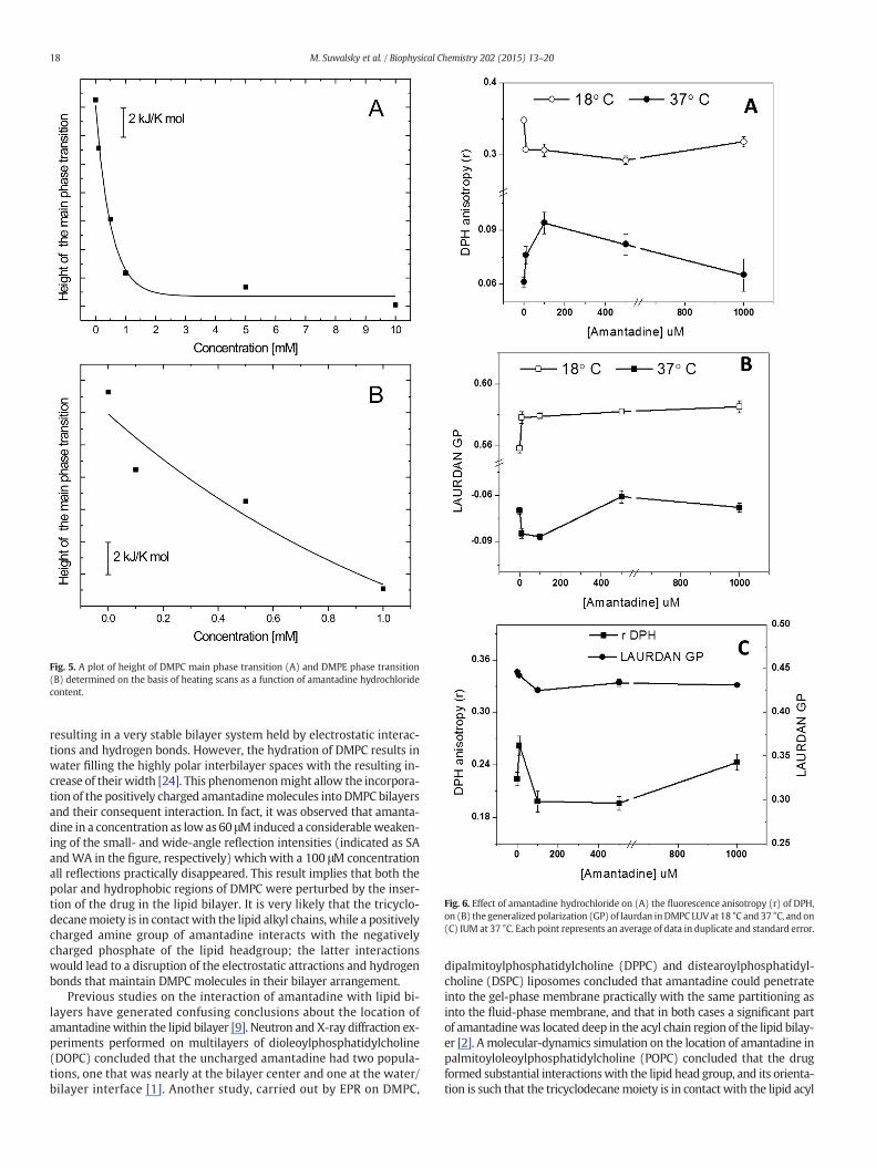
Fig. 5. A plot of height of DMPC main phase transition (A) and DMPE phase transition(B) determined on the basis of heating scans as a function of amantadine hydrochloridecontent.
Fig. 6. Effect of amantadine hydrochloride on (A) the fluorescence anisotropy (r) of DPH,on (B) the generalizedpolarization (GP) of laurdan inDMPC LUVat 18 °C and 37 °C, andon(C) IUM at 37 °C. Each point represents an average of data in duplicate and standard error.
18 M. Suwalsky et al. / Biophysical Chemistry 202 (2015) 13–20
resulting in a very stable bilayer system held by electrostatic interac-tions and hydrogen bonds. However, the hydration of DMPC results inwater filling the highly polar interbilayer spaces with the resulting in-crease of their width [24]. This phenomenonmight allow the incorpora-tion of the positively charged amantadinemolecules into DMPC bilayersand their consequent interaction. In fact, it was observed that amanta-dine in a concentration as low as 60 μM induced a considerableweaken-ing of the small- and wide-angle reflection intensities (indicated as SAandWA in the figure, respectively) which with a 100 μM concentrationall reflections practically disappeared. This result implies that both thepolar and hydrophobic regions of DMPC were perturbed by the inser-tion of the drug in the lipid bilayer. It is very likely that the tricyclo-decanemoiety is in contactwith the lipid alkyl chains, while a positivelycharged amine group of amantadine interacts with the negativelycharged phosphate of the lipid headgroup; the latter interactionswould lead to a disruption of the electrostatic attractions and hydrogenbonds that maintain DMPC molecules in their bilayer arrangement.
Previous studies on the interaction of amantadine with lipid bi-layers have generated confusing conclusions about the location ofamantadine within the lipid bilayer [9]. Neutron and X-ray diffraction ex-periments performed on multilayers of dioleoylphosphatidylcholine(DOPC) concluded that the uncharged amantadine had two popula-tions, one that was nearly at the bilayer center and one at the water/bilayer interface [1]. Another study, carried out by EPR on DMPC,
dipalmitoylphosphatidylcholine (DPPC) and distearoylphosphatidyl-choline (DSPC) liposomes concluded that amantadine could penetrateinto the gel-phase membrane practically with the same partitioning asinto the fluid-phase membrane, and that in both cases a significant partof amantadinewas located deep in the acyl chain region of the lipid bilay-er [2]. Amolecular-dynamics simulation on the location of amantadine inpalmitoyloleoylphosphatidylcholine (POPC) concluded that the drugformed substantial interactionswith the lipid head group, and its orienta-tion is such that the tricyclodecanemoiety is in contact with the lipid acyl

Fig. 7. Effect of amantadine on the morphology of human erythrocytes. Images obtained by scanning electron microscopy (SEM) of (A) control, (B) incubated with 50 μM, and (C) with100 μM amantadine hydrochloride.
19M. Suwalsky et al. / Biophysical Chemistry 202 (2015) 13–20
chains, and the ammonium group is in contact with the lipid head group,in particular the glycerol oxygens [3]. Our X-ray diffraction results tend toagreewith the conclusion reachedbyChewet al. [3]. However,webelievethat phosphate instead of glycerol oxygens interact with the amantadinecharged amine group; as explained above, this interaction would be re-sponsible for the structural perturbation induced by the drug to DMPCpolar region. Moreover, our fluorescence spectroscopy results coincidedin that amantadine located in both the polar head and acyl chain regionsof DMPC in both the gel and liquid crystalline phases.
To the best of our knowledge, little is reported about thermotropicbehavior of membrane models in the presence of adamantanes, andeven more so there are no DSC studies on the influence of amantadineon phospholipids. Calorimetric measurements performed on DMPC li-posomes showed that amantadine was capable of distortion of bothpretransition and main phase transition of lipids that form a bilayer.As a small molecule, with its experimentally determined log P value of2.44 [29], amantadine is a very lipophilic compound and could pene-trate easily into the core of the lipid bilayer. This leads to reduction of in-terfacial tension and affects the lateral interaction between the apolarfatty acid chains. The presence of the polar head group on the phospho-lipids gives rise to specific and direct interactions between DMPCmole-cules and amine group of a drug. As a result, we observed the decreaseof molar heat capacity and broadening of the main phase transition aswell as its shift to the lower values. Significantly, smaller effect of aman-tadine on DMPE liposomes shown in the calorimetric studies was prob-ably related to the tight packing of this lipid,whosehead group occupiesa molar area smaller by about 8 Å2 in comparison with that of DMPC[30]. BeingdistributedwithinDMPCbilayer homogenously, amantadinemolecules despite small size and lipophilic properties were unable topenetrate the hydrophobic core of DMPE. It can then be assumed thatsmaller miscibility of amantadine with DMPE liposomes may arisefrom the fact that van der Waals interactions between the lipid acylchains themselves are much stronger than those in which amantadinemolecules participate.
SEM in vitro observations showed that amantadine induced mor-phological alterations to human red cells from their normal discoidshape to echinocytes. According to the bilayer couple hypothesis [31,32] shape changes induced in erythrocytes by foreign molecules aredue to differential expansion of the two monolayers of the red cellmembrane. Thus, cup-shaped stomatocytes are formed when the com-pound inserts into the inner monolayer whereas spiculated-shapedechinocytes are produced when it locates into the outer moiety. Thefinding that amantadine induced the formation of echinocytes indicatesthat it was inserted in the outer leaflet of the erythrocyte membrane.This conclusion is supported by X-ray and calorimetric experimentscarried out in DMPC and DMPE bilayers, In fact, results showed thatamantadinemostly interactedwith DMPC,which is preferentially locatedin the outer monolayer of the human erythrocyte membrane. There arevery scanty reports related tomorphological changes induced by amanta-dine to human erythrocytes. In one of them, Tverdislov et al. [10] foundthat amantadine induced the formation of stomatocytes. The differencewith our results might be due to the higher drug concentration they
used (2.66 mM and 5.32 mM). In general, plasma amantadine levelsrange from about 7 μM after therapeutic doses [33], about 16 μM inpatients with toxic symptoms, and up to about 0.13mM in lethal concen-tration [34]. It is important to emphasize that the effects of amantadinedetected in the present work, particularly at the erythrocyte membranelevel, were observed at a concentration range of 10–50 μM, which issomewhat higher than the plasma therapeutic concentration. How-ever, it must be taken into account that these were biophysical stud-ies in vitro carried out, which usually require high concentrations inorder to detect drug effects.
In conclusion, our experimental findings are certainly of interest asthey have demonstrated that amantadine interacts with the humanerythrocyte membrane affecting the cell morphology. It must be con-sidered that alteration of the normal biconcave shape of red blood cells in-creases their resistance to entry into capillaries, which could contribute toa decreased bloodflow, loss of oxygen, and tissue damage throughmicro-vascular occlusion [35,36]. Functions of ion channels, receptors and en-zymes immersed in cell membrane lipid moieties also might be affected.
Acknowledgments
To C.P. Sotomayor, L.F. Aguilar and F. Neira for technical assis-tance. This work was supported by FONDECYT (project 1130043).Calorimetric measurements were carried out using the instrumentpurchased thanks tofinancial support of EuropeanRegional DevelopmentFund (contract no. POIG.02.01.00-12-167/08, project Malopolska Centreof Biotechnology).
References
[1] K.C. Duff, A.J. Cudmore, J.P. Bradshaw, The location of amantadine hydrochloride andfree base within phospholipid mltilayers: a neutron and X-ray diffraction study,Biochim. Biophys. Acta 1145 (1993) 149–156.
[2] W.K. Subczynski, J. Wojas, V. Pezeshk, A. Pezeshk, Partitioning and localization ofspin-labeled amantadine in lipid bilayers: an EPR study, J. Pharm. Sci. 87 (1998)1249–1254.
[3] C.F. Chew, A. Guy, P.C. Biggin, Distribution and dynamics of adamantanes in a lipidbilayer, Biophys. J. 95 (2008) 5627–5636.
[4] M. Föller, C. Geiger, H. Mahmud, J. Nicolay, F. Lang, Stimulation of suicidal erythro-cyte death by amantadine, Eur. J. Pharmacol. 581 (2008) 13–18.
[5] N. Nishikawa, M. Nagai, T. Moritoyo, H. Yabe, M. Nomoto, Parkinsonism and relateddisorders, Parkinsonism Relat. Disord. 15 (2009) 351–353.
[6] L.H. Pino, L.J. Holsinger, R.A. Lamb, Influenza virus M2 protein has ion channel activ-ity, Cell 69 (1992) 517–528.
[7] J. Wang, J.R. Schnell, J.J. Chou, Amantadine partition and localization in phospholipidmembrane: a solution NMR study, Biochem. Biophys. Res. Commun. 324 (2004)212–217.
[8] R.M. Epand, R.F. Epand, R.C. McKenzie, Effects of viral chemotherapeutic agents onmembrane properties. Studies of cyclosporine A, benzyloxycarbonyl-D-Phe-L-Phe-Gly and amantadine, J. Biol. Chem. 262 (1987) 1526–1529.
[9] C. Li, M. Yi, J. Hu, H.-X. Zhou, T.A. Cross, Solid-state and MD simulations of the anti-viral drug amantadine solubilized in DMPC bilayers, Biophys. J. 94 (2008)1295–1302.
[10] V.A. Tverdislov, S. El-Karadagi, I.G. Kharitonenkov, R. Glaser, E. Donath, A. Herrmann,P. Lentzsch, J. Donath, Interaction of the antivirus agents remantadine and amanta-dine with lipid membranes and the influence on the curvature of human red cells,Gen. Physiol. Biophys. 5 (1986) 61–75.

20 M. Suwalsky et al. / Biophysical Chemistry 202 (2015) 13–20
[11] J.Y. Chen, W.H. Huestis, Role of membrane lipid distribution in chlorpromazine-induced shape change of human erythrocytes, Biochim. Biophys. Acta 1323(1997) 299–309.
[12] J.M. Boon, B.D. Smith, Chemical control of phospholipid distribution across bilayermembranes, Med. Res. Rev. 22 (2000) 251–281.
[13] P.F. Devaux, A. Zachowsky, Maintenance and consequences of membrane phospho-lipids asymmetry, Chem. Phys. Lipids 73 (1994) 107–120.
[14] M. Suwalsky, I. Sánchez, M. Bagnara, C.P. Sotomayor, Interaction of antiarrhythmicdrugs with model membranes, Biochim. Biophys. Acta 1195 (1994) 189–196.
[15] M. Suwalsky, J. Belmar, F. Villena, M.J. Gallardo, M. Jemiola-Rzeminska, K. Strzalka,Acetylsalicylic acid (aspirin) and salicylic acid interaction with the human erythro-cyte membrane bilayer induce in vitro changes in the morphology of erythrocytes,Arch. Biochem. Biophys. 539 (2013) 9–19.
[16] M. Suwalsky, P. Zambrano, S. Mennickent, F. Villena, C. Sotomayor, L.F. Aguilar, S.Bolognin, Effects of phenylpropanolamine (PPA) on in vitro human erythrocytemembranes and molecular models, Biochem. Biophys. Res. Commun. 406 (2011)320–325.
[17] M. Suwalsky, M. Manrique, F. Villena, C.P. Sotomayor, Structural effects in vitro ofthe anti-inflammatory drug diclofenac on human erythrocytes and molecularmodels of cell membranes, Biophys. Chem. 141 (2009) 34–40.
[18] T. Parasassi, E. Gratton, Membrane lipid domains and dynamics as detected bylaurdan fluorescence, J Fluoresc. 5 (1995) 59–69.
[19] J.T. Dodge, C. Mitchell, C.D. Hanahan, The preparation and chemical characterizationof haemoglobin-free ghosts of human erythrocytes, Arch. Biochem. Biophys. 100(1963) 119–130.
[20] J.R. Lakowicz, Principles of Fluorescence Spectroscopy, Plenum, New York, 1999.[21] T. Parasassi, G. De Stasio, A. D'Ubaldo, E. Gratton, Phase fluctuation in phospholipid
membranes revealed by laurdan fluorescence, Biophys. J. 57 (1990) 1179–1186.[22] B. Zimmermann, D.M. Soumpasis, Effects of monovalent cations on red cell shape
and size, Cell Biophys. 7 (1985) 115–127.[23] S.V.P. Malheiros, M.A. Brito, D. Brites, M.N. Correa, Membrane effects of trifluopera-
zine, dibucaine and praziquantel on human erythrocytes, Chem. Biol. Interact. 126(2000) 79–95.
[24] M. Suwalsky, Phospholipid bilayers, in: J.C. Salamone (Ed.)Polymeric Materials En-cyclopedia, vol. 7, CRC, Boca Raton, FL 1996, pp. 5073–5078.
[25] B. Tenchov, On the reversibility of the phase transitions in lipid–water systems,Chem. Phys. Lipids 57 (1991) 165–177.
[26] D. Marsh, General features of phospholipid phase transitions, Chem. Phys. Lipids 57(1991) 109–120.
[27] R. Koynova, M. Caffrey, Phases and phase transitions of the phosphatidylcholines,Biochim. Biophys. Acta 1376 (1998) 91–145.
[28] R.N. Lewis, R.N. McElhaney, Calorimetric and spectroscopic studies of the polymorphicphase behavior of a homologous series of n-saturated 1,2-diacyl phosphatidylethanol-amines, Biophys. J. 64 (1993) 1081–1096.
[29] http://www.drugbank.ca.[30] A. Sujak, K. Strzalka, W.I. Gruszecki, Thermotropic phase behaviour of lipid bilayers
containing carotenoid pigment canthaxanthin: a differential scanning calorimetrystudy, Chem. Phys. Lipids 145 (2007) 1–12.
[31] M.P. Sheetz, S.J. Singer, Biological membranes as bilayer couples. A molecular mech-anism of drug-erythrocyte induced interactions, Proc. Natl. Acad. Sci. U. S. A. 71(1974) 4457–4461.
[32] G. Lim, M. Wortis, Stomatocyte–discocyte–echinocyte sequence of the human redblood cell: evidence for the bilayer-couple hypothesis from membrane mechanics,Proc. Natl. Acad. Sci. U. S. A. 99 (2002) 16766–16769.
[33] F.G. Hayden, H.E. Hoffman, D.A. Spyker, Differences in side effects of amantadine hy-drochloride and rimantadine hydrochloride relate to differences in pharmacokinet-ics, Antimicrob. Agents Chemother. 23 (1983) 458–464.
[34] R. Regenthal, M. Krueger, C. Koeppel, R. Preiss, Drug levels: therapeutic and toxicserum/plasma concentrations of common drugs, J. Clin. Monit. Comput. 15 (1999)529–544.
[35] S.L. Winski, D.E. Carter, Arsenate toxicity in human erythrocytes: characterization ofmorphological changes and determination of the mechanism of damage, J. Toxicol.Environ. Health A 53 (1998) 345–355.
[36] S. Svetina, D. Kuzman, R.E. Waugh, P. Zibert, B. Zeks, The cooperative role of mem-brane skeleton and bilayer in the mechanical behaviour of red blood cells,Bioelectrochemistry 62 (2004) 107–113.