THE EFFECT OF WONDERFUL COLA ON LIVER HISTOLOGY IN ALLOXAN INDUCED DIABETIC WISTAR RATS

The in-vitro effects of alloxan and the product of its reduction dialuric acid (alone or in combination withcopper ions) on lipid peroxidation, carbonyl content, GSH level and antioxidant enzyme activities in ratliver and kidney have been studied. The effects of Cu2+/alloxan and Cu2+/dialuric acid were comparedwith those of Fe3+/alloxan and Fe3+/dialuric acid. Unlike alloxan, dialuric acid increased liver and kidneylipid peroxidation; similar effects were registered in the presence of Fe3+. In the presence of Cu2+/dialuricacid, the lipid peroxidation was strongly inhibited and vice versa – the liver protein oxidation wasincreased. Alloxan and dialuric acid, as well as their combinations with Fe3+ had no effect on the totalGSH level. Both substances did not affect the Cu2+-induced changes in GSH level, glucose-6-phosphatedehydrogenase and gluthatione reductase activities. In contrast, Cu2+ had no effect on dialuric-acidinduced changes in gluthatione peroxidase and superoxide dismutase activities. The present in vitroresults, concerning the metal dependence of the effects of alloxan and dialuric acid, are a premise forin vivo study of alloxan effects in metal-loaded animals.
Keywords: Alloxan – dialuric acid – copper – lipid peroxidation – antioxidant enzymes
INTRODUCTION
The cytotoxic action of alloxan (A), used for inducing of experimental diabetes, isconnected with the participation of reactive oxygen species (ROS); A is reduced todialuric acid (AH2), the oxidation of the latter leading to production of alloxan radi-cal (AH.), superoxide radical (O2
–) and hydrogen peroxide (H2O2) [7, 13, 20]. In thepresence of transition metals (mainly iron), a production of hydroxyl radicals (.OH)is also registered [24; this cell-damaging radical is believed to be the main toxicagent in A-induced diabetes, 7, 8, 16, 20, 32.]
Acta Biologica Hungarica 58 (4), pp. 359–367 (2007)DOI: 10.1556/ABiol.58.2007.4.3
0236-5383/$ 20.00 © 2007 Akadémiai Kiadó, Budapest
IN VITRO EFFECTS OF ALLOXAN/COPPERCOMBINATIONS ON LIPID PEROXIDATION,
PROTEIN OXIDATION AND ANTIOXIDANT ENZYMESALBENA ALEXANDROVA,* L. PETROV, MILA KESSIOVA and MARGARITA KIRKOVA
Institute of Physiology, Bulgarian Academy of Sciences,23 Acad. G. Bonchev St., 1113 Sofia, Bulgaria
(Received: September 5, 2006; accepted: December 15, 2006)
* Corresponding author; e-mail: [email protected]
360 ALBENA ALEXANDROVA et al.
Acta Biologica Hungarica 58, 2007
Using the deoxyribose test [12], we found that the AH2-dependent deoxyribosedegradation was changed in a different manner by the presence of metal ions (Fe3+,Cu2+, V5+ or V4+), H2O2, metal reducers or metal chelators [1, 17]. The copper effectson A- and AH2-induced inhibition of O2
–-provoked nitro blue tetrazolium (NBT)reduction [2] and the vanadium effects onAH2-induced lipid peroxidation in rat liverand kidney [3] were also found to differ from those of iron. In addition, an insulin-mimetic activity of some metals (vanadium, lithium, selenium, molybdenum) hasbeen reported [5, 21, 22, 25, 28–30]. All these findings justify our investigations onthe role of the different transition metals in the A-action.
The aim of this study was to investigate the in vitro effects of alloxan and itsreduction product – dialuric acid, on lipid peroxidation, protein oxidation and antiox-idant enzyme activities in rat liver and kidney preparations in the presence of copper.The results were compared with those, observed in the presence of iron.
MATERIALS AND METHODS
Chemicals
Alloxan was purchased from BDH; dialuric acid – from Riedel-de Haen AG; NBT –from Serva; H2O2 and riboflavin – from Merck. All other reagents were analyticalgrade; all solutions were prepared with de-ionized water.
Animals
Male Wistar rats, weighing 180–200 g, were given free access to tap water and astandard lab diet. The animals were starved 24 hr and then killed under light etheranaesthesia.
Tissue preparations
Liver, perfused with drilled 0.15 M KCl, and kidney were homogenized in drilled0.15 M KCl –10 mM potassium phosphate buffer, pH 7.4. After centrifugation of the10% liver and kidney homogenates at 600 g for 10 min, “post-nuclear” preparationswere obtained. Part of these homogenates and the supernatants, obtained after cen-trifugation of homogenates at 12,000 g for 20 min (“post mitochondrial” super-natant), were used in the present experiments; samples (2 mg protein/ml buffer)were incubated for 15 min at 0 °C in the presence and in the absence of adding allox-an, dialuric acid, FeCl3 or CuCl2.

Analytical methods
Protein content was measured by the method of Lowry et al. [19].Protein carbonyl content (in “post-nuclear” homogenates) was measured at 366
nm, according to Reznick and Parker [27].Lipid peroxidation (LP) was determined by the amount of thiobarbituric acid
reactive substances (TBARS), formed in fresh “post-nuclear” homogenates (mgprotein/ml) after 60 min-incubation at 37 °C and expressed in nmoles malondialde-hyde (MDA)/mg protein [15]. The 600 nm absorbance was considered to be a non-specific baseline and was subtracted from A532.
Glutathione (GSH) level in “post-nuclear” homogenates was measured at 412 nmby the method of Tietze [33].
Glutathione peroxidase (GSH-PER) activity in “post-mitochondrial” supernatant(with t-butylhydroperoxide as a substrate) was measured by the method of Gunzleret al. [9].
Glutathione reductase (GSSG-RED) activity in “post-mitochondrial” supernatantwas measured by the method of Pinto and Bartley [26].
Glucose-6-phosphate dehydrogenase (Glu-6-P-DH) activity in “post-mitochond-rial” supernatant was determined after Cartier et al. [6].
Superoxide dismutase (SOD) activity in “post-mitochondrial” supernatant wasdetermined according to Beauchamp and Fridovich [4]. The samples were lighted for5 min with a 250-W HgL lamp and the inhibition of O2
–-provoked nitro blue tetra-zolium (NBT) reduction was measured at 560 nm; one unit of SOD activity is theamount of the enzyme, inhibiting the NBT reduction by 50%.
Statistical analysis
The results were statistically processed by Student t-test; P < 0.05 being accepted asthe minimum level of statistical significance of the established differences. The num-ber of rats used during the experiments is given under the tables and figure.
The experiments have been performed according to the “Principles of laboratoryanimal care” (NIH publication No. 85-23, revised 1985), and the rules of the EthicsCommittee of the Institute of Physiology, Bulgarian Academy of Sciences (registra-tion FWA 00003059 by the US Department of Health and Human Services).
RESULTS
AH2, the reduction product of A, increased the amount of TBARs (an index of lipidperoxidation) formed in rat liver and kidney homogenates (Table 1); the A-effectwas similar to that of AH2, but only in the presence of GSH (a metal and alloxanreducer). Both AH2
– and A/GSH-induced LP was intensified after Fe3+-, but not afterCu2+-addition to the reaction mixture. Unlike Fe3+ (a good LP-inductor), Cu2+ (in the
Alloxan/copper combinations and antioxidant system 361
Acta Biologica Hungarica 58, 2007

362 ALBENA ALEXANDROVA et al.
Acta Biologica Hungarica 58, 2007
concentration used) strongly decreased the TBARs formation in both tissue prepara-tions, AH2 and A/GSH failing to change it.AH2 increased the carbonyl content (an index of protein oxidation) in liver, but the
effect of AH2/Cu2+ was stronger (Fig. 1). The Cu2+/AH2 effect on protein oxidation
Table 1Effects of alloxan and dialuric acid on lipid peroxidation
Additions – 100 µM Fe3+ 100 µM Cu2+
Liver lipid peroxidationControls 1.8 ± 0.09 5.0 ± 0.14* 0.3 ± 0.03*+100 µM alloxan 2.0 ± 0.14 5.6 ± 0.19* 0.3 ± 0.02*+100 µM dialuric acid 3.4 ± 0.10* 7.9 ± 0.16** 0.3 ± 0.03*+100 µM alloxan/100 µM GSH 3.2 ± 0.07* 7.4 ± 0.37** 0.4 ± 0.03*+100 µM GSH 1.0 ± 0.08* 5.2 ± 0.23* 0.3 ± 0.04*
Kidney lipid peroxidationControls 1.1 ± 0.12 2.1 ± 0.17* 0.3 ± 0.03*+100 µM alloxan 1.4 ± 0.11 2.2 ± 0.22* 0.3 ± 0.02*+100 µM dialuric acid 2.4 ± 0.11* 3.6 ± 0.16** 0.4 ± 0.07*+100 µM alloxan/100 µM GSH 1.7 ± 0.04* 4.8 ± 0.08** 0.3 ± 0.03*+100 µM GSH 1.3 ± 0.14 3.6 ± 0.15** 0.3 ± 0.03*
The values represent the mean ± SEM of 7 animals and are expressed in nmoles MDA/mg protein,using a molar extinction coefficient of 1.56 × 10⁵M–1cm–1. Statistically significant differences versus cor-responding controls (*in the absence of metal ions and **in the presence of metal ions) at P < 0.05.
Fig. 1. Effects of dialuric acid on carbonyl content in rat liver. The values represent the mean ± SEM of5 animals and are expressed in nmoles carbonyls/mg protein, using a molar extinction coefficient
of 2.2×10⁴M–1cm–1. *Statistically significant differences versus controls at P < 0.05

Alloxan/copper combinations and antioxidant system 363
Acta Biologica Hungarica 58, 2007
Table 2Effects of alloxan and dialuric acid on GSH level in rat tissues
Additions – 100 µM Fe3+ 100 µM Cu2+
Liver GSH levelControls 3131 ± 195 2721 ± 220 2434 ± 104*+100 µM alloxan 2836 ± 119 2843 ± 292 2688 ± 164*+100 µM dialuric acid 2864 ± 225 2787 ± 221 2463 ± 226*
Kidney GSH levelControls 1139 ± 65 1338 ± 149 897 ± 54*+100 µM alloxan 1201 ± 101 1191 ± 131 854 ± 91*+100 µM dialuric acid 1066 ± 42 979 ± 114 871 ± 62*
The values represent the mean ± SEM of 7 animals and are expressed in ng GSH/mg protein.* Statistically significant differences versus controls (in the absence of metal ions) at P < 0.05.
Table 3Effects of alloxan and dialuric acid on antioxidant enzyme activities in rat liver
Additions Controls 100 µM Fe3+ 100 µM Cu2+
GSH-PER activityControls 218 ± 13.4 219 ± 11.3 207 ± 17.1+100 µM alloxan 5* 9* 9*+100 µM dialuric acid 4* 7* 7*
GSSG-RED activityControls 36 ± 2.5 39 ± 4.6 28 ± 1.9*+100 µM alloxan 33 ± 4.2 38 ± 4.2 29 ± 1.8*+100 µM dialuric acid 43 ± 3.2 44 ± 5.7 18 ± 1.4**
Glu-6-P-DH activityControls 29 ± 2.9 29 ± 2.5 14 ± 2.2*+100 µM alloxan 33 ± 4.2 28 ± 3.4 15 ± 1.6*+100 µM dialuric acid 38 ± 4.9 34 ± 4.0 13 ± 1.2*
SOD activityControls 74 ± 5.6 80 ± 3.7 92 ± 6.8+100 µM alloxan 155 ± 5.5* 144 ± 9.2* 136 ± 11.9*+100 µM dialuric acid 131 ± 15.9* 133 ± 12.1* 132 ± 11.5*
The values represent the mean ± SEM of 7 animals. GSH-PER, GSSG-RED and Glu-6-P-DH activi-ties are expressed in in nmoles NADP(H)/min/mg protein, using a molar extinction coefficient of6.22 × 106M–1cm–1 and SOD is expressed in U/mg protein. Statistically significant differences versus cor-responding controls (* in the absence of metal ions and ** in the presence of metal ions) at P < 0.05.
364 ALBENA ALEXANDROVA et al.
Acta Biologica Hungarica 58, 2007
was similar to that of Cu2+/H2O2; it might be mentioned that the latter combinationpresents Fenton-like system, generating .OH radicals.
The effects of A and AH2 on GSH-level and the antioxidant enzyme activities inliver and kidney preparations in the absence and in the presence of Fe3+ and Cu2+
were also measured. A and AH2, alone or in combinations with Fe3+, had no effecton the total GSH level; besides, the both substances did not change the Cu2+-induceddecrease of GSH level (Table 2). Unlike Fe3+, Cu2+ inhibited Glu-6-P-DH andGSSG-RED activity but this Cu2+-induced inhibition was not affected by A and AH2;only an additional decrease of Glu-6-P-DH activity in the presence of AH2 wasobserved (Table 3). Conversely, Cu2+ had no effect on A- and AH2-induced decreaseof GSH-PER activity and AH2-induced increase of SOD activity.
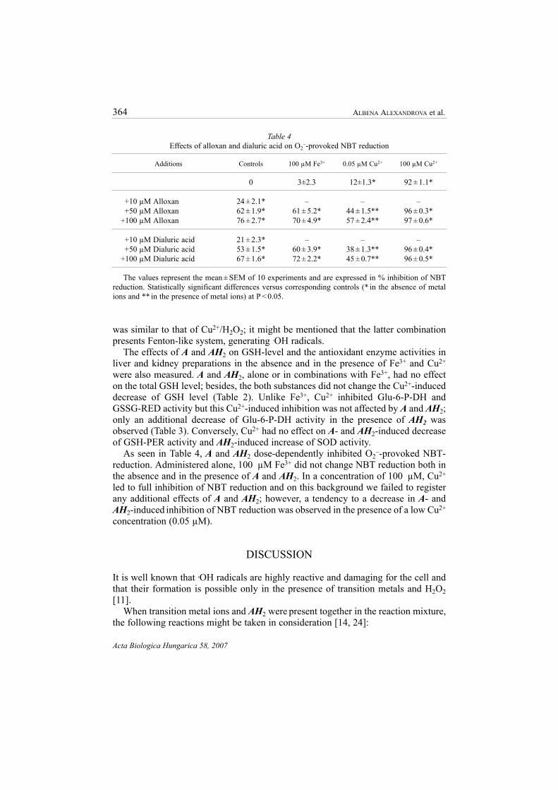
As seen in Table 4, A and AH2 dose-dependently inhibited O2–-provoked NBT-
reduction. Administered alone, 100 µM Fe3+ did not change NBT reduction both inthe absence and in the presence of A and AH2. In a concentration of 100 µM, Cu2+
led to full inhibition of NBT reduction and on this background we failed to registerany additional effects of A and AH2; however, a tendency to a decrease in A- andAH2-induced inhibition of NBT reduction was observed in the presence of a low Cu2+
concentration (0.05 µM).
DISCUSSION
It is well known that .OH radicals are highly reactive and damaging for the cell andthat their formation is possible only in the presence of transition metals and H2O2[11].
When transition metal ions and AH2 were present together in the reaction mixture,the following reactions might be taken in consideration [14, 24]:
Table 4Effects of alloxan and dialuric acid on O2
–-provoked NBT reduction
Additions Controls 100 µM Fe3+ 0.05 µM Cu2+ 100 µM Cu2+
0 3±2.3 12±1.3* 92 ± 1.1*
+10 µM Alloxan 24 ± 2.1* – – –+50 µM Alloxan 62 ± 1.9* 61 ± 5.2* 44 ± 1.5** 96 ± 0.3*
+100 µM Alloxan 76 ± 2.7* 70 ± 4.9* 57 ± 2.4** 97 ± 0.6*
+10 µM Dialuric acid 21 ± 2.3* – – –+50 µM Dialuric acid 53 ± 1.5* 60 ± 3.9* 38 ± 1.3** 96 ± 0.4*
+100 µM Dialuric acid 67 ± 1.6* 72 ± 2.2* 45 ± 0.7** 96 ± 0.5*
The values represent the mean ± SEM of 10 experiments and are expressed in % inhibition of NBTreduction. Statistically significant differences versus corresponding controls (* in the absence of metalions and ** in the presence of metal ions) at P < 0.05.

AH2 + O2→ O2– + AH. + H+
AH2 + O2– + H+ → AH.+ H2O2
AH. + O2– + H+ → A + H2O2
AH2 + Men+1 → AH. + Men + H+
AH. + Men+1 → A. + Men + H+
Men + H2O2 → Men+1 + OH– + .OH
In O2-presence, the formed .OH radicals lead to peroxidation of numerous organ-ic molecules, including proteins [10, 11]; these radicals attack the side chains ofamino acid residues at the metal binding site, leading to protein modification andincrease in carbonyl content [31]. Hence, the increase of AH2-evoked carbonyl con-tent in Cu2+ presence (Fig. 1) might be explained with .OH radicals formation. Theidentity in the effects of Cu2+/ AH2 and Cu2+/H2O2 (Fenton-like system, generating.OH radicals) on liver carbonyl content is an additional confirmation for .OH-forma-tion.
It is known that copper ions display high affinity for both NH2- and SH-groups,occurring in proteins. Specialized proteins containing clusters of these groups trans-port and store copper ions, hampering their potential toxicity; this mechanism maybe overwhelmed under copper-overload conditions, in which copper ions bind to theSH-groups of proteins, non-related to copper metabolism. Thus, indiscriminate cop-per binding may damage protein structure, modifying its biological functions [18]. Itmight be suggested that the protein damage induced by copper in µM concentrationsis a reason for the decreased activity of GSH transferase [18], GSH reductase andGlucose-6-P-DH (Table 3). In addition, the results presented on Tab. 4 suggested thatthe increase in tissue SOD activity in the presence of A and AH2 (Table 3) is puta-tive (SOD-like activity). These findings must be taken into consideration, when dis-cussing the in-vitro effects of other redox-cycling substances, like A/AH2, on tissueSOD activity.
Probably, the strong inhibition of liver and kidney lipid peroxidation in the pres-ence of Cu2+ or Cu2+/AH2 (Table 1) is due to the high Cu2+-concentration (100 µM)used in the present study. This is in accordance with data of Letelier et al. [18] thatCu2+ in nM concentrations range induces a significant lipoperoxidation, while it isnon-detectable at increasing in Cu2+-concentrations to ≥50 µM. Unlike Cu2+/AH2,the combination of AH2 (O2
–-generating system) with Fe3+ led to a higher formationof TBARs in liver and kidney than AH2 and Fe3+ alone (Table 1). This is in accor-dance with the results obtained in the system – xanthine oxidase + ADP/Fe3+, wherethe O2
– and H2O2, produced from xanthine oxidase support iron-catalyzed lipid per-oxidation through their participation in redox reactions of iron [23].
In conclusion, the present in-vitro results appeared to be a premise for studyingand comparing the in-vivo effects of alloxan in animals, pretreated with copper oriron. Thus, further in-vitro and in-vivo studies could be useful for understanding therole of these metal ions in A-induced diabetes.
Alloxan/copper combinations and antioxidant system 365
Acta Biologica Hungarica 58, 2007

366 ALBENA ALEXANDROVA et al.
Acta Biologica Hungarica 58, 2007
ACKNOWLEDGMENTS
This study was supported by Grant MU-B-1001 from the National Research Fund, Bulgaria.
REFERENCES
1. Alexandrova, A., Georgieva, A., Kirkova, M. (2006) Alloxan and dialuric acid. Effects on .OH-pro-voked degradation of deoxyribose in the presence of different metal ions. C.R. Acad. Bulg. Sci. 59,305–312.
2. Alexandrova, A., Kessiova, M., Tsvetanova, E., Kirkova, M. (2006) Alloxan. Effects on O2–-pro-
voked inhibition of nitro-blue tetrazolium reduction in the presence of different metal ions. C.R.Acad. Bulg. Sci. 59, 201–206.
3. Alexandrova, A., Kirkova, M., Russanov, E. (1998) In vitro effects of alloxan-vanadium combina-tion on lipid peroxidation and on antioxidant enzyme activity. Gen. Pharmac. 31, 489–493.
4. Beauchamp, C., Fridovich, I. (1971) Superoxide dismutase: Improved assays and assay applicable toacrylamide gels. Anal. Biochem. 44, 276–287.
5. Becker, D. J., Reul, B., Ozcelikay, A. T., Buchet, J. P., Henquin, J. C., Brichard, S. M. (1996) Oralselenates improves glucose homeostasis and partly reverses abnormal expression of liver glycolyticand gluconeogenesis enzymes in diabetic rats. Diabetologia 39, 3–11.
6. Cartier, P., Leroux, J. P., Marchand, J. Cl. (1967) Techniques de dosage des enzymes glycocytiquestissulaires. Ann. Biol. Clin. 25, 109–136.
7. Fischer, L. J., Harman, A. W. (1982) Oxygen free radicals and diabetogenic action of alloxan. In:Author A. P. (ed.) Pathology of oxygen. Academic Press, New York. p. 261.
8. Grankvist, K., Marklund, S. L., Schlin, J., Taljedal, I. B. (1979) Superoxide dismutase, catalase andscavengers of hydroxyl radical protect against the toxic action of alloxan on pancreatic islet cells invitro. Biochem. J. 182, 17–25.
9. Gunzler, W. A., Vergin, H., Muller, I., Flohe, L. (1972) Glutathion peroxidase. VI. Die reaction derglutathion peroxidase mit Verschieden hydroperoxiden. Hoppe-Seyler’s Z. Physiol. Chem. 353,1001–1004.
10. Halliwell, B., Gutteridge, J. M. C. (1985) Free radicals in biology and medicine. Calderon Press,Oxford.
11. Halliwell, B., Gutteridge, J. M. C. (1984) Oxygen toxicity, oxygen radicals, transition metals and dis-ease. Biochem. J. 219, 1–14.
12. Halliwell, B., Gutteridge, J. M. C., Aruoma, O. I. (1987) The deoxyribose method: a simple “test-tube” assay for determination of free constants for reactions of hydroxyl radicals. Anal. Biochem.165, 215–219.
13. Heikkila, R. E., Winston, B., Cohen, G., Barden, H. (1976) Alloxan-induced diabetes: evidence forhydrohyl radical as a cytotoxic intermediate. Biochem. Pharmac. 25, 1085–1092.
14. Houee-Levin, C., Gardes-Albert, M., Ferradini, C., Pucheault, J. (1981) Radiolysis study of the allox-an-dialuric acid couple II: the autooxidation of dialuric acid. Radiat. Res. 88, 20–28.
15. Hunter, F., Gebinski, J., Hoffstein, P., Weinstein, J., Scott, A. (1963) Swelling and lysis of rat livermitochondria by ferrous ions. J. Biol. Chem. 238, 828–835.
16. Ishibashi, F., Howard, B. V. (1981) Alloxan and H2O2 action on glucose metabolisis in culturedfibroblasts. Generation of oxygen – containing free radicals as a mechanism of alloxan action. J. Biol.Chem. 256, 12134–12139.
17. Kirkova, M., Karakashev, P., Russanov, E. (1998) Hydroxyl radicals production in the vanadiumions/dialuric acid systems. Gen. Pharmac. 31, 247–251.
18. Letelier, M. E., Lepe, A. M., Faundez, M., Salazar, J., Marin, R., Aracena, P., Speiski, H. (2005)Possible mechanisms underlying copper-induced damage in biological membranes leading to cellu-lar toxicity. Chem. Biol. Interact. 151, 71–82.
19. Lowry, O. H., Rosenbrough, N. J., Farr, A. L., Randal, R. J. (1951) Protein measurement with theFolin phenol reagent. J. Biol. Chem. 193, 265–278.
20. Malaisse, W. J. (1982) Alloxan toxicity to the pancreatic β-cell. A new hypothesis. Biochem.Pharmac. 31, 3527–3534.
21. McNeill, J. H., Delgatty, H. L. M., Battell, M. L. (1991) Insulin-like effects of sodium selenate instreptozotocin induced diabetic rats. Diabetes 40, 1675–1678.
22. Meyerovitch, J., Farfel, Z., Sack, J., Schechter, Y. (1987) Oral administration of vanadate normalizesblood glucose levels in streptozotocin-treated rats. J. Biol. Chem. 262, 6658–6662.
23. Miller, D. M., Grover, A., Nayini, N., Aust, S. D. (1993) Xanthine oxidase- and iron-dependent lipidperoxidation. Arch. Biochem. Biophys. 301, 1–7.
24. Munday, R. (1988) Effects of transition metals on the reaction rate and on the generation of “activeoxygen” species. Biochem. Pharmac. 37, 409–413.
25. Ozcelikay, A. T., Becker, D. J., Ongemba, L. N., Pottier, A. M., Henquin, J. L., Buchard, S. M. (1996)Improvement of glucose and lipid metabolism in diabetic rats treated with molybdate. Am. J. Physiol.270, E344–E352.
26. Pinto, R. E., Bartley, W. (1969) The effect of age and sex on glutathione reductase and glutathioneperoxidase activities and on aerobic glutathione oxidation in rat liver homogenates. Biochem. J. 112,109–115.
27. Reznick, A. Z., Parker, L. (1994) Oxidative damage to ptoteins: Spectrophotometric method for car-bonyl assay. Meth. Enzymol. 233, 357–363.
28. Rodriguez-Gil, J. E., Fernandez-Novell, J. M., Barbera, A., Guinovart, J. J. (2000) Lithium effects onrat glucose metabolism in vivo. Arch. Biochem. Biophys. 375, 377–384.
29. Samura, S., Theresa, A. B., Jndith, H. W. (1984) A novel mechanism for the insulin-like effects ofvanadate on glycogen synthase rat adipocytes. J. Biol. Chem. 259, 6650–6658.
30. Schechter, Y. (1990) Insulin-mimetic effects of vanadate. Possible implications for future treatmentof diabetes. Diabetes 39, 1–5.
31. Stadtman, E. R., Oliver, C. N. (1991) Metal-catalyzed oxidation of proteins. J. Biol. Chem. 266,2005–2008.
32. Tibaldi, J., Benjamin, J., Cabbat, F. S., Heikkila, R. E. (1979) Protection against alloxan-induced dia-betes by various urea derivatives: relationship between protective effects and reactivity with thehydroxyl radical. J. Pharmac. Exp. Ther. 211, 415–418.
33. Tietze, F. (1969) Enzymic method for quantitative determination of nanogram amounts of total andoxidized glutathione: Applications to mammalian blood and other tissues. Anal. Biochem. 27,502–522.
Alloxan/copper combinations and antioxidant system 367
Acta Biologica Hungarica 58, 2007

Copyright © 2022 FDOKUMEN