Bovine Papillomavirus Type 2 (BPV-2) E5 Oncoprotein Binds to the Subunit D of the V1-ATPase Proton...
13
Bovine Papillomavirus Type 2 (BPV-2) E5 Oncoprotein Binds to the Subunit D of the V 1 -ATPase Proton Pump in Naturally Occurring Urothelial Tumors of the Urinary Bladder of Cattle Sante Roperto 1 *, Valeria Russo 2 , Giuseppe Borzacchiello 2 , Chiara Urraro 2 , Roberta Luca ` 2 , Iolanda Esposito 2 , Marita Georgia Riccardi 2 , Cinzia Raso 2¤ , Marco Gaspari 3 , Dora Maria Ceccarelli 2 , Rocco Galasso 4 , Franco Roperto 5 1 Dipartimento di Medicina Veterinaria e Produzioni Animali, Settore Malattie Infettive, Universita ` di Napoli Federico II, Napoli, Italia, 2 Dipartimento di Medicina Veterinaria e Produzioni Animali, Settore Patologia Generale, Universita ` di Napoli Federico II, Napoli, Italia, 3 Dipartimento di Medicina Clinica e Sperimentale, Universita ` di Catanzaro ‘‘Magna Graecia’’, Catanzaro, Italia, 4 Unit of clinical epidemiology, biostatistic and cancer registry, IRCCS CROB, Rionero in Vulture (Potenza), Italia, 5 Dipartimento di Biologia, Universita ` di Napoli Federico II, Napoli, Italia Abstract Background: Active infection by bovine papillomavirus type 2 (BPV-2) was documented for fifteen urinary bladder tumors in cattle. Two were diagnosed as papillary urothelial neoplasm of low malignant potential (PUNLMP), nine as papillary and four as invasive urothelial cancers. Methods and Findings: In all cancer samples, PCR analysis revealed a BPV-2-specific 503 bp DNA fragment. E5 protein, the major oncoprotein of the virus, was shown both by immunoprecipitation and immunohistochemical analysis. E5 was found to bind to the activated (phosphorylated) form of the platelet derived growth factor b receptor. PDGFbR immunoprecipitation from bladder tumor samples and from normal bladder tissue used as control revealed a protein band which was present in the pull-down from bladder cancer samples only. The protein was identified with mass spectrometry as ‘‘V 1 -ATPase subunit D’’, a component of the central stalk of the V 1 -ATPase vacuolar pump. The subunit D was confirmed in this complex by coimmunoprecipitation investigations and it was found to colocalize with the receptor. The subunit D was also shown to be overexpressed by Western blot, RT-PCR and immunofluorescence analyses. Immunoprecipitation and immunofluorescence also revealed that E5 oncoprotein was bound to the subunit D. Conclusion: For the first time, a tri-component complex composed of E5/PDGFbR/subunit D has been documented in vivo. Previous in vitro studies have shown that the BPV-2 E5 oncoprotein binds to the proteolipid c ring of the V 0 -ATPase sector. We suggest that the E5/PDGFbR/subunit D complex may perturb proteostasis, organelle and cytosol homeostasis, which can result in altered protein degradation and in autophagic responses. Citation: Roperto S, Russo V, Borzacchiello G, Urraro C, Luca ` R, et al. (2014) Bovine Papillomavirus Type 2 (BPV-2) E5 Oncoprotein Binds to the Subunit D of the V 1 - ATPase Proton Pump in Naturally Occurring Urothelial Tumors of the Urinary Bladder of Cattle. PLoS ONE 9(2): e88860. doi:10.1371/journal.pone.0088860 Editor: Zhi-Ming Zheng, National Institute of Health - National Cancer Institute, United States of America Received November 5, 2013; Accepted January 10, 2014; Published February 24, 2014 Copyright: ß 2014 Roperto et al. This is an open-access article distributed under the terms of the Creative Commons Attribution License, which permits unrestricted use, distribution, and reproduction in any medium, provided the original author and source are credited. Funding: This work was sponsored by grants from the Ministero Italiano dell’Universita ` e della Ricerca Scientifica (MIUR), Ministero Italiano delle Politiche Agricole, Agroalimentari e Forestali (MIPAAF) and Assessorato alla Sanita ` della Regione Basilicata. The funders had no role in study design, data collection and analysis, decision to publish, or preparation of the manuscript. Competing Interests: The authors have declared that no competing interests exist. * E-mail: [email protected] ¤ Current address: Systems Biology Ireland, University College Dublin, Belfield, Dublin, Republic of Ireland Introduction Urinary bladder tumors are very rare in cattle, representing approximately 0.01% of all bovine malignancies [1]. However, these tumors occur endemically in adult cattle reared in hilly/ mountain pasturelands rich in bracken fern (Pteridium spp.) [2–4]. The fern contains immunosuppressive, mutagenic, clastogenic and carcinogenic chemicals; therefore, it is believed to be the only higher plant naturally causing cancer in animals [5]. It has been suggested that toxic substances of fern have an important synergistic role in concert with infectious agents in bovine urinary bladder carcinogenesis [6,7]. Furthermore, bovine papillomavirus type 2 (BPV-2) has a crucial role in bovine bladder carcinogenesis; BPV-2 DNA was found in 80% of naturally occurring cancers of the urinary bladder in cattle [4,8–11]. Indeed, BPV-2 appears to be the most important infectious agent involved in bovine and bubaline urinary bladder carcinogenesis [9,10,12–18]. It has been suggested that BPV-2, a closely related serotype to BPV-1 [19], causes a latent infection of the urothelium, which can be activated by the chemical carcinogens of bracken fern ultimately resulting in bladder cancer [7]. PLOS ONE | www.plosone.org 1 February 2014 | Volume 9 | Issue 2 | e88860
Transcript of Bovine Papillomavirus Type 2 (BPV-2) E5 Oncoprotein Binds to the Subunit D of the V1-ATPase Proton...

Bovine Papillomavirus Type 2 (BPV-2) E5 OncoproteinBinds to the Subunit D of the V1-ATPase Proton Pump inNaturally Occurring Urothelial Tumors of the UrinaryBladder of CattleSante Roperto1*, Valeria Russo2, Giuseppe Borzacchiello2, Chiara Urraro2, Roberta Luca2,
Iolanda Esposito2, Marita Georgia Riccardi2, Cinzia Raso2¤, Marco Gaspari3, Dora Maria Ceccarelli2,
Rocco Galasso4, Franco Roperto5
1 Dipartimento di Medicina Veterinaria e Produzioni Animali, Settore Malattie Infettive, Universita di Napoli Federico II, Napoli, Italia, 2 Dipartimento di Medicina
Veterinaria e Produzioni Animali, Settore Patologia Generale, Universita di Napoli Federico II, Napoli, Italia, 3 Dipartimento di Medicina Clinica e Sperimentale, Universita di
Catanzaro ‘‘Magna Graecia’’, Catanzaro, Italia, 4 Unit of clinical epidemiology, biostatistic and cancer registry, IRCCS CROB, Rionero in Vulture (Potenza), Italia,
5 Dipartimento di Biologia, Universita di Napoli Federico II, Napoli, Italia
Abstract
Background: Active infection by bovine papillomavirus type 2 (BPV-2) was documented for fifteen urinary bladder tumors incattle. Two were diagnosed as papillary urothelial neoplasm of low malignant potential (PUNLMP), nine as papillary and fouras invasive urothelial cancers.
Methods and Findings: In all cancer samples, PCR analysis revealed a BPV-2-specific 503 bp DNA fragment. E5 protein, themajor oncoprotein of the virus, was shown both by immunoprecipitation and immunohistochemical analysis. E5 was foundto bind to the activated (phosphorylated) form of the platelet derived growth factor b receptor. PDGFbRimmunoprecipitation from bladder tumor samples and from normal bladder tissue used as control revealed a proteinband which was present in the pull-down from bladder cancer samples only. The protein was identified with massspectrometry as ‘‘V1-ATPase subunit D’’, a component of the central stalk of the V1-ATPase vacuolar pump. The subunit Dwas confirmed in this complex by coimmunoprecipitation investigations and it was found to colocalize with the receptor.The subunit D was also shown to be overexpressed by Western blot, RT-PCR and immunofluorescence analyses.Immunoprecipitation and immunofluorescence also revealed that E5 oncoprotein was bound to the subunit D.
Conclusion: For the first time, a tri-component complex composed of E5/PDGFbR/subunit D has been documented in vivo.Previous in vitro studies have shown that the BPV-2 E5 oncoprotein binds to the proteolipid c ring of the V0-ATPase sector.We suggest that the E5/PDGFbR/subunit D complex may perturb proteostasis, organelle and cytosol homeostasis, whichcan result in altered protein degradation and in autophagic responses.
Citation: Roperto S, Russo V, Borzacchiello G, Urraro C, Luca R, et al. (2014) Bovine Papillomavirus Type 2 (BPV-2) E5 Oncoprotein Binds to the Subunit D of the V1-ATPase Proton Pump in Naturally Occurring Urothelial Tumors of the Urinary Bladder of Cattle. PLoS ONE 9(2): e88860. doi:10.1371/journal.pone.0088860
Editor: Zhi-Ming Zheng, National Institute of Health - National Cancer Institute, United States of America
Received November 5, 2013; Accepted January 10, 2014; Published February 24, 2014
Copyright: � 2014 Roperto et al. This is an open-access article distributed under the terms of the Creative Commons Attribution License, which permitsunrestricted use, distribution, and reproduction in any medium, provided the original author and source are credited.
Funding: This work was sponsored by grants from the Ministero Italiano dell’Universita e della Ricerca Scientifica (MIUR), Ministero Italiano delle PoliticheAgricole, Agroalimentari e Forestali (MIPAAF) and Assessorato alla Sanita della Regione Basilicata. The funders had no role in study design, data collection andanalysis, decision to publish, or preparation of the manuscript.
Competing Interests: The authors have declared that no competing interests exist.
* E-mail: [email protected]
¤ Current address: Systems Biology Ireland, University College Dublin, Belfield, Dublin, Republic of Ireland
Introduction
Urinary bladder tumors are very rare in cattle, representing
approximately 0.01% of all bovine malignancies [1]. However,
these tumors occur endemically in adult cattle reared in hilly/
mountain pasturelands rich in bracken fern (Pteridium spp.) [2–4].
The fern contains immunosuppressive, mutagenic, clastogenic and
carcinogenic chemicals; therefore, it is believed to be the only
higher plant naturally causing cancer in animals [5]. It has been
suggested that toxic substances of fern have an important
synergistic role in concert with infectious agents in bovine urinary
bladder carcinogenesis [6,7]. Furthermore, bovine papillomavirus
type 2 (BPV-2) has a crucial role in bovine bladder carcinogenesis;
BPV-2 DNA was found in 80% of naturally occurring cancers of
the urinary bladder in cattle [4,8–11]. Indeed, BPV-2 appears to
be the most important infectious agent involved in bovine and
bubaline urinary bladder carcinogenesis [9,10,12–18].
It has been suggested that BPV-2, a closely related serotype to
BPV-1 [19], causes a latent infection of the urothelium, which can
be activated by the chemical carcinogens of bracken fern
ultimately resulting in bladder cancer [7].
PLOS ONE | www.plosone.org 1 February 2014 | Volume 9 | Issue 2 | e88860

The major transforming protein encoded by BPV-2 is the 44-
amino acid polypeptide E5. Bovine and human papillomavirus E5
proteins appear to be localized in the membranes of the
endoplasmic reticulum, the Golgi apparatus and in the plasma
membrane of the host cell [20]. It has been shown that E5
oncoprotein of bovine papillomavirus is responsible for cell
transformation via several pathways [21,22] including the
impairment of the V0-ATPase [23]. Furthermore, papillomavirus
E5 protein is a powerful proteotoxic factor causing severe swelling
and fragmentation of the Golgi apparatus and extensive vacuol-
ization of the cytoplasm [24].
In vitro studies have revealed that BPV E5 oncoprotein can
impair the vacuolar H+-ATPase proton pump as it is able to bind
to its component, the cellular protein 16 k ductin/subunit c of the
V0 domain [25]. This pump is essential for the acidification of the
intracellular organelle compartments and may have an important
role in protein sorting and processing [26]. Dysfunction of the H+-
ATPase proton pump can result in the perturbation of acidifica-
Figure 1. PCR amplification and sequencing of BPV-2 DNA. Lane M, Molecular mass marker (1 kb DNA Ladder, Microtech). Lanes 1–3: Threenormal (control) samples from healthy cows. Lanes 4–6: three representative tumor samples. Lane C+: positive control containing a cloned BPV-2DNA. Lane C-. negative control (no DNA added). The lower part of the figure shows 100% identity between the sequence of the amplicons in lanes 4–6 and the sequence of BPV-2 deposited in GenBank (M20219.1).doi:10.1371/journal.pone.0088860.g001
BPV-2 E5 Interacts with the V1-ATPase Proton Pump
PLOS ONE | www.plosone.org 2 February 2014 | Volume 9 | Issue 2 | e88860

tion of the endomembrane components and the cytosol. Further-
more, it has been suggested that the 16 k protein allows E5 to bind
to the PDGFbR, the activation of which has a central role in
bovine bladder carcinogenesis [15,18,21,26,27].
Herein we present in vivo data showing that E5 binds to the
subunit D of the V1-ATPase proton pump in naturally occurring
urothelial bladder tumors in cattle.
Materials and Methods
Ethics StatementIn this study we did not perform any animal experiments. We
collected the samples directly from public slaughterhouses; the
animals were slaughtered following a mandatory clinical ante-
mortem examination as required by European Union legislation.
Tumor SamplesFifteen bovine urothelial tumor samples and three normal
(control) bladder samples were collected with the permission of the
medical authorities in public and private slaughterhouses named
‘‘Macello Comunale’’ of Muro Lucano (PZ), ‘‘Barbara Rocco sas’’
of Simbario (VV), ‘‘Real Beef srl’’ of Flumeri (AV).
Bladder samples were routinely divided into several parts. Some
parts were fixed in 10% buffered formalin for microscopic
investigations. The remaining parts were immediately frozen in
liquid nitrogen and stored at 280uC for subsequent biomolecular
analysis.
HistopathologyThe tissues fixed in 10% buffered formalin were routinely
paraffin embedded. Histologic diagnosis was assessed on 5-mm-
thick hematoxylin-eosin (HE)–stained sections using morphologic
criteria suggested in the recent report on the new histological
classification of urothelial tumors of the urinary bladder of cattle
[4].
ImmunohistochemistryAll samples were stained and sections of normal bovine urinary
bladder mucosa were tested in parallel as controls. Briefly, sections
were deparaffinized, and blocked for endogenous peroxidase in
0.3% H2O2 in methanol for 20 min. Antigen enhancement was
performed by pretreating with microwave heating (twice for 5 min
each at 750 W). The slides were washed three times with
phosphate buffered saline (PBS, pH 7.4, 0.01 M). They were
incubated for 1 h at room temperature with donkey serum (Santa
Cruz Biotechnology Inc., CA, USA) diluted at 1 in 20 in PBS for
the E5 detection and with protein block serum-free (DakoCyto-
mation, Denmark) for V1-ATPase subunit D and pPDGFbR
detection. The following primary antibodies were used: a purified
polyclonal sheep anti-BPV-2 E5 (a kind gift by Dr. L. Nasir,
Glasgow University), a monoclonal mouse anti-V1-ATPase
subunit D (Santa Cruz Biotechnology Inc., CA, USA) and a
polyclonal goat anti-pPDGFbR (phosphorylated at Tyr770) (Santa
Cruz Biotechnology Inc., CA, USA). They were diluted at 1 in
5000, at 1 in 50, at 1 in 200 in phosphate buffered saline (PBS;
Figure 2. BPV-2 E5 immunoprecipitation. The presence of E5protein detected by immunoprecipitation. a) Lanes 1–3: urinarybladders from healthy cows. Lanes 4–6: three representative urothelialtumors of the urinary bladder in cows. Lanes 7: positive control (bovineplacenta infected with BPV-2).doi:10.1371/journal.pone.0088860.g002
Figure 3. BPV-2 E5 immunohistochemistry. Urothelial carcinoma. Immunohistochemical detection of cytoplasmic E5 in neoplastic urothelialcells. E5-expressing cancer cells are scattered both in basal and suprabasal layers. Magnification, 6550. Insert: normal (control) urothelium fromhealthy cows. Magnification, 6550.doi:10.1371/journal.pone.0088860.g003
BPV-2 E5 Interacts with the V1-ATPase Proton Pump
PLOS ONE | www.plosone.org 3 February 2014 | Volume 9 | Issue 2 | e88860

pH 7.4, 0.01 M) for E5, V1-ATPase subunit D and pPDGFbR
(phosphorylated at Tyr770) respectively and were applied overnight
at room temperature in a humified chamber. All the slides were
washed for 20 min with PBS. Then the slides were incubated for
30 min with a secondary donkey anti-sheep antibody (Santa Cruz
Biotechnology Inc., CA, USA) diluted at 1 in 100 in PBS for the
E5 detection and with appropriate biotinylated secondary
antibody (labelled streptavidin-biotin (LSAB) Kit; DakoCytoma-
tion, Denmark) for V1-ATPase subunit D and pPDGFbR
detection. Sections were washed three times with PBS and then
incubated with streptavidin-conjugated to horseradish peroxidase
(LSAB Kit, DakoCytomation, Denmark). Color development was
obtained by treatment with diaminobenzidine (DakoCytomation,
Denmark) for 5–20 min. Sections were counterstained with
Mayer’s haematoxylin.
Figure 4. Total and phosphorylated (activated) PDGFbR expression. (x) Total protein extracts from tissue lysates were generated and used inWestern blot analysis with an antibody specific for total PDGFbR and a phosphospecific PDGFbR antibody that recognized pPDGFbR phosphorylatedat Tyr770. Lanes 1–3: urinary bladder from healthy animals. Lanes 4–6: representative neoplastic tissues from three cows with papillomavirus-associated tumors of the urinary bladder. Actin protein levels were detected to ensure equal protein loading. (y) Quantitative densitometric analysisof the filters was performed with Image Lab software (ChemiDoc; Bio-Rad Laboratories) and significance determined by the Student T-test (***, p,0.001).doi:10.1371/journal.pone.0088860.g004
BPV-2 E5 Interacts with the V1-ATPase Proton Pump
PLOS ONE | www.plosone.org 4 February 2014 | Volume 9 | Issue 2 | e88860

ImmunofluorescenceAll samples were stained and sections of normal bovine urinary
bladder mucosa were tested in parallel as control. For two-color
immunofluorescence, sections were deparaffinized, rehydrated
and heated in a microwave oven in citrate buffer (twice for
5 min each at 750 W) to allow antigen unmasking. Briefly, the
sections were rinsed in PBS, pre-incubated for 1 h with normal
donkey serum (diluted at 1 in 20) and then overlaid with the
purified polyclonal sheep anti-BPV-2 E5 primary antibody diluted
at 1 in 500 in phosphate buffered saline (PBS; pH 7.4, 0.01 M) (a
kind gift by Dr. L. Nasir, Glasgow University) and the polyclonal
goat anti-pPDGFbR (phosphorylated at Tyr770) primary antibod-
ies (Santa Cruz Biotechnology Inc., CA, USA) diluted at 1 in 25 in
phosphate buffered saline (PBS; pH 7.4, 0.01 M) were applied
overnight at room temperature in a humified chamber.
Before the exposure to secondary antibodies, all the slides were
washed for 20 min with PBS. A secondary antibody Alexa Fluor
488 donkey anti-sheep (Invitrogen, Molecular Probes) and a
secondary antibody Alexa Fluor 546 donkey anti-goat (Invitrogen,
Molecular Probes), diluted at 1 in 50 in PBS, were applied for 2 h
at room temperature.
After washing 3 times with PBS, the slides were mounted under
aqueous medium (Sigma-Aldrich, Milan, Italy).
An immunofluorescence staining was performed to detect V1-
ATPase subunit D. The sections were treated as above, then the
monoclonal mouse anti-V1-ATPase subunit D primary antibody
(Santa Cruz Biotechnology Inc., CA, USA) diluted at 1 in 20 in
phosphate buffered saline (PBS; pH 7.4, 0.01 M) was applied
overnight at room temperature in a humid chamber. Before the
exposure to secondary antibodies, all the slides were washed for
20 min with PBS. A secondary antibody Alexa Fluor 546 donkey
anti-mouse (Invitrogen, Molecular Probes), diluted at 1 in 50 in
PBS, was applied for 2 h at room temperature. After washing 3
times with PBS, the slides were mounted under aqueous medium
(Sigma-Aldrich, Milan, Italy).
For two-color immunofluorescence staining of BPV-2 E5 and
V1-ATPase subunit D, the sections were treated as above, then the
polyclonal sheep anti-BPV-2 E5 (a kind gift by Dr. L. Nasir,
Glasgow University) and the monoclonal mouse anti-V1-ATPase
subunit D primary antibodies (Santa Cruz Biotechnology Inc.,
CA, USA) diluted respectively at 1 in 50 and 1 in 20 in phosphate
buffered saline (PBS; pH 7.4, 0.01 M) were applied overnight at
room temperature in a humid chamber. Before the exposure to
secondary antibodies, all the slides were washed for 20 min with
PBS. A secondary antibody Alexa Fluor 488 donkey anti-sheep
Figure 5. pPDGFbR immunohistochemistry. Urothelial carcinoma. Immunohistochemical detection of phosphorylated PDGFbR in neoplasticurothelial cells and in normal (control) urothelial cells as shown in the insert. Magnification, 6550.doi:10.1371/journal.pone.0088860.g005
Figure 6. BPV-2 E5 and PDGFßR co-immunoprecipitation. Thepresence of phosphorylated and total PDGFßR was detected in E5immunoprecipitates. Lanes 1–3: urinary bladder from healthy cows.Lanes 4–6: cancer tissue from three cows with papillomavirus-associated tumors of the urinary bladder.doi:10.1371/journal.pone.0088860.g006
BPV-2 E5 Interacts with the V1-ATPase Proton Pump
PLOS ONE | www.plosone.org 5 February 2014 | Volume 9 | Issue 2 | e88860

(Invitrogen, Molecular Probes) and a secondary antibody Alexa
Fluor 546 donkey anti-mouse (Invitrogen, Molecular Probes),
diluted at 1 in 50 in PBS, were applied for 2 h at room
temperature.
After washing 3 times with PBS, the slides were mounted under
aqueous medium (Sigma-Aldrich, Milan, Italy).
For two-color immunofluorescence staining of V1-ATPase
subunit D and pPDGFbR (phosphorylated at Tyr770), the sections
were treated as above, then the monoclonal mouse anti-V1-
ATPase subunit D primary antibody (Santa Cruz Biotechnology
Inc., CA, USA) diluted at 1 in 20 in phosphate buffered saline
(PBS; pH 7.4, 0.01 M) and the polyclonal goat anti-pPDGFbR
(phosphorylated at Tyr770) primary antibody (Santa Cruz
Biotechnology Inc., CA, USA) diluted at 1 in 25 in phosphate
buffered saline (PBS; pH 7.4, 0.01 M) were applied overnight at
room temperature in a humid chamber. Before the exposure to
secondary antibodies, all the slides were washed for 20 min with
PBS. A secondary antibody Alexa Fluor 546 donkey anti-mouse
(Invitrogen, Molecular Probes) and a secondary antibody Alexa
Fluor 488 donkey anti-goat (Invitrogen, Molecular Probes), diluted
at 1 in 50 in PBS, were applied for 2 h at room temperature.
After washing 3 times with PBS, the slides were mounted under
aqueous medium (Sigma-Aldrich, Milan, Italy).
For all immunofluorescence observations and photography, a
laser scanning confocal microscope LSM-510 (Zeiss, Gottingen,
Germany) was used.
In-gel Digestion of IP ProteinGel bands for mass spectrometric analysis were basically
processed according to Shevchenko et al. [39]. Sliced gel pieces
were washed with 100 mM NH4HCO3 and acetonitrile (1:1, v/v)
(buffer A). HPLC-grade acetonitrile was obtained from Sigma-
Aldrich (St. Louis, MO). Proteins were in-gel reduced by 10 mM
DTT, and subsequently alkylated with 20 mM iodoacetamide.
After a washing step with buffer A, the gel pieces were dried in a
vacuum centrifuge, and rehydrated at 4uC in digestion buffer
Figure 7. E5 and pPDGFbR colocalization. Urothelial carcinoma. Immunofluorescence detection of E5 and pPDGFbR and their colocalization(yellow in the merged images) in urothelial cancer cells (upper) vs normal (control) urothelial cells (lower). Magnification 6550.doi:10.1371/journal.pone.0088860.g007
Figure 8. PDGFßR and V1-ATPase co-immunoprecipitation.PDGFbR interaction with V1-ATPase subunit D is increased inimmunoprecipitates derived from pathological tissues. V0-ATPase csubunit does not co-immunoprecipitate with PDGFbR. Lanes 1–3:urinary bladder from healthy cows. Lanes 4–6: cancer tissue from threecows with papillomavirus-associated tumors of the urinary bladder.doi:10.1371/journal.pone.0088860.g008
BPV-2 E5 Interacts with the V1-ATPase Proton Pump
PLOS ONE | www.plosone.org 6 February 2014 | Volume 9 | Issue 2 | e88860
(50 mM NH4HCO3, 5 mM CaCl2) containing 25 ng/ml trypsin.
After overnight incubation, the peptides were extracted from the
gel using three separate washings with a mixture of acetonitrile/
water/formic acid 70/25/5 (v/v/v). The extracts were combined
and dried down in a vacuum centrifuge. The lyophilized digest
was reconstituted in 30 ml of loading pump solvent (see nano LC-
MS/MS Section). Ten ml of the solution were then injected for
nano LC-MS/MS analysis.
Nano LC-MS/MS and Database SearchChromatography was performed using an Ultimate nanoscale
liquid chromatography (nano LC) system from Dionex (Sunny-
vale, CA). The analytical nano LC column used was an in-house
packed 75 mm i.d., 40 cm long Integra FritTM column obtained
from New Objective (Cambridge, MA), filled with 4 mm C12 silica
particles Jupiter Proteo from Phenomenex (Torrence, CA). Ten
mL of the peptide mixture were loaded onto an in-house packed
150 mm i.d., 3 cm long Integra FritTM (New Objective) trapping
column (packing bed length 1 cm) at 12 mL/min of loading pump
solvent, consisting of H2O/acetonitrile/trifluoroacetic acid (TFA)
97.95:2:0.05 (v/v/v). After 2 minutes washing, the trapping
column was switched on-line to the analytical column, and
gradient separation started at 200 nL/min.
A binary gradient was used for peptide elution. Mobile phase A
was H2O/acetonitrile/formic acid/TFA 97.9:2:0.09:0.01 (v/v/v/
v); mobile phase B was H2O/acetonitrile/formic acid/TFA
29.9:70:0.09:0.01 (v/v/v/v). Gradient was from 5 to 45% B in
60 minutes at 200 nL/min flow rate. After 10 minutes at 95% B,
the column was re-equilibrated at 5% B for 30 minutes before the
following injection.
MS detection was performed on a QSTAR XL hybrid mass
spectrometer from Applied Biosystems (Foster City, CA) operating
in positive ion mode, with nanoelectrospray (nESI) potential at
1800 V, curtain gas at 15 units, CAD gas at 3 units. nESI
ionization was achieved via distal coated Pico TipsTM 20 mm ID,
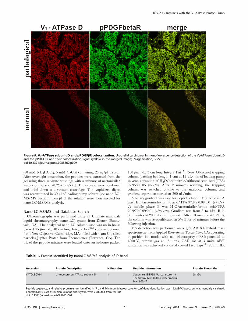
Figure 9. V1-ATPase subunit D and pPDGFbR colocalization. Urothelial carcinoma. Immunofluorescence detection of the V1-ATPase subunit Dand the pPDGFbR and their colocalization signal (yellow in the merged image). Magnification, 6550.doi:10.1371/journal.pone.0088860.g009
Table 1. Protein identified by nanoLC-MS/MS analysis of IP band.
Accession Protein Description N.Peptides Peptide Information Protein Theor.Mw
VATD_BOVIN V1-type proton ATPase subunit D 1 Sequence: IEIFPSR Mascot score: 14Theoretical Mw: 860.48 ExperimentalMw: 860.47
28 kDa
Peptide sequence, and relative protein entry, identified in IP band. Minimum Mascot score for confident identification was 14. MS/MS spectrum was manually validated.Contaminants such as human keratins and trypsin were excluded from the list.doi:10.1371/journal.pone.0088860.t001
BPV-2 E5 Interacts with the V1-ATPase Proton Pump
PLOS ONE | www.plosone.org 7 February 2014 | Volume 9 | Issue 2 | e88860

10 mm tip ID (New Objective). Information-dependent acquisition
(IDA) was performed by selecting the two most abundant peaks for
MS/MS analysis after a full TOF-MS scan from 400 to 1600 m/z
lasting 4 seconds. Both MS/MS analyses were performed in
enhanced mode (3 seconds/scan). Threshold value for peak
selection for MS/MS was 20 counts.
Data were searched on the Mascot search engine (www.
matrixscience.com) against the Swiss Prot database using the
following parameters: MS tolerance 10 ppm; MS/MS tolerance
0.3 Da; fixed modifications carbamidomethyl cysteine; enzyme
trypsin; max. missed cleavages 1; taxonomy other mammalia.
Protein hits based on two successful peptide identifications were
considered valid. Protein hits based on a single peptide identifi-
cation with Mascot score higher than the significance level (.14)
were retained after manual validation.
BPV-2 E5 and PDGFbR ImmunoprecipitationTissues were lysed in ice-cold buffer containing 50 mM Tris-
HCl (pH 7.5), 1% (v/v) Triton X-100, 150 mM NaCl, 2 mM
PMSF, 1.7 mg/ml Aprotinin, 50 mM NaF, and 1 mM sodium
orthovanadate. The protein concentration was measured using the
Bradford assay (Bio-Rad Laboratories, Milan, Italy). Proteins
(1000 mg) were immunoprecipitated using 2 mg of anti- E5
antibody (a kind gift of Dr L. Nasir, Glasgow University) or
anti-PDGFbR antibody (Santa Cruz Biotechnology, CA, USA)
and 30 ml of Protein A/G-Plus Agarose (Santa Cruz Biotechnol-
ogy, CA, USA). Immunoprecipitates were washed four times in
complete lysis buffer (above), finally heated in 1X Laemmli sample
buffer at 100uC for 10 minutes. Immunoprecipitates were
separated on polyacrylamide gels and transferred to nitrocellulose
filter membranes (Ge Healthcare Life Sciences, Chalfont St Giles,
UK) for 16 h at 30 mA in 192 mM glycine/25 mM Tris-HCl
(pH 7.5)/10% methanol. Membranes were blocked for 1 h at
room temperature in 5% nonfat dry milk, incubated with anti-E5
antibody, anti-PDGFbR, anti-pPDGFbR (phosphorylated at
Tyr770), anti-V1-ATPase subunit D (Santa Cruz Biotechnology,
CA, USA), and anti V0-ATPase c subunit (Cosmo Bio CO, Japan)
overnight at 4uC. After three washes in Tris-buffered saline,
membranes were incubated with rabbit anti-sheep IgG-horserad-
ish peroxidase (HRP) (Santa Cruz Biotechnology, CA, USA) or
with goat anti-rabbit or anti-mouse IgG (Bio-Rad Laboratories,
Milan, Italy) for 60 min at room temperature. Proteins were
visualized by enhanced chemiluminescence system (Western
Blotting Luminol Reagent, Santa Cruz Biotechnology, CA,
USA) and ChemiDoc XRS Plus (Bio-Rad Laboratories, Milan,
Italy). Images were acquired with Image Lab Software version
2.0.1 (Bio-Rad Laboratories, Milan, Italy).
BPV-2 DNA Detection and SequencingDNA was extracted from urinary bladder samples from frozen
pathological and normal (control) bladder samples using the
DNeasy Tissue kit (Qiagen, Milan, Italy) according to the
manufacturer’s protocol. All the samples were lysed using
proteinase K. Lysates were loaded onto DNeasy spin columns.
After two washing steps pure DNA was eluted with low salt buffer.
To amplify the entire BPV-2 genome, the purified DNA was
subjected to multiply primed rolling-circle amplification using a
reaction mixture containing 20 ng sample DNA, 12.5 mM of each
primer, 4 mM dNTPs and 10 U phi 29 DNA polymerase
(Fermentas, Milan, Italy). The resulting linear dsDNA product
was purified using MinElute PCR Purification kit (Qiagen, Milan,
Italy). For the detection of BPV-2 DNA, specific primers for a
503 bp DNA amplicon encompassing the BPV-2 E5-L2 ORF
sequence (nt 3723–4225) were designed by Primer BLAST
Figure 10. V1-ATPase subunit D expression. (x) Western blotanalysis showing overexpression of V1-ATPase subunit D. Lanes 1–3:urinary bladder from healthy animals. Lanes 4–6: neoplastic tissue fromrepresentative three cows with papillomavirus-associated tumors of theurinary bladder. Actin protein levels were detected to ensure equalprotein loading. (y) Quantitative densitometric analysis of the filters wasperformed with Image Lab software (ChemiDoc; Bio-Rad Laboratories)and significance determined by the Student T-test (**, p,0.01).doi:10.1371/journal.pone.0088860.g010
Figure 11. V1-ATPase subunit D immunofluorescence. Urothelialcarcinoma. The overexpression of the subunit D of the V1-domain isalso detected by immunofluorescence. The subunit D appears to beoverexpressed both in the membrane and in the cytoplasm of urothelialcancer cells compared to urothelial normal cells (insert). Magnification,6550.doi:10.1371/journal.pone.0088860.g011
BPV-2 E5 Interacts with the V1-ATPase Proton Pump
PLOS ONE | www.plosone.org 8 February 2014 | Volume 9 | Issue 2 | e88860

software (forward, 59-TCAGGCACAGATCTTGATCA-39; re-
verse, 59-TCATAGACATTTGCACGTT-39). To evaluate the
adequacy of the DNA samples, a control PCR for bovine b-actin
sequence was performed using a set of primers designed by Primer
BLAST software (forward, 59-GAGCGTGGCTACAGCTT-
CAC-39; reverse, 59-CATTGCCGATGGTGATGA-39). Aliquots
50–100 ng of purified DNA were amplified in 25 ml of reaction
mixture containing 2 mM MgCl2, 200 mM each dNTP, 480 nM
of each primer and 2.5 U of AmpliTaq Gold DNA Polymerase
(Applied Biosystems, Monza, Italy). The reaction was carried out
in a thermocycler (Veriti, Applied Biosystems, Monza, Italy) with
an initial denaturation step of 3 min. Then, 35 cycles of
amplification were carried out with a denaturation step at 94uCfor 40 sec, an annealing step at 60uC, 30 sec, for b-actin or at
50uC,40 sec, for BPV-2, and an extension step at 72uC for 1 min.
A final extension step at 72uC for 7 min was performed in each
PCR assay. Detection of the amplified products was carried out by
electrophoresis on ethidium bromide-stained agarose gel. In each
experiment, a blank sample consisting of reaction mixture without
DNA and a positive sample consisting of cloned BPV-2 (a kind gift
by Dr. A. Venuti) were included. The amplified DNA was
subjected to direct sequencing in an automated apparatus (ABI
Prism 3100 Genetic Analyzer; Applied Biosystems, Monza, Italy).
Western Blot AnalysisHealthy and diseased bladders were solubilized at 4uC in lysis
buffer containing 50 mM Tris-HCl pH 7.5, 150 mM NaCl, 1%
Triton X-100. Immediately prior to use, the following reagents
were added: 1 mM DTT, 2 mM PMSF, 1.7 mg/ml Aprotinin,
25 mM NaF, 1 mM Na3VO4 (Sigma-Aldrich, Milan, Italy).
Lysates were clarified at 5006g for 20 min. The protein
concentration was measured using the Bradford assay (Bio-Rad
Laboratories, Milan, Italy). For Western blotting, 50 mg of lysate
proteins were heated at 100uC in 4X premixed Laemmli sample
buffer (Bio-Rad Laboratories, Milan, Italy). Proteins were
subjected to sodium dodecyl sulfate–polyacrylamide gel electro-
phoresis (SDS–PAGE) under reducing conditions.
After electrophoresis, proteins were transferred onto nitrocellu-
lose filter membranes (GE Healthcare Life Sciences, Chalfont St
Giles, UK) for 1 h at 10 V in 192 mM glycine/25 mM Tris-HCl
(pH 7.5)/10% methanol using a Trans-Blot SD Semi Dry cell
(Bio-Rad Laboratories, Milan, Italy) according to the manufac-
turer’s instructions. The membranes were blocked with 5% non-
fat dry milk in Tris-buffered saline (TBS, pH 7.5) for 1 h at room
temperature, washed with TBS-0.1% Tween. Then, filters were
probed both with anti-PDGFbR, anti-pPDGFbR (phosphorylated
at Tyr770), and anti-V1-ATPase subunit D antibody (Santa Cruz
Figure 12. RT PCR for subunit D. The relative expression levels of V1-ATPase subunit D in neoplastic tissues. V1-ATPase subunit D mRNA levelswere determined by qRT-PCR. Relative mRNA levels, calculated using the DDCT method, represent fold changes in comparison to urinary bladdersamples from healthy cows. All values were normalized to the internal control b-actin. Results represent the means and standard deviations of threeindependent experiments performed in triplicate. (*, p,0.05, vs urinary bladder from healthy cows).doi:10.1371/journal.pone.0088860.g012
Figure 13. V0-ATPase c subunit expression. Western blot analysisshowing similar expression levels of V0-ATPase c subunit among all thesamples. Lanes 1–3: urinary bladder from healthy animals. Lanes 4–6:neoplastic tissue from representative three cows with papillomavirus-associated tumors of the urinary bladder. Actin protein levels weredetected to ensure equal protein loading.doi:10.1371/journal.pone.0088860.g013
Figure 14. BPV-2 E5 and V1-ATPase subunit D co-immunopre-cipitation. The presence of V1-ATPase subunit D was detected in E5immunoprecipitates. Lanes 1–3: urinary bladder from healthy cows.Lanes 4–6: neoplastic tissue from three cows with papillomavirus-associated tumors of the urinary bladder.doi:10.1371/journal.pone.0088860.g014
BPV-2 E5 Interacts with the V1-ATPase Proton Pump
PLOS ONE | www.plosone.org 9 February 2014 | Volume 9 | Issue 2 | e88860

Biotechnology, CA, USA) for an overnight incubation at 4uC.
After three washes in Tris-buffered saline, membranes were
incubated with horseradish peroxidase-conjugated anti-rabbit IgG
or anti-mouse IgG (Bio-Rad Laboratories, Milan, Italy) and anti-
goat IgG (Santa Cruz Biotechnology, CA, USA), for 1 h at room
temperature. After appropriate washing steps, protein detection
and image acquisition were performed as above reported.
RNA ExtractionTotal RNA was extracted from urinary bladders of cows using
the RNeasy Mini Kit (Qiagen, Milan, Italy), according to the
manufacturer’s instructions. The RNA quality was determined by
agarose gel electrophoresis and ultraviolet spectrophotometer
analysis. The RNA was treated with RNase-free DNase I
Fermentas Life Sciences (Dasit, Milan, Italy) to remove potential
DNA contamination.
cDNA Synthesis and Real Time-PCR Analysis (RT-PCR) forV1-ATPase Subunit D
For Real Time-PCR analysis, 500 ng RNA were reverse-
transcribed using the iScript cDNA Synthesis Kit (Bio-Rad
Laboratories, Milan, Italy) and the reaction was incubated at
25uC for 5 min, 42uC for 30 min, 85uC for 5 min, and then kept
at 4uC for 5 min. Real Time reactions were performed using
SsoFast EvaGreen Supermix (Bio-Rad Laboratories, Milan, Italy).
For the detection of V1-ATPase subunit D specific primers
(forward primer, 59-AAGACTCAGTGGCTGGGTTG -39; re-
verse primer, 59-AGGTTTCGACCTGTCTGTGC-39) were
used. All reactions were performed in triplicate and b-actin was
used as the internal standard (forward primer 59- TAGCA-
CAGGCCTCTCGCCTTCG-39; reverse primer 59- GCA-
CATGCCGGAGCCGTTGT-39).
Figure 15. V1-ATPase subunit D and BPV-2 E5 colocalization. Urothelial carcinoma. Immunofluorescence detection of E5 and the subunit D.The proteins appear to colocalize (yellow in the merged image).doi:10.1371/journal.pone.0088860.g015
Figure 16. BPV-2 E5, PDGFßR and V1-ATPase D subunit co-immunoprecipitation. The presence of PDGFßR and V1-ATPase Dsubunit was detected in E5 immunoprecipitates. Lanes 1–3: urinarybladder from healthy cows. Lanes 4–6: tissue from three cows withpapillomavirus-associated tumors of the urinary bladder.doi:10.1371/journal.pone.0088860.g016
BPV-2 E5 Interacts with the V1-ATPase Proton Pump
PLOS ONE | www.plosone.org 10 February 2014 | Volume 9 | Issue 2 | e88860

Results
Microscopical Pattern of the TumorsHistological patterns of urothelial tumors of the urinary bladder
of cattle were consistent with the diagnosis of papillary urothelial
neoplasm of low malignant potential (PUNLMP) (two cases),
papillary and invasive urothelial cancers (nine and four cases,
respectively).
PCR Analysis and BPV-2 SequencingPCR yielded BPV-2 DNA fragments of anticipated size (503 bp)
for all neoplastic lesions. No BPV-2 DNA was detected in normal
(control) bladder samples (Figure 1). The presence of BPV-2 DNA
was also confirmed by sequencing (Figure 1) according to BPV-2
sequence M20219.1.
Immunoprecipitation and Immunohistochemistry forBPV-2 E5 Protein
The expression of E5 was detected by immunoprecipitation and
immunohistochemistry in tumor samples. E5 immunoreactivity
was evident in cells located in basal and suprabasal urothelial
layers (Figure 2 and 3).
Coimmunoprecipitation and Colocalization of E5Oncoprotein with PDGFbR
Previous in vivo studies have shown that E5 binds to the
activated (phosphorylated) form of PDGFbR in bladder tumors
[18,21,27]. Indeed, PDGFbR appeared to be constitutively
expressed and its phosphorylation was increased in the tumor
samples compared to the healthy ones as detected in total lysates
(Figure 4). The activation of the phosphorylated PDGFbR was
also documented by immunohistochemical investigations
(Figure 5).
A coimmunoprecipitation experiment using an anti-E5 anti-
body was carried out and PDGFbR and pPDGFbR were detected
by Western blot in the immunoprecipitates (Figure 6). Morpho-
logically, the E5/pPDGFbR complex was shown by confocal
microscopy: E5 and the activated form of the PDGFbR appeared
to co-localize as judged by the yellow fluorescence of the merged
images (Figure 7). Normal urothelium yielded no E5 signal.
PDGFbR Binds to the Component D of V1-ATPase:Proteomic Analysis
In vitro, PDGFbR can bind to the proteolipid ring of V0 sector
of the proton pump in absence of E5, it has been suggested that E5
binds to the PDGFbR via the subunit c of the ring. However,
these data have been obtained for medium-adapted cultured cells
and hence may not reflect the authentic in vivo situation, for
which no information is available thus far [26].
Accordingly, we investigated whether a similar complex could
take place in vivo and performed immunoprecipitation of
PDGFbR on bladder carcinoma tissue and on control normal
bladder tissue from healthy cattle.
Proteins contained in the two PDGFbR pull-downs from
bladder tumor samples and from normal bladder tissue, were
separated with SDS-PAGE and detected with Coomassie staining.
Differential analysis between the protein bands contained in the
two immunoprecipitates revealed a single band present in the
PDGFbR pull-down from bladder tumor samples only. The
differential band was excised and in-gel digested with trypsin. The
peptides were injected for nano LC-MS/MS analysis. Database
search of MS/MS spectra allowed the identification of the protein
‘‘V1-ATPase subunit D’’ (Table 1). Because the protein has been
identified by a single peptide hit having a low Mascot score, this
result needed to be validated by further investigations. For this
purpose, we performed Western blot analysis using an anti-subunit
D antibody on the PDGFbR immunoprecipitates. This allowed us
to detect the presence of the subunit D (Figure 8). We also
performed Western blot analysis using an anti-subunit c antibody
on these immunoprecipitates as the PDGFbR/subunit c complex
was documented in in vitro studies [25]. We did not detect the
subunit c of the V0-ATPase domain (Figure 8). The complex
pPDGFbR/subunit D of the V1-ATPase was shown by immuno-
fluorescence studies as the two proteins appeared to colocalize by
confocal microscopy (Figure 9).
Western Blot Analysis of the Subunit DWe performed immunoblotting to reveal the total level of the
subunit D of the V1-domain. Overexpression of subunit D could
be shown by immunoblotting (Figure 10) and morphologically
documented by immunofluorescence (Figure 11). Furthermore, a
statistically significant increase of the transcripts of this subunit was
also shown by RT-PCR (Figure 12). Normal levels of the
constitutively expressed subunit c were detected by Western blot
both in healthy and tumor samples (Figure 13).
Coimmunoprecipitation and Colocalization of E5Oncoprotein with the Subunit D of the V1-ATPase
Using Western blot, we detected the subunit D of the V1-
domain of the proton pump in E5 immunoprecipitates (Figure 14).
Morphologically, this complex was demonstrated by confocal
microscopy as E5 and subunit D appeared to colocalize (Figure 15).
Normal urothelium yielded no E5 signal.
Ultimately, both pPDGFbR and subunit D of the V1-ATPase
were found by Western blot in E5 immunoprecipitates (Figure 16).
Discussion
Our results indicate, for the first time, that a ternary complex
composed of BPV-2 E5 oncoprotein/PDGFbR/subunit D of V1-
ATPase is present in urothelial cells of naturally occurring tumors
of the bovine urinary bladder.
It has been shown that bovine papillomavirus E5 interacts with
the subunit c of the V0 domain in cultured cells [25]. As PDGFbR
can bind to the subunit c in the absence of E5, it has been
suggested that E5 binds to PDGFbR via its association with
subunit of V0-ATPase proton pump [26].
Our in vivo findings appear to be different from previous
in vitro results and show that the complex composed of E5 and the
activated form of PDGFbR is associated with the overexpressed
subunit D of the V1 domain, which catalyzes ATP hydrolysis, but
not with the subunit c of the V0 domain responsible for H+
translocation. Furthermore, the subunit c appeared to be
constitutively expressed, as normal levels of expression were
shown to occur both in normal and neoplastic tissues.
It has been suggested that cell perturbation resulting from
binding of the subunit c of the proton pump and E5 oncoprotein is
responsible for Golgi alkalinization which, in turn, leads to the
activation of Golgi-associated Src molecules. Indeed, Golgi
alkalinization and c-Src are involved in a common mechanism
leading to E5-dependent NIH3T3 cell transformation [28].
The subunit D of the V1 complex belongs to a central rotor
stalk, which is composed also of F subunit. These proteins are
bound directly to the subunit d of the V0 sector that links to the c
ring. Therefore, the central stalk (DFd complex) connects the V1
and V0 domains. It serves as a rotor that couples the energy that is
released from the hydrolysis of ATP to the rotation of the
BPV-2 E5 Interacts with the V1-ATPase Proton Pump
PLOS ONE | www.plosone.org 11 February 2014 | Volume 9 | Issue 2 | e88860

proteolipid c ring of the V0-ATPase pump and causes active
transport of protons thus regulating the pH (acidification) of
intracellular organelles and cytosol [29–31]. The controlled pH of
the intracellular compartments is crucial for many biological
processes, including membrane trafficking and protein degrada-
tion. It is conceivable that in naturally occurring bovine bladder
cancers, the complex E5/PDGFbR/subunit D could have an
important role in perturbing proteostasis network as well as
organelle and cytosol homeostasis. It is worthwhile noting that in
bovine urothelial tumors we detected an overexpression of some of
the most important markers of proteostasis stress such as heat
shock proteins (HSPs) [32]. The latter are known to act as
molecular chaperones to restore protein homeostasis [33,34].
Furthermore, we found an overexpression of the co-chaperone
BAG3 (Bcl-2 associated athanogene 3), which depends on an
altered degradation of the protein rather than the upregulation of
gene transcription [35]. It is worth remembering that BAG3
protein degradation occurs via proteasome system only.
Cytosolic pH has been identified as a novel regulator that
mediates the formation of proteasome storage granule (PSGs) and
other protein aggregates. The regulation of proper partitioning of
the proteasome into PSGs is essential for maintaining the correct
level of the proteasome in the cytosol. It has been shown that the
impaired ability of V-ATPase to regulate intracellular pH affects
the kinetics of the PSG formation [30].
In our cases, the impairment of the vacuolar pump induced by
E5 oncoprotein can be responsible for a proteasomal dysfunction
resulting in a reduced clearance of specific protein such as
proteasome-degraded BAG3 protein, known to be involved in a
plethora of biological processes including the key role in mitigating
the proteotoxicity via selective autophagy [36]. It has been shown
that autophagy is activated when the proteasome function is
reduced, thus constituting a strong functional link between
autophagy and proteasome systems [37]. Our findings are
consistent with several in vitro studies showing that the impair-
ment of V-ATPase can induce autophagic responses and increase
the formation of autophagosomes thus the autophagy represents a
mechanism to overcome alteration of pH homeostasis mediated by
proton pump perturbation [38].
Finally, the selective autophagy occurring in bovine urothelial
tumor cells transformed by E5 oncoprotein that we have been
studying (Roperto, unpublished data) seems to strengthen this
suggestion and the emerging concept that the molecular chaper-
ones, the ubiquitin-proteasome system (UPS) and the autophagy
machinery are central elements of the proteostasis network in
which the V-ATPase proton pump is also involved.
Acknowledgments
We are grateful to Dr M.S. Campo, Professor Emeritus, University of
Glasgow, UK, for reading the manuscript; Dr L. Nasir, University of
Glasgow, UK, for providing us the anti-BPV-2 E5 antibody and Dr A.
Venuti, Istituto dei Tumori ‘‘Regina Elena’’, Rome, for giving us the BPV-
2 DNA plasmid.
Author Contributions
Conceived and designed the experiments: SR VR FR. Performed the
experiments: SR VR CU RL IE MGR MG DMC. Analyzed the data: SR
CR GB FR. Wrote the paper: SR RG FR.
References
1. Meuten DJ (2002) Tumors of the urinary system. In Meuten, D.J. (Ed) Tumors
in Domestic Animals, Blackwell Publishing Company, Iowa State Press, Ames,
524–546.
2. Ozkul IA, Aydin Y (1996) Tumours of the urinary bladder in cattle and water
buffalo in the Black Sea region of Turkey. Br Vet J 152: 473–475.
3. Pamukcu AM, Price JM, Bryan GT (1976) Naturally occurring and bracken
fern-induced bovine urinary bladder tumors. Clinical and morphological
characteristics. Vet Pathol 13: 110–122.
4. Roperto S, Borzacchiello G, Brun R, Leonardi L, Maiolino P, et al. (2010a). A
review of bovine urothelial tumours and tumour-like lesions of the urinary
bladder. J Comp Pathol 142: 95–108.
5. Evans IA, Mason J (1965) Carcinogenic activity of bracken. Nature 208: 913–
914.
6. Cairney M, Campo MS (1995) The synergism between bovine papillomavirus
type 4 and quercetin is dependent on the timing of exposure. Carcinogenesis 16:
1997–2001.
7. Campo MS (1997) Bovine papillomavirus and cancer. Vet J 154: 175–188.
8. Borzacchiello G, Iovane G, Marcante ML, Poggiali F, Roperto F, et al. (2003)
Presence of bovine papillomavirus type 2 and expression of the viral oncoprotein
E5 in naturally occurring urinary bladder tumours in cows. J Gen Virol 84:
2921–2926.
9. Campo MS, Jarrett WFH, Barron RJ, O’Neil BW, Smith KT (1992) Association
of bovine papillomavirus type 2 and bracken fern with bladder cancer in cattle.
Cancer Res 52: 6898–6904.
10. Roperto S, Brun R, Paolini F, Urraro C, Russo V, et al. (2008) Detection of
bovine papillomavirus type 2 (BPV-2) in the peripheral blood of cattle with
urinary bladder tumours: possible biological role. J Gen Virol 89: 3027–3033.
11. Wosiaki SR, Barreiros MAB, Alfieri AF, Alfieri AA (2005) Semi-nested PCR for
detection and typing of bovine papillomavirus type 2 in urinary bladder and
whole blood from cattle with enzootic haematuria. J Virol Methods 126: 215–
219.
12. Balcos LG, Borzacchiello G, Russo V, Popescu O, Roperto S, et al. (2008)
Association of bovine papillomavirus type-2 and urinary bladder tumours in
cattle from Romania. Res Vet Sci 85: 145–148.
13. Borzacchiello G, Roperto F (2008) Bovine papillomaviruses, papillomas and
cancer in cattle. Vet Res 39: 45–63.
14. Maiolino P, Ozkul A, Sepici-Dincel A, Roperto F, Yucel G, et al. (2013) Bovine
papillomavirus type 2 infection and microscopic patterns of urothelial tumors of
the urinary bladder in water buffaloes. Biomed Res Int 2013: 937918, doi:
10.1155/2013/937918.
15. Martano M, Roperto F, de Cassia Stocco R, Russo V, Borzacchiello G, (2013)
Bovine papillomavirus type 2 (BPV-2) infection and a series of mesenchymal
tumors of the urinary bladder in cattle. Biomed Res Int 2013: 814635, doi:
10.1155/2013/814635.
16. Resendes AR, Roperto S, Trapani F, Urraro C, Rodrigues A et al. (2011)
Association of bovine papillomavirus type 2 (BPV-2) and urinary bladder
tumours in cattle from the Azores archipelago. Res Vet Sci 90: 526–529.
17. Roperto S, Comazzi S, Ciusani E, Paolini F, Borzacchiello G, et al. (2011)
PBMCs are additional sites of productive infection of bovine papillomavirus type
2. J Gen Virol 92: 1787–1794.
18. Roperto S, Russo V, Ozkul A, Sepici-Dincel A, Maiolino P, et al. (2013a) Bovine
papillomavirus type 2 infects the urinary bladder of water buffalo (Bubalus
bubalis) and plays a crucial role in bubaline urothelial carcinogenesis. J Gen
Virol 94: 403–408.
19. Shafti-Keramat S, Schellenbacher C, Handisurya A, Christensen N, Reininger
B, et al. (2009) Bovine papillomavirus type 1 (BPV1) and BPV2 are closely
related serotypes. Virology 393:1–6.
20. Conrad M, Bubb VJ, Schlegel R (1993) The human papillomavirus type 6 and
16 E5 proteins are membrane-associated proteins which associate with the 16-
kilodalton pore-forming protein. J Virol 67: 6170–6178.
21. Borzacchiello G, Russo V, Gentile F, Roperto F, Venuti A, et al. (2006) Bovine
papillomavirus E5 oncoprotein binds to the activated form of the platelet-derived
growth factor ß receptor in naturally occurring bovine urinary bladder tumours.
Oncogene 25: 1251–1260.
22. Roperto S, De Tullio R, Raso C, Stifanese R, Russo V, et al. (2010b) Calpain 3is expressed in a proteolitically active form in papillomavirus-associated
urothelial tumors of the urinary bladder in cattle. PLoS One 5, e10299.
23. Andresson T, Sparkowski J, Goldstein DJ, Schlegel R. (1995) Vacuolar H+
ATPase mutants transform cells and define a binding site for papillomavirus E5
oncoprotein. J Biol Chem 270, 6830–6837.
24. Ashrafi GH, Tsirimonaki E, Marchetti BN, O’Brien PM, Sibbet GJ, et al (2002).
Downregulation of MHC class I by bovine papillomavirus E5 oncoproteins.
Oncogene 21, 248–259.
25. Goldstein DJ, Schlegel R (1990) The E5 oncoprotein of bovine papillomavirus
binds to a 16 kd cellular protein EMBO J 9: 137–146.
26. Goldstein DJ, Andresson T, Sparkowski JJ, Schlegel R (1992) The BPV-1 E5
protein, the 16 kDa membrane pore-forming protein and the PDGF receptor
exist in a complex that is dependent on hydrophobic transmembrane
interactions. EMBO J 11: 4851–4859.
27. Roperto S, Russo V, Ozkul A, Corteggio A, Sepici-Dincel A, et al. (2013b)Productive infection of bovine papillomavirus type 2 in the urothelial cells of
BPV-2 E5 Interacts with the V1-ATPase Proton Pump
PLOS ONE | www.plosone.org 12 February 2014 | Volume 9 | Issue 2 | e88860

naturally occurring urinary bladder tumors in cattle and water buffaloes. PLoS
One 8: e62227.28. Suprynowicz FA, Campo MS, Schlegel R (2006) Biological activities of
papillomavirus E5 proteins. In: Campo, M.S. (Ed.), Papillomavirus Research –
from natural history to vaccines and beyond, Caister Academic Press, Norfolk,97–113.
29. Forgac M (2007) Vacuolar ATPases: rotary proton pumps in physiology andpathophysiology. Nat Rev Mol Cell Biol 8: 917–929.
30. Peters LZ, Hazan R, Breker M, Schulcliner M, Ben-Aroya S (2013) Formation
and dissociation of proteasome storage granules are regulated by cytosolic pH.J Cell Biol 201: 663–671.
31. Saijo S, Aral S, Hossain KMM, Yamato I, Suzuki K, et al. (2011) Crystalstructure of the central axis DF complex of the prokaryotic V-ATPase. Proc Natl
Acad Sci 108: 19955–19960.32. Romanucci M, Malatesta D, Ciccarelli A, Bongiovanni L, Palmieri C, et al.
(2012) Expression of heat shock proteins in premalignant and malignant
urothelial lesions of bovine urinary bladder. Cell Stress Chaperones 17: 683–692.
33. Hartl EU, Bracher A, Hayer-Hartl M (2011) Molecular chaperones in protein
folding and proteostasis. Nature 475: 324–332.34. Powers EV, Balch WE (2013) Diversity in the origins of proteostasis networks – a
driver for protein function in evolution. Nat Rev Mol Cell Biol 14: 237–248.
35. Roperto S, Paciello O, Borzacchiello G, Esposito I, Riccardi M, et al. (2013c) IsBag 3 protein involved in autophagic mechanisms in papillomavirus-associated
urothelial tumors in cattle? J Comp Pathol 148: 49.36. Rapino F, Jung M, Fulda S (2013) BAG3 induction is required to mitigate
proteotoxicity via selective autophagy following inhibition of constitutive protein
degradation pathways. Oncogene, doi:10.1038/onc.2013.110.37. Ding WX, Ni HM, Gao W, Yoshimori T, Stolz DB, et al. (2007) Linking of
autophagy to ubiquitin-proteasome system is important for the regulation ofendoplasmic reticulum stress and cell viability. Am J Pathol 171: 513–524.
38. Juhasz G (2012) Interpretation of bafilomycin, pH neutralizing or proteaseinhibitor treatments in autophagy flux experiments. Autophagy 12: 1875–1876.
39. Shevchenko A, Wilm M, Vorm O, Mann M (1996) Mass spectrometric
sequencing of proteins silver-stained polyacrylamide gels. Anal Chem 68: 850–858.
BPV-2 E5 Interacts with the V1-ATPase Proton Pump
PLOS ONE | www.plosone.org 13 February 2014 | Volume 9 | Issue 2 | e88860