Calpain3 Is Expressed in a Proteolitically Active Form in Papillomavirus-Associated Urothelial...
9
Calpain3 Is Expressed in a Proteolitically Active Form in Papillomavirus-Associated Urothelial Tumors of the Urinary Bladder in Cattle Sante Roperto 1 *, Roberta De Tullio 2 , Cinzia Raso 3 , Roberto Stifanese 2 , Valeria Russo 4 , Marco Gaspari 3 , Giuseppe Borzacchiello 4 , Monica Averna 2 , Orlando Paciello 4 , Gianni Cuda 3 , Franco Roperto 4 1 Department of Pathology and Animal Health, Division of Infectious Diseases, Naples University Federico II, Naples, Italy, 2 Department of Experimental Medicine (DIMES), Biochemistry Section and Centre of Excellence for Biomedical Research (CEBR), University of Genova, Genova, Italy, 3 Department of Experimental Medicine and Clinics, University of Catanzaro ‘Magna Graecia’, Catanzaro, Italy, 4 Department of Pathology and Animal Health, Division of General Pathology, Naples University Federico II, Naples, Italy Abstract Background: Calpain 3 (Capn3), also named p94, is a skeletal muscle tissue-specific protein known to be responsible for limb-girdle muscular dystrophy type 2A (LGMD2A). Recent experimental studies have hypothesized a pro-apoptotic role of Capn3 in some melanoma cell lines. So far the link between calpain3 and tumors comes from in vitro studies. The objective of this study was to describe Capn3 activation in naturally occurring urothelial tumors of the urinary bladder in cattle. Methods and Findings: Here we describe, for the first time in veterinary and comparative oncology, the activation of Capn3 in twelve urothelial tumor cells of the urinary bladder of cattle. Capn3 protein was initially identified with nanoscale liquid chromatography coupled with tandem mass spectrometry (nano LC-MS/MS) in a co-immunoprecipitation experiment on E2F3, known to be a transcription factor playing a crucial role in bladder carcinogenesis in humans. Capn3 expression was then confirmed by reverse transcription polymerase chain reaction (RT-PCR). Finally, the Ca 2+ -dependent proteolytic activity of Capn3 was assayed following ion exchange chromatography. Morphologically, Capn3 expression was documented by immunohistochemical methods. In fact numerous tumor cells showed an intracytoplasmic immunoreactivity, which was more rarely evident also at nuclear level. In urothelial tumors, bovine papillomavirus type 2 (BPV-2) DNA was amplified by PCR and the expression of E5 protein, the major oncogenic protein of BVP-2, was detected by western blotting, immunohistochemistry, and immunofluorescence. E2F3 overexpression and pRb protein downregulation were shown by western blotting. Conclusion: The role of capn3 protein in urothelial cancer of the urinary bladder remains to be elucidated: further studies would be required to determine the precise function of this protease in tumor development and progression. However, we suggest that activated Capn3 may be involved in molecular pathways leading to the overexpression of E2F3, which in turn could be responsible for urothelial tumor cell proliferation also in cattle, though other mechanisms are likely to exist. If further studies corroborate the important role of Capn3 in urothelial tumors of the urinary bladder, cattle with urinary tumors may prove useful as animal model for bladder carcinogenesis. Citation: Roperto S, De Tullio R, Raso C, Stifanese R, Russo V, et al. (2010) Calpain3 Is Expressed in a Proteolitically Active Form in Papillomavirus-Associated Urothelial Tumors of the Urinary Bladder in Cattle. PLoS ONE 5(4): e10299. doi:10.1371/journal.pone.0010299 Editor: Wanda Markotter, University of Pretoria, South Africa Received January 4, 2010; Accepted March 26, 2010; Published April 22, 2010 Copyright: ß 2010 Roperto et al. This is an open-access article distributed under the terms of the Creative Commons Attribution License, which permits unrestricted use, distribution, and reproduction in any medium, provided the original author and source are credited. Funding: This work was supported in part by grants from the Ministero delle Politiche Agricole, Alimentari, Forestali, as well as from the Assessorato alla Sanita ` della Regione Campania e della Regione Basilicata, from the Legge Regionale n. 5 and PRIN 2007 and 2008. The funders had no role in study design, interpretation of data, writing of the paper, and decision to submit it for publication. Competing Interests: The authors have declared that no competing interests exist. * E-mail: [email protected] Introduction The calpain system is composed of a family of Ca 2+ -dependent cysteine proteases, their activity being regulated by cytosolic calcium [1,2]. Calpains originally comprised two proteases, calpain 1 (Capn1) or m-calpain (micromolar) and calpain 2 (Capn2) or m-calpain (millimolar). Also referred to as the ‘‘conventional’’ or ‘‘ubiquitous’’ calpains, they are specifically inhibited by calpastatin [1]. Calpains appear to be involved in cell motility, signal transduction, cell cycle progression, gene expres- sion regulation and apoptosis [1,3]. Calpains are known to play an important role in macroauthophagy, being regulators of autopha- gosome formation [4,5], and in some viral oncogenetic mecha- nisms [6]. Furthermore, calpains represent a promising target for cancer therapy, since they appear to play a key role in metastatic cell migration and angiogenesis [7]. Calpain 3 (Capn3), also known as calpain p94, is a member of the so-called tissue-specific subfamily, being predominantly expressed in skeletal muscle. Its structure is similar to that of the other members of this family but it presents three specific sequences not found in any other calpain: a novel sequence (NS) at the N-terminus, insertion sequence 1 (IS1) within the catalytic PLoS ONE | www.plosone.org 1 April 2010 | Volume 5 | Issue 4 | e10299
Transcript of Calpain3 Is Expressed in a Proteolitically Active Form in Papillomavirus-Associated Urothelial...

Calpain3 Is Expressed in a Proteolitically Active Form inPapillomavirus-Associated Urothelial Tumors of theUrinary Bladder in CattleSante Roperto1*, Roberta De Tullio2, Cinzia Raso3, Roberto Stifanese2, Valeria Russo4, Marco Gaspari3,
Giuseppe Borzacchiello4, Monica Averna2, Orlando Paciello4, Gianni Cuda3, Franco Roperto4
1 Department of Pathology and Animal Health, Division of Infectious Diseases, Naples University Federico II, Naples, Italy, 2 Department of Experimental Medicine (DIMES),
Biochemistry Section and Centre of Excellence for Biomedical Research (CEBR), University of Genova, Genova, Italy, 3 Department of Experimental Medicine and Clinics,
University of Catanzaro ‘Magna Graecia’, Catanzaro, Italy, 4 Department of Pathology and Animal Health, Division of General Pathology, Naples University Federico II,
Naples, Italy
Abstract
Background: Calpain 3 (Capn3), also named p94, is a skeletal muscle tissue-specific protein known to be responsible forlimb-girdle muscular dystrophy type 2A (LGMD2A). Recent experimental studies have hypothesized a pro-apoptotic role ofCapn3 in some melanoma cell lines. So far the link between calpain3 and tumors comes from in vitro studies. The objectiveof this study was to describe Capn3 activation in naturally occurring urothelial tumors of the urinary bladder in cattle.
Methods and Findings: Here we describe, for the first time in veterinary and comparative oncology, the activation of Capn3in twelve urothelial tumor cells of the urinary bladder of cattle. Capn3 protein was initially identified with nanoscale liquidchromatography coupled with tandem mass spectrometry (nano LC-MS/MS) in a co-immunoprecipitation experiment onE2F3, known to be a transcription factor playing a crucial role in bladder carcinogenesis in humans. Capn3 expression wasthen confirmed by reverse transcription polymerase chain reaction (RT-PCR). Finally, the Ca2+-dependent proteolytic activityof Capn3 was assayed following ion exchange chromatography. Morphologically, Capn3 expression was documented byimmunohistochemical methods. In fact numerous tumor cells showed an intracytoplasmic immunoreactivity, which wasmore rarely evident also at nuclear level. In urothelial tumors, bovine papillomavirus type 2 (BPV-2) DNA was amplified byPCR and the expression of E5 protein, the major oncogenic protein of BVP-2, was detected by western blotting,immunohistochemistry, and immunofluorescence. E2F3 overexpression and pRb protein downregulation were shown bywestern blotting.
Conclusion: The role of capn3 protein in urothelial cancer of the urinary bladder remains to be elucidated: further studieswould be required to determine the precise function of this protease in tumor development and progression. However, wesuggest that activated Capn3 may be involved in molecular pathways leading to the overexpression of E2F3, which in turncould be responsible for urothelial tumor cell proliferation also in cattle, though other mechanisms are likely to exist. Iffurther studies corroborate the important role of Capn3 in urothelial tumors of the urinary bladder, cattle with urinarytumors may prove useful as animal model for bladder carcinogenesis.
Citation: Roperto S, De Tullio R, Raso C, Stifanese R, Russo V, et al. (2010) Calpain3 Is Expressed in a Proteolitically Active Form in Papillomavirus-AssociatedUrothelial Tumors of the Urinary Bladder in Cattle. PLoS ONE 5(4): e10299. doi:10.1371/journal.pone.0010299
Editor: Wanda Markotter, University of Pretoria, South Africa
Received January 4, 2010; Accepted March 26, 2010; Published April 22, 2010
Copyright: � 2010 Roperto et al. This is an open-access article distributed under the terms of the Creative Commons Attribution License, which permitsunrestricted use, distribution, and reproduction in any medium, provided the original author and source are credited.
Funding: This work was supported in part by grants from the Ministero delle Politiche Agricole, Alimentari, Forestali, as well as from the Assessorato alla Sanitadella Regione Campania e della Regione Basilicata, from the Legge Regionale n. 5 and PRIN 2007 and 2008. The funders had no role in study design, interpretationof data, writing of the paper, and decision to submit it for publication.
Competing Interests: The authors have declared that no competing interests exist.
* E-mail: [email protected]
Introduction
The calpain system is composed of a family of Ca2+-dependent
cysteine proteases, their activity being regulated by cytosolic
calcium [1,2]. Calpains originally comprised two proteases,
calpain 1 (Capn1) or m-calpain (micromolar) and calpain 2
(Capn2) or m-calpain (millimolar). Also referred to as the
‘‘conventional’’ or ‘‘ubiquitous’’ calpains, they are specifically
inhibited by calpastatin [1]. Calpains appear to be involved in cell
motility, signal transduction, cell cycle progression, gene expres-
sion regulation and apoptosis [1,3]. Calpains are known to play an
important role in macroauthophagy, being regulators of autopha-
gosome formation [4,5], and in some viral oncogenetic mecha-
nisms [6]. Furthermore, calpains represent a promising target for
cancer therapy, since they appear to play a key role in metastatic
cell migration and angiogenesis [7].
Calpain 3 (Capn3), also known as calpain p94, is a member of
the so-called tissue-specific subfamily, being predominantly
expressed in skeletal muscle. Its structure is similar to that of the
other members of this family but it presents three specific
sequences not found in any other calpain: a novel sequence (NS)
at the N-terminus, insertion sequence 1 (IS1) within the catalytic
PLoS ONE | www.plosone.org 1 April 2010 | Volume 5 | Issue 4 | e10299
domain, and insertion sequence 2 (IS2) upstream of the Ca2+-
binding domain [8]. It has been suggested that Capn3 may play a
role in sarcomere remodelling and in mitochondrial protein
turnover [9]. Point mutations in the Capn3 gene are responsible
for limb girdle muscular dystrophy type 2A (LGMD2A), an
autosomal recessive disease characterized by progressive atrophy
and weakness of the proximal limb muscle [10].
Furthermore, several variants of Capn3 have been reported in
many tissues [11] including the eye [12,13], peripheral blood
mononuclear cells [14], and astrocytes [15], thus suggesting that
Capn3 is important for tissues other than skeletal muscle [2].
Recently, novel Capn3 isoforms in white blood cells have been
sequenced and are being investigated to develop a new approach
for performing the molecular diagnosis of LGMD2A at mRNA
level [16].
To our knowledge, no investigation has so far been carried out
on Capn3 expression in veterinary and comparative spontaneous
carcinogenesis.
The aim of the present paper is to report, for the first time in
medical literature, the activation of the Capn3 protease in
urothelial tumors of the urinary bladder in cattle. Such tumors
are very commonly found in adult cattle grazing on lands rich in
bracken fern. Furthermore, a strong relationship between bracken
fern and bovine papillomavirus type 2 (BPV-2) has been
established [17], so that BPV-2 infection appears to be a pivotal
event in the bladder carcinogenesis of cattle [17,18,19].
Methods
Ethics StatementIn our cases we didn’t perform any experimentation as we
collected tissue samples directly in public slaughterhouses. All the
animals we studied were slaughtered after a mandatory clinical ante-
mortem examination, as required by European Union legislation.
Bladder samplesSamples of bladder neoplastic urothelium were collected at
public slaughterhouses from twelve 4- to 24-year-old cows that had
suffered from chronic enzootic hematuria for several years.
Samples of bladder mucosa without any apparent gross lesions
due to tumor proliferations were also obtained from the same
animals. All animals had been raised in hilly/mountain cattle
households in the South of Italy and were known to have grazed
on pastures rich in bracken fern. Normal bladder mucosa was
obtained from five 4- to 14-year-old healthy cows which had
grazed on pastures in which no bracken was present. Bladder
samples were routinely divided into four parts. One part was fixed
in 10% buffered formalin. Two parts were immediately frozen in
liquid nitrogen, stored at 280uC and utilized for molecular
procedures. The remaining part was frozen in isopentane pre-
cooled in liquid nitrogen and stored at 280uC until further
processed for immunohistochemistry.
HistopathologyThe tissues fixed in 10% buffered formalin were routinely
processed for paraffin embedding. Histologic diagnosis was
assessed on 5-mm-thick haematoxylin-eosin (HE)–stained sections
using morphological criteria suggested in a recent report on the
new histological classification of urothelial tumors of the urinary
bladder of cattle [20].
E2F3 immunoprecipitation for proteomic analysisE2F3 overexpression is known to play a crucial role in bladder
carcinogenesis [21]. To investigate its potential molecular
partners, a co-immunoprecipitation experiment with E2F3 was
performed. Tissues were lysed in ice-cold buffer containing
50 mM Tris-HCl (pH 7.5), 1% (v/v) Triton X-100, 150 mM
NaCl, 2 mM PMSF, 1.7 mg/ml Aprotinin, 50 mM NaF, and
1 mM sodium orthovanadate. Lysates were clarified by centrifu-
gation at 11,000 g for 30 minutes. Supernatants were collected,
and protein concentration was determined by a modified Bradford
assay (Bio-Rad). One mg per sample of proteins was immunopre-
cipitated using, in a first step, IgG mouse (Sigma) with 30 ml of G-
sepharose (Ge Healthcare) for preclearing, and, in a second step,
2 mg of anti-E2F3 antibody (Upstate) with the same amount of G-
sepharose. Immunoprecipitates were washed four times in a
complete lysis buffer and finally heated in Laemmli buffer
composed of glycerol 40% (Sigma), b-mercaptoethanol 0,35 M
(Sigma), SDS 5% (Sigma), and Blue Bromophenol (Roche).
Immunoprecipitates were separated on 4–12% polyacrylamide
gels and then submitted to in-gel digestion.
In-gel digestion of IP protein bandsGel bands for mass spectrometric analysis were basically
processed according to Shevchenko et al. [22]. Sliced gel pieces
were washed with 100 mM NH4HCO3 and acetonitrile (1:1, v/v)
(buffer A). HPLC-grade acetonitrile was obtained from Sigma-
Aldrich (St. Louis, MO). Proteins were in-gel reduced by 10 mM
DTT, and subsequently alkylated with 20 mM iodoacetamide.
After a washing step with buffer A, the gel pieces were dried in a
vacuum centrifuge, and rehydrated at 4uC in a digestion buffer
(50 mM NH4HCO3, 5 mM CaCl2) containing 25 ng/ml trypsin.
After overnight incubation, peptides were extracted from the gel
using three separate washings with a mixture of acetonitrile/
water/formic acid 70/25/5 (v/v/v). Extracts were combined and
dried down in a vacuum centrifuge.
The lyophilized digests were reconstituted in 30 ml of loading
pump solvent (see Nanoscale LC-MS/MS Section). An aliquot of
the solution (10 ml) was then injected for nanoscale LC-MS/MS
analysis.
Nanoscale Liquid Chromatography coupled with tandemMass Spectometry (nano LC-MS/MS)
Chromatography was performed on an Ultimate nano LC
system from Dionex (Sunnyvale, CA). The analytical nano LC
column used was an in-house packed 75 mm i.d., 40 cm long
Integra FritTM, column from New Objective (Cambridge, MA),
filled with 4 mm C12 silica particles Jupiter Proteo from
Phenomenex (Torrence, CA); a mixture of the peptide (10 mL)
was loaded onto an in-house packed 150 mm i.d., 3 cm long
Integra FritTM (New Objective) trapping column (packing bed
length 1 cm) at 12 mL/min of loading pump solvent, consisting of
H2O/acetonitrile/trifluoroacetic acid (TFA) 97.95:2:0.05 (v/v/v).
After a 2-minute washing, the trapping column was switched on-
line to the analytical column, and gradient separation started at
200 nL/min.
A binary gradient was used for peptide elution. Mobile phase A
was H2O/acetonitrile/formic acid/TFA 97.9:2:0.09:0.01 (v/v/v/
v); mobile phase B was H2O/acetonitrile/formic acid/TFA
29.9:70:0.09:0.01 (v/v/v/v). Gradient was from 5 to 45% B in
60 minutes at 200 nL/min flow rate. After 10 minutes at 95% B,
the column was re-equilibrated at 5% B for 30 minutes before
injection. MS detection was performed on a QSTAR XL hybrid
LC-MS/MS from Applied Biosystems (Foster City, CA) operating
in positive ion mode, with nESI potential at 1800 V, curtain gas at
15 units, CAD gas at 3 units. Nanoelectrospray ionization was
achieved via distal coated Pico TipsTM 20 mm ID, 10 mm tip ID
(New Objective). Information-dependent acquisition (IDA) was
Calpain3 Activation in Cancer
PLoS ONE | www.plosone.org 2 April 2010 | Volume 5 | Issue 4 | e10299

performed by selecting the two highest peaks for MS/MS analysis
after a full TOF-MS scan from 400 to 1600 m/z lasting 4 seconds.
Both MS/MS analyses were performed in enhanced mode (3
second/scan). Threshold value for peak selection for MS/MS was
20 counts.
RNA isolation, cDNA synthesis, and PCRTotal RNA was isolated both from normal and neoplastic
mucosa. Furthermore, RNA was also isolated from the same
neoplastic bladders in an apparently unaffected mucosa region
(since it did not show any gross neoplastic lesions), according to
manufacturer’s instructions (Paris kit, Ambion). cDNAs were
synthesized from bovine bladders or human PBMC starting from
total RNA (4 mg), using Thermoscript RT-PCR system (Invitro-
gen) [14]. Calpain 3 transcript was amplified from cDNAs using
PCRx Taq Polymerase Enhancer System (Invitrogen). A pair of
primers flanking a region of the protease sequence known to
contain calpain 3 (Bos Taurus calpain 3 p94, CAPN3 GenBank
Accession No NM_174260.2) specific insertion sequence 1 (IS1)
were used: forward primer (Sn 831) 59-CTGCTGGA-
GAAGGCTTATGC-39, reverse primer (Asn 1845) 59-
CCCGCATGTTGATGTAGGTT-39. Another pair of primers
flanking a region of the protease sequence known to contain
calpain 3 specific insertion sequence 2 (IS2) was also used: forward
primer (Sn 1884) 59-GTCATCGTGCCCTCCACC-39, reverse
primer (Asn 2669) 59-TCAGGCATACATGGTGAGCTGCAG-
39. PCR was performed using the following parameters: a
denaturation step for 2 min at 98uC; then 95uC for 40 s, 55uCfor 40 s and 72uC for 2 min, for 40 cycles. To determine the
relative levels of calpain 3 transcript in different animals, equal
amounts of each cDNA sample were amplified in the presence of
primers (forward primer 59-ACGACCCCTTCATTGACC-39,
reverse primer 59-TGCTTCACCACCTTCTTG) specific for
glyceraldehyde-3-phosphate dehydrogenase (GAPDH). PCR con-
ditions were the following: a denaturation step for 2 min at 98uC;
then 95uC for 30 s, 55uC for 30 s and 72uC for 1 min, for 22
cycles. PCR products were separated by electrophoresis on 1.2%
agarose gel (Biorad).
Detection of calpain 3 activity in bovine bladdersBovine bladder samples collected as above described were
stored at 280uC until further processed. Bladders from healthy or
pathological animals were minced, homogenized and lysed by
sonication (six bursts, 30 sec each) in five volumes of ice-cold
50 mM Na Acetate buffer pH 6.7, containing 1 mM EDTA,
0.5 mM 2-mercaptoethanol. The particulate material was dis-
carded by centrifugation (100,000 g for 15 min, 4uC) and the
soluble fraction (35 mg protein) was loaded onto a ion-exchange
DE52 column (4 ml), previously equilibrated in 50 mM Na
Acetate buffer pH 6.7 containing 0.1 mM EDTA, 0.5 mM 2-
mercaptoethanol. The absorbed proteins were eluted with a linear
gradient (70 ml) 0–0.4 M NaCl in 1 ml fractions. Calpain activity
was assayed on aliquots (150 ml) of each eluted fraction as
previously described [23].
Immunohistochemical examinationSix mm-sectioned frozen samples were fixed in acetone at 4uC
for 5 min, then blocked for endogenous peroxidase in 0.3% H2O2
in methanol for 20 min. Tissue sections were then incubated
overnight at room temperature with an anti-rabbit calpain p94
mouse monoclonal antibody (Chemicon International, Billerica,
MA, USA), diluted 1:100 and 1:200. Slides were washed three
times with PBS, then labelled with streptavidin biotin (LSAB kit;
DakoCytomation, Denmark) for 30 min, followed by incubation
with streptavidin conjugated to horseradish peroxidase (LSAB Kit;
DakoCytomation, Denmark). Color development was obtained
following 5–20 min of diaminobenzidine (DakoCytomation,Denmark) treatment. Sections were finally counterstained with
Mayer’s hematoxylin.
Viral DNA analysisA small fragment of frozen tissue of the urothelial tumors was
digested by Proteinase-K in a lysis buffer (50 mM KCl, 10 mM
Tris HCl, pH 8.3, 2.5 mM MgCl2, 100 mg/ml gelatin, 0.45%
NP-40, and 0.45% Tween-20), in order to recover the genomic
DNA. Ten microliters of each sample were amplified by PCR
utilizing one unit of Taq polymerase (PlatinumTaq Invitrogen,
Milan, Italy) in 50 ml of the buffer provided by the manufacturer
with 3 mM MgCl2, and 2.5 mM of each dNTP. The reaction was
carried out in an iCycler (Bio-Rad Laboratories, Milan, Italy)
using the 59-TACTGTTTCTGCTGCTATTT-39 forward prim-
er and the 59-ACAAATCAAATCCACATAATAGTA-39 reverse
primer that amplify a small fragment of BPV-2 (125 bp). PCR
conditions were as follows: denaturation for 2 min at 95uC,
followed by 35 cycles of denaturation at 95uC for 30 s, annealing
at 50uC for 30 s, and extension at 72uC for 1 min [19]. The final
PCR products were electrophoresed in 2% agarose gel and
visualized by ethidium bromide stain. Each experiment included a
blank sample consisting of reaction mixture without DNA and a
positive sample consisting of a recombinant plasmid carrying the
genomic sequence of BPV-2 (kindly provided by Dr. M. S.
Campo, Glasgow University, Scotland). A band corresponding to
the size of the amplified sequences of BPV-2 was detected in the
examined cancer samples. To confirm the PCR data, the
amplified products were purified through silicagel membranes by
the QIAquick PCR quantification kit according to the manufac-
turer’s instructions (QIAgen, Milan, Italy) and then subjected to
direct sequencing in an automated apparatus (Biogen, Rome,
Italy).
Immunoprecipitation and immunofluorescence for BPV-2E5 protein
Tissues were lysed in ice-cold buffer containing 50 mM Tris-
HCl (pH 7.5), 1% (v/v) Triton X-100, 150 mM NaCl, 2 mM
PMSF, 1.7 mg/ml Aprotinin 50 mM NaF, and 1 mM sodium
orthovanadate. Lysates were clarified by centrifugation at
10,000 g for 30 minutes. Supernatants were collected, and protein
concentration was determined by a modified Bradford assay (Bio-
Rad). One mg per sample of proteins was immunoprecipitated
using 2 mg of anti-E5 antibody (kindly provided by Dr. Campo,
Glasgow University, Scotland) and 30 ml of G-sepharose (Ge
Healthcare). Immunoprecipitates were washed four times in
complete lysis buffer and finally heated in LDS loading buffer
4X (Invitrogen) at 70uC for 10 minutes according to the
manufacturer’s protocol. Immunoprecipitates were separated on
4–12% polyacrylamide gels and transferred to nitrocellulose filter
membranes (Biorad). Membranes were blocked in 5% nonfat dry
milk, incubated with primary antibodies, detected by the
appropriate secondary antibodies, and revealed with an enhanced
chemiluminescence system (Amersham Biosciences).
For immunofluorescence, paraffin sections were deparaffinized,
rehydrated and heated in a microwave oven (twice, for 5 min each
at 750 W) to allow antigen to be unmasked. Slides were then
incubated with a 1:50 dilution of rabbit anti-E5 antiserum (kindly
provided by Dr Schlegel, Georgetown University, USA) and,
thereafter, with a FITC-conjugated secondary antibody (Chemi-
con). Immunofluorescence was analyzed with a Zeiss LSM 510
Calpain3 Activation in Cancer
PLoS ONE | www.plosone.org 3 April 2010 | Volume 5 | Issue 4 | e10299

laser scanning confocal microscope (Carl Zeiss GmbH, Jena,
Germany).
Western blot analysisIn order to validate protein identification based on a single
protein as detected by proteomic approach and also investigate the
expression levels of E2F3 and retinoblastoma tumor suppressor
protein (pRb), the latter known to regulate E2F activities, western
blot analysis was performed. Briefly, tissues were lysed in a buffer
containing 50 mM Tris-HCl (pH 7.5), 1% (v/v) Triton X-100,
150 mM NaCl, 2 mM PMSF, 1.7 mg/ml Aprotinin, 50 mM
NaF, and 1 mM sodium orthovanadate, using an Ultra Turrax
(Ika-Werke). LDS loading buffer 4X (Invitrogen) was added to the
protein samples (40 mg) and they were then heated at 70uC for
10 min as indicated in manufacturer’s instructions. Electrophore-
sis of the proteins was carried out in a MOPS 4–20% gradient gel
(Invitrogen). Proteins were blotted on PVDF membranes and
subsequently incubated with the appropriate antibodies. Protein
bands were detected using ECL (Amersham). The following
antibodies (Abs) were used: anti-E2F3 mouse monoclonal Ab
(Upstate), anti-pRB goat polyclonal Ab (Santa Cruz), anti-serpin3
mouse monoclonal Ab (Sigma), and anti-actin and anti-tubulin
mouse monoclonal Abs (Sigma) as controls. Appropriate second-
ary antibodies were also utilized (Amersham Biosciences).
Results
Microscopical morphology of the tumorsThe histological patterns of examined tumors were diagnosed as
low grade papillary carcinoma (three cases), high grade papillary
carcinoma (one case), low grade invasive carcinoma (two cases),
high grade invasive carcinoma (two cases), primary carcinoma in situ
(CIS) (two cases), papillary urothelial neoplasm of low malignant
potential (PUNLMP) (one case), and papilloma (one case).
Proteomic analysisNano LC-MS/MS analysis and database search of tryptic
peptides generated by in-gel digestion of protein bands immuno-
precipitated with E2F3 allowed the identification of the proteins
reported in Table 1.
Data were searched on the Mascot search engine (www.
matrixscience.com) against the MSDB database (updated in April
2008) using the following parameters: MS tolerance 10 ppm; MS/
MS tolerance 0.3 Da; fixed modifications carbamidomethyl
cysteine; enzyme trypsin; max. missed cleavages 1; taxonomy other
mammalia.
Protein hits based on two successful peptide identifications were
considered valid. Protein hits based on a single peptide
identification with Mascot score higher than the significance level
(.15) were retained after manual validation.
Detection of calpain 3 transcript in cDNAs from bovinebladders
The presence of calpain 3 transcript was determined in bovine
neoplastic urothelium, on bladder cDNAs obtained from different
animals. As shown in Figure 1, all neoplastic bladder samples
contained calpain 3 transcripts although at different levels. Theses
transcripts were not present in bladders from healthy animals; only
in one sample trace amounts of calpain 3 mRNA were detected.
We have also observed that calpain 3 transcripts were detectable in
apparently unaffected bladder portions, belonging to the same
affected animals. Although the amounts of cDNA used for PCR
were normalized with respect to GAPDH, quantification of
calpain 3 transcripts cannot be precisely determined due to the
high number of PCR cycles performed. In fact, the PCR fragment
detected following 40 PCR cycles is much less intense in
pathological bovine bladders than in human PBMC cDNA,
known to express a Capn3-like protease [14] (Figure 2). The data
reported in Figure 2 indicate also that pathological bovine
bladders express a calpain 3 transcript lacking both IS1
(Figure 2A) and IS2 (Figure 2B) inserts typical of the CAPN3
gene. In conclusion, the calpain 3 expressed in pathological
Table 1. Proteins identified by nanoLC-MS/MS analysis of IP bands.
Accession Protein Description N. Peptides Peptide sequences* Theor. Mw (kDa)
AAD05333 Calpain p94 1 MVRNMDNSR(17) 37
Q3SZQ8_BOVINE SerpinA3-7 (formerly endopin 2B) 1 LAVSHVIHK(30) 47
HSHU4 Histone H4 3 VFLENVIR(34)AVTYTEHAK(33)ISGLIYEETR(40)
11
(*) The Mascot score for each individual peptide is reported in parenthesis. Significance threshold (p,0.05) corresponded to a Mascot score of 15. Protein identificationsbased on a single peptide above the significance threshold were validated by careful visual inspection of MS/MS data and western blot analysis.doi:10.1371/journal.pone.0010299.t001
Figure 1. Expression of activated Capn3 in urothelial tumors.Detection of Capn3 transcript in bovine bladders. PCR analysis wasperformed on the cDNAs synthesized from bladders isolated from differentanimals as reported in Methods. The primer pair utilized to detect Capn3was Sn831/Asn1845 and the conditions are reported in Methods. PCRproducts were separated on 1.2% agarose gel. Each lane corresponds to adifferent animal. The lanes having the same letter refer to the same animal.cDNA amounts are normalized with respect to equal GAPDH levels.doi:10.1371/journal.pone.0010299.g001
Calpain3 Activation in Cancer
PLoS ONE | www.plosone.org 4 April 2010 | Volume 5 | Issue 4 | e10299

bladders shows peculiar properties due to its low level of
transcription and to the absence of IS1 and IS2 structures.
The absence of IS1 internal structure prevents the auto-
inactivation step catalyzed by the protease following its exposure
to Ca2+ [24], whereas the lack of IS2 containing a putative nuclear
localization signal, may alter the cellular protease trafficking [25].
Detection of calpain 3 activity in bovine bladdersTo determine the presence of active or activable calpain 3 in the
pathological bladder we submitted crude extracts of bovine
bladders isolated from both normal and pathological animals to
ion-exchange chromatography in order to separate calpain 3 from
the ubiquitous m- and m-calpains, and from calpastatin. As shown
in Figure 3, a peak of Ca2+-dependent activity eluted from the
chromatographic column of the pathological bladder at a ionic
strength different from that of m-, m-calpain and calpastatin. The
elution of this calpain 3 protease in this ion-exchange chromatog-
raphy is slightly different from that previously observed with
PBMC-calpain 3 [14], probably because of the difference in the
protein structure and in the pH employed in the analysis. In crude
extracts from healthy bladders no calpain 3 activity was detectable
both in normal and pathological bladders the level of m-calpain
(Figure 3) and calpastatin (data not shown) were comparable.
Thus, the presence of calpain 3 mRNA and protein is a
characteristic feature of these urinary bladder tumors.
Immunohistochemically, Capn3 was not manifest in normal
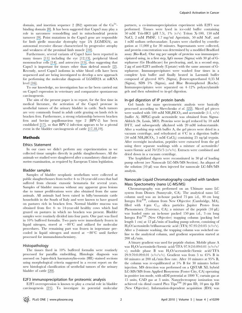
urothelial cells from healthy cattle (Figure 4). Capn3 was weakly
detected in urothelial cells from apparently unaffected mucosa of
the neoplastic bladders (Figure 5). In urothelial cancer cells, Capn3
was observed mostly in the cytoplasm. Nuclear positivity was also
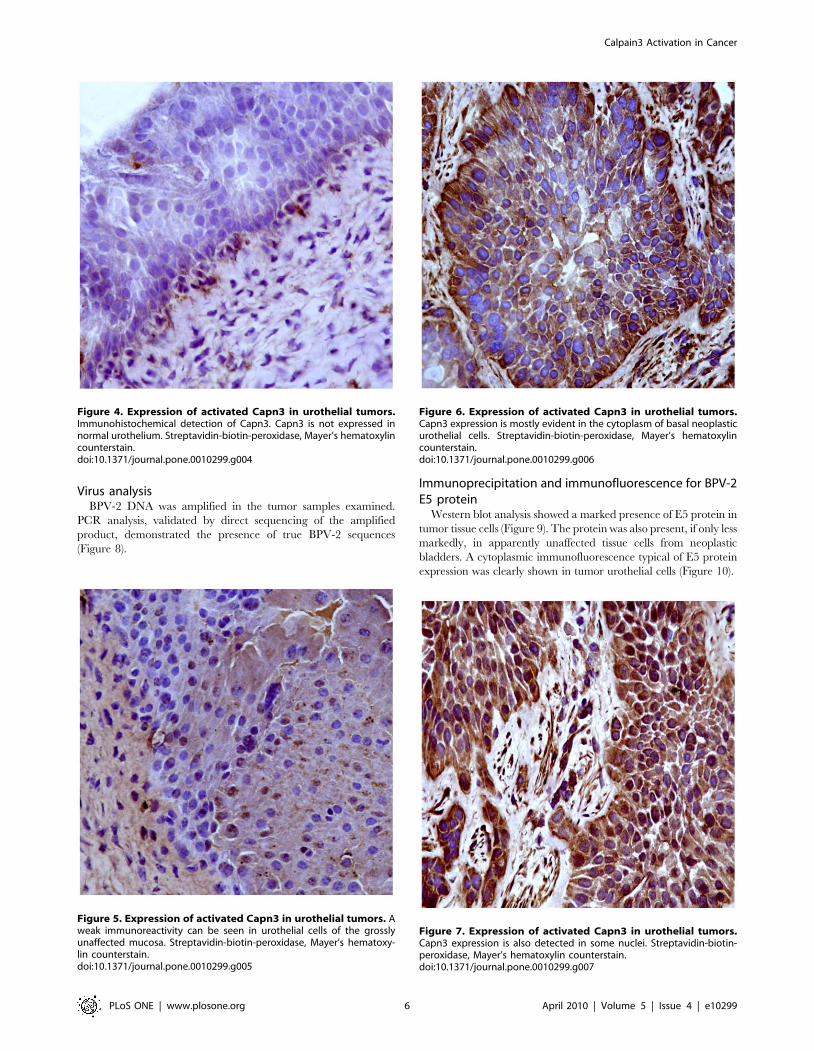
manifest. Basal cells were the predominant ones showing a strong
immunoreactivity for Capn3 (Figures 6 and 7).
Figure 2. Expression of activated Capn3 in urothelial tumors. Detection of IS1 and IS2 inserts in Capn3 transcript. PCR analysis was performedon the cDNAs synthesized from bladders isolated from pathological animals or human PBMC as reported in Methods. The primer pair utilized todetect Capn3 IS1 insert was Sn831/Asn1845 (A) and that utilized to detect IS2 insert was Sn1884/Asn2669 (B). PCR conditions are reported inMethods. PCR products were separated by electrophoresis on 1.2% agarose gel. Marker sizes: GeneRuler 100 bp DNA ladder (Fermentas). cDNAamounts are normalized with respect to equal GAPDH levels. The expected sizes for the PCR fragments in (A) are 1018 bp with IS1, and 829 bpwithout IS1. The expected sizes for the PCR fragments in (B) are 805 bp with IS2, and 577 bp without IS2.doi:10.1371/journal.pone.0010299.g002
Figure 3. Expression of activated Capn3 in urothelial tumors.Ion-exchange chromatography on bovine bladders. Crude extracts wereprepared from normal (full circles) or pathological (empty circles) bovinebladders as reported in methods. Aliquots (35 mg) were submitted toion-exchange chromatography and calpain activity was assayed onaliquots (100 ml) of the eluted fractions as described in Methods.doi:10.1371/journal.pone.0010299.g003
Calpain3 Activation in Cancer
PLoS ONE | www.plosone.org 5 April 2010 | Volume 5 | Issue 4 | e10299
Virus analysisBPV-2 DNA was amplified in the tumor samples examined.
PCR analysis, validated by direct sequencing of the amplified
product, demonstrated the presence of true BPV-2 sequences
(Figure 8).
Immunoprecipitation and immunofluorescence for BPV-2E5 protein
Western blot analysis showed a marked presence of E5 protein in
tumor tissue cells (Figure 9). The protein was also present, if only less
markedly, in apparently unaffected tissue cells from neoplastic
bladders. A cytoplasmic immunofluorescence typical of E5 protein
expression was clearly shown in tumor urothelial cells (Figure 10).
Figure 4. Expression of activated Capn3 in urothelial tumors.Immunohistochemical detection of Capn3. Capn3 is not expressed innormal urothelium. Streptavidin-biotin-peroxidase, Mayer’s hematoxylincounterstain.doi:10.1371/journal.pone.0010299.g004
Figure 5. Expression of activated Capn3 in urothelial tumors. Aweak immunoreactivity can be seen in urothelial cells of the grosslyunaffected mucosa. Streptavidin-biotin-peroxidase, Mayer’s hematoxy-lin counterstain.doi:10.1371/journal.pone.0010299.g005
Figure 6. Expression of activated Capn3 in urothelial tumors.Capn3 expression is mostly evident in the cytoplasm of basal neoplasticurothelial cells. Streptavidin-biotin-peroxidase, Mayer’s hematoxylincounterstain.doi:10.1371/journal.pone.0010299.g006
Figure 7. Expression of activated Capn3 in urothelial tumors.Capn3 expression is also detected in some nuclei. Streptavidin-biotin-peroxidase, Mayer’s hematoxylin counterstain.doi:10.1371/journal.pone.0010299.g007
Calpain3 Activation in Cancer
PLoS ONE | www.plosone.org 6 April 2010 | Volume 5 | Issue 4 | e10299

Western blot analysisTo establish the expression levels of E2F3 protein, we
performed a western blot analysis (Figure 11) and we observed
that E2F3 was markedly overexpressed in tumor samples. Since
E2F3 interacts with the retinoblastoma tumor suppressor protein
(pRb), we also investigated pRb expression and detected a severe
downregulation, to actual inactivation of pRb protein in the same
cancer samples (Figure 12). Finally, we performed an investigation
to validate the effective presence of SerpinA3-7 protein identified
by a single peptide by means of proteomic analysis. We showed
that the molecular complex obtained using an anti-E2F3 antibody
was also composed of SerpinA3-7 (Figure 12).
Discussion
Although various calpain substrate proteins are associated with
carcinogenesis [2], the molecular identities of these substrates are
largely unknown [26]. It has been suggested that Capn3 and the
conventional calpains have common substrate specificities, as
several proteins known to be potential targets of Capn3 appear the
same substrates of conventional calpains [9,26].
Recently, calpains have been shown to be involved in proteolysis
of NORE1A, a potential Ras effector, and of Ras-association
domain family 1 (RASSF1A) proteins. Downregulation of NORE1A
and RASSF1A proteins might be involved in some carcinogenesis
mechanisms [27]. Calpain 4 (Capn4) has been found to be associated
with metastasis and recurrence of hepatocellular carcinoma (HCC).
Therefore, it has been proposed that Capn4 might be a target for
cancer therapy [28]. In addition, Calpain 6 (Capn6) expression was
found increased in some uterine tumors [29].
High levels of Capn3 without any protease activity were
detected in human melanoma cell lines. Therefore, it has been
suggested that Capn3 may play a pro-apoptotic role in melanoma
cells, and it could be a useful diagnostic marker for monitoring
melanoma development and progression [30,31].
In our cases, Capn3 expression was detected in twelve
papillomavirus-positive neoplastic urothelial lesions. Capn3 pro-
tein was initially identified with nanoscale liquid chromatography
coupled with tandem mass spectrometry (nano LC-MS/MS) in a
co-immunoprecipitation experiment on E2F3. Capn3 expression
was confirmed by reverse transcription polymerase chain reaction
(RT-PCR) and its Ca2+-dependent proteolytic activity was assayed
following ion exchange chromatography. Morphologically, Capn3
expression was documented by immunohistochemical examina-
tion. To our knowledge, this is the first study showing Capn3
activation in spontaneous oncogenesis in medical literature. We
did not investigate whether any oncogenic proteins of BPV-2 could
be involved in Capn3 activation. It has been shown that human
papillomavirus (HPV) E7 oncoprotein is able to down-regulate
pRb expression [32], since it binds to m-calpain and activates its
proteolytic activity, resulting in cleavage of pRb [6,33]. Other
mechanisms are likely to exist in animal cancers in which BPV-2
infection may play a central role. We have also shown that
papillomavirus-associated urothelial cancers of cattle are charac-
terized by an overexpression of sigma 2 receptors [34]. These
receptors are known to play a crucial role in modulating
cytoplasmic [Ca2+] in cancer cells [35]. It is reasonable to suggest
that sigma 2 receptors may be involved in Capn3 activation, as it
has been shown that even small increases of resting cytoplasmic
[Ca2+] are responsible for Capn3 proteolytic activity [36,37].
The role of the Capn3 protein in urothelial bladder cancer still
remains to be elucidated. We have shown also E2F3 protein
overexpression and a dramatic decrease of Rb protein in bladder
cancers. Taken together our data allow us to suggest that pRb may
be a calpain substrate also in bovine urothelial tumors. E2F3 levels
are known to be regulated by pRb and have a crucial role in
activating cell proliferation in human bladder cancers [21,38–41].
Figure 8. Expression of activated Capn3 in urothelial tumors.PCR amplification of urinary bladder samples. Lanes:1–3, tumorsamples; 4, positive control of BPV-2 plasmid; 5–6, negative controlwith no DNA added; M, molecular mass marker #8 (Roche, Milan, Italy).doi:10.1371/journal.pone.0010299.g008
Figure 9. Expression of activated Capn3 in urothelial tumors.E5 oncoprotein expression detected by immunoprecipitation. Lane 1,negative normal bladder tissue from healthy cattle; lanes 2–6 neoplasticsamples showing an evident E5 expression except in lane 2.doi:10.1371/journal.pone.0010299.g009
Figure 10. Expression of activated Capn3 in urothelial tumors.E5 oncoprotein documented by scanning laser confocal microscope.Immunofluorescence is evident in the cytoplasm of several neoplasticcells (arrows).doi:10.1371/journal.pone.0010299.g010
Figure 11. Expression of activated Capn3 in urothelial tumors.Western blot analysis to characterize the expression of E2F3. Lane 1,healthy animal as control; lanes 2–7, E2F3 protein appears to beoverexpressed in all examined tumor samples.doi:10.1371/journal.pone.0010299.g011
Calpain3 Activation in Cancer
PLoS ONE | www.plosone.org 7 April 2010 | Volume 5 | Issue 4 | e10299

As Capn 3 was only detected in molecular complexes immuno-
precipitated with E2F3, it can be argued that the proteolitically
active form of Capn3 protein might be directly involved in
molecular pathways leading to the overexpression of E2F3
transcription factors, very likely via Rb protein degradation.
Furthermore, Capn3 is known to be a regulator of the
conventional calpain system [42]. Therefore, it cannot be excluded
that Capn3 may play a role in regulating E2F3 protein expression
in an indirect manner, as conventional m-calpain is known to
promote the degradation of Rb protein [6]. It has been suggested
that inactivation of Rb pathway and overexpression of E2F3 are
obligate events in some human bladder tumors [21]. Our
suggestion appears to be strengthened by the proteomic profiles
of the other complexes from the same samples, immunoprecipi-
tated with antibodies, vs proteins known to play a role in BPV-2
infection, such as PDGF receptor beta [43]. No Capn3 was
detected in them. Indeed, extracellular matrix components such as
lumican and decorin were predominantly detected in these
complexes (data not shown). Our observations appear to be
consistent with the very interesting emerging evidence of a broader
role of PDGFRs in tumor stromogenesis [44], and with the
increasing interest in the role of decorin and lumican via the
tyrosine kinase receptor family in cancer biology [45].
Further studies are needed to better understand the role of the
calpain system in bladder carcinogenesis. If our results are
validated by other studies, Capn3 will definitely appear to play
an important role in the molecular pathway of bovine urinary
bladder tumors. As a consequence, it may prove useful as a
diagnostic biomarker for monitoring urothelial tumor develop-
ment and progression and as a potential target for cancer therapy.
Cattle suffering from urothelial tumors, whose incidence may be
,90% in adult animals [20,46], may serve as an animal model
useful for gaining insight into new molecular pathways involved in
naturally occurring bladder carcinogenesis and for evaluating in
vivo potential new drugs against specific targets, or for proposing
novel therapeutic strategies that are urgently needed nowadays
[47]. It is also worth to remember that the establishment of reliable
and reproducible animal models for bladder cancer remains an
ongoing challenge, since developing therapeutic agents requires in
vivo models [47]. Furthermore, the UICC Study Group suggested
that biological models may still be trail blazing for the natural
history of cancer, although molecular models have fostered an
impressive progress over the last decades [48]. Finally, it is
worthwhile noting that cattle has already been investigated and, it
has been found to be a good animal model for several other
human diseases [49,50].
Author Contributions
Conceived and designed the experiments: SR FR. Performed the
experiments: SR. Analyzed the data: SR RDT CR RS VR MG GB MA
OP GC FR. Wrote the paper: SR FR.
References
1. Goll DE, Thompson VF, Li H, Wei W, Cong J (2003) The calpain system.
Physiol Rev 83: 731–801.
2. Suzuchi K, Hata S, Kawabata Y, Sorimachi H (2004) Structure, activation, and
biology of calpain. Diabetes 53 (Suppl. 1): S12–S18.
3. Stifanese R, Averna M, De Tullio R, Salamino F, Cantoni C, et al. (2008) Role
of calpain-calpastatin system in the density-dependent growth arrest. Arch
Biochem Biophys 479: 145–152.
4. Demarchi F, Bertoli C, Copetti T, Tanica I, Brancolini C, et al. (2006) Calpain
is required for macroautophagy in mammalian cells. J Cell Biol 175: 595–605.
5. Demarchi F, Bertoli C, Copetti T, Eskelinen E-L, Schneider C (2007) Calpain as
a novel regulator of autophagosome formation. Autophagy 3: 235–237.
6. Darnell GA, Schroder WA, Antalis TM, Lambley E, Major L, et al. (2007)
Human papillomavirus E7 requires the protease calpain to degrade the
retinoblastoma protein. J Biol Chem 282: 37492–37500.
7. Demarchi F, Schneider C (2007) The calpain system as a modulator of stress/
damage response. Cell Cycle 6: 136–138.
8. Sorimachi H, Imajoh-Ohmi Y, Kawasaki H, Ohno S, Minami Y, et al. (1989)
Molecular cloning of a novel mammalian calcium-dependent protease distinct
from both m- and m-types. Specific expression of the mRNA in skeletal muscle.
J Biol Chem 264: 20106–20111.
9. Cohen N, Kudryashova E, Kramerova I, Anderson LVB, Beckmann JS, et al.
(2006) Identification of putative in vivo substrates of calpain 3 by comparative
proteomics of overexpressing transgenic and nontransgenic mice. Proteomics 6:
6075–6084.
10. Richard I, Broux O, Allamand V, Fougerousse F, Chiannilkulchai N, et al.
(1995) Mutations in the proteolytic enzyme calpain 3 cause limb-girdle muscular
dystrophy type 2A. Cell 81: 27–40.
11. Kawabata Y, Hata S, Ono Y, Ito Y, Suzuki K, et al. (2003) Newly identified
exons encoding novel variants of p94/calpain 3 are expressed ubiquitously and
overlap the a-glucosidase C gene. FEBS Lett 555: 623–630.
12. Azuma M, Fukiage C, Higashine M, Nakajima T, Ma H, et al. (2000)
Identification and characterisation of a retina-specific calpain (Rt88) form rat.
Curr Eye Res 21: 710–720.
13. Ma H, Shih M, Hata I, Fukiage C, Azuma M, et al. (2000) Lp85 calpain is an
enzymatically active rodent-specific isozyme of lens Lp82. Curr Eye Res 20:
183–189.
14. De Tullio R, Stifanese R, Salamino F, Pontremoli S, Melloni E (2003)
Characterization of a new p94-like calpain form in human lymphocytes.
Biochem J 375: 689–696.
15. Konig N, Raynaud F, Feane H, Durand M, Mestre-Frances N, et al. (2003)
Calpain 3 is expressed in astrocytes of rat and Microcebus brain. J Chem
Neuroanat 25: 129–136.
16. Blazquez L, Azpitarte M, Saenz A, Goicoechea M, Otaegui D, et al. (2008)
Characterization of novel CAPN3 isoforms in white blood cells: an alternative
approach for limb-girdle muscular dystrophy 2A diagnosis. Neurogenetics 9:
173–182.
17. Campo MS, Jarrett WFH, Barron RJ, O’Neil BW, Smith KT (1992) Association
of bovine papillomavirus type 2 and bracken fern with bladder cancer in cattle.
Cancer Res 52: 6898–6904.
18. Borzacchiello G, Roperto F (2008) Bovine papillomaviruses, papillomas and
cancer in cattle. Vet Res 39: 45–63.
19. Roperto S, Brun R, Paolini F, Urraro C, Russo V, et al. (2008) Detection of
bovine papillomavirus type 2 (BPV-2) in the peripheral blood of cattle with
urinary bladder tumours: possible biological role. J Gen Virol 89: 3027–3033.
20. Roperto S, Borzacchiello G, Brun R, Leonardi L, Maiolino P, et al. (2009) A
review of bovine urothelial tumours and tumour-like lesions of the urinary
bladder. J Comp Pathol doi:10.1016/j.jcpa.2009.08.156.
21. Hurst CD, Tomlinson DC, Williams SV, Platt FM, Knowles MA (2008)
Inactivation of the Rb pathway and overexpression of both isoforms of E2F3 are
obligate events in bladder tumours with 6p22 amplification. Oncogene 27:
2716–2727.
22. Shevchenko A, Wilm M, Vorm O, Mann M (1996) Mass spectrometric
sequencing of proteins silver-stained polyacrylamide gels. Anal Chem 68:
850–858.
23. Pontremoli S, Melloni E, Viotti PL, Michetti M, Salamino F, et al. (1991)
Identification of two calpastatin forms in rat skeletal muscle and their
susceptibility to digestion by homologous calpains. Arch Biochem Biophys
288: 646–652.
24. Rey MA, Davies PL (2002) The protease core of the muscle-specific calpain p94
undergoes Ca2+2dependent intramolecular autolysis. FEBS Lett 532: 401–406.
25. Sorimachi H, Kinbara K, Kimura S, Takahashi M, Ishiura S, et al. (1995)
Muscle-specific calpain, p94, responsible for limb girdle muscular dystrophy type
Figure 12. Expression of activated Capn3 in urothelial tumors.Western blot analysis to characterize the expression of pRb andconstitutive SerpinA3-7 protein. A downregulation of pRb proteinexpression is severely evident in all tumor samples. The SerpinA3-7protein was normally expressed in tumor samples.doi:10.1371/journal.pone.0010299.g012
Calpain3 Activation in Cancer
PLoS ONE | www.plosone.org 8 April 2010 | Volume 5 | Issue 4 | e10299

2A, associates with connectin through IS2, a p94-specific sequence. J Biol Chem
270: 31158–31162.26. Ono Y, Hayashi C, Doi N, Kitamura F, Shindo M, et al. (2007) Comprehensive
survey pf p94/calpain 3 substrates by comparative proteomics-Possible
regulation of protein synthesis by p94. Biotechnol J 2: 565–576.27. Kuznetsov S, Khokhlatchev AV (2008) The growth and tumor suppressor
NORE1A and RASSF1A are target for calpain-mediated proteolysis. PloS ONE3: e3997. doi: 101371/journal.pone.0003997.
28. Bai D-S, Dai Z, Zhou J, Liu Y-K, Qiu S-J, et al. (2009) Capn4 overexpression
underlies tumor invasion and metastasis after liver transplantation forhepatocellular carcinoma. Hepatology 49: 460–470.
29. Lee S-J, Choi Y-L, Lee E-J, Kim B-G, Bae D-S, et al. (2007) Increasedexpression of calpain 6 in uterine sarcomas and carcinosarcomas: an
immunohistochemical analysis. Int J Gynecol Cancer 17: 248–253.30. Huynh KM, Kim G, Kim D-J, Yang S-J, Park S-m, et al. (2009) Gene
expression analysis of terminal differentiation of human melanoma cells
highlights global reductions in cell cycle-associated genes. Gene 433: 32–39.31. Moretti D, del Bello B, Cosci E, Biagioli M, Miracco C, et al. (2009) Novel
variants of muscle calpain 3 identified in human malenoma cells: cisplastin-induced changes in vitro and differential expression in melanocytic lesions.
Carcinogenesis 30: 960–967.
32. Zhang W, Li J, Kanginakudru S, Zhao W, Yu X, et al. (2009) The humanpapillomavirus type 58 E7 oncoprotein modulates cell cycle regulatory proteins
and abrogates cell cycle checkpoints. Virology, doi.10.1016/j.virol.2009.10.051.33. Tonnetti L, Netzel-Arnett S, Darnell GA, Hayes T, Buzza MS, et al. (2008)
SerpinB2 protection of retinoblastoma protein from calpain enhances tumourcell survival. Cancer Res 68: 5648–5657.
34. Roperto S, Colabufo NA, Inglese C, Urraro C, Brun R, et al. (2010) Sigma-2
receptor expression in bovine papillomavirus-associated urinary bladdertumours. J Comp Pathol 142: 19–26.
35. Zeng C, Vangveravong S, Xu J, Chang KC, Hotchkiss RS, et al. (2007)Subcellular localization of sigma-2 receptors in breast cancer cells using two-
photon and confocal microscopy. Cancer Res 67: 6708–6716.
36. Diaz BEG, Gauthier S, Davies PL (2006) Ca2+-dependency of calpain 3 (p94)activation. Biochemistry 45: 3714–3722.
37. Murphy RM, Lamb GD (2009) Endogenous calpain-3 is primarily governed bysmall increases in resting cytoplasmic [Ca2+] and is not dependent on stretch.
J Biol Chem 284: 7811–7819.
38. Feber A, Clark J, Goodwin G, Dodson AR, Smith PH, et al. (2004)
Amplification and overexpression of E2F3 in human bladder cancer. Oncogene
23: 1627–1630.
39. Oeggerli M, Tomovska S, Schraml P, Calvano-Forte D, Schafroth S, et al.
(2004) E2F3 amplification and overexpression is associated with invasive tumor
groth and rapid tumor cell proliferation in urinary bladder cancer. Oncogene
23: 5616–5623.
40. Olsson AY, Feber A, Edwards S, te Poele R, Giddings I, et al. (2007) Role of
E2F3 expression in modulating cellular proliferation rate in human bladder and
prostate cancer cells. Oncogene 26: 1028–1037.
41. Wu L, Timmers C, Malti B, Saavedra HI, Sang L, et al. (2001) The E2F1-3
transcription factors are essential for cellular proliferation. Nature 414: 457–462.
42. Ono Y, Kakinuma K, Tori F, Irie A, Nakagawa K, et al. (2004) Possible
regulation of the conventional calpain system by skeletal muscle-specific calpain,
p94/Calpain 3. J Biol Chem 279: 2761–2771.
43. Borzacchiello G, Russo V, Gentile F, Roperto F, Venuti A, et al. (2006) Bovine
papillomavirus E5 oncoprotein binds to the activated form of the platelet-derived
growth factor b receptor in naturally occurring bovine urinary bladder tumours.
Oncogene 25: 1251–1260.
44. Andrae J, Gallini R, Betsholtz C (2008) Role of platelet-derived growth factors in
physiology and medicine. Genes Dev 22: 1276–1312.
45. Goldoni S, Iozzo RV (2008) Tumor microenvironment: Modulation by decorin
and related molecules harbouring leucin-rich tandem motifs. Int J Cancer, 2008;
123. pp 2473–2479.
46. Pamukcu AM, Price JM, Bryan GT (1976) Naturally occurring and bracken
fern-induced bovine urinary bladder tumors. Vet Pathol 13: 110–122.
47. Chan E, Patel A, Heston W, et al. (2009) Mouse orthotopic models for bladder
cancer research. BJU Int, 2009; 104: 1286–1291.
48. Burger MM (2000) UICC Study Group on basic and clinical cancer research:
animal models for the natural history of cancer. Int J Cancer, 2000, 85:
303–305.
49. Besser TE, Potter KA, Bryan GM, et al. (1990) An animal model of the Marfan
syndrome. Am J Med Genet, 1990, 37: 159–165.
50. Drogemuller C, Drogemuller M, Leeb T (2008) Identification of a missense
mutation in the bovine ATP2A1 gene in congenital pseudomyotonia of
Chianina cattle: an animal model of human Brody disease. Genomics, 2008;
92: 474.477.
Calpain3 Activation in Cancer
PLoS ONE | www.plosone.org 9 April 2010 | Volume 5 | Issue 4 | e10299