Nucleic acid vaccination of Brucella abortus ribosomal L7 L12 gene elicits immune response
Upload
khangminh22Category
view
3download
0
Yale UniversityEliScholar – A Digital Platform for Scholarly Publishing at Yale
Yale Medicine Thesis Digital Library School of Medicine
1990
Detection of human papillomavirus by nucleic acidhybridization as an adjunct to the papanicolaousmearJulia Ann SchillingerYale University
Follow this and additional works at: http://elischolar.library.yale.edu/ymtdl
This Open Access Thesis is brought to you for free and open access by the School of Medicine at EliScholar – A Digital Platform for ScholarlyPublishing at Yale. It has been accepted for inclusion in Yale Medicine Thesis Digital Library by an authorized administrator of EliScholar – A DigitalPlatform for Scholarly Publishing at Yale. For more information, please contact [email protected].
Recommended CitationSchillinger, Julia Ann, "Detection of human papillomavirus by nucleic acid hybridization as an adjunct to the papanicolaou smear"(1990). Yale Medicine Thesis Digital Library. 3137.http://elischolar.library.yale.edu/ymtdl/3137

YALE MEDICAL LIBRARY
3 9002 08627 9545

2 Permission for photocoping or microfilming of "
(Title of thesis)
for the purpose of individual scholarly consultation or reference
is hereby granted by the author. This permission is not to be
interpreted as affecting publication of this work or otherwise
placing it in the public domain, and the author reserves all rights
of ownership guaranteed under common law protection of unpublished
manuscripts.
MR- ‘U *
Date

Digitized by the Internet Archive in 2017 with funding from
The National Endowment for the Humanities and the Arcadia Fund
https://archive.org/details/detectionofhumanOOschi


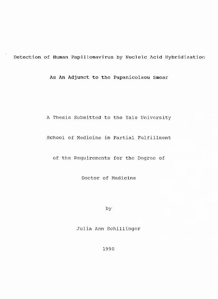
Detection of Human Papillomavirus by Nucleic Acid Hybridization
As An Adjunct to the Papanicolaou Smear
A Thesis Submitted to the Yale University
School of Medicine in Partial Fulfillment
of the Requirements for the Degree of
Doctor of Medicine
by
Julia Ann Schillinger
1990

Med . I 'b
T M B> f-r ^

ABSTRACT
DETECTION OF HUMAN PAPILLOMAVIRUS BY NUCLEIC ACID HYBRIDIZATION
AS AN ADJUNCT TO THE PAPANICOLAOU SMEAR
JULIA ANN SCHILLINGER
1990
We compared methods for detecting Human
Papillomavirus [HPV] deoxyribonucleic acid [DNA] in cervical
cytology specimens. Endocervical swabs were obtained from 40
women referred to a Gynecologic oncology clinic for a
previously abnormal Papanicolaou [Pap] smear. We employed a
modified filter in situ method [ViraPap] for the detection of
HPV DNA, followed by HPV typing [ViraType]. Sixteen of 40
specimens were positive for the presence of HPV DNA by
hybridization with probes specific to HPV of types 6, 11, 16,
18, 31, 33, and 35. Of these, 5 patients had negative Pap
smears but evidence of disease by other parameters
(endocervical curettage [ECC], cervical biopsy [CBx],
colposcopy) . Four of these 5 patients with negative Pap
smears were infected with a type of HPV associated with a high
risk of developing cervical disease; types 16/18. 7 patients
had positive Pap smears with evidence of disease on cervical
biopsy, but had negative ViraPap results. We then used the


polymerase chain reaction [PCR] to amplify segments of the
HPV-16 E6 and E7 open reading frames [ORFs] . The E6 ORF was
found to be the most sensitive. In three independent PCR
assays, 13 specimens contained the HPV-16 E6 DNA. These
included all of those typed as 16/18 by the ViraType method
as well as 5 additional samples not detected by the
hybridization methods; 3 with abnormal Pap smears, and 2 with
normal Pap smears but evidence of disease by CBx. We conclude
that in a population at high risk for cervical disease, the
ViraPap can be a useful adjunct to the Pap smear in the
detection of disease and that PCR of the HPV-16 E6 ORF may be
used as a sensitive, but more labor intensive, confirmatory
test.

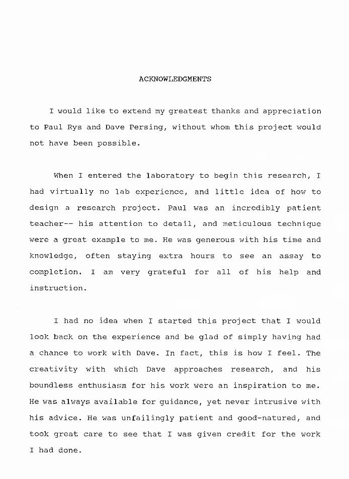
ACKNOWLE DGMENTS
I would like to extend my greatest thanks and appreciation
to Paul Rys and Dave Persing, without whom this project would
not have been possible.
When I entered the laboratory to begin this research, I
had virtually no lab experience, and little idea of how to
design a research project. Paul was an incredibly patient
teacher— his attention to detail, and meticulous technigue
were a great example to me. He was generous with his time and
knowledge, often staying extra hours to see an assay to
completion. I am very grateful for all of his help and
instruction.
I had no idea when I started this project that I would
look back on the experience and be glad of simply having had
a chance to work with Dave. In fact, this is how I feel. The
creativity with which Dave approaches research, and his
boundless enthusiasm for his work were an inspiration to me.
He was always available for guidance, yet never intrusive with
his advice. He was unfailingly patient and good-natured, and
took great care to see that I was given credit for the work
I had done.


In addition to all of Paul and Dave's wonderful personal
and intellectual qualities it was just downright fun to work
with them. The lab was a great place to be— full of laughter
and wit at the same time that serious work was being done. It
was a real haven for me during a year that was otherwise
somewhat fragmented, and I feel very lucky to have been a part
of it.
I would also like to extend my thanks to my thesis advisor
Stephen Edberg, for his support of this project, and to Stuart
Flynn in the Pathology Department for his interest, and for
his contributions of time and energy in rescreening all the
Pap smears in this study.
Joseph and Setsuko Chambers very kindly obtained the
specimens used in this study, and were available for
discussion of the ramifications of using the proposed test.


DEDICATION
I dedicate this thesis to my friends at the Yale School
of Medicine who have helped to make these four years a joyous
time for me. Medical school is undoubtedly an innately
interesting and challenging experience—it is the people with
whom one works which make it something more. My close friends
during medical school were a great source of strength, humor,
and joy during these past four years. I have a great deal of
respect and appreciation for them, and I dedicate this thesis
to their collective friendship.

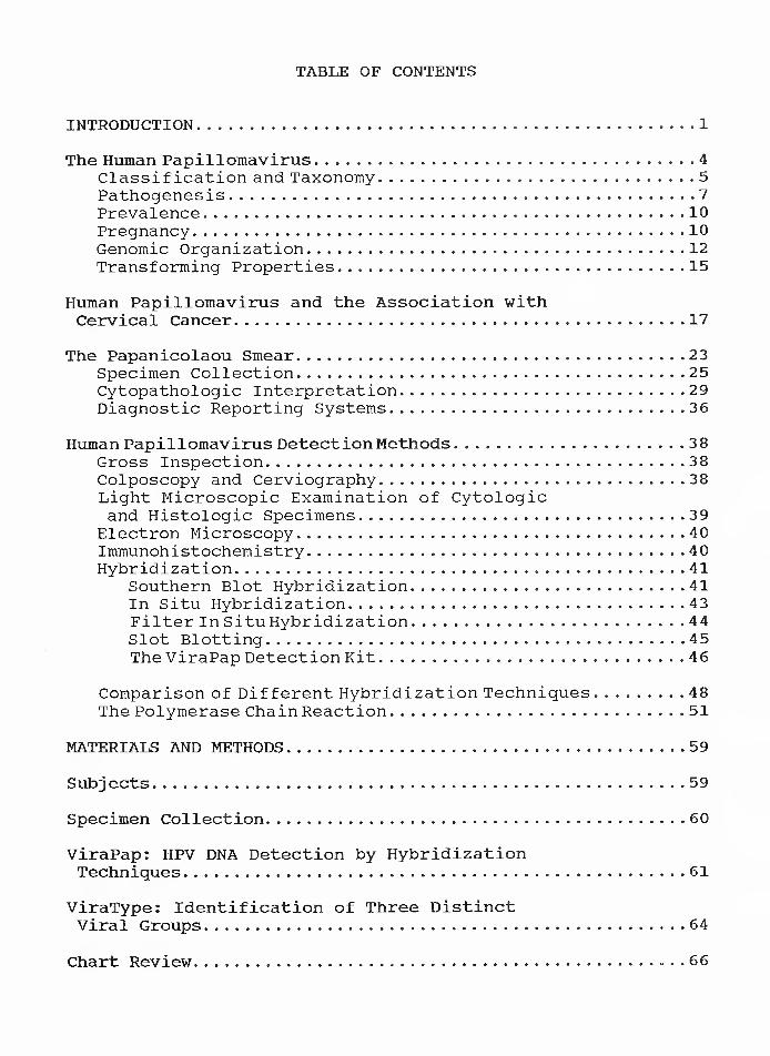
TABLE OF CONTENTS
INTRODUCTION.1
The Human Papillomavirus.4 Classification and Taxonomy.5 Pathogenesis.7 Prevalence...10 Pregnancy.10 Genomic Organization.12 Transforming Properties...15
Human Papillomavirus and the Association with Cervical Cancer.17
The Papanicolaou Smear.23 Specimen Collection.25 Cytopathologic Interpretation.29 Diagnostic Reporting Systems. ..36
Human Papillomavirus Detection Methods..38 Gross Inspection.38 Colposcopy and Cerviography.38 Light Microscopic Examination of Cytologic and Histologic Specimens.39
Electron Microscopy.40 Immunohistochemistry.40 Hybridization.41
Southern Blot Hybridization.41 In Situ Hybridization.43 Filter In Situ Hybridization.44 Slot Blotting.45 The ViraPap Detection Kit.46
Comparison of Different Hybridization Techniques.48 The Polymerase Chain Reaction. 51
MATERIALS AND METHODS. 59
Subjects. 59
Specimen Collection.60
ViraPap: HPV DNA Detection by Hybridization Techniques. 61
ViraType: Identification of Three Distinct Viral Groups.64
Chart Review 66


Polymerase Chain Reaction. 67
DNA Extraction of Remaining ViraPap Specimens.69
Restriction Digest on Specimens Identified as
16/18 Positive by Either ViraType or PCR.71
Determination of Specimen Adequacy.72
RESULTS.77
Chart Review.77
ViraPap Results.79
ViraType Results...80
PCR Results.82
Restriction Digest. 84
Sample Adequacy...86
DISCUSSION.87
Subjects. 87
ViraPap and Pap Smear. ..88
Typing. 98
Polymerase Chain Reaction.100
CONCLUSIONS. ..104
APPENDIX. 106


1
INTRODUCTION
In 1989, 6,000 women died of cervical cancer, and 13,000
new cases of invasive cervical cancer were diagnosed (43).
Many studies have established a strong association between
infection with the Human Papillomavirus (HPV) and the
development of cervical cancer and its' precursor lesions (10,
14, 15, 16, 18, 33, 36, 38, 57, 65, 83); over 90% of cervical
cancers examined have been found to contain HPV DNA seguences
integrated into the host cell genome (38, 64). There has been
tremendous interest and attention focussed on the Human
Papillomavirus in recent years as a model for viral
oncogenesis. To date more than 60 different types of HPV have
been identified. The various types show an apparent cellular
tropism, with twenty-three preferentially infecting the
genital tract. Among these types there appears to be a range
of oncogenic potential.
Current understanding of the biological behavior of
cervical cancer suggests that these malignancies are the most
severe in a range of precursor lesions usually described as
cervical dysplasias. Because the prognosis for a woman with
cervical cancer is determined by the stage at which the
disease is detected, early diagnosis is the best means of


2
reducing mortality. The Papanicolaou (Pap) smear has been
extremely effective in identifying occult carcinomas and
premalignant lesions of the cervix, and the mortality and
morbidity associated with cervical cancer have decreased
dramatically in the past 40 years. Nonetheless, a percentage
of invasive cancers and its precursor lesions go undetected
by Pap smears each year. HPV is notorious for confounding the
interpretation of Pap smears, blurring the distinction between
non-neoplastic proliferative processes and preneoplastic
changes, and contributing to ambiguity in the grading of
lesions. Increasing recognition of the prevalence of HPV
infection in the general population has spurred an interest
in a more reliable means of detection.
In 1989 the Food and Drug Administration licensed a test
kit for the detection of HPV in endocervical swabs and
cervical biopsy specimens. This test, known as ViraPap and
manufactured by Bethesda Research Laboratories, utilizes
filter hybridization techniques to detect virus in processed
specimens. The ViraPap test uses P labelled probes composed
of RNA sequences complementary to the DNA of HPV types 6, 11,
16, 18, 31, 33, and 35; the seven types most frequently found
to infect the genital tract.
This study was designed to assess the clinical usefulness
of detecting HPV in specimens of exfoliated cervical cells.


3
Because screening and diagnostic tests already exist for
recognition of the morphologic changes leading to cervical
cancer, the usefulness would have to hinge on the tests'
ability to provide information the alternative methods could
not. In this study the detection of HPV in an endocervical
swab specimen by hybridization technigue was compared to the
results of concurrently taken Pap smears, biopsies, and
colposcopy in 40 patients. Because of the apparent range in
the oncogenic potential of different HPV types, typing was
undertaken on all the specimens shown to contain HPV DNA.
Specimens shown to contain HPV DNA of type 16 were confirmed
by use of the polymerase chain reaction, a highly sensitive
means of DNA amplification.
An assessment of a diagnostic test reguires an in depth
understanding of the phenomenon being examined, as well as an
appreciation for how the proposed test adds to or complements
the existing methodology. With these ends in mind, this paper
will first address the current understanding of the Human
Papillomavirus and its proposed relation to cervical cancer,
and then will review the uses and limitations of existing
screening and diagnostic tests, with particular attention to
the Pap smear and hybridization technique.


4
The Human Papillomavirus
The Human Papillomavirus is one of the genus
Papillomavirus, belonging to the family of viruses known as
Papovaviridae, which include the polyomaviruses and
vacuolating viruses. Papillomaviruses derive their name from
the Latin word 'papilla', meaning nipple or pustule, and the
Greek suffix -oma, denoting tumor. This is a befitting
etymology for a virus known to cause warts. The viral
etiology of warts was first demonstrated in 1905, when Ciuffo
demonstrated viral transmission using a cell-free filtrate
(10). The first Papillomavirus was described by Richard E.
Shope, when he identified the Cottontail Rabbit Papillomavirus
as the etiologic agent in infectious Papillomatosis in rabbits
(44) .
Transmissible Papillomaviruses have now been described in
over twenty species of animal, including cows, horses, deer,
rabbits, dogs, birds, elephants, and primates, including man.
Transmission frequently occurs between members of the same
species, a principle which has had major economic
ramifications for cattle raisers and for kennel owners who
must vaccinate their dogs with a prophylactic vaccine prepared
from a suspension of Canine Oral Papillomavirus.


5
Papillomaviruses from cattle, deer, sheep and Cottontail
rabbits can induce tumors when experimentally transmitted to
other species, but naturally occurring infectivity between
species seems only a slight possibility (8) . A unique HPV,
type 7, has been identified in the hand warts of butchers and
veterinarians. Hybridization studies using probes made from
BPV1 and HPV1, 2, 3, 4, and 5, were unable to detect the viral
DNA, and restriction analysis showed it to be distinct from
known HPVs and BPV1, 2, 3, and 4. Nonetheless, it is possible
that this viral type represents an example of cross-species
infectivity with an unrecognized bovine, porcine, or ovine
type (45).
Classification and Taxonomy
The Human Papillomavirus, like all others of its type,
is an icosahedral nonenveloped virus containing only DNA and
proteins. Each virion contains a single DNA molecule which
is double stranded and circular. The HPV genomes are all
approximately 8000 base pairs in length, ranging from around
7200 to 8000 base pairs.
Study of the structure and behavior of the Human
Papillomavirus was hampered for many years by the inability
to culture infected cells in vitro. The virus infects
epithelial surfaces, causing proliferations which are usually


6
benign, but which can lead to malignant growth. Replication
of the virus apparently relies on factors present in
differentiating keratinocytes; efforts to create a culture
system capable of supporting such growth have not been
successful as yet (8) . The development of recombinant DNA
technology, with the creation of cloned plasmid containing
HPV DNA, has enabled researchers to delve into the intricacies
of this elusive virus. HPVs have been classified into types
and subtypes based on liquid phase hybridization followed by
SI nuclease digestion. If the viruses being compared show
homology of less than 50% by this method, the unknown is
considered a new type. A subtype is defined as an unknown
which shows more than 50% but less than 100% homology with the
prototype under these conditions. A reliance on known
prototypes clearly limits the ability to identify new,
distantly related viral types. It is important to understand
that when the nucleotide sequences of different viral types
are compared by heteroduplex analysis, far more than 50%
homology may be found (10). The complete genomes of HPV types
1, 5, 6, 8, 11, 16, 18, 31, 33, and 52 have been, or are in
the process of being sequenced (9) . As more extensive
sequence data for all the types emerges, a sequence based
classification system may prove to be clinically useful.
It has long been recognized that different HPV types have
a predilection for infection of different parts of the body.


7
HPV-1 and HPV-2 typically cause plantar warts on the soles of
the feet and hands respectively. Types 6 and 11 are most
frequently found in benign exophytic genital warts, or
condylomata, on the external genitalia and surrounding the
anus. Type 16 and 18 have a preference for the epithelium of
the uterine cervix, and HPV type 13 has only been described
in the oral papillomas of Heck's disease. These observations
have led to a clinical classification of the HPVs by a type-
specific preference for different cell types and anatomic
locations. The broadest categorization groups the types into
those that infect cutaneous surfaces and those that infect
mucous membranes. The preference of different types for
different locations on the body has been suggested to be an
evolutionary response to different types of keratinization
within the body (4 6) .
Pathogenesis
Twenty-three of the 60 HPVs identified thus far have been
found to infect the genital tract:6, 11, 16, 18, 33, 35, 30,
34, 39, 40, 42-45, 51-59 (9). Of these, types 6,11,16, and
18 are most frequently identified. The sexually transmitted
nature of HPV infection was first described in 1954 by Barrett
et al.(30). The authors reported on a cohort of 24 women, all
of whom had first noticed the appearance of genital
condylomata within 4-6 weeks of their respective husbands'


8
return from a military tour in the Far East. When questioned,
all the men admitted to a sexual encounter in the Far East,
and all had recently had penile warts. In 1990, HPV infection
is acknowledged to be the most common sexually transmitted
disease in the United States (84, 87).
The virus is thought to enter the epithelium through any
minor abrasions or breaks in the skin. Infection does not
usually result when innoculums are introduced onto intact skin
(31). The virus infects the basal cells, which rest against
the basement membrane in the lowest strata of the epithelium.
Apparently the virus requires a cell which is still capable
of dividing to initiate infection (8) . The basal cells are
nonpermissive for vegetative viral growth, and HPV DNA has
been detected only in small quantities in the basal and
parabasal cells (32, 33). Papillomavirus DNA is detected with
increasing frequency in the differentiated cells above the
basal layer (33). The virus probably replicates and is
assembled in these keratinizing cells, with the infectious
virions being shed in the desquamating squames. This model
would help to explain why the cervical squamocolumnar
junction, with its exposed junction of rapidly proliferating
cells, would serve as a likely site of viral entry, and is the
site at which around 90% of cervical carcinomas originate (8).
This model also supports the notion that the presence of other
sexually transmitted diseases which inflame or otherwise


9
compromise the integrity of the epithelium (herpes, syphilitic
chancre, trichomonas etc.), may facilitate infection with
viruses such as HPV.
Latent infection may persist by episomal replication in
the basal cells, without progression through the keratinizing
process (10). HPV DNA has been detected in normal appearing
epithelium both adjacent and distant to observed lesions (18,
29, 59). It is not known how long the virus may remain in such
a state or what might instigate a productive infection.
Spontaneous regression of disease is freguently observed,
although whether this a true regression or simply a reversion
to latency is not clear.
There are a number of morphologic features associated
with HPV infection on histologic examination. The findings
associated with productive viral replication are an appearance
of hyperplastic proliferations of the basal and parabasal
cells (acanthosis), a degenerative cytoplasmic vacuolization
(koilocytosis), and variable thickening of the more
superficial cell layers (hyperkeratosis and parakeratosis).
Other findings include nuclear wrinkling, pyknosis,
binucleation, and more severe forms of nuclear atypia
including enlargement, irregular chromatin clumping, and
hyperchromasia.

10
Prevalence
The prevalence of genital tract infection with Human
Papillomavirus is not known. Estimates vary according to the
study design, the subjects and population, the type of
specimen selected for analysis (ie. tissue or cytology
specimen), and most significantly, the detection method used.
Estimates of prevalence rates in the population with normal
cytology range from 9% using filter in situ hybridization to
80% using the recently developed polymerase chain reaction
(18, 29). Any consideration of prevalence must take the above
mentioned variables into account.
Pregnancy
A study examining the prevalence of HPV amongst pregnant
women arrived at an rate of 28% (19). In this study cervical
smears from 92 pregnant and 96 nonpregnant women were tested
for the presence of HPV DNA by Southern blot hybridization and
probes for HPV types 6,11,16 and 18. Pap smears taken
simultaneously with the sample smear showed no evidence of CIN
or HPV infection, and none of the women had a history of a
previously abnormal Pap smear. The subjects were age matched
and were all sexually active.
HPV DNA was detected in 26(28%) of the pregnant subjects,


11
and in 12(12.5%) of the nonpregnant subjects, making the
detection of HPV DNA 2.3 times more frequent in the pregnant
population tested. HPV of type 16 was the type most frequently
identified, accounting for 42.3% of the HPV positive smears
amongst the pregnant women, but only 25% of the positive
smears among the nonpregnant group. When estimates of the
amount of viral DNA in a specimen were made, 85% of those from
pregnant women contained more than 1.0 pg. of HPV DNA, while
the majority of specimens from nonpregnant women (80%),
contained less than 1.0 pg. In both groups of subjects the
amount of viral DNA in HPV 16 positive specimens was higher
than that found in specimens containing other types. The
authors draw on these results to suggest that the state of
pregnancy allows for the reactivation of HPV and increased
viral replication. It must also be considered that an enhanced
ability to sample the squamo-columnar junction during
pregnancy due to columnar eversion might also contribute to
the increased rate of and viral recovery detection.
It is commonly observed that condyloma acuminata often
flourish during pregnancy, only to regress in the post partum
period (47). Both hormonal influences and an altered state of
immunity have been proposed as contributing factors (22).
Studies of immunosuppressed or immunodeficient populations
also show a higher rate of HPV infection and associated
neoplasia. Renal transplant patients on chronic


12
immunosuppressive therapy, and HIV positive patients form the
patient populations for most of these investigations (48, 49) .
Epidermodysplasia Verruciformis (EV) is a rare disease
with an apparently autosomal recessive pattern of inheritance.
It is characterized by an impaired cell mediated immunity, and
by widespread skin lesions induced by HPV of specific types.
Patients with EV have a high incidence of cutaneous cancers,
usually in sun-exposed areas. This disease has served as a
model in which to study the interaction between immunologic
function and HPV infection (8) .
Genomic Organization
Most of the initial work on the genomic organization of
the HPV was worked out using the BPV. The details that follow
concern the BPV1 genome, but will be related to the HPV. All
the Papillomaviruses have the same basic organization; early
transcriptional units E1-E8, two late transcriptional units
LI and L2, as well as a long noncoding region also known as
the upstream regulatory region (URR). The open reading
frames (ORFs) of the Papillomavirus are all located on the
same DNA strand and show considerable overlap. Some function
has been localized to each of the 8 ORFs, with the exception
of E3. The E3, E5, and E8 ORFs are not as highly conserved as
other portions of the genome and may not be present in all


13
Papillomaviruses. Over 90% of the genome is accounted for by
coding regions.
The transforming (early) segment of the BPV1 genome
contains the eight ORFs, E1-E8. The El ORF is the largest of
the eight and overlaps slightly with E2. It has the capacity
to encode a polypeptide of 68 kilodaltons (K). This protein
has been shown by mutational analysis to have a critical role
in episomal replication; cells transformed by virus
selectively mutated in the El ORF show Papillomavirus
integration (50). The E2 ORF encodes a 45K polypeptide, and
overlaps completely with E3, which lacks its own start codon,
and must be spliced to an upstream exon if it is to be
expressed as a functional polypeptide. The BPV E4 also
overlaps with E2 and codes for a 12K polypeptide. Both the E2
and E4 proteins appear to be involved in the transforming
process. The E5 is found at the 38 end of the early region.
It encodes a very hydrophobic polypeptide, a 44 amino acid
membrane-associated transforming protein. The E8 ORF has a
variable location in different Papillomaviruses and is
probably not functional. The E3 and E5 ORF8s are not found in
all the Papillomaviruses.
The E6 and E7 ORFs are found at the 58 part of the
transforming region where they overlap slightly. The two code
for polypeptides of 16K and 14K respectively. These gene


14
products seem to be important in transformation; Harlow et al.
showed that the HPV16 E7 gene product, a nuclear
phosphoprotein, binds to the retinoblastoma protein, the
product of a cellular antioncogene (13). In addition, the
HPV16 E7 protein can cooperate with ras to transform baby rat
kidney cells and transactivates the adenovirus E2 promoter
(51) .
The LI and L2 ORFs of the late region code for
polypeptides with predicted weights of 55.5K and 50.IK
respectively. Both these ORF's code for proteins present in
the Papillomavirus capsid, although LI appears to code for the
majority of the protein, VP1 (8). The noncoding or upstream
regulatory region has several portions which are AT rich, and
several nearly perfect palindromes. It is presumed to contain
control elements for DNA replication and transcription (8).
As mentioned earlier, the Human Papillomaviruses have an
organization very similar to the Bovine Papillomaviruses.
When compared to HPV1, the E8 ORF was not in a corresponding
position, although the other ORFs were in equivalent
locations. Portions of the El, E2, LI and L2, contained
notable homologies. Within the E6 and E7 ORFs, although there
was less homology, there were certain common characteristics.
These common findings were also true of HPV6. For instance,
the amino acid sequence Cys-X-X-Cys is repeated four times in


15
the predicted E6 polypeptide of both genomes (BPV1 and HPVla) .
In the E7 ORF the same motif is repeated twice in both
genomes. There are also similarities in the noncoding regions.
Transforming Properties
The HPV detected in malignant lesions is usually in an
integrated form, while that detected in benign lesions is
typically in an episomal state (10, 41). HPV 16 and 18, which
together account for more than 90% of all the HPVs identified
in cervical tumor specimens, are usually found to be
integrated in higher grade lesions and carcinomas, while other
lower grade lesions harbor HPV DNA in episomal form, with
proportionately more infections with types 6 and 11 (8, 38,
41, 88). Several immortal cell lines, Hela, SW756, C4-1,
SiHa, and Caski contain integrated HPV DNA and are derived
from cervical carcinomas. The first three contain HPV type
18, and the latter two, DNA of type 16 (8). It appears that
integration results in a deregulation of cellular controls,
however, integration does not necessarily lead to
transformation; several benign lesions have been shown to
contain integrated HPV DNA, and rarely a cervical carcinoma
will contain HPV DNA in episomal form (52) . It is possible
that integration is not the transforming event, and rather,
that transformed cells are somehow permissive of integration.


16
During integration the El, E2, and E5 as well as the late
ORFs are disrupted. This may lead to deregulation of the E6
and E7 ORFs, which remain intact during the integration event.
The upstream regulatory region is also reliably conserved
despite integration. Both the E6 and E7 ORFs undergo
transcription and translation into viral proteins and appear
to be required for the transforming properties (35, 53). As
mentioned above, the E7 protein has recently been shown to
bind a protein product of the Retinoblastoma gene, a model
antioncogene. When inactivated, the RB protein is effectively
eliminated from the cell, and increased cell proliferation and
oncogenesis may be allowed (34).
A recent paper discussed the differences between the E7
protein of HPV type 16, associated with a high percentage of
cervical carcinomas, and type 6b, a nononcogenic type (15).
The E7 proteins differed in a number of ways. First, they had
different electrophoretic mobilities in SDS-PAGE; type 6b
resolved into three distinct species, the slowest of which was
phosphorylated. Second, the HPV16 and 6b E7 proteins had
different sedimentation properties in nondenaturing glycerol
gradients. Third, and most significant, the E7 proteins of
these two types had different capacities to bind the RB
protein; the HPV6b product bound much less extensively than
the type 16 product. Apparently binding only occurred with the


17
phosphorylated species of type 6b. These findings suggest that
the oncogenic capacity of various types of HPV may be
partially determined by the affinity of the E7 gene product
for the RB protein. The authors postulate that cellular
changes such as enhanced activity or higher levels of kinase
in host cells containing HPV6b could lead to more effective
binding of the RB protein, and more effective oncogenesis.
This may account for the rare finding of type 6b in
malignancies (34).
Human Papillomavirus and the Association with Cervical Cancer
The epidemiology of cervical cancer suggests an
infectious,
sexually transmitted agent. There are several pieces of
evidence to support this model. First, cervical cancer is rare
in nuns and sexually abstinent women (54, 55). Second, a
positive association exists between the development of
cervical cancer and early age at first coitus, multiple sexual
partners, and the presence of other sexually transmitted
diseases (56). Third, a woman whose husband has been
previously married to a woman with cervical neoplasia, has
herself a four-fold greater risk of developing cervical cancer
(57) . Fourth, an increased incidence of cervical cancer has
been documented in the wives of men with penile carcinoma
(58) . Fifth, the incidence rates of cervical and penile

18
carcinoma are proportional, even across wide variations in
incidence.
Until recently the Herpes Simplex Virus 2 (HSV-2) was
considered a likely etiologic agent for cervical cancer. This
notion was based primarily on seroepidemiologic studies which
consistently showed patients with cervical cancer to have
higher HSV-2 antibody titers than matched controls, and also
on the demonstrated oncogenic potential of partially
inactivated HSV-2 in rodent cells. Using DNA-RNA
hybridization techniques some workers have found HSV RNA in
malignant and premalignant cervical tissues, but in only one
case has HSV-2 DNA been detected in a cervical cancer specimen
(86) .
The demonstration of HPV DNA in the majority (90%) of
cervical cancer specimens, as well as in 75- 95% of CIN
lesions of all grades (CIN1-3), suggests that HPV may be a
cofactor in the development of this disease (35, 38, 64). HPV
DNA has also been detected in other genital tract tumors (17,
39, 60). In a recent series of 53 Penile carcinomas, 27/53
(51%) were found to contain integrated HPV DNA. 26 of the 27
were type 16 and one specimen was type 18 (39). The authors
suggested that this might be an underestimation of the actual
presence of HPV; when two tissue samples from a known 16/18-
positive specimen were analyzed, only 33% were positive in

19
both samples. Another study examined 16 vulvar carcinomas for
the presence of HPV DNA (17). Fourteen of 16 (88%) contained
HPV DNA, 13/16 (81%) type 18. HPV has also been detected in
carcinomas of the vagina, and anus (60). The recent attention
focussed on HPV as an oncogenic agent has led investigators
to look for the virus in malignancies outside the genital
tract (40) .
HPV also exists in a multitude of benign lesions, most
notably condyloma accuminatum. The DNA can also be found in
normal epithelium in genital areas of patients with
condylomata, and in the normal cervix (61, 59). There seems
to be a range in oncogenic potential amongst the HPV types,
with type 6 and 11 representing a more benign agent, and types
16 and 18, seemingly more aggressive forms of the virus. A
recent review of four studies demonstrated an inverse
relations ship between the detection of types 6 and 11 and
increasing grade of cervical intraepithelial neoplasia, and
a positive association between types 16 and 18 and increasing
grade (10, 64). Type 16 is the HPV most commonly found in
cervical neoplasias and CIN lesions. It has been suggested
that type 18 is found more frequently in adenocarcinoma of the
cervix (62). Southern blot analysis of 11 primary
adenocarcinomas detected HPV18 in 5 (45%) of the tumor
specimens, and type 16 in 2 of 11 (18%) (36). It has been
proposed that type 18 is associated with approximately 35% of

20
cases of a "rapidly progressive cervical cancer," defined as
cervical cancer occurring within three years of the last true
negative Pap smear. This type of tumor affects a population
which is distinguished by younger age, higher socioeconomic
class, more advanced disease at time of diagnosis, and greater
reported frequency of benign gynecologic conditions (uterine
leiomyomata, vaginitis) when compared to a control cervical
cancer group (63). However, when Koutsky et al. conducted a
review of ten studies of squamous cell carcinomas and seven
studies of adenocarcinoma, HPV type 18 was detected in 32/281
(11%) of the squamous carcinomas but only 6/31 (19%) of the
adenocarcinomas (10) . This phenomenon clearly requires further
investigation, and control for sampling error (see section on
Pap smear collection).
On the basis of broad epidemiologic studies of the
association between different types of HPV and cervical
lesions of all grades, types 16 and 18 can be considered "high
risk" for carcinogenesis and types 6 and 11 can be considered
"low risk." Types 31, 33, and 35 may be viewed as
"intermediate-high risk" (64). Assignments of associated risk
are valuable as general prognostic indicators, but it is
important to realize that types 6 and 11 have been identified
in some cervical cancers and types 16 and 18 do exist in
normal epithelium without evidence of disease (64, 88) . Thus,
infection with any of the types of HPV should be considered


21
to carry some risk of carcinogenesis. Coinfection with more
than one viral type has been reported to occur in between 3
and 20% of cases of infection (66) . It has been proposed that
coinfection may result in a synergistically enhanced
progression of dysplasia. It is also possible that
recombination occurs between different viral types, resulting
in a more oncogenic form.
A patient at high risk for cervical neoplasia has been
described in the literature (56-58). Early age at first
coitus, early age at first pregnancy, multiple sexual
partners, a partner with penile condylomata or penile cancer,
and a history of an abnormal Pap smear can all be considered
risk factors. A history of HPV infection, infection with other
sexually transmitted diseases, and immunosuppression or
deficiency have all been linked to the development of cervical
disease.
In addition, several cofactors have been suggested for the
oncogenesis of HPV on the cervix. Among these are HIV, HSV,
GC, Chlamydia, bacterial vaginoses, bacterial metabolites, and
mutagenic degradation products. Cigarette smoking, vitamin
deficiencies, and the use of oral contraceptives have also
been proposed (10).
The association between cervical cancer and early age at

X

22
first intercourse, early age at first pregnancy, and the use
of oral contraceptive agents may all be explained by the fact
that the transformation zone (squamocolumnar junction) is most
exposed during adolescence and in excess estrogenic states
(ie. pregnancy and oral contraceptive use) , therefore creating
an easy venue for viral entry. It is also possible that the
increased prevalence in populations with these "risk factors"
is a result not of increased rates of infection, but rather,
of increased rates of detection due to columnar eversion and
enhanced sampling, and is a truer estimate of prevalence.
As discussed earlier, HPV DNA exists in episomal form in
condyloma accuminatum and in all grades of CIN. When cervical
carcinoma specimens are examined, the DNA is found to be
present in an integrated form. The eight immortal cell lines
described earlier also all contain HPV DNA in its integrated
form. Integration is currently believed to be a critical and
determinant step in malignant transformation by DNA animal
viruses in vitro and in tumor production in vivo. The E6 and
E7 ORFs are always maintained intact during integration, and
code for proteins whose transforming properties are presently
being investigated. The fact that HPV DNA exists in the
integrated form in cancer specimens is strong evidence in
support of a causal relationship.
There are, of course, several possible explanations for


23
the close association between HPV and cervical cancer other
than a direct causal relationship. It is possible that HPV
is simply an eager opportunist of cell dysgenesis, or is made
more easily detectible by dysplastic change. Perhaps
integration is permitted only by a transformed cell, and the
integrated HPV DNA represents a result rather than a cause of
cell transformation. Despite these explanations, at present
there is substantial evidence in support of a etiologic role
for HPV in cervical carcinogenesis.
The Pap Smear
In 1928, George Papanicolaou and Aureli Babes, a Rumanian
pathologist, independently introduced the notion of using
exfoliated uterine cervix cells found in the vaginal pool as
a means of detecting cervical cancer. Over the next two
decades Papanicolaou furthered his studies of this cytologic
phenomenon and in 1943 he published the monograph, Diagnosis
of Uterine Cancer by Vaginal Smear. In 1947, J. Ernest Ayre,
a Canadian gynecologist, determined that a sample of cervical
cells obtained by direct sampling from the cervix with a
wooden spatula produced a smear which was more efficient and
simpler to examine (67). Hence the Pap smear and one of its'
implements, the Ayre spatula, were born.
The use of the Pap smear to sample cells from the uterine


24
cervix, with subsequent interpretation by a cytologist, has
enabled physicians to detect occult cervical carcinomas and
its precursor lesions. Because the prognosis for a patient
with cervical cancer is determined by the clinical stage of
disease at the time of diagnosis, the Pap smear has assumed
a highly important role as a cancer screening test. In the
United States, mortality from cervical cancer has decreased
over 70% in the past 40 years (43). This dramatic improvement
can be attributed in large measure to the use of the Pap
smear, leading to detection of cancers at earlier stages of
disease, and to the regular visits to the physician that
accompany the use of this screening test.
Despite striking improvements in early cancer detection,
there are many flaws in the application of the Pap smear. The
use of the pap smear has never been evaluated in a prospective
fashion for obvious ethical considerations. Nor has the
efficacy of different screening intervals been subjected to
prospective analysis; different medical organizations have
advised different schedules for routine screening. The use of
a varied nomenclature for describing cervical cytopathology
has contributed to confusion in the referring physician's
interpretation and management of potentially precancerous
lesions. Finally, false negative readings of abnormal pap
smears and poor reproduceability of interpretation, even by
a single reviewer, has plagued the test.


25
It is not unusual for a patient diagnosed with cervical
cancer to have a recent history of normal Pap smears (3, 71,
85). In one study of 264 women who were evaluated and treated
for primary epithelial carcinoma of the cervix (64% had stage
I disease) , 97 women (37%) had a history of a normal pap smear
within three years of diagnosis; of these, 48 (18% of the
total) had had the normal smear within the past year. Twenty-
one of the patients presenting with primary epithelial
carcinoma (8%) had never been screened, and the results of
previous pap smears were not available for 81 (31%) of the
women. Finally, 8 of the women had normal Pap smears at the
time of histologic diagnosis of cancer (3). This study was
designed to assess the impact of different Pap smear screening
intervals on the prevention of advanced disease. Nonetheless,
it highlights the potential weaknesses in a reliance on the
Pap smear as a screening test. There are a number of reasons
that the Pap smear is not an infallible screening test. Some
of these relate to the very nature of cervical cancer itself,
and others to the collection, interpretation, and reporting
of results.
Specimen Collection
The reliability of cervical cytology specimens is largely
dependent on the skill of the clinician collecting the


26
specimen, the area sampled, and the method of specimen
collection used. The majority of cervical squamous carcinomas
arise at the squamocolumnar junction, or transformation zone.
A mechanism for preferential infection of this portion of the
cervix by the human papillomavirus has been proposed (see
earlier text). In adolescents this zone is well exposed on
the vaginal portion of the cervix, but as a women ages this
cell junction moves rostrally, into the cervical os.
Consequently, an adequate sampling of this region becomes more
difficult in older women. A number of studies have correlated
the absence of endocervical cells with a lower rate of
detection of malignancy (68). It is generally accepted,
therefore, that a Pap smear that does not include endocervical
columnar cells does not represent a sampling of the
transformation zone, and is unsatisfactory.Using this
criteria, as high as 30% of all gynecologic cytology specimens
received by some laboratories would be considered inadequate
(69). It is possible that the so-called "rapidly progressive
carcinoma", is a result of a type specific predilection for
endocervical cells. While the cellular tropism of this
particular HPV type remains unestablished, if in fact the
virus infects the columnar cells of the endocervix, it may be
escaping early detection because of poor sampling as described
above.
A recent development which may greatly enhance specimen


27
collection is the use of the endocervical "cytobrush" in
conjunction with the Ayre spatula. In one study of 5716 Pap
smears which were obtained using an Ayre spatula and
cytobrush, 98% were found to contain endocervical cells. This
was compared to 24,496 slides which were obtained with spatula
alone, only 84% of which included endocervical cells (69).
Another study compared the use of cytobrush and spatula to
specimen collection by spatula and cotton swab. When 510
slides prepared in each manner were examined, 12% of those
using the spatula and swab lacked endocervical cells, while
only 1.7% of those utilizing the cytobrush lacked these cells
(5). Potential drawbacks to the use of the cytobrush include
the need to reorient cytopathologists to interpreting the
slides which can contain whole sheets of endocervical cells,
as well as revealing an increased number of minor cytologic
atypias. The abrasive effect of the cytobrush in the
endocervical canal can also produce more bleeding than the
commonly used cotton swab, and the resultant blood can obscure
cells on the Pap smear. Another means of sampling cervical and
vaginal cells is cervical aspiration, and cervicovaginal
lavage (26).
Because the Pap smear consists of only a sampling of
cells, it cannot be considered truly demonstrative of the
histologic state of the cervix. Even when Pap smears have been
properly collected, there may be precancerous lesions on the


28
cervix which are represented only by a few atypical cells.
Different lesions shed cells in a different fashion, and may
be over or under represented on the Pap smear. In invasive
cancer, the outer surface of the lesion can become necrotic,
yielding a Pap smear obscured by debris, and failing to show
any obvious cellular abnormalities. It has been suggested
that all patients in a high risk group (such as adolescents
at a sexually transmitted diseases clinic) be given routine
colposcopy at the time of Pap smear (anecdotal) . Lesions which
might escape detection by Pap smear would be recognized, and
could then be confirmed by biopsy specimen. However,
colposcopy is a time consuming procedure, which must be
performed by highly trained personnel—the implementation of
routine colposcopic screening would make for an expensive and
time consuming evaluation in clinical settings already
overburdened by patient load. Patients undergoing colposcopy,
while at a high risk for cervical disease, would also have a
greater likelihood of more innocent genital tract infections
which could present falsely positive findings on colposcopy,
This would lead to biopsy, thus generating a lot of additional
pathologic material requiring interpretation. Also of note,
the colposcopic examination is far more uncomfortable for the
patient than a simple speculum exam and Pap smear. Patient
compliance is already an impediment to annual screening by Pap
smear alone; if routine colposcopy was added to annual
checkups, compliance could decrease further.


29
Filter hybridization for HPV DNA has also been suggested
as a screening test for patients at high risk for cervical
neoplasia. This possibility will be discussed extensively in
the following section.
Cytopathologic Interpretation
The evaluation of Pap smears is a poorly standardized and
regulated process, without extensive and effective quality
control. Recent reports in the popular press (Wall Street
Journal, November 1, 1987), focussed on the Pap smear
"industry", where the number of Pap smears which could be
evaluated by a cytotechnologist within a certain time frame
was largely unregulated. The presumption is that this results
in an increased risk of abnormal Pap smears being read as
normal. There has been little study of the time dependency of
an accurate Pap smear evaluation, but it is generally believed
that a thorough screening of a single slide requires at least
five minutes, and that the recognition of cytologic
abnormalities would suffer were less time allotted.
Before issuing a license for interstate commerce, the
Centers for Disease Control requires that a laboratory observe
a quality control measure whereby 10% of all Pap smears read
as negative be rescreened. Given the low rate of abnormal


30
smears (approximately 10% of all smears taken), this method
could fail to detect inadequate evaluation. There are other
quality control efforts made by dedicated laboratories. A
histopatholoqic correlation can be made when biopsies have
been taken, and a review of a patient's previous pap smears
is often undertaken when a current smear is evaluated as
questionable or abnormal. The 10% rescreening measure remains
the only regulated quality control measure. The
implementation of other measures is at the discretion of a
particular laboratory. Only a few states require proficiency
examination by those reading cytology. In New York, a
competency examination has been required of personnel reading
Pap smears. An analysis of a fourteen year period demonstrated
a significant improvement in the performance of the
pathologists (70) .
There are clearly many improvements which could be made
in the evaluation process at an administrative and regulatory
level. At the foundation of these problems rests the fact that
even when performed by expert pathologists, the interpretation
of a Pap smear is extremely difficult. As described above, a
sample of cells may not reflect underlying disease, and at
best, the interpretation of cellular changes is a subjective
process.
A false negative Pap smear is one read as normal despite


31
the presence of disease. As discussed, the failure to detect
disease can be a consequence of tumor behavior, sample
collection, or smear interpretation. A review of five studies
using a comparison of two smears to assess false negativity,
found rates to range between 0.0% and 29.7% (71-75).
Sensitivity rates calculated from these same studies ranged
between 77.1% and 100% These studies were based on
populations from whom both an endo and ectocervical specimen
had been collected, and in whom disease was diagnosed within
three years of the "negative" smear. Smears defined as false
negative were those which were found to represent sampling or
interpretive errors when reevaluated following a subsequently
abnormal pap smear. These results are confounded by the fact
that different instruments were used in specimen collection,
the fact that cervical lesions can regress, persist, or
progress, and that some cervical lesions will continue to be
undetected even by the second Pap smear. Nonetheless, these
studies confirm the fallibility of the Pap smear as a cancer
screening test.
Several studies have addressed the issue of inter¬
observer variability in the interpretation of Pap smears. One
study investigated 339 cases of tissue proven cervical
malignancy (carcinoma in situ, invasive squamous carcinoma,
endocervical adenocarcinoma, and lymphoid malignancy involving
the cervix) (25). A total of 66 (20%) of patients had had a


32
Pap smear interpreted as normal within a year of diagnosis.
These cytology slides were obtained and rescreened by two
pathologists who had no previous knowledge of the present
diagnosis; "true negative" slides were interspersed amongst
the study slides as a measure of internal control. The
reviewers assigned the slides to one of three false-negative
categories; (1)Sampling error—no malignant or dysplastic
cells seen on review, (2)Screening error—malignant or
dysplastic cells present but not recognized by the cytologist,
and (3)Interpretation error—dysplastic or malignant cells
marked by the screening cytologist but not interpreted as
significant by the overseeing cytopathologist. Overall, these
Pap smears represented a 20% false negative rate for the
detection of malignant disease. After rescreening, sampling
error was thought to account for 62% of these cases, screening
errors accounted for 16% and interpretation accounted for 22%
of the false negatives. For the purposes of this discussion
"screening" and "interpretation" errors can be considered
together, as they represent a failure on the part of the
personnel responsible for recognizing malignancies. When these
errors are summed, interpretive errors account for 38% of the
false negatives. It should be noted that slides which the
reviewing pathologists considered "unsatisfactory" were
attributed to interpretive error, rather than being included
in the "sampling error" group. Slides considered
unsatisfactory contained few cells, had drying artifacts,


33
obscuring inflammation or necrosis, or showed heavy clumps of
cells; slides which did not contain endocervical cells were
not considered unsatisfactory.
Another study by Horn et al. concerned the reliability
of the characterization of HPV infection on a Pap smear (23).
Two pathologists well experienced with the cytologic
manifestations of HPV infection were asked to read 87 slides,
a proportion of which were known to contain evidence of HPV
infection as described by Meisels criteria (24). Approximately
25% of these slides were then reintroduced into the screening
system in an attempt to assess intraobserver variability in
interpretation. The pathologists classified the smears on the
basis of (1) the presence of absence of HPV infection, and if
HPV was thought to be present, (2) the degree of certainty
with which the diagnosis of HPV was made.
The two pathologists agreed on the presence of absence
of HPV infection 74% of the time. The kappa statistic was used
to control for agreement which could be expected by chance
alone; the value 0.38 reflects only a fair degree of
agreement, but far more than would be expected by chance
alone. Intraobserver reliability was found to be 96% for one
pathologist, and 79% for the other.
It is very significant that in Horn's study. Twenty


34
percent of the slides were considered to be borderline or
questionable diagnoses of HPV, ie. one pathologist classified
the slide as lacking evidence of HPV, and the other considered
some evidence of infection to be present. In their paper on
the cytologic patterns of condylomatous lesions of the cervix
and vagina, Meisels et al. lament the ambiguity of
interpretation of cytologic findings associated with HPV
infection, and express concern that these findings are often
misinterpreted as CIN1 (24) . In light of the unreliability in
detecting HPV, and difficulties in interpreting the findings
when present, a more sensitive and reliable means of
identifying HPV infection is needed.
Two recent studies examined both inter and intra-observer
variation in the interpretation of cervical intraepithelial
neoplasia in histopathological specimens. Analysis was
conducted using the Kappa statistic; both studies found
overall poor interobserver agreement. One study found
agreement between reading pathologists was best on immature
squamous metaplasia and CIN3, and that there was only poor
agreement on CIN1 and CIN2 (7) . Importantly, it was found that
the ability to distinguish morphologic changes typically
associated with HPV infection resulted in the poorest degree
of agreement in the study; little more than what would be
expected by chance. It was concluded that HPV may induce
morphologic changes which may exaggerate the apparent severity


35
of dysplastic changes in cervical epithelium. Analysis of
intraobserver variation also showed poor agreement, but this
parameter was assessed with unusually difficult cases, and may
reflect some selection bias. The second study found that
agreement was best with regard to invasive lesions, mediocre
in grade III lesions, and poor for lesions of grade I and II
(42) . The results of this study also revealed that even
experienced histopathologists had difficulty distinguishing
reactive proliferations of the epithelium (including changes
due to the human papillomavirus), from CIN1.
The use of the term "atypia" is often used to describe Pap
smears.In theory, this diagnosis is made when the epithelial
cells show slight to moderate alterations in nuclear size and
morphology. These changes can include a nucleus 1.5-2 x normal
size, binucleation, (finely granular and evenly distributed
chromatin, chromacenters) and an indistinct perinuclear halo
in an orangeophilic cytoplasm. There has been a great deal of
controversy surrounding the appropriate management of patients
with such findings. Several studies have shown that there is
a high rate of neoplasia in patients with such smears (77-80).
In one study, 406 women with "atypical'1 pap smears were
referred for repeat Pap smear and colposcopic evaluation
within six 6 weeks to 3 months of the original Pap smear (1).
Biopsies and endocervical curettage were performed if
appropriate. In 18.7% of the patients, CIN was documented

36
histopathologically at this return visit. While these findings
may partly reflect problems with sample collection and the
ability of cervical lesions to progress in severity over time,
they also reflect the ambiguity in the interpretation of
cervical smears, and the danger inherent in a vague
classification system.
Diagnostic Reporting Systems
Three methods of describing cervical cytology specimens
are currently in use and a new system has recently been
proposed (6). The Papanicolaou classification (class I-class
V) system is based on the certainty with which a
cytopathologist can describe a smear as containing malignant
cells. It does not provide for description of non-cancerous
findings. The World Health Organization developed a system for
describing Pap smears along a continuum from "normal", to
"invasive squamous carcinoma," with the premalignant lesions
designated as mild, moderate, or severe dysplasia. Pap smears
have also been reported as ranging from normal, through grades
of cervical intraepithelial neoplasia (CIN1, 2, and 3) to
invasive squamous cell carcinoma. Both of the latter two
systems had the advantage of describing the findings in
morphologic terms, but years of experience showed that there
was a great deal of variability in the assignation of these
terms, and poor reproduceability among readers. In an effort


37
to minimize the ambiguity of the terminology, and to reduce
the confusion generated for clinicians using these reports for
clinical management decisions, the participants of a National
Cancer Institute Workshop held in December, 1988, proposed a
new system for reporting Pap smears. This system, known as the
Bethesda System, classifies all precancerous lesions as "Low
grade Squamous Intraepithelial Lesion" (SIL), and "High Grade
Squamous Intraepithelial Lesion." These terms encompass the
descriptive terms which have been used previously. Low grade
squamous Intraepithelial lesions are those showing cellular
changes associated with HPV infection, and mild dysplasia
(CIN1). High grade SIL encompasses morphologic changes
previously described as moderate, and severe dysplasia, and
carcinoma in situ.
The Bethesda system limits the use of the term "atypia"
to describe those findings which are of "undetermined
significance." Specific mention was made in the report from
the NCI workshop that the term not be attached to otherwise
defined inflammatory, preneoplastic, or neoplastic cellular
changes. The guidelines for cytologic description include all
these categories. The workshop also recommended that a
cytopathology report provide a statement on the adequacy of
the specimen.

38
Detection Methods
There are a number of means of detecting HPV infection
of the genital tract. Some of these rely on morphology and
are used clinically, and others use nucleic acid hybridization
and other molecular biology techniques to recognize the
presence of the virus.
Gross Inspection
Condyloma acuminata can usually be detected by gross
inspection of the genitalia. An application of 3% acetic acid
and the use of a magnifying hand lens will reveal more subtle
lesions. Raised, condylomatous lesions are typically found in
the region of the external genitalia, but are often in the
mucous membranes of the anus, vulva, introitus, and vagina,
and sometimes appear on the cervix. The fact that HPV DNA has
been demonstrated in normal appearing epithelium clearly
illustrates the limitations of gross examination.
Colposcopy and Cerviography
HPV-associated lesions on the cervix are often invisible
to the naked eye, but can be seen with colposcopic examination
after the application of acetic acid. Such lesions can be
recognized by their white appearance, often with a slightly


39
raised, mosaic, or punctate pattern.
Cerviography is a recently developed diagnostic method
which combines the principles of colposcopy with a simple
photograph of the cervix. The cervix is photographed after
application of 5% acetic acid: abnormal cervical patterns can
be recognized in the photographs by trained reviewers. A
comparison of the Pap smear and cerviogram found the
cerviogram to be significantly more sensitive, but less
specific than the Pap smear for the detection of CIN lesions
(76) . As with other means of detection, the inability to
detect nonproductive (latent) lesions with these techniques
limits their usefulness. In addition, both colposcopy and
cerviography require highly trained personnel to carry out the
examination and interpret the findings.
Light Microscopic Examination of Cytologic and Histologic
Specimens
As has been discussed in some detail, pathologists
examining cytology and tissue specimens using light
microscopes have defined characteristic morphologic changes
associated with HPV infection. The koilocyte is considered
pathognomonic, but viral changes are often difficult to detect
in the higher grade lesions of CIN, and there is substantial
interobserver variation in the identification of viral

40
associated findings (23).
Electron Microscopy
The electron microscope has been used to examine
specimens for the presence of HPV viral particles. Although
particles of the appropriate size and structure have been
identified in the cells of condyloma accuminatum, flat
condylomas, and dysplasias, they have only been seen in 10-
50% of the specimens thought to show evidence of HPV infection
by clinical, cytologic, or histologic criteria. In addition
to the apparently low sensitivity of this method, it is a time
consuming process reguiring a skilled technician (81, 82).
Immunohistochemistry
Immunohistochemical techniques have been applied to
testing for HPV. Anti-Papillomavirus antibodies have been
raised in rabbits; the antiserum is commercially available and
reacts with VP1, the major capsid protein encoded by the LI
ORF of the Papillomavirus. Unfortunately, this technique is
only able to detect capsid protein in approximately 50% of
histologically positive specimens, and is limited to the
detection of productive lesions. Because the LI gene product
is expressed primarily in the koilocyte, the antigen becomes
less detectible in the progressively higher grades of CIN as

41
koilocytes themselves are fewer in number. In addition,
because commercially available antiserum is produced against
BPV1 virions, different HPV types do not react equally well;
for instance, BPV1 and HPV6 share more homology in the genus
specific epitope of VP1 than BPV1 and HPV 16. "the genus
specific epitope of the major capsid antigen is more closely
conserved between BPV1 and HPV6 than between BPV1 and HPV16"
(10). Thus, HPV16 is less detectible using this antisera.
Antibodies to other viral proteins such as early region gene
products will greatly enhance the sensitivity of this
technique (10).
Hybridization
Nucleic acid hybridization has proven to be a far more
sensitive means of detecting HPV DNA than any of the methods
discussed thus far. A multitude of studies have documented the
detection of HPV DNA in specimens with normal cytology using
hybridization techniques (18, 19, 20, 25, 26, 27, 28). The use
of these methods has added enormously to the understanding of
the epidemiology and pathogenesis of HPV.
Southern Blot Hybridization
Southern blot hybridization is generally accepted as the
most sensitive of the hybridization tests, and is considered

42
the "gold standard" for typing studies. Briefly, total
cellular DNA is extracted from a specimen, and cut with a
restriction enzyme known to have multiple restriction sites
within the HPV genome. The resultant fragments are separated
by electrophoresis on an agarose gel, denatured, neutralized,
and transferred to a nitrocellulose or nylon filter according
to a method described by Southern (83) . The filter is then
hybridized with radiolabelled probes complementary to the DNA
sequence being sought. Autoradiographic exposure will reveal
a series of fragments of different lengths.
Southern blotting is considered highly sensitive, capable
of detecting as few as 0.1 copies of HPV DNA per cell when
using 10 micrograms of cellular DNA as starting material (11).
The procedure can be carried out with conditions of low and
high stringency, and can thus be used to screen for the
presence of HPV DNA and then used to test for more specific
typing information. Human Papillomaviruses of different types
will produce a characteristic pattern when cut with specific
restriction enzymes. This has obvious applications for typing
assays, but it also has the more general advantage of
confirming that a positive result is in fact due to the virus
in question, and not to nonspecific hybridization. The pattern
of the restriction digest also permits differentiation between
integrated and episomal forms of the virus, and allows for the
identification of particular genomic fragments.


43
One of the disadvantages of this detection method is that
it requires a relatively large amount of DNA. Such quantities
of DNA can be recovered from a biopsy specimen, but less
reliably from a cervical cytology specimen, thus necessitating
more invasive sampling. The procedure is time consuming and
labor intensive, and does not provide any information
concerning the localization of HPV DNA within the specimen.
Finally, the assay requires fresh tissue, and cannot be used
for retrospective analysis.
In Situ Hybridization
In situ hybridization techniques probe tissue or a smear
directly, without disrupting the normal tissue or cellular
architecture. DNA or RNA probes can be generated, and can be
made type-specific. The great advantage of this technique is
that it provides information on the location and frequency of
HPV infection within a specimen. This method enabled
researchers to identify HPV sequences in normal and dysplastic
tissue contiguous with cancerous regions of cervical tissue
(84). It has also been possible to demonstrate the paucity of
HPV sequences in the early basal and parabasal cells at the
basement membrane, and the much higher copy number seen in
more fully differentiated keratinocytes (33). Small samples
are adequate for testing, and the technique can be applied to

44
paraffin embedded specimens, enabling retrospective study.
In situ hybridization can also be used to detect mRNA,
enabling the one to detect the expression of specific viral
genes.
Despite the advantages of in situ hybridization, its'
principle limitation is that it is not as sensitive as other
detection methods. In specific instances where only a few
cells in a lesion harbor the HPV DNA, this method of detection
may be more sensitive than techniques such as Southern
blotting which essentially average the amount of HPV DNA
present in a sample. In general, however, this method is not
nearly as sensitive as other means, especially in frankly
neoplastic specimens (19).
Filter In Situ Hybridization
Use of the filter in situ hybridization bypasses the time
consuming DNA extraction process; cells collected by smears
or cervical scrape are applied directly to a nylon membrane.
After application, the cells are chemically disrupted, the DNA
denatured, and the filter is probed with complementary
radiolabelled or biotinylated nucleic acids. This is a very
fast, simple means of detection which can be easily applied
to large screening studies. Only a small amount of DNA is
required.


45
This technique has a number of limitations. First, the
results of this test provides no information on the
localization of DNA sequences within the specimen. Like the
Southern blot, it is essentially a DNA averaqinq assay.
Second, non-specific siqnals from cellular debris may give
false positive results (10). Third, a higher rate of dual
infection has been reported using this technique; high levels
of HPV of one type may result in cross reaction with another
type-specific probe (10). Last, while superior to the in situ
method, the sensitivity of this detection method is less than
that of the Southern blot(19).
Slot Blotting
The slot, or dot blot method can be considered a hybrid
of the Southern blot and filter in situ methods. First total
DNA is extracted from the specimens. The DNA is denatured,
neutralized and applied to a nitrocellulose or nylon filter
using a slot or dot blot manifold. This apparatus effectively
concentrates the applied DNA into a defined area. Labelled
probes can then be hybridized to the filter. The slot blot
method has a sensitivity comparable to the Southern blot when
RNA probes are used (85). It is estimated that 1 virus/10 host
cells could be detected by this method (20) . Only a small
amount of DNA is required for this assay;this quantity is


46
easily provided by a cervical cytology specimen. The
technique is readily applicable to large screening studies.
Because of the possibility of false positives and
nonspecific hybridization, this test must be carried out under
highly stringent conditions. As with the filter in situ
technique, specimens with large quantities of HPV DNA of one
type may cross react with probes directed against a second
type. Like the other techniques making use of DNA extraction,
slot blot technique does not yield any information about the
distribution or localization of HPV DNA in the specimen.
The ViraPap Detection Kit
The ViraPap Human Papillomavirus Detection Kit represents
the first hybridization technique to be developed into an FDA
approved detection kit for HPV. The method used in this study
is essentially a modification of the filter in situ
hybridization technique. Samples are collected from the
endocervix using a dacron tipped swab and the exfoliated cells
are stored in a transport media. The cells are chemically
disrupted and the DNA denatured, and the solution is applied
to a nylon membrane using vacuum filtration through a dot blot
manifold. 32P labelled HPV RNA probes prepared by
transcription from recombinant plasmid are then incubated
with the membrane and hybridization occurs between the

47
membrane-bound sample DNA and the complementary RNA probes.
The kit provides two positive controls which are dilutions of
cells from the Hela cell line known to contain integrated
HPV18 DNA, and a negative control of HTB-31 cells which
contain no HPV DNA.
The ViraPap test has been evaluated in comparison to
Southern blotting technique by the manufactures. In a
collaborative study conducted by four laboratories, cervical
cytology specimens from 830 patients were tested for the
presence of HPV DNA using the ViraPap and Southern blot
technique. The ViraPap was shown to have a sensitivity of
94.5% and a specificity of 95.5% when compared to Southern
blot analysis (89). Intra and inter-laboratory
reproduceability using a control panel prepared from the Hela
cell line were 98.4% and 98% respectively.
The ViraPap kit uses probes complementary to the DNA of HPV
types 6,11,16,18,31,33, and 35. The advantage of using RNA
rather than DNA probes lies in the greater stability of the
RNA-DNA complex (when compared to a DNA-DNA complex), thus al¬
lowing the use of more stringent conditions. However, since
the kit uses high stringency conditions, only these specific
types will result in a positive test result.


48
Comparison of Different Hybridization Techniques
It is important to understand that not only do these
various hybridization tests have different sensitivities in
the detection of histologically or cytologically documented
disease, they also differ tremendously in their ability of
detect HPV DNA in normal specimens with no evidence of
disease. In addition, a given test method may have different
sensitivities when applied to lesions of different grades.
A study by Caussy et al. compared Southern blotting,
filter in situ hybridization, and in situ hybridization for
their sensitivity and specificity in the detection of HPV DNA
in condylomas, cancers, and normal cervical specimens (19).
Sampling of the normal and cancerous cervices was not
colposcopically directed, but was obtained from gross
hysterectomy specimens. The diagnoses of condyloma and
cervical cancer were confirmed histologically, as was the
absence of lesions in the group of normal cervical specimens.
It is of note that of the 33 normal cervical specimens 9 were
found to have cervicitis, and 11 had metaplasia. The remaining
13 specimens were unremarkable. There was no evidence of CIN
or HPV infection in any of these 33 specimens.
Probes capable of detecting HPV DNA of types 6,11,16,and
18 were used in this study. When the condylomas were assayed


49
by the three hybridization techniques described above, HPV DNA
was detected in 82% by Southern blot, 62% by filter in situ,
and 72% by in situ testing. When cancerous specimens were
tested, HPV DNA was detected in 70%, 89%, and 30% of samples
by Southern blot, filter in situ, and in situ hybridization
respectively. Specimens which showed no histologic evidence
of neoplasia or condylomatous changes were 13% HPV DNA-
positive by Southern blot, 9.5% positive by filter in situ,
and 6% positive by in situ. Condylomas and cancerous lesions
were considered together for the purposes of calculating the
sensitivities and specificities of disease detection. The
Southern blot method had a sensitivity of 76%, and a
specificity of 87%. The filter in situ technique had a
sensitivity of 72% and a specificity of 90%, and the in situ
technique, a sensitivity of 50% and a specificity of 93%. The
sensitivity of the in situ method varied significantly between
disease categories, with a sensitivity of 72% for condylomas,
and only 30% for invasive cancer specimens.
Finally, the filter in situ and in situ techniques were
compared to the Southern blot method as a standard. This
comparison yielded sensitivities of 66 and 61% and
specificities of 88 and 86% for the filter in situ and in situ
hybridization techniques respectively.
The authors carried out experiments to determine the

50
lower limits of detection for each method. They concluded
that the Southern method was capable of detecting 0.2 pg. of
HPV DNA (0.2-0.3 copies per cell). The filter in situ method
was capable od detecting as little as 0.1 pg. of HPV DNA. It
was difficult to establish a lower limit in the in situ
method, although, the authors were unable to detect Siha cells
which contain 10 copies of HPV DNA per cell. They were able
to detect Caski cells, which contain 500 copies per cell.
Clearly, any attempts at establishing the prevalence of
HPV infection in the general population (with and without
evidence of disease), will be greatly confounded by the
variability in testing technigues. Conservative estimates of
the prevalence of HPV infection in the sexually active
population between the ages of 15 and 49, propose a figure of
10% (11). In a large population based study of cervical smears
collected from 9,295 women attending gynecology clinics for
routine screening, 8,755 (94.2%) of the smears were
cytologically normal. 196 (2.1%) of the smears showed
koilocytosis; 162 (1.7%) CIN1/2, 120(1.3%) CIN3, and 62 (0.7%)
invasive cervical cancer. This study employed the filter in
situ method to detect HPV DNA of types 6,11,16 and 18 in
samples of exfoliated cells obtained at the same time as the
cervical smear. HPV was detected in 9% of the cytologically
normal smears; when CIN and invasive carcinoma were considered
together, 40% contained HPV DNA (18). Only 30% of those


51
initially HPV—positive were detected by a follow up test 3
months later, and a number of patients whose specimens were
initially negative were found to have HPV DNA on follow up.
In view of this finding, and the 40% detection rate amongst
invasive cancers using the filter in situ method, the authors
propose that the prevalence rate is likely to be 2-3 fold
greater than that detected by their study.
The Polymerase Chain Reaction
The polymerase chain reaction is a recently developed
method of nucleic acid amplification which permits the
detection of extremely small quantities of target DNA. Since
its description in 1985, this method has been applied to a
wide range of pathogens ranging from chlamydia to HIV.
The polymerase chain reaction requires a knowledge of the
nucleotide sequence being amplified. A region of particular
interest within a genome is selected for amplification. In
the case of HPV this would most likely be a region which is
highly conserved between different types, as this would give
the widest range of detection. Once the target nucleotide
sequence has been selected, stretches of nucleotides are
identified, 25 to 30 base pairs long, and flanking the target
region at the 5' and 3' ends. These flanking oligonucleotides
are known as amplification primers and may be synthesized


52
using an automated DNA synthesizer.
A sample presumed to contain the target DNA is mixed with
the amplification primers, free nucleotides (A,T,G, and C) ,
and a thermostable DNA polymerase (such as TaqI). The mixture
is subjected to a series of thermal cyclings. During each
cycle the target DNA is denatured, the amplification primers
anneal to their complementary sites, and the DNA polymerase
extends the strand toward the other primers. After roughly
30 cycles the target DNA concentration has increased
exponentially, over 1 million fold. This so-called "PCR
product" consists of approximately 1 microgram of DNA, and can
be analyzed by dot blot or Southern blot techniques, or even
run on a gel and stained with ethidium bromide.
Theoretically primers can be designed which are type-
specific, although this may be difficult within highly
conserved regions. More typing information can be attained
by a restriction enzyme digestion or by transferring the PCR
product to nitrocellulose paper and hybridizing with type-
specific probes. It has not yet been determined how many
primer sets can be used simultaneously.
There are a number of advantages to the use of this
amplification technique. It is highly sensitive, minute
quantities of DNA can be used as starting material, and the

53
reaction can be carried out on paraffin-embedded specimens
without DNA extraction. This provides a good means of
retrospective analysis. The procedure is relatively easy to
perform, (although preparation can be labor intensive), and
lends itself to possible automation.
One disadvantage of the PCR is that it necessitates a
knowledge of the genome being investigated. Whether new virus
types or subtypes will be identified by this method remains
to be seen. The clinical significance of the presence of
viral DNA at a level only detectible following PCR has not
been established. Finally, contamination of specimens or
reagents with PCR amplification fragments can result in false
positive results. Given the high sensitivity of this
procedure, a single target fragment contaminating an otherwise
negative specimen could easily produce a discernable
amplification product. Contamination could also occur between
specimens before processing, with a similar consequence. As
a result of these contamination risks it has become crucial
to run a large number of negative controls with each specimen
being tested, and to take great care to avoid the dispersion
of fragments in the laboratory by containment under a hood or
use of a separate room.
A number of studies have investigated the presence of HPV
in clinical specimens using this reaction. Of particular


54
interest are recent estimates of HPV prevalence based on
detection following the PCR. A study by Tidy et al. used the
polymerase chain reaction to look for HPV16 infection women
with normal cytology, "dyskaryotic" smears, or invasive
cervical carcinoma (29) . The authors designed two sets of
primers, A and B, which flanked a region in the upstream
regulatory region, and C and D which recognized a segment
within the E6E7 open reading frames. Each experiment included
three controls, a positive control consisting of a known
HPV16-positive cervical carcinoma, a negative control of DNA
from the human cell lymphoblastoid line BJAB (HPV-negative),
and water (to check for contamination). One hundred and forty
normal cervical samples were tested with primers A and B; 84%
of these specimens contained HPV DNA. Sixty-seven percent of
the "dyskaryotic" smears contained HPV DNA, and 100% of the
carcinomas yielded a positive amplification product. Twenty
patients who had undergone laser treatment for CIN in the past
and who were without evidence of disease at follow-up all
showed a positive amplification product with the primers for
HPV16. Primers C and D were used to confirm the findings in
22 samples. All the samples considered negative with primers
A and B were also negative with the second primer set. Five
of the 18 specimens positive with primers A and B were
negative on testing with C and D. The authors attributed the
failure to detect HPV DNA in these five specimens to the lower
sensitivity of a longer amplification product. The

55
amplification product produced with each set of primers was
of the appropriate size as determined by gel electrophoresis,
and gave the appropriate pattern when cut with a restriction
enzyme.
This study proposes a prevalence rate of HPV infection
in the cytologically normal population far greater than that
predicted using other detection methods. In addition it
reports a greater rate of detection of HPV16 DNA in cervical
carcinomas than has previously been described. Given the
limitations of the PCR method discussed above, a study such
as this one should be interpreted with caution. The primer
sets used in this study flanked stretches within highly
conserved regions of the HPV genome. Since there were no
controls using samples containing HPV of different types,
there is no assurance that these primers aren't amplifying a
variety of different types of HPV, not simply type 16. Even
so, the detection of HPV DNA of any type in such a large
number of specimens from patients with normal cytology is an
unprecedented finding. It is possible that HPV DNA does in
fact exist in such a large percentage of the "normal"
population, and has gone undetected because of the lower
sensitivity of previously applied techniques. If HPV DNA is
present in such a large proportion of the population should
the understanding of the association between HPV and cervical
cancer be revised? The clinical significance of detection of


56
HPV DNA by PCR is not at all clear. How much DNA should
constitute an infection; is HPV a normal flora in some sense?
It is also possible that the high rate of detection in this
study reflects specimen contamination. Confirmation with a
second primer set cannot allay fears that contamination
occurred between specimens prior to PCR. The five specimens
which were PCR positive by primers A and B only, may
illustrate contamination with specific PCR fragments
(fragments from the amplification with A and B, but not C and
D) .
A recent study analyzing the 210 smears which had served
as the basis for the study described above found that the
majority (63%) of women with normal smears were infected with
type 16b, a previously unidentified subtype (15). This
discovery was based on a slight difference in the molecular
weight of the amplification products observed amongst the
specimens following PCR. When the authors sequenced each of
these products a new subtype was identified, differing from
type 16 by a 21 bp deletion and 3 point mutations within the
URR target being used for amplification. The authors found
that less than 1% of the women with normal smears were
infected with type 16a, and that dual infection with both 16a
and 16b occurred in 20% of the normal specimens. Amongst the
dyskaryotic smears, 43% had type 16a only, 20% were infected
with type 16b only, and 7% showed dual infection. One hundred


57
percent of the carcinomas (22) contained type 16a alone.
The authors propose that the deletion in type 16b. leads
to the loss of the consensus E2 binding site with subsequent
reduction in the transcription of E6 and E7, and altered
oncogenicity.
Both these studies serve to demonstrate the need for
caution in interpreting the results of any new detection
technique. Nonetheless, the PCR may allow for new methods of
detection and may enlarge our understanding of disease
transmission. An example of one such study is that done by
Melchers et al., in which urine specimens were obtained from
17 male patients known to have condyloma acuminata in the
meatus urethrae (21). Urine was also collected from 14 male
volunteers with no evidence of HPV related disease. After the
urine was centrifuged, a DNA extraction was performed on the
sediment, and was found to contain between 100 and 500 ng. of
chromosomal DNA by agarose gel electrophoresis. The
polymerase chain reaction was carried out with primers
flanking portions of the type 6 and type 11 genomes, and
subsequently analyzed with agarose gel electrophoresis and
Southern blot hybridization. HPV DNA of types 6 and/or 11
could be detected in 88% of the patients with condylomata
acuminatum, but in none of the specimens from the control
group. The authors conclude that urine specimens might provide

58
a means of screening for HPV infection, although such a method
may only detect those men with infection in the urinary tract,
and may miss those individuals with lesions of the squamous
epithelium of the penis. The finding of HPV DNA in specimens
presumably containing cells exfoliated from the genito-urinary
tract also supports the notion that HPV could be transmitted
to women in the semen of infected males.


59
MATERIALS AND METHODS
Subjects
The participants in this study were forty
women who were referred to the Yale Gynecologic Oncology
Clinic for evaluation of an abnormal Pap smear. The women
ranged in age from 18 to 64 with an mean age of 30. Seven of
the 40 women were pregnant at the time the study specimen was
collected.
The cytologic abnormalities that prompted referral to the
Yale Oncology Clinic ranged in severity from "some features
suggestive of condylomatous changes," to "carcinoma in situ."
In most cases the study specimen was obtained at the time of
the subject's initial evaluation at Yale; in a few instances
the subject was already being followed at the Oncology Clinic,
and the sample was collected during a return visit. Both
initial and follow-up evaluation at the Oncology Clinic
consisted of a pertinent history, and physical examination
including a Pap smear, colposcopy, and endocervical curettage
and colposcopically directed cervical biopsies whenever
appropriate. The investigational use of the diagnostic test
was explained to each subject, and with her consent an
endocervical cytology specimen was obtained.


60
Specimen Collection
Specimens were collected by Obstetrician-Gynecologists at
the Yale Gynecologic Oncology Clinic using the ViraPap
Specimen Collection kit. After visualization of the cervix
and collection of a Pap smear using a cotton tipped swab and
Ayre spatula, a dacron tipped swab was used to sample
exfoliated cells from the endocervical canal. Immediately
after sampling the swab was placed in 1 milliliter (ml.) of
transport medium containing a cheiotropic agent and 0.05% w/v
Sodium Azide to retard bacterial growth. Specimens were
stored at room temperature for no more than one week before
being frozen at -20 degrees Celsius for up to two months.
In most cases colposcopy was performed immediately after
ViraPap specimen collection. Any colposcopic findings (aceto-
white change, abnormal vascularity, punctation, mosaicism,
visible condylomata) were noted, and colposcopically directed
cervical biopsies and endocervical curettage were carried out
when appropriate. Cervical biopsies and endocervical
curettage specimens were transported to the Department of
Pathology in a 10% Formalin solution, prepared for light
microscopy with Hematoxylin and Eosin staining, and read by
pathologists in the Yale department of Pathology. Pap smears
were screened by certified cytotechnologists and those with
abnormal findings were referred to department pathologists for


61
interpretation.
After approximately fifty specimens were collected,
frozen samples were thawed and assayed. A technical service
representative from Life Technologies supervised the
processing of the specimens. In instances where the clinician
had substituted a wood-shafted swab for the plastic-shafted
swab provided in the manufactured collection kit, the specimen
was discarded because of the possibility of a false negative
result after wooden material has been stored in the transport
media. Specimens which were not accompanied by a concurrently
taken Pap smear were also disqualified from the study. In
all, 40 specimens were deemed acceptable. Each specimen was
numbered, and the date of collection, clinician attending the
patient, as well as the subject's name, unit number and date
of birth were recorded on a tally sheet. Special note was
taken of the specimen appearance, and yellow, dark yellow or
visibly bloody specimens were identified.
ViraPap: HPV DNA Detection by Hybridization Techniques
Specimens were prepared for hybridization according to
the instructions provided in the ViraPap Human Papillomavirus
DNA Detection Kit, and utilizing reagents provided in the same
self-contained kit. The kit contains positive controls made
up of disrupted Hela cells in ViraPap transport medium. Hela

62
cells are a transformed human cell line derived from a
cervical carcinoma containing integrated copies of the HPV-
18 genome. The high positive control contains 2.0 ml. of 1.0
x 105 cells/ml., and the low positive control a higher
dilution at 1.0 x 10⁴ cells/ml. A negative control consists of
1.0 ml of 5 x 10⁵ cells/ml. disrupted HTB-31 cells, a
transformed human cell line derived from a cervical carcinoma
which does not contain any HPV sequences. All three controls
were processed in the same manner as the patient specimens.
Two drops of blue "Sample Preparation Reagent" were added
to each specimen tube. This reagent is a bacterial protease
which serves primarily to disrupt the integrity of the
endocervical cells in solution and release the DNA into the
medium. Each tube was recapped and vortexed and then
incubated at 37 degrees Celsius for 1 hour. The samples were
vortexed again following incubation and the dacron swabs
removed after care was taken to express as much of the medium
as possible. Two hundred and fifty microliters (ul.) of the
sample was removed and placed in a 12 x 75 millimeter (mm)
test tube and 750 ul. of "Sample Diluent," a dilute base
solution, was added and the specimens were incubated at room
temperature for five minutes. This basic solution effectively
denatures any DNA present. Each 1.0 ml specimen was then
placed in a well in the ViraPap Filter Manifold, and filtered
through a presoaked nylon membrane with a vacuum pressure of


63
13mm Hg.
The nylon membrane was then incubated at 60 degrees
Celsius with a prehybridization reagent containing formamide,
blocking reagents, stabilizers, and a kinetic enhancer. After
a 30 minute incubation the membrane was blotted dry and the
"Hybridization Reagent" was added. The hybridization reagent
■ • • 32 contains 5 microcurie of P labelled RNA probes complementary
to the DNA of HPV types 6, 11, 16, 18, 31, 33, and 35. The
membrane was then allowed to incubate at 60 degrees for two
hours.
When the hybridization incubation was complete the
membrane was washed with two high stringency buffers and
treated with an RNAse reagent to reduce nonspecific binding
of the RNA probes. Wash #1 was a 0.26 M phosphate buffered
saline solution containing 0.026 M EDTA and 4.8 M NaCl. The
membrane was covered with 50 ml. of buffer and incubated at
room temperature with gentle agitation for three minutes.
These washes were carried out a total of three times. RNase
reagent was then added to cover the membrane and allowed to
incubate with shaking for 15 minutes at 37 degrees Celsius.
One hundred and fifty ml. of Wash #2, a 100 mM phosphate
buffered saline solution with 10 mM EDTA and 1% w/v SDS, was
added and the membrane was incubated with shaking for 5
minutes at 60 degrees. This wash was also carried out a total

64
of three times. When the washing procedure was completed the
membrane was blotted dry and placed in a film cassette
adjacent to Kodak type 57 film with intensifying screens, and
allowed to expose at -70 degrees. The film was developed
after 7 days (Figure 1).
ViraType: Identification of three distinct viral groups
The protocol accompanying the ViraType Human
Papillomavirus DNA Typing Kit was followed during the typing
procedure. Three type specific controls were provided in this
kit. The controls were prepared from HPV DNA added to
inactivated HTB-31 cells in transport media. One positive
typing control contained HPV DNA of type 6 and 11, the second,
HPV DNA of types 16 and 18, and a third, HPV DNA of types 31,
33, and 35. A negative control contained only inactivated
HTB-31 cells in transport media.
Only those specimens which had yielded a positive ViraPap
test were passed through the typing procedure, with the
exception of two specimens which served as internal negative
controls (Specimens #8 and #22). There were approximately 750
ul. of the original specimen remaining after the protease
treatment step carried out in the ViraPap protocol. Four
hundred and fifty ul. of this specimen was transferred to a
clean 12x 75 mm test tube. Two milliliters of Sample Diluent


65
(a chemical denaturing reagent) was added to each specimen,
mixed thoroughly and allowed to incubate at room temperature
for 15 minutes. Three separate nylon membranes were prepared
for filtration and labelled 6/11, 16/18, and 31/33/35
respectively. Each membrane was subsequently placed in the
ViraPap Filtration Manifold and 800 ul. aliquots of each
specimen were placed in corresponding wells on each membrane.
The three positive controls and the negative control were
applied to each of the three filters as well. After an
aliquot of each sample had been loaded in the filter manifold,
vacuum pressure was applied to 13 mm Hg. The manifold was
washed well with soap and water after each filtering.
Prehybridization was carried out using a formamide
solution containing blocking agents and a kinetic enhancer.
The three filters were incubated at 60 degrees for 30 minutes,
and then blotted dry. There were three separate hybridization
mixtures, each containing viral group-specific probes. The
RNA probes in the ViraType kit were prepared by in vitro
transcription of recombinant plasmid which each contain
. . • • 32 essentially the entire genome of a particular viral type. P
labelled RNA sequences complementary to types 6 and 11 were
added to one hybridization reagent, RNA complementary to types
16 and 18 were added to a second, and RNA complementary to
types 31, 33, and 35 to a third hybridization mixture. A
separate membrane was incubated with each of the


66
hybridization reagents for two hours at 60 degrees Celsius.
After the hybridization incubation the membranes were washed
with the same high stringency washes used in the ViraPap kit,
and likewise treated with an RNase reagent to minimize
nonspecific probe binding. The schedule and seguence of steps
were identical to those used in the ViraPap protocol. The
membranes were then placed in a film cassette with Kodak type
57 film and intensifying screens and exposed at -70 degrees
Celsius. The film was developed after 7 days (Figure 2).
Chart Review
A chart review was made of all the gynecologic oncology
records of participating subjects. The objectives were to
ascertain gravity and parity, and most importantly, to
document the colposcopic findings at the time of specimen
collection and to review the final pathology reports
concerning any concurrently taken cytology and pathology
specimens.
Pap smears taken at the time of ViraPap specimen
collection were scored as positive (indicative of disease) if
the cytologic interpretation included "atypia," "viral
atypia," koilocytosis, "condylomatous changes," or any grade
of CIN. Cervical biopsy and endocervical curettage specimens
were scored similarly. In instances where the pathologist read


67
the cytology or histology as representing a range of pathology
(ie. CIN1-CIN2), the more severe grade was recorded. Likewise,
when multiple biopsies taken from a single patient yielded
varying degrees of pathology, the most severe grade was
recorded. Smears and biopsies showing "inflammatory atypia"
were not scored as positive (see Appendix).
Polymerase Chain Reaction
Using the Wisc-5 computer program (Version 5, Sequence
Management Software), the entire genomes of type 16 and 18
were aligned and staggered so as to provide for the greatest
degree of homology. The regions containing the E6 and E7 open
reading frames (ORF's) were outlined on each genome and an
effort was made to select stretches of nucleotides within each
of the type 16 open reading frames that had little if any
homology to the corresponding regions of type 18. Four
oligonucleotides were chosen, each between 23 and 25 base
pairs in length, and subsequently named HPV16-1, HPV16-2,
HPV16-3, and HPV16-4. HPV16-1 and 2 lay within the type 16
E6 ORF, and framed a sequence of nucleotides 183 base pairs
long (including the length of the primers themselves). HPV16-
3 and 4 lay within the type 16 E7 ORF and flanked a sequence
163 base pairs long. The oligonucleotide primers were
synthesized on a DNA synthesizer with Trityl on, and purified
by oligopurification cartridge (Figure 3a).


68
The annealing temperatures of the primers were
approximated based on the G, T, A, and C content of each
oligonucleotide, and the PCR machine was programmed to cycle
with an annealing temperature of 55 degrees Celsius. These
conditions were thought to be stringent enough to minimize
nonspecific binding, while still allowing for effective
elongation. The Polymerase chain reaction was carried out
using a protocol modified from the original Elmer Cetus form.
A "master mix" was prepared containing all the components
necessary for a DNA synthesis reaction except the DNA template
itself: free nucleotides (Promega), Taq polymerase (a
thermostable DNA polymerase, manufactured by Perkin Elmer
Cetus), oligonucleotide primers flanking the region to be
amplified (manufactured as described above), a detergent
(Tween), lOx Buffer with a magnesium concentration of 15 mM,
and water. In order to test the viability of the HPV primers
before applying them to patient specimens, the polymerase
chain reaction was first run with serial dilutions of pBR322
plasmid containing HPV-16, kindly provided by Dan DiMaio's
laboratory. A 1:10 serial dilution series was made of
pBR322/HPV-16 plasmid (1.065 micrograms/ microliter) ranging
o o
from 1.0 x 10 to 1.0 x 10 . 5 microl. of each plasmid
dilution and 45 ul. of "per master mix" were added to a 1.5
ml. sterile siliconized Eppendorf tube. A drop of mineral oil
was placed on top of each reaction mixture to minimize

69
evaporation during the thermal cycling, and the tubes were
placed in a Perkins Elmer Cetus DNA Thermal Cycler. The
samples were passed thirty times through a cycle of successive
incubation steps: denaturation at 94 degrees Celsius for 45
seconds, annealing at 55 degrees for 45 seconds, and extension
at 72 degrees for two minutes. After the cycling was
complete, 5 microl. of the reaction mixture was drawn up and
run on a 4% agarose gel with a KB ladder serving as a
standard. The gel was then stained in ethidium bromide for 45
minutes (Figure 3b).
DNA Extraction of Remaining ViraPap Specimens
Once the assay was optimized, we applied the technique to
patient specimens. To provide an accessible, concentrated DNA
template, a DNA extraction was carried out on the remaining
aliquot of the original ViraPap specimen. A protocol for
extracting sample DNA from transport medium for Southern blot
analysis was provided by Life Technologies, Inc. This
protocol was followed with minor modifications.
A 250 ul. aliquot of each of the original cytology
specimens (or as much as was remaining) was transferred to a
1.5 ml. Eppendorf tube. Kit positive controls were also
processed in the same manner. If less than 250 ul. of specimen
was available, the sample was augmented with ViraPap transport


70
medium. An equal volume of phenol/chloroform/isoamyl alcohol
(water-saturated and neutralized) was added and the mixture
vortexed for 10 seconds. The specimens were then centrifuged
in a microfuge (12,500 x g) at room temperature, and the
resultant aqueous upper phase transferred to a clean
Eppendorf. The lower phenol/chloroform phase was reextracted
with 250 ul. of TE (10 mM This-HCl, pH 7.5, 1 mM EDTA) ,
vortexed, centrifuged, and the upper aqueous phase pooled with
the previously separated phase. 500 ul. of chloroform/isoamyl
alcohol (49:1) was added to these pooled phases, and the
mixture vortexed, then centrifuged for 1 minute, 12,500 x g.,
at room temperature. The resultant upper aqueous phase was
transferred to a clean Eppendorf, and 50 ul. of 2.75 M NaAc
and 600 ul. of isopropanol were added. This mixture was
vortexed, and then centrifuged for 30 minutes in a microfuge
(12,500 x g) at 4 degrees Celsius. When the centrifuging was
complete, the supernatant was drawn off, and the tube contents
were allowed to dry overnight at 37 degrees. In most cases
there was a visible DNA pellet. The pellet was then
resuspended in 50 ul. of TE. Five microliters of each
specimen were passed through the PCR with primers HPV16-1,
2, 3, and 4; producing amplification products corresponding
to the E6, E7 and E6E7 targets. Figure 4 shows the results of
the E6 amplifications.


71
Restriction Digest on Specimens Identified as 16/18 Positive
by Either ViraType or PCR
The type 16 and type 18 genomes were entered into the
Wisc-5 program and a map was produced indicating sites of
restriction enzyme cutting. The regions corresponding to the
E6 amplification product were examined within each genome; for
each type,one enzyme was selected which cleaved the DNA within
that region, but which had no activity in the alternate type.
Taq I (manufactured by Bethesda Research Laboratories [BRL])
was selected as the type-16 specific enzyme; Taq I recognizes
a site within the type-16 DNA sequence corresponding to the
E6 amplification product which would be expected to yield two
fragments of 134 and 49 nucleotides. Hae III (BRL) was chosen
as the type 18 specific enzyme; this enzyme would be expected
to produce two fragments 139 and 63 nucleotides long.
All the specimens ViraType-positive for type 16/18, as
well as any specimens which had produced a discernable E6
amplification product by PCR were included in this experiment.
Also subjected to restriction digest were all three positive
ViraType controls, a 10"4 dilution of pBR322-HPV-16 plasmid,
a negative control (master mix), and one 31/33/35-positive and
one 6/11 positive specimen. Using the PCR with primers HPV16-
1 and HPV16-2, new E6 amplification product was made from each
specimen in preparation for the restriction digest


72
experiments. Five microliters of the resultant products were
run on a 4% agarose gel for one hour at 100 volts, and the DNA
stained with ethidium bromide. The specimens could be easily
grouped into those which produced a bright, easily discernible
signal, and those with a fainter band at the appropriate
position. In the subsequent digest, 5 ul. of the former
specimens were entered into the digestion, while 10 ul. of the
latter specimens were used.
5 or 10 ul. of PCR product, 1 ul. of TaqI, 2 ul. of
buffer (React 2), and water were mixed in a .5 ml. Eppendorf
to bring the volume of the reaction mixture to 13 ul. The
samples were allowed to incubate overnight in a 37 degree
water bath. The entire specimen was then run on a 4% agarose
gel at 100 volts for approximately two hours and stained with
ethidium bromide (figure 5a).
This procedure was then repeated using Hae III, the type-
18 specific enzyme (figure 5b).
Determination of Specimen Adequacy
We developed a random primed genome probe for human DNA
with the intention of determining whether human DNA was
present in all the samples. Because of their easy
accessibility, white blood cells were selected as a source of


73
DNA for creating a probe as well as a target with which to
test probe sensitivity.
The target DNA consisted of known dilutions of white blood
cells processed in a manner mimicking the ViraPap protocol for
endocervical specimens. The buffy coat was isolated from 1 and
1/2 ml. of whole blood with a (Ficoll-Pague and PBS gradient) .
The white blood cells were then washed with five volumes of
PBS and centrifuged at 7 x g. for five minutes. This wash was
repeated, and the final supernatant was decanted, leaving 1
and 1/2 ml. A 100 ul. aliguot of the white cell solution was
then read in a Ortho Diagnostic Laser cell counter; the white
cell number was calculated to be 0.9 cells/ul. The solution
was centrifuged, and the pellet was then resuspended in 1.5
ml. of the ViraPap transport medium. Two drops of the Sample
Preparation Reagent were added and a serial dilution was made
corresponding to 2.25 x 10⁵, 1.125 x 10⁵, 1.125 x 10⁴, 1.125
x 10 , 1.125 x 10², and 1.125 x 10^ cells. Each dilution, in
a 250 ul. aliquot, was then applied to a nylon membrane using
the ViraPap Filter Manifold in the same manner used in loading
patient specimens. After filtration the membrane was baked at
55 degrees for 5 minutes and then exposed to ultraviolet (302
nanometer wavelength) for five minutes to cross link any
unbound DNA.
In preparing the probe, white blood cells were isolated


74
from whole blood by the same method described above, and
■ • 32 labeled by incorporating P labeled adenosine triphosphate
using denatured WBC DNA as a template. The labeling was
• • • TM carried out using the Pnmagene labeling system, which
employs a mixture of random hexanucleotides to prime DNA
synthesis in vitro.
The isolated white cells in solution were centrifuged,
and the pellet was resuspended in 0.45 ml.Proteinase K Digest
with 50 ul. of Proteinase. The mixture was incubated at 55
degrees for 1 and 1/2 hours to assure digestion, and then a
DNA extraction was performed using the Chloroform/Phenol
method described earlier for DNA extraction from ViraPap
cytology specimens. The WBC DNA product was washed with
ethanol, dried overnight, and resuspended in 25 ul. of TE. A
calculation of DNA content was made using spectrophotometer
measurements, and a volume estimated to contain 25 nanograms
(ng.) of DNA was entered into the Primagene assay.
25 ng. of WBC DNA dissolved in TE (lx) were denatured by
heating at 95 degrees for 2 minutes. The sample was
immediately placed on ice to prevent reannealing, and the
Primagene reaction mixture was assembled. The components of
the Primagene system include a "labeling buffer" which
contains a mixture of random hexanucleotides which will serve
as primers for DNA synthesis, a mixture of unlabeled


75
nucleotides, A, T, G, and C, and Klenow enzyme, a DNA
32 polymerase. P labeled Adenosine was chosen as the labeled
nucleotide (50 microcuries, 3000 curies/mmole). 10 ul. of
labeling buffer, 2 ul. of nonlabelling dNTPs, 1 ul. of
denatured WBC DNA template, 2 ul. of nuclease-free BSA, 5 ul.
32 of P-dATP, 5 units of Klenow enzyme, and 25 ul.of sterile
water were added to a microcentrifuge tube, mixed gently, and
allowed to incubate at room temperature for 1 hour. The
reaction was terminated by heating at 95 degrees for 2 minutes
and subseguent chilling. EDTA was added to 20 mM, and the
probe was stored at -20 degrees.
The probe was initially tested against the membrane
prepared with known dilutions of whole WBCs. The membrane was
placed in a "seal-a-meal" bag with 15 ml of prehybridizing
solution containing salmon sperm DNA, and incubated overnight
• . • 32 in a 55 degree waterbath. 5 ul. (5 microcuries) of ^P-dATP
were added, and the solution was allowed to hybridize for 48
hours. The hybridization solution was decanted and the
membrane was washed twice with a low stringency buffer (Wash
#1, 2 x SSC/0.1% SDS), at room temperature, for 10 minutes.
The membrane was then washed with a slightly more stringent
buffer (Wash #2, lx SSC/0.1% SDS) at room temperature for 30
minutes. The membrane was blotted dry and placed in a film
cassette with Kodak film and intensifying screens, and was
allowed to expose for 7 days (figure 6).


76
The ViraPap membranes were stripped of previous RNA probes
by a shaking incubation at 90 degrees with a high stringency
wash (50% Formamide). The membranes were subseguently washed
twice with a low stringency wash for 10 minutes at room
temperature. Prehybridization of the membranes was carried out
. 3 2 overnight at 55 degrees, and 5 ul. of P labeled probe were
added to the solution in the morning. After a 24 hour
incubation, the membranes were washed with Wash #1 and Wash
#2 as described above, and the membrane was placed in a film
cassette with intensifying screens. The film was allowed to
expose for 6 hours at -70 degrees (figure 7).


77
RESULTS
Chart Review
Forty gynecologic oncology charts were reviewed in this
portion of the study. Parity at the time of specimen
collection was reviewed, and 18 of 40 (45%) of the subjects
were found to be nulliparous, 14 of 40 (35%) were parous, 7
(17%) were pregnant. For 1 subject there was no parity
documented.
Twenty-one of the 40 cervical smears (Pap smears) taken
at the time of ViraPap specimen collection were noted to have
abnormal findings (53%). Of these, 10 were described at
showing atypia or "koilocytic atypia," and 11 smears were read
as displaying some degree of dysplasia or CIN 1, 2, or 3. The
remaining 19 Pap smears were read as normal.
Of those subjects who had abnormal findings on pap
smears, 11 (55%) had positive ViraPap tests. There were 5
cases in which a normal pap smear was accompanied by a
positive ViraPap test. In all of these cases there was
evidence of disease by other parameters (cervical biopsy or
colposcopy.)
When cervical biopsy pathology was examined, there were

78
19 patients with evidence of disease ranging from koilocytic
atypia to invasive Ca. The ViraPap test detected HPV DNA in
the endocervical swab specimens of 9 (47%) of these cases.
There were 10 cases of cervical pathology for which the
ViraPap test was negative. There were, however, 9 subjects in
whom cervical biopsy was not performed despite abnormal
findings on colposcopy. The ViraPap detected HPV DNA in 4 of
these cases, 1 of which had a normal accompanying Pap smear.
Interestingly, there was one case in which a positive ViraPap
test was obtained despite a normal cervical specimen: this
patient had also had a vaginal biopsy performed, and this
proved to be positive for dysplasia. Another subject had both
negative ViraPap and cervical biopsy results, but had a
vaginal biopsy which showed koilocytic atypia. Both these
subjects had normal Pap smears.
With one exception, all the patients in the study had a
Pap smear taken at the time of ViraPap specimen collection.
The exception was a patient who was evaluated at the
gynecologic oncology clinic, with simultaneous referral to
the Pathology department of previously taken biopsy specimens
showing invasive cancer. This patient had a nodular, cancerous
cervix visible on gross inspection, and no Pap smear was
taken. For the purposes of this study, this patient was scored
as having had a positive Pap smear because her cervical
disease would have been evident to any clinician taking a Pap


79
smear.
In this study, cervical biopsies were not obtained in
a number of instances in which there were colposcopic findings
suggestive of disease. There were two reasons biopsy was not
carried out; first, several of the patients were pregnant, and
cervical biopsy was thought to pose an unnecessary risk, and
second, a number of patients had had previous colposcopic and
histologic evaluation before presenting to the YNHH
gynecologic oncology clinic, and had their pathology specimens
referred and reread by the Yale Pathology department. When
these specimens showed evidence of disease, it was sometimes
not necessary to repeat biopsies as it was not felt that the
information provided would alter management. Because these
biopsies were not obtained at the same time as the rest of
the material analyzed in this study, the findings were not
included in our data.
ViraPap Results
Any specimen whose signal was equal to or of greater
intensity than the signal corresponding to the kit low
positive control was considered to be positive. A positive
result indicates the presence of HPV DNA of at least one of
types 6, 11, 16, 18, 31, 33, and 35. ViraPap kit protocol
recommends that a specimen which yields a signal which is more

80
distinct than the negative control, but less than the low
positive control should be considered "borderline" and that
another specimen be obtained from that patient, and the assay
repeated.
Sixteen of 40 specimens (40%) tested positive for the
presence of HPV DNA of types 6, 11, 16, 18, 31, 33, or 35.
Many of the autoradiograph signals produced by the samples
were of far greater intensity than even the high positive
controls, and there was virtually no background. (note: The
signal produced at position A3, corresponding to specimen #8,
was thought to represent a rim of HPV DNA-positive fluid drawn
over from the adjacent, highly positive specimen (#13). This
specimen was not counted among the positives, and was
subsequently established to be negative.)
The ViraPap protocol includes a disclaimer with regard
to bloody specimens, stating that "blood may cause false
negative results." Six specimens among the total 40 had been
noted to be dark yellow or brown at the time of ViraPap assay,
suggesting the presence of blood in the solution. Only one of
these, #40, resulted in a positive ViraPap test (figure 1) .
ViraType Results
Each of the three membranes represented a different viral


81
group typing. Any signal generated by a patient specimen
which was equal to or less intense than the kit negative
control was interpreted as negative; a relative absence of HPV
DNA of the type being probed for on that particular membrane.
A signal of greater intensity than the negative control was
scored as positive. If a patient specimen produced a signal
greater than the negative control on more than one membrane,
the possibility of infection with multiple types of HPV was
considered, and interpreted in light of the potential cross
reactivity described below.
Interpretation of the ViraType autoradiograph was a
straightforward process confounded only by slight cross
reactivity between the positive controls for types 16/18 and
31/33/35. A region of homology between type 16 and type 31
allows for some cross-reactivity between these probes when
large amounts of HPV DNA are present. For this reason, a
specimen strongly positive for HPV DNA of types 16/18 may show
a signal on the 31/33/35 membrane as well. If the signal on
this second membrane is of equal or lesser intensity than the
31/33/35 positive control on the 16/18 membrane, cross¬
reactivity, and not a true 31/33/35 positive should be
suspected. It was not possible to determine whether more than
one viral type within a viral group was present, as the
ViraType probes were mixtures of different viral types.


82
In all, 21 specimens were assayed with the ViraType kit.
Of these, 16 had previously tested positive with ViraPap. Of
the 16 specimens, 1 was typed as 6/11; 6 were typed as 16/18;
6 were typed as 31/33/35; and 2 specimens (#17 and #28)
appeared to have multiple infection with HPVs from groups
16/18 and 31/33/35, as these specimens had a strongly positive
result on the 16/18 membrane, and also produced a signal on
the 31/33/35 membrane of greater intensity than the
corresponding control on the 16/18 filter. One specimen (#40),
which had produced a positive signal by ViraPap, did not
produce a positive signal on any one of the three viral-group
membranes, and hence was interpreted as "untypeable". The
additional 5 specimens assayed by ViraType included 2 used as
internal negative controls (#22,25), a third questionable
specimen (#8), and two specimens not previously run on
ViraPap, (#45,and 46). All five of these additional specimens
had a negative result by ViraType assay (figure 2).
PCR Results
The prime objective in carrying out the Polymerase Chain
Reaction was to verify HPV type 16 positive specimens by a
very sensitive means, and to identify any additional positives
not detectible by the nucleic acid hybridization methods
already used. The ViraType assay does not differentiate
between types 16 and 18, so starting samples were known only

X

83
to be 16/18 positive.
When the polymerase chain reaction was carried out on the
serial dilutions of pBR322-HPV16 plasmid, clearly defined
bands were seen at the predicted molecular weights. Primers
HPV16-1 and HPV16-4, which flank a stretch of genome including
both E6 and E7, produced a 398 nucleotide amplification
product from dilutions as high as 10 (-7). Primers HPV16-1 and
HPV16-2 produced a 183 nucleotide amplification product with
equal sensitivity. The E7 amplification product, flanked by
HPV16-3 and HPV16-4 was visible from a starting dilution of
10 (-6). This last reaction series was of interest because of
the formation of "primer-dimers" by oligonucleotide primers
not consumed in the amplification reaction (Figure 3b).
The study samples were divided into two sets and
subjected to the PCR. The first reaction series included four
specimens which had tested negative by ViraPap and were not
believed to contain any HPV DNA, the six specimens identified
as type 16/18 by Viratype, the two specimens demonstrating
coinfection, as well as one 6/11-positive specimen, and two
31/33/35 specimens. DNA extracted from the three kit controls
for 6/11, 16/18, and 31/33/35 was also included.
All the specimens identified as type 16/18 by ViraType
produced a discernable band at the position of the E6 product


84
when amplified using the HPV16-specific primer for E6. Neither
the four specimens originally negative by ViraPap, nor the
type 6/11 specimen produced a positive result. One of the
31/33/35-positive specimens (#44) gave a weakly positive
signal, while the other did not. DNA extracted from the kit
controls for type 16/18 gave a strongly positive band on the
gel, and the kit control for type 6/11 produced a weakly
positive result. The kit control for types 31/33/35 did not
produce a visible signal.
The second reaction series was not known to include any
specimens containing HPV 16/18 DNA. Four additional positive
specimens were readily identifiable after PCR. Two of the four
were strongly positive; one specimen (#40), had been
previously categorized as "untypeable" following a positive
ViraPap result and negative ViraType test, and the other had
been negative by both ViraPap and ViraType testing. The
remaining two positive specimens (#s 21,22) could be
considered weakly positive, and produced bands with an
intensity equal to or less than that of the 6/11 control on
the first gel (Figure 4).
Results; Restriction Digest
Subjecting the specimens to the PCR identified several
additional 16/18 positive specimens. While the oligonucleotide


85
primers used in the PCR were designed with type 16 specificity
in mind, all the specimens identified as 16/18 positive by
ViraType had also been successfully amplified by the PCR, so
the primer set’s ability to differentiate between type 16 and
18 by preferentially binding to type 16 sequences had not been
demonstrated. A restriction digest with type-specific enzymes
was undertaken to differentiate between type 16 and type 18
positive specimens as a means of ascertaining primer
specificity.
Taq I cut all the 16/18 positive specimens (as identified
by ViraType and /or PCR), producing visible fragments of
approximately 140 nucleotides in length, as predicted by
restriction mapping of HPV type 16. The specimens which had
been weakly positive by PCR (17, 21, 22, 44, and C-l) were
indistinguishable after digestion (Figure 5a).
Digestion with HAE III, the type 18 specific enzyme, did
not result in any alteration in fragment size from uncut form.
The PCR weak positives were still faintly visible at a
position corresponding to their uncut molecular weight (Figure
5b) .


86
Results; Sample Adequacy
The ViraPap kit uses probes which are capable of
detecting the DNA of HPV types 6/11, 16/18, and 31/33/35 only.
When a specimen is assayed and a negative result is obtained
no inference can be made with regard to the possibility of
infection with another type of HPV, nor can one be assured
that an adequate specimen was initially obtained from the
patient. Generally. 10-100 labelled copies will produce a
autoradiographic signal. This number is thought to represent
the lower limit of resolution by this technique.
Use of the human DNA probe on known dilutions of white
blood cells demonstrated that the probe was capable of
detecting as few as (1000) cells after a seven day exposure.
When the probe was applied to the ViraPap membranes and
allowed to expose for 6 hours, every patient specimen produced
a signal darker than the high positive control, known to
contain 2 x 10⁵ disrupted Hela cells. The intensity of the
signal produced by a given specimen with the human DNA probe
did not correspond to the signal intensity of the same
specimen when probed with either the ViraPap or ViraType
probes (Figure 7).

87
DISCUSSION
Subjects
The patients in this study could all be considered to have
a risk factor for the later development of cervical cancer on
the basis of a previously abnormal Pap smear. Information
concerning age at first coitus, number and health status of
sexual partners, and histories of sexually transmitted
diseases was not universally reported in the gynecologic
oncology records, and hence could not be analyzed in a further
consideration of risk status. A large proportion (17%) of the
patients were pregnant; as discussed previously, pregnancy may
be considered an altered state of immunity and may lead to a
reactivation and increased rate of viral production. For these
reasons, the patients in this study are not representative of
the general population and the results of this study must be
interpreted with reference to a "high risk" population.
While most of the oncology records recorded number of
sexual partners, and it can be assumed that if a patient had
not been sexually active, this fact would have been recorded
as remarkable, there was not universal documentation of
virginal/nonvirginal status among the study patients. In light
of recent anecdotal reports of HPV transmission to the


88
genitals without sexual contact, future studies with a patient
population referred for abnormal cervical cytology might
investigate sexual activity.
ViraPap and the Pap Smear
Studies assessing a detection method must choose some
standard definition of disease by which to measure the new
test's sensitivity. Most investigations of hybridization
methods for HPV DNA detection have used histopathologic
evidence of disease as the standard for comparison. In this
study, all abnormal findings on Pap smears, ranging from
koilocytic atypia and other cytologic findings suggestive of
HPV infection, to CIN3 were taken to represent "cervical
disease". Such a broad classification system, while failing
to distinguish between simple viral infection and CIN, is
acceptable in this investigation because all the specimens
thereby included warrant follow-up and further diagnostic
interventions such as colposcopy and cervical biopsy.
Overall, cytologic methods (Pap smear) detected cervical
disease with the greatest frequency. Twenty-one of the 40 Pap
smears obtained on the day of study (52.5%) showed evidence
of disease as defined above. It is of note that all (100%) of
the subjects in this study were referred for an abnormal Pap
smear in the recent past, thus highlighting the limitations


89
of relying on the Pap smear alone to demonstrate disease. The
Pap smears with cytologic abnormalities were not necessarily
those found to contain HPV DNA; 11/21 of the abnormal Pap
smears (52%) were positive for the presence of HPV DNA.
The ViraPap detected HPV DNA in 16/40 (40%) of
concurrently taken endocervical swabs. The specimens in which
HPV DNA was detected were not necessarily those which showed
cytologic abnormalities; Most significantly, 5/19 (26%) of
the cytologically normal Pap smears were found to contain HPV
DNA. These latter specimens represent five instances in which
a woman with an increased risk of developing cervical cancer
may have failed to be identified. Typing of the virus found
in these five instances revealed four infections with the
"high risk" group, types 16 and 18, and one infection with the
group 31/33/35, which is associated with an intermediate risk
of developing cervical neoplasia or a precursor lesion (20,
26, 27, 28, 64). When the four 16/18-positive specimens were
subjected to PCR and restriction digestion, they were
ascertained to be type 16. It is significant that these five
cases all had evidence of disease by another more invasive
parameter (cervical biopsy, or colposcopy). Colposcopy and
biopsy are not performed in routine screening, so those
patients with normal Pap smears may have been missed by
ordinary screening methods, but were identified as harboring
HPV DNA by the ViraPap test.

90
A single pathologist (S.F.) was asked to rescreen the 40
Pap smears obtained on the day of study; while he was aware
of the purposes of this study, S.F. did not have any knowledge
of the previous interpretation given the Pap smears, nor did
he know of the ViraPap results for each patient. Because the
rescreening was not carried out in a completely blind manner,
calculations of interobserver variability would not be
accurate. Nonetheless, an important finding emerged from the
rescreening efforts; in the 5 cases in which a negative Pap
smear had a corresponding positive ViraPap test, 4 of the Pap
smears were described as inadequate on review. Two smears were
described as unsatisfactory because of the absence of
endocervical cells, one was of unacceptable quality as a
result of airdrying, and one had obscuring purulence and
cytolysis. The fifth smear, considered adequate, was read as
CIN3 on rescreening—it had been interpreted as normal on
initial evaluation. These findings serve to highlight the
importance of specifying "sample adequacy" when reporting
cytology interpretations, and also demonstrate the role that
an HPV DNA detection test such as ViraPap could play in
identifying patients in need of further evaluation.
A positive ViraPap was found to be a good predictor of
the presence of cytologic abnormalities. In 11/16 (69%) of
instances in which HPV DNA was detected, cytologic


91
abnormalities were present, ranging from atypia to CIN3. The
ViraPap appeared more sensitive in the detection of dysplasias
than in cases of atypical cytology. Six of 7 (86%) of patients
with CIN 1, CIN2, or CIN3 had HPV DNA detected by ViraPap,
while in only 4/13 (31%) of patients with atypia, or
condylomatous changes, was HPV DNA detected. Because of the
small number of specimens in each of these categories, actual
sensitivities were not calculated.
There were 10 cases of abnormal Pap smears in which HPV
DNA was not detected by ViraPap. In nine of these cases there
was evidence of disease by another parameter, either tissue
biopsy or colposcopy. In one case neither biopsy or
colposcopy was performed for reasons which are not clear.
These 10 specimens could be considered false negatives. If a
false negative rate is calculated, taking as the numerator
those cases in which disease is present but HPV DNA is not
detected by ViraPap, and as the denominator, all cases in
which disease is present as shown by cytologic examination,
a value of 46.7% is obtained. This is slightly better than
some of the estimates of false negativity rates for the
detection of cervical disease by Pap smear (86). The factors
contributing to the high rate of false negative Pap smears has
been discussed in some detail (see section on the Pap smear).
There are a number of reasons a ViraPap test could yield a
false negative result. First, the ViraPap only contains HPV

92
RNA probes complementary to the DNA of types 6, 11, 16, 18,
31, 33, and 35, and the assay is run under highly stringent
conditions. Any other types of HPV will not be recognized by
the probes, and thus will be undetected in this test.
Heretofore unidentified HPV types are unlikely to hybridize
under the stringent conditions used, even with significant
homology. Second, an extremely low level of infection with few
copies of HPV DNA, may escape detection by this test. The low
positive control provided in the kit contains 4 x 10 (4)
disrupted Hela cells, which each contain (10) copies of
integrated HPV DNA per cell. This is the lower limit of
detection representative of HPV infection as guaranteed by the
kit manufacturers. Samples containing less HPV DNA than that
in the low positive control may not be detected by the test.
Third, the specimen obtained for analysis may not be
representative of the actual cervical pathology. As with the
Pap smear, these specimens are subject to a variety of
"sampling errors". While using a probe for human DNA can
assure us that a sample of appropriate quantity was obtained,
we can make no assumptions about whether the transformation
zone or any potential lesions on the cervix were sampled.
Unlike the Pap smear, with which the presence or absence of
endocervical cells can be used to assess for adequate
sampling, with this assay we are limited to an assessment of
specimen quantity.


93
Finally, according to the kit manufacturers, bloody
specimens may yield false negatives. Six of the specimens in
this study were considered "bloody” at the time of testing.
Only one of these yielded a positive result by ViraPap, but
four of the remaining five had abnormal cytology on the
accompanying Pap smears. Had they been less bloody, HPV DNA
may have been detected in these specimens; were these samples
to be eliminated from the false negatives, the false negative
rate would improve to 28.6%.
A molecular diagnostic test should not be allowed to
substitute for clinical judgement. There were two instances
in this study in which a vaginal biopsy was obtained. In both
cases, the subject had a normal Pap smear and a cervical
biopsy without evidence of disease on the day of study, but
a vaginal biopsy which showed disease—koilocytic atypia in
one case, and mild dysplasia in another. Only one of these
subjects had HPV DNA detected by ViraPap. While these are only
two examples, these disparate findings suggest that HPV DNA
may move freely enough within the genital tract (in
desguamated cells, etc.) to be picked up by a swab at a
location other than the infected site. These findings should
also serve to caution the clinician against a reliance on
molecular diagnostic tests as a substitute for other means of
evaluation.


94
The results of this study are similar to those found in
other studies comparing cytologic evidence of disease with the
detection of HPV DNA by hybridization methods. Morse et al.
conducted a study using high stringency dot blot hybridization
to detect HPV DNA in specimens obtained from 164 cervical
scrapes (Ayre spatula) obtained from 143 women (27). The
authors of this study classified smears showing CIN
separately from those simply showing evidence of viral
infection.
HPV DNA was detected in 64 (39%) of the specimens, while
cytologic features suggestive of HPV infection were found in
67 (41%) of smears. As was the case in the present study, the
same specimens did not necessarily contain both cytologic and
hybridization-based evidence of disease. Sixteen (10%) of the
samples which had normal cytology were found to contain HPV
DNA. Our study showed a substantially higher percentage of
such cases (25%) . One explanation for this discrepancy is that
specimens in our study were obtained from a "high risk"
population, with a higher expected disease prevalence. The
subjects in Morse's study were not preselected to be "high
risk", although the high rate of abnormal cytologic findings
and HPV DNA detection may suggest otherwise. There were 19
cases in which there was cytologic evidence of HPV infection,
but no HPV DNA detected by hybridization. This yields a false
negative rate of 28%, which may be explained in part by the


95
fact that the investigators used probes to only three
different types of HPV; types 6,16, and 18.
When the specimens showing CIN were considered, 30/40
(75%) were found to harbor HPV DNA; 5 contained HPV type 6,
and 21 contained HPV type 16. The hybridization test seemed
to be a good indicator of infection in this group, as 27/30
(90%) of the HPV DNA-positive specimens showed evidence of HPV
infection.
Another study which compared findings on Pap smears to
the results of an HPV DNA hybridization test used
cervicovaginal lavage as means of collecting the specimen for
hybridization studies (26). Burk et al. carried out a
prospective study of 60 women referred to a colposcopy clinic
because of a previously abnormal Pap smear. Following the
collection of a Pap smear, the cervix was swabbed with a
normal saline solution, followed by 5% acetic acid. The cervix
was then lavaged with 7-8 ml. of saline solution, and the
washings aspirated from the posterior vaginal fornix. The
solution was centrifuged, and a DNA extraction was performed
on the sedimented cells. Southern blot analysis was carried
out, and the membranes were probed with HPV types 6, 11, 16,
and 18.
On the day of study, 28/60 (47%) of the women had an

96
abnormal Pap smear, and 32/69 (53%) had normal cytology. 21/28
(75%) of patients with ClassII-IV smears had HPV DNA detected
by Southern blot on cervicovaginal lavage specimens. Of the
32 women with negative Pap smears, 9 (28%) had HPV DNA
detected. A retrospective review of the Pap smear results
within a one year period for this group of patients revealed
that 6/9 (67%) had had a Class III or Class IV smear in
previous or subsequent specimens. Amongst the 30 patients who
tested positive for the presence of HPV DNA, 21 (70%) were
found to have cytologic abnormalities; HPV detection by
Southern analysis on lavage specimens was predictive of a
dysplastic cervical lesion in at least 95% of the patients
who underwent colposcopically directed biopsy. Pap smears done
on the day of study detected only 68% of these biopsy-proven
dysplasias. There were 7 patients in whom HPV DNA was not
detected, but whom were found to have ClassII-IV smears. A
false negative rate of 25% can be calculated for the HPV DNA
detection method using the Pap smear result as the indicator
of disease. Among the Pap smears showing cytologic
abnormalities,(ClassI-IV) , typing revealed type 6 in 2 cases,
type 11 in 2 cases, type 16 in 7 cases, type 18 in 5 cases,
mixed infection with types 11 and 18 in 1 case, and 4 cases
with an unidentified type.
McNichol et al. used the filter in situ hybridization
method to detect the presence of HPV DNA in cellular specimens


97
obtained from the Ayre spatula and endocervical swab after
preparation of the Pap smear (28) . In a patient population of
98 women referred to colposcopy clinic for a previously
abnormal Pap smear, the authors detected HPV DNA of types
6/11/16 or 18 in 79(81%) of the specimens. Only 30 (31%) of
the women had HPV infection recognized by cytologic means. The
authors conclude that filter in situ hybridization is far more
sensitive than cytology as means of recognizing HPV infection,
and recommend that such a method be used to augment present
Pap smear screening.
Differences in study design make a direct comparison
between the results from these studies and our own impossible.
There were some similarities; Burk et al. and McNichol et al.
selected a patient population very similar to that in our
study. Like our study, all three of these studies were
designed in a prospective fashion, and included some
comparisons between cytology specimens and hybridization
methods as a means of detecting HPV infection. However, in
each study the methods of sample collection and hybridization
were different, and this may have affected the results
substantially. A comparison of the sensitivities of HPV DNA
detection cannot be made directly between these
investigations.
Despite the differences in methodology and patient


98
population, one finding has emerged consistently from these
and other studies; the use of hybridization techniques has
identified HPV DNA in a large proportion of specimens found
to be abnormal by other means (cytology, histopathology, or
colposcopy) . In addition, HPV DNA has been found in a
significant percentage of samples otherwise considered to be
free of disease. The sensitivity of HPV detection is clearly
not high enough to justify abandoning the Pap smear, but
detection of HPV DNA may assist in the interpretation of an
otherwise normal smear.
Typing
Determinations of the viral type found in different
lesions has provided a great deal of information on the tissue
tropism, prevalence, and apparent oncogenic potential of
various forms of HPV. On the basis of such typing studies
"high risk" and "lower risk" HPV types have been suggested.
The clinical utility of knowing the viral type with which a
patient is infected has not yet been established. The ultimate
usefulness of this technique will be determined by whether or
not a knowledge of viral type affects patient management.
Conceivably, infection with HPV-16, the type most highly
associated with cervical carcinoma, could lead clinicians to
recommend that a patient pursue more frequent monitoring of
cervical cytology, or further diagnostic measures to assure

99
the absence of cervical disease. On a research level, viral
typing continues to be an important way to study the
oncogenicity, and to follow trends in the epidemiology of this
genus of viruses.
In this study typing was performed on the specimens which
had produced a positive result by ViraPap. These 16 specimens
were predominantly of types 16/18 (6) , and types 31/33/35 (6) .
There was one specimen of type 6/11, two instances of
coinfection with types from groups 16/18 and 31/33/35, and one
specimen which was untypeable by the ViraType method.
There was some variation in the degree of cytologic
abnormality associated with different viral groupings. The
specimen typed as 6/11 had a corresponding Pap smear
interpreted as koilocytic atypia. The specimens typed as 16/18
had corresponding smears read as negative (4), CIN2 (1), and
CIN3 (1). Smears corresponding to the type 31/33/35 specimens
were interpreted as negative (1), koilocytic atypia (2),
ungraded CIN (1), CIN2 (1), and CIN3 (1). The coinfected
specimens had corresponding Pap smears of CIN2 and CIN3.
The sample size in this study is too small to analyze for
type associated trends. An investigation of viral types found
in different grades and kinds of cervical malignancy would
ideally select specimens in both a retrospective and

100
prospective fashion, have a large sample size, and use
multiple probes with both high and low stringency conditions
to allow for the identification of new viral types.
Polymerase Chain Reaction
All the specimens in this study were carried through the
polymerase chain reaction using HPV type 16 specific primers.
All the specimens which had been identified as type 16/18 by
ViraType testing produced an E6 amplification product. This
finding suggested that HPV type 16 was present in all six
specimens, but provided no information concerning type 18.
Restriction analysis with a type 16 specific enzyme
demonstrated that the PCR products were in fact amplifications
of the type 16 E6 ORE. While this confirms the specificity of
the amplification primers used, no conclusions can be drawn
regarding the additional presence or absence of type 18 in
the specimens.
Four additional specimens which had not been previously
identified as containing HPV DNA produced amplification
products. Two of the samples produced strong signals equal
in intensity to those of the known 16/18-positive specimens.
One of these samples (#33) had tested negative with the
ViraPap screen, subsequent typing was not done. This subject
had a normal Pap smear on the day of study; a corresponding

101
cervical biopsy showed koilocytic atypia, and colposcopy had
revealed a characteristic punctate lesion extending into the
endocervical canal. Thus, this specimen had evidence of HPV-
related disease by two other parameters, but had escaped
detection by both cytologic methods and the ViraPap screening
test.
Specimen #40 yielded a positive ViraPap result, but had
not hybridized with any of the type specific probes in the
ViraType kit, and was therefore classified as "untypeable".
This subject had a nodular cervix on gross inspection, and
histopathology described as poorly differentiated, invasive
squamous cell carcinoma. Of note, the endocervical sample from
this subject had been described as "bloody" at the time of
collection.
Given the intensity of the signal produced by the
amplification products from these two specimens, and the
evidence of disease by other parameters, it is unlikely that
contamination is responsible for these results. It is likely
that the HPV DNA exists in episomal form in the first of these
cases, and in an integrated form in the invasive carcinoma.
The fact that amplification proceeded in both these cases
suggests that the PCR is not limited in its ability to detect
HPV DNA in its varied states.


102
Specimens #21, and #22 produced weakly positive signals
after PCR. Neither of these specimens had tested positive by
ViraPap, but both had evidence of disease by Pap smear, biopsy
and colposcopy. It is possible that these are examples of
contamination with PCR fragments. It is unlikely that
contamination occurred by carry over of raw material from
other specimens, as the specimens adjacent to these were
negative by ViraPap testing and show no evidence of
amplification by PCR.
One specimen typed as 31/33/35, and the kit control for
type 6/11 both produced weakly positive amplification
products. This is of considerable interest in light of the
fact that comparable specimens (one positive for 6/11 and five
others positive for 31/33/35) did not give positive results.
While contamination must always be considered in explanation,
it is also possible that small differences in the base pair
sequences within the highly conserved E6 ORF are responsible
for the failure or success in amplification amongst these
samples. Such subtle variation might not be evident with
hybridization techniques using essentially the entire genome,
but may be asserted when specific oligonucleotide primers are
used to recognize sequences.
As described earlier, investigators using the PCR have
reported detecting HPV DNA in as many as 84% of cytologically


103
normal cervical smears, a majority of dysplastic smears, and
100% of cervical carcinomas (29). Our study is distinguished
from this and other studies using PCR by the relatively few
specimens found to be positive for HPV 16 DNA. Our results
suggest that when careful technique is observed, and primers
are designed with specificity to the type being sought, the
polymerase chain reaction can be a sensitive confirmatory test
following hybridization studies, and may detect additional
cases missed by these methods. Whether this labor intensive
test will become commonly utilized will depend in part on
whether further studies establish that detection of HPV DNA
by this means is clinically significant in the absence of
positive hybridization studies. Subjecting specimens to the
PCR with a panel of primers specific to the higher risk types
(16, 18, 31, 33, and 35) may prove useful.


104
CONCLUSIONS
A number of investigators have described a history of
previously normal Pap smears in patients presenting with
invasive cervical carcinoma. Frequently, the patient had a
normal Pap smear within a year of diagnosis. Although the
decrease in mortality due to cervical cancer in the United
States can be attributed in large part to this simple
screening test, a proportion of women with cervical disease
are clearly going undetected.
The association between infection with HPV and cervical
cancer is very strong. Detailed investigation on the
molecular level continues to add evidence for its role in
oncogenesis, and as the likely etiologic agent in cervical
cancer.
Hybridization methods and newer amplification techniques
have demonstrated HPV DNA in cytology and histology specimens
without any evidence of disease, as well as in all grades of
CIN and invasive cancer. These detection methods provide means
of investigating the epidemiology and pathogenesis of this
virus. Most importantly, they allow for the identification of
individuals with HPV infection, perhaps leading to closer or
more frequent monitoring.


105
Until an etiologic role for HPV in cervical
carcinogenesis is definitively established, the presence of
the virus can at least be considered a marker for an increased
risk of developing cervical disease. This study undertook to
assess the usefulness of applying a hybridization test for
detecting HPV in conjunction with the Pap smear, as compared
to a reliance on the Pap smear alone.
Our data showed that while cytologic examination revealed
HPV-associated changes and dysplasias most frequently, the
ViraPap detected HPV DNA in a significant proportion of the
Pap smears not otherwise identified as abnormal. When the
viral DNA present in these cases was typed, 80% of the cases
were found to contain "high-risk" HPV types; the remaining
case contained an HPV type associated with an "intermediate"
risk of developing cervical disease. Given the fallibility of
cytologic screening programs in the recognition of cervical
malignancies and precursor lesions, we conclude that in a high
risk population the use of an HPV detection test such as
ViraPap serves as a useful adjunct to the Pap smear in
identifying women at risk for developing cervical cancer.


106
APPENDIX
s# Coord VPap Vtype PCR Pap CBx ECC Colp Preg
1 2 (D1) _ ND _ _ ND ND _ _
2 2 (El) - ND - CIN3 ND ND + -
3 2 (A2) - ND - + CIN2 ND + + 5 2(C2)/A2 + 31/33/35 - CIN2 CIN3 CIN + - 6 2(D2)/A3 + 16/18 + CIN2 CIN3 - + - 7 2(E2)/A4 + 31/33/35 - + CIN2 ND + +
8 2(A3)/A5 - - - - ND ND - -
9 2(B3)/B2 + 31/33/35 - + + ND + + 10 2 (C3) - ND - + + + + -
11 2(D3)/B3 + 16/18 + - -(*) - + -
12 2(E3)/B3 + 16/18 + - - + + -
13 2(A4)/B5 + 16/18 + - ICC ND + -
14 2(B4)/C2 + 31/33/35 - + ND ND + +
15 2(C4)/C3 + 6/11 - + + - + -
16 2(D4)/C4 + 16/18 + - ND ND + +
17 2(E4J/C5 + 31/33/35 16/18
+ CIN2 CIN1 '
+ '
18 2(A5)/D2 + 16/18 + CIN3 CIN3 ND + + 19 2 (B5) - ND - - ND - + - 20 2 (C5) - ND - - - - + -
21 2 (D5) - ND + + + - + - 22 2 (E5)/D3 - - + + + - + - 23 1 (A1) - ND - - CIN1 - + - 25 1(C1)/D - - - + CIN2 - + - 26 1 (Dl) - ND - - "(*) - + -
27 1 (A2) - ND - - CIN1 - + -
28 1(B2J/D5 + 16/18 31/33/35
+ CIN3 ND ND +
29 1(C2)/El + 31/33/35 - CIN ND ND + + 30 1 (D2) - ND - + - - + - 31 1 (A3) - ND - - ND ND + - 32 1 (B3) - ND - - ND - + - 33 1 (C3) - ND + - + - + - 34 1 (D3) - ND - - ND ND - - 35 1(A4) - ND - CIN1 ND ND ND - 36 1 (B4) - ND - - ND ND ND - 38 1(D4) - ND - + + + - -
39 1 (E4) - ND - - ND ND - -
40 1(A5)/E2 + - + + (@) ND ND ND - 41 1 (B5) - ND - + CIN1 ND + - 43 1 (D5) - ND - - ND + + - 44 1(E5J/E3 + 31/33/35 + - + - + -
Key: S# (Specimen number), Coord (Coordinates;refers to positions on ViraPap/ViraType filters), VPap (ViraPap), VType (ViraType), PCR


107
(Polymerase Chain Reaction), Pap (Papanicolaou Smear), CBx (Cervical Biopsy), ECC (Endocervical Curettage), Colp (Colposcopy), Preg (Gravity at time of specimen collection), ND (Not Done). "+" under Pap, CBx, and ECC headings denotes koilocytosis or condylomatous changes. (*) specimens #11 and #26 both had negative cervical biopsies but vaginal biopsies which showed CIN1 and koilocytosis respectively. (@) specimen #40 did not have a Pap smear but had a grossly nodular cervix, so was scored as positive.


103
(Figure 1) ViraPap Results. Filters #1 (top) and #2 (bottom). The high and low positive controls on filter #1 correspond to coordinates El and E2 respectively. On filter #2 high and low positive controls are at positions A1 and B1 respectively. For specific patient specimen information refer to the data listing in the appendix.


109
(Figure 2) See legend on following page.
6/11
16/18


110
31/33/35
(Figure 2 continued) ViraType Results. Filters from ViraType procedure. Top filter probed with type 6/11 specific probes, middle filter with type 16/18 probes, and bottom filter with type 31/33/35 specific probes. Positive controls can be seen at positions Al, Bl, and Cl on the 6/11, 16/18, and 31/33/35 respectively.
PCR PRIMER SEQUENCES
PRIMER SEQUENCE STARTING BASE PAIR
HPV16-1 5 ' CAGCAATACAACAAACCGTTGTGTG 3 ' 371
HPV16-2 5 ' GCTGGGTTTCTCTACGTGTTCTTG 3 ' 554
HPV16-3 5 ' GCAACCAGAGACAACTGATCTCTAC 3 ' 606
HPV16-4 5 ' GTACGCACAACCGAAGCGTAGAG 3 ' 769
(Figure 3a) Nucleotide sequences of the synthesized primers.


Ill
E6E7 E6 E7
(Figure 3b) PCR products using serial dilutions of pBR322/HPV16 plasmid ranging from 1.0 x 10"“ to 1.0 x 10" . Primers HPV16-1 and 4, 1 and 2, and 3 and 4 were used to produce the E6E7, E6 and E7 products as shown. KB standard is at far right.

#

112
(Figure 4) PCR Results. HPV-X6 E6 amplification of patient specimens using primers HPV16-1 and HPV16-2. Band corresponding to approximately 183 base pairs can be clearly seen at level of the arrow.


113
(b)
(Figure 5a and b) Restriction Digests. In (a) specimens cut with type 16 specific enzyme, TaqI. Remnants of uncut form are present at higher molecular weight. Cut form is visible as lower band. In (b) , digested with a type 18 specific enzyme, Hae III, all the specimens remained in uncut form. There were no visible bands of lower molecular weight.


114
(Figure 6) Serial dilutions with labelled white blood cells

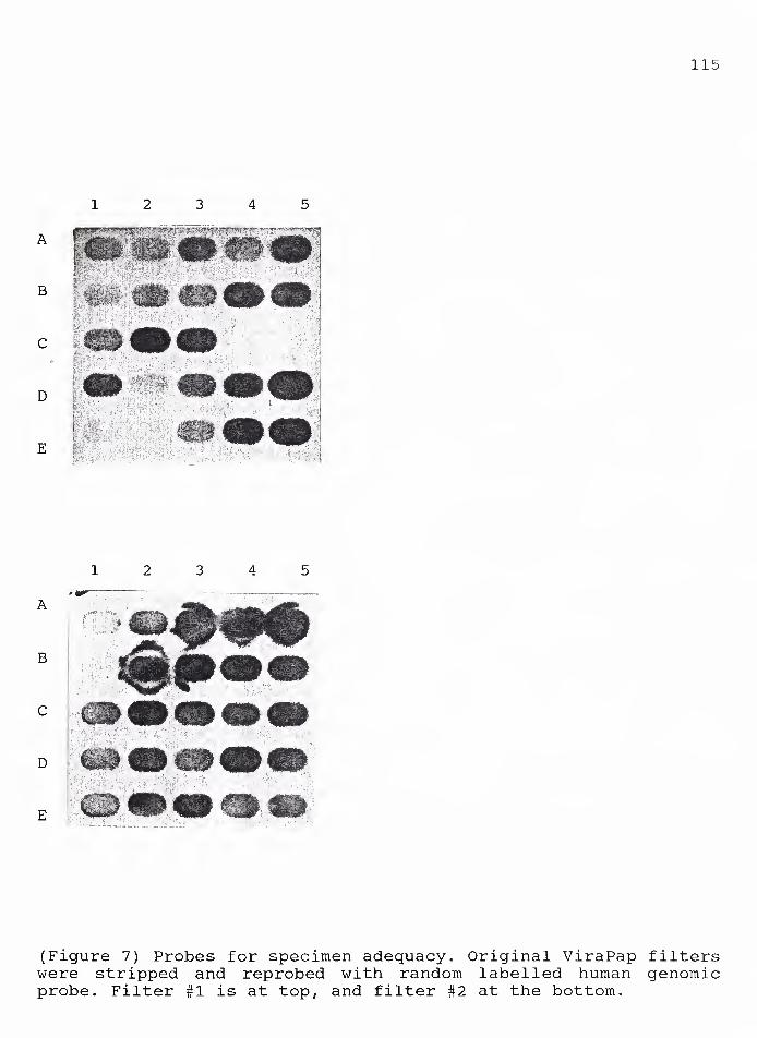
115
(Figure 7) Probes for specimen adequacy. Original ViraPap filters were stripped and reprobed with random labelled human genomic probe. Filter #1 is at top, and filter #2 at the bottom.


116
REFERENCES
1. Davis GL, Hernandez E, Davis JL, Miyazawa K. Aytpical Squamous
Cells in Papanicolaou Smears. Obstet Gynecol. 69(l):43-46, 1987.
2. Lunberg GD, ed. Toward Optimal Laboratory Use/Council Report:
Quality Assurance in Cervical Cytology: The Papanicolaou Smear.
JAMA. 262(12):1672-1679, 1989.
3. Bearman DM, MacMillan JP, Creasman WT. Papanicolaou Smear
History of Patients Developing Cervical Cancer: An Assessment of
Screening Protocols. Obstet Gynecol. 69(2):151-155, 1987.
4. Koss LG. State of the Art/Review: The Papanicolaou Test for
Cervical Cancer Detection: A Triumph and a Tragedy. JAMA.
261(5):737-743, 1989.
5. Taylor PT, Andersen WA, Barber SR, Coveil JL, et al. The
Screening Papanicolaou Smear: Contribution of the Endocervical
Brush. Obstet Gynecol. 70(5):734-737, 1987.
6. Lunberg GD, ed. Toward Optimal Laboratory Use: The 1988 Bethesda
System for Reporting Cervical/Vaginal Cytological Diagnoses. JAMA.
262(7):931-934, 1989.
7. Robertson AJ, Anderson JM, Swanson Beck J, Burnett RA, et al.
Observer Variability in Histopoathogical Reporting of Cervical
Biopsy Specimens. J Clin Pathol. 42:231-238, 1989.
8. Salzman NP, Howley PM, eds. The Papovaviridae. Vol.2.: The
Papillomaviruses. Plenum Press, New York, 1987.
9. de Villiers EM. Minireview: Heterogeneity of the Human
Papillomavirus Group. J Virol. 63(11):4898-4903, 1989.
10. Koutsky LA, Galloway DA, Holmes KK. Epidemiology of Genital
Human Papillomavirus Infection. Epidemiologic Reviews. 10:122-163,
1988.
11. Roman A, Fife KH. Human Papillomaviruses: Are We Ready to Type?
Clin Micro Rev. 2(2):166-190, 1989.
12. Lowy DR, Schiller JT. Transforming Genes of Bovine and Human
Papillomaviruses. In
13. Dyson N, Howley PM, Munger K, Harlow E. The Human Papilloma
Virus—16 E7 Oncoprotein Is Able to Bind to the Retinoblastoma Gene
Product. Science. 243:934-937, 1989.
14. Campion MJ, Singer A, Clarkson PK, McCance DJ. Increased Risk
of Cervical Neoplasia in Consorts of Men with Penile Condylomata

117
Acuminata. Lancet. 943-946, April, 1985.
15. Tidy JA, Vousden KH, Farrell PJ. Relation Between Infection With A Subtype of HPV16 and Cervical Neoplasia. Lancet. 1225-1227, June, 1989.
16. Singer A, McCance DJ. The Wart Virus and Genital Neoplasia; A Casual or Causal Association. Commentary. Br J Obstet Gynecol. 92(11):1083-1085, 1985.
17. Venuti A, Marcante ML. Presence of Human Papillomavirus Type 18 DNA in Vulvar Carcinomas and Its Integration into the Cell Genome. J Gen Virol. 70:1587-1592, 1989.
18. de Villiers EM, Schneider A, Miklaw H, Papendick U, et al. Human Papillomavirus Infections in Women With and Without Abnormal Cervical Cytology. Lancet. 703-706, September, 1987.
19. Caussy D, Orr W, Daya AD, Roth P, et al. Evaluation of Mehods for Detecting Human Papillomavirus Deoxyribonucleotide Sequences in Clinical Specimens. J Clin Micro. 26(2):236-243, 1988.
20. Lorincz, AT. Human Papillomvirus Testing. Diag and Clin Testing. 27:28-37, 1989.
21. Melchers WJG, Schift R, Stolz E, Lindeman J, Quint WGV. Human Papillomavirus Detection in Urine Samples from Male Patients by the Polymerase Chain Reaction. J Clin Micro. 27(8):1711-1714, 1989.
22. Schneider A, Hotz M, Gissman L. Increased Prevalence of Human Papillomaviruses in the Lower Genital Tract of Pregnant Women. Int J Cancer. 40:198-201, 1987.
23. Horn PL, Lowell DM, LiVolsi VA, Boyle CA. Reproducibility of the Cytologic Diagnosis of Human Papillomavirus Infection. Acta Cytologica. 29 (5) :692-694, 1985.
24. Meisels A, Fortin R. Condylomatous Lesions of the Cervix and Vagina: Cytologic Patterns. Acta Cytologica. 20(6):505-509, 1976.
25. Day JD, Donaldson LD, Goellner JR. False-Negative Results in Cervical Cytologic Studies. Acta Cytologica. 29(6):1043-1046, 1985.
26. Burk RD, Radish AS, Calderin S, Romney SL. Human Papillomavirus Infection of the Cervix Detected by Cervicovaginal Lavage and Molecular Hybridization: Correlation with Biopsy Results and Papanicolaou Smear. Am J Obstet Gynecol. 154(5):982-989, 1986.
27. Morse AR, Wickenden C, Byrne M, Taylor-Robinson D, et al. DNA


118
Hybridization of Cervical Scrapes: Comparison with Cytological Findings in Papanicolaou Smears. J Clin Path. 41:296-299, 1988.
28. McNichol PJ, Guijon FB, Paraskevas M, Brunham RC. Comparison of Filter In Situ Deoxyribonucleic Acid Hybridization with Cytologic, Colposcopic, and Histopathologic Examination for Detection of Human Papillomavirus Infection in Women with Cervical Intraepithelial Neoplasia. Am J Obstet Gynecol. 160(1):265-270, 1989.
29. Tidy JA, Parry GCN, Ward P Coleman DV, et al. High Rate of Human Papillomavirus Type 16 Infection in Cytologically Normal Cervices. Lancet. 434, February, 1989.
30. Barrett TJ, Silbar JD. Genital Warts—A Venereal Disease. JAMA.154(4):333-334, 1954.
31. White DO, Huebner RJ, Rowe WP, Traub R. Studies on the Virus of Rabbit Papilloma: Methods of Assay. Austral J Exp Biol. 41:41- 50, 1963.
32. Beckmann AM, Myerson D, Baling JR, Kiviat NB, et al. Detection and Localization of Human Papillomavirus DNA in Human Genital Condylomas by In Situ Hybridization With Biotinylated Probes. J Med Virol. 16:265-273, 1985.
33. Stoler MH, Broker TR. In Situ Hybridization Detection of Human Papillomavirus DNAs and Messenger RNAs in Genital Condylomas and a Cervical Carcinoma. Human Pathol. 17(12):1250-1258, 1986.
34. Gage JR, Meyers C, Wettstein FO. The E7 Proteins of the Nononcogenic Human Papillomaviurs Type 6b (HPV-6b) and of the Oncogenic HPV-16 Differ in Retinoblastoma Protein Binding and Other Properties. J Virol. 64(2):723-730, 1990.
35. Bedell MA, Jones KH, Laimins LA. The E6-E7 Region of Human Papillomavirus Type 18 Is Sufficient for Transformation of NIH 3T3 and Rat-1 Cells. J Virol. 61(11):3635-3640, 1987.
36. Wilczynski SP, Walker J, Liao SY, Bergen S, et al. Adenocarcinoma of the Cervix Associated With Human Papillomavirus. Cancer. 62:1331-1336, 1988.
37. Skrabanek P. Cervical Cancer in Nuns and Prostitutes: A Plea for Scientific Continence. J Clin Epidemiol. 41(6):577-582, 1988.
38. McCance DJ, Clarkson PK, Chesters PM, Jenkins D, Singer A. Prevalence of Human Papillomavirus type 16 DNA sequences in cervical intraepithelial neoplasia and invasive carcinoma of the cervix. Br J Obstet Gynecol. 92:1101-1105, 1985.
39. McCance DJ, Kalache A, Ashdown K, Andrade L, et al. Human

119
Papillomavirus Types 16 and 18 In Carcinomas of the Penis From Brazil. Int J Cancer. 37:55-59, 1986.
40. Ostrow RS, Manias DA, Fong WJ, Zachow KR. A Survey of Human Cancers for Human Papillomavirus DNA by Filter Hybridization. Cancer 59:429-434, 1987.
41. Durst M, Kleinheinz A, Hotz M, Gissman L. The Physical State of Human Papillomavirus Type 16 DNA in Benign and Malignant Genital Tumors. J Gen Virol. 66:1515-1522, 1985.
42. Ismail, SM, Colclough AB, Dinnen JS, Eakins D, et al. Observer Variation in Histopathological Diagnosis and Grading of Cervical Intraepithelial Neoplasia. Br Med J. 298:707-710, March, 1989.
43. Cancer Facts and Figures-1989. American Cancer Society, New York, NY. 1989.
44. Shope RE. Infectious Papillomatosis of Rabbits. J Exp Med. 58:607-624, 1933.
45. Orth G, et al. Identification of Papillomaviruses in Butcher's Warts. J Invest Derm. 76:97-102, 1981.
46. Danos O, Giri I, Thierry F, Yaniv M. Papillomavirus Genome: Seguences and Consequences. J Invest Derm. 83 (1) :7s-lls, 198**.
47. Woodruff JD, Peterson WF. Condyloma Acuminatum of the Cervix. Am J Obstet Gynecol. 75:1354-1362, 1958.
48. Schneider V, Kay S, Lee HM. Immunosuppression as a High Risk Factor in the Development of Condyloma Acuminatum and Squamous Neoplasia of the Cervix. Acta Cytologica. 27(3):220-224, 1983.
49. Halpert R, Fruchter RG, Sedlis A, Butt K, et al. Human Papillomavirus and Lower Genital Neoplasms in Renal Transplant Patients. Obstet Gynecol. 68(2):251-258, 1986.
50. Sarver N, Rabson MS, Yang YC, Byrne JC, Howley PM. Localization and Analysis of Bovine Papillomavirus Type 1 Transforming Functions. J Virol. 52(2):377-388, 1984.
51. Phelps WC, Yee CL, Munger K, Howley PM. The Human Papillomavirus Type 16 E7 Gene Encodes Transactivation and Transformation Functions Similar To Those of Adenovirus E1A. Cell. 53:539-547, 1988.
52. Schneider-Maunoury S, Croissant 0, Orth G. Integration of Human Papillomavirus Type 16 DNA Sequences: A Possible Early Event in the Progression of Genital Tumors. J Virol. 61(1): 3295-3298, 1987.


120
53. Smotkin D, Wettstein FO. Transcription of Human Papillomavirus Type 16 Early Genes in a Cervical Cancer and a Cancer-Derived Cell Line and Identification of the E7 Protein. Proc Nat Acad Sci. 83:4680-4684, 1986.
54. Fraumeni JF, Lloyd JW, Smith EM, Wagoner JK. Cancer Mortality Among Nuns: Role of Marital Status in Etiology of Neoplastic Disease in Women. J Nat Cane Insti. 42:455-468, 1969.
55. Towne JE. Carcinoma of the Cervix in Nulliparous and Celibate Women. Am J Obstet Gynecol. 69 (3):606-613, 1955.
56. Harris RWC, Brinton LA, Cowdell RH, Skegg DCG et al. Characteristics of Women with Dysplasia or Carcinoma In Situ of the Cervix Uteri. Br J Cancer. 42:359-369, 1980.
57. Kessler II. Human Cervical Cancer as a Venereal Disease. Cancer Res. 36:783-791, 1976.
58. Martinez I. Relationship of Sguamous Cell Carcinoma of the Cervix Uteri to Sguamous Cell Carcinoma of the Penis Among Puerto Rican Women Married to Men with Penile Carcinoma. Cancer. 24:777- 780, 1969.
59. Toon PG, Arrand JR, Wilson LP, Sharp DS. Human Papillomavirus Infection of the Uterine Cervix of Women Without Cytological Signs of Neoplasia. Br Med J. 293:1261-1254, 1986.
60. Daling JR, Weiss WS, Hislop TG, Maden C et al. Sexual Practices, Sexually Transmitted Diseases, and the Incidence of Anal Cancer. NEJM. 317(16)*;973-977, 1987.
61. Ferenczy A, Mitao M, Nagar N, Silverstein SJ, Crum CP. Latent Papillomavirus and Recurring Genital Warts. NEJM. 313(13):784-788, 1985.
62. Barnes W, Delgado G, Kurman RJ, Petrilli ES et al. Possible Prognostic Significance of Human Papillomavirus Type in Cervical Cancer. Gynecol One. 29:267-273, 1988.
63. Hadjimichael 0, Janerich D, Lowell DM, Meigs JW, Merino MJ, Schwartz PE. Histologies and Clinical Characteristics Associated with Rapidly Progressive Invasive Cervical Cancer: A Preliminary Report From the Yale Cancer Control Research Unit. Yale J Bio Med. 62:345-350, 1989.
64. Lorincz AT, Temple GF, Kurman RJ, Jenson AB et al. Oncogenic Association of Specific Human Papillomavirus Types with Cervical Neoplasia. J Nat Cane Inst. 79 (4):671-678, 1987.

121
65. Zachow KR, Ostrow RS, Bender M et al. Detection of Human Papillomavirus DNA in Anogenital Neoplasias. Nature. 300:771-773, 1982.
66. Campion MJ, McCance DJ, Cuzick J, Singer A. Progressive Potential of Mild Cervical Atypia: Prospective Cytological, Colposcopic, and Virological Study. Lancet. ii:237-240, 1986.
67. Ayre JE. Selective Cytology Smears for Diagnosis of Cancer. Am J Obstet Gynecol. 53:609-617, 1947.
68. Elias A. Linthorst G, Bekker B, Vooijs PG. The Significance of Endocervical Cells in the Diagnosis of Cervical Epithelial Changes. Acta Cytologica. 27:225-229, 1983.
69. Boons ME, Alons-van Kordelaar JJM, Rietveld-Scheffers PEM. Consequences of the Introduction of Combined Spatula and Cytobrush Sampling for Cervical Cytology. Acta Cytologica. 30:264-270, 1986.
70. Collins DN, Patacsil DP. Proficiency Testing in Cytology in New York: Analysis of a 14-Year State Program. Acta Cytologica. 30:633- 642, 1985.
71. Gay JD, Donaldson LD, Goellner JR. False-Negative Results in Cervical Cytologic Studies. Acta Cytologica. 29:1043-1046, 1985.
72. Husain OAN, Butler B, Evans DMD, McGregor JF et al. Quality Control in Cervical Cytology. J Clin Pathol. 27:935-946, 1976.
73. Reagen JW, Wcott RB. The Detection of Cancer of the Uterine Cervix by Cytologic Study. Am J Obstet Gynecol. 62:1347-1352, 1951.
74. Richart RM, Vaillant HM. Influence of Cell Collection Techniques upon Cytological Diagnosis. Cancer. 65:1474-1478, 1965.
75. van der Graaf Y, Vooijs GP, Gaillard HLJ, Daisy M. Screening Errors in Cervical Cytology. Acta Cytologica. 31:434-438, 1987.
76. Tawa K, Forsythe A, Cove JK, Saltz A et al. A Comparison of the Papanicolaou Smear and the Cervigram: Sensitivity, Specificity, and Cost Analysis. Obstet Gynecol. 71(2):229-235, 1988.
77. Nyirjesy I. Atypical or Suspicious Cervical Smears. An Aggressive Diagnostic Approach. JAMA. 222:691-693, 1972.
78. Sandmere HF, Austin SD, Bechtel RL. Experience with 40,000 Papanicolaou Smears. Obstet Gynecol. 48:56-60, 1976.
79. Stafl A, Mattingly RF. Colposcopic Diagnosis of Cervical Neoplasia. Obstet Gynecol. 41:168-176, 1973.
80. Reiter RC. Management of Initial Atypical Cytology: A


122
Randomized Prospective Study. Obstet Gynecol.68:237, 1986.
81. Pilloti S, Rilke F, Shah KV, DelleTorre G et al. Immunohistochemical and Ultrastructural Evidence of Papillomavirus Infection Associated with In Situ and Micro-Invasive Squamous Cell Carcinoma of the Vulva. Am J Surg Path. 8(10):661-670, 1981.
82. Southern EM. Detection of Specific Sequences Among DNA Fragments Separated by Gel Electrophoresis. J Mol Bio. 98:503-517, 1975.
83. Wilbur DC, Bontiglio TA, Stoler MH. Continuity of Human Papillomavirus (HPV) Type Between Neoplastic Precursors and Invasive Cervical Cancer: An In Situ Study. Am J Surg Path. 12(3):182-186, 1988.
84. Warford AL, Levy RA. Use of Commercial DNA Probes. Clin Lab Sci. 2:105-108, 1989.
85. Fetherston WC. False-Negative Cytology in Invasive Cancer of the Cervix. Clin Obstet Gynecol. 26(4):929-937, 1983.
86. Frenkel N, Roizman B, Cassai JE, Nahmias A. A DNA Fragment of Herpes Simplex 2 and Its Transcription in Human Cervical Cancer Tissue. Proc Nat Acad Sci. 69:3734-3789, 1972.
87. Sexually Transmitted Disease Statistics. United States Department of Health and Human Services. Public Health Service. Centers for Disease Control, Atlanta, GA., 1987.
88. Reid R, Greenberg M, Jenson AB, Husain M, et al. Sexually Transmitted Papillomaviral Infections: The Anatomic Distribution and Pathologic Grade of Neoplastic Lesions Associated with Different Viral Types. Am J Obstet Gynecol. 156(1):212-222, 1987.
89. ViraPap Test Kit Instruction Booklet. Bethesda Research Laboratories: Molecular Diagnostics Division. 1989.


YALE MEDICAL LIBRARY
Manuscript Theses
Unpublished theses submitted for the Masterfs and Doctor's degrees and deposited in the Yale Medical Library are to be used only with due regard to the rights of the authors. Bibliographical references may be noted, but passages must not be copied without permission of the authors, and without proper credit being given in subsequent written or published work.
This thesis by has been used by the following persons, whose signatures attest their acceptance of the above restrictions.
NAME AND ADDRESS DATE


Copyright © 2022 FDOKUMEN