High-Risk Human Papillomavirus and Epstein–Barr Virus ...
23
biology Review High-Risk Human Papillomavirus and Epstein–Barr Virus Coinfection: A Potential Role in Head and Neck Carcinogenesis Rancés Blanco 1 , Diego Carrillo-Beltrán 1 , Alejandro H. Corvalán 2 and Francisco Aguayo 3, * Citation: Blanco, R.; Carrillo-Beltrán, D.; Corvalán, A.H.; Aguayo, F. High-Risk Human Papillomavirus and Epstein–Barr Virus Coinfection: A Potential Role in Head and Neck Carcinogenesis. Biology 2021, 10, 1232. https://doi.org/10.3390/ biology10121232 Received: 6 November 2021 Accepted: 18 November 2021 Published: 26 November 2021 Publisher’s Note: MDPI stays neutral with regard to jurisdictional claims in published maps and institutional affil- iations. Copyright: © 2021 by the authors. Licensee MDPI, Basel, Switzerland. This article is an open access article distributed under the terms and conditions of the Creative Commons Attribution (CC BY) license (https:// creativecommons.org/licenses/by/ 4.0/). 1 Programa de Virología, Instituto de Ciencias Biomédicas (ICBM), Facultad de Medicina, Universidad de Chile, Santiago 8380000, Chile; [email protected] (R.B.); [email protected] (D.C.-B.) 2 Advanced Center for Chronic Diseases (ACCDiS), Pontificia Universidad Católica de Chile, Santiago 8320000, Chile; [email protected] 3 Universidad de Tarapacá, Arica 1000000, Chile * Correspondence: [email protected] Simple Summary: A subset of carcinomas that arise in the head and neck region show a viral etiology. In fact, a subgroup of oropharyngeal cancers are caused by some types of human papillomavirus (HPV), so-called high-risk (HR)-HPVs, whereas undifferentiated nasopharyngeal carcinomas are etiologically related to Epstein–Barr virus (EBV). However, studies have reported the presence of both HR-HPV and EBV in some types of head and neck cancers. In this review, we discuss the potential contribution and role of HR-HPV/EBV coinfection in head and neck carcinogenesis, as well as the mechanisms that are potentially involved. In addition, HR-HPV/EBV interaction models are proposed. Abstract: High-risk human papillomaviruses (HR-HPVs) and Epstein–Barr virus (EBV) are recog- nized oncogenic viruses involved in the development of a subset of head and neck cancers (HNCs). HR-HPVs are etiologically associated with a subset of oropharyngeal carcinomas (OPCs), whereas EBV is a recognized etiological agent of undifferentiated nasopharyngeal carcinomas (NPCs). In this review, we address epidemiological and mechanistic evidence regarding a potential cooperation between HR-HPV and EBV for HNC development. Considering that: (1) both HR-HPV and EBV infections require cofactors for carcinogenesis; and (2) both oropharyngeal and oral epithelium can be directly exposed to carcinogens, such as alcohol or tobacco smoke, we hypothesize possible inter- action mechanisms. The epidemiological and experimental evidence suggests that HR-HPV/EBV cooperation for developing a subset of HNCs is plausible and warrants further investigation. Keywords: Epstein–Barr virus; human papillomavirus; head and neck cancer 1. Introduction Head and neck cancers (HNCs) encompass tumors of a variety of subsites, including the lips, oral cavity, nasopharynx, oropharynx, hypopharynx, larynx, and salivary glands. Head and neck squamous cell carcinomas (HNSCCs) constitute more than 90% of all HNCs [1]. These malignancies were the sixth leading cancer worldwide, with 931,931 new cases in 2020, excluding non-melanoma skin neoplasms. Additionally, HNCs represented the seventh highest cause of cancer-related deaths in the same year, with 467,125 deaths [2]. Alcohol consumption and tobacco smoking are the two most important risk factors for HNC development. Infection with oncogenic viruses, such as the human papillomavirus (HPV) or the Epstein–Barr virus (EBV), have been reported to be associated with the development and progression of these neoplasms. In fact, high-risk (HR)-HPV infection is a risk factor for developing a subset of oropharyngeal tumors, including the tonsils and the base of the tongue [3]. Similarly, EBV infection is related to the development of nasopharyngeal (NPC) and salivary gland cancers [4]. However, neither HR-HPV nor EBV infection alone Biology 2021, 10, 1232. https://doi.org/10.3390/biology10121232 https://www.mdpi.com/journal/biology
-
Upload
khangminh22 -
Category
Documents
-
view
1 -
download
0
Transcript of High-Risk Human Papillomavirus and Epstein–Barr Virus ...

biology
Review
High-Risk Human Papillomavirus and Epstein–Barr VirusCoinfection: A Potential Role in Head and Neck Carcinogenesis
Rancés Blanco 1, Diego Carrillo-Beltrán 1, Alejandro H. Corvalán 2 and Francisco Aguayo 3,*
�����������������
Citation: Blanco, R.; Carrillo-Beltrán,
D.; Corvalán, A.H.; Aguayo, F.
High-Risk Human Papillomavirus
and Epstein–Barr Virus Coinfection:
A Potential Role in Head and Neck
Carcinogenesis. Biology 2021, 10, 1232.
https://doi.org/10.3390/
biology10121232
Received: 6 November 2021
Accepted: 18 November 2021
Published: 26 November 2021
Publisher’s Note: MDPI stays neutral
with regard to jurisdictional claims in
published maps and institutional affil-
iations.
Copyright: © 2021 by the authors.
Licensee MDPI, Basel, Switzerland.
This article is an open access article
distributed under the terms and
conditions of the Creative Commons
Attribution (CC BY) license (https://
creativecommons.org/licenses/by/
4.0/).
1 Programa de Virología, Instituto de Ciencias Biomédicas (ICBM), Facultad de Medicina,Universidad de Chile, Santiago 8380000, Chile; [email protected] (R.B.);[email protected] (D.C.-B.)
2 Advanced Center for Chronic Diseases (ACCDiS), Pontificia Universidad Católica de Chile,Santiago 8320000, Chile; [email protected]
3 Universidad de Tarapacá, Arica 1000000, Chile* Correspondence: [email protected]
Simple Summary: A subset of carcinomas that arise in the head and neck region show a viral etiology.In fact, a subgroup of oropharyngeal cancers are caused by some types of human papillomavirus(HPV), so-called high-risk (HR)-HPVs, whereas undifferentiated nasopharyngeal carcinomas areetiologically related to Epstein–Barr virus (EBV). However, studies have reported the presence ofboth HR-HPV and EBV in some types of head and neck cancers. In this review, we discuss thepotential contribution and role of HR-HPV/EBV coinfection in head and neck carcinogenesis, aswell as the mechanisms that are potentially involved. In addition, HR-HPV/EBV interaction modelsare proposed.
Abstract: High-risk human papillomaviruses (HR-HPVs) and Epstein–Barr virus (EBV) are recog-nized oncogenic viruses involved in the development of a subset of head and neck cancers (HNCs).HR-HPVs are etiologically associated with a subset of oropharyngeal carcinomas (OPCs), whereasEBV is a recognized etiological agent of undifferentiated nasopharyngeal carcinomas (NPCs). Inthis review, we address epidemiological and mechanistic evidence regarding a potential cooperationbetween HR-HPV and EBV for HNC development. Considering that: (1) both HR-HPV and EBVinfections require cofactors for carcinogenesis; and (2) both oropharyngeal and oral epithelium canbe directly exposed to carcinogens, such as alcohol or tobacco smoke, we hypothesize possible inter-action mechanisms. The epidemiological and experimental evidence suggests that HR-HPV/EBVcooperation for developing a subset of HNCs is plausible and warrants further investigation.
Keywords: Epstein–Barr virus; human papillomavirus; head and neck cancer
1. Introduction
Head and neck cancers (HNCs) encompass tumors of a variety of subsites, includingthe lips, oral cavity, nasopharynx, oropharynx, hypopharynx, larynx, and salivary glands.Head and neck squamous cell carcinomas (HNSCCs) constitute more than 90% of allHNCs [1]. These malignancies were the sixth leading cancer worldwide, with 931,931 newcases in 2020, excluding non-melanoma skin neoplasms. Additionally, HNCs representedthe seventh highest cause of cancer-related deaths in the same year, with 467,125 deaths [2].Alcohol consumption and tobacco smoking are the two most important risk factors for HNCdevelopment. Infection with oncogenic viruses, such as the human papillomavirus (HPV)or the Epstein–Barr virus (EBV), have been reported to be associated with the developmentand progression of these neoplasms. In fact, high-risk (HR)-HPV infection is a risk factorfor developing a subset of oropharyngeal tumors, including the tonsils and the base ofthe tongue [3]. Similarly, EBV infection is related to the development of nasopharyngeal(NPC) and salivary gland cancers [4]. However, neither HR-HPV nor EBV infection alone
Biology 2021, 10, 1232. https://doi.org/10.3390/biology10121232 https://www.mdpi.com/journal/biology

Biology 2021, 10, 1232 2 of 23
is a sufficient condition for tumorigenesis. In most immunocompetent subjects, HPVinfection is ultimately controlled [5], whereas EBV is present during the lifetime of theindividual without any clinical manifestation, all of which suggesting that additionalcofactors are required for promoting HR-HPV or EBV-driven cancers. Indeed, some studieshave reported the simultaneous presence of HR-HPV and EBV in HNCs [6–8], proposingthat HR-HPV/EBV coinfection could play a fundamental role in the development ofthese malignancies [6,9,10]. However, potential mechanisms involved in such HPV/EBVcooperation need further clarification. Here, we review the current literature regarding HR-HPV/EBV coinfection in HNSCCs, as well as its potential contribution to carcinogenesisand the progression of these tumors. Finally, we propose a mechanism by which HR-HPV-mediated alterations facilitate EBV infection in epithelial tissues.
2. Human Papillomavirus and Epstein–Barr Virus Tropism
Epithelial tissues are susceptible and permissive to both HPV (cutaneous andmucosal) [11] and EBV (mucosal) infections [12]. Basal epithelial cells are susceptibleto HPV infection, although only upper highly differentiated cells in the stratified epithe-lium are permissive [13,14]. HPV entry into epithelial cells requires the involvement ofheparan sulphate proteoglycans (HSPGs) for initial tethering to basal cells [15]. Integrins(α6 and α3) and CD151 are also required to form an “entry receptor complex” with annexinA2, CD63, and EGFR/GFR (reviewed in [16]). Interestingly, multiple studies have reportedthat HPV can regulate integrin levels. For instance, Werner et al. (2011) reported increasedlevels of αv, α5, β1, β4, and β6 integrins in HPV-positive cervical cells when compared withnon-tumor cells, which was attributable to HPV infection [17]. Overexpression of HPV16E6*, the smaller splice isoform of the E6 oncogene, increased the levels of the β1 integrin incervical cancer cells [18]. A 2.44-fold increase in the expression of the ITGB5 gene, whichencodes the β5 integrin subunit, was reported in cervical cells upon L2 overexpression [19].In contrast, it was reported that HPV38 E6/E7 proteins downregulate the expression ofαvβ8 integrin in human keratinocytes [20].
EBV exhibits a similar epithelial tropism, although EBV cell entry occurs throughboth apical and basolateral routes in mucosal epithelia [21], and this virus shows anadditional and preferential tropism for B cells, which constitute the EBV reservoir [22].EBV lytic infections occur in highly differentiated cells, as demonstrated by Temple et al.(2014) [23]. EBV entry to epithelial cells is mediated by the ephrin receptor tyrosine kinaseA2 (EphA2). Furthermore, the increased expression of EphA2 leads to a more efficientEBV infection of epithelial cells [24,25]. Of note, elevated EphA2 expression was associatedwith progression and poor prognosis in HNSCC patients [26]. Notably, some EBV-encodedproteins (e.g., LMP1) can regulate the expression of the Eph family [27]. Studies byChesnokova et al. suggested that the interaction of EBV glycoproteins gH/gL with αvβ5,αvβ6, and αvβ8 integrins is associated with EBV fusion with gastric carcinoma cells [28,29],though Chen et al. (2018) reported a lack of effect of these surface integrins on EBV entryin HEK293 cells [24]. Interestingly, the expression of αvβ5 integrin was significantlyincreased in oral and laryngeal SCCs when compared to normal counterparts [30,31]. Inaddition, αvβ6 integrin was evidenced in almost all HNCs, including oral, oropharyngeal,hypopharyngeal, laryngeal, and nasopharyngeal carcinomas, but not in normal oral andtongue mucosa [32]. The expression of αvβ8 integrin was also found to be increased inlaryngeal carcinoma compared with normal tissues [31]. A comparison between HPV andEBV tropism is shown in Table 1.

Biology 2021, 10, 1232 3 of 23
Table 1. Comparison between HPV and EBV epithelial cell entry.
Biological Process Human Papillomavirus Epstein–Barr Virus References
Route of entryDirect epithelial contact,
apical entry(microlesions)
Salivary transmission,apical, basolateral, or
basal entry[21,33]
Tropism Epithelial cells (mucosalor cutaneous)
Epithelial cells (mucosal),B cells, T cells, NK cells [33,34]
Entry mechanism Endocytosis Membrane fusion [35,36]
Receptors Entry receptor complex,HSPGs, integrins EphA2 [24,37]
HSPGs, heparan sulphate proteoglycans; EphA2, ephrin receptor tyrosine kinase A2.
3. Epidemiology of HPV/EBV Coinfection in HNSCCs
As in epidemiological studies, the cellular location of viruses is generally not con-sidered, and the term “copresence” is assumed when HPV and EBV are both detectedin tumors. However, copresence in tumors is not synonymous with coinfection, whichoccurs when both HPV and EBV infects the same epithelial cell. Molecular methodologies,such as a polymerase chain reaction (PCR), cannot determine the coinfection or coexpres-sion of viral products, although this test is frequently used in epidemiological studies.HPV/EBV copresence has been evidenced in 31.2% of HNCs, which includes larynx, lip,and nasopharynx malignancies [9]. Similarly, Drop et al. found HPV/EBV copresencein 34.1% of oral cavity, oropharyngeal, and laryngeal tumors [6], and EBV infection wassignificantly associated with HPV16, 18, 45, and 58 presence in HNSCCs [10]. Remarkably,the expression of both LMP1 and E6 oncoproteins in HNCs was associated with an in-creased cell invasiveness and advanced tumor stage [9,10]. HPV/EBV copresence was alsoassociated with alcohol abuse and an increased histological grade of HNCs, although itwas more frequent in early-stage disease [6]. In the following subsections, we will analyzethe HPV/EBV copresence in HNCs from different anatomical sites.
3.1. Oral Cavity
More than 90% of oral tumors are squamous cell carcinomas (OSCCs), which arisefrom the mucosal epithelium lining the lips, anterior tongue, gingivae, floor of mouth,palate, and other areas of the mouth [38]. In 2020, there were an estimated 377,713 new casesand 177,757 deaths from lip and oral cavity tumors worldwide [2]. Tobacco smoking andalcohol consumption are the two most important risk factors associated with oral cancer de-velopment. Additionally, the involvement of some viral infections has also been suggestedin oral carcinogenesis (reviewed in [39]). HPV infection in OSCCs ranges between 6% and58% worldwide [40–42]. A meta-analysis including 62 studies and 4852 OSCCs found that38.1% of cancers contained HPV DNA [43]. It was also discovered that HPV infectionincreased the probability of OSCC development in the Chinese population (OR 12.7, 95% CI:8.0–20.0) by more than 12 times [44]. However, a recent meta-analysis published by Meloet al. (2021) found E6/E7 mRNA expression in only 4.4% of OSCCs [45]. HPV16 and 18are the most frequent HPV genotypes detected in these tumors [45,46]. In addition, thepresence of HPV16 has been related to an increased histological grade of oral tumors [47].Meanwhile, EBV infection has been detected in 25.9% to 82.5% of OSCCs [48–50]. EBNA2and LMP1 were also detected in 50.2% and 10.7% of OSCCs, respectively [51]. In particular,the expression of LMP1 was increased in OSCCs when compared to normal mucosa andoral leukoplakia [52]. She et al. (2017) conducted a meta-analysis that included 13 studiespublished between 1995 and 2016. In this report, OSCC development was shown to be5 times more likely to occur in the presence of EBV (OR 5.03, 95% CI: 1.80–14.01) [53].Another meta-analysis also found an association between EBV infection and OSCC. In fact,this study reported that the probability of OSCC development was 2.5 (OR 2.5, 95% CI:1.2–5.36) times increased in EBV-positive cases [54]. Regarding HPV/EBV coinfection, this

Biology 2021, 10, 1232 4 of 23
was evidenced in 6.5–37.5% of OSCCs [6,10,55,56]. In one study, which included 155 OSCCsfrom eight countries, HPV/EBV was detected in 21% of cases, making it the most frequentcoinfection in these tumors [55]. The coexpression of HR-HPV E6 and LMP1 was alsodemonstrated in OSCCs by immunohistochemistry (IHC) [10]. In particular, HPV16 andEBV copresence was detected in 5.6% of cases [57]. Moreover, EBV infection was associatedwith HR-HPV16, 18, 31, 35, 45, 51, and 58 [10]. Conversely, an absence of EBV/HPVcopresence has also been reported in oral cancers [58,59].
3.2. Nasopharynx
Nasopharyngeal carcinoma (NPC) is a HNC with an estimated 133,354 new casesand 80,008 deaths in 2020 [2]. NPC usually exhibits a heterogeneous racial and geo-graphic distribution. Its frequency is high in Southern China, intermediate in SoutheastAsia and Northern Africa, and low in most areas of the word, with 15–20.5 and <1 per100,000 habitants, respectively [60]. According to the World Health Organization (WHO)pathologic classification, NPCs are divided into keratinizing squamous cell carcinoma(WHO type I) and non-keratinizing carcinoma (WHO types II and III) [61]. In endemicareas, the keratinizing subtype is relatively rare, whereas the non-keratinizing subtypecan reach more than 99% of total cases [62,63]. Diverse factors, such as salted fish con-sumption, smoking, alcohol consumption, air pollution exposure (e.g., NO2), and geneticsusceptibility, are among the risk factors related to NPC development [64–66]. EBV isa well-established etiological agent of undifferentiated (non-keratinizing) NPC [67–69],which is detected in almost 100% of cases [70,71]. Overall, 57.6% to 100% of NPCs areEBV-positive [72–76]. A meta-analysis reported that EBV prevalence was higher in WHOtype II/III NPC compared to WHO type I tumors (83.2% vs. 21.3%) [77]. Importantly,increased levels of plasma EBV DNA are related to a more aggressive biological behaviorof this cancer [78–81]. However, an association was reported between the EBV viral loadand a better prognosis in NPC patients [82]. The expression of LMP1, a latent EBV protein,was associated with the TNM stage and lymph node metastasis in NPC patients [61]. In ameta-analysis conducted by Zhao et al. (2012), the occurrence of metastasis in NPC was1.98 times increased in the presence of LMP1 (OR 1.98, 95% CI: 1.38–2.83) [83]. On the otherhand, Bam HI rightward Frame 1 (BARF1), which is considered an exclusively epithelialoncoprotein, is detected in 69–100% of NPCs [84]. Regarding HPV infection, the pooledprevalence worldwide was 21%, and it increased 4.77-fold in tumor samples compared tocontrol cases [85]. HPV16 and HPV18 are the most common genotypes detected in HPV-positive NPCs [77]. Of note, an increased prevalence of HR-HPV was evidenced in WHOtype I NPC when compared with WHO type II/III cases (39.9% vs. 23.3%) [77]. Moreover,patients with HPV-positive tumors displayed a decreased overall survival, progression-freesurvival, and locoregional control when compared to exclusively EBV positive patients [86].In the meta-analysis conducted by Tham et al. (2020), an increased frequency of HPV/EBVcopresence was found in WHO type I NPCs when compared with WHO type II/III tumors(7.6% vs. 1.0%) [77]. In addition, HPV/EBV copresence was evidenced mainly in NPC-endemic regions, ranging from 15% to 47.7% of the tumors [8,87,88]. Of note, about 80% ofHPV16 or HPV18-positive NPCs were also positive for EBV DNA [87]. The overexpres-sion of p16, a surrogate biomarker of oncogenic HPV infection, was associated with animproved progression-free survival and locoregional control in EBV-positive NPCs [89].However, in non-endemic areas, HPV/EBV coinfection is less common. Indeed, someauthors reported that HPV and EBV infections are mutually exclusive in populations witha low NPC incidence [86,90].
3.3. Oropharynx
An estimated 98,412 new cases and 48,143 cancer deaths worldwide were attributableto oropharyngeal tumors in 2020 [2]. Most oropharyngeal tumors are squamous cell carcino-mas (OPSCC), which arise from epithelial cells lining the oropharynx [91]. Etiologically, twodifferent types of OPSCCs have been described: HPV-driven and HPV-negative carcinomas,

Biology 2021, 10, 1232 5 of 23
characterized by a distinct biological and clinical behavior [92]. In fact, patients with HPV-driven OPSCCs usually show a better prognosis compared to those with HPV-negativetumors [93,94]. Furthermore, patients with HPV16-driven OPSCC showed an increasedoverall survival rate when compared to patients with non-HPV16-positive carcinomas [95].HPV-negative OPSCCs are mainly related to other risk factors, such as tobacco smokeand alcohol consumption [96]. The prevalence of HPV in OPSCC displays a geographicheterogeneity. For instance, a systematic review showed HPV positivity ranging from 18%to 65% in OPSCC patients from European countries [97]. In addition, Mariz et al. (2020)found a 44.8% pooled prevalence of HPV-driven OPSCC, with higher percentages found inNew Zealand (74.5%), Sweden (70.0%), and Denmark (61.7%) when compared with theNetherlands (30.3%), Germany (25.0%), and Brazil (11.1%) [98]. Regarding the oropharynxsubsites, HPV infection is more frequent in OPSCCs from the base of the tongue (40%) andtonsils (56%) when compared with other sites, such as the soft palate (12%) and posteriorwall (19%) [99]. HPV16 is the most frequent genotype detected in OPSCC, reaching 88% ofHPV-positive cases [100]. However, other authors reported significant differences in theprevalence of HPV16-positive OPSCCs in the United States (59.3%) and Europe (31.1%)in comparison with Brazil (4.1%) [101]. Other genotypes, such as HPV18, HPV33, HPV35,and HPV58, have also been detected, although only in a small percentage of cases [102].Interestingly, EBV infection was evidenced in 53.3–86% of OPSCCs [56,103], suggesting itscontribution to carcinogenesis. In normal oropharyngeal tissues, an absence of HPV/EBVcoinfection was evidenced [7]. Conversely, the copresence of HPV and EBV was detectedin 20% and 25% of tonsillar and base of tongue SCCs, respectively, while the absence ofHPV/EBV coinfection in soft palate tumors was reported [7]. Similarly, Drop et al. identi-fied HPV/EBV copresence in 28.6% of oropharyngeal tumors [6], whereas other authorsonly found HPV/EBV infections in 8–9% [56]. Additionally, Broccolo et al. reported HPVinfection in 25% of OPSCCs, whereas EBV infection was found in 45% of tumors. However,coinfection was only evidenced in 4% of OPSCCs [58].
3.4. HPV and EBV Infection in Other HNSCCs
HNCs also include tumors arising from the hypopharynx, larynx, paranasal sinuses,nasal cavity, and salivary glands. However, data regarding HPV/EBV coinfection inthese neoplasms are scarce. HPV/EBV coinfection was reported in 25% and 33.3% ofhypopharyngeal and paranasal sinus carcinomas, respectively [104]. In addition, thecopresence of HPV and EBV was evidenced in 12.8% [105] and 21.4% [6] of laryngealcarcinomas. Of interest, a three-fold increase was reported in the invasiveness propertiesof HPV+/EBV+ NOK and FaDu hypopharyngeal cells in the presence of lysophosphatidicacid (LPA) compared to HPV-/EBV- and HPV-/EBV+ cells [7]. Tables 2 and 3 showmeta-analysis data for HPV and EBV detection in HNSCCs.
Table 2. HPV prevalence in HNSCC according to meta-analysis data.
Tumor Type No. of Cases HPV PooledPrevalence (%) OR (95% CI) Comments Refs
OSCC 4680 46.5 37.6–55.5HPV infection was 4.7 timesmore likely to be detected in
OSCC than in normal mucosa.[106]
OSCC 610 58.0 54.1–61.9HPV positivity increased the
probability of OSCCdevelopment by 12.7 times.
[44]
OSCCHNSCC
(other sites)
32384852
38.134.5
30.0–46.228.4–40.6
The pooled HPV prevalencewas greater in OSCC (38.1%)
than in other HNSCCs(24.1%).
[43]

Biology 2021, 10, 1232 6 of 23
Table 2. Cont.
Tumor Type No. of Cases HPV PooledPrevalence (%) OR (95% CI) Comments Refs
NPC 1748 21.0 1.69–13.45
HPV prevalence was higher incases outside of China than in
cases from regions in China(23% vs. 19%; p < 0.001).
[85]
NPC 2453 19.9 13.6–27.1
HR-HPV infection was higherin WHO type I NPCs (39.9%)
compared to WHO typesII/III tumors (23.3%).
[77]
OPSCC 6009 44.8 36.4–53.5
HPV pooled prevalence wasmore increased in New
Zealand (74.5%), Sweden(70.0%), and Denmark (61.7%)
than in Brazil (11.1%),Germany (25.0%), and the
Netherlands (30.3%).
[98]
OPSCCHNSCC (all sites)
9255681
41.021.9
38.0–44.021.0–23.0
HR-HPV (any genotype) andHPV16 increased the
probability of HNSCCdevelopment by 1.83 and 4.44
times, respectively.
[107]
OPSCCHNSCC (other
sites)
539613,972
47.721.8
42.9–52.518.9–25.1 - [108]
OSCCOPSCCLSCC
HNSCC (all sites)
2642969
14355046
23.535.624.025.9
21.9–25.132.6–38.721.8–26.324.7–27.2
HPV prevalence wassignificantly higher in OPSCC
than OSCC or LSCC.[109]
OSCCOPSCC
LSCC/HPSCCHNSCC (all sites)
547839462739
12,163
24.245.822.129.5
18.7–30.238.9–52.916.4–28.325.5–33.6
HPV16 prevalence was moreincreased in OPSCC (40.6%)
than in OSCC (14.9%) orLSCC (13.4%).
[102]
OSCCOPSCCLSCC
HNSCC (all sites)
315327688566777
37.540.523.637.0
35.9–39.238.7–42.322.1–25.036.0–38.0
The association of HPV withcancer development was
increased for OPSCC (OR:14.7) compared to OSCC (OR:
4.1) or LSCC (OR: 3.2).
[110]
LSCC 2559 28.0 23.5–32.9
HPV infection wassignificantly associated with
the risk of LSCC development(OR: 5.4).
[111]
HNSCC, head and neck squamous cell carcinoma; OSCC, oral squamous cell carcinoma; OPSCC, oropharyngeal squamous cell carcinoma;LSCC, laryngeal squamous cell carcinoma; HPSCC, hypopharyngeal squamous cell carcinoma; NPC, nasopharyngeal carcinoma.
Table 3. EBV prevalence in HNSCCs.
Tumor Type Method No. of Cases EBV Positivity (%) Comments Refs
OSCC EBV-chiphybridization 57 82.5 - [48]
OSCC PCR for BNLF1 91 45.1
EBV was significantlyassociated with the probability
of OSCC development(OR 3.76).
[49]

Biology 2021, 10, 1232 7 of 23
Table 3. Cont.
Tumor Type Method No. of Cases EBV Positivity (%) Comments Refs
OSCC EBER in situhybridization 165 41.2 - [50]
NPC EBER in situhybridization 92 57.6 - [72]
NPC EBER in situhybridization 62 85.5 - [73]
NPC EBER in situhybridization 150 62.0
Overall survival was increasedin EBV-positive patients
(p = 0.005).[74]
NPC EBER in situhybridization 19 84.2 - [75]
NPC EBER in situhybridization 82 87.8
EBV positivity was evidencedin 92.6% of non-keratinizing
carcinoma.[76]
NPC EBER in situhybridization 56 73.2
EBV infection was notassociated with the
histopathological type of NPC.[112]
NPC - 2329 76.7
Meta-analysis. EBV prevalencewas increased in WHO Type
II/III (83.2%) compared toWHO Type I cases (21.3%).
[77]
OPSCC Nested PCR 62 29.0 - [56]
OPSCC PCR-ELISA 28 85.7 - [103]
OSCCNPC
OPSCCHPSCCLSCC
EBER in situhybridization
3720745028
060.01.400
EBER positivity wassignificantly higher in NPCs
compared to non-NPC HNSCCs(p < 0.001).
[113]
HNSCC (OSCC,PSCC, and LSCC) PCR for LMP1 98 69.4
LMP1 protein was alsoexpressed in PSCC (100%)
followed by OSCC (76.0%) andLSCC (33.3%).
[10]
HNSCC, head and neck squamous cell carcinoma; OSCC, oral squamous cell carcinoma; OPSCC, oropharyngeal squamous cell carcinoma;LSCC, laryngeal squamous cell carcinoma; HPSCC, hypopharyngeal squamous cell carcinoma; NPC, nasopharyngeal carcinoma; PSCC,pharyngeal carcinoma.
4. Possible Mechanisms of HR-HPV/EBV Cooperation4.1. HPV Facilitates EBV Entry in Epithelial Cells
The cluster of differentiation 21 (CD21), also known as the complement receptortype 2 (CR2), is considered to be the major attachment receptor for EBV in B cells [114].However, CD21 is variably detected in epithelial cells. Interestingly, an increased CD21expression was detected in the tonsil epithelium, comparable to what was detected inperipheral B cells, Burkitt lymphoma cells [115], and OSCCs [116]. Furthermore, a CD21mRNA increase is associated with the grade of oral epithelial dysplasia, and correlateswith the presence of EBER1 transcripts [116]. Additionally, epithelial cells ectopicallyexpressing CD21 become susceptible and permissive to EBV infection [117,118]. In fact,both human keratinocytes and skin cancer cells were susceptible to EBV infection after CD21gene transfection, whereas un-transfected cells remained EBV-negative [117]. CD21 gene-transfected epithelial cells also increase the expression of EBERs and EBNA1, along with alimited LMP1 expression [117,118], which resembles the EBV latency program detected inNPCs [119,120]. Low levels of CD21 expression are sufficient for leading to EBV infectionin human epithelial cells [121]. Taken together, these findings suggest a role of CD21 in EBV

Biology 2021, 10, 1232 8 of 23
entry into epithelial cells. Conversely, an absence of CD21 expression was evidenced in oraltongue and mucosa, soft palate, and the uvula [115], and a decreased CD21 expression wasfound in NPCs when compared to normal nasopharynx [122]. Interestingly, CD21 levelswere associated with HPV and EBV infection in tonsillar and base of tongue carcinomas [7].A high level of CD21 was found in HPV+/EBV+ NPCs, when compared to EBV-/HPV-tumors (p = 0.0194), although no difference was observed between HPV+/EBV+ NPCs andthose harboring EBV or HPV single infections [7]. Furthermore, increased levels of EphA2,the epithelial EBV receptor, was evidenced in CIN and cervical carcinomas when comparedto normal cervical epithelium, and also correlated with CDK6 protein expression [123]. Inaddition, a 15.28-fold increase in EphA2 receptor expression was observed in non-tumorcervical cells transfected with the HPV18 E2 gene [124]. Similarly, transfection with theHPV18 E6 gene resulted in a 3.2-fold increase in EphA2 gene expression in esophagealcancer cells [125]. These facts suggest that HPV could potentially facilitate EBV entry intohead and neck epithelial cells by promoting increased levels of proteins involved in thisprocess. Interestingly, the fact that persistent viruses promote the expression of moleculesinvolved in the entry of another virus is not new. In fact, it was recently reported thatEBV infection promotes increased levels of the angiotensin-converting enzyme 2 (ACE2)receptor, facilitating SARS-CoV-2 entry to epithelial cells [126]. However, further functionalstudies are warranted in order to validate this possibility.
4.2. HPV Infection Could Promote EBV Latency Establishment and Lytic Cycle Activation in Headand Neck Epithelial Cells
Cultures of normal epithelial cells in which the EBV lytic cycle is preferentially es-tablished appear unable to sustain latent EBV infection [51,127,128]. For instance, Tem-ple et al. (2014) demonstrated that EBV establishes a predominantly productive infectionin organotypic cultures of primary oral keratinocytes, characterized by the expressionof latent cycle proteins (e.g., EBNA1, LMP1, EBNA2) and lytic cycle proteins (e.g., gB,gp350). Nevertheless, cells expressing exclusively latent proteins were not detected [23].It is well-known that EBV lytic infection occurs in normal differentiated oral cells fromimmunosuppressed patients, causing oral hairy leukoplakia (OHL) [129]. Although thepresence of EBV latent infection was reported in morphologically normal mucosa, thesesamples corresponded to tongue and gingival fibrous overgrowths, which are character-ized by abnormal enlargement of tissues [51]. Accordingly, it was suggested that normalepithelial cells from the periodontium could be latently infected by EBV [130], but patientsconsidered as “healthy donors” displayed mild periodontal disease (clinical attachmentlevel 1 ≤ x < 3 mm) [131,132]. Additionally, the absence of EBER1 mRNA expression wasobserved in the normal epithelium adjacent to oral dysplasia [116]. Indeed, an EBV latentinfection can be established in nasopharyngeal cells displaying previous genetic alterations,such as hTERT, cyclin D1, or Bmi-1 overexpression, as well as p16 inactivation [133–135].These molecular changes are commonly detected in dysplastic epithelial cells [136,137],which are also more susceptible to EBV infection [116]. Interestingly, the pooled preva-lence of HPV infection was 27.2% in oral epithelial dysplasia [138], whereas other authorsfound a 50% positivity in oral cavity and oropharyngeal dysplasia [139]. Guidry and Scott(2018) reported a significant reduction in EBV DNA replication in both HPV-positive andE7-immortalized human keratinocytes when compared with the EBV DNA replicationobserved in primary keratinocytes cultured in organotypic rafts [140], suggesting the con-tribution of HPV to the establishment of EBV latency. Later, a significant decrease in theexpression of both EBV immediate early (BZLF1 and BRLF1) and early (BALF5 and BMRF1)genes was identified in HPV-positive tonsillar epithelial rafts when compared with HPV-negative rafts. In addition, a significant increase in the expression of EBER1 (a noncodingRNA highly expressed in latently EBV-infected cells) was evidenced in HPV-positive raftscompared with HPV-negative rafts [141]. On the other hand, the differentiation-dependentcellular transcription factors KLF4 and BLIMP1 can activate BZLF1 and BRLF1 promoters,inducing a differentiation-dependent EBV lytic infection [142]. However, HPV16 E6 andE7 proteins reduce the expression levels of some late differentiation markers, such as FLG,

Biology 2021, 10, 1232 9 of 23
LOR, and WNT5A (which are KLF4 transcriptional targets), blocking EBV replicationin human foreskin keratinocytes [141]. Previous studies demonstrated the capacity ofTGF-β/Smad signaling to activate the EBV latent-lytic switch, promoting BZLF1 geneexpression [143,144]. Regarding this issue, HPV16 E7 can downregulate TGF-βRII expres-sion in a transgenic mouse model [145]. HPV16 E5 also downregulates the expression ofTGF-βRII, which disrupts TGFβ1/Smad signaling in human epithelial cells [146]. Addi-tionally, HPV18 can stabilize the maintenance of EBV genomes in monolayer cultures oforal epithelial cells [147]. Interestingly, HPV integration, a very important event duringHPV-driven carcinogenesis, has been related to EBV infection. In fact, EBV presence wasassociated with a seven-fold (OR 7.11, 95% CI 1.70–29.67) increased probability of theoccurrence of HPV16 genome integration [148]. Moreover, EBV infection was found to in-crease the probability of HPV16 genome integration in the host genome by five-fold (OR 5,95% CI: 1.15–21.8) [149]. Taken together, these findings suggest the possibility that HR-HPV infection and HR-HPV genome integration can promote EBV latency establishment,which is a very important initial event during EBV-driven carcinogenesis. Meanwhile, anincreased percentage of oral cells expressing the Zta protein was evidenced in EBV/HPV18-coinfected raft cultures when compared with HPV18-negative raft cultures [147]. This factsuggests the capacity of HPV18 to promote EBV lytic reactivation in the suprabasal layersof raft cultures, which was also associated with E6 and E7 expression [147].
4.3. Immune Evasion Orchestrated by HPV Facilitates the EBV Second Infection
Both HPV and EBV demonstrate a broad range of mechanisms to evade both theinnate and adaptive antiviral host response for virus replication (reviewed in [150,151]).Interestingly, some mechanisms of immune evasion in HPV infection are also used by EBV.This fact leads us to hypothesize that primary HPV infection in head and neck epithelialcells could create a more favorable environment for EBV secondary infection. In thefollowing sections, we will describe the mechanisms by which both HPV and EBV evadethe immune response to their infection. In general, that response involves the followingsteps: first, toll-like receptors (TLRs) recognize pathogen-associated molecular patterns(PAMPs) to initiate the antiviral innate immune response [152,153]; next, the nuclear factorkappa light chain enhancer of activated B cells (NF-κB) and interferon regulatory factors(IRFs) are activated, inducing the expression of IFNs and proinflammatory cytokines.
4.3.1. Modulation of Toll-Like Receptors (TLRs)
An increased expression of TLR3, TLR7, and TLR8 has been associated with HPV16clearance/control in cervical specimens [154]. However, HPV16 can inhibit TLR2, TLR3,TLR7, TLR8, and TLR9 expression, thus contributing to viral persistence in the cervicalmucosa [155]. In HPV-positive OPSCCs, TLR5 and TLR9 expression was reduced comparedto HPV-negative tumors [156]. In addition, the expression of HPV16 E6 and E7 oncoproteinswas found to be related to TLR9 downregulation at both mRNA and protein levels, whereasHPV6 E6 and E7 proteins were unable to repress the activity of the TLR9 promoter [157].This finding was also associated with the capacity of HPV16 E7 to induce the recruitmentof an inhibitory NF-κBp50–p65 complex in the TLR9 promoter, suggesting that TLR9 isinvolved in cervical carcinogenesis [158]. EBV also disrupts the TLRs sensing in order tomodulate the host´s innate immune response. For instance, BGLF5, an alkaline exonucleaseexpressed during the EBV lytic phase, decreases the expression of TLR2 and TLR9 [159,160].The large tegument protein BPLF1 also suppresses the TLR2-mediated activation of NF-κBsignaling, as well as IL-18 production [161]. Furthermore, EBV can downregulate theexpression of TLR9 through LMP1-mediated NF-κB activation [162]. However, LMP1induces TLR3 expression, leading to an increased NF-κB p65 signaling activation in NPCcells [163]. Interestingly, the expression of nuclear TLR2 and TLR5 was significantlydecreased in EBV-positive NPC tumors when compared with HPV-positive and EBV/HPV-negative groups [164]. Since TLR2, TLR3, TLR7, and TLR9 are able to sense EBV infectionby monocytes and plasmacytoid dendritic cells, consequently activating the host antiviral

Biology 2021, 10, 1232 10 of 23
response [152,153,165,166], it could be hypothesized that TLR downregulation promotedby HPV infection may disrupt the innate immune recognition of EBV, thus facilitatingthis infection. In this regard, a polymorphism in the TLR9 1635 locus, related to the HIVacquisition, was also associated with an increased risk of EBV infection [167]. Finally,single nucleotide polymorphisms (SNPs) in TLR4 and TLR9 genes were associated with anincreased risk of infectious mononucleosis (IM), an acute disease typically caused by EBVinfection [168].
4.3.2. Regulation of IRF Signaling and IFNs Production
IFNs constitute one of the first lines of host defense against virus infections. Theproduction of IFN-α and IFN-β is activated mainly by the IRFs 3 and 7 (reviewed in: [169]).The repression of IRF and Type I IFN production is a mechanism used by HR-HPV todisrupt an effective antiviral response. Specifically, HPV16 E6 binds to IRF3, inhibiting theexpression of IFN-β [170]. Additionally, E6 can decrease the expression of IFN-α [171]. Arole was also reported for HPV16 E5 in the suppression of stromal Type I IFNs signaling,which involved the function of IRF3 and IRF7 [172]. The capacity of HR-HPVs to inhibitthe expression of IFN-stimulated genes (ISG), such as IFIT1, MX1, STAT1, RIG-I, andMDA5, consequently impairing the production of IFN-β and IFN-λ, has previously beenreported [173]. Additionally, HPV16 E6 can reduce IFN-κ at mRNA and protein levelsin human keratinocytes [173–175]. The production of IFN-κ is also decreased by HPV16and HPV18 E2 through STING downregulation [176]. Similarly, the immediate-earlyEBV BZLF1 and BRLF1 proteins reduce the activation of IRF3 and IRF7, decreasing thesynthesis of IFN-α and IFN-β [177,178]. BZLF1 also reduces the activation of the IRF1 andSTAT1 phosphorylation mediated by IFN-γ [179]. LF2 (BILF4) inhibits IRF7, leading tothe suppression of IFN-α production [180], whereas BGLF4 interferes with IRF3 activity,reducing the synthesis of IFN-β [181]. The microRNA (miR)-BART16-5p downregulatesthe expression of the CREB-binding protein (CBP), a transcriptional coactivator in IFNsignaling, abrogating the production of ISGs (IFIT1 and ISG15) in response to IFN-αstimulation [182]. Finally, the miR-BART6-3p suppresses the IFN-β production, targetingthe RIG-I-like receptor [183].
4.3.3. Interference with Other Innate Effector Molecules
The host innate response to viral infection is composed of the production and releaseof pro-inflammatory cytokines, which are also hijacked by viral proteins. For instance,HPV E2, E6, and E7 oncoproteins induce the production of interleukin-10 (IL-10), which,in turn, activates the Janus kinase-1/tyrosine kinase-2/signal transducer and activatorof transcription 3 (JAK1/Tyk2/STAT3). The stimulation of the STAT3 pathway inhibitsmacrophage activation and the production of some inflammatory cytokines, such as TNF-α,IL-6, and IL-1β [184]. Other pro-inflammatory cytokines, such as IL-2 and IL-23, are down-regulated in HPV-positive cervical lesions [185]. HPV16 E6 induces the downregulationof the pleiotropic IL-18 [186], which also induces the production of IFN-γ [187]. Reducedlevels of IL-1β and IL-18 are associated with an increased risk of developing cervicalcancer [188]. HPV E6 reduces the mRNA and protein levels of GM-CSF and TNF-α, inter-fering with the NF-κB signaling pathway [189]. Similarly, the EBV BCRF1 gene encodesfor viral interleukin-10 (vIL-10), a homolog of human IL-10. vIL-10 inhibits IFN-γ, TNF-α,and GM-CSF production by monocytes [190]. BARF1 acts as a soluble receptor for themacrophage colony-stimulating factor (M-CSF), reducing the differentiation and activationof macrophages and inhibiting both the synthesis and release of IFN-α [191,192]. Moreover,BARF1 induces the downregulation of other human cytokines, such as IL-1α and IL-8 [193].The expression of IL-1α on the cell membrane was found to be related to an anti-tumorimmune response [194]. The EBV miR-BHRF1-2-5p targets IL-1 receptor 1 (IL1R1), blockingthe activation of NF-κB mediated by IL-1β [195]. HPV16 E6 reduces the level of IL-1βby the proteasomal degradation of pro-IL-1β [196]. However, both tumor-inhibiting and

Biology 2021, 10, 1232 11 of 23
tumor-promoting effects have been reported for IL-1β. This cytokine activates the Th1CD4+ T cells, which, in turn, secrete IFN-γ and TNF-α inflammatory cytokines [197,198].
4.3.4. Disruption of NF-κB Signaling Pathway
It has been reported that HPV16 E7 decreased the activity of the IKK complex (α andβ), which induces the IκBα phosphorylation and degradation of the NF-κB inhibitor [199].Furthermore, HPV16 E6 interferes with the NF-κB RelA/p65-dependent transcriptionalactivity, cooperating with the disruption of TNFα-induced NF-κB signaling [199]. HPV16E6 and E7 increase the p105 (NFKB1) and p100 (NFKB2) levels, thus inhibiting the transcrip-tional activity of NF-κB complexes [200]. E6 also inhibits the activation of NF-κB inducedby the CBP/p300 coactivator [201], although both HPV E6 and E7 bind to CBP/p300(a p65 transcriptional coactivator) and p300/CBP-associated factor (P/CAF), disruptingIL-18 transcription [202]. Additionally, EBV genes are involved in disrupting NF-κB signal-ing. For instance, BZLF1 suppresses the NF-κB pathway, inducing p65 nuclear transloca-tion but disrupting its transcriptional function [203]. Additionally, BPLF1 deubiquitinatesthe TNF receptor-associated factor 6 (TRAF6), inhibiting NF-κB signaling during lyticinfection [161]. EBNA1 inhibits the phosphorylation of IKKα/β, disrupting the canonicalNF-κB pathway [204]. EBV BGLF4 downregulates NF-κB transactivation by disruptingthe interaction of the NF-κB coactivator UXT and p65 [205]. BGLF2 downregulates TNFα-induced NF-κB activity, blocking the nuclear translocation of p65 [206]. Taken together,these facts suggest that both HPV and EBV have the capacity to disrupt the antiviralimmune response in the head and neck region, which potentially facilitates secondary EBVinfection. Interestingly, both HPV and EBV-encoded proteins are also able to activate theNF-κB signaling pathway, promoting cancer progression [207,208]. Indeed, NF-κB is a piv-otal link between chronic inflammation and cancer [209]. Conversely, it was suggested thatNF-κB has tumor suppressor properties after HPV infection, and that its downregulationby E6/E7 viral proteins contributes to cervical cell immortalization [210].
4.3.5. Downregulation of Major Histocompatibility Complex (MHC) by HPV Oncoproteins
It has been reported that the HR-HPV E5 oncoprotein can disrupt antigen presentationby downregulating MHC/HLA class I membrane expression with accumulation in theGolgi’s apparatus [211,212]. This effect results in a reduced recognition of HPV-infectedcells by CD8+ T cells [212]. Additionally, the HR-HPV E7 oncoprotein has been shownto repress the MHC class I heavy chain promoter [213], contributing to escaping fromimmune surveillance.
4.4. Other Mechanisms Involved in EBV/HPV-Associated HNCs
It has been proposed that HR-HPV and EBV interactions could contribute to cancerEMT and cancer progression, including HNC [214,215]. However, mechanistic studiesthat are focused on the role of EBV/HPV co-infection and HNC progression are scarce.In this section, we describe additional pathways and mechanisms involved in a potentialcooperation between both viruses for HNC development.
4.4.1. Cooperation between HR-HPV E6 and EBV LMP1
Al-Thawadi et al. found an association between the LMP1/E6 co-expression andupregulation of the inhibitors of DNA binding 1 (Id-1) in cervical carcinomas [216]. Indeed,LMP1 has been reported to induce Id-1 by inactivating the Foxo3a function in nasopharyn-geal cells [217]. Id-1 can activate the NF-κB/survivin and phosphoinositide 3-Kinase/Aktsignaling pathways, promoting HNC cell growth [218]. Additionally, LMP1 alone, or incombination with HPV16 E6, was able to induce the expression of γH2AX, which is con-sidered a DNA damage biomarker. Moreover, a HPV16 E6 and LMP1 copresence reducesthe expression of p53, RB, p27, and ChK1, suggesting a synergism between E6/LMP1proteins and DDR [219]. Similarly, under genotoxic conditions, a reduction in DDR pro-teins, such as ATM, ATR, Chk1, and Chk2, as well as an increased resistance to apoptosis,

Biology 2021, 10, 1232 12 of 23
was evidenced in HPV16 E6+/EBV LMP1+ cells compared to those expressing only theHPV16 E6 protein [220]. In fact, the expression of both HPV16 E6 and EBV LMP1 wasable to increase the proliferation rates of MEFs cells compared to those cells expressingthe HPV16 E6 oncoprotein alone, which was associated with NF-κB activation and p53suppression [219,220]. In addition, increased soft agar colonies, as well as tumor formationin nude mice, were evidenced when HPV16 E6+/EBV LMP1+ cells were compared to mockcells. This finding was associated with increased levels of AKT and adhesion molecules(MMP2, CAT-1, and paxillin) in HPV16 E6 +/ EBV LMP1+ cells [220]. However, no effect inthe proliferation rates was evidenced when MEFs expressing EBNA1 or EBNA1/E6 wereanalyzed [219]. The expression of HR-HPV E6 and EBV LMP1 oncoproteins was associatedwith grade 3 infiltrating ductal carcinoma, as well as with the occurrence of one or morelymph node metastasis when compared with HPV-, EBV-, or HPV-/EBV- tumors [221].
4.4.2. Gene Promoter Methylation Promoted by HR-HPV/EBV Infection
An increased frequency of TP53 promoter methylation was evidenced in HR-HPV/EBV-infected OPSCCs compared to HR-HPV or EBV infection alone [222]. Additionally,HPV/EBV coinfection was associated with the E-cadherin gene (CDH1) and RB1 promotermethylation in cervical lesions [223]. However, HR-HPV/EBV coinfection was not associ-ated with the methylation status of the death-associated protein kinase (DAPK) gene inHSILs [224]. DAPK is a family of serine/threonine kinases with tumor suppressor functionsthat operate by regulating apoptosis and autophagy, which are frequently downregulatedin cancer by gene promoter methylation [225].
4.4.3. Reduced Activity of Detoxifying Enzymes Promoted by HR-HPV/EBV Infection
In OPSCC, the activity of glutathione peroxidase (GPx) and superoxide dismutase(SOD) antioxidant enzymes was significantly decreased in patients with HPV/EBV coin-fection when compared with HPV or EBV infection alone. A low GPx and SOD activitywas observed in HPV/EBV-coinfected patients with poorly differentiated tumors and anadvanced stage of disease [226].
4.4.4. Gene Polymorphisms and HR-HPV/EBV Coinfection
Sharma et al. (2019) studied the association of the TLR 9 (−1486 T/C) genotype, andwild (TT), hetero (CT), and homozygous (CC) allele, with HPV/EBV coinfection in pre-malignant and OSCC samples [227]. Interestingly, in HPV+/EBV+ patients, the occurrenceof the TLR 9 (−1486 T/C) hetero and homozygous (CT + CC) genotype was associated witha 22 (OR 22.5, 95% CI: 1.608 to 314.77) and 30 (OR 30.0, 95% CI: 2.188–411.25) times increasedprobability of pre-malignant and OSCC development, respectively [227]. An associationwas also identified between TLR 4 (+896A/G) polymorphism and the development of oralpremalignant lesions in HPV/EBV-coinfected patients [227]. TLR polymorphisms inducemodification in both cytokines and chemokines patterns, thus increasing the probabilityof NPC development [228,229]. Furthermore, evidence suggested that high levels ofEBER1 may contribute to HPV-mediated oncogenesis by modulating both IFN-relatedgenes and pro-inflammatory cytokine genes [230]. A hypothetical model of HR-HPV/EBVcooperation is shown in Figure 1.

Biology 2021, 10, 1232 13 of 23Biology 2021, 10, x FOR PEER REVIEW 13 of 24
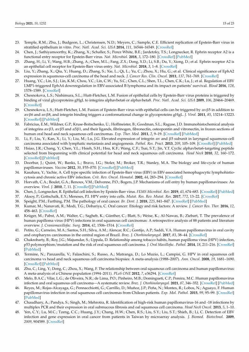
Figure 1. A hypothetical model of HPV and EBV cooperation for the development of HNCs. (1), HR-HPV infection and viral genome integration promote DNA damage in head and neck epithelial cells; (2), previous DNA alterations, as well as the increased expression of cyclin D1 and hTERT induced by HR-HPV, can promote the establishment of EBV latency; (3), HR-HPV E6/E7 oncopro-teins induce the expression of BZLF1, favoring the expression of EBV lytic genes with oncogenic properties, such as BARF1 (abortive lytic cycle); (4), HR-HPV infection induces CD21 (CR2), which, in turn, facilitates EBV cell entry; (5), the expression of HR-HPV oncoproteins inhibits protagonists of the anti-viral immune response, inducing immune evasion. Green shapes with red text represent HR-HPV oncoproteins. Brown shapes symbolize EBV proteins. Created by BioRender.com (ac-cessed on 5 November 2021).
5. Conclusions and Remarks Single HR-HPV or EBV infections are insufficient for carcinogenesis, suggesting the
involvement of additional factors. Both HR-HPV and EBV have been detected in HNCs, most notably in oropharyngeal tumors. The HPV/EBV copresence in HNCs is highly var-iable worldwide, ranging from 6.5% to 37.5% of OSCCs, 15% to 47.7% of NPCs in endemic regions, and 53.3% to 86% of OPSCCs. Potential mechanisms for HPV/EBV cooperation remain unclear, although some possibilities have been suggested. First, HPV could facili-tate EBV entry by promoting an increased expression of integrins and CD21. Second, HPV could facilitate EBV latency establishment by promoting genetic alterations due to HR-HPV genome integration into the host genome. Third, local immune evasion promoted by HPV infection could facilitate secondary EBV infection in epithelial cells. Additionally, HPV and EBV viral oncoproteins can synergistically cooperate for head and neck carcin-ogenesis. Additional studies are warranted in order to evaluate the mechanisms involved in the cooperation between HPV and EBV for head and neck carcinogenesis.
Author Contributions: Conceptualization, R.B. and F.A.; writing—original draft preparation, R.B.; writing—review and editing, R.B., F.A., D.C.-B., and A.H.C.; supervision, F.A. All authors have read and agreed to the published version of the manuscript.
Funding: This research was funded by ANID-FONDECYT Postdoctoral Grant 3190723 (R.B.), and CONICYT-FONDAP 15130011 (A.H.C.).
Institutional Review Board Statement: Not applicable.
Informed Consent Statement: Not applicable.
Data Availability Statement: Not applicable.
Figure 1. A hypothetical model of HPV and EBV cooperation for the development of HNCs. (1), HR-HPV infection and viral genome integration promote DNA damage in head and neck epithelial cells;(2), previous DNA alterations, as well as the increased expression of cyclin D1 and hTERT inducedby HR-HPV, can promote the establishment of EBV latency; (3), HR-HPV E6/E7 oncoproteins inducethe expression of BZLF1, favoring the expression of EBV lytic genes with oncogenic properties,such as BARF1 (abortive lytic cycle); (4), HR-HPV infection induces CD21 (CR2), which, in turn,facilitates EBV cell entry; (5), the expression of HR-HPV oncoproteins inhibits protagonists of theanti-viral immune response, inducing immune evasion. Green shapes with red text represent HR-HPV oncoproteins. Brown shapes symbolize EBV proteins. Created by BioRender.com (accessed on5 November 2021).
5. Conclusions and Remarks
Single HR-HPV or EBV infections are insufficient for carcinogenesis, suggesting theinvolvement of additional factors. Both HR-HPV and EBV have been detected in HNCs,most notably in oropharyngeal tumors. The HPV/EBV copresence in HNCs is highlyvariable worldwide, ranging from 6.5% to 37.5% of OSCCs, 15% to 47.7% of NPCs inendemic regions, and 53.3% to 86% of OPSCCs. Potential mechanisms for HPV/EBVcooperation remain unclear, although some possibilities have been suggested. First, HPVcould facilitate EBV entry by promoting an increased expression of integrins and CD21.Second, HPV could facilitate EBV latency establishment by promoting genetic alterationsdue to HR-HPV genome integration into the host genome. Third, local immune evasionpromoted by HPV infection could facilitate secondary EBV infection in epithelial cells.Additionally, HPV and EBV viral oncoproteins can synergistically cooperate for head andneck carcinogenesis. Additional studies are warranted in order to evaluate the mechanismsinvolved in the cooperation between HPV and EBV for head and neck carcinogenesis.
Author Contributions: Conceptualization, R.B. and F.A.; writing—original draft preparation, R.B.;writing—review and editing, R.B., F.A., D.C.-B. and A.H.C.; supervision, F.A. All authors have readand agreed to the published version of the manuscript.
Funding: This research was funded by ANID-FONDECYT Postdoctoral Grant 3190723 (R.B.), andCONICYT-FONDAP 15130011 (A.H.C.).
Institutional Review Board Statement: Not applicable.
Informed Consent Statement: Not applicable.

Biology 2021, 10, 1232 14 of 23
Data Availability Statement: Not applicable.
Acknowledgments: To Brianna Johnson for extensive manuscript revision and comments.
Conflicts of Interest: The authors declare no conflict of interest.
References1. Alam, M.S.; Siddiqui, S.A.; Perween, R. Epidemiological profile of head and neck cancer patients in Western Uttar Pradesh and
analysis of distributions of risk factors in relation to site of tumor. J. Cancer Res. Ther. 2017, 13, 430–435. [CrossRef]2. Sung, H.; Ferlay, J.; Siegel, R.L.; Laversanne, M.; Soerjomataram, I.; Jemal, A.; Bray, F. Global cancer statistics 2020: GLOBOCAN
estimates of incidence and mortality worldwide for 36 cancers in 185 countries. CA Cancer J. Clin. 2021, 71, 209–249. [CrossRef][PubMed]
3. Chaturvedi, A.K.; Engels, E.A.; Pfeiffer, R.M.; Hernandez, B.Y.; Xiao, W.; Kim, E.; Jiang, B.; Goodman, M.T.; Sibug-Saber, M.;Cozen, W.; et al. Human papillomavirus and rising oropharyngeal cancer incidence in the United States. J. Clin. Oncol. 2011, 29,4294–4301. [CrossRef]
4. Chien, Y.C.; Chen, J.Y.; Liu, M.Y.; Yang, H.I.; Hsu, M.M.; Chen, C.J.; Yang, C.S. Serologic markers of Epstein-Barr virus infectionand nasopharyngeal carcinoma in Taiwanese men. N. Engl. J. Med. 2001, 345, 1877–1882. [CrossRef] [PubMed]
5. Zhang, S.; Batur, P. Human papillomavirus in 2019: An update on cervical cancer prevention and screening guidelines. Clevel.Clin. J. Med. 2019, 86, 173–178. [CrossRef]
6. Drop, B.; Strycharz-Dudziak, M.; Kliszczewska, E.; Polz-Dacewicz, M. Coinfection with Epstein-Barr Virus (EBV), HumanPapilloma Virus (HPV) and Polyoma BK Virus (BKPyV) in Laryngeal, Oropharyngeal and Oral Cavity Cancer. Int. J. Mol. Sci.2017, 18, 2752. [CrossRef] [PubMed]
7. Jiang, R.; Ekshyyan, O.; Moore-Medlin, T.; Rong, X.; Nathan, S.; Gu, X.; Abreo, F.; Rosenthal, E.L.; Shi, M.; Guidry, J.T.; et al.Association between human papilloma virus/Epstein-Barr virus coinfection and oral carcinogenesis. J. Oral Pathol. Med. 2015, 44,28–36. [CrossRef]
8. Laantri, N.; Attaleb, M.; Kandil, M.; Naji, F.; Mouttaki, T.; Dardari, R.; Belghmi, K.; Benchakroun, N.; El Mzibri, M.; Khyatti,M. Human papillomavirus detection in moroccan patients with nasopharyngeal carcinoma. Infect. Agents Cancer 2011, 6, 3.[CrossRef]
9. Gupta, I.; Ghabreau, L.; Al-Thawadi, H.; Yasmeen, A.; Vranic, S.; Al Moustafa, A.E.; Malki, M.I. Co-incidence of HumanPapillomaviruses and Epstein-Barr Virus Is Associated With High to Intermediate Tumor Grade in Human Head and NeckCancer in Syria. Front. Oncol. 2020, 10, 1016. [CrossRef] [PubMed]
10. Al-Thawadi, H.; Gupta, I.; Jabeen, A.; Skenderi, F.; Aboulkassim, T.; Yasmeen, A.; Malki, M.I.; Batist, G.; Vranic, S.; Al Moustafa,A.E. Co-presence of human papillomaviruses and Epstein-Barr virus is linked with advanced tumor stage: A tissue microarraystudy in head and neck cancer patients. Cancer Cell Int. 2020, 20, 361. [CrossRef]
11. Egawa, N.; Egawa, K.; Griffin, H.; Doorbar, J. Human Papillomaviruses; Epithelial Tropisms, and the Development of Neoplasia.Viruses 2015, 7, 3863–3890. [CrossRef] [PubMed]
12. Möhl, B.S.; Chen, J.; Sathiyamoorthy, K.; Jardetzky, T.S.; Longnecker, R. Structural and Mechanistic Insights into the Tropism ofEpstein-Barr Virus. Mol. Cells 2016, 39, 286–291. [CrossRef]
13. McBride, A.A. Human papillomaviruses: Diversity, infection and host interactions. Nat. Rev. Microbiol. 2021, 1–14. [CrossRef]14. Doorbar, J.; Egawa, N.; Griffin, H.; Kranjec, C.; Murakami, I. Human papillomavirus molecular biology and disease association.
Rev. Med. Virol. 2015, 25, 2–23. [CrossRef] [PubMed]15. Surviladze, Z.; Sterkand, R.T.; Ozbun, M.A. Interaction of human papillomavirus type 16 particles with heparan sulfate and
syndecan-1 molecules in the keratinocyte extracellular matrix plays an active role in infection. J. Gen. Virol. 2015, 96, 2232–2241.[CrossRef] [PubMed]
16. Ozbun, M.A.; Campos, S.K. The long and winding road: Human papillomavirus entry and subcellular trafficking. Curr. Opin.Virol. 2021, 50, 76–86. [CrossRef]
17. Werner, J.; Decarlo, C.A.; Escott, N.; Zehbe, I.; Ulanova, M. Expression of integrins and Toll-like receptors in cervical cancer: Effectof infectious agents. Innate Immun. 2011, 18, 55–69. [CrossRef] [PubMed]
18. Evans, W.; Filippova, M.; Filippov, V.; Bashkirova, S.; Zhang, G.; Reeves, M.E.; Duerksen-Hughes, P. Overexpression of HPV16 E6*Alters β-Integrin and Mitochondrial Dysfunction Pathways in Cervical Cancer Cells. Cancer Genom. Proteom. 2016, 13, 259–273.
19. Eckhardt, M.; Zhang, W.; Gross, A.M.; Von Dollen, J.; Johnson, J.R.; Franks-Skiba, K.E.; Swaney, D.L.; Johnson, T.L.; Jang, G.M.;Shah, P.S.; et al. Multiple Routes to Oncogenesis Are Promoted by the Human Papillomavirus-Host Protein Network. CancerDiscov. 2018, 8, 1474–1489. [CrossRef]
20. Romero-Medina, M.C.; Venuti, A.; Melita, G.; Robitaille, A.; Ceraolo, M.G.; Pacini, L.; Sirand, C.; Viarisio, D.; Taverniti, V.; Gupta,P.; et al. Human papillomavirus type 38 alters wild-type p53 activity to promote cell proliferation via the downregulation ofintegrin alpha 1 expression. PLoS Pathog. 2020, 16, e1008792. [CrossRef]
21. Hutt-Fletcher, L.M. The Long and Complicated Relationship between Epstein-Barr Virus and Epithelial Cells. J. Virol. 2017,91, e01677-16. [CrossRef]
22. Hatton, O.L.; Harris-Arnold, A.; Schaffert, S.; Krams, S.M.; Martinez, O.M. The interplay between Epstein-Barr virus and Blymphocytes: Implications for infection, immunity, and disease. Immunol. Res. 2014, 58, 268–276. [CrossRef] [PubMed]
Biology 2021, 10, 1232 15 of 23
23. Temple, R.M.; Zhu, J.; Budgeon, L.; Christensen, N.D.; Meyers, C.; Sample, C.E. Efficient replication of Epstein-Barr virus instratified epithelium in vitro. Proc. Natl. Acad. Sci. USA 2014, 111, 16544–16549. [CrossRef]
24. Chen, J.; Sathiyamoorthy, K.; Zhang, X.; Schaller, S.; Perez White, B.E.; Jardetzky, T.S.; Longnecker, R. Ephrin receptor A2 is afunctional entry receptor for Epstein-Barr virus. Nat. Microbiol. 2018, 3, 172–180. [CrossRef] [PubMed]
25. Zhang, H.; Li, Y.; Wang, H.B.; Zhang, A.; Chen, M.L.; Fang, Z.X.; Dong, X.D.; Li, S.B.; Du, Y.; Xiong, D.; et al. Ephrin receptor A2 isan epithelial cell receptor for Epstein-Barr virus entry. Nat. Microbiol. 2018, 3, 1–8. [CrossRef]
26. Liu, Y.; Zhang, X.; Qiu, Y.; Huang, D.; Zhang, S.; Xie, L.; Qi, L.; Yu, C.; Zhou, X.; Hu, G.; et al. Clinical significance of EphA2expression in squamous-cell carcinoma of the head and neck. J. Cancer Res. Clin. Oncol. 2011, 137, 761–769. [CrossRef]
27. Huang, Y.C.; Lin, S.J.; Lin, K.M.; Chou, Y.C.; Lin, C.W.; Yu, S.C.; Chen, C.L.; Shen, T.L.; Chen, C.K.; Lu, J.; et al. Regulation of EBVLMP1-triggered EphA4 downregulation in EBV-associated B lymphoma and its impact on patients’ survival. Blood 2016, 128,1578–1589. [CrossRef]
28. Chesnokova, L.S.; Nishimura, S.L.; Hutt-Fletcher, L.M. Fusion of epithelial cells by Epstein-Barr virus proteins is triggered bybinding of viral glycoproteins gHgL to integrins alphavbeta6 or alphavbeta8. Proc. Natl. Acad. Sci. USA 2009, 106, 20464–20469.[CrossRef]
29. Chesnokova, L.S.; Hutt-Fletcher, L.M. Fusion of Epstein-Barr virus with epithelial cells can be triggered by αvβ5 in addition toαvβ6 and αvβ8, and integrin binding triggers a conformational change in glycoproteins gHgL. J. Virol. 2011, 85, 13214–13223.[CrossRef] [PubMed]
30. Fabricius, E.M.; Wildner, G.P.; Kruse-Boitschenko, U.; Hoffmeister, B.; Goodman, S.L.; Raguse, J.D. Immunohistochemical analysisof integrins αvβ3, αvβ5 and α5β1, and their ligands, fibrinogen, fibronectin, osteopontin and vitronectin, in frozen sections ofhuman oral head and neck squamous cell carcinomas. Exp. Ther. Med. 2011, 2, 9–19. [CrossRef] [PubMed]
31. Li, F.; Liu, Y.; Kan, X.; Li, Y.; Liu, M.; Lu, J.G. Elevated expression of integrin αv and β5 subunit in laryngeal squamous-cellcarcinoma associated with lymphatic metastasis and angiogenesis. Pathol. Res. Pract. 2013, 209, 105–109. [CrossRef] [PubMed]
32. Hsiao, J.R.; Chang, Y.; Chen, Y.L.; Hsieh, S.H.; Hsu, K.F.; Wang, C.F.; Tsai, S.T.; Jin, Y.T. Cyclic alphavbeta6-targeting peptideselected from biopanning with clinical potential for head and neck squamous cell carcinoma. Head Neck 2010, 32, 160–172.[CrossRef] [PubMed]
33. Doorbar, J.; Quint, W.; Banks, L.; Bravo, I.G.; Stoler, M.; Broker, T.R.; Stanley, M.A. The biology and life-cycle of humanpapillomaviruses. Vaccine 2012, 30, F55–F70. [CrossRef] [PubMed]
34. Kasahara, Y.; Yachie, A. Cell type specific infection of Epstein-Barr virus (EBV) in EBV-associated hemophagocytic lymphohistio-cytosis and chronic active EBV infection. Crit. Rev. Oncol. Hematol. 2002, 44, 283–294. [CrossRef]
35. Horvath, C.A.; Boulet, G.A.; Renoux, V.M.; Delvenne, P.O.; Bogers, J.P. Mechanisms of cell entry by human papillomaviruses: Anoverview. Virol. J. 2010, 7, 11. [CrossRef] [PubMed]
36. Chen, J.; Longnecker, R. Epithelial cell infection by Epstein-Barr virus. FEMS Microbiol. Rev. 2019, 43, 674–683. [CrossRef] [PubMed]37. Aksoy, P.; Gottschalk, E.Y.; Meneses, P.I. HPV entry into cells. Mutat. Res. Rev. Mutat. Res. 2017, 772, 13–22. [CrossRef]38. Speight, P.M.; Farthing, P.M. The pathology of oral cancer. Br. Dent. J. 2018, 225, 841–847. [CrossRef] [PubMed]39. Kumar, M.; Nanavati, R.; Modi, T.G.; Dobariya, C. Oral cancer: Etiology and risk factors: A review. J. Cancer Res. Ther. 2016, 12,
458–463. [CrossRef]40. Krüger, M.; Pabst, A.M.; Walter, C.; Sagheb, K.; Günther, C.; Blatt, S.; Weise, K.; Al-Nawas, B.; Ziebart, T. The prevalence of
human papilloma virus (HPV) infections in oral squamous cell carcinomas: A retrospective analysis of 88 patients and literatureoverview. J. Craniomaxillofac. Surg. 2014, 42, 1506–1514. [CrossRef]
41. Petito, G.; Carneiro, M.A.; Santos, S.H.; Silva, A.M.; Alencar, R.C.; Gontijo, A.P.; Saddi, V.A. Human papillomavirus in oral cavityand oropharynx carcinomas in the central region of Brazil. Braz. J. Otorhinolaryngol. 2017, 83, 38–44. [CrossRef]
42. Chakrobarty, B.; Roy, J.G.; Majumdar, S.; Uppala, D. Relationship among tobacco habits, human papilloma virus (HPV) infection,p53 polymorphism/mutation and the risk of oral squamous cell carcinoma. J. Oral Maxillofac. Pathol. 2014, 18, 211–216. [CrossRef][PubMed]
43. Termine, N.; Panzarella, V.; Falaschini, S.; Russo, A.; Matranga, D.; Lo Muzio, L.; Campisi, G. HPV in oral squamous cellcarcinoma vs head and neck squamous cell carcinoma biopsies: A meta-analysis (1988–2007). Ann. Oncol. 2008, 19, 1681–1690.[CrossRef] [PubMed]
44. Zhu, C.; Ling, Y.; Dong, C.; Zhou, X.; Wang, F. The relationship between oral squamous cell carcinoma and human papillomavirus:A meta-analysis of a Chinese population (1994–2011). PLoS ONE 2012, 7, e36294. [CrossRef]
45. Melo, B.A.C.; Vilar, L.G.; de Oliveira, N.R.; de Lima, P.O.; Pinheiro, M.B.; Domingueti, C.P.; Pereira, M.C. Human papillomavirusinfection and oral squamous cell carcinoma—A systematic review. Braz. J. Otorhinolaryngol. 2021, 87, 346–352. [CrossRef] [PubMed]
46. Reyes, M.; Rojas-Alcayaga, G.; Pennacchiotti, G.; Carrillo, D.; Muñoz, J.P.; Peña, N.; Montes, R.; Lobos, N.; Aguayo, F. Humanpapillomavirus infection in oral squamous cell carcinomas from Chilean patients. Exp. Mol. Pathol. 2015, 99, 95–99. [CrossRef][PubMed]
47. Chaudhary, A.; Pandya, S.; Singh, M.; Mehrotra, R. Identification of high-risk human papillomavirus-16 and -18 infections bymultiplex PCR and their expression in oral submucous fibrosis and oal squamous cell carcinoma. Head Neck Oncol. 2013, 5, 1–10.
48. Yen, C.Y.; Lu, M.C.; Tzeng, C.C.; Huang, J.Y.; Chang, H.W.; Chen, R.S.; Liu, S.Y.; Liu, S.T.; Shieh, B.; Li, C. Detection of EBVinfection and gene expression in oral cancer from patients in Taiwan by microarray analysis. J. Biomed. Biotechnol. 2009,2009, 904589. [CrossRef]

Biology 2021, 10, 1232 16 of 23
49. Acharya, S.; Ekalaksananan, T.; Vatanasapt, P.; Loyha, K.; Phusingha, P.; Promthet, S.; Kongyingyoes, B.; Pientong, C. Associationof Epstein-Barr virus infection with oral squamous cell carcinoma in a case-control study. J. Oral Pathol. Med. 2014, 44, 252–257.[CrossRef]
50. Heawchaiyaphum, C.; Iizasa, H.; Ekalaksananan, T.; Burassakarn, A.; Kiyono, T.; Kanehiro, Y.; Yoshiyama, H.; Pientong, C.Epstein-Barr Virus Infection of Oral Squamous Cells. Microorganisms 2020, 8, 419. [CrossRef]
51. Kikuchi, K.; Noguchi, Y.; de Rivera, M.W.G.-N.; Hoshino, M.; Sakashita, H.; Yamada, T.; Inoue, H.; Miyazaki, Y.; Nozaki, T.;González-López, B.S.; et al. Detection of Epstein-Barr virus genome and latent infection gene expression in normal epithelia,epithelial dysplasia, and squamous cell carcinoma of the oral cavity. Tumor Biol. 2016, 37, 3389–3404. [CrossRef]
52. Rahman, R.; Poomsawat, S.; Juengsomjit, R.; Buajeeb, W. Overexpression of Epstein-Barr virus-encoded latent membraneprotein-1 (LMP-1) in oral squamous cell carcinoma. BMC Oral Health 2019, 19, 142. [CrossRef] [PubMed]
53. She, Y.; Nong, X.; Zhang, M.; Wang, M. Epstein-Barr virus infection and oral squamous cell carcinoma risk: A meta-analysis.PLoS ONE 2017, 12, e0186860. [CrossRef] [PubMed]
54. de Lima, M.A.P.; Teodoro, I.P.P.; de Galiza, L.E.; Filho, P.H.B.M.; Marques, F.M.; Pinheiro Junior, R.F.F.; Macedo, G.E.C.; Facundo,H.T.; da Silva, C.G.L.; Lima, M.V.A. Association between Epstein-Barr Virus and Oral Carcinoma: A Systematic Review withMeta-Analysis. Crit. Rev. Oncog. 2019, 24, 349–368. [CrossRef]
55. Jalouli, J.; Jalouli, M.M.; Sapkota, D.; Ibrahim, S.O.; Larsson, P.A.; Sand, L. Human papilloma virus, herpes simplex virus andepstein barr virus in oral squamous cell carcinoma from eight different countries. Anticancer Res. 2012, 32, 571–580. [PubMed]
56. Polz-Gruszka, D.; Morshed, K.; Jarzynski, A.; Polz-Dacewicz, M. Prevalence of Polyoma BK Virus (BKPyV), Epstein-Barr Virus(EBV) and Human Papilloma Virus (HPV) in Oropharyngeal Cancer. Pol. J. Microbiol. 2015, 64, 323–328. [CrossRef]
57. Vanshika, S.; Preeti, A.; Sumaira, Q.; Vijay, K.; Shikha, T.; Shivanjali, R.; Shankar, S.U.; Mati, G.M. Incidence OF HPV and EBV inoral cancer and their clinico-pathological correlation—A pilot study of 108 cases. J. Oral Biol. Craniofac. Res. 2021, 11, 180–184.[CrossRef]
58. Broccolo, F.; Ciccarese, G.; Rossi, A.; Anselmi, L.; Drago, F.; Toniolo, A. Human papillomavirus (HPV) and Epstein-Barr virus(EBV) in keratinizing versus non- keratinizing squamous cell carcinoma of the oropharynx. Infect. Agents Cancer 2018, 13, 32.[CrossRef]
59. Naqvi, S.U.; Khan, S.; Ahmed, A.; Lail, A.; Gul, S.; Ahmed, S. Prevalence of EBV, CMV, and HPV in oral squamous cell carcinomapatients in the Pakistani population. J. Med. Virol. 2020, 92, 3880–3883. [CrossRef]
60. Tang, L.L.; Chen, W.Q.; Xue, W.Q.; He, Y.Q.; Zheng, R.S.; Zeng, Y.X.; Jia, W.H. Global trends in incidence and mortality ofnasopharyngeal carcinoma. Cancer Lett. 2016, 374, 22–30. [CrossRef]
61. Wang, A.; Zhang, W.; Jin, M.; Zhang, J.; Li, S.; Tong, F.; Zhou, Y. Differential expression of EBV proteins LMP1 and BHFR1 inEBV-associated gastric and nasopharyngeal cancer tissues. Mol. Med. Rep. 2016, 13, 4151–4158. [CrossRef]
62. Pan, J.J.; Ng, W.T.; Zong, J.F.; Chan, L.L.; O’Sullivan, B.; Lin, S.J.; Sze, H.C.; Chen, Y.B.; Choi, H.C.; Guo, Q.J.; et al. Proposal for the8th edition of the AJCC/UICC staging system for nasopharyngeal cancer in the era of intensity-modulated radiotherapy. Cancer2016, 122, 546–558. [CrossRef] [PubMed]
63. Tang, L.L.; Chen, Y.P.; Mao, Y.P.; Wang, Z.X.; Guo, R.; Chen, L.; Tian, L.; Lin, A.H.; Li, L.; Sun, Y.; et al. Validation of the 8thEdition of the UICC/AJCC Staging System for Nasopharyngeal Carcinoma From Endemic Areas in the Intensity-ModulatedRadiotherapy Era. J. Natl. Compr. Cancer Netw. 2017, 15, 913–919. [CrossRef] [PubMed]
64. Okekpa, S.I.; Mydin, R.B.S.M.N.; Mangantig, E.; Azmi, N.S.A.; Zahari, S.N.S.; Kaur, G.; Musa, Y. Nasopharyngeal Carcinoma(NPC) Risk Factors: A Systematic Review and Meta-Analysis of the Association with Lifestyle, Diets, Socioeconomic andSociodemographic in Asian Region. Asian Pac. J. Cancer Prev. 2019, 20, 3505–3514. [CrossRef] [PubMed]
65. Bei, J.X.; Zuo, X.Y.; Liu, W.S.; Guo, Y.M.; Zeng, Y.X. Genetic susceptibility to the endemic form of NPC. Chin. Clin. Oncol. 2016,5, 15. [CrossRef]
66. Fan, H.C.; Chen, C.Y.; Hsu, Y.C.; Chou, R.H.; Teng, C.J.; Chiu, C.H.; Hsu, C.Y.; Muo, C.H.; Chang, M.Y.; Chang, K.H. Increasedrisk of incident nasopharyngeal carcinoma with exposure to air pollution. PLoS ONE 2018, 13, e0204568. [CrossRef]
67. Zhang, H.; Wang, J.; Yu, D.; Liu, Y.; Xue, K.; Zhao, X. Role of Epstein-Barr Virus in the Development of Nasopharyngeal Carcinoma.Open Med. 2017, 12, 171–176. [CrossRef]
68. Fan, C.; Tang, Y.; Wang, J.; Xiong, F.; Guo, C.; Wang, Y.; Xiang, B.; Zhou, M.; Li, X.; Wu, X.; et al. The emerging role of Epstein-Barrvirus encoded microRNAs in nasopharyngeal carcinoma. J. Cancer 2018, 9, 2852–2864. [CrossRef]
69. Shannon-Lowe, C.; Rickinson, A. The Global Landscape of EBV-Associated Tumors. Front. Oncol. 2019, 9, 713. [CrossRef]70. Asante, D.B.; Asmah, R.H.; Adjei, A.A.; Kyei, F.; Simpong, D.L.; Brown, C.A.; Gyasi, R.K. Detection of Human Papillomavirus
Genotypes and Epstein-Barr Virus in Nasopharyngeal Carcinomas at the Korle-Bu Teaching Hospital, Ghana. Sci. World J. 2017,2017, 2721367. [CrossRef]
71. Breda, E.; Catarino, R.J.; Azevedo, I.; Lobão, M.; Monteiro, E.; Medeiros, R. Epstein-Barr virus detection in nasopharyngealcarcinoma: Implications in a low-risk area. Braz. J. Otorhinolaryngol. 2010, 76, 310–315. [CrossRef]
72. Dogan, S.; Hedberg, M.L.; Ferris, R.L.; Rath, T.J.; Assaad, A.M.; Chiosea, S.I. Human papillomavirus and Epstein-Barr virus innasopharyngeal carcinoma in a low-incidence population. Head Neck 2014, 36, 511–516. [CrossRef] [PubMed]
73. Svajdler, M.; Kaspirkova, J.; Mezencev, R.; Laco, J.; Torday, T.; Dubinsky, P.; Straka, L.; Ondic, O.; Michal, M.; Skalova, A. Humanpapillomavirus and Epstein-Barr virus in nasopharyngeal carcinoma in a non-endemic eastern european population. Neoplasma2016, 63, 107–114. [CrossRef] [PubMed]

Biology 2021, 10, 1232 17 of 23
74. Ruuskanen, M.; Irjala, H.; Minn, H.; Vahlberg, T.; Randen-Brady, R.; Hagström, J.; Syrjänen, S.; Leivo, I. Epstein-Barr virus andhuman papillomaviruses as favorable prognostic factors in nasopharyngeal carcinoma: A nationwide study in Finland. HeadNeck 2019, 41, 349–357. [CrossRef]
75. Rumayor Piña, A.; Dos Santos, H.T.; Carlos, R.; Altemani, A.; de Almeida, O.P. Epstein-Barr Virus in Nasopharyngeal Carcinomaof Guatemalan and Brazilian Patients. Int. J. Surg. Pathol. 2017, 25, 304–309. [CrossRef]
76. Tatlı Dogan, H.; Kılıçarslan, A.; Dogan, M.; Süngü, N.; Güler Tezel, G.; Güler, G. Retrospective analysis of oncogenic humanpapilloma virus and Epstein-Barr virus prevalence in Turkish nasopharyngeal cancer patients. Pathol. Res. Pract. 2016, 212,1021–1026. [CrossRef]
77. Tham, T.; Machado, R.; Russo, D.P.; Herman, S.W.; Teegala, S.; Costantino, P. Viral markers in nasopharyngeal carcinoma: Asystematic review and meta-analysis on the detection of p16INK4a, human papillomavirus (HPV), and Ebstein-Barr virus (EBV).Am. J. Otolaryngol. 2021, 42, 102762. [CrossRef]
78. Zhang, J.; Shu, C.; Song, Y.; Li, Q.; Huang, J.; Ma, X. Epstein-Barr virus DNA level as a novel prognostic factor in nasopharyngealcarcinoma: A meta-analysis. Medicine 2016, 95, e5130. [CrossRef] [PubMed]
79. Peng, H.; Chen, L.; Zhang, Y.; Guo, R.; Li, W.F.; Mao, Y.P.; Tan, L.L.; Sun, Y.; Zhang, F.; Liu, L.Z.; et al. Survival analysis of patientswith advanced-stage nasopharyngeal carcinoma according to the Epstein-Barr virus status. Oncotarget 2016, 7, 24208–24216.[CrossRef]
80. Liu, T.B.; Zheng, Z.H.; Pan, J.; Pan, L.L.; Chen, L.H. Prognostic role of plasma Epstein-Barr virus DNA load for nasopharyngealcarcinoma: A meta-analysis. Clin. Investig. Med. 2017, 40, E1–E12. [CrossRef]
81. Du, Y.Y.; Luo, D.H.; Sun, X.S.; Tang, L.Q.; Mai, H.Q.; Chen, Q.Y.; Zhong, J.H.; Mai, D.M.; Zhang, W.R.; Chen, W.H.; et al.Combining pretreatment plasma Epstein-Barr virus DNA level and cervical node necrosis improves prognostic stratification inpatients with nasopharyngeal carcinoma: A cohort study. Cancer Med. 2019, 8, 6841–6852. [CrossRef] [PubMed]
82. Nilsson, J.S.; Forslund, O.; Andersson, F.C.; Lindstedt, M.; Greiff, L. Intralesional EBV-DNA load as marker of prognosis fornasopharyngeal cancer. Sci. Rep. 2019, 9, 15432. [CrossRef] [PubMed]
83. Zhao, Y.; Wang, Y.; Zeng, S.; Hu, X. LMP1 expression is positively associated with metastasis of nasopharyngeal carcinoma:Evidence from a meta-analysis. J. Clin. Pathol. 2012, 65, 41–45. [CrossRef]
84. Hu, C.; Wei, W.; Chen, X.; Woodman, C.B.; Yao, Y.; Nicholls, J.M.; Joab, I.; Sihota, S.K.; Shao, J.Y.; Derkaoui, K.D.; et al. A globalview of the oncogenic landscape in nasopharyngeal carcinoma: An integrated analysis at the genetic and expression levels. PLoSONE 2012, 7, e41055. [CrossRef]
85. Luo, F.-F.; Li, Z.-Y.; Huang, S.-N.; Chen, G.; Xie, T.-T.; LI, Y.-Q.; Xing, W.-W.; Li, W.-Y.; Lu, Y.-K.; Ding, H. Prevalence of humanpapillomavirus in patients with nasopharyngeal carcinoma: A meta-analysis. Int. J. Clin. Exp. Med. 2017, 10, 9837–9847.
86. Stenmark, M.H.; McHugh, J.B.; Schipper, M.; Walline, H.M.; Komarck, C.; Feng, F.Y.; Worden, F.P.; Wolf, G.T.; Chepeha, D.B.;Prince, M.E.; et al. Nonendemic HPV-positive nasopharyngeal carcinoma: Association with poor prognosis. Int. J. Radiat. Oncol.Biol. Phys. 2014, 88, 580–588. [CrossRef]
87. Tung, Y.C.; Lin, K.H.; Chu, P.Y.; Hsu, C.C.; Kuo, W.R. Detection of human papilloma virus and Epstein-Barr virus DNA innasopharyngeal carcinoma by polymerase chain reaction. Kaohsiung J. Med. Sci. 1999, 15, 256–262.
88. Mirzamani, N.; Salehian, P.; Farhadi, M.; Tehran, E.A. Detection of EBV and HPV in nasopharyngeal carcinoma by in situhybridization. Exp. Mol. Pathol. 2006, 81, 231–234. [CrossRef]
89. Jiang, W.; Chamberlain, P.D.; Garden, A.S.; Kim, B.Y.; Ma, D.; Lo, E.J.; Bell, D.; Gunn, G.B.; Fuller, C.D.; Rosenthal, D.I.; et al.Prognostic value of p16 expression in Epstein-Barr virus-positive nasopharyngeal carcinomas. Head Neck 2016, 38, E1459–E1466.[CrossRef]
90. Simon, J.; Schroeder, L.; Ingarfield, K.; Diehl, S.; Werner, J.; Brenner, N.; Liu, Z.; Pawlita, M.; Pring, M.; Butt, J.; et al. Epstein-Barrvirus and human papillomavirus serum antibodies define the viral status of nasopharyngeal carcinoma in a low endemic country.Int. J. Cancer 2020, 147, 461–471. [CrossRef]
91. Osborne, R.F.; Brown, J.J. Carcinoma of the oral pharynx: An analysis of subsite treatment heterogeneity. Surg. Oncol. Clin. N. Am.2004, 13, 71–80. [CrossRef]
92. Pan, C.; Issaeva, N.; Yarbrough, W.G. HPV-driven oropharyngeal cancer: Current knowledge of molecular biology and mecha-nisms of carcinogenesis. Cancers Head Neck 2018, 3, 12. [CrossRef] [PubMed]
93. Kaplon, A.W.; Galloway, T.J.; Bhayani, M.K.; Liu, J.C. Effect of HPV Status on Survival of Oropharynx Cancer with DistantMetastasis. Otolaryngol. Head Neck Surg. 2020, 163, 372–374. [CrossRef] [PubMed]
94. Wendt, M.; Hammarstedt-Nordenvall, L.; Zupancic, M.; Friesland, S.; Landin, D.; Munck-Wikland, E.; Dalianis, T.; Näsman, A.;Marklund, L. Long-Term Survival and Recurrence in Oropharyngeal Squamous Cell Carcinoma in Relation to Subsites, HPV, andp16-Status. Cancers 2021, 13, 2553. [CrossRef] [PubMed]
95. Mazul, A.L.; Rodriguez-Ormaza, N.; Taylor, J.M.; Desai, D.D.; Brennan, P.; Anantharaman, D.; Gheit, T.; Tommasino, M.; Abedi-Ardekani, B.; Olshan, A.F.; et al. Prognostic significance of non-HPV16 genotypes in oropharyngeal squamous cell carcinoma.Oral Oncol. 2016, 61, 98–103. [CrossRef]
96. Smith, E.M.; Rubenstein, L.M.; Haugen, T.H.; Hamsikova, E.; Turek, L.P. Tobacco and alcohol use increases the risk of bothHPV-associated and HPV-independent head and neck cancers. Cancer Causes Control. 2010, 21, 1369–1378. [CrossRef]
97. Stjernstrøm, K.D.; Jensen, J.S.; Jakobsen, K.K.; Grønhøj, C.; von Buchwald, C. Current status of human papillomavirus positivityin oropharyngeal squamous cell carcinoma in Europe: A systematic review. Acta Oto-Laryngol. 2019, 139, 1112–1116. [CrossRef]

Biology 2021, 10, 1232 18 of 23
98. Mariz, B.A.L.A.; Kowalski, L.P.; William, W.N.; de Castro, G.; Chaves, A.L.F.; Santos, M.; de Oliveira, T.B.; Araújo, A.L.D.;Normando, A.G.C.; Ribeiro, A.C.P.; et al. Global prevalence of human papillomavirus-driven oropharyngeal squamous cellcarcinoma following the ASCO guidelines: A systematic review and meta-analysis. Crit. Rev. Oncol. Hematol. 2020, 156, 103116.[CrossRef]
99. Haeggblom, L.; Ramqvist, T.; Tommasino, M.; Dalianis, T.; Näsman, A. Time to change perspectives on HPV in oropharyngealcancer. A systematic review of HPV prevalence per oropharyngeal sub-site the last 3 years. Papillomavirus Res. 2017, 4, 1–11.[CrossRef]
100. Donà, M.G.; Spriano, G.; Pichi, B.; Rollo, F.; Laquintana, V.; Covello, R.; Pellini, R.; Giuliani, M.; Pescarmona, E.; Benevolo, M.Human papillomavirus infection and p16 overexpression in oropharyngeal squamous cell carcinoma: A case series from 2010 to2014. Future Microbiol. 2015, 10, 1283–1291. [CrossRef]
101. Anantharaman, D.; Abedi-Ardekani, B.; Beachler, D.C.; Gheit, T.; Olshan, A.F.; Wisniewski, K.; Wunsch-Filho, V.; Toporcov, T.N.;Tajara, E.H.; Levi, J.E.; et al. Geographic heterogeneity in the prevalence of human papillomavirus in head and neck cancer. Int. J.Cancer 2017, 140, 1968–1975. [CrossRef]
102. Ndiaye, C.; Mena, M.; Alemany, L.; Arbyn, M.; Castellsagué, X.; Laporte, L.; Bosch, F.X.; de Sanjosé, S.; Trottier, H. HPV DNA,E6/E7 mRNA, and p16INK4a detection in head and neck cancers: A systematic review and meta-analysis. Lancet Oncol. 2014, 15,1319–1331. [CrossRef]
103. Szkaradkiewicz, A.; Kruk-Zagajewska, A.; Wal, M.; Jopek, A.; Wierzbicka, M.; Kuch, A. Epstein-Barr virus and human papillo-mavirus infections and oropharyngeal squamous cell carcinomas. Clin. Exp. Med. 2002, 2, 137–141. [CrossRef] [PubMed]
104. Tyan, Y.S.; Liu, S.T.; Ong, W.R.; Chen, M.L.; Shu, C.H.; Chang, Y.S. Detection of Epstein-Barr virus and human papillomavirus inhead and neck tumors. J. Clin. Microbiol. 1993, 31, 53–56. [CrossRef] [PubMed]
105. Vazquez-Guillen, J.M.; Palacios-Saucedo, G.C.; Rivera-Morales, L.G.; Alonzo-Morado, M.V.; Burciaga-Bernal, S.B.; Montufar-Martinez, M.; Ortiz-Lopez, R.; Gonzalez-Villasana, V.; Martinez-Torres, A.C.; Serna-Hernandez, J.C.; et al. Infection andcoinfection by human papillomavirus, Epstein-Barr virus and Merkel cell polyomavirus in patients with squamous cell carcinomaof the larynx: A retrospective study. PeerJ 2018, 6, e5834. [CrossRef] [PubMed]
106. Miller, C.S.; Johnstone, B.M. Human papillomavirus as a risk factor for oral squamous cell carcinoma: A meta-analysis, 1982–1997.Oral Surg. Oral Med. Oral Pathol. Oral Radiol. Endod. 2001, 91, 622–635. [CrossRef]
107. Dayyani, F.; Etzel, C.J.; Liu, M.; Ho, C.H.; Lippman, S.M.; Tsao, A.S. Meta-analysis of the impact of human papillomavirus (HPV)on cancer risk and overall survival in head and neck squamous cell carcinomas (HNSCC). Head Neck Oncol. 2010, 2, 15. [CrossRef]
108. Mehanna, H.; Beech, T.; Nicholson, T.; El-Hariry, I.; McConkey, C.; Paleri, V.; Roberts, S. Prevalence of human papillomavirus inoropharyngeal and nonoropharyngeal head and neck cancer–systematic review and meta-analysis of trends by time and region.Head Neck 2013, 35, 747–755. [CrossRef]
109. Kreimer, A.R.; Clifford, G.M.; Boyle, P.; Franceschi, S. Human papillomavirus types in head and neck squamous cell carcinomasworldwide: A systematic review. Cancer Epidemiol. Biomark. Prev. 2005, 14, 467–475. [CrossRef]
110. Shaikh, M.H.; McMillan, N.A.; Johnson, N.W. HPV-associated head and neck cancers in the Asia Pacific: A critical literaturereview & meta-analysis. Cancer Epidemiol. 2015, 39, 923–938. [CrossRef] [PubMed]
111. Li, X.; Gao, L.; Li, H.; Gao, J.; Yang, Y.; Zhou, F.; Gao, C.; Li, M.; Jin, Q. Human papillomavirus infection and laryngeal cancer risk:A systematic review and meta-analysis. J. Infect. Dis. 2013, 207, 479–488. [CrossRef] [PubMed]
112. Altekin, I.; Tas, A.; Yalcin, O.; Guven, S.G.; Aslan, Z.; Adali, M.K.; Karasalihoglu, A.R. Frequency of Epstein-Barr virus andhuman papilloma virus in patients with nasopharyngeal carcinoma. Eur. Arch. Otorhinolaryngol. 2020, 277, 2041–2047. [CrossRef]
113. Deng, Z.; Uehara, T.; Maeda, H.; Hasegawa, M.; Matayoshi, S.; Kiyuna, A.; Agena, S.; Pan, X.; Zhang, C.; Yamashita, Y.; et al.Epstein-Barr virus and human papillomavirus infections and genotype distribution in head and neck cancers. PLoS ONE 2014,9, e113702. [CrossRef]
114. Hutt-Fletcher, L.M. Epstein-Barr virus entry. J. Virol. 2007, 81, 7825–7832. [CrossRef] [PubMed]115. Jiang, R.; Gu, X.; Nathan, C.O.; Hutt-Fletcher, L. Laser-capture microdissection of oropharyngeal epithelium indicates restriction
of Epstein-Barr virus receptor/CD21 mRNA to tonsil epithelial cells. J. Oral Pathol. Med. 2008, 37, 626–633. [CrossRef] [PubMed]116. Jiang, R.; Gu, X.; Moore-Medlin, T.N.; Nathan, C.A.; Hutt-Fletcher, L.M. Oral dysplasia and squamous cell carcinoma: Correlation
between increased expression of CD21, Epstein-Barr virus and CK19. Oral Oncol. 2012, 48, 836–841. [CrossRef]117. Li, Q.X.; Young, L.S.; Niedobitek, G.; Dawson, C.W.; Birkenbach, M.; Wang, F.; Rickinson, A.B. Epstein-Barr virus infection and
replication in a human epithelial cell system. Nature 1992, 356, 347–350. [CrossRef]118. Knox, P.G.; Li, Q.X.; Rickinson, A.B.; Young, L.S. In vitro production of stable Epstein-Barr virus-positive epithelial cell clones
which resemble the virus:cell interaction observed in nasopharyngeal carcinoma. Virology 1996, 215, 40–50. [CrossRef]119. Brooks, L.; Yao, Q.Y.; Rickinson, A.B.; Young, L.S. Epstein-Barr virus latent gene transcription in nasopharyngeal carcinoma cells:
Coexpression of EBNA1, LMP1, and LMP2 transcripts. J. Virol. 1992, 66, 2689–2697. [CrossRef]120. Murono, S.; Yoshizaki, T.; Park, C.S.; Furukawa, M. Association of Epstein-Barr virus infection with p53 protein accumulation but
not bcl-2 protein in nasopharyngeal carcinoma. Histopathology 1999, 34, 432–438. [CrossRef]121. Fingeroth, J.D.; Diamond, M.E.; Sage, D.R.; Hayman, J.; Yates, J.L. CD21-Dependent infection of an epithelial cell line, 293, by
Epstein-Barr virus. J. Virol. 1999, 73, 2115–2125. [CrossRef]122. Zhou, Y.; Yang, W.; Mei, X.; Hu, H. Transcriptomic Analyses Reveal Gene Expression Profiles and Networks in Nasopharyngeal
Carcinoma. Biomed Res. Int. 2021, 2021, 8890176. [CrossRef] [PubMed]

Biology 2021, 10, 1232 19 of 23
123. Huang, C.; Chen, Z.; He, Y.; He, Z.; Ban, Z.; Zhu, Y.; Ding, L.; Yang, C.; Jeong, J.H.; Yuan, W.; et al. EphA2 promotes tumorigenicityof cervical cancer by up-regulating CDK6. J. Cell. Mol. Med. 2021, 25, 2967–2975. [CrossRef]
124. Fuentes-González, A.M.; Muñoz-Bello, J.O.; Manzo-Merino, J.; Contreras-Paredes, A.; Pedroza-Torres, A.; Fernández-Retana, J.;Pérez-Plasencia, C.; Lizano, M. Intratype variants of the E2 protein from human papillomavirus type 18 induce different geneexpression profiles associated with apoptosis and cell proliferation. Arch. Virol. 2019, 164, 1815–1827. [CrossRef] [PubMed]
125. Ganguly, P.; Ganguly, N. Transcriptomic analyses of genes differentially expressed by high-risk and low-risk human papillomavirus E6 oncoproteins. Virusdisease 2015, 26, 105–116. [CrossRef]
126. Verma, D.; Church, T.M.; Swaminathan, S. Epstein-Barr Virus Lytic Replication Induces ACE2 Expression and Enhances SARS-CoV-2 Pseudotyped Virus Entry in Epithelial Cells. J. Virol. 2021, 95, e0019221. [CrossRef] [PubMed]
127. Tsang, C.M.; Deng, W.; Yip, Y.L.; Zeng, M.S.; Lo, K.W.; Tsao, S.W. Epstein-Barr virus infection and persistence in nasopharyngealepithelial cells. Chin. J. Cancer 2014, 33, 549–555. [CrossRef] [PubMed]
128. Sixbey, J.W.; Vesterinen, E.H.; Nedrud, J.G.; Raab-Traub, N.; Walton, L.A.; Pagano, J.S. Replication of Epstein-Barr virus in humanepithelial cells infected in vitro. Nature 1983, 306, 480–483. [CrossRef] [PubMed]
129. Herrmann, K.; Frangou, P.; Middeldorp, J.; Niedobitek, G. Epstein-Barr virus replication in tongue epithelial cells. J. Gen. Virol.2002, 83, 2995–2998. [CrossRef]
130. Vincent-Bugnas, S.; Vitale, S.; Mouline, C.C.; Khaali, W.; Charbit, Y.; Mahler, P.; Prêcheur, I.; Hofman, P.; Maryanski, J.L.; Doglio,A. EBV infection is common in gingival epithelial cells of the periodontium and worsens during chronic periodontitis. PLoS ONE2013, 8, e80336. [CrossRef]
131. Rheu, G.B.; Ji, S.; Ryu, J.J.; Lee, J.B.; Shin, C.; Lee, J.Y.; Huh, J.B.; Shin, S.W. Risk assessment for clinical attachment loss ofperiodontal tissue in Korean adults. J. Adv. Prosthodont. 2011, 3, 25–32. [CrossRef] [PubMed]
132. Tonetti, M.S.; Greenwell, H.; Kornman, K.S. Staging and grading of periodontitis: Framework and proposal of a new classificationand case definition. J. Periodontol. 2018, 89, S159–S172. [CrossRef]
133. Tsang, C.M.; Zhang, G.; Seto, E.; Takada, K.; Deng, W.; Yip, Y.L.; Man, C.; Hau, P.M.; Chen, H.; Cao, Y.; et al. Epstein-Barr virusinfection in immortalized nasopharyngeal epithelial cells: Regulation of infection and phenotypic characterization. Int. J. Cancer2010, 127, 1570–1583. [CrossRef]
134. Tsang, C.M.; Yip, Y.L.; Lo, K.W.; Deng, W.; To, K.F.; Hau, P.M.; Lau, V.M.; Takada, K.; Lui, V.W.; Lung, M.L.; et al. Cyclin D1overexpression supports stable EBV infection in nasopharyngeal epithelial cells. Proc. Natl. Acad. Sci. USA 2012, 109, E3473–E3482.[CrossRef]
135. Yip, Y.L.; Tsang, C.M.; Deng, W.; Cheung, P.Y.; Jin, Y.; Cheung, A.L.; Lung, M.L.; Tsao, S.W. Expression of Epstein-Barr virus-encoded LMP1 and hTERT extends the life span and immortalizes primary cultures of nasopharyngeal epithelial cells. J. Med.Virol. 2010, 82, 1711–1723. [CrossRef]
136. Raghunandan, B.N.; Sanjai, K.; Kumaraswamy, J.; Papaiah, L.; Pandey, B.; Jyothi, B.M. Expression of human telomerase reversetranscriptase protein in oral epithelial dysplasia and oral squamous cell carcinoma: An immunohistochemical study. J. OralMaxillofac. Pathol. 2016, 20, 96–101. [CrossRef] [PubMed]
137. Moharil, R.B.; Khandekar, S.; Dive, A.; Bodhade, A. Cyclin D1 in oral premalignant lesions and oral squamous cell carcinoma: Animmunohistochemical study. J. Oral Maxillofac. Pathol. 2020, 24, 397. [CrossRef] [PubMed]
138. de la Cour, C.D.; Sperling, C.D.; Belmonte, F.; Syrjänen, S.; Verdoodt, F.; Kjaer, S.K. Prevalence of human papillomavirus in oralepithelial dysplasia: Systematic review and meta-analysis. Head Neck 2020, 42, 2975–2984. [CrossRef]
139. Sarkar, S.; Alam, N.; Chakraborty, J.; Biswas, J.; Mandal, S.S.; Roychoudhury, S.; Panda, C.K. Human papilloma virus (HPV)infection leads to the development of head and neck lesions but offers better prognosis in malignant Indian patients. Med.Microbiol. Immunol. 2017, 206, 267–276. [CrossRef]
140. Guidry, J.T.; Birdwell, C.E.; Scott, R.S. Epstein-Barr virus in the pathogenesis of oral cancers. Oral Dis. 2018, 24, 497–508. [CrossRef]141. Guidry, J.T.; Myers, J.E.; Bienkowska-Haba, M.; Songock, W.K.; Ma, X.; Shi, M.; Nathan, C.O.; Bodily, J.M.; Sapp, M.J.; Scott, R.S.
Inhibition of Epstein-Barr Virus Replication in Human Papillomavirus-Immortalized Keratinocytes. J. Virol. 2019, 93, e01216-18.[CrossRef]
142. Nawandar, D.M.; Wang, A.; Makielski, K.; Lee, D.; Ma, S.; Barlow, E.; Reusch, J.; Jiang, R.; Wille, C.K.; Greenspan, D.; et al.Differentiation-Dependent KLF4 Expression Promotes Lytic Epstein-Barr Virus Infection in Epithelial Cells. PLoS Pathog. 2015,11, e1005195. [CrossRef]
143. Liang, C.L.; Chen, J.L.; Hsu, Y.P.; Ou, J.T.; Chang, Y.S. Epstein-Barr virus BZLF1 gene is activated by transforming growthfactor-beta through cooperativity of Smads and c-Jun/c-Fos proteins. J. Biol. Chem. 2002, 277, 23345–23357. [CrossRef] [PubMed]
144. Iempridee, T.; Das, S.; Xu, I.; Mertz, J.E. Transforming growth factor beta-induced reactivation of Epstein-Barr virus involvesmultiple Smad-binding elements cooperatively activating expression of the latent-lytic switch BZLF1 gene. J. Virol. 2011, 85,7836–7848. [CrossRef] [PubMed]
145. Diaz-Chavez, J.; Hernandez-Pando, R.; Lambert, P.F.; Gariglio, P. Down-regulation of transforming growth factor-beta type IIreceptor (TGF-betaRII) protein and mRNA expression in cervical cancer. Mol. Cancer 2008, 7, 3. [CrossRef]
146. French, D.; Belleudi, F.; Mauro, M.V.; Mazzetta, F.; Raffa, S.; Fabiano, V.; Frega, A.; Torrisi, M.R. Expression of HPV16 E5down-modulates the TGFbeta signaling pathway. Mol. Cancer 2013, 12, 38. [CrossRef] [PubMed]
147. Makielski, K.R.; Lee, D.; Lorenz, L.D.; Nawandar, D.M.; Chiu, Y.F.; Kenney, S.C.; Lambert, P.F. Human papillomavirus promotesEpstein-Barr virus maintenance and lytic reactivation in immortalized oral keratinocytes. Virology 2016, 495, 52–62. [CrossRef]

Biology 2021, 10, 1232 20 of 23
148. Szostek, S.; Zawilinska, B.; Kopec, J.; Kosz-Vnenchak, M. Herpesviruses as possible cofactors in HPV-16-related oncogenesis. ActaBiochim. Pol. 2009, 56, 337–342. [CrossRef]
149. Kahla, S.; Oueslati, S.; Achour, M.; Kochbati, L.; Chanoufi, M.B.; Maalej, M.; Oueslati, R. Correlation between ebv co-infection andHPV16 genome integrity in Tunisian cervical cancer patients. Braz. J. Microbiol. 2012, 43, 744–753. [CrossRef]
150. Ressing, M.E.; van Gent, M.; Gram, A.M.; Hooykaas, M.J.; Piersma, S.J.; Wiertz, E.J. Immune Evasion by Epstein-Barr Virus. Curr.Top. Microbiol. Immunol. 2015, 391, 355–381. [CrossRef]
151. Lo Cigno, I.; Calati, F.; Albertini, S.; Gariglio, M. Subversion of Host Innate Immunity by Human Papillomavirus Oncoproteins.Pathogens 2020, 9, 292. [CrossRef]
152. Gaudreault, E.; Fiola, S.; Olivier, M.; Gosselin, J. Epstein-Barr virus induces MCP-1 secretion by human monocytes via TLR2. J.Virol. 2007, 81, 8016–8024. [CrossRef] [PubMed]
153. Iwakiri, D.; Zhou, L.; Samanta, M.; Matsumoto, M.; Ebihara, T.; Seya, T.; Imai, S.; Fujieda, M.; Kawa, K.; Takada, K. Epstein-Barrvirus (EBV)-encoded small RNA is released from EBV-infected cells and activates signaling from Toll-like receptor 3. J. Exp. Med.2009, 206, 2091–2099. [CrossRef] [PubMed]
154. Scott, M.E.; Ma, Y.; Farhat, S.; Moscicki, A.B. Expression of nucleic acid-sensing Toll-like receptors predicts HPV16 clearanceassociated with an E6-directed cell-mediated response. Int. J. Cancer 2015, 136, 2402–2408. [CrossRef] [PubMed]
155. Daud, I.I.; Scott, M.E.; Ma, Y.; Shiboski, S.; Farhat, S.; Moscicki, A.B. Association between toll-like receptor expression and humanpapillomavirus type 16 persistence. Int. J. Cancer 2011, 128, 879–886. [CrossRef] [PubMed]
156. Jouhi, L.; Mohamed, H.; Mäkitie, A.; Remes, S.M.; Haglund, C.; Atula, T.; Hagström, J. Toll-like receptor 5 and 7 expressionmay impact prognosis of HPV-positive oropharyngeal squamous cell carcinoma patients. Cancer Immunol. Immunother. 2017, 66,1619–1629. [CrossRef] [PubMed]
157. Hasan, U.A.; Bates, E.; Takeshita, F.; Biliato, A.; Accardi, R.; Bouvard, V.; Mansour, M.; Vincent, I.; Gissmann, L.; Iftner, T.; et al.TLR9 expression and function is abolished by the cervical cancer-associated human papillomavirus type 16. J. Immunol. 2007, 178,3186–3197. [CrossRef] [PubMed]
158. Hasan, U.A.; Zannetti, C.; Parroche, P.; Goutagny, N.; Malfroy, M.; Roblot, G.; Carreira, C.; Hussain, I.; Müller, M.; Taylor-Papadimitriou, J.; et al. The human papillomavirus type 16 E7 oncoprotein induces a transcriptional repressor complex on theToll-like receptor 9 promoter. J. Exp. Med. 2013, 210, 1369–1387. [CrossRef]
159. van Gent, M.; Gram, A.M.; Boer, I.G.J.; Geerdink, R.J.; Lindenbergh, M.F.S.; Lebbink, R.J.; Wiertz, E.J.; Ressing, M.E. Silencing theshutoff protein of Epstein-Barr virus in productively infected B cells points to (innate) targets for immune evasion. J. Gen. Virol.2015, 96, 858–865. [CrossRef]
160. van Gent, M.; Griffin, B.D.; Berkhoff, E.G.; van Leeuwen, D.; Boer, I.G.; Buisson, M.; Hartgers, F.C.; Burmeister, W.P.; Wiertz, E.J.;Ressing, M.E. EBV lytic-phase protein BGLF5 contributes to TLR9 downregulation during productive infection. J. Immunol. 2011,186, 1694–1702. [CrossRef]
161. van Gent, M.; Braem, S.G.; de Jong, A.; Delagic, N.; Peeters, J.G.; Boer, I.G.; Moynagh, P.N.; Kremmer, E.; Wiertz, E.J.; Ovaa, H.;et al. Epstein-Barr virus large tegument protein BPLF1 contributes to innate immune evasion through interference with toll-likereceptor signaling. PLoS Pathog. 2014, 10, e1003960. [CrossRef] [PubMed]
162. Fathallah, I.; Parroche, P.; Gruffat, H.; Zannetti, C.; Johansson, H.; Yue, J.; Manet, E.; Tommasino, M.; Sylla, B.S.; Hasan, U.A. EBVlatent membrane protein 1 is a negative regulator of TLR9. J. Immunol. 2010, 185, 6439–6447. [CrossRef] [PubMed]
163. Zhou, L.; Hang, X.; Xie, L. Toll-like receptor 3 (TLR3) functions as a pivotal target in latent membrane protein 1 (LMP1)-mediatednasopharyngeal carcinoma cell proliferation. Int. J. Clin. Exp. Pathol. 2020, 13, 153–162.
164. Ruuskanen, M.; Leivo, I.; Minn, H.; Vahlberg, T.; Haglund, C.; Hagström, J.; Irjala, H. Expression of toll-like receptors innon-endemic nasopharyngeal carcinoma. BMC Cancer 2019, 19, 624. [CrossRef] [PubMed]
165. Fiola, S.; Gosselin, D.; Takada, K.; Gosselin, J. TLR9 contributes to the recognition of EBV by primary monocytes and plasmacytoiddendritic cells. J. Immunol. 2010, 185, 3620–3631. [CrossRef] [PubMed]
166. Quan, T.E.; Roman, R.M.; Rudenga, B.J.; Holers, V.M.; Craft, J.E. Epstein-Barr virus promotes interferon-alpha production byplasmacytoid dendritic cells. Arthritis Rheum. 2010, 62, 1693–1701. [CrossRef]
167. Beima-Sofie, K.; Wamalwa, D.; Maleche-Obimbo, E.; Lingappa, J.R.; Mackelprang, R.; Gantt, S.; John-Stewart, G.; Casper, C.;Slyker, J.A. Toll-like receptor 9 polymorphism is associated with increased Epstein-Barr virus and Cytomegalovirus acquisition inHIV-exposed infants. AIDS 2018, 32, 267–270. [CrossRef]
168. Jabłonska, A.; Studzinska, M.; Szenborn, L.; Wisniewska-Ligier, M.; Karlikowska-Skwarnik, M.; Gesicki, T.; Paradowska, E. TLR4896A/G and TLR9 1174G/A polymorphisms are associated with the risk of infectious mononucleosis. Sci. Rep. 2020, 10, 13154.[CrossRef]
169. Chiang, H.S.; Liu, H.M. The Molecular Basis of Viral Inhibition of IRF- and STAT-Dependent Immune Responses. Front. Immunol.2018, 9, 3086. [CrossRef]
170. Ronco, L.V.; Karpova, A.Y.; Vidal, M.; Howley, P.M. Human papillomavirus 16 E6 oncoprotein binds to interferon regulatoryfactor-3 and inhibits its transcriptional activity. Genes Dev. 1998, 12, 2061–2072. [CrossRef]
171. Nees, M.; Geoghegan, J.M.; Hyman, T.; Frank, S.; Miller, L.; Woodworth, C.D. Papillomavirus type 16 oncogenes downregulateexpression of interferon-responsive genes and upregulate proliferation-associated and NF-kappaB-responsive genes in cervicalkeratinocytes. J. Virol. 2001, 75, 4283–4296. [CrossRef] [PubMed]

Biology 2021, 10, 1232 21 of 23
172. Raikhy, G.; Woodby, B.L.; Scott, M.L.; Shin, G.; Myers, J.E.; Scott, R.S.; Bodily, J.M. Suppression of Stromal Interferon Signaling byHuman Papillomavirus 16. J. Virol. 2019, 93, e00458-19. [CrossRef] [PubMed]
173. Reiser, J.; Hurst, J.; Voges, M.; Krauss, P.; Münch, P.; Iftner, T.; Stubenrauch, F. High-risk human papillomaviruses repressconstitutive kappa interferon transcription via E6 to prevent pathogen recognition receptor and antiviral-gene expression. J. Virol.2011, 85, 11372–11380. [CrossRef]
174. DeCarlo, C.A.; Severini, A.; Edler, L.; Escott, N.G.; Lambert, P.F.; Ulanova, M.; Zehbe, I. IFN-κ, a novel type I IFN, is undetectablein HPV-positive human cervical keratinocytes. Lab. Investig. 2010, 90, 1482–1491. [CrossRef]
175. Rincon-Orozco, B.; Halec, G.; Rosenberger, S.; Muschik, D.; Nindl, I.; Bachmann, A.; Ritter, T.M.; Dondog, B.; Ly, R.; Bosch, F.X.;et al. Epigenetic silencing of interferon-kappa in human papillomavirus type 16-positive cells. Cancer Res. 2009, 69, 8718–8725.[CrossRef]
176. Sunthamala, N.; Thierry, F.; Teissier, S.; Pientong, C.; Kongyingyoes, B.; Tangsiriwatthana, T.; Sangkomkamhang, U.; Ekalak-sananan, T. E2 proteins of high risk human papillomaviruses down-modulate STING and IFN-κ transcription in keratinocytes.PLoS ONE 2014, 9, e91473. [CrossRef]
177. Hahn, A.M.; Huye, L.E.; Ning, S.; Webster-Cyriaque, J.; Pagano, J.S. Interferon regulatory factor 7 is negatively regulated by theEpstein-Barr virus immediate-early gene, BZLF-1. J. Virol. 2005, 79, 10040–10052. [CrossRef]
178. Bentz, G.L.; Liu, R.; Hahn, A.M.; Shackelford, J.; Pagano, J.S. Epstein-Barr virus BRLF1 inhibits transcription of IRF3 and IRF7and suppresses induction of interferon-beta. Virology 2010, 402, 121–128. [CrossRef]
179. Morrison, T.E.; Mauser, A.; Wong, A.; Ting, J.P.; Kenney, S.C. Inhibition of IFN-gamma signaling by an Epstein-Barr virusimmediate-early protein. Immunity 2001, 15, 787–799. [CrossRef]
180. Wu, L.; Fossum, E.; Joo, C.H.; Inn, K.S.; Shin, Y.C.; Johannsen, E.; Hutt-Fletcher, L.M.; Hass, J.; Jung, J.U. Epstein-Barr virus LF2:An antagonist to type I interferon. J. Virol. 2009, 83, 1140–1146. [CrossRef]
181. Wang, J.T.; Doong, S.L.; Teng, S.C.; Lee, C.P.; Tsai, C.H.; Chen, M.R. Epstein-Barr virus BGLF4 kinase suppresses the interferonregulatory factor 3 signaling pathway. J. Virol. 2009, 83, 1856–1869. [CrossRef]
182. Hooykaas, M.J.G.; van Gent, M.; Soppe, J.A.; Kruse, E.; Boer, I.G.J.; van Leenen, D.; Groot Koerkamp, M.J.A.; Holstege, F.C.P.;Ressing, M.E.; Wiertz, E.J.H.J.; et al. EBV MicroRNA BART16 Suppresses Type I IFN Signaling. J. Immunol. 2017, 198, 4062–4073.[CrossRef] [PubMed]
183. Lu, Y.; Qin, Z.; Wang, J.; Zheng, X.; Lu, J.; Zhang, X.; Wei, L.; Peng, Q.; Zheng, Y.; Ou, C.; et al. Epstein-Barr Virus miR-BART6-3pInhibits the RIG-I Pathway. J. Innate Immun. 2017, 9, 574–586. [CrossRef]
184. Berti, F.C.B.; Pereira, A.P.L.; Cebinelli, G.C.M.; Trugilo, K.P.; Brajão de Oliveira, K. The role of interleukin 10 in human papillomavirus infection and progression to cervical carcinoma. Cytokine Growth Factor Rev. 2017, 34, 1–13. [CrossRef] [PubMed]
185. Petrini, C.G.; Bastos, L.B.; Duarte, G.; Dos Santos Melli, P.P.; Alves-Filho, J.C.; Quintana, S.M. Downregulation of IL-2 and IL-23 inCervical Biopsies of Cervical Intraepithelial Lesions: A Cross-Sectional Study. Acta Cytol. 2020, 64, 442–451. [CrossRef]
186. Cho, Y.S.; Kang, J.W.; Cho, M.; Cho, C.W.; Lee, S.; Choe, Y.K.; Kim, Y.; Choi, I.; Park, S.N.; Kim, S.; et al. Down modulation ofIL-18 expression by human papillomavirus type 16 E6 oncogene via binding to IL-18. FEBS Lett. 2001, 501, 139–145. [CrossRef]
187. Yasuda, K.; Nakanishi, K.; Tsutsui, H. Interleukin-18 in Health and Disease. Int. J. Mol. Sci. 2019, 20, 649. [CrossRef]188. Matamoros, J.A.; da Silva, M.I.F.; de Moura, P.M.M.F.; Leitão, M.D.C.G.; Coimbra, E.C. Reduced Expression of IL-1β and IL-18
Proinflammatory Interleukins Increases the Risk of Developing Cervical Cancer. Asian Pac. J. Cancer Prev. 2019, 20, 2715–2721.[CrossRef]
189. Havard, L.; Delvenne, P.; Fraré, P.; Boniver, J.; Giannini, S.L. Differential production of cytokines and activation of NF-kappaB inHPV-transformed keratinocytes. Virology 2002, 298, 271–285. [CrossRef]
190. Niiro, H.; Otsuka, T.; Abe, M.; Satoh, H.; Ogo, T.; Nakano, T.; Furukawa, Y.; Niho, Y. Epstein-Barr virus BCRF1 gene product(viral interleukin 10) inhibits superoxide anion production by human monocytes. Lymphokine Cytokine Res. 1992, 11, 209–214.
191. Cohen, J.I.; Lekstrom, K. Epstein-Barr virus BARF1 protein is dispensable for B-cell transformation and inhibits alpha interferonsecretion from mononuclear cells. J. Virol. 1999, 73, 7627–7632. [CrossRef]
192. Shim, A.H.; Chang, R.A.; Chen, X.; Longnecker, R.; He, X. Multipronged attenuation of macrophage-colony stimulating factorsignaling by Epstein-Barr virus BARF1. Proc. Natl. Acad. Sci. USA 2012, 109, 12962–12967. [CrossRef] [PubMed]
193. Wiech, T.; Nikolopoulos, E.; Lassman, S.; Heidt, T.; Schöpflin, A.; Sarbia, M.; Werner, M.; Shimizu, Y.; Sakka, E.; Ooka, T.; et al.Cyclin D1 expression is induced by viral BARF1 and is overexpressed in EBV-associated gastric cancer. Virchows Arch. 2008, 452,621–627. [CrossRef] [PubMed]
194. Voronov, E.; Dotan, S.; Krelin, Y.; Song, X.; Elkabets, M.; Carmi, Y.; Rider, P.; Cohen, I.; Romzova, M.; Kaplanov, I.; et al. UniqueVersus Redundant Functions of IL-1α and IL-1β in the Tumor Microenvironment. Front. Immunol. 2013, 4, 177. [CrossRef] [PubMed]
195. Skinner, C.M.; Ivanov, N.S.; Barr, S.A.; Chen, Y.; Skalsky, R.L. An Epstein-Barr Virus MicroRNA Blocks Interleukin-1 (IL-1)Signaling by Targeting IL-1 Receptor 1. J. Virol. 2017, 91, e00530-17. [CrossRef] [PubMed]
196. Niebler, M.; Qian, X.; Höfler, D.; Kogosov, V.; Kaewprag, J.; Kaufmann, A.M.; Ly, R.; Böhmer, G.; Zawatzky, R.; Rösl, F.; et al.Post-translational control of IL-1β via the human papillomavirus type 16 E6 oncoprotein: A novel mechanism of innate immuneescape mediated by the E3-ubiquitin ligase E6-AP and p53. PLoS Pathog. 2013, 9, e1003536. [CrossRef]
197. Szabo, S.J.; Sullivan, B.M.; Peng, S.L.; Glimcher, L.H. Molecular mechanisms regulating Th1 immune responses. Annu. Rev.Immunol. 2003, 21, 713–758. [CrossRef]

Biology 2021, 10, 1232 22 of 23
198. Haabeth, O.A.; Lorvik, K.B.; Yagita, H.; Bogen, B.; Corthay, A. Interleukin-1 is required for cancer eradication mediated bytumor-specific Th1 cells. Oncoimmunology 2016, 5, e1039763. [CrossRef]
199. Spitkovsky, D.; Hehner, S.P.; Hofmann, T.G.; Möller, A.; Schmitz, M.L. The human papillomavirus oncoprotein E7 attenuatesNF-kappa B activation by targeting the Ikappa B kinase complex. J. Biol. Chem. 2002, 277, 25576–25582. [CrossRef]
200. Havard, L.; Rahmouni, S.; Boniver, J.; Delvenne, P. High levels of p105 (NFKB1) and p100 (NFKB2) proteins in HPV16-transformedkeratinocytes: Role of E6 and E7 oncoproteins. Virology 2005, 331, 357–366. [CrossRef]
201. Patel, D.; Huang, S.M.; Baglia, L.A.; McCance, D.J. The E6 protein of human papillomavirus type 16 binds to and inhibitsco-activation by CBP and p300. EMBO J. 1999, 18, 5061–5072. [CrossRef]
202. Huang, S.M.; McCance, D.J. Down regulation of the interleukin-8 promoter by human papillomavirus type 16 E6 and E7 througheffects on CREB binding protein/p300 and P/CAF. J. Virol. 2002, 76, 8710–8721. [CrossRef] [PubMed]
203. Morrison, T.E.; Kenney, S.C. BZLF1, an Epstein-Barr virus immediate-early protein, induces p65 nuclear translocation whileinhibiting p65 transcriptional function. Virology 2004, 328, 219–232. [CrossRef] [PubMed]
204. Valentine, R.; Dawson, C.W.; Hu, C.; Shah, K.M.; Owen, T.J.; Date, K.L.; Maia, S.P.; Shao, J.; Arrand, J.R.; Young, L.S.; et al. Epstein-Barr virus-encoded EBNA1 inhibits the canonical NF-kappaB pathway in carcinoma cells by inhibiting IKK phosphorylation.Mol. Cancer 2010, 9, 1. [CrossRef] [PubMed]
205. Chang, L.S.; Wang, J.T.; Doong, S.L.; Lee, C.P.; Chang, C.W.; Tsai, C.H.; Yeh, S.W.; Hsieh, C.Y.; Chen, M.R. Epstein-Barr virusBGLF4 kinase downregulates NF-κB transactivation through phosphorylation of coactivator UXT. J. Virol. 2012, 86, 12176–12186.[CrossRef] [PubMed]
206. Chen, T.; Wang, Y.; Xu, Z.; Zou, X.; Wang, P.; Ou, X.; Li, Y.; Peng, T.; Chen, D.; Li, M.; et al. Epstein-Barr virus tegument proteinBGLF2 inhibits NF-κB activity by preventing p65 Ser536 phosphorylation. FASEB J. 2019, 33, 10563–10576. [CrossRef] [PubMed]
207. Liu, H.; Zheng, H.; Duan, Z.; Hu, D.; Li, M.; Liu, S.; Li, Z.; Deng, X.; Wang, Z.; Tang, M.; et al. LMP1-augmented kappaintron enhancer activity contributes to upregulation expression of Ig kappa light chain via NF-kappaB and AP-1 pathways innasopharyngeal carcinoma cells. Mol. Cancer 2009, 8, 92. [CrossRef]
208. Chang, M.S.; Kim, D.H.; Roh, J.K.; Middeldorp, J.M.; Kim, Y.S.; Kim, S.; Han, S.; Kim, C.W.; Lee, B.L.; Kim, W.H.; et al. Epstein-Barr virus-encoded BARF1 promotes proliferation of gastric carcinoma cells through regulation of NF-κB. J. Virol. 2013, 87,10515–10523. [CrossRef]
209. Karin, M. NF-kappaB as a critical link between inflammation and cancer. Cold Spring Harb. Perspect. Biol. 2009, 1, a000141.[CrossRef]
210. Vandermark, E.R.; Deluca, K.A.; Gardner, C.R.; Marker, D.F.; Schreiner, C.N.; Strickland, D.A.; Wilton, K.M.; Mondal, S.;Woodworth, C.D. Human papillomavirus type 16 E6 and E 7 proteins alter NF-kB in cultured cervical epithelial cells andinhibition of NF-kB promotes cell growth and immortalization. Virology 2012, 425, 53–60. [CrossRef]
211. de Freitas, A.C.; de Oliveira, T.H.A.; Barros, M.R.; Venuti, A. hrHPV E5 oncoprotein: Immune evasion and related immunothera-pies. J. Exp. Clin. Cancer Res. 2017, 36, 71. [CrossRef] [PubMed]
212. Campo, M.S.; Graham, S.V.; Cortese, M.S.; Ashrafi, G.H.; Araibi, E.H.; Dornan, E.S.; Miners, K.; Nunes, C.; Man, S. HPV-16 E5down-regulates expression of surface HLA class I and reduces recognition by CD8 T cells. Virology 2010, 407, 137–142. [CrossRef]
213. Georgopoulos, N.T.; Proffitt, J.L.; Blair, G.E. Transcriptional regulation of the major histocompatibility complex (MHC) class Iheavy chain, TAP1 and LMP2 genes by the human papillomavirus (HPV) type 6b, 16 and 18 E7 oncoproteins. Oncogene 2000, 19,4930–4935. [CrossRef] [PubMed]
214. Cyprian, F.S.; Al-Farsi, H.F.; Vranic, S.; Akhtar, S.; Al Moustafa, A.E. Epstein-Barr Virus and Human Papillomaviruses Interactionsand Their Roles in the Initiation of Epithelial-Mesenchymal Transition and Cancer Progression. Front. Oncol. 2018, 8, 111.[CrossRef] [PubMed]
215. Al Moustafa, A.E.; Chen, D.; Ghabreau, L.; Akil, N. Association between human papillomavirus and Epstein-Barr virus infectionsin human oral carcinogenesis. Med. Hypotheses 2009, 73, 184–186. [CrossRef]
216. Al-Thawadi, H.; Ghabreau, L.; Aboulkassim, T.; Yasmeen, A.; Vranic, S.; Batist, G.; Al Moustafa, A.E. Co-Incidence of Epstein-BarrVirus and High-Risk Human Papillomaviruses in Cervical Cancer of Syrian Women. Front. Oncol. 2018, 8, 250. [CrossRef]
217. Lo, A.K.; Dawson, C.W.; Lo, K.W.; Yu, Y.; Young, L.S. Upregulation of Id1 by Epstein-Barr virus-encoded LMP1 confers resistanceto TGFbeta-mediated growth inhibition. Mol. Cancer 2010, 9, 155. [CrossRef]
218. Lin, J.; Guan, Z.; Wang, C.; Feng, L.; Zheng, Y.; Caicedo, E.; Bearth, E.; Peng, J.R.; Gaffney, P.; Ondrey, F.G. Inhibitor of differen-tiation 1 contributes to head and neck squamous cell carcinoma survival via the NF-kappaB/survivin and phosphoinositide3-kinase/Akt signaling pathways. Clin. Cancer Res. 2010, 16, 77–87. [CrossRef]
219. Shimabuku, T.; Tamanaha, A.; Kitamura, B.; Tanabe, Y.; Tawata, N.; Ikehara, F.; Arakaki, K.; Kinjo, T. Dual expression ofEpstein-Barr virus, latent membrane protein-1 and human papillomavirus-16 E6 transform primary mouse embryonic fibroblaststhrough NF-κB signaling. Int. J. Clin. Exp. Pathol. 2014, 7, 1920–1934.
220. Uehara, K.; Tanabe, Y.; Hirota, S.; Higa, S.; Toyoda, Z.; Kurima, K.; Kina, S.; Nakasone, T.; Arasaki, A.; Kinjo, T. Co-expression oflow-risk HPV E6/E7 and EBV LMP-1 leads to precancerous lesions by DNA damage. BMC Cancer 2021, 21, 688. [CrossRef]
221. Al Moustafa, A.E.; Al-Antary, N.; Aboulkassim, T.; Akil, N.; Batist, G.; Yasmeen, A. Co-prevalence of Epstein-Barr virus andhigh-risk human papillomaviruses in Syrian women with breast cancer. Hum. Vaccin. Immunother. 2016, 12, 1936–1939. [CrossRef][PubMed]

Biology 2021, 10, 1232 23 of 23
222. Jarzynski, A.; Boguszewska, A.; Kliszczewska, E.; Rolniak, L.; Dworzanska, A.; Polz-Dacewicz, M. TP53 promoter methylation inEBV and HPV associated oropharyngeal carcinoma—A pilot study. Curr. Issues Pharm. Med. Sci. 2017, 30, 119–122. [CrossRef]
223. McCormick, T.M.; Canedo, N.H.; Furtado, Y.L.; Silveira, F.A.; de Lima, R.J.; Rosman, A.D.; Almeida Filho, G.L.; Carvalho, M.a.G.Association between human papillomavirus and Epstein—Barr virus DNA and gene promoter methylation of RB1 and CDH1 inthe cervical lesions: A transversal study. Diagn. Pathol. 2015, 10, 59. [CrossRef] [PubMed]
224. Lattario, F.; Furtado, Y.L.; Fonseca, R.; Silveira, F.A.; do Val, I.C.; Almeida, G.; Carvalho, M.G. Analysis of human papillomavirusand Epstein-Barr virus infection and aberrant death-associated protein kinase methylation in high-grade squamous intraepitheliallesions. Int. J. Gynecol. Cancer 2008, 18, 785–789. [CrossRef]
225. Singh, P.; Ravanan, P.; Talwar, P. Death Associated Protein Kinase 1 (DAPK1): A Regulator of Apoptosis and Autophagy. Front.Mol. Neurosci. 2016, 9, 46. [CrossRef] [PubMed]
226. Strycharz-Dudziak, M.; Fołtyn, S.; Dworzanski, J.; Kiełczykowska, M.; Malm, M.; Drop, B.; Polz-Dacewicz, M. GlutathionePeroxidase (GPx) and Superoxide Dismutase (SOD) in Oropharyngeal Cancer Associated with EBV and HPV Coinfection. Viruses2020, 12, 1008. [CrossRef]
227. Sharma, U.; Singhal, P.; Bandil, K.; Patle, R.; Kumar, A.; Neyaz, K.; Bose, S.; Kumar Dewan, A.; Mehrotra, R.; Sharma, V.; et al.Genetic variations of TLRs and their association with HPV/EBV, co-infection along with nicotine exposure in the development ofpremalignant/malignant lesions of the oral cavity in Indian population. Cancer Epidemiol. 2019, 61, 38–49. [CrossRef]
228. Yang, Z.H.; Dai, Q.; Gu, Y.J.; Guo, Q.X.; Gong, L. Cytokine and chemokine modification by Toll-like receptor polymorphisms isassociated with nasopharyngeal carcinoma. Cancer Sci. 2012, 103, 653–658. [CrossRef]
229. Dai, Q.; Li, X.P.; Chai, L.; Long, H.A.; Yang, Z.H. Polymorphisms of Toll-like receptor 9 are associated with nasopharyngealcarcinoma susceptibility. Tumor Biol. 2014, 35, 3247–3253. [CrossRef]
230. Aromseree, S.; Middeldorp, J.M.; Pientong, C.; van Eijndhoven, M.; Ramayanti, O.; Lougheed, S.M.; Pegtel, D.M.; Steenbergen,R.D.; Ekalaksananan, T. High Levels of EBV-Encoded RNA 1 (EBER1) Trigger Interferon and Inflammation-Related Genes inKeratinocytes Expressing HPV16 E6/E7. PLoS ONE 2017, 12, e0169290. [CrossRef]