Inhibitory activities of microalgal extracts against Epstein-Barr virus DNA release from...
16
Arch. Biol. Sci., Belgrade, 66 (3), 1009-1024, 2014 DOI:10.2298/ABS1403009K 1009 INHIBITORY ACTIVITIES OF MICROALGAL EXTRACTS AGAINST EPSTEIN-BARR VIRUS (EBV) ANTIGEN EXPRESSION IN LYMPHOBLASTOID CELLS YIH YIH KOK 1* , WAN LOY CHU 1 , SIEW MOI PHANG 2 , SHAR MARIAM 1 MOHAMED, NAIDU RAKESH 3,4 , PEY JIUN LAI 3 , SHUI NYUK LING 1 , JOON WAH MAK 1 , PATRICIA KIM CHOOI LIM 1,5 , BALRAJ PAULINE 5 and ALAN SOO BENG KHOO 5 1 International Medical University, No. 126 Jalan Jalil Perkasa 19, Bukit Jalil, 57000 Kuala Lumpur, Malaysia 2 Institute of Biological Sciences & Institute of Ocean and Earth Sciences (IOES), University of Malaya, 50603 Kuala Lumpur, Malaysia 3 Faculty of Medicine, University of Malaya, 50603 Kuala Lumpur, Malaysia 4 School of Medicine and Health Sciences, Monash University, Sunway Campus, Malaysia 5 Molecular Pathology Unit, Institute for Medical Research, Jalan Pahang, 50588 Kuala Lumpur, Malaysia Corresponding author: [email protected] Abstract – e inhibitory activities of microalgal extracts against the expression of three EBV antigens, latent membrane protein (LMP)1, Epstein-Barr nuclear antigen (EBNA)1 and Z Epstein-Barr reactivation activator (ZEBRA) were assessed by immunocytochemistry. e observation that the methanol extracts and their fractions from Ankistrodesmus convolu- tus, Synechococcus elongatus and Spirulina platensis exhibited inhibitory activity against EBV proteins in three Burkitt’s lymphoma cell lines at concentrations as low as 20 µg/ml suggests that microalgae could be a potential source of antiviral compounds against EBV. Keywords: microalgae; Epstein-Barr virus (EBV); Ankistrodesmus convolutes; Synechococcus elongatus INTRODUCTION Algae are a potential source of yet to be fully ex- plored antiviral compounds. e sulfated polysac- charides from the red algae Porphyridium sp., and the blue green algae Spirulina platensis were found to inhibit the replication of herpes simplex virus-1 and -2 (HSV-1 and HSV-2), varicella zoster virus (VZV) and human immunodeficiency virus type 1 (HIV-1) (Hayashi et al., 1996; Huleihel et al. 2001; Huleihel et al., 2002; Barron et al., 2008). e com- pounds griffithsin and fucoidan isolated from the red seaweed Griffithsia sp. and brown algae, respectively, were found to inhibit the entry of human immu- nodeficiency virus-1 (HIV-1) into host cells (Wang and Ng 2001; Hayashi et al., 2008; Hidari et al., 2008; Micewicz et al., 2010). Despite many antiviral studies on algal compounds, there are few reports about the effects of these compounds against the Epstein-Barr virus (EBV). e EBV is an etiological factor in Burkitt’s lymphoma (BL) and other EBV-related malignan- cies, such as nasopharyngeal carcinoma, Hodgkin’s disease and infectious mononucleosis (Rickinson and Kieff, 2006; Rezk and Weiss, 2007). During la- tent infection, several of the nine viral latent gene products, such as the Epstein-Barr nuclear antigen
-
Upload
independent -
Category
Documents
-
view
0 -
download
0
Transcript of Inhibitory activities of microalgal extracts against Epstein-Barr virus DNA release from...
Arch. Biol. Sci., Belgrade, 66 (3), 1009-1024, 2014 DOI:10.2298/ABS1403009K
1009
INHIBITORY ACTIVITIES OF MICROALGAL EXTRACTS AGAINST EPSTEIN-BARR VIRUS (EBV) ANTIGEN EXPRESSION IN LYMPHOBLASTOID CELLS
YIH YIH KOK1*, WAN LOY CHU1, SIEW MOI PHANG2, SHAR MARIAM1 MOHAMED, NAIDU RAKESH3,4, PEY JIUN LAI3, SHUI NYUK LING1, JOON WAH MAK1,
PATRICIA KIM CHOOI LIM1,5, BALRAJ PAULINE5 and ALAN SOO BENG KHOO5
1International Medical University, No. 126 Jalan Jalil Perkasa 19, Bukit Jalil, 57000 Kuala Lumpur, Malaysia 2Institute of Biological Sciences & Institute of Ocean and Earth Sciences (IOES), University of Malaya, 50603 Kuala
Lumpur, Malaysia 3Faculty of Medicine, University of Malaya, 50603 Kuala Lumpur, Malaysia
4School of Medicine and Health Sciences, Monash University, Sunway Campus, Malaysia 5Molecular Pathology Unit, Institute for Medical Research, Jalan Pahang, 50588 Kuala Lumpur, Malaysia
Corresponding author: [email protected]
Abstract – The inhibitory activities of microalgal extracts against the expression of three EBV antigens, latent membrane protein (LMP)1, Epstein-Barr nuclear antigen (EBNA)1 and Z Epstein-Barr reactivation activator (ZEBRA) were assessed by immunocytochemistry. The observation that the methanol extracts and their fractions from Ankistrodesmus convolu-tus, Synechococcus elongatus and Spirulina platensis exhibited inhibitory activity against EBV proteins in three Burkitt’s lymphoma cell lines at concentrations as low as 20 µg/ml suggests that microalgae could be a potential source of antiviral compounds against EBV.
Keywords: microalgae; Epstein-Barr virus (EBV); Ankistrodesmus convolutes; Synechococcus elongatus
INTRODUCTION
Algae are a potential source of yet to be fully ex-plored antiviral compounds. The sulfated polysac-charides from the red algae Porphyridium sp., and the blue green algae Spirulina platensis were found to inhibit the replication of herpes simplex virus-1 and -2 (HSV-1 and HSV-2), varicella zoster virus (VZV) and human immunodeficiency virus type 1 (HIV-1) (Hayashi et al., 1996; Huleihel et al. 2001; Huleihel et al., 2002; Barron et al., 2008). The com-pounds griffithsin and fucoidan isolated from the red seaweed Griffithsia sp. and brown algae, respectively, were found to inhibit the entry of human immu-
nodeficiency virus-1 (HIV-1) into host cells (Wang and Ng 2001; Hayashi et al., 2008; Hidari et al., 2008; Micewicz et al., 2010). Despite many antiviral studies on algal compounds, there are few reports about the effects of these compounds against the Epstein-Barr virus (EBV).
The EBV is an etiological factor in Burkitt’s lymphoma (BL) and other EBV-related malignan-cies, such as nasopharyngeal carcinoma, Hodgkin’s disease and infectious mononucleosis (Rickinson and Kieff, 2006; Rezk and Weiss, 2007). During la-tent infection, several of the nine viral latent gene products, such as the Epstein-Barr nuclear antigen

1010 YIH YIH KOK ET AL.
1 (EBNA1) and latent membrane proteins 1 (LMP1), are expressed to transform and immortalize lym-phoblastoid cell lines (Farrell 1995; Thorley-Lawson et al., 2008). LMP1 plays a crucial role in enhancing cell proliferation and providing an oncogenic effect by activating nuclear factor (NF) kappa B (κB) which upregulates anti-apoptotic genes (Fries et al., 1996; Luftig et al., 2004). On the other hand, EBNA1 acti-vates the initiation of EBV DNA replication in host cells, and partitions the viral episomes during cell di-vision for long-term plasmid maintenance (Hung et al., 2001). EBNA1 also confers resistance to apopto-sis by upregulating the apoptotic suppressor protein gene (Lu et al., 2011). The reactivation of EBV from latency is initiated via activation of the expression of the immediate-early BZLF1 gene that encodes for the Z Epstein-Barr replication activator (ZEBRA) or Zta (Countryman and Miller, 1985).
At present, there is no specific approved drug against EBV (Gershburg and Pagano, 2005). In clini-cal applications, non-EBV specific antiviral drugs are used to treat EBV-associated diseases. Acyclovir is used to inhibit replication of the virus by target-ing DNA polymerase (Elion, 1993). Hence, there is great interest in exploring natural compounds for their anti-EBV potential. To date, several molecules that display anti-EBV properties have been isolated from higher plants. Epigallocatechin gallate from green tea, auraptene and umbelliprenin from Ferula sp., resveratrol from grapes, as well as the ethanol extracts from Polygonum cuspidatum and Chrysan-themum indicum, inhibit the EBV lytic cascade by in-hibiting EBV proteins (Chang et al., 2003; Iranshahi et al., 2008; Yiu et al., 2010; Yiu et al., 2011; Kim et al., 2012).
Our previous report showed the microalgae also contained bioactive anti-EBV compounds (Kok et al., 2011). The methanol extracts from Ankistrodes-mus convolutus, Synechococcus elongatus and Spiruli-na platensis were found to inhibit the release of EBV from BL cell lines, Akata, B95-8 and P3HR-1. We in-vestigated whether these algal extracts inhibited the release of cell-free EBV after viral antigen expression in BL cells. The objective of this study was to assess
the inhibitory activities of methanol extracts and their fractions from Ankistrodesmus convolutus, Syn-echococcus elongatus and Spirulina platensis against the expression EBV proteins EBNA1, LMP1 and ZE-BRA in lymphoblastoid cells.
MATERIALS AND METHODS
Microalgae cultures
Three strains of microalgae were obtained from the University Malaya Algae Culture Collection (UMACC): Spirulina (Arthrospira) platensis UMACC 161, Synechococcus elongatus UMACC 105 and An-kistrodesmus convolutus UMACC 101 (Phang and Chu, 1999). Spirulina platensis and Synechococcus elongatus were grown in Kosaric medium. and An-kistrodesmus convolutus UMACC 101 was grown in Bold’s Basal Medium (BBM) (Nicols and Bold, 1965; Zarrouk, 1996). The cultures were placed on shelves illuminated with fluorescent lamps (42 µmol/m2/s, 12:12 h light-dark cycle) in a controlled-temperature (28oC) room.
Extract preparation
The algal extracts were prepared as described previ-ously (Kok et al., 2011). A bioassay-guided fractiona-tion approach was used to test the antiviral activity of the extracts. Only extracts and fractions that exhib-ited strong inhibitory activities against the release of cell-free EBV shown in the previous study were fur-ther tested for their inhibitory activities against the expression of EBV antigens in BL cells (Table 1). The concentrations tested were much lower than the val-ues for the inhibition concentration (IC)50 obtained previously. Cells treated with commercial antiviral drugs such as acyclovir and foscarnet (Sigma) (0-100 µg/ml) served as positive controls.
Lymphoblastoid cell cultures
Three EBV-positive Burkitt’s lymphoma (BL) cell lines were used: Akata, B95-8, and P3HR-1. All cell lines were maintained in Roswell Park Memorial Institute (RPMI) 1640 medium supplemented with
INHIBITORY ACTIVITY OF MICROALGAL EXTRACTS AGAINST THE EXPRESSION OF EPSTEIN-BARR VIRUS (EBV) 1011
10% (v/v) fetal bovine serum (FBS) and 50 IU pen-icillin-streptomycin, and grown in a CO2 incubator. B95-8 and P3HR-1 cells were chemically induced using 0.2% (v/v) phorbol 12-myristate-13-acetate (32 µM) and 1% (v/v) sodium N-butyrate (0.3 µM). Akata cells were induced into a lytic cycle with 0.8% (v/v) rabbit antiserum to human IgG (ICN Pharma-ceuticals, USA).
Immunocytochemistry analysis
After 72 h of incubation with the microalgal extracts, the BL cells were washed twice with phosphate buffer saline (PBS). A small drop of the suspension (106 cells/ml) was smeared on a Teflon-coated multi-welled slide and dried fixed with cold acetone. The
fixed slides were boiled in 10 mM citrate buffer, pH 6.0 for 4 min and rinsed with distilled water. Hydro-gen peroxide (3%) was added to the wells and then washed with distilled water followed by PBS.
Monoclonal mouse anti-Epstein-Barr virus an-tibodies, LMP1 clone S12 (BD Bioscience, USA), EBNA1 (Calbiochem, USA) and BZLF1 protein, ZEBRA clone BZ.1 (DakoCytomation, Denmark) were diluted to 1:100 with PBS containing 10% nor-mal goat serum before addition to the samples in the wells. The samples were left at 37oC for 4 h to allow hybridization to occur. The slides were then washed in PBS thrice (5 min each). HRP-conjugated anti-mouse polymer (DakoCytomation, Denmark) was added to the wells and left at 37oC for 1 h. The cells
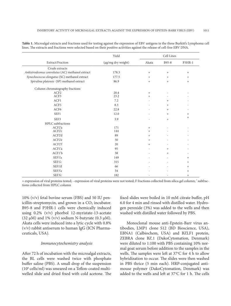
Table 1. Microalgal extracts and fractions used for testing against the expression of EBV antigens in the three Burkitt’s Lymphoma cell lines. The extracts and fractions were selected based on their positive activities against the release of cell-free EBV DNA.
Yield Cell Lines
Extract/Fraction (µg/mg dry weight) Akata B95-8 P3HR-1
Crude extractsAnkistrodesmus convolutus (AC) methanol extract 178.3 + + +
Synechococcus elongatus (SC) methanol extract 177.5 + + +Spirulina platensis (SP) methanol extract 86.9 + + +
Column chromatography fractionsACF2 20.4 + - -ACF3 23.2 + - -ACF1 7.2 - + -ACF5 8.3 - + -ACF6 22.8 - + -SEF1 12.0 - + +
SEF3 3.9 - + +
HPLC subfractionsACF2’a 171 + - -ACF2’c 144 + - -ACF2’d 89 + - -ACF2’e 50 + - -ACF2’f 20 + - -ACF1’a 95 - + -ACF1’b 38 - + -SEF1’a 149 - - +SEF1’c 315 - - +SEF1’d 66 - - +SEF3’a 54 - - +SEF3’c 182 - - +
+ expression of viral proteins tested; - expression of viral proteins were not tested; F fractions collected from silica gel column; ’ subfrac-tions collected from HPLC column
1012 YIH YIH KOK ET AL.
were washed in PBS with slow shaking for 5 min. The substrate 3,3-diaminobenzidine (DAB) (DakoCyto-mation, Denmark) was used for color development. Unstained cells were counter-stained with hematoxy-lin. After washing, the excess hematoxylin, the slides were dipped in ammonia water (10%) for 10 s. The dried slides were mounted using Depax and covered with cover slips. The slides were examined under the light microscope (Nikon Eclipse 80i). A minimum of 400 cells were scored. The percentage of cells ex-pressing the targeted of protein was calculated using the following equation:
Percentage of cells that express the targeted pro-tein (%) = number of positively stained cells x 100% Total number of cells counted in a population
Statistical analysis
Data are presented as the means with standard devia-tion derived from duplicate samples from two inde-pendent experiments. The data were analyzed using one-way analysis of variance (ANOVA), followed by Duncan’s Multiple Range test using Statistica soft-ware (Version 5). A p value < 0.05 was regarded as statistically significant.
RESULTS
Inhibitory activity of the methanol extracts
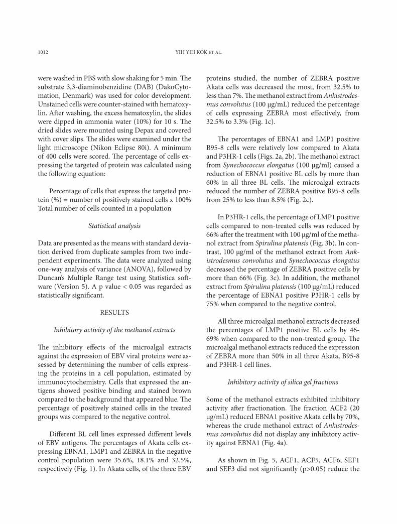
The inhibitory effects of the microalgal extracts against the expression of EBV viral proteins were as-sessed by determining the number of cells express-ing the proteins in a cell population, estimated by immunocytochemistry. Cells that expressed the an-tigens showed positive binding and stained brown compared to the background that appeared blue. The percentage of positively stained cells in the treated groups was compared to the negative control.
Different BL cell lines expressed different levels of EBV antigens. The percentages of Akata cells ex-pressing EBNA1, LMP1 and ZEBRA in the negative control population were 35.6%, 18.1% and 32.5%, respectively (Fig. 1). In Akata cells, of the three EBV
proteins studied, the number of ZEBRA positive Akata cells was decreased the most, from 32.5% to less than 7%. The methanol extract from Ankistrodes-mus convolutus (100 µg/mL) reduced the percentage of cells expressing ZEBRA most effectively, from 32.5% to 3.3% (Fig. 1c).
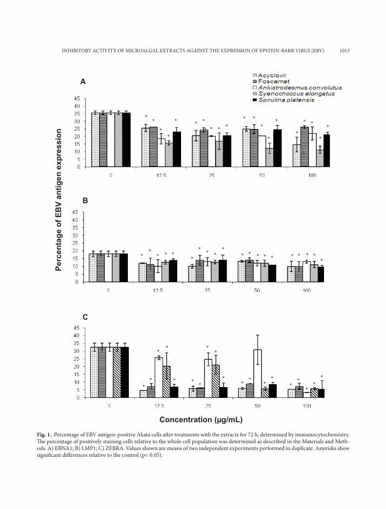
The percentages of EBNA1 and LMP1 positive B95-8 cells were relatively low compared to Akata and P3HR-1 cells (Figs. 2a, 2b). The methanol extract from Synechococcus elongatus (100 µg/ml) caused a reduction of EBNA1 positive BL cells by more than 60% in all three BL cells. The microalgal extracts reduced the number of ZEBRA positive B95-8 cells from 25% to less than 8.5% (Fig. 2c).
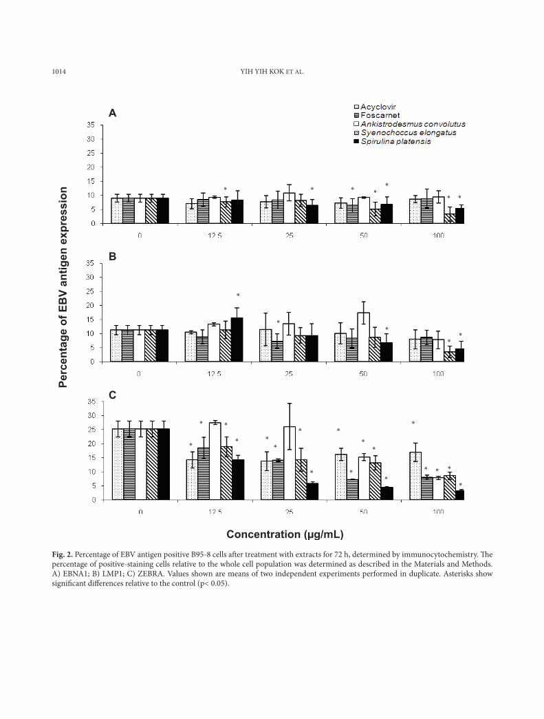
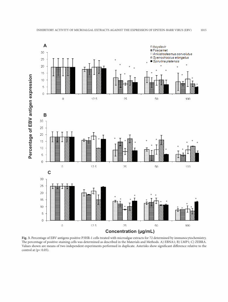
In P3HR-1 cells, the percentage of LMP1 positive cells compared to non-treated cells was reduced by 66% after the treatment with 100 µg/ml of the metha-nol extract from Spirulina platensis (Fig. 3b). In con-trast, 100 µg/ml of the methanol extract from Ank-istrodesmus convolutus and Synechococcus elongatus decreased the percentage of ZEBRA positive cells by more than 66% (Fig. 3c). In addition, the methanol extract from Spirulina platensis (100 µg/mL) reduced the percentage of EBNA1 positive P3HR-1 cells by 75% when compared to the negative control.
All three microalgal methanol extracts decreased the percentages of LMP1 positive BL cells by 46-69% when compared to the non-treated group. The microalgal methanol extracts reduced the expression of ZEBRA more than 50% in all three Akata, B95-8 and P3HR-1 cell lines.
Inhibitory activity of silica gel fractions
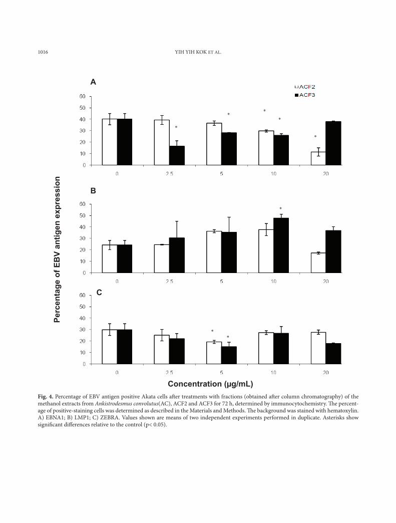
Some of the methanol extracts exhibited inhibitory activity after fractionation. The fraction ACF2 (20 µg/mL) reduced EBNA1 positive Akata cells by 70%, whereas the crude methanol extract of Ankistrodes-mus convolutus did not display any inhibitory activ-ity against EBNA1 (Fig. 4a).
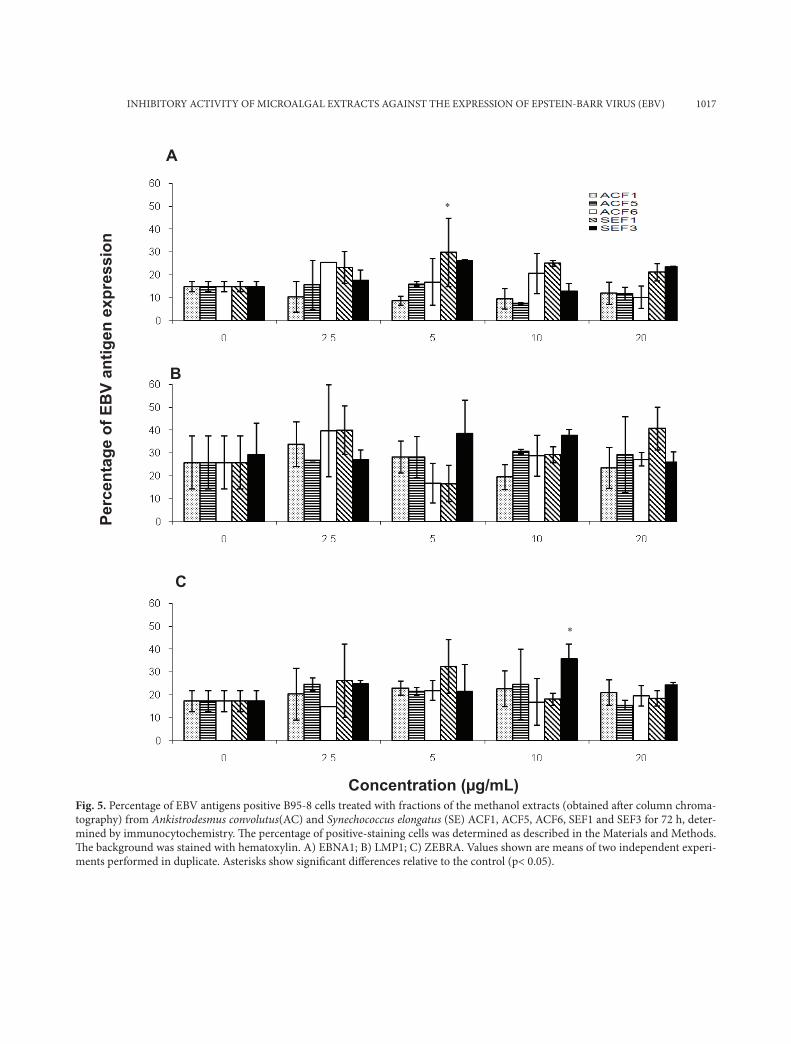
As shown in Fig. 5, ACF1, ACF5, ACF6, SEF1 and SEF3 did not significantly (p>0.05) reduce the
INHIBITORY ACTIVITY OF MICROALGAL EXTRACTS AGAINST THE EXPRESSION OF EPSTEIN-BARR VIRUS (EBV) 1013
*
Perc
enta
ge o
f EB
V an
tigen
exp
ress
ion
A
B
C
*
*
*
*
*
*
*
*
*
*
*
*
*
*
*
*
*
*
*
*
*
*
*
*
*
*
*
*
*
*
*
*
*
*
*
*
*
*
*
*
*
*
* *
* * *
* * * *
* * * * * * * * * *
Concentration (µg/mL)
Fig. 1. Percentage of EBV antigen-positive Akata cells aft er treatments with the extracts for 72 h, determined by immunocytochemistry. Th e percentage of positively staining cells relative to the whole cell population was determined as described in the Materials and Meth-ods. A) EBNA1; B) LMP1; C) ZEBRA. Values shown are means of two independent experiments performed in duplicate. Asterisks show signifi cant diff erences relative to the control (p< 0.05).
1014 YIH YIH KOK ET AL.
Figure 1:
Perc
enta
ge o
f EB
V an
tigen
exp
ress
ion
A
B
C
*
*
*
*
*
*
*
*
*
*
*
*
*
*
*
*
*
*
*
*
*
*
*
*
*
*
*
*
*
*
Concentration (µg/mL) Fig. 2. Percentage of EBV antigen positive B95-8 cells aft er treatment with extracts for 72 h, determined by immunocytochemistry. Th e percentage of positive-staining cells relative to the whole cell population was determined as described in the Materials and Methods. A) EBNA1; B) LMP1; C) ZEBRA. Values shown are means of two independent experiments performed in duplicate. Asterisks show signifi cant diff erences relative to the control (p< 0.05).
INHIBITORY ACTIVITY OF MICROALGAL EXTRACTS AGAINST THE EXPRESSION OF EPSTEIN-BARR VIRUS (EBV) 1015
Fig. 3. Percentage of EBV antigens positive P3HR-1 cells treated with microalgae extracts for 72 determined by immunocytochemistry. Th e percentage of positive-staining cells was determined as described in the Materials and Methods. A) EBNA1; B) LMP1; C) ZEBRA. Values shown are means of two independent experiments performed in duplicate. Asterisks show signifi cant diff erence relative to the control at (p< 0.05).
*
*
*
*
*
*
*
*
*
*
*
*
*
*
*
Perc
enta
ge o
f EB
V an
tigen
exp
ress
ion
A
B
C
*
*
*
*
*
*
*
*
*
*
*
*
*
*
*
*
*
*
*
*
*
*
*
*
*
*
*
*
*
*
Concentration (µg/mL)
1016 YIH YIH KOK ET AL.
*
*
*
* * *
*
)
*
Perc
enta
ge o
f EB
V an
tigen
exp
ress
ion
A
B
C
Concentration (µg/mL) Fig. 4. Percentage of EBV antigen positive Akata cells aft er treatments with fractions (obtained aft er column chromatography) of the methanol extracts from Ankistrodesmus convolutus(AC), ACF2 and ACF3 for 72 h, determined by immunocytochemistry. Th e percent-age of positive-staining cells was determined as described in the Materials and Methods. Th e background was stained with hematoxylin. A) EBNA1; B) LMP1; C) ZEBRA. Values shown are means of two independent experiments performed in duplicate. Asterisks show signifi cant diff erences relative to the control (p< 0.05).
INHIBITORY ACTIVITY OF MICROALGAL EXTRACTS AGAINST THE EXPRESSION OF EPSTEIN-BARR VIRUS (EBV) 1017
Fig. 5. Percentage of EBV antigens positive B95-8 cells treated with fractions of the methanol extracts (obtained aft er column chroma-tography) from Ankistrodesmus convolutus(AC) and Synechococcus elongatus (SE) ACF1, ACF5, ACF6, SEF1 and SEF3 for 72 h, deter-mined by immunocytochemistry. Th e percentage of positive-staining cells was determined as described in the Materials and Methods. Th e background was stained with hematoxylin. A) EBNA1; B) LMP1; C) ZEBRA. Values shown are means of two independent experi-ments performed in duplicate. Asterisks show signifi cant diff erences relative to the control (p< 0.05).
*
*
Perc
enta
ge o
f EB
V an
tigen
exp
ress
ion
A
B
C
Concentration (µg/mL)
1018 YIH YIH KOK ET AL.
* *
* *
* * *
*
*
* *
Perc
enta
ge o
f EB
V an
tigen
exp
ress
ion
A
B
C
Concentration (µg/mL)
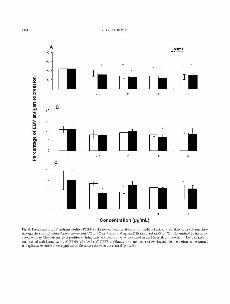
Fig. 6. Percentage of EBV antigens positive P3HR-1 cells treated with fractions of the methanol extracts (obtained aft er column chro-matography) from Ankistrodesmus convolutus(AC) and Synechococcus elongatus (SE) SEF1 and SEF3 for 72 h, determined by immuno-cytochemistry. Th e percentage of positive-staining cells was determined as described in the Materials and Methods. Th e background was stained with hematoxylin. A) EBNA1; B) LMP1; C) ZEBRA. Values shown are means of two independent experiments performed in duplicate. Asterisks show signifi cant diff erences relative to the control (p< 0.05).
INHIBITORY ACTIVITY OF MICROALGAL EXTRACTS AGAINST THE EXPRESSION OF EPSTEIN-BARR VIRUS (EBV) 1019
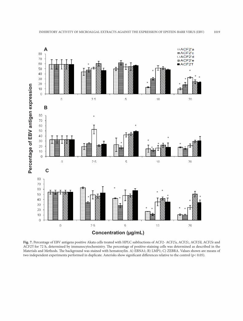
Fig. 7. Percentage of EBV antigens positive Akata cells treated with HPLC subfractions of ACF2- ACF2’a, ACF2’c, ACF2’d, ACF2’e and ACF2’f for 72 h, determined by immunocytochemistry. Th e percentage of positive-staining cells was determined as described in the Materials and Methods. Th e background was stained with hematoxylin. A) EBNA1; B) LMP1; C) ZEBRA. Values shown are means of two independent experiments performed in duplicate. Asterisks show signifi cant diff erences relative to the control (p< 0.05).
Perc
enta
ge o
f EB
V an
tigen
exp
ress
ion
A
B
C
*
*
*
*
*
* * *
*
*
*
*
* * *
*
*
*
* * *
* *
* * *
* *
*
Concentration (µg/mL)
1020 YIH YIH KOK ET AL.
* *
*
*
*
*
*
*
*
Perc
enta
ge o
f EB
V an
tigen
exp
ress
ion
A
B
C
Concentration (µg/mL)
* *
*
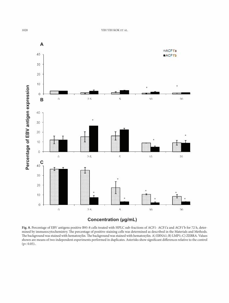
Fig. 8. Percentage of EBV antigens positive B95-8 cells treated with HPLC sub-fractions of ACF1- ACF1’a and ACF1’b for 72 h, deter-mined by immunocytochemistry. Th e percentage of positive-staining cells was determined as described in the Materials and Methods. Th e background was stained with hematoxylin. Th e background was stained with hematoxylin. A) EBNA1; B) LMP1; C) ZEBRA. Values shown are means of two independent experiments performed in duplicates. Asterisks show signifi cant diff erences relative to the control (p< 0.05)..
INHIBITORY ACTIVITY OF MICROALGAL EXTRACTS AGAINST THE EXPRESSION OF EPSTEIN-BARR VIRUS (EBV) 1021
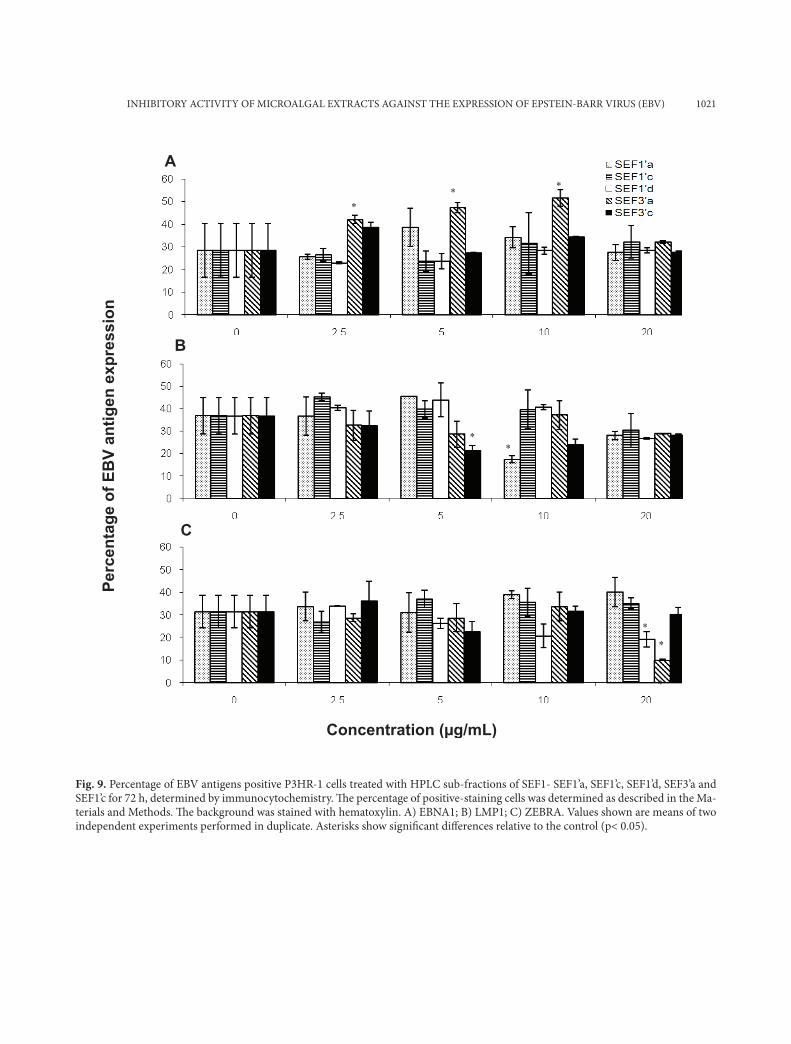
Fig. 9. Percentage of EBV antigens positive P3HR-1 cells treated with HPLC sub-fractions of SEF1- SEF1’a, SEF1’c, SEF1’d, SEF3’a and SEF1’c for 72 h, determined by immunocytochemistry. Th e percentage of positive-staining cells was determined as described in the Ma-terials and Methods. Th e background was stained with hematoxylin. A) EBNA1; B) LMP1; C) ZEBRA. Values shown are means of two independent experiments performed in duplicate. Asterisks show signifi cant diff erences relative to the control (p< 0.05).
A
* * *
Perc
enta
ge o
f EB
V an
tigen
exp
ress
ion
* *
*
*
B
C
Concentration (µg/mL)
*
1022 YIH YIH KOK ET AL.
expression of the three antigens in B95-8 cells, al-though they showed antiviral activity in the real time PCR assay in our previous work (Kok et al., 2011). SEF3 reduced significantly (p>0.05) the number of EBNA1 positive P3HR-1 cells at 2.5-20 µg/ml, while SEF1 (20 µg/ml) reduced the number of EBNA1 and ZEBRA positive P3HR-1 cells by 40.4% and 41.4%, respectively. (Figs. 6a and 6c, re-spectively).
Inhibitory activities of HPLC subfractions
The HPLC subfractions of ACF2 showed different activities against the expression of the three antigens in Akata cells (Fig. 7). The bioactivities of ACF2’a, ACF2’c, ACF2’d, ACF2’e and ACF2’f were highest at 20 µg/ml, as they reduced by 45-83% the number of EBNA1 positive Akata cells as compared to the con-trol (Fig. 7a). All the HPLC sub-fractions (10 µg/ml) significantly reduced (p<0.05) the percentage of LMP1-positive Akata cells by 45-63% (Fig. 7b). ACF2’a and ACF2’c at 10 µg/ml caused a considera-ble 65% reduction in the number of ZEBRA positive Akata cells (Fig. 7c). Clearly, the inhibitory activities in Ankistrodesmus convolutus against ZEBRA were preserved after fractionation.
The HPLC subfractions of the extract from Ank-istrodesmus convolutus, ACF1’a and ACF1’b, signifi-cantly reduced (p<0.05) the number of ZEBRA-posi-tive B95-8 cells, but not EBNA1- and LMP1-contain-ing cells (Fig. 8). Nevertheless, the percentage of ZE-BRA-positive B95-8 cells was lower after treatment with ACF1’b compared to ACF1’a (Fig. 8c). None of the of the HPLC subfractions of the extracts of Syn-echococcus elongatus were capable of lowering the expression of the three antigens in P3HR-1 cells (Fig. 9). The only active HPLC subfraction that showed remarkable inhibition activity was SEF3’a (20 µg/ml), which reduced the number of ZEBRA-positive P3HR-1 cells by 67.8% compared to the control (Fig. 9c). Overall, the subfractions from the microalgal extracts, ACF1’a, ACF1’b, ACF2’a, ACF2’c, SEF1’d and SEF3’a, seemed to be more active in inhibiting the expression of the EBV lytic protein ZEBRA in BL cells (Figs. 7c, 8c, 9c).
DISCUSSION
The microalgal extracts tested herein previously showed inhibitory activity against the release of cell-free EBV DNA. Our aim was to examine further the antiviral effects of the the extracts. Generally, they showed inhibitory activities against the expression of EBV proteins, observed as a reduction in the num-bers of cells expressing the EBV proteins. The per-sistency, replication and gene expression of EBV is dependent on the binding of EBNA1 to the origin of plasmid replication (OriP), which serves to main-tain the EBV episome in dividing cells (Hung et al., 2001). Therefore, EBNA1 is an attractive target for potential EBV inhibitors because of its critical role in EBV-associated disorders. In this study, the methanol extract from Ankistrodesmus convolutus, Synechococ-cus elongatus and Spirulina platensis (100 µg/mL) exhibited a significant reduction of EBNA1-positive BL cells (by more than 60%). These microalgal ex-tracts could decrease the presence of EBV episomes in new daughter cells and terminate latent EBV in-fection by inhibiting EBV episome replication. Per-sistence of the EBV genome and the lytic cascade can be obstructed via downregulation of the latency-associated promoter Qp (Kieff and Rickinson, 2006). This causes fewer EBV virions to be assembled and released. In our previous study where we assessed the copy number of cell-free EBV DNA, ACF2 and its HPLC subfractions reduced the number of cell-free EBV DNA by 50% at concentrations between 1.6-4.2 µg/ml (Kok et al., 2011).
Recently, LMP1 was proposed as a therapeutic target in EBV-associated disorders (Hannigan and Wilson, 2010). The microalgal crude extract exhib-ited >30% inhibitory activity against LMP1 in all three cell lines, except Ankistrodesmus convolutus in B95-8 cells. These compounds could block the NF-κB pathway and disrupt cell survival, leading to cell death (Lavorgna and Harhaj, 2012). This is in agree-ment with our previous study where the HPLC sub-fractions ACF2’c and ACF2’d reduced the number of viable Akata cells by about 50% (Kok et al., 2011). We also speculate that the reduction of LMP1 activates the DNA repair pathway in host cells, leading to deg-

INHIBITORY ACTIVITY OF MICROALGAL EXTRACTS AGAINST THE EXPRESSION OF EPSTEIN-BARR VIRUS (EBV) 1023
radation of EBV viral DNA and the resulting release of lower amounts of cell-free EBV. This was shown in our previous work where the methanol crude ex-tracts, ACF2’a, ACF2’c, ACF2’d, ACF2’e and ACF2’f, inhibited the release of cell-free EBV DNA by 50% at concentrations lower than 20 µg/ml.
All three microalgal extracts and some of their HPLC subfractions lowered the number of ZEBRA-positive cells by about 60% in all three Akata, B95-8 and P3HR-1 cell lines. ZEBRA is a crucial lytic trans-activator in the EBV life cycle. Therefore, these ex-tracts could switch off the lytic transactivation cycle in BL cells by blocking the activation of the BZLF1 gene, thus preventing the switch from latent to lytic cycle gene expression. Consequently, this leads to a decrease in the numbers of assembled and released viral particles (Feederle et al., 2000; El-Guindy et al., 2006). This explains the lower copy number of cell-free EBV released by the host cell after exposure to the methanol extract of Ankistrodesmus convolutus that was reported previously.
The mechanisms responsible for the induction of lytic EBV are complex and involve calcium-depend-ent mechanisms, protein kinase C (PKC), p38, c-jun N-terminal kinase (JNK) and mitogen activated pro-tein kinases (MAPK) (Israel and Kenney, 2003). Be-side BZLF1, BRLF1 mediates the switch form latent to lytic viral replication (Miller and El-Guindy, 2002). Therefore, the effects of the microalgal extracts on the cell cycle of BL cells warrants further studies.
CONCLUSION
Methanolic microalgal extracts and their subfrac-tions obtained after HPLC reduced the number of LMP1-, EBNA1- and ZEBRA-positive BL cells, sug-gesting that these preparations could serve as sources of potential anti-EBV agents.
Acknowledgments - This study was supported by a grant (IRPA: 06-05-01-0000-PR0054105-04) from the Ministry of Science, Technology and Innovation (MOSTI). We thank Prof. Kenzo Takada and Prof. Sam Choon Kook for providing the cell lines. The technical support from the NPC Labora-
tory, University of Malaya and Cancer Research Initiatives Foundation (CARIF) is gratefully acknowledged. We wish to thank the Director General for Health of Malaysia for his per-mission to publish this paper, and the Director of the Institute for Medical Research for her support.
REFERENCES
Barron, B.L., Torres-Valencia, M., Chamorro-Cevallos, G. and A. Zuniga-Estrada (2008). Spirulina as an antiviral agent, In: Spirulina in Human Nutrition and Health (Eds. M.E. Ger-shwin, A Belay) , 71-99. CRC Press, Boca Raton.
Chang, L.K., Wei, T.T., Chiu, Y.F., Tung, C.P., Chuang, J.Y., Hung, S.K., Li, C. and S.T. Liu (2003). Inhibition of Epstein-Barr virus lytic cycle by (-)-epigallocatechin gallate. Biochem. Biophys. Res. Commun. 301(4): 1062-8.
Countryman, J. and G. Miller (1985). Activation of expression of latent Epstein-Barr herpesvirus after gene transfer with a small cloned subfragment of heterogeneous viral DNA. Proc Natl Acad Sci USA 82(12): 4085-9.
El-Guindy, A.S. Paek, S.Y. Countryman, J. and G. Miller (2006). Identification of constitutive phosphorylation sites on the Epstein-Barr virus ZEBRA protein. J. Biol. Chem. 281(6): 3085-95.
Elion, G.B. (1993). Acyclovir: discovery, mechanism of action and selectivity. J. Med. Virol. 41 Suppl 1: 2-6.
Farrell, P. (1995). Epstein-Barr virus immortalizing genes. Trends Microbiol 3(3): 105-9.
Feederle, R. Kost, M. Baumann, M., Janz, A., Drouet, E., Ham-merschmidt, W. and H.J. Delecluse (2000). The Epstein-Barr virus lytic program is controlled by the cooperative functions of two transactivators. EMBO J 19(12): 3080-9.
Fries, K.L. Miller, W.E. and N. Raab-Traub(1996). Epstein-Barr virus latent membrane protein 1 blocks p53 mediated apoptosis through the induction of the A20 gene. J. Virol. 70(12): 8653-9.
Gershburg, E. and J.S. Pagano (2005). Epstein-Barr virus infec-tions: prospects for treatment. J. Antimicrob. Chemother. 56(2): 227-281.
Hannigan, A. and J.B. Wilson (2010). Evaluation of LMP1 of Epstein-Barr virus as atherapeutic target by its inhibition. Molecular Cancer 9: 184.
Hayashi, K. Hayashi, T. and I. Kojima (1996). A natural sulfated polysaccharide, calcium spirulan, isolated from Spirulina platensis: in vitro and ex vivo evaluation of anti-herpes simplex virus and anti-human immunodeficiency virus activities. AIDS Res. Hum. Retrovir. 12(15): 1463-71.
Hayashi, K. Nakano, T. Hashimoto, M., Kanekiyo, K. and T. Hayashi (2008). Defensive effects of a fucoidan from
1024 YIH YIH KOK ET AL.
brown alga Undaria pinnatifida against herpes simplex vi-rus infection. Int Immunopharmacol 8(1): 109-16.
Hidari, K.I. Takahashi, N. Arihara, M., Nagaoka, M., Morita, K.,and T. Suzuki (2008). Structure and anti-dengue virus activity of sulfated polysaccharide from a marine alga. Bio-chem Biophys Res Commun 376(1): 91-5.
Huleihel, M. Ishanu, V. Tal, J. and S.M. Arad (2002). Activity of Porphyridium sp. polysaccharide against herpes simplex viruses in vitro and in vivo. J Biochem Biophys Methods 50(2-3): 189-200.
Huleihel, M. Ishanu,V. Tal, J. and S.M. Arad (2001). Antiviral ef-fect of red microalgal polysaccharides on Herpes simplex and Varicella zoster viruses. J Appl Phycology 13(2): 127-34.
Hung, S.C. Kang, M.S. and E. Kieff (2001). Maintenance of Epstein-Barr virus (EBV) oriP-based episomes requires EBV-encoded nuclear antigen-1 chromosome binding do-mains, which can be replaced by high-mobility group-1 or histone H1. PNAS 98(4): 1865-70.
Iranshahi, M., Kalategi, F. Rezaee, R., Shahverdi, A.R., Ito, C., Fu-rukawa, H., Tokuda, H. and M. Itoigawa (2008). Cancer chemopreventive activity of terpenoid coumarins from Ferula species. Planta Med 74 (2):147-50.
Israel, B.F. and S.C. Kenney (2003). Virally targeted therapies for EBV-associated malignancies. Oncogene 22(33): 5122–5130.
Kieff, E. and A.B. Rickinson (2006). Epstein-Barr virus and its replication. In: Fields Virology, 5th Edition (Vol. 1), (Eds. D.M. Knipe, P.M. Howley), 2603-54. Lippincott-Raven, Philadelphia.
Kim, J.E. Jun, S., Song, M., Kim, J.H.,and Y.J. Song (2012). The extract of Chrysanthemum indicum Linne inhibits EBV LMP1-induced NF-κB activation and the viability of EBV-transformed lymphoblastoid cell lines. Food and Chemical Toxicology 50(5): 1524-1528.
Kok, Y.Y. Chu, W.L. Phang, S.M., Mohamed, S.M., Naidu, R., Lai, P.J., Ling, S.N., Mak, J.W., Lim, P.K.C., Balraj, P. and A.S.B. Khoo (2011). Inhibitory activities of microalgal extracts against Epstein-Barr virus DNA release from lymphoblas-toid cells. J. Zhejiang UnivSci B 12(5): 335–345.
Lavorgna, A. and E.W. Harhaj (2012). EBV LMP1: New and shared pathways to NF-κB activation. PNAS 109(7): 2188-2189.
Lu, J. Murakami, M. Verma, S.C. Cai, Q.L., Haldar, S., Kaul, R., Wasik, M.A., Middeldorp, J. and E.S. Robertson (2011). Epstein-Barr virus nuclear antigen 1 (EBNA1) confers resistance to apoptosis in EBV-positive B-lymphoma cells
through up-regulation of surviving. Virology 410(1): 64-75.
Luftig, M. Yasui, T., Soni, V., Kang, M.S., Jacobson, N., McFarland, E.C., Seed, B. and E. Kieff (2004). Epstein-Barr virus latent infection membrane protein 1 TRAF-binding site induces NIK/IKKα-dependent noncanonical NF-ΚB activation. Proc Natl Acad Sci USA 101(1): 141-6.
Micewicz, E.D. Cole, A.L. Jung, C.L., Luong, H., Phillips, M.L., Pratikhya, P., Sharma, S., Waring, A.J., Cole, A.M. and P. Ruchala (2010). Grifonin-1: A small HIV-1 entry inhibi-tor derived from the algal lectin, griffithsin. PLOS ONE 5(12): e14360.
Miller, G.I. and A.S. El-Guindy (2002). Regulation of Epstein-Barr virus lytic cycle activation in malignant and nonma-lignant disease. J Natl Cancer Inst 94(23): 1733-5.
Nicols, H.W. and H.C. Bold (1965). Trichosarcina polymorpha Gen et Sp. J Phycol 1: 34-8.
Phang, S.M. and W.L. Chu (1999). University of Malaya algae culture collection (UMACC): Catalogue of strains. Institute of postgraduate studies and research. University Malaya, Kuala Lumpur, 77 Pp.
Rezk, S.A. and L.M. Weiss (2007). Epstein-Barr virus-associated lymphoproliferative disorders. Hum Pathol 38(9): 1293-304.
Rickinson, A.B. and E. Kieff (2006). Epstein-Barr virus. In: Fields Virology 5th Edition (Vol. 1), (Eds. D.M. Knipe and P.M. Howley), 2655-700, Lippincott-Raven, Philadelphia.
Thorley-Lawson, D.A., Duca, K.A. and M. Shapiro (2008). Ep-stein-Barr virus: A paradigm for persistent infection- for real and in virtual reality. Trends Immunol 29(4): 195-201.
Wang, H.X. and T.B. Ng (2001). Examination of lectins, poly-saccharopeptide, polysaccharide, alkaloid, coumarin and trypsin inhibitors for inhibitory activity against human immunodeficiency virus reverse transcriptase and glyco-hydrolases. Planta Medica 67(7): 669-72.
Yiu, C.Y. Chen, S.Y. Chang, L.K., Chiu, Y.F. and T.P. Lin (2010). Inhibitory Effects of Resveratrol on the Epstein-Barr Virus Lytic Cycle. Molecules 15(10): 7115-7124.
Yiu, C.Y. Chen, S.Y., Huang, C.W., Yeh, D.B. and T.P. Lin (2011). Inhibitory effects of Polygonum cuspidatum on the Epstein-Barr Virus lytic cycle. Journal of Food and Drug Analysis, 19(2): 107-113.
Zarrouk, C. (1996). Contribution à l’étude d’unecyano-phycée. Influence de divers facteurs physiques et chimiquessur la croissance et la photosynthèse de Spirulina maxima, Ph. D. Thesis, Université de Paris.