Binding of the Heterogeneous Ribonucleoprotein K (hnRNP K) to the Epstein-Barr Virus Nuclear Antigen...
18
Binding of the Heterogeneous Ribonucleoprotein K (hnRNP K) to the Epstein-Barr Virus Nuclear Antigen 2 (EBNA2) Enhances Viral LMP2A Expression Henrik Gross 1 , Christine Hennard 2 , Ilias Masouris 2 , Christian Cassel 1 , Stephanie Barth 1 , Ute Stober- Gra ¨ sser 1 , Alfredo Mamiani 1 , Bodo Moritz 3 , Dirk Ostareck 4 , Antje Ostareck-Lederer 4 , Nils Neuenkirchen 5 , Utz Fischer 5 , Wen Deng 6 , Heinrich Leonhardt 6 , Elfriede Noessner 2 , Elisabeth Kremmer 2 , Friedrich A. Gra ¨ sser 1 * 1 Institute of Virology, Saarland University Medical School, Homburg/Saar, Germany, 2 Institute of Molecular Immunology, Helmholtz Zentrum Mu ¨ nchen, German Research Center for Environmental Health, Munich, Germany, 3 Institute of Biochemistry and Biotechnology, Martin-Luther-University Halle-Wittenberg, Halle (Saale), Germany, 4 Experimental Research Unit, Department of Intensive Care and Intermediate Care, University Hospital Aachen, RWTH Aachen University, Aachen, Germany, 5 Department of Biochemistry, Biocenter of the University of Wu ¨ rzburg, Wu ¨ rzburg, Germany, 6 Department of Biology, Center for Integrated Protein Science Munich, Ludwig Maximilians University Munich, Planegg-Martinsried, Germany Abstract The Epstein-Barr Virus (EBV) -encoded EBNA2 protein, which is essential for the in vitro transformation of B-lymphocytes, interferes with cellular processes by binding to proteins via conserved sequence motifs. Its Arginine-Glycine (RG) repeat element contains either symmetrically or asymmetrically di-methylated arginine residues (SDMA and ADMA, respectively). EBNA2 binds via its SDMA-modified RG-repeat to the survival motor neurons protein (SMN) and via the ADMA-RG-repeat to the NP9 protein of the human endogenous retrovirus K (HERV-K (HML-2) Type 1). The hypothesis of this work was that the methylated RG-repeat mimics an epitope shared with cellular proteins that is used for interaction with target structures. With monoclonal antibodies against the modified RG-repeat, we indeed identified cellular homologues that apparently have the same surface structure as methylated EBNA2. With the SDMA-specific antibodies, we precipitated the Sm protein D3 (SmD3) which, like EBNA2, binds via its SDMA-modified RG-repeat to SMN. With the ADMA-specific antibodies, we precipitated the heterogeneous ribonucleoprotein K (hnRNP K). Specific binding of the ADMA- antibody to hnRNP K was demonstrated using E. coli expressed/ADMA-methylated hnRNP K. In addition, we show that EBNA2 and hnRNP K form a complex in EBV- infected B-cells. Finally, hnRNP K, when co-expressed with EBNA2, strongly enhances viral latent membrane protein 2A (LMP2A) expression by an unknown mechanism as we did not detect a direct association of hnRNP K with DNA-bound EBNA2 in gel shift experiments. Our data support the notion that the methylated surface of EBNA2 mimics the surface structure of cellular proteins to interfere with or co-opt their functional properties. Citation: Gross H, Hennard C, Masouris I, Cassel C, Barth S, et al. (2012) Binding of the Heterogeneous Ribonucleoprotein K (hnRNP K) to the Epstein-Barr Virus Nuclear Antigen 2 (EBNA2) Enhances Viral LMP2A Expression. PLoS ONE 7(8): e42106. doi:10.1371/journal.pone.0042106 Editor: Dong-Yan Jin, University of Hong Kong, Hong Kong Received March 15, 2012; Accepted July 2, 2012; Published August 3, 2012 Copyright: ß 2012 Gross et al. This is an open-access article distributed under the terms of the Creative Commons Attribution License, which permits unrestricted use, distribution, and reproduction in any medium, provided the original author and source are credited. Funding: This work was supported by Deutsche Forschungsgemeinschaft through grant KR2218/2-1 to EK, OS290/3-2 to AO-L and DHO and GR950/12-1 to FG and the BioImaging Network Munich to HL. WD was supported by the Chinese Scholarship Council. The funders had no role in study design, data collection and analysis, decision to publish, or preparation of the manuscript. Competing Interests: The authors have declared that no competing interests exist. * E-mail: [email protected] Introduction The Epstein-Barr virus (EBV) is associated with various human malignancies [1] and growth-transforms primary human B- lymphocytes which are the in vitro correlate of EBV-associated post-transplant lymphoproliferative disease (PTLD) (for review, see [2]). In EBV-transformed lymphocytes, 11 so-called latent genes are expressed. Of these, only the nuclear antigens EBNA1, -2, -3a, -3c and the latent membrane protein LMP1 are necessary for transformation (reviewed in [3]). EBNA2 is a multifunctional transcriptional activator that does not bind directly to DNA but is tethered to promoter elements by interacting with DNA-bound transcription factors. For example, it associates through a Trp-Trp-Pro (‘‘WWP 325 ’’) motif at position 323–325 (see Figure 1) with the DNA-bound repressor RBPjk [4,5,6]. EBNA2 is the viral functional homologue to the cellular transmembrane receptor Notch which also activates gene expres- sion via RBPjk (reviewed in [7]). Binding of EBNA2 or Notch converts the repressor RBPjk to the transcriptionally active form. Figure 1 shows a schematic representation of EBNA2. A virus encoding an EBNA2 protein with a mutation in the WWP-motif is unable to immortalise B-lymphocytes and does not activate the viral oncogene LMP1 [8]. In addition to RBPjk, EBNA2 binds to a variety of basal transcription factors [2] and forms complexes with proteins involved in RNA metabolism like the DEAD-box protein DDX20 (DP103/Gemin3) [9] or the survival of motor neurons (SMN) protein [10,11]. The binding of EBNA2 to a variety of other host proteins is reflected by its presence in high molecular weight complexes of different composition [12,13,14]. Adjacent to the WWP-motif, EBNA2 contains an Arginine- PLoS ONE | www.plosone.org 1 August 2012 | Volume 7 | Issue 8 | e42106
-
Upload
independent -
Category
Documents
-
view
4 -
download
0
Transcript of Binding of the Heterogeneous Ribonucleoprotein K (hnRNP K) to the Epstein-Barr Virus Nuclear Antigen...
Binding of the Heterogeneous Ribonucleoprotein K(hnRNP K) to the Epstein-Barr Virus Nuclear Antigen 2(EBNA2) Enhances Viral LMP2A ExpressionHenrik Gross1, Christine Hennard2, Ilias Masouris2, Christian Cassel1, Stephanie Barth1, Ute Stober-
Grasser1, Alfredo Mamiani1, Bodo Moritz3, Dirk Ostareck4, Antje Ostareck-Lederer4, Nils Neuenkirchen5,
Utz Fischer5, Wen Deng6, Heinrich Leonhardt6, Elfriede Noessner2, Elisabeth Kremmer2,
Friedrich A. Grasser1*
1 Institute of Virology, Saarland University Medical School, Homburg/Saar, Germany, 2 Institute of Molecular Immunology, Helmholtz Zentrum Munchen, German
Research Center for Environmental Health, Munich, Germany, 3 Institute of Biochemistry and Biotechnology, Martin-Luther-University Halle-Wittenberg, Halle (Saale),
Germany, 4 Experimental Research Unit, Department of Intensive Care and Intermediate Care, University Hospital Aachen, RWTH Aachen University, Aachen, Germany,
5Department of Biochemistry, Biocenter of the University of Wurzburg, Wurzburg, Germany, 6Department of Biology, Center for Integrated Protein Science Munich,
Ludwig Maximilians University Munich, Planegg-Martinsried, Germany
Abstract
The Epstein-Barr Virus (EBV) -encoded EBNA2 protein, which is essential for the in vitro transformation of B-lymphocytes,interferes with cellular processes by binding to proteins via conserved sequence motifs. Its Arginine-Glycine (RG) repeatelement contains either symmetrically or asymmetrically di-methylated arginine residues (SDMA and ADMA, respectively).EBNA2 binds via its SDMA-modified RG-repeat to the survival motor neurons protein (SMN) and via the ADMA-RG-repeat tothe NP9 protein of the human endogenous retrovirus K (HERV-K (HML-2) Type 1). The hypothesis of this work was that themethylated RG-repeat mimics an epitope shared with cellular proteins that is used for interaction with target structures.With monoclonal antibodies against the modified RG-repeat, we indeed identified cellular homologues that apparently havethe same surface structure as methylated EBNA2. With the SDMA-specific antibodies, we precipitated the Sm protein D3(SmD3) which, like EBNA2, binds via its SDMA-modified RG-repeat to SMN. With the ADMA-specific antibodies, weprecipitated the heterogeneous ribonucleoprotein K (hnRNP K). Specific binding of the ADMA- antibody to hnRNP K wasdemonstrated using E. coli expressed/ADMA-methylated hnRNP K. In addition, we show that EBNA2 and hnRNP K forma complex in EBV- infected B-cells. Finally, hnRNP K, when co-expressed with EBNA2, strongly enhances viral latentmembrane protein 2A (LMP2A) expression by an unknown mechanism as we did not detect a direct association of hnRNP Kwith DNA-bound EBNA2 in gel shift experiments. Our data support the notion that the methylated surface of EBNA2 mimicsthe surface structure of cellular proteins to interfere with or co-opt their functional properties.
Citation: Gross H, Hennard C, Masouris I, Cassel C, Barth S, et al. (2012) Binding of the Heterogeneous Ribonucleoprotein K (hnRNP K) to the Epstein-Barr VirusNuclear Antigen 2 (EBNA2) Enhances Viral LMP2A Expression. PLoS ONE 7(8): e42106. doi:10.1371/journal.pone.0042106
Editor: Dong-Yan Jin, University of Hong Kong, Hong Kong
Received March 15, 2012; Accepted July 2, 2012; Published August 3, 2012
Copyright: � 2012 Gross et al. This is an open-access article distributed under the terms of the Creative Commons Attribution License, which permitsunrestricted use, distribution, and reproduction in any medium, provided the original author and source are credited.
Funding: This work was supported by Deutsche Forschungsgemeinschaft through grant KR2218/2-1 to EK, OS290/3-2 to AO-L and DHO and GR950/12-1 to FGand the BioImaging Network Munich to HL. WD was supported by the Chinese Scholarship Council. The funders had no role in study design, data collection andanalysis, decision to publish, or preparation of the manuscript.
Competing Interests: The authors have declared that no competing interests exist.
* E-mail: [email protected]
Introduction
The Epstein-Barr virus (EBV) is associated with various human
malignancies [1] and growth-transforms primary human B-
lymphocytes which are the in vitro correlate of EBV-associated
post-transplant lymphoproliferative disease (PTLD) (for review, see
[2]). In EBV-transformed lymphocytes, 11 so-called latent genes
are expressed. Of these, only the nuclear antigens EBNA1, -2, -3a,
-3c and the latent membrane protein LMP1 are necessary for
transformation (reviewed in [3]).
EBNA2 is a multifunctional transcriptional activator that does
not bind directly to DNA but is tethered to promoter elements by
interacting with DNA-bound transcription factors. For example, it
associates through a Trp-Trp-Pro (‘‘WWP325’’) motif at position
323–325 (see Figure 1) with the DNA-bound repressor RBPjk
[4,5,6]. EBNA2 is the viral functional homologue to the cellular
transmembrane receptor Notch which also activates gene expres-
sion via RBPjk (reviewed in [7]). Binding of EBNA2 or Notch
converts the repressor RBPjk to the transcriptionally active form.
Figure 1 shows a schematic representation of EBNA2. A virus
encoding an EBNA2 protein with a mutation in the WWP-motif is
unable to immortalise B-lymphocytes and does not activate the
viral oncogene LMP1 [8]. In addition to RBPjk, EBNA2 binds to
a variety of basal transcription factors [2] and forms complexes
with proteins involved in RNA metabolism like the DEAD-box
protein DDX20 (DP103/Gemin3) [9] or the survival of motor
neurons (SMN) protein [10,11]. The binding of EBNA2 to
a variety of other host proteins is reflected by its presence in high
molecular weight complexes of different composition [12,13,14].
Adjacent to the WWP-motif, EBNA2 contains an Arginine-
PLoS ONE | www.plosone.org 1 August 2012 | Volume 7 | Issue 8 | e42106

Glycine (RG-) repeat element at aa 339–354 with methylated
arginine residues [10,15]. The deletion of the RG-repeat results in
a five-fold higher ability of EBNA2 to stimulate LMP1 expression,
but a recombinant virus featuring this deletion in EBNA2 has
a reduced transforming activity and needs an extended time span
to induce outgrowth of transformed cell clones [16]. The EBNA2A
protein from type A isolates was originally shown to confer a higher
transforming capacity than EBNA2B derived from type B isolates
of EBV [17]. Recently, it was demonstrated that the RG- repeat,
among other C-terminal sequences, is important to confer the
higher transforming activity of EBNA2A vs. EBNA2B [18].
Methylation is a post-translational modification that affects
protein-protein interactions [19]. Methylation at arginine residues
[20] may lead to three known forms in higher eukaryotes: v-NG
MonoMethyl-arginine (MMA), v-NG,NG-Asymmetric DiMethyl-
arginine (ADMA) and v -NG,N’G-Symmetric DiMethyl-arginine
(SDMA). The methylation is carried out by two types of Protein-
Arginine-Methyl-Transferases (PRMTs): Type I enzymes
(PRMT1, 2, 3, 4, 6 and -8) catalyse the formation of ADMA
whereas type II enzymes (PRMT5, -7 and -9) account for the
formation of SDMA ([21,22,23].
hnRNP K was originally detected as a polycytidylic acid binding
protein purified from heterogeneous nuclear ribonucleoprotein
particles [24]. Later on, it was found that hnRNP K is involved in
various cellular processes such as chromatin reorganisation,
mRNA translation, transcriptional regulation, splicing, RNA
shuttling and cell survival (for review, see [25,26]). Recently, it
was proposed that hnRNP K activates the VEGF-A promoter by
binding to unwound superhelical single stranded C-rich sequences
upstream of the transcription start site and to support association
of transcription initiation factors [27]. hnRNP K is composed of
modular regions that confer binding both to RNA or DNA as well
as protein-protein interaction domains [28]. It binds to tyrosine
kinases like c- Src and Lck as well as transcription factors such as
C/EBP. The interaction with c-Src and its activation by hnRNP K
is modulated by asymmetric dimethylation of five arginine residues
catalysed by PRMT1 [29]. Reduced PRMT1 expression in
induced erythroid maturation of human K562 cells leads to
a decreased methylation of newly synthesised hnRNP K. This
correlates with hnRNP K tyrosine phosphorylation and activation
of the human reticulocyte 15-lipoxygenase (r15-LOX) mRNA
translation, which is inhibited by hnRNP K in early erythroid
maturation [30,31].
We have previously developed monoclonal antibodies against
the methylated RG-repeat of EBNA2 and found that it contains
either SDMA or ADMA residues but does not exist in non-
methylated (NMA) form [15]. SDMA-modified EBNA2 (SDMA-
EBNA) forms a complex with the SMN protein [10], while
ADMA-modified EBNA2 (ADMA-EBNA2) preferentially binds to
the NP9 protein encoded by the human endogenous retrovirus
HERV-K (HML-2) Type 1 [32]. It is known that SMN binds only
to SDMA-modified proteins [33;34], for example to the SDMA-
modified SmD3 protein which in turn is part of the SMN complex
[35]. We therefore reasoned that the methylated surface of
EBNA2 at the RG-repeat resembles cellular proteins engaging in
similar interactions and that the antibodies should recognize these
conserved epitopes. These cellular proteins in turn should be able
to interact with proteins bound to EBNA2. Here we show that the
immunoprecipitation of cellular proteins using the SDMA-specific
antibodies indeed yields SmD3. Likewise, the ADMA-antibodies
precipitated, among other proteins, hnRNP K. Further analysis
demonstrated that EBNA2 does not only share a conserved surface
epitope with hnRNP K but that it also forms a complex with
hnRNP K. Most importantly, hnRNP K, when co-expressed with
EBNA2, strongly increases the ability of EBNA2 to activate the
viral LMP2A promoter.
Results
Monoclonal Antibodies Against SDMA- and ADMA-EBNA2 Precipitate Cellular Proteins with MethylatedArginine ResiduesWe employed previously developed monoclonal antibodies
directed against ADMA- or SDMA-modified EBNA2 [15] for
the precipitation of cellular and/or viral proteins from EBV-
infected cells. The precipitated proteins were analysed by mass
spectrometry as previously described [36] and are listed in
Table 1. Also indicated in Table 1 is the methylation status
where known. Among the proteins precipitated by the SDMA-
antibody, we identified SmD3 known to be a core component
of spliceosomal U snRNPs and a major component of the SMN
complex (see, for instance, [37,38]). SmD3 was precipitated
both from EBV-infected and non-infected cells as SmD3 has an
RG-containing repeat structure comparable to that of EBNA2
which attaches to the SMN protein [35]. The SMN protein was
not found, presumably because the binding to the SDMA-
modified protein(s) precluded the reaction with the SDMA-
Figure 1. Schematic representation of the Epstein-Barr virus nuclear antigen 2 (EBNA2). EBNA2 of the standard B95.8 strain (accessionnumber: AJ507799) of EBV consists of 487 amino acids (aa) present in an A-type virus. The N-terminal dimerization domain (‘‘Dim’’) is located next toa poly-Proline stretch (‘‘Pro’’). The variable region (‘‘variable’’) differs between the A-type viruses and B-type viruses. B-type viruses have a reduced invitro transformation potential. The binding site for RBPjk (‘‘WWP’’) is located around a Trp-Trp-Pro motif at aa 323–325. The adjacent Arginine-Glycinerepeat (‘‘RG’’) between aa 339–354 confers binding to the survival of motor neurons (SMN) protein and represents the second nuclear localizationsignal (‘‘NLS’’) in addition to the canonical NLS found at the extreme C-terminus between aa 468–487. The C-terminal acidic transactivation domain(‘‘TAD’’) between aa 424–468 interacts with various basal transcription factors.doi:10.1371/journal.pone.0042106.g001
hnRNP-K Binds and Co-Activates EBNA2
PLoS ONE | www.plosone.org 2 August 2012 | Volume 7 | Issue 8 | e42106

Figure 2. SmD3 is precipitated by the SDMA- EBNA2 specific antibody. (A) Monoclonal antibodies (mAbs) directed against the SDMA- andADMA- containing Arginine-Glycine (RG)-repeat of EBNA2 were tested by precipitation using extracts of HEK 293-T cells expressing EBNA2-wt andHA- SmD3. For each antibody, an appropriate isotype control was tested in parallel to exclude unspecific binding to the protein G Sepharose used forprecipitation. Precipitated HA- SmD3 protein was visualised using the HA -specific mAb 3F10. The position of HA- SmD3 is indicated by an arrow. (B)Immunoprecipitation of EBNA2 from transiently transfected cells. HEK 293-T cells expressing EBNA2-wt and HA- SmD3 were precipitated withmonoclonal antibodies directed against the SDMA- and ADMA- containing Arginine-Glycine (RG)-repeat of EBNA2 using appropriate isotype controlantibodies. Precipitated EBNA2 protein was visualised using the EBNA2 mAb R3. The position of EBNA2 is indicated by an arrow.doi:10.1371/journal.pone.0042106.g002
Table 1. Proteins precipitated by the EBNA2- SDMA antibody.
Protein Size Accession number Methylationstatus
Pre-mRNA processing splicing factor 8 ,200 kDa Q6P2Q9 unknown
U5 small nuclear ribonucleoprotein 200 kDahelicase
,200 kDa O75643 unknown
Gem-associated protein 5 (gemin 5) ,150 kDa Q8TEQ6 unknown
U1 snRNP 70 K ,55 kDa P08621 sDMA
SmD3 ,14 kDa P62318 sDMA
Proteins precipitated by the EBNA2- ADMA antibody
Protein Size Accession number Methylationstatus
ATP dependent RNA helicase ,140 kDa Q08211 aDMA
Caprin-1 ,80 kDa Q14444 unknown
Ras-GTPase-activating protein (SH3-domain-binding protein variant)
,55 kDa Q53HH4 unknown
hnRNP K ,55 kDa Q5T6W5 aDMA
doi:10.1371/journal.pone.0042106.t001
hnRNP-K Binds and Co-Activates EBNA2
PLoS ONE | www.plosone.org 3 August 2012 | Volume 7 | Issue 8 | e42106

specific antibody. To confirm this result, we generated an
expression vector for HA-tagged SmD3. In cell extract contain-
ing HA-SmD3, we could precipitate SmD3 with the SDMA-
but not the ADMA-specific antibody or the EBNA2- specific R3
antibody. The staining of the precipitated SmD3 with the HA-
specific antibody is shown in Figure 2A. In the control
experiment depicted in Figure 2B, EBNA2 was precipitated
with both methylation-specific antibodies and R3 but not with
the isotype control antibodies. Note that due to the use of rat
and mouse monoclonal antibodies, not all the heavy chains
(‘‘IgG-H’’) were stained by the secondary antibodies; however,
the light chains (‘‘IgG-L’’) were clearly present. These data show
that the antibodies specific for the methylated surface of EBNA2
react with epitopes on cellular proteins that interact with the
same interaction partners, in this case the SMN protein. We
were mainly interested in the analysis of proteins identified with
the ADMA-specific antibody because our previous analysis had
shown that ADMA-EBNA2 is predominantly present at
EBNA2-regulated viral promoters [15]. The proteins reactive
with the SDMA-antibody were thus not pursued further.
EBNA2 Forms a Complex with hnRNP K in EBV-infectedCellsWe used the ADMA-specific monoclonal antibodies for the
precipitation of methylated proteins from non-infected BL41 and
EBV-infected EBNA2-containing Raji Burkitt’s lymphoma cells.
Among the proteins identified by the mass spectrometric analysis,
we found hnRNP K in both EBNA2-positive and EBNA2-
negative cell extracts. To confirm this result, we first subjected
extract of non-infected BL41 cells to precipitation using the
EBNA2-specific monoclonal antibody R3 which binds at the C-
terminus outside of the methylation region [39], the SDMA- and
ADMA-antibodies and the hnRNP K specific monoclonal
antibody D6. As can be seen in Figure 3A, only the ADMA- or
the hnRNP K-specific antibody D6, but not the SDMA-specific
antibody precipitated hnRNP K. As expected, R3 did not yield
a signal, and the absence of EBNA2 in the BL41 extract was
confirmed by western blot (data not shown).
We decided to confirm the reactivity of the ADMA-antibody
using hnRNP K synthesised and asymmetrically dimethylated in
E. coli by coexpressed PRMT1. Soluble E. coli extract containing
ADMA- methylated hnRNP K was subjected to immunoprecip-
itation with either an hnRNP K-specific monoclonal antibody, the
ADMA-specific antibody or the previously described monoclonal
antibody against the non-methylated RG-repeat of EBNA2
(NMA) [15]. As shown in Figure 3B, the hnRNP K-specific as
well as the ADMA-specific antibody clearly precipitated hnRNP K
from the E. coli extract confirming the reactivity of the ADMA-
specific antibody with hnRNP K in the absence of other
eukaryotic proteins. Furthermore, we excluded binding to
Figure 3. hnRNP K is precipitated by the ADMA- specific antibody. (A) Immunoprecipitation of hnRNP K from BL- 41 cells. EBV negative BL-41 cells were precipitated with monoclonal antibodies directed against the SDMA- and ADMA- containing Arginine-Glycine (RG)-repeat of EBNA2 andan hnRNP K specific antibody using appropriate isotype control antibodies. The EBNA2 specific mAb R3 served as a negative control. The position ofhnRNP K is indicated by an arrow. (B) ADMA- modified hnRNP K is precipitated by the ADMA- specific antibody. Soluble extract containing ADMA-hnRNP K methylated in E. coli with the type I methyltransferase PRMT1 was subjected to immunoprecipitation using either a hnRNP K- specific mAb,the ADMA- specific mAb and the NMA- specific mAb. Precipitated hnRNP K was visualised using the hnRNP K mAb.doi:10.1371/journal.pone.0042106.g003
hnRNP-K Binds and Co-Activates EBNA2
PLoS ONE | www.plosone.org 4 August 2012 | Volume 7 | Issue 8 | e42106

hnRNP-K Binds and Co-Activates EBNA2
PLoS ONE | www.plosone.org 5 August 2012 | Volume 7 | Issue 8 | e42106

PRMT1 which is complexed to hnRNP K [29,40] (data not
shown). We also observed binding of a small fraction of hnRNP K
by the NMA-antibody. Our previous experiments had shown that
this antibody only reacted with (non-methylated) EBNA2 when
the EBV-infected cell was treated with the methylation inhibitor
Adox [15]. The binding of this antibody to hnRNP K again
indicated that non-methylated hnRNP K apparently has a surface
at its methylation site similar to the one of EBNA2.
We next subjected Raji cells to precipitation either with the
ADMA-, SDMA- or the methylation-independent EBNA2-specific
monoclonal antibody R3, and the hnRNP K specific monoclonal
antibody D6. As can be seen in Figure 4A, upper panel, the
methylation-specific antibodies and R3 precipitated EBNA2, while
the control antibody did not. When the same extracts were probed
with the hnRNP K specific antibody D6, hnRNP K was found co-
precipitated by R3, the SDMA- and ADMA as well as the hnRNP
K antibody (Figure 4A, lower panel). Conversely, the hnRNP K-
specific antibody D6 co-precipitated EBNA2. The fact that R3
and also the SDMA-antibody precipitated hnRNP K from the
EBNA2-containing extract indicated that both proteins are in
a complex with each other, because only EBNA2 but not hnRNP
K contains SDMA residues [29].
The EBNA2-hnRNP K-interaction is Dependent ofMethylation of hnRNP KTo further investigate if the binding of the two proteins is
dependent of the methylation status of hnRNP K, we carried out
co- immunoprecipitations from 293T cells either transfected with
wild-type GFP- hnRNP K and EBNA2 or the methylation
deficient mutant GFP- hnRNP K 5RG and EBNA2. As can be
seen in Figure 4B, the binding of EBNA2 to hnRNP K is
unaffected by the GFP- tag of hnRNP K and EBNA2 is co-
precipitated by the hnRNP K specific antibody and vice versa.
These findings confirm the results from EBV positive Raji cells
(Figure 4A). In contrast, the methylation deficient 5RG mutant of
hnRNP K is unable to bind to EBNA2 whereas the binding of
EBNA2 to endogenous hnRNP K is unaffected. Furthermore the
ADMA- specific antibody is not able to precipitate GFP - hnRNP
K 5RG, which further highlights its specificity (Figure 4C).
To corroborate the above results, GST-pull-down assays were
carried out. For this purpose, we generated a GST-EBNA2 fusion
protein containing aa 300–400 of EBNA2 including the methyl-
ation site at 339–354. This fusion protein was then treated either
with E. coli-expressed His- tagged PRMT1 to generate ADMA-
EBNA2 or Baculovirus expressed PRMT5/WD45 to generate
SDMA-EBNA2. The reaction products were then precipitated
with the antibodies to see whether the correct products were
formed as the SDMA-specific antibody does not react in a western
blot [15]. Treatment with PRMT1 yielded a GST-EBNA2 fusion
protein that reacted with the NMA and the ADMA but not the
SDMA antibody as shown in Figure S2. The detection of E.coli-
expressed PRMT1 with the novel PRMT1-specific antibody 7D2
(see Materials and Methods) is shown in Figure S1. This antibody
was raised against a peptide encompassing amino acids 250–264 of
the human PRMT1 which are divergent from all other known
PRMTs. The clone 7D2 (Rat IgG2a) reacted only with a single
band in a western blot using DG75 cell extract and migrated to the
same position as E.coli-expressed PRMT1 (Figure S1). Methyla-
tion with the PRMT5/WD45 complex expressed in Baculovirus-
infected cells, however, yielded a protein that reacted with the
three antibodies indicating that the PRMT5 preparation con-
tained a contamination with a type I PRMT that catalysed the
generation of ADMA residues. The precipitation from the
untreated extract yielded, in addition to a strong band of non-
methylated fusion protein, a faint band reactive with ADMA
antibody (Figure S2). We then employed the bacterial fusion
proteins for a pull-down of hnRNP K protein from extract of non-
infected DG75 lymphoma cells. The extract from these cells was
treated with AdOx after lysis to prevent additional and unspecific
methylation of the GST-fusion proteins. As shown in Figure 5A,
we observed binding of hnRNP K to non-methylated as well as
methylated GST-EBNA2 fusion protein. In the control experi-
ment using GST-protein alone, no binding was observed. The
observation that non-methylated EBNA2 also binds hnRNP K
indicated that residues adjacent to the RG-repeat of EBNA2 might
also be involved in binding to hnRNP K. As outlined above, the
SDMA-specific antibody co-precipitated hnRNP K from EBNA2-
containing cell extract but not from non-infected cells showing that
hnRNP K interacted both with SDMA- and ADMA-EBNA2 in
vivo. The same results (Figure 5B) were obtained with a GST-
EBNA2 (aa 300–400) mutant lacking the RG repeat (DRG). This
indicated also that residues adjacent to the RG-repeat are also
involved in the interaction with hnRNP K.
To further characterise the binding of EBNA2 to hnRNP K,
a GST- tagged EBNA2 fragment containing the C-terminal amino
acids 400–487 were created. As can be seen in Figure 5C, the
fragment containing the aa 400–487 is not able to bind hnRNP K
in contrast to the EBNA2 fragment consisting of aa 300–400
encompassing the RG-repeat. These results suggest that hnRNP K
interacts with EBNA2 via its amino acids 300–400 regardless of
the presence of the methylated RG- repeat. A similar observation
was previously made for the interaction of the SMN protein with
the SDMA-modified RG-repeat of EBNA2, where the main but
not exclusive binding region for SMN on EBNA2 was located at
and around the RG-repeat [10]. In contrast, the methylation of
hnRNP K appears to be necessary for binding to EBNA2 (see also
below).
EBNA2 and hnRNP K Co-localiseToshowthatEBNA2also formsacomplexwithhnRNPKinintact
cells, we carried out a co-localisation study using confocal laser
scanning microscopy as described previously [32]. For this purpose,
EGFP-EBNA2 [32] was expressed in HeLa cells. hnRNP K was
visualised using the D6 antibody and secondary Alexa 647 -labelled
goat anti-mouse IgG. Co-localisation of EBNA2with hnRNPKwas
observed in 35.7% of the cells that expressed both proteins (a
representative image is shown in Figure 6). hnRNP K and EBNA2
showed clear co- localisation at numerous spots along the inner
Figure 4. EBNA2 is co-precipitated with wild- type hnRNP K but not with the methylation deficient hnRNP K 5RG mutant. (A) Co-immunoprecipitation of EBNA2 and hnRNP K from EBV positive Raji cells. Raji cells expressing EBNA2 were precipitated with monoclonal antibodiesdirected against the SDMA- and ADMA- containing Arginine-Glycine (RG)-repeat of EBNA2, an EBNA2 specific mAB (R3) and an hnRNP K specific mAB(D6). The positions of EBNA2 and hnRNP K are indicated by arrows. (B) Co-immunoprecipitation of EBNA2 and GFP - hnRNP K from transfected 293Tcells. The cells were precipitated with monoclonal antibodies directed against the SDMA- and ADMA- containing Arginine-Glycine (RG)-repeat ofEBNA2, an EBNA2 specific mAB (R3) and an hnRNP K specific mAB (D6). The positions of EBNA2 and hnRNP K are indicated by arrows. (C) No co-immunoprecipitation of EBNA2 and GFP - hnRNP K 5RG is observed from transfected 293T cells. The cells were precipitated with monoclonalantibodies directed against the SDMA- and ADMA- containing Arginine-Glycine (RG)-repeat of EBNA2, an EBNA2 specific mAB (R3) and an hnRNP Kspecific mAB (D6). The positions of EBNA2 and hnRNP K are indicated by arrows.doi:10.1371/journal.pone.0042106.g004
hnRNP-K Binds and Co-Activates EBNA2
PLoS ONE | www.plosone.org 6 August 2012 | Volume 7 | Issue 8 | e42106

Figure 5. hnRNP K binds to the amino acids 300 – 400 of EBNA2 regardless of the methylation or presence of the RG- repeat. (A) Invitro methylated (SDMA and ADMA) and unmethylated (NMA) GST- EBNA2 fusion protein containing amino acids 300–400 of EBNA2 and GST alonewere coupled to glutathione sepharose and were incubated with DG75 cell extract treated with methylation inhibitor AdOX. Precipitated hnRNP Kwas visualised using the hnRNP K mAb D-6. (B) GST- EBNA2DRG fusion protein containing amino acids 300–400 without the RG- Repeat of EBNA2 andGST alone were coupled to glutathione sepharose and were incubated with DG75 cell extract. Precipitated hnRNP K was visualised using the hnRNP KmAb D-6. (C) GST- EBNA2 aa400–487 fusion protein containing amino acids 400- 487 of EBNA2, GST- EBNA2 aa300–400 and GST alone were coupledto glutathione sepharose and were incubated with DG75 cell extract. Precipitated hnRNP K was visualised using the hnRNP K mAb D-6.doi:10.1371/journal.pone.0042106.g005
hnRNP-K Binds and Co-Activates EBNA2
PLoS ONE | www.plosone.org 7 August 2012 | Volume 7 | Issue 8 | e42106

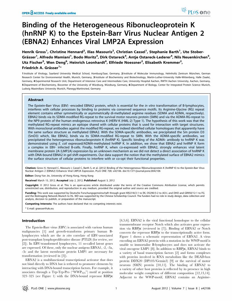
Figure 6. hnRNP K and EBNA2 co-localize in transiently transfected cells. (A) HeLa cells transfected with EGFP- EBNA2 were analysed byconfocal laser scanning microscopy. Endogenous hnRNP K was detected using the monoclonal D-6 antibody and an Alexa 647 coupled anti mouseantibody. The signals for hnRNP K (red) or EBNA2 (green) are shown. The merged signals show co-localisation of hnRNP K and EBNA2, resulting ina yellow color. Also shown is the DAPI staining of DNA. The fluorescence profiles of hnRNP K and EBNA2 (B) at a co-localisation hotspot (indicated bythe line, left picture - lower lane) were analysed with the Leica MMAF software. The signals for hnRNP K and EBNA2 show the same progression ofintensity at the inner nuclear membrane.doi:10.1371/journal.pone.0042106.g006
hnRNP-K Binds and Co-Activates EBNA2
PLoS ONE | www.plosone.org 8 August 2012 | Volume 7 | Issue 8 | e42106
hnRNP-K Binds and Co-Activates EBNA2
PLoS ONE | www.plosone.org 9 August 2012 | Volume 7 | Issue 8 | e42106

nuclear membrane. A representative image of the distribution of
fluorescence intensity across a line through thenucleus (‘‘linescan’’) is
pictured in Figure 6B. The linescan shows that the intensities for the
EBNA2 and hnRNP K signals overlap and further supports the
notion that the two proteins interact.
The same results were obtained for endogenous proteins using the
293EBVcell line.Theendogenous (i.e. non-transfected)EBNA2was
visualised by the EBNA2- specific R3 antibody and goat- anti- rat
TRITC -labelled antibody. hnRNP K was visualised using the D6
antibody and secondary Alexa 647 -labelled goat anti-mouse IgG. A
representative cell is pictured in Figure 7A, a linescan showing the
same intensity in fluorescence is shown inFigure 7B.These results, in
conjunction with the GST-pull-down study and the co-immunopre-
cipitation experiments (see above), strongly suggest thatADMA- and
SDMA-modified EBNA2 and ADMA-methylated hnRNP K form
(a) functional unit(s) in EBV-infected cells.
hnRNP K Interacts with EBNA2 in vivo in a Subset of CellsTo further investigate this interaction we used a cell based protein
interaction assay. We immunocaptured the GFP-EBNA2 fusion
protein (bait) with theGBP- lacI at the chromosomal lacOarray, that
becomesvisibleasdistinctnuclear spot (Figure8A). Inaboutone third
of all transfected cells we observed a clear co-localisation of theCFP-
hnRNPK fusion protein (prey) at the lacO spot which is indicative of
Figure 7. hnRNP K and EBNA2 co-localize in EBV positive cells. (A) 293-EBV cells were analysed by confocal laser scanning microscopy.Endogenous hnRNP K was detected using the monoclonal D-6 antibody and an Alexa 647 coupled anti mouse antibody. Endogenous EBNA2expressed from the viral episome was detected using the monoclonal R3 antibody and an TRITC coupled anti rat antibody. The signals for hnRNP K(red) or EBNA2 (green) are shown. The merged signals show co-localisation of hnRNP K and EBNA2, resulting in a yellow color. Also shown is the DAPIstaining of DNA. The fluorescence profiles of hnRNP K and EBNA2 (B) at a co-localization hotspot (indicated by the line, left picture - lower lane) wereanalysed with the Leica MMAF software. The signals for hnRNP K and EBNA2 show the same progression of intensity at the inner nuclear membrane.doi:10.1371/journal.pone.0042106.g007
Figure 8. hnRNP K interacts with EBNA2 in a cell based interaction system. Cells containing a lac operator (lacO) array inserted in thegenome were transfected with expression vectors for a lac repressor fused with a GFP binding protein (GBP) and the indicated fluorescent fusionproteins as indicated. For comparison and orientation the nucleus was stained with PI or DAPI. The GFP fusion proteins are captured at the lacoperator array by the LacI-GBP and the co-localization of cyan or red fusion proteins visualized. Clear and weak interactions at lacO array spots aremarked with filled and open arrow tips, respectively. The displayed cells represent the different patterns observed in several independentexperiments. GFP expression vectors were used as negative control. Scale bar is 5 mm.doi:10.1371/journal.pone.0042106.g008
hnRNP-K Binds and Co-Activates EBNA2
PLoS ONE | www.plosone.org 10 August 2012 | Volume 7 | Issue 8 | e42106

hnRNP-K Binds and Co-Activates EBNA2
PLoS ONE | www.plosone.org 11 August 2012 | Volume 7 | Issue 8 | e42106

a direct or indirect protein interaction. Another third showed aweak
interaction and the remaining cells did not show any clearly
detectable interaction and were indistinguishable fromGFP control
cells. We also performed the reciprocal experiments by switching
fluorescent proteins and immobilizing GFP-hnRNP K at the lacO
array spot (Figure 8B). In this combination we also observed co-
localisation of the DsRed-EBNA2 at the lacO spot although with
generally weaker signals. These cell based interaction assay results
provide further evidence for an interactionofhnRNPKwithEBNA2
in vivo. The displayed cells give a representative overview of the
observed variability and indicate that this interaction does not occur
in all cells or at least not to an equal extent. These results suggest that
the hnRNP K interaction with EBNA2 is not constitutive but likely
subjected to some additional regulation.
hnRNP K Enhances EBNA2-mediated Activation of theViral LMP2A PromoterhnRNP K has repeatedly been shown to directly activate
[41,42,43,44] or inactivate [45,46] transcription. To test whether
the interactionofEBNA2andhnRNPKchanged the transactivation
of a viral promoter byEBNA2,we co-expressed EBNA2-wt, hnRNP
K-wt, the methylation-deficient mutants EBNA2-DRG [16] and
hnRNP K-5RG [31] in all possible combinations together with
a luciferase reporter drivenby thepromoter of the viral LMP2A[32].
As shown in Figure 9A, EBNA2-wt activated the promoter by about
500-fold (p = 0.0000000016), while the activation by EBNA2-DRGwas lowerbut still highly significant (p = 0.000009975). hnRNPK-wt
or the5RG-mutantaloneexerteda small but significant activationon
thepromoter construct (p = 0.000173andp= 0.00529, respectively),
while co-expression of hnRNP K-wt enhanced the EBNA2-wt-
mediated activation by up to three-fold (p = 0.001675). The hnRNP
K- 5RG mutant co-activated EBNA2-wt to a smaller degree than
hnRNP K-wt (p = 0.00833). The activity of EBNA2-DRG was only
slightly increased by hnRNP K- wt (p = 0.00261), and no co-
activation was observed for the combination of both mutants
(p = 0.875). We determined the relative levels of EBNA2 in the
presence or absence of hnRNP K expression. The co-expression of
hnRNP K did not change the EBNA2 level excluding a trivial
explanation for the observed effect (Figure S3). This result and the
results from interaction analysis using the hnRNP K-5RG mutant
(Figure 4C) strongly support the notion that the interaction between
EBNA2andhnRNPKismainly (butnot exclusively)mediatedby the
methylation of the two proteins. The expression levels of transfected
EBNA2-wt, EBNA2-DRG, hnRNP K-wt and hnRNP K-5RG are
shown in Figure S4. We used the co-expression of SmD3 which was
precipitated by the SDMA antibody (Table 1) as an additional
negative control. SmD3which does not bind to EBNA2was not able
to co-activate EBNA2 in this assay as shown in Figure 9B.
hnRNP K is not Present in EBNA2-DNA BindingComplexesTo see whether hnRNP K is present in EBNA2-DNA
complexes, we carried out a gel-shift experiment employing cell
extract from EBV/EBNA2-positive Raji cells. As indicated in
Figure 10, we added either HA- hnRNP K or HA- RBPjk to the
Raji cell extract. EBNA2 binds DNA via the repressor RBPjk [47],
and this interaction can be inhibited by the antibody 6C8 directed
against the WWP-repeat of EBNA2 [48] that is used for its
interaction with RBPjk (reviewed in [49]). We have previously
shown that the ADMA-form of EBNA2 is preferentially present at
EBNA2-regulated promoters [15]. We thus tested whether the
hnRNP K- antibody D6 which co-precipitates EBNA2 (see
Figure 4A) would induce a super- shift as observed for the
EBNA2-specific antibody R3 with a DNA-fragment derived from
the viral LMP2A promoter. To exclude a possible unsuitability of
the D6 antibody for EMSA we also tested the HA antibody in the
samples which included HA- tagged hnRNP K or RBPjk, the
latter serving as internal positive control. As shown in Figure 10,
the EBNA2/ RBPjk -containing complex designated ‘‘IV’’ was
super-shifted by R3, while the antibody D6 against hnRNP K and
the HA- antibody did not. In contrast, the HA- antibody was able
to bind to and super-shift the HA- RBPjk bound to DNA
(‘‘Supershift*’’). As internal control we used the antibody 6C8
which interferes in the EBNA2- RBPjk interaction [48]. As can be
seen, this antibody diminished the signal from complex IV. The
absence of hnRNP K in the EBNA2/DNA-complex indicates that
the observed co-activation by hnRNP K is not mediated by direct
promoter binding of hnRNP K. The interaction of the two sub-
forms of EBNA2 (SDMA and ADMA) which differ in their
presence in EBNA2- DNA-binding complexes [15] to hnRNP K
hints at the possibility that EBNA2 and hnRNP K co-operate in
other activities, for instance in post-transcriptional processing of
mRNA. The latter possibility will have to be addressed in
a different set of experiments beyond the scope of this
communication.
Discussion
The hypothesis underlying our analysis was that EBNA2, as
demonstrated for its interaction with RBPjk via its ‘‘WWP’’-motif,
uses the methylated RG-repeat to attach to cellular factors to use
or interfere with their functions. For instance, EBNA2 binds with
its SDMA- modified RG- repeat to the SMN protein and with the
ADMA-modified RG-repeat to the HERV-K (HML-2) NP9
protein [10,32]. We therefore employed recently developed
monoclonal antibodies against the SDMA and ADMA-repeat of
EBNA2 for the identification of cellular proteins with a similar
surface structure. Of the proteins precipitated by the SDMA-
EBNA2-specific antibodies, we notably detected SmD3 known to
be SDMA-modified by PRMT5 [33,34] and PRMT7 [50]. The
precipitation of SmD3 strengthened our hypothesis that the
antibody recognised not only the methylated arginine residues of
EBNA2 but also the tertiary structure of the RG-motif. The RG-
motif confers binding of both the SDMA-SmD3 and the SDMA-
EBNA2 to SMN [10,35]. Importantly, the ADMA-EBNA2-
antibody, in contrast to the SDMA-antibody, did not bind
SmD3 as it does not contain ADMA residues [51]. Because
SmD3 is a common component of spliceosomal U snRNP it was
not surprising that the SDMA-specific antibody also precipitated
components of the spliceosome, namely the U1 snRNP-specific
70K protein as well as the U5 snRNP component PRPF8 [52].
Since it was likely that these proteins co-precipitated with SmD3
Figure 9. hnRNP K but not SmD3 enhances the EBNA2-mediated promoter activation at the viral LMP2a promoter. (A) A LMP2apromoter luciferase construct was co- transfected into DG75 cells with EBNA2, EBNA2-DRG, hnRNP K, and hnRNP K-5RG expression constructs in theindicated combinations. The luciferase value (RLU) obtained with empty pSG5 and the reporter construct was set to 100%. The graph represents thevalues obtained from 5 independent experiments carried out in duplicate. (B) A LMP2a promoter luciferase construct was co- transfected into DG75cells with EBNA2 and SmD3 expression constructs in the indicated combinations. The luciferase value (RLU) obtained with empty pSG5 and thereporter construct was set to 100%. The graph represents the values obtained from 4 independent experiments carried out in duplicate.doi:10.1371/journal.pone.0042106.g009
hnRNP-K Binds and Co-Activates EBNA2
PLoS ONE | www.plosone.org 12 August 2012 | Volume 7 | Issue 8 | e42106

due to being part of the same RNP complexes we did not pursue
the SDMA-precipitated proteins further.
Of the proteins identified by the ADMA-specific antibodies,
nothing is known about the Ras-GTPase-activating protein SH3-
domain-binding protein variant (gi: 62896771) identified in our
study. Its splice variant, G3BP1, was shown to be associated with
SMN and with Caprin-1 which was formerly known as GPI-
anchored membrane protein 1 or cell cycle associated protein
Figure 10. hnRNP K is not present in EBNA2-containing DNA complexes. EBNA2-containing Raji cell extract was incubated with in vitrotranslated hnRNP K and RBP- Jk and antibodies as indicated above and then assayed in a gel shift assay. R3 recognizes EBNA2 regardless of itsmethylation status and induces a ‘‘super- shift’’ indicated by the upper arrow, the mAb 6C8 directed against the ‘‘WWP’’-repeat of EBNA2 destroys theEBNA2/RBPjk-complex IV. Control antibodies corresponded to the respective IgG-subtype of each antibody. To efficiently separate the highmolecular weight complexes, the electrophoresis was carried out for an extended time. Therefore, uncomplexed 32P-labelled probe ran out of the gel.The position of the RBPjk-containing complexes I-IV as described in the text are indicated; the arrow (‘‘Supershift’’) points at the EBNA2-containingcomplex IV that is supershifted by R3 but not by the hnRNP K specific D6 antibody or the HA- specific antibody. The arrow (‘‘Supershift *’’) indicatesthe RBP- Jk containing complex which is supershifted by the HA- specific antibody and served as an internal control.doi:10.1371/journal.pone.0042106.g010
hnRNP-K Binds and Co-Activates EBNA2
PLoS ONE | www.plosone.org 13 August 2012 | Volume 7 | Issue 8 | e42106

[53,54]. Caprin-1 was also detected in our analysis. The role of
Caprin-1 and the G3BP1 variant in EBV transcription or
replication is unknown, however, a role of Caprin-1 in Vaccinia
virus replication was previously demonstrated [55]. The G3BP1
variant contains several RGG and RG motifs at its C-terminus
and might thus also be arginine-methylated. The ATP-dependent
RNA helicase A (DHX9) contains ADMA and its arginine
methylation is a prerequisite for nuclear localisation [56]. Like
hnRNP K, DHX9 was previously found to be associated with the
EBV-encoded nuclear antigen 5 (EBNA5 or EBNA-LP) [57]. As
EBNA2 binds to the RNA helicase DDX20 (DP103/Gemin3) [9],
it is possible that DHX9 and EBNA2 also form a complex. This is
presently being investigated.
hnRNP K is highly conserved in eukaryotic cells and plays a role
in various cellular processes like chromatin remodelling, tran-
scriptional regulation, splicing, translation or signal transduction
(see, for example, [30,44,58,59,60,61]). hnRNP K interacts
directly or indirectly with a large number of cellular proteins
[62], most notably with proteins involved in RNA metabolism.
Because hnRNP K also plays a role in transcriptional activation
and since the ADMA-form of EBNA2 is predominantly bound to
EBNA2-responsive promoters [15], we decided to analyse the
precipitation of hnRNP K by the ADMA-specific antibody in
greater detail. As a precedent, the cross-reactivity between an
epitope on hnRNP K and the PTB-associated splicing factor was
demonstrated recently [63]. Through the use of bacterial
expressed hnRNP K, which contained exclusively ADMA-
methylated arginine residues we could clearly show that the
ADMA-specific antibody binds to methylated hnRNP K. In-
terestingly, an antibody directed against non-methylated EBNA2
also detected hnRNP K indicating that both proteins share
a common surface structure that is most likely used for the
interaction with cellular partner proteins. Most importantly,
hnRNP K and EBNA2 bind to each other, presumably via the
methylated regions as protein arginine methylation is used either
for protein-RNA or protein-protein interactions [21]. However,
the GST-pull-down analysis showed that the non-methylated
EBNA2 and the DRG mutant also bind to hnRNP K indicating
that the region surrounding the RG-repeat is also involved in
binding. This is in line with the previously described association of
EBNA2 with SMN, where the binding to SMN is mainly but not
exclusively mediated via the RG-repeat of EBNA2 [10]. In the
living cell, the EBNA2-hnRNP K-interaction might be regulated
via methylation or another secondary modification, as we observed
an interaction in the lacO-based assay system only in about 60%
of the cells that expressed the GFP-labelled proteins (Figure 6).
While EBNA2 does not exist in non-methylated form [15], it is
possible that newly synthesized hnRNP K might undergo cell cycle
–dependent differences in methylation. The functional significance
of the EBNA2-hnRNP K-interaction was emphasised by the
observation that hnRNP K enhanced the EBNA2-mediated
activation of the viral LMP2A promoter by more than 3-fold.
Interestingly, the activation of the viral C promoter by the hnRNP
protein AUF1 was described by Ling and co-workers; however,
the interaction domains between EBNA2 and AUF1 were not
mapped [64].
A previous report showed that hnRNP K is present in
transcriptionally active sites in EBV-transformed cells and that
hnRNP K was highly enriched at loci with high EBV viral RNA
content [65]. This is reflected by the fact that hnRNP K strongly
co-activated EBNA2. The observation that the splicing machinery
was distributed randomly vis-a-vis the viral DNA but was enriched
at the transcript site [65] indicated that there is a recruitment of
splicing factors to nascent viral transcripts. The role of EBNA2 in
this process remains unclear. However, we assume that the
interaction between EBNA2 and hnRNP K indicates a co-
operation during transcription and that the binding of EBNA2 to
proteins of the splicing machinery reflects the close link between
transcription and splicing [66,67]. However, the lack of hnRNP K
at EBNA2- containing DNA complexes indicates that the
enhancement of LMP2A expression might take place at a post-
transcriptional level. Further studies will be needed to address the
question whether the binding of EBNA2 influences other activities
of hnRNP K, i.e. the known interaction with c- Src or its activity in
mRNA translation, i.e. the c-myc gene, which is a target for both
hnRNP K and EBNA2 [43,68,69,70].
Materials and Methods
Cell Lines and Tissue CultureHEK 293-T, 293-EBV and HeLa cells were cultured in DMEM
medium (GIBCO), supplemented with 10% FCS and antibiotics,
non-adherent cell lines were grown in RPMI 1640 medium
(GIBCO), supplemented with 10% FCS, Na-Pyruvate and
antibiotics. The EBV-infected cell lines Raji and 293-EBV, the
EBV- negative cell lines DG75 [71] and BL- 41 as well as 293-T
and HeLa cells were previously described [32,72].
Transfection/Electroporation/Luciferase AssayFor transient expression of the various proteins, 56106 293-T
cells were transfected with 8 mg/10 cm dish of the expression
vectors and combinations thereof using NanofectinH (PAA, Colbe,
Germany). Western blotting by the ECLH-method (GE Health-
care, Munich, Germany) was carried out as described. Electro-
poration and luciferase analysis was carried out as described [32].
DG75 cells were electroporated using a Bio-Rad Gene Pulser at
250 V and 950 mF. Briefly, 107 cells were washed once and
resuspended in 0.25 ml of ice-cold RPMI 1640 without supple-
ments and placed on ice. Then, 4 mg of reporter plasmid, 10 mg ofeach respective effector plasmid, and 2 mg of peGFP-C1
(Clontech, Palo Alto, CA, USA) were added. Parental pSG5
vector (Agilent Technologies, Waldbronn, Germany) was used to
adjust DNA amounts. After electroporation, cells were kept on ice
for 10 min, suspended in 10 ml of RPMI with 20% fetal calf
serum, and grown for 48 h. To determine the transfection
efficiency, 100 ml of the cells were fixed and analysed in a Becton
Dickinson FACScan analyser for eGFP-positive cells, gated on the
living population. The remainder of cells were washed in PBS and
lysed in 100 ml 16 CCLR-buffer (Promega, Mannheim, Ger-
many). The luciferase activity of the supernatants was determined
in a Lumat LB9501 (Berthold, Bad Wildbad, Germany) by using
the Promega luciferase assay systemH (Promega) as recommended
by the manufacturer.
PlasmidsToexpress quantitatively asymmetricallymethylatedhnRNPK in
E. coli the plasmid pET28-PRMT1, a kind gift of X. Cheng, Emory
University, Atlanta, GA, [73] was completed with a second Shine-
Dalgarno- and PRMT1 coding sequence (PCR primers 59
AAACTCGA GAACTTTAAGAAGGAGATATACCATG 39; 59
TTTCTCGAGTTCAGCGCATCCGGTA GTCGG 39) inserted
into Xho I and a Shine-Dalgarno- (His6)-hnRNP K sequence [74]
(PCR primers 59 TTTGTCGACAAC TTTAAGAAGGAGATA-
TACCATG 39; 59 TTTGTCGACCCGGATCATCAGTGGTG
39) in the Sal I site. peGFP- EBNA2 was described previously [11].
pSG5 –HA - hnRNP K and pSG5- HA- hnRNP K 5RG were
constructedusingthepeGFP-hnRNPKandpeGFP-hnRNPK5RG
plasmids [29]. dsRed- EBNA2 was constructed using the peGFP-
hnRNP-K Binds and Co-Activates EBNA2
PLoS ONE | www.plosone.org 14 August 2012 | Volume 7 | Issue 8 | e42106

EBNA2plasmid [11] and thedsRedMonomerC1vector (Clontech).
peCFP- hnRNP K was constructed using the peGFP- hnRNP K
plasmid [29] and the peCFP- C1 vector (Clontech). GST- EBNA2
fragment fusion proteins were constructed using the pGEX- 4T1
Vector (Amersham). The complete coding sequence of PRMT1was
amplified by PCR from a HeLa-cDNA library with primers
59PRMT1-TACAGGATCCATGGAGGTGTCCTG
TGGCCAGGCG G-39 and 39PRMT1 59-GACGGGATCC-
GAATTCAGCGCATCCGGTAGTCGGTGGAGCAG -39 and
cloned into theBamHI-digested eukaryotic expression vectors pSG5
or the BamHI-digested pGEX-4T1 vector for expression of a GST-
PRMT1 fusion protein in E.coli.
Preparation of Native Whole Cell ExtractRaji or DG75 cells were lysed for 30 min on ice in PBS
supplemented with 0.5% IGEPAL (Sigma) and 0.15 M NaCl and
protease inhibitors (Complete miniH, Roche). The lysate was
centrifuged at 15,0006g for 15 min, and the supernatant was used
for further analysis.
AntibodiesThe rat mAb 8C3 (IgG2b) reacts with NMA-EBNA2, the
mouse mAb 13B10 (IgG2c) recognises SDMA-EBNA2, the mouse
mAb 6F12 (IgG2b) binds to ADMA-EBNA2 [15], and the rat
mAb R3 (IgG2a) binds to a C-terminal epitope outside the
methylation region of EBNA2 [39]. Monoclonal anti-hnRNP K
antibody (D-6) was from Santa Cruz (Heidelberg, Germany), goat-
anti- mouse Alexa 647 was from Life Technologies (Invitrogen,
Darmstadt, Germany), peroxidase-coupled anti-rat or anti-rabbit
IgG were from Sigma (Munich, Germany). The monoclonal
antibody 3F10 (Roche, Penzberg, Germany) binds to the HA-tag.
For production of anti-PRMT1 monoclonal antibodies, a peptide
encompassing amino acids G250MRPNAKNNRDL264 of human
PRMT1 coupled to BSA was used to immunize Lou/C rats
according to a standard protocol [75]. A clone designated 7D2
(Rat IgG2a) that reacted with GST-PRMT1 but not an irrelevant
GST-fusion protein in a western blot was stably subcloned and
used for further analysis. The reactivity of this antibody with E.coli-
expressed non-fused PRMT1 and GST-PRMT1 as well as
endogenous cellular PRMT1 from the human cell line DG75
[71] is shown in Figure S2.
Confocal Immunofluorescence MicroscopyHeLa cells were seeded on microscopy cover slips. Cells were
transfected with a plasmid encoding EGFP-EBNA2 [32] and
endogenous hnRNP K was visualized with the D6 antibody and
secondary Alexa 647-labeled goat anti-mouse IgG2a (Invitrogen,
Molecular Probes). Nuclei were stained with DAPI. Slides were
mounted using Vectashield (Vector Laboratories). Fluorescence
images (Figure 6) were captured with a laser scanning microscope,
Leica TCS SP2 (Leica Microsystems, Heidelberg, Germany)
equipped with an HCX PL APO 6361.40 NA oil immersion
objective lens using scan settings of pinhole 1.0 Airy units,
5126512 pixel image format, four frame averages, and a TD488/
543/633 dichromatic beam splitter. Fluorescence spill-over was
excluded by using sequential image recording and tightly
controlled excitation power and detection channel settings
(EGFP-EBNA2 excitation: 44% of 488-nm laser; Alexa 647
excitation: 81% of 633-nm laser, DAPI excitation: 49% of 405-nm
laser). The co-localisation of endogenous (i.e. non-transfected)
EBNA2 and hnRNP K was carried out in 293-EBV cells [76].
EBNA2 expressed from the viral episome was detected using the
monoclonal R3 antibody and a TRITC-coupled anti-rat antibody.
Endogenous hnRNP K was visualized with the D6 antibody and
secondary Alexa 647-labeled goat anti-mouse IgG2a. Secondary
antibodies were highly cross-adsorbed and showed not cross-
recognition. Images were captured using the TCS SP5 II/AOBS
Leica confocal system (Figure 7). Fluorescence images were
acquired in a sequential scan mode with HyD detectors with
tightly controlled laser powers and acquisition windows to prevent
spill-over (scan 1:4% 405-nm with 3% 561-nm; scan 2:6% 488-nm
with 16% 633-nm). All images were recorded as stacked series of
confocal single z-planes (step size: 488 nm using magnification
with 46 frame average of 6306with zoom factor of at least 2.5.
Editing of contrast and brightness was applied to the whole image
using Leica LAS AF software. For EBNA2-hnRNP K co-
localisation, 56 double-positive cells expressing both fusion
proteins were evaluated. Co-localisation was analysed using the
Leica Lite software profile tool. Co-localisation hotspots were
defined as regions with coinciding high fluorescence intensity of
hnRNP K and EBNA2 in the same optical z-plane. The
percentage of cells showing co-localisation was calculated among
the cells expressing both proteins. Additional de-convolution was
performed using the Autoquant plug-in of the Leica MMAF
Software (Leica Microsystems, Heidelberg, Germany).
Cell Based Protein Interaction AssayFluorescent two-hybrid assays [77] were performed with a few
modifications to visualize and test protein interactions. BHK cells
containing a lac operator repeat array inserted in the genome [78]
were seeded on coverslips and cultured in DMEM medium with
10% FCS. After attachment cells were co-transfected with
expression vectors for the indicated fluorescent fusion proteins
and a GBP- LacI fusion [79] using polyethylenimine (Sigma). After
about 16 h cells were fixed with 3.7% formaldehyde in PBS for
10 min, washed with PBST (PBS with 0.02% Tween), stained with
DAPI or PI and mounted in Vectashield medium (Vector
Laboratories) (Figure 8). For PI staining RNA was eliminated by
RNase treatment after fixation.
ImmunoprecipitationThe rat monoclonal antibody (mAb) R3 (rat IgG2a) recognises
a C-terminal epitope of EBNA2 while the clone 6C8 (rat IgG2a)
binds to the Trp-Trp-Pro motif of EBNA2 and interferes with
binding to RBPjk [48]. For immunoprecipitation appropriate
mouse or rat IgG isotype controls were used. For precipitation,
400 ml of mAb supernatant were coupled to 100 ml of settled
protein-G-sepharose (PGS, GE Healthcare, Munchen, Germany)
for 1 h at 4uC under agitation, sedimented at 5.000 rpm and
washed once with 1 ml of lysis buffer 1. For precipitation
experiments either 400 mg protein of native whole cell extract or
100 mg protein of native nuclear extract was added and incubated
for 2 h at 4uC under agitation, washed three times with lysis buffer
2 (PBS with 0.5% IGEPAL and 0.5 M NaCl) and once with lysis
buffer 1. The pellet was resuspended in 26SDS sample-buffer and
incubated for 10 min at RT or heated at 98uC.
Co-immunoprecipitation AnalysisRaji cells were lysed for 30 min on ice in PBS supplemented
with 0.5% IGEPAL (Sigma) and 0.15 M NaCl containing protease
inhibitors (Complete miniH, Roche, Penzberg, Germany). After
incubation, the solution was sonicated with a 10 s pulse and
centrifuged at 13.0006g for 15 min, and the supernatant was used
for further incubation with antibody immobilised on 30 ml of
settled protein G SepharoseH (GE Healthcare). The cells were
washed twice with ice-cold PBS and lysed for 30 min on ice in
buffer 1 (PBS supplemented with 0.5% IGEPAL (Sigma) and
0.15 M NaCl) containing protease inhibitors (Complete miniH,
hnRNP-K Binds and Co-Activates EBNA2
PLoS ONE | www.plosone.org 15 August 2012 | Volume 7 | Issue 8 | e42106

Roche). After incubation, the solution was sonicated with a 10 s
pulse, centrifuged at 13,0006g and 4uC and incubated for 4 h at
4uC with antibody immobilised on 30 ml of settled protein G
sepharose (GE Healthcare). The beads were collected and washed
repeatedly with lysis buffer. The immune complexes were
dissolved in SDS-gel buffer and separated in 10% polyacrylamide
gel electrophoresis and transferred to a nitrocellulose membrane.
The antibody R3 binds to the C-terminus of EBNA2. The
antibodies 6F12 and 13B10 which recognise the asymmetrically
and symmetrically di-methylated arginine-Glycine repeat of
EBNA2, respectively, and the GST-specific monoclonal antibody
6G9 have been described recently. hnRNP K was detected using
monoclonal antibody D6 (sc-13133, Santa Cruz, Heidelberg,
Germany).
In vitro Methylation AssaysCompetent E. coli BL21- bacteria were transformed with
appropriate expression vectors, protein expression was induced
with IPTG and soluble extracts were purified with NAPTM25
columns (GE Healthcare, Freiburg, Germany) as described
previously [32]. In vitro methylation assays were carried out using
20 ml of His-PRMT1 extract or 5 ml of PRMT5/WD45 extract,
20 ml of GST- EBNA2 (aa300-400) fusion protein extract and 5 mlof 0.5 M SAM. The mixture was incubated for 1 h at 37uC.GST- pull- down assays: For GST- EBNA2 and GST competent
E. coli BL21- bacteria were transformed and protein expression
was induced with IPTG as described previously [32]. The GST
fusion proteins were adsorbed to glutathione-Sepharose beads (GE
Healthcare, Freiburg, Germany) for 2 h at 4uC with rotation, and
subsequently washed twice with lysis buffer containing 0.15 M
NaCl. For binding of cellular proteins to the GST fusion proteins,
typically 500 ml of AdOX- treated native whole cell extract (see
above) was added to the mixture and incubated for 2 h at 4uCwith rotation and subsequently washed 5 times with lysis buffer
containing 0.5 M NaCl. The beads were suspended in SDS-gel
electrophoresis buffer, boiled and separated by 10% SDS-PAGE.
GST-EBNA2 and GST were detected in western-blotting using
6G9 antibody. hnRNP K was detected using the D-6 monoclonal
antibody (Santa Cruz , D-6, sc-28380).
Electrophoretic Mobility Shift Assay (EMSA)Nuclear cell extracts were prepared essentially as described [80].
Shortly, approx. 108 cells were collected for 5 minutes at
1200 rpm and 4uC and washed twice with cold PBS. The pellet
was resuspended in a 4-fold volume of buffer A (10 mM HEPES
pH 7.9, 10 mM KCl, 1.5 mM MgCl2) [80] and kept on ice for at
least 20 minutes. Cells were broken up by several strokes in
a dounce homogenizer until the lysate contained about 50% intact
nuclei by staining with Trypan blue. The lysate was centrifuged for
15 seconds at 14.000 rpm and 4uC and washed once with a 2-fold
volume of buffer A. The resulting nuclear pellet was resuspended
a 1-fold volume of buffer B (20 mM HEPES pH 7.9, 420 mM
NaCl, 1.5 mM MgCl2, 2 mM EDTA pH 8.5) and kept on ice for
30 minutes. The lysate was centrifuged for 20 minutes at
15.000 rpm and 4uC and the supernatant was used for further
experiments or stored at –80uC. Electrophoretic mobility shift
assays were carried out exactly as described [4,81]. The probe
used for EMSA is derived from the viral LMP2a promoter and
contains two RBPjk–binding sites. For supershift experiments, we
employed the EBNA2-specific rat monoclonal antibody R3 [39] or
an appropriate isotype (rat IgG2a) control. The monoclonal
antibody 6C8 binds to the Trp-Trp-Pro (‘‘WWP’’) motif of
EBNA2 interferes with binding to RBKJk and destroys the
EBNA2-containing DNA-RBPjk-EBNA2-complex [48]. In vitro
transcription-translation of HA-tagged hnRNP K and HA-tagged
RBPjk using vector AJ247 [82] was performed using the TNTHCoupled Reticulocyte Lysate System (Promega, Mannheim,
Germany) as described [10,15] following the instruction of the
manufacturer. Typically, 50 ml of the transcription-translation mix
were programmed with 1 mg of vector DNA using T7 RNA
polymerase.
Supporting Information
Figure S1 Expression control of His- PRMT1 andcharacterization of the PRMT1 specific rat monoclonalantibody 7D2. E.Coli extract containing His- tagged PRMT1,
E.Coli extract containing GST- tagged PRMT1 and DG75 whole
cell extract was analysed by western blotting.
(TIF)
Figure S2 In vitro methylation of GST-EBNA2-300-400.The E.coli-expressed unmethylated GST-EBNA2-300-400 fusion
protein was subjected to in vitro methylation by PRMT5/WD45
purified from a baculovirus expression system or PRMT1
expressed in E.coli. The methylated fusion proteins as well as an
unmethylated control were immunoprecipitated with the EBNA2-
methylation specific antibodies (NMA, SDMA and ADMA) and
the appropriate isotype controls. Precipitated GST-EBNA2-300-
400 fusion protein was detected in a western blot using the GST-
specific 6G9 monoclonal antibody.
(TIF)
Figure S3 EBNA2 expression is not affected by hnRNPK. DG75 cells were transfected with pSG5 – EBNA2 and pSG5-
HA- hnRNP K and the cell extract was analysed by western
blotting. EBNA2 was visualized using the R3 antibody, hnRNP K
was visualized with the D6 antibody.
(TIF)
Figure S4 Expression control of the plasmids used inluciferase activity assays. DG75 cells were transfected with
pSG5 – EBNA2, pSG5 – EBNA2DRG, pSG5-HA- hnRNP K and
pSG5-HA- hnRNP K 5RG and the cell extract was analysed by
western blotting. EBNA2 was visualized using the R3 antibody,
HA- hnRNP K and HA- hnRNP K 5RG was visualized with the
HA antibody.
(TIF)
Acknowledgments
We thank Ruth Nord for expert technical assistance.
Author Contributions
Conceived and designed the experiments: FG EK . Performed the
experiments: HG CH CC AM IM US-G WD SB. Analyzed the data: HG
CH HL EN EK FG. Contributed reagents/materials/analysis tools: DHO
AO-L BM NN UF. Wrote the paper: HG FG.
References
1. Crawford DH (2001) Biology and disease associations of Epstein-Barr virus.
Philos Trans R Soc Lond B Biol Sci 356: 461–473.
2. Kieff E, Rickinson AE (2007) Epstein-Barr Virus and its replication. In: Knipe
D, Griffin DE, Lamb RA, Strauss SE, Howley PM, et al., editors. Fields
Virology. 5 ed. Philadelphia: Lippincott-Raven. 2603–2654.
hnRNP-K Binds and Co-Activates EBNA2
PLoS ONE | www.plosone.org 16 August 2012 | Volume 7 | Issue 8 | e42106

3. Bornkamm GW, Hammerschmidt W (2001) Molecular virology of Epstein-Barrvirus. Philos Trans R Soc Lond B Biol Sci 356: 437–459.
4. Zimber Strobl U, Kremmer E, Grasser F, Marschall G, Laux G, et al. (1993)
The Epstein-Barr virus nuclear antigen 2 interacts with an EBNA2 responsivecis-element of the terminal protein 1 gene promoter. EMBO J 12: 167–175.
5. Ling PD, Hayward SD (1995) Contribution of conserved amino acids in
mediating the interaction between EBNA2 and CBF1/RBPJk. J Virol 69: 1944–1950.
6. Henkel T, Ling PD, Hayward SD, Peterson MG (1994) Mediation of Epstein-
Barr virus EBNA2 transactivation by recombination signal-binding protein Jkappa. Science 265: 92–95.
7. Zimber Strobl U, Strobl LJ (2001) EBNA2 and Notch signalling in Epstein-Barr
virus mediated immortalization of B lymphocytes. Semin Cancer Biol 11: 423–434.
8. Cohen JI, Wang F, Kieff E (1991) Epstein-Barr virus nuclear protein 2 mutations
define essential domains for transformation and transactivation. J Virol 65:2545–2554.
9. Grundhoff AT, Kremmer E, Tureci O, Glieden A, Gindorf C, et al. (1999)
Characterization of DP103, a novel DEAD box protein that binds to theEpstein-Barr virus nuclear proteins EBNA2 and EBNA3C. J Biol Chem 274:
19136–19144.
10. Barth S, Liss M, Voss MD, Dobner T, Fischer U, et al. (2003) Epstein-Barr virus
nuclear antigen 2 binds via its methylated Arginine- glycine repeat to the survival
motor neuron protein. J Virol 77: 5008–5013.
11. Voss MD, Hille A, Barth S, Spurk A, Hennrich F, et al. (2001) Functional
cooperation of Epstein-Barr virus nuclear antigen 2 and the survival motor
neuron protein in transactivation of the viral LMP1 promoter. J Virol 75:11781–11790.
12. Tsui S, Schubach WH (1994) Epstein-Barr virus nuclear protein 2A forms
oligomers in vitro and in vivo through a region required for B-celltransformation. J Virol 68: 4287–4294.
13. Wu DY, Krumm A, Schubach WH (2000) Promotor-specific targeting of humanSWi-SNF complex by Epstein-Barr virus nuclear protein 2. J Virol 74: 8893–
8903.
14. Grasser FA, Haiss P, Gottel S, Mueller Lantzsch N (1991) Biochemicalcharacterization of Epstein-Barr virus nuclear antigen 2A. J Virol 65: 3779–
3788.
15. Gross H, Barth S, Palermo RD, Mamiani A, Hennard C, et al. (2010)Asymmetric Arginine dimethylation of Epstein-Barr virus nuclear antigen 2
promotes DNA targeting. Virology 397: 299–310.
16. Tong X, Yalamanchili R, Harada S, Kieff E (1994) The EBNA-2 Arginine-glycine domain is critical but not essential for B-lymphocyte growth trans-
formation; the rest of region 3 lacks essential interactive domains. J Virol 68:6188–6197.
17. Rickinson AB, Young LS, Rowe M (1987) Influence of the Epstein-Barr virus
nuclear antigen EBNA 2 on the growth phenotype of virus-transformed B cells.J Virol 61: 1310–1317.
18. Cancian L, Bosshard R, Lucchesi W, Karstegl CE, Farrell PJ (2011) C-Terminal
Region of EBNA-2 Determines the Superior Transforming Ability of Type 1Epstein-Barr Virus by Enhanced Gene Regulation of LMP-1 and CXCR7.
PLoS Pathog 7: e1002164.
19. Gary JD, Clarke S (1998) RNA and protein interactions modulated by proteinArginine methylation. Prog Nucleic Acid Res Mol Biol 61: 65–131.
20. Paik WK, Kim S (1967) Enzymatic methylation of protein fractions from calf
thymus nuclei. Biochem Biophys Res Commun 29: 14–20.
21. Bedford MT, Richard S (2005) Arginine methylation an emerging regulator of
protein function. Mol Cell 18: 263–272.
22. Cook JR, Lee JH, Yang ZH, Krause CD, Herth N, et al. (2006) FBXO11/PRMT9, a new protein Arginine methyltransferase, symmetrically dimethylates
Arginine residues. Biochem Biophys Res Commun 342: 472–481.
23. Lee J, Sayegh J, Daniel J, Clarke S, Bedford MT (2005) PRMT8, a newmembrane-bound tissue-specific member of the protein Arginine methyltrans-
ferase family. J Biol Chem 280: 32890–32896.
24. Swanson MS, Dreyfuss G (1988) Classification and purification of proteins ofheterogeneous nuclear ribonucleoprotein particles by RNA-binding specificities.
Mol Cell Biol 8: 2237–2241.
25. Bomsztyk K, Denisenko O, Ostrowski J (2004) hnRNP K: one protein multipleprocesses. Bioessays 26: 629–638.
26. Ostareck-Lederer A, Ostareck DH (2004) Control of mRNA translation and
stability in haematopoietic cells: the function of hnRNPs K and E1/E2. Biol Cell96: 407–411.
27. Uribe DJ, Guo K, Shin YJ, Sun D (2011) Heterogeneous nuclear ribonucleo-
protein K and nucleolin as transcriptional activators of the vascular endothelialgrowth factor promoter through interaction with secondary DNA structures.
Biochemistry 50: 3796–3806.
28. Ostareck-Lederer A, Ostareck DH, Hentze MW (1998) Cytoplasmic regulatory
functions of the KH-domain proteins hnRNPs K and E1/E2. Trends Biochem
Sci 23: 409–411.
29. Ostareck-Lederer A, Ostareck DH, Rucknagel KP, Schierhorn A, Moritz B, et
al. (2006) Asymmetric Arginine dimethylation of heterogeneous nuclear
ribonucleoprotein K by protein-Arginine methyltransferase 1 inhibits itsinteraction with c-Src. J Biol Chem 281: 11115–11125.
30. Naarmann IS, Harnisch C, Flach N, Kremmer E, Kuhn H, et al. (2008) mRNA
silencing in human erythroid cell maturation: heterogeneous nuclear ribonu-
cleoprotein K controls the expression of its regulator c-Src. J Biol Chem 283:
18461–18472.
31. Naarmann IS, Harnisch C, Muller-Newen G, Urlaub H, Ostareck-Lederer A, et
al. (2010) DDX6 recruits translational silenced human reticulocyte 15-
lipoxygenase mRNA to RNP granules. Rna 16: 2189–2204.
32. Gross H, Barth S, Pfuhl T, Willnecker V, Spurk A, et al. (2011) The NP9 protein
encoded by the human endogenous retrovirus HERV-K(HML-2) negatively
regulates gene activation of the Epstein-Barr virus nuclear antigen 2 (EBNA2).
Int J Cancer 129: 1105–1115.
33. Meister G, Eggert C, Buhler D, Brahms H, Kambach C, et al. (2001)
Methylation of Sm proteins by a complex containing PRMT5 and the putative
U snRNP assembly factor pICln. Curr Biol 11: 1990–1994.
34. Friesen WJ, Massenet S, Paushkin S, Wyce A, Dreyfuss G (2001) SMN, the
product of the spinal muscular atrophy gene, binds preferentially to
dimethylArginine-containing protein targets. Mol Cell 7: 1111–1117.
35. Friesen WJ, Dreyfuss G (2000) Specific sequences of the Sm and Sm-like (Lsm)
proteins mediate their interaction with the spinal muscular atrophy disease gene
product (SMN). J Biol Chem 275: 26370–26375.
36. Hennard C, Pfuhl T, Buettner M, Becker KF, Knofel T, et al. (2006) The
antibody 2B4 directed against the Epstein-Barr virus (EBV)-encoded nuclear
antigen 1 (EBNA1) detects MAGE-4: implications for studies on the EBV
association of human cancers. J Pathol 209: 430–435.
37. Meister G, Buhler D, Pillai R, Lottspeich F, Fischer U (2001) A multiprotein
complex mediates the ATP-dependent assembly of spliceosomal U snRNPs. Nat
Cell Biol 3: 945–949.
38. Liu Q, Fischer U, Wang F, Dreyfuss G (1997) The spinal muscular atrophy
disease gene product, SMN, and its associated protein SIP1 are in a complex
with spliceosomal snRNP proteins. Cell 90: 1013–1021.
39. Kremmer E, Kranz B, Hille A, Klein K, Eulitz M, et al. (1995) Rat monoclonal
antibodies differentiating between the Epstein-Barr virus nuclear antigens 2A
(EBNA2A) and 2B (EBNA2B). Virology 208: 336–342.
40. Chan JY, Hsieh TY, Liu ST, Chou WY, Chung MH, et al. (2009) Physical and
functional interactions between hnRNP K and PRMT family proteins. FEBS
Lett 583: 281–286.
41. Li H, Liu J (2010) Identification of heterogeneous nuclear ribonucleoprotein K
as a transactivator for human low density lipoprotein receptor gene
transcription. J Biol Chem 285: 17789–17797.
42. Ritchie SA, Pasha MK, Batten DJ, Sharma RK, Olson DJ, et al. (2003)
Identification of the SRC pyrimidine-binding protein (SPy) as hnRNP K:
implications in the regulation of SRC1A transcription. Nucleic Acids Res 31:
1502–1513.
43. Takimoto M, Tomonaga T, Matunis M, Avigan M, Krutzsch H, et al. (1993)
Specific binding of heterogeneous ribonucleoprotein particle protein K to the
human c-myc promoter, in vitro. J Biol Chem 268: 18249–18258.
44. Michelotti GA, Michelotti EF, Pullner A, Duncan RC, Eick D, et al. (1996)
Multiple single-stranded cis elements are associated with activated chromatin of
the human c-myc gene in vivo. Mol Cell Biol 16: 2656–2669.
45. Hsieh TY, Matsumoto M, Chou HC, Schneider R, Hwang SB, et al. (1998)
Hepatitis C virus core protein interacts with heterogeneous nuclear ribonucleo-
protein K. J Biol Chem 273: 17651–17659.
46. Lau JS, Baumeister P, Kim E, Roy B, Hsieh TY, et al. (2000) Heterogeneous
nuclear ribonucleoproteins as regulators of gene expression through interactions
with the human thymidine kinase promoter. J Cell Biochem 79: 395–406.
47. Zimber Strobl U, Strobl LJ, Meitinger C, Hinrichs R, Sakai T, et al. (1994)
Epstein-Barr virus nuclear antigen 2 exerts its transactivating function through
interaction with recombination signal binding protein RBP-J kappa, the
homologue of Drosophila Suppressor of Hairless. EMBO J 13: 4973–4982.
48. Sauder C, Gotzinger N, Schubach WH, Horvath GC, Kremmer E, et al. (1996)
Mutational analysis of the Epstein-Barr virus nuclear antigen 2 by far-Western
blotting and DNA-binding studies. J Gen Virol 77: 991–996.
49. Zimber-Strobl U, Strobl LJ (2001) EBNA2 and Notch signalling in Epstein-Barr
virus mediated immortalization of B lymphocytes. Semin Cancer Biol 11: 423–
434.
50. Lee JH, Cook JR, Yang ZH, Mirochnitchenko O, Gunderson S, et al. (2004)
PRMT7: A new protein Arginine methyltransferase that synthesizes symmetric
dimethylArginine. J Biol Chem 19: 19.
51. Brahms H, Raymackers J, Union A, de Keyser F, Meheus L, et al. (2000) The C-
terminal RG dipeptide repeats of the spliceosomal Sm proteins D1 and D3
contain symmetrical dimethylArginines, which form a major B-cell epitope for
anti-Sm autoantibodies. J Biol Chem 275: 17122–17129.
52. Jurica MS, Moore MJ (2003) Pre-mRNA splicing: awash in a sea of proteins.
Mol Cell 12: 5–14.
53. Zou T, Yang X, Pan D, Huang J, Sahin M, et al. (2011) SMN deficiency reduces
cellular ability to form stress granules, sensitizing cells to stress. Cell Mol
Neurobiol 31: 541–550.
54. Hua Y, Zhou J (2004) Survival motor neuron protein facilitates assembly of stress
granules. FEBS Lett 572: 69–74.
55. Rosales R, Sutter G, Moss B (1994) A cellular factor is required for transcription
of vaccinia viral intermediate-stage genes. Proc Natl Acad Sci U S A 91: 3794–
3798.
56. Smith WA, Schurter BT, Wong-Staal F, David M (2004) Arginine methylation
of RNA helicase a determines its subcellular localization. J Biol Chem 279:
22795–22798.
hnRNP-K Binds and Co-Activates EBNA2
PLoS ONE | www.plosone.org 17 August 2012 | Volume 7 | Issue 8 | e42106

57. Forsman A, Ruetschi U, Ekholm J, Rymo L (2008) Identification of intracellular
proteins associated with the EBV-encoded nuclear antigen 5 using an efficient
TAP procedure and FT-ICR mass spectrometry. J Proteome Res 7: 2309–2319.
58. Mikula M, Bomsztyk K (2011) Direct recruitment of ERK cascade components
to inducible genes is regulated by heterogeneous nuclear ribonucleoprotein
(hnRNP) K. J Biol Chem 286: 9763–9775.
59. Wei CC, Zhang SL, Chen YW, Guo DF, Ingelfinger JR, et al. (2006)
Heterogeneous nuclear ribonucleoprotein K modulates angiotensinogen gene
expression in kidney cells. J Biol Chem 281: 25344–25355.
60. Adolph D, Flach N, Mueller K, Ostareck DH, Ostareck-Lederer A (2007)
Deciphering the cross talk between hnRNP K and c-Src: the c-Src activation
domain in hnRNP K is distinct from a second interaction site. Mol Cell Biol 27:
1758–1770.
61. Marchand V, Santerre M, Aigueperse C, Fouillen L, Saliou JM, et al. (2011)
Identification of protein partners of the human immunodeficiency virus 1 tat/rev
exon 3 leads to the discovery of a new HIV-1 splicing regulator, protein hnRNP
K. RNA Biol 8: 325–342.
62. Mikula M, Dzwonek A, Karczmarski J, Rubel T, Dadlez M, et al. (2006)
Landscape of the hnRNP K protein-protein interactome. Proteomics 6: 2395–
2406.
63. Garcia-Jurado G, Llanes D, Moreno A, Soria B, Tejedo JR (2011) Monoclonal
antibody that recognizes a domain on heterogeneous nuclear ribonucleoprotein
K and PTB-associated splicing factor. Hybridoma (Larchmt) 30: 53–59.
64. Fuentes Panana EM, Peng R, Brewer G, Tan J, Ling PD (2000) Regulation of
the Epstein-Barr virus C promoter by AUF1 and the cyclic AMP/protein kinase
A signaling pathway. J Virol 74: 8166–8175.
65. Melcak I, Cermanova S, Jirsova K, Koberna K, Malinsky J, et al. (2000) Nuclear
pre-mRNA compartmentalization: trafficking of released transcripts to splicing
factor reservoirs. Mol Biol Cell 11: 497–510.
66. Steinmetz EJ (1997) Pre-mRNA processing and the CTD of RNA polymerase II:
the tail that wags the dog? Cell 89: 491–494.
67. Neugebauer KM, Roth MB (1997) Distribution of pre-mRNA splicing factors at
sites of RNA polymerase II transcription. Genes Dev 11: 1148–1159.
68. Kaiser C, Laux G, Eick D, Jochner N, Bornkamm GW, et al. (1999) The proto-
oncogene c-myc is a direct target gene of Epstein-Barr virus nuclear antigen 2.
J Virol 73: 4481–4484.
69. Alfieri C, Birkenbach M, Kieff E (1991) Early events in Epstein-Barr virus
infection of human B lymphocytes. Virology 181: 595–608.
70. Lee MH, Mori S, Raychaudhuri P (1996) trans-Activation by the hnRNP K
protein involves an increase in RNA synthesis from the reporter genes. J BiolChem 271: 3420–3427.
71. Ben-Bassat H, Goldblum N, Mitrani S, Goldblum T, Yoffey JM, et al. (1977)
Establishment in continuous culture of a new type of lymphocyte from a ‘‘Burkittlike’’ malignant lymphoma (line D.G.-75). Int J Cancer 19: 27–33.
72. Barth S, Pfuhl T, Mamiani A, Ehses C, Roemer K, et al. (2008) Epstein-Barrvirus-encoded microRNA miR-BART2 down-regulates the viral DNA poly-
merase BALF5. Nucleic Acids Res 36: 666–675.
73. Zhang X, Cheng X (2003) Structure of the predominant protein Argininemethyltransferase PRMT1 and analysis of its binding to substrate peptides.
Structure 11: 509–520.74. Ostareck DH, Ostareck-Lederer A, Shatsky IN, Hentze MW (2001) Lipox-
ygenase mRNA silencing in erythroid differentiation: The 39UTR regulatorycomplex controls 60S ribosomal subunit joining. Cell 104: 281–290.
75. Szczyrba J, Nolte E, Wach S, Kremmer E, Stohr R, et al. (2011) Down-
regulation of Sec23A Protein by miRNA-375 in Prostate Carcinoma. MolCancer Res 9: 791–800.
76. Delecluse HJ, Hilsendegen T, Pich D, Zeidler R, Hammerschmidt W (1998)Propagation and recovery of intact, infectious Epstein-Barr virus from
prokaryotic to human cells. Proc Natl Acad Sci U S A 95: 8245–8250.
77. Zolghadr K, Mortusewicz O, Rothbauer U, Kleinhans R, Goehler H, et al.(2008) A fluorescent two-hybrid assay for direct visualization of protein
interactions in living cells. Mol Cell Proteomics 7: 2279–2287.78. Tsukamoto T, Hashiguchi N, Janicki SM, Tumbar T, Belmont AS, et al. (2000)
Visualization of gene activity in living cells. Nat Cell Biol 2: 871–878.79. Dambacher S, Deng W, Hahn M, Sadic D, Frohlich J, et al. (2012) CENP-C
facilitates the recruitment of M18BP1 to centromeric chromatin. Nucleus 3.
80. Dignam JD, Lebovitz RM, Roeder RG (1983) Accurate transcription initiationby RNA polymerase II in a soluble extract from isolated mammalian nuclei.
Nucleic Acids Res 11: 1475–1489.81. Sauder C, Haiss P, Grasser FA, Zimber Strobl U, Mueller Lantzsch N (1994)
DNA-binding studies of the Epstein-Barr virus nuclear antigen 2 (EBNA-2):
evidence for complex formation by latent membrane protein gene promoter-binding proteins in EBNA-2-positive cell lines. J Gen Virol 75: 3067–3079.
82. Maier S, Santak M, Mantik A, Grabusic K, Kremmer E, et al. (2005) A somaticknockout of CBF1 in a human B-cell line reveals that induction of CD21 and
CCR7 by EBNA-2 is strictly CBF1 dependent and that downregulation ofimmunoglobulin M is partially CBF1 independent. J Virol 79: 8784–8792.
hnRNP-K Binds and Co-Activates EBNA2
PLoS ONE | www.plosone.org 18 August 2012 | Volume 7 | Issue 8 | e42106