Temporal Approach for Resection of Juvenile Nasopharyngeal Angiofibromas

JOURNAL OF VIROLOGY, Nov. 2005, p. 13326–13337 Vol. 79, No. 210022-538X/05/$08.00�0 doi:10.1128/JVI.79.21.13326–13337.2005Copyright © 2005, American Society for Microbiology. All Rights Reserved.
In Nasopharyngeal Carcinoma Cells, Epstein-Barr Virus LMP1Interacts with Galectin 9 in Membrane Raft Elements
Resistant to SimvastatinCatherine Pioche-Durieu,1† Cecile Keryer,1† Sylvie Souquere,2 Jacques Bosq,3
Wolfgang Faigle,4 Damarys Loew,4 Mitsuomi Hirashima,5 Nozomu Nishi,6Jaap Middeldorp,7 and Pierre Busson1*
UMR 8126 CNRS, Institut Gustave Roussy, Villejuif, France1; CNRS UPR 1983, Institut Andre Lwoff,Villejuif, France2; Departement d’Anatomie Pathologique, Institut Gustave Roussy, Villejuif, France3;
Laboratoire de Spectrometrie de Masse, Institut Curie, 75005 Paris, France4; Departmentof Immunopathology, Faculty of Medicine, Kagawa University, Japan5; Department of
Endocrinology, Faculty of Medicine, Kagawa University, Japan6; and Departmentof Pathology, Free University Hospital, De Boelelaan 1117,
1081 HV Amsterdam, The Netherlands7
Received 1 May 2005/Accepted 22 July 2005
Nasopharyngeal carcinomas (NPC) are etiologically related to the Epstein-Barr virus (EBV), and malignantNPC cells have consistent although heterogeneous expression of the EBV latent membrane protein 1 (LMP1).LMP1 trafficking and signaling require its incorporation into membrane rafts. Conversely, raft environmentis likely to modulate LMP1 activity. In order to investigate NPC-specific raft partners of LMP1, rafts derivedfrom the C15 NPC xenograft were submitted to preparative immunoprecipitation of LMP1 combined with massspectrometry analysis of coimmunoprecipitated proteins. Through this procedure, galectin 9, a beta-galacto-side binding lectin and Hodgkin tumor antigen, was identified as a novel LMP1 partner. LMP1 interaction withgalectin 9 was confirmed by coimmunoprecipitation and Western blotting in whole-cell extracts of NPC andEBV-transformed B cells (lymphoblastoid cell lines [LCLs]). Using mutant proteins expressed in HeLa cells,LMP1 was shown to bind galectin 9 in a TRAF3-independent manner. Galectin 9 is abundant in NPC biopsiesas well as in LCLs, whereas it is absent in Burkitt lymphoma cells. In subsequent experiments, NPC cells weretreated with Simvastatin, a drug reported to dissociate LMP1 from membrane rafts in EBV-transformed Bcells. We found no significant effects of Simvastatin on the distribution of LMP1 and galectin 9 in NPC cellrafts. However, Simvastatin was highly cytotoxic for NPC cells, regardless of the presence or absence of LMP1.This suggests that Simvastatin is a potentially useful agent for the treatment of NPCs although it has distinctmechanisms of action in NPC and LCL cells.
Nasopharyngeal carcinoma (NPC) is one of the best exam-ples of a human solid tumor which is consistently associatedwith a virus (7). NPC is rare in Europe and North America (�3cases/100,000 persons/year), but it remains a major publichealth problem in several areas of Asia and Africa. Very-high-incidence foci are found in south China, especially in Guan-dong and Guangxi provinces (25 to 40 cases per 100,000 per-sons per year) and also in other populations of southeast Asia,for example, in the Sarawak people of Borneo island (14).Intermediate risk areas include the Philippines, Vietnam,Indonesia, and several countries of North and West Africa(incidence of 4 to 8 cases/100,000 persons/year). Most NPCshave minimal epithelial maturation and are classified as undif-ferentiated (World Health Organization [WHO] type III) orpoorly differentiated (WHO type II). A few cases are differ-entiated (WHO type I). Epstein-Barr virus (EBV) associationis constant regardless of patient origin and tumor differentia-tion except for some very rare cases of differentiated NPC
(type I) in western countries (47). Another striking constantfeature of NPC is the presence of a massive lymphoid infiltratein the primary tumor. This infiltrate contains mostly T lympho-cytes and a minority of B cells, monocytes, eosinophils, anddendritic cells. It results at least in part from a massive pro-duction of inflammatory cytokines by malignant cells, includinginterleukin 1�, interleukin 1� and macrophage inhibitory pro-tein 1 (6, 26, 57).
EBV infection in NPC cells is mainly latent. Several copiesof the EBV genome (about 170 kb) are contained in the nucleiof malignant cells, generally in the form of circular indepen-dent DNA molecules called episomes. Most of the about 80EBV genes are silent in NPC cells, but some of them areconsistently expressed (50). Two of these genes encode smalluntranslated EBV-encoded RNAs (28). Other EBV genes con-sistently transcribed in NPC encode viral proteins with provenor suspected oncogenic properties like Epstein-Barr nuclearantigen 1 (EBNA1), latent membrane protein 1 (LMP1) andLMP2, and the BARF1 protein (5, 23, 55). Currently, onlyEBNA1 and LMP1 are detected in NPC cells by routineimmunohistochemistry (detection of LMP2 requires specialtechniques to enhance antibody staining) (23). EBNA1 is achromatin-associated protein that is involved in EBV genome
* Corresponding author. Mailing address: Institut Gustave Roussy,CNRS UMR 8126, Villejuif Cedex 94805, France. Phone: 33 1 42 11 4583. Fax: 33 1 42 11 54 94. E-mail: [email protected].
† C.P.-D. and C.K. contributed equally to this work.
13326
on July 26, 2015 by guesthttp://jvi.asm
.org/D
ownloaded from
maintenance and is also suspected to have an oncogenic role(34). LMP1, which is regarded as the main EBV oncoprotein,is a membrane-associated protein trafficking in the internaland plasma membranes. Its role in NPC oncogenesis has beenquestioned because its expression is heterogenous, variablefrom one biopsy to another and within a given specimen (16)(36). However, extensive sequence analysis of EBV tumor iso-lates reveals frequent invalidation of LMP1 T-cell epitopes andstrongly suggests that it is expressed in NPC cells despite anegative pressure of the immune system (17). This observationsupports the notion that LMP1 has a role in the malignantphenotype of NPC cells. In addition, we have found that LMP1expression is more consistent and abundant in the juvenileform of North African NPCs, which differs from the adult formby several clinical and biological characteristics (35, 36).
LMP1 is a 386-amino-acid protein with a short N-terminalintracytoplasmic domain (24 residues), six transmembrane seg-ments joined by short internal and external loops, and a longC-terminal intracytoplasmic domain (190 residues). The N-terminal domain and the transmembrane segments have a keyrole in membrane anchoring, intracellular trafficking, and self-aggregation of the protein (19, 29, 65). On the other hand, theC-terminal intracytoplasmic domain can bind a series of cellu-lar signaling adaptors including TRAF3, tumor necrosis factorreceptor-associated death domain (TRADD) protein, and thegp85 subunit of the phosphatidylinositol 3-kinase (46), (37),(13). There is a suspicion that the N terminus and transmem-brane portions can also bind signaling adaptors, but so far nonehas been formally identified (33, 49, 65). LMP1 has the abilityto activate a wide range of signaling pathways. The assortmentof these pathways and the resulting effects are dependent onthe context of the host cell, the nature of LMP1 strain poly-morphisms, and LMP1 cellular concentration (18, 27, 39, 45,66). In EBV-transformed B cells, LMP1 signaling pathways areadjusted in order to draw cells through the G1/S checkpointand simultaneously prevent apoptosis in a majority of cells(15). There is a consensus that three types of molecular pro-cesses are required for optimal activation of most LMP1 sig-naling pathways: its own oligomerization; the capture of itssignaling adaptors, especially TRAF3, TRAF6, and TRADD;and its incorporation into membrane rafts (19, 32, 43, 64).Membrane rafts are microdomains of the cell membrane net-work characterized by a high content in cholesterol and glyco-sphingolipids. These microdomains are sites of preferentialattachment for glycosylphosphatidylinisitol-anchored (externalmembrane layer) and nonreceptor tyrosine kinases (internalmembrane layer). Most raft components are recovered asbuoyant complexes through a procedure of cell fractionationand flotation on a density gradient. Most authors simply iden-tify membrane rafts to buoyant complexes, as we will do our-selves in the rest of this report. However, one needs to keep inmind that there are several categories of rafts that are notdiscriminated by flotation assays although they have distinctcontent in situ (48). The exact nature of the raft-like complexescarrying LMP1 has remained elusive since we first reported theassociation of LMP1 with these subcellular elements (11). Al-though we along with others have shown that LMP1 recruits itsmain signaling adaptor TRAF3 in raft-like complexes, so farwe have not been able to identify additional cellular partners ofLMP1 in these structures (1, 2, 24, 32, 65). Kaykas et al. (32)
have reported that artificial targeting of the LMP1 C-terminaldomain to classical membrane rafts only partially restores itssignaling activity. As noted by these authors, there is something“special” in natural rafts carrying LMP1 that is required for itsmaximal signaling ability.
In order to progress in the characterization of these com-plexes, we undertake to identify novel partner proteins associ-ated with LMP1 in membrane rafts using a direct approachbased on preparative immunoprecipitation and mass spec-trometry (MS) analysis. This approach was applied to NPCcells having spontaneous LMP1 expression. We report thatLMP1 interacts with galectin 9, a protein previously identifiedin Hodgkin’s disease, which appears to be extremely abundantin NPC cells. Association of LMP1 and galectin 9 with the raftswas not inhibited by the raft-modifying agent Simvastatin, incontrast with a previous report regarding EBV-transformed Bcells (31). However, Simvastatin appeared to be highly cyto-toxic for NPC cells by an LMP1-independent mechanism.
MATERIALS AND METHODS
Tumor and cell lines. C15 and C17 are EBV-positive NPC tumor lines per-manently propagated by subcutaneous passage into nude mice (6). C666-1 is anEBV-positive NPC cell line which can be propagated by in vitro culture (9). Itwas kindly provided by D. P. Huang and K. W. Lo (Chinese University of HongKong). C15 cells consistently produces LMP1, whereas LMP1 production isundetectable at the protein level in C17 and C666-1 cells (9, 11). Non-NPCmalignant epithelial cell lines were also used: HeLa (cervix carcinoma), A431(vulvar epidermoid carcinoma), and HT 29 and HCT 116 (both colonic carci-nomas). EBV1 and NAD�C15 are lymphoblastoid cell lines (LCL) resultingfrom EBV transformation of normal B lymphocytes and are infected with theB95-8 and the C15 strain of EBV, respectively (1). Several Burkitt lymphoma celllines were used: Ramos (EBV negative), BL2 (EBV negative), and Daudi (EBVpositive without endogenous LMP1 expression). BL2-B95 resulted from theconversion of BL2 by the B95-8 strain of EBV. Daudi-LMP6 is a subclone ofDaudi transfected with the B95-8 LMP1 gene under the control of the humanmetallothionein promoter (63).
Eukaryotic expression vectors and transfections. All transfected genes wereexpressed by short-term transfections, except for the previously described HL8clone derived from HeLa cells and having stable expression of the full-lengthB95-8 LMP1 under the control of the metallothionein promoter (2). Short-termtransfections were also done in HeLa cells using DOTAP (N-[1-(2,3-diole-oyloxy)propyl]-N,N,N-trimethylammonium methylsulfate) cationic liposomes(Roche Molecular, Meylan, France). Cells were harvested 24 h after transfection.The full-length galectin 9 cDNA encoding the shortest isoform lacking exons 5and 10 was expressed under the control of the cytomegalovirus (CMV) promoterin the pBK-CMV plasmid from Stratagene (La Jolla, CA). LMP1 cDNA fromthe B95-8 strain, either full-length or deleted of the PXQXT motif at codons 204to 208, was expressed from the pcDNA3 plasmid (Invitrogen) (2).
Antibodies. LMP1 was detected using three types of reagents. CS1-4 (Dako-Cytomation, Denmark) is a pool of four monoclonal antibodies directed to theC terminus of LMP1; its target epitopes map in the variable 11 amino acidrepeats between residues 205 to 308 and close to residue 386 at the veryC terminus (44, 52). CS1-4 was used under the form of hybridoma culturesupernatant, as provided by the manufacturer. OT21C is a monoclonal antibodythat reacts with a conformational epitope mapping at residues 290 to 318, thusoverlapping the 11 amino acid repeats of LMP1 (44). OT21C was affinity purifiedand used mainly for immunoprecipitation. OT22CN is a monoclonal antibodythat reacts with an epitope contained in residues 1 to 23 in the amino-terminalintracytoplasmic portion of LMP1 (44). It was also affinity purified prior to its usein this study. Galectin 9 was detected using an affinity-purified polyclonal anti-body raised against the C-terminal carbohydrate recognition domain of humangalectin 9, whose central motif maps at residues 287 to 293. Affinity-purifiedantibodies against TRAF3 (H122), Lyn (clone 42), poly(ADP-ribose) polymer-ase (PARP; clone C-2-10) and CD40 (monoclonal antibody [MAb] 89) werefrom Santa Cruz (Heidelberg, Germany), Transduction Laboratories (BectonDickinson, Le Pont de Claix, France), Merck Biosciences (Darmstadt,Germany), and Beckman Coulter (Villepinte, France), respectively. The anti-CD40 antibody G28-5 was produced using the corresponding hybridoma
VOL. 79, 2005 LATENT MEMBRANE PROTEIN 1 INTERACTIONS WITH GALECTIN 9 13327
on July 26, 2015 by guesthttp://jvi.asm
.org/D
ownloaded from

(American Type Culture Collection; clone HB 9110) and purified by protein Achromatography.
Whole-tumor or cultured cell protein extraction. Small tumor pieces weretransferred in a small volume of RIPA buffer (150 mM NaCl, 25 mM Tris-HCl,pH 7.5, 5 mM EDTA, 0.5% sodium deoxycholate, 0.5% NP 40, 0.1% sodiumdodecyl sulfate [SDS]) supplemented with Complete protease inhibition mix-ture according to the manufacturer’s instructions (Roche Molecular, Meylan,France). Tumor pieces were homogenized in a tight-fitting conical glass homog-enizer and sonicated (three times for 10 s each time) on ice. Extracts wereclarified by centrifugation for 15 min at 16,000 � g at 4°C. Cultured cells weresimply solubilized in RIPA buffer with Complete protease inhibition mixture,sonicated, and clarified in the same way as for tumor extracts.
Isolation of rafts (flotation assay). Rafts were isolated by Triton X-100 ex-traction and flotation through a sucrose step gradient as previously described(1, 2, 11). Briefly, tumor pieces (500 mg) or cell suspensions (about 108 cells)were homogenized at 4°C in 3 ml of MES (morpholineethanesulfonic acid)-buffered saline (MBS) containing 1% Triton X-100 and the protease inhibitionmixture (Complete). A 100-�l aliquot of this unfractionated extract was saved forWestern blot analysis (designated initial extract). The remainder was clarified bycentrifugation at 50 � g for 2 min. The pellet was collected and named LSP (lowspeed pellet). The clarified homogenate was made up to 4 ml in 1.2 M (40%)sucrose–MBS–Triton at 4°C, transferred to an SW41 ultracentrifuge tube, andoverlaid with 4.5 ml of 0.9 M (30%) sucrose-MBS buffer (without Triton) and athird layer of 2.7 ml of MBS (without sucrose). This step gradient was centri-fuged at 180,000 � g at 4°C in an SW41 Ti Beckman rotor for 18 to 20 h. The raftfraction, visible as an opaque band 5 mm below the interface of the upper andmiddle gradient layers, was harvested. The sample was diluted to a final volumeof 3 ml in MBS and pelleted in a TLA 100.3 Beckman rotor (300,000 � g at 4°Cfor 60 min). The amount of proteins in the upper and the 30% sucrose layerswere consistently negligible. The 40% sucrose layer was collected and namedfraction F40. Finally, a pellet of heavy Triton-insoluble material was retained andnamed fraction HSP (high speed pellet). For protein assays and Western blotanalysis, the LSP, HSP, and raft pellets were solubilized in approximately 10volumes of RIPA buffer supplemented with SDS (6% final concentration). Sim-ilarly, aliquots of the unfractionated lysate and the F40 fractions were furtherdiluted and homogenized in 6% SDS-RIPA buffer. Rafts were used for prepar-ative or analytical immunoprecipitations. In other experiments, all fractions—initial extract, LSP, HSP, rafts, and F40—were analyzed by direct polyacrylamidegel electrophoresis (PAGE) and Western blotting. The relative concentration ofvarious protein species in the rafts by comparison with the initial extract wasestimated by the ratio of the densitometric “volumes” (V) of the correspondingbands: V raft/V initial.
Protein concentration assay. In all tumor and cell extracts or fractions, proteinconcentration was assayed by the Lowry method using a detergent-compatiblemicroassay system (Bio-Rad, Marnes-la-Coquette, France).
Western blotting. Western blotting was performed on polyvinylidene difluo-ride membranes (Immobilon P; Millipore, St. Quentin en Yvelines, France)according to standard protocols, using horseradish peroxidase-conjugated sec-ondary antibodies and the ECL system (Amersham, Les Ulis, France). In oneinstance (see Fig. 2c), a Supersignal Westfemto kit (Pierce, Brebieres, France)was used for revelation of horseradish peroxidase-conjugated secondary antibod-ies instead of the ECL system. For a given PAGE gel, the amounts of totalproteins loaded in each lane were identical (for example, 30 �g). When required,densitometry of the chemiluminescence films was done using a GS-710 cali-brated imaging densitometer with Quantity One software (Bio-Rad, Marnes-la-Coquette, France).
Immunoprecipitations. Affinity-purified antibodies were loaded on magneticbeads according to the manufacturer’s instructions (Dynal Biotech ASA, Com-piegne, France). Pan-mouse immunoglobulin G (IgG) or protein A Dynabeadswere used for murine monoclonal or rabbit polyclonal antibodies, respectively.For some experiments, antibodies were covalently cross-linked on magneticbeads using DMP (dimethyl pimelimidate dihydrochloride) according to themanufacturer’s instructions (Dynal Biotech).
(i) Preparative immunoprecipitations. Preparative immunoprecipitationswere made on raft complexes prior to tandem MS (MS/MS) analysis, using 45 �gof OT21C monoclonal antibody covalently cross-linked on 4.5 � 108 pan-mouseIgG Dynabeads. Purified mouse IgG1 was used for some of the negative controls(Sigma). Antigen capture was performed by overnight incubation of 4.5 � 108
IgG-loaded beads with a raft sample corresponding to 180 �g of protein dilutedin 1.8 ml of immunoprecipitation buffer (10 mM Tris-HCl, pH 7.4, 150 mM NaCl,1% Triton X-100). Magnetic beads carrying the immune complexes were sub-sequently washed six times in washing buffer (10 mM Tris-HCl, pH 7.4, NaCl150 mM, 1% Triton X-100, 60 mM octyl �-D-glucopyranoside). Precipitated
proteins were eluted by boiling the beads for 5 min in 120 �l of a modified formof Laemmli buffer with a high content of SDS (50 mM Tris, 75 mM NaCl, 2.5 mMEDTA, 10% glycerol, 5% SDS). A small aliquot of eluted proteins (6 �l) wassaved for quality controls of the immunoprecipitation step based on Western blotdetection of LMP1 and TRAF3. The rest was further processed for MS analysis.
(ii) Analytical immunoprecipitations. Analytical immunoprecipitations weredone using the same basic procedure, with some modifications according to thetype of precipitating antibodies and the type of tumor or cell extract. Analyticalimmunoprecipitations were made on the rafts with anti-LMP1 monoclonal an-tibodies using the above-mentioned conditions, except that only 5 �g of anti-bodies was loaded on 5 � 107 beads in order to save reagents (without covalentcross-linking unless otherwise mentioned). Loaded beads were then incubatedwith a raft sample containing 60 �g of protein diluted in 600 �l of immunopre-cipitation buffer (unless otherwise mentioned). Eluted proteins were analyzed byWestern blotting. The same procedure was carried out for immunoprecipitationon whole-tumor extracts except for two points: loaded beads were incubated witha sample of tumor extract containing 120 �g of protein in 600 �l of immuno-precipitation buffer, and protease inhibition mixture (Complete) was added tothe immunoprecipitation buffer. For direct immunoprecipitation of galectin 9, anaffinity-purified polyclonal antibody was bound to protein A Dynabeads at a ratioof 10 �g to 80 �l of beads. Rabbit nonspecific purified immunoglobulins wereused for the control experiments (Sigma). Antigen capture was performed by a4-h incubation of 20 �l of Ig-loaded beads with a raft sample corresponding to30 �g of protein or 30 �l of loaded beads with 120 �g of whole-cell extract.
MS analysis of proteins coimmunoprecipitated with LMP1. (i) SDS-PAGEseparation and protein digestion. Precipitated proteins eluted from beads wereloaded in a 5% SDS-PAGE gel for a short migration (about 1 cm) in order toremove detergents incompatible with MS analysis and further reduce the com-plexity of the eluted samples. Unstained segments of gel lanes containing pro-teins were cut in nine slices (width, about 1 mm) from low to high molecularweights. Each slice was treated as a distinct source of protein digests. Practically,slices were cut in 1- by 1-mm pieces which were washed three times by sequentialimmersions in 25 mM NH4 · HCO3 and plain acetonitrile and then dried. Driedgel pieces were reduced and alkylated by using dithiothreitol and iodoacetamid,respectively. Gel pieces were dried again after a new cycle of washes as describedabove. In-gel trypsin digestion was performed overnight at 30°C (20 ng/�l in NH4 ·HCO3 solution; Sigma). Finally, protein digests were extracted by the addition togel pieces of a mixture of acetonitrile–H2O–HCOOH (60:35:5 [vol/vol/vol]),followed by sonication and centrifugation. Supernatants containing tryptic pep-tides were vacuum dried to a very small volume (1 or 2 �l) and then diluted againin 5% acetonitrile (H2O, 95%).
(ii) Liquid chromatography and MS/MS analysis. Concentrated peptides wereseparated on an LC Packings system (Dionex) coupled to the nanoelectrosprayII ionization interface of a QSTAR/Pulsar I (Applied Biosystems). The MS/MSdata from the different experiments were analyzed using MASCOT software onan internal server (Matrix Science, London, United Kingdom). The proteinsearch was done in three rounds, one without taxonomic restrictions, one againstthe NCBI human database, and one against the murine database.
Confocal immunofluorescence analysis. Cells were fixed on poly-L-lysine slidesat room temperature using 4% paraformaldehyde for 15 min, permeabilized in0.1% SDS in phosphate-buffered saline (PBS) for 10 min, and then incubated for20 min with 10% FCS–0.2% bovine serum albumin–PBS for saturation of non-specific binding sites. Antibodies were diluted in PBS–0.2% bovine serum albu-min. The mouse monoclonal OT22CN directed to the N-terminal domain ofLMP1 was diluted to 2 �g/ml. The rabbit polyclonal antibody against galectin 9was the same as for Western blotting, used at 5 �g/ml. Secondary anti-mouse andanti-rabbit antibodies were conjugated to Alexa 546 and Alexa 488, respectively(Molecular Probes, Invitrogen, Cergy-Pontoise, France). Observations weremade with a confocal microscope (Zeiss LSM510).
Immunostaining of fresh NPC biopsies. Tissue sections were dewaxed, rehy-drated, and microwaved at 98°C for 20 min in citrate buffer (10 mM, pH 7.3).After endogenous peroxidase was blocked with H2O2, they were incubated withthe affinity-purified anti-galectin 9 rabbit antibody at 5 �g/ml for 60 min at roomtemperature. In the next step, sections were incubated with the EnVision poly-mer (DakoCytomation, Trappes, France) conjugated to peroxidase for 45 min atroom temperature and subsequently with diaminobenzidine used as a chromo-genic substrate of peroxidase. Finally, sections were counterstained with Mayer’shematoxylin for 8 s, dehydrated, and mounted.
Electron microscopy. Cell pellets were fixed with 1.6% glutaraldehyde at 4°C,followed by treatment with osmium tetroxide, and then dehydrated and embed-ded in Epon resin. Ultrathin sections were cut on an LKB-III ultra-microtome,stained for contrast with uranyl acetate and lead citrate, and examined with aZeiss EM 902 transmission electron microscope.
13328 PIOCHE-DURIEU ET AL. J. VIROL.
on July 26, 2015 by guesthttp://jvi.asm
.org/D
ownloaded from

In vitro pharmacological assays on NPC cells. Prior to in vitro experiments,xenografted C15 tumors were minced and treated with type II collagenase forcell dispersion as previously described (54). Residual cell aggregates were re-moved by filtration on a nylon cell strainer with 100-�m pores. C15 cell suspen-sions were grown in HEPES-buffered RPMI medium with 7.5% fetal calf serumon plastic coated with poly(2-hydroxyethylmethacrylate) (polyHEMA; Sigma,Saint-Quentin Fallavier, France), an antiadhesive polymer that inhibits cell at-tachment (60). Using this coating procedure, proliferation of murine fibroblastswas completely inhibited, whereas NPC cells grew as nonanchored spheroids oraggregates about 150 �m in diameter. Preactivated Simvastatin was solubilized indimethylsulfoxide (DMSO; 10 mM stock solution) (Calbiochem/Merck). Forbiochemical experiments, C15 cells were seeded in 24-well plates at 1 millioncells/well in 1.5 ml of culture medium and incubated with Simvastatin for 5 daysat a final concentration of 5 �M. Control cells were incubated in culture mediumwith 0.05% DMS0. To provide a control for PARP cleavage, C15 cells weretreated for 12 h with the CD95 agonist antibody 7C11 (250 ng/ml) (Becton-Dickinson Biosciences, Le Pont de Claix, France). For toxicity assays, NPC cellswere seeded in 96-well plastic microplates coated with polyHEMA at 75 � 103
cells/well in 150 �l of culture medium. Cell viability was evaluated using theWST-1 assay based on a soluble form of MTT [3-(4,5-dimethylthiazol-2-yl)22,5-diphenyl tetrazolium bromide] according to the manufacturer’s instructions(Roche Molecular, Meylan, France). Cells were incubated with 10 �l of theWST-1 reagent added to the culture medium for 4 h at 37°C. The plates weresubsequently read on an enzyme-linked immunosorbent assay reader (DynatechMR7000) using a 450-nm filter. The mean and standard deviation were deter-mined for quadruplicate samples.
RESULTS
Determination of optimal conditions for preparative immu-noprecipitations of LMP1. We intended to identify novel pro-teins associated with LMP1 into lipid rafts derived from NPCcells including indirect partners and proteins whose expressionwas restricted to the NPC cell lineage. We chose a strategybased on preparative immunoprecipitation of LMP1-carryingcomplexes. Total raft complexes were isolated from C15 NPCtumor tissue by extraction in Triton lysis buffer and flotation ona density gradient. In the next step, LMP1-carrying complexes
FIG. 1. Assessment of preparative LMP1 immunoprecipitationfrom C15 rafts using the OT21C antibody. Samples of C15 rafts cor-responding to 60 �g of protein were submitted to immunoprecipitationusing 15 �g of purified OT21C or irrelevant mouse IgG1 covalentlybound to 1.5 � 108 magnetic beads. Parallel blots were stained withanti-LMP1 (CS1-4), anti-TRAF3, and anti-Lyn. The last blot wasrestained with �-actin antibodies. Input, raft-derived proteins savedprior to the immunoprecipitation step (5 �g, LMP1; 10 �g, TRAF3,Lyn, and �-actin); mIgG1, one-third of the eluted proteins recoveredfrom control immunoprecipitation (irrelevant mouse IgG1); OT21C,one-third of the eluted proteins recovered from LMP1 immunopre-cipitation. Using the above-mentioned ratios of magnetic beads, anti-bodies, and raft proteins, about 100% of LMP1 and 50% of TRAF3were recovered by immunoprecipitation with OT21C. All the experi-ments depicted in this and subsequent figures were performed at leasttwice.
TA
BL
E1.
Exam
plesof
proteinsidentified
byM
S/MS
following
coimm
unoprecipitationw
iththe
anti-LM
P1antibody
OT
21Capplied
onm
embrane
raftsderived
fromthe
C15
NPC
xenograft
ProteinA
ccessionno. a
MW
Peptideno.
Peptidesequence(s)
HSP90beta
NP_031381
835541
AD
LIN
NL
GT
IAK
HSP70
PRO
T5
(BiP)
NP_005338
724026
DA
GT
IAG
LN
VM
RT
WN
DPSV
DIK
TF
APE
EISA
MV
LT
KIT
PSYV
AF
TPE
GE
RM
KL
SLV
AA
ML
LL
LSA
AR
IINE
PTA
AA
IAY
GL
DK
R
HSP70.1A
NP_005336
702806
DA
GT
IAG
LN
VL
RT
TPSY
VA
FT
DT
ER
AF
YPE
EISSM
VL
TK
IINE
PTA
AA
IAY
GL
DR
NQ
VA
LN
PQN
TV
FD
AK
RR
ibophorin1
NP_002941
686415
FPL
FG
GW
KA
LT
SEIA
LL
QSR
SED
LL
DY
GPF
RA
TSF
LL
AL
EPE
LE
AR
YD
YQ
RQ
PDSG
ISSIRC
alnexinN
P_00173767982
2IV
DD
WA
ND
GW
GL
KK
KIPN
FE
DL
EPF
RT
RA
F3
Q13114
660152
NT
GL
LE
SQL
SRH
IGD
FK
PDP
Galectin-9
NP_033665
398353
FE
DG
GY
VV
CN
TR
SILL
SGT
VL
PSAQ
RN
TQ
IDN
SWG
SEE
R
aN
CB
IR
efSeqor
Swiss.Prot
database.
VOL. 79, 2005 LATENT MEMBRANE PROTEIN 1 INTERACTIONS WITH GALECTIN 9 13329
on July 26, 2015 by guesthttp://jvi.asm
.org/D
ownloaded from

were captured using anti-LMP1 antibodies adsorbed on mag-netic beads. This procedure was performed in experimentalconditions compatible with preservation of raft lipid-proteininteractions (1% Triton at 4°C in the absence of ionic deter-gents). During preliminary experiments OT21C was selected asthe best precipitating antibody, being more efficient than S12or OT22CN (data not shown). OT21C has been shown to binda conformational epitope in the C-terminal intracytoplasmicregion of LMP1 within residues 290 to 318 (44). Optimal ratiosof antibody, magnetic beads, raft material, and reaction buffervolumes were determined in subsequent experiments. Wefound that it was possible to immunoprecipitate 100% LMP1from a sample of C15 rafts corresponding to 60 �g of proteins,using 1.5 � 108 magnetic beads coated with 15 �g of affinity-purified OT21C, in a 600-�l reaction volume (Fig. 1). Thesame ratios of reagents and targets were used for subsequentpreparative immunoprecipitations. In the same series of exper-iments, we were able to show that TRAF3 was coprecipitatedwith LMP1 but not the Lyn tyrosine-kinase, although it wasabundant in the C15 raft fraction (Fig. 1). This was a goodindication that we were able to perform selective capture ofLMP1-carrying complexes from a composite mixture of mem-brane rafts.
FIG. 2. Bidirectional coimmunoprecipitation of galectin 9 andLMP1 from NPC and LCL cell extracts. (a) Coimmunoprecipitation ofgalectin 9 and TRAF3 with the anti-LMP1 antibody OT21C reactingon NPC and LCL rafts. Raft samples (60 �g of protein) were preparedfrom the C15 NPC tumor line (NPC) and two EBV-transformed B-celllines (EBV1 and NAD�C15) and submitted to analytical immunopre-cipitation using 5 �g of purified IgG covalently bound to 5 � 107
magnetic beads. Precipitated proteins were analyzed in two parallelblots, one stained successively with anti-LMP1 (CS1-4) and anti-galec-tin 9 and the other stained with anti-TRAF3 antibodies. Input, 5 �g ofraft-derived proteins saved prior to the immunoprecipitation step;mIgG1, control immunoprecipitation performed with mouse non-specific IgG1; OT21C, LMP1 immunoprecipitation performed withOT21C (NPC, one-third of the eluted proteins; LCL, one-half of theeluted proteins). In the case of NAD�C15, there was a better recoveryfor the low-molecular-weight isoform of galectin 9. (b) Coimmunopre-cipitation of galectin 9 with the anti-LMP1 OT21C and OT22CNantibodies reacting on whole-tumor extracts. Samples of C15 whole-tumor extracts (120 �g of protein) were submitted to immunoprecipi-tation using 5 �g of purified IgG coated on 5 � 107 magnetic beads(without covalent binding). Precipitated proteins were analyzed insuccessive Western blots stained with anti-LMP1 (CS1-4) and anti-
galectin 9 antibodies. Because precipitating IgG was not covalentlybound to magnetic beads, it was recovered in the eluate and stained onthe Western membrane with the anti-mouse conjugate, just below theLMP1 band. Input, crude sample of C15 whole-tumor extract contain-ing 30 �g of protein saved prior to the immunoprecipitation step;mIgG1, control immunoprecipitation; OT21C, LMP1 immunoprecipi-tation performed with the OT21C antibody directed to the large C-terminal intracytoplasmic region (residues 290 to 318) (one-half of theeluted proteins); OT22CN, LMP1 immunoprecipitation performedwith the OT22CN antibody directed to the short N-terminal intracy-toplasmic region (residues 1 to 23) (one-half of the eluted proteins).(c) Lack of coimmunoprecipitation of galectin 9 with an anti-CD40antibody reacting on whole-tumor extracts. Samples of C15 whole-tumor extracts (120 �g of protein) were submitted to immunoprecipi-tation using 5 �g of purified IgG coated on 5 � 107 magnetic beads.Precipitated proteins were analyzed in parallel Western blots stainedwith anti-CD40 (MAb 89) and anti-galectin 9 antibodies. Protein gelsprepared for CD40 detection were run in nondenaturing conditions asrequired for MAb 89 staining, thereby giving several bands and pre-cluding molecular weight determination (8). Input, crude sample ofC15 whole-tumor extract containing 40 �g of protein saved prior to theimmunoprecipitation step; mIgG1, control immunoprecipitation;OT21C, LMP1 immunoprecipitation performed with the OT21C an-tibody (one-half of the eluted proteins); G28-5, CD40 immunoprecipi-tation performed with the G28-5 antibody (one-half of the elutedproteins). (d) Coimmunoprecipitation of LMP1 with an anti-galectin 9antibody reacting on NPC rafts and whole-tumor extracts. C15 rafts(30 �g of protein) were submitted to immunoprecipitation using 2.5 �gof purified rabbit anti-galectin 9 Ig (loaded on 20 �l of protein Abeads). Whole-tumor extracts (120 �g of protein) were treated with3.75 �g of Ig loaded on 30 �l of beads. Precipitated proteins wereanalyzed in parallel Western blots stained with anti-galectin 9 (samerabbit polyclonal) and anti-LMP1 (CS1-4). Input, 10 �g of raft-derivedprotein or 30 �g of whole-tumor extract saved prior to the immuno-precipitation step; rIg, control immunoprecipitation performed withnonspecific rabbit Ig; gal9, galectin 9 immunoprecipitation performedwith rabbit polyclonal antibodies directed to the C-terminal carbohy-drate recognition domain of human galectin 9. This experiment wasperformed twice in duplicate. In each case, 25% and 100% of theeluted proteins were loaded in the gel for galectin 9 and LMP1 detec-tion, respectively.
13330 PIOCHE-DURIEU ET AL. J. VIROL.
on July 26, 2015 by guesthttp://jvi.asm
.org/D
ownloaded from

Preparative immunoprecipitation of LMP1 and MS/MSidentification of coimmunoprecipitated proteins. In the nextstep, we performed MS/MS analysis of the raft complexesimmunoprecipitated from 180 �g of rafts using 4.5 � 108
magnetic beads coated with 45 �g of OT21C antibody. Twotypes of negative controls were used: C15 raft complexes sub-mitted to immunoprecipitation with nonspecific IgG1 andcomplexes derived from the LMP1-negative C17 tumor linetreated with OT21C. In order to reduce sample complexity,precipitated proteins eluted from the beads were submitted toa short migration (1 cm) on a small PAGE gel which was cut innine slices. Proteins extracted from each slice were submittedto nano-liquid chromatography-electrospray ionization-quadryde time-of-flight analysis. This whole procedure wasperformed twice using either the C17/OT21C or the C15/IgG1precipitates as controls. Proteins identified at least once in theC15/OT21C precipitate but not in the controls were retained aspotential LMP1 partners (Table 1). TRAF3 was detected oncein the OT21C precipitate whereas LMP1 itself was never de-tected, probably because it has only a few trypsin digestion sites(9 sites for LMP1 in contrast to 75 sites for TRAF3).
Demonstration of LMP1-galectin 9 interactions in NPC andLCL cells. Subsequent investigations were focused on galectin9 for two main reasons: (i) this galactosyl binding protein isknown to be very abundant in malignant cells of Hodgkin’sdisease, which is frequently associated with EBV and has in-tense LMP1 expression (21, 59); (ii) another human galectin,galectin 4, has been shown to play a role in the stability of lipidrafts (4, 22). A series of investigations was performed to con-firm the interactions of LMP1 with galectin 9 using immuno-precipitation and Western blotting (Fig. 2). Galectin 9 wascoprecipitated from NPC and LCL rafts by OT21C and re-vealed by using a specific polyclonal antibody. Depending onthe experiment, two to four isoforms of galectin 9 were visu-alized in the crude raft extract as well as in the precipitate(Fig. 2a). These isoforms are generated by alternative splicingof exons 5 and 10, and possibly by posttranslational modifica-
tions (25, 41). Coprecipitation of galectin 9 was observed forboth the EBV1 and NAD�C15 LCLs which carry the B95-8and C15 strains of EBV, respectively. Galectin 9 was alsocoprecipitated from NPC whole-tumor extracts not only withthe OT21C antibody (directed to the LMP1 C terminus) butalso with OT22CN (directed to the LMP1 N terminus) (Fig.2b). However, there was a higher relative amount of galectin 9coprecipitated with OT21C (about 20% instead of 10% forOT22CN). To further assess the specificity of galectin 9 copre-cipitation with LMP1, another type of control experiment wasdone using an antibody directed to the CD40 receptor, a cel-lular membrane protein which like LMP1 is constitutively ac-cumulated in membrane rafts (2, 32). As shown in Fig. 2c, nogalectin 9 was coimmunoprecipitated with CD40. The LMP1-galectin 9 interaction was also documented by doing coimmu-noprecipitations of LMP1 with anti-galectin 9 antibodies.These experiments were performed on NPC rafts and whole-tumor extracts (Fig. 2d). Although the LMP1 band obtainedfrom whole-tumor extract was faint, it was consistent in severalexperiments. Apparently, a smaller proportion of LMP1 mol-ecules was coprecipitated with galectin 9 from whole-tumorextract compared to membrane rafts. One possible explanationcould be that LMP1 and galectin 9 molecules interact morefrequently in membrane rafts than in other cell compartments.To provide additional evidence of LMP1 interactions with ga-lectin 9 at the single-cell level, we performed immunofluores-cence staining of fixed cells and confocal analysis with anti-LMP1 and galectin 9 antibodies. Partial colocalization of thesetwo proteins was observed in C15 NPC cells (Fig. 3).
TRAF3 binding to LMP1 is not required for LMP1-galectin9 interactions. TRAF3 is a major partner protein of LMP1. Wealong with others have previously shown that it is recruited by
FIG. 3. Partial colocalization of LMP1 and galectin 9 in NPC cells.C15 NPC cells were double stained with a mouse anti-LMP1(OT22CN; red secondary antibody, Alexa 546) (lower left) and a rabbitanti-Galectin 9 (green secondary antibody, Alexa 488) (upper right).Overlay (lower right). Scale bar, 1 �m. In this typical C15 cell, galectin9 and LMP1 colocalize in dots where galectin 9 staining is prominent.
FIG. 4. LMP1-TRAF3 binding is not required for LMP1-galectin 9interactions. (a) Coimmunoprecipitation of LMP1 and galectin 9 fromrafts of HeLa cells stably transfected with the LMP1 gene (HL8 clone)and transiently transfected with the galectin 9 gene. (b) Coimmunopre-cipitation of mutated LMP1 (deletion of the PXQXT motif at residues204 to 208) and galectin 9 from rafts of transiently transfected HeLacells. In both cases, raft samples (60 �g of protein) were treated with5 �g of purified IgG coated on 0.5 � 108 magnetic beads (control IgG1or OT21C). Precipitated proteins were analyzed in parallel Westernblots stained with antibodies directed to LMP1 (CS1-4) and galectin 9(rabbit polyclonal). WT, wild type.
VOL. 79, 2005 LATENT MEMBRANE PROTEIN 1 INTERACTIONS WITH GALECTIN 9 13331
on July 26, 2015 by guesthttp://jvi.asm
.org/D
ownloaded from

LMP1 from the cytosol into membrane rafts (1, 2, 24). Todetermine whether TRAF3-LMP1 binding is required forLMP1 interaction with galectin 9, LMP1-galectin 9 associationwas investigated in HeLa cells transfected with the galectin 9gene and either wild-type B95-8 LMP1 or a mutant LMP1deleted of the critical TRAF3-binding motif PXQXT (residues204 to 208) (45). As shown in Fig. 4, galectin 9 was coprecipi-
tated with wild-type or mutant LMP1 with the same efficiency,providing evidence that TRAF3 is dispensable for galectin9-LMP1 interaction. In addition, galectin 9 was also readilycoprecipitated with another mutant form of LMP1 deleted notonly of the PXQXT motif but also of the YDD motif (residues384 to 386) which is required for LMP1-TRADD binding (datanot shown) (37).
FIG. 5. Cell lineage and subcellular distribution of galectin 9. (a) Constitutive expression of galectin 9 in various types of lymphoid andepithelial cells. EBV1 and NAD�C15 are EBV-transformed B-cell lines (LCL). Ramos, BL2, and Daudi are Burkitt lymphoma cell lines. BL2-B95was obtained by EBV conversion of BL2 cells infected with the B95-8 strain. Regarding epithelial cells, all protein extracts were prepared fromxenografted tumors (C15 and C17 are permanently propagated into nude mice; other epithelial cells were specially grown as tumors into nude micefor the consistency of this analysis). For each cell type, 20 �g of whole-cell protein extract was analyzed by Western blotting. (b) Association ofgalectin 9 with membrane rafts in both LMP1-positive and -negative NPC cells. C15 and C17 tumor pieces were homogenized in MBS-Tritonbuffer, subjected to a raft flotation assay, and analyzed by Western blotting. The flotation assay generates three cell fractions in addition to the raftfraction: an LSP and an HSP, containing mostly cytoskeletal elements, and fraction F40 recovered in the 40% sucrose layer of the step gradient.F40 contains most components of the cytosol and nonraft membranes (11). Ten micrograms of protein was loaded per lane. In agreement withthe data presented in panel a, galectin 9 was much more abundant in C15 than in C17 material (corresponding films were exposed 2 and 10 min,respectively). However, the ratio of galectin 9 concentration in the rafts compared to the initial extract was in the same range for the C15(LMP1-positive) and the C17 (LMP1-negative) xenografts. (c) Galectin 9 expression in clinical specimens of NPC. NPC tissue sections from ninepatients were stained with a primary anti-galectin 9 and a secondary peroxidase-labeled antibody and finally counterstained with Mayer’shematoxylin. Galectin 9 expression by malignant cells was found in all 9 biopsies; it was very intense for seven out of nine cases. Results from twopatients (patients 2 and 3) are presented in this figure. A very strong expression of galectin 9 is visible in tumor cells (T). In contrast, galectin 9is at a low abundance in the lymphoid stroma (LS) and the adjacent nonmalignant mucosa (PM; parakeratosic mucosa). In the case of patient 3,the staining of malignant cells is predominant at the plasma membrane.
13332 PIOCHE-DURIEU ET AL. J. VIROL.
on July 26, 2015 by guesthttp://jvi.asm
.org/D
ownloaded from

Cell and tissue distribution of galectin 9. According to pre-vious reports, galectin 9 expression is restricted to some nor-mal tissues and some special types of malignancies (25). Toobtain more information regarding the relationship betweenEBV infection and galectin 9 expression, we investigated itsexpression in a series of lymphoid and epithelial cells. Veryintense expression was recorded in EBV-positive NPC tumorlines and EBV-transformed B-cell lines (EBV1 and NAD�C15) (Fig. 5a). In contrast, all Burkitt cell lines were negativefor galectin 9, including one originally infected by EBV(Daudi) and one converted by in vitro EBV infection (BL2-B95) (Fig. 5a). Although it was not as high as in C15, galectin9 expression was very high in the LMP1-negative NPC tumorlines C17 and C666-1 (Fig. 5a). For both C15 and C17, therewas a preferential association of galectin 9 with membranerafts (Fig. 5b). In contrast, no galectin 9 expression or a muchweaker expression was recorded in non-NPC EBV-negativeepithelial cell lines (Fig. 5b). In summary, in vitro investiga-tions suggested that a very high level of galectin 9 expressionwas a specific feature of NPC and LCL cells. To confirmgalectin 9 expression by NPC cells in the patients, a series offresh NPC biopsies was analyzed by immunohistochemistry
with our anti-galectin 9 antibody. Intense galectin 9 expressionby malignant cells was found in seven out of nine biopsies. Twocharacteristic examples are displayed in Fig. 5c. Two of ninespecimens had weaker galectin 9 expression levels; they werepeculiar, differentiated forms of NPC from French patients.Staining on infiltrating lymphocytes was much weaker in allspecimens. Adjacent normal mucosa was either completelynegative or weakly stained.
LMP1 and galectin 9 accumulation into NPC cell rafts is notinhibited by Simvastatin. It has been reported by Katano et al.that Simvastatin, an HMG (3-hydroxy-3-methylglutaryl)-coen-zyme A reductase inhibitor and raft-modifying agent, induceddissociation of LMP1 from lipid rafts and subsequent apopto-sis of LCL cells in about 5 days (31). Therefore, it was decidedto assess the impact of Simvastatin on the raft distribution ofLMP1 and galectin 9 in NPC cells. First, the range of activedrug concentrations was determined in vitro on cells resultingfrom dispersion of the C15 and C17 xenografts using an assayof cell viability and proliferation. Significant decreases in cellviability were obtained when cells were treated for 5 days withconcentrations of Simvastatin as low as 2 �M. These toxiceffects were similar for the LMP1-positive and -negative NPC
FIG. 6. Cytotoxic effects of Simvastatin on NPC cells. (a) C15 and C17 cells derived from xenografted tumors were short-term cultured for 5days on a polyHEMA matrix in culture medium supplemented with vehicle (DMSO) only or various concentrations of Simvastatin. Cell viabilitywas measured at days 1, 3, and 5 using a soluble form of MTT (see Materials and Methods). (b) Low-magnification electron micrographs showingone example of a C15 cell treated for 5 days with 5 �M Simvastatin and several control cells incubated with the vehicle (DMSO) by itself. Amongthe morphological changes induced by Simvastatin, note the loss of cellular connections, the transition to round-shaped cells and nuclei, the intensevacuolization, and the partial chromatin condensation at the periphery of the nucleus. (c) Assessment of PARP cleavage in C15 cells treated bySimvastatin (Sim) or the vehicle (DMSO) by itself. Thirty micrograms of protein derived from whole-cell extracts was loaded per lane. A positivecontrol was provided by C15 cells treated with a Fas agonist for 12 h (7C11; 250 ng/ml); this sample contains the cleaved (89 kDa) in addition tothe uncleaved (115 kDa) form of PARP.
VOL. 79, 2005 LATENT MEMBRANE PROTEIN 1 INTERACTIONS WITH GALECTIN 9 13333
on July 26, 2015 by guesthttp://jvi.asm
.org/D
ownloaded from
cells derived from the C15 and C17 xenografts, respectively(Fig. 6a). C15 cells treated with 5 �M Simvastatin for 5 dayswere examined by electron microscopy. About 50% of treatedcells displayed a morphology suggestive of necrosis (data notshown). Another subset of about 25% of cells underwent dis-tinctive morphological changes: a general loss of cell connec-tions, a transition to round-shaped cells and nuclei, and intensevacuolization (Fig. 6b). These alterations were reminiscent ofmorphological changes previously reported for pancreatic car-cinoma cells treated with HMG-coenzyme A reductase inhib-itors (38). In some cells, partial chromatin condensation at theperiphery of the nucleus was suggestive of apoptosis-like pro-grammed cell death, also called “caspase-independent apopto-sis” (40) (Fig. 6b). Simvastatin did not stimulate PARP cleav-age in C15 cells, providing additional evidence that drugtoxicity was not mediated by massive caspase-dependent apo-ptosis (Fig. 6c). In subsequent experiments, raft distributionsof LMP1, galectin 9, and TRAF3 were investigated in C15 cellstreated with Simvastatin for 5 days. We could see no reduction
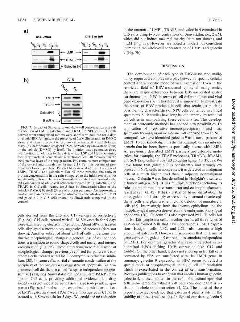
in the amount of LMP1, TRAF3, and galectin 9 contained inC15 rafts using two concentrations of Simvastatin, i.e., 2 �M,which did not induce maximal toxicity (data not shown), and5 �M (Fig. 7a). However, we noted a modest but consistentincrease in the whole-cell concentration of LMP1 and galectin9 (Fig. 7b).
DISCUSSION
The development of each type of EBV-associated malig-nancy requires a complex interplay between a specific cellularcontext and a specific mode of viral expression. Even in therestricted field of EBV-associated epithelial malignancies,there are major differences between EBV-associated gastriccarcinomas and NPC in terms of cell differentiation and viralgene expression (56). Therefore, it is important to investigatethe status of EBV products in cells that retain, as much aspossible, the characteristics of NPC cells contained in clinicalspecimens. Such studies have long been hampered by technicaldifficulties in manipulating these cells in vitro. The develop-ment of proteomic methods has opened new possibilities. Byapplication of preparative immunoprecipitation and massspectrometry analysis on membrane rafts derived from an NPCxenograft, we have identified galectin 9 as a novel partner ofLMP1. To our knowledge, it is the first example of a membraneprotein that has been shown to specifically interact with LMP1.All previously described LMP1 partners are cytosolic mole-cules, for example, the TRAF molecules, TRADD, BRAM1,and SCF (Skp-cullin-F box) E3 ubiquitin ligase (10, 37, 58). Wehave found that galectin 9 is consistently and strongly ex-pressed in NPC cells; in most cases, it is detected in malignantcells at a much higher level than in adjacent nonmalignantmucosa. Galectin 9 was first described in Hodgkin’s disease asa tumor antigen (59). It has multiple functions including itsrole as a membrane urate transporter and eosinophil chemoat-tractant (25, 41, 42). It has a restricted tissue distribution. Inmice, galectin 9 is strongly expressed by postnatal thymic epi-thelial cells and plays a role in clonal deletion of immature Tcells (62). Interestingly, both the thymus epithelium and thenasopharyngeal mucosa derive from the embryonic pharyngealendoderm (20). Galectin 9 is also expressed by LCL cells butnot Burkitt lymphoma cells. In other words, all three types ofEBV-transformed cells that have spontaneous LMP1 expres-sion—Hodgkin cells, NPC, and LCL—also contain a highamount of galectin 9. However, it is obvious that, in terms ofgene expression, galectin 9 expression is somehow independentof LMP1. For example, galectin 9 is readily detected in xe-nografted NPCs lacking LMP1-expression like C17 andC666-1. On the other hand, it does not show up in Burkitt cellsconverted by EBV or transfected with the LMP1 gene. Insummary, galectin 9 expression in NPC seems to reflect aspecial mode of nasopharyngeal epithelial cell differentiationwhich is exacerbated in the context of cell transformation.Previous publications have shown that another human galectin,galectin 4, is accumulated in the rafts of intestinal epithelialcells, more precisely within a raft core component that is re-sistant to cholesterol extraction (4, 22). The latest of thesereports provides evidence that galectin 4 plays a role in thestability of these structures (4). In light of our data, galectin 9
FIG. 7. Impact of Simvastatin on whole-cell concentration and raftdistribution of LMP1, galectin 9, and TRAF3 in NPC cells. C15 cellsderived from xenografted tumors were short-term cultured for 5 dayson a polyHEMA matrix in the presence of 5 �M Simvastatin or DMSOalone and then subjected to protein extraction and a raft flotationassay. (a) Raft flotation assay of C15 cells treated by Simvastatin (Sim)or the vehicle (DMSO) by itself. The flotation assay generates threecell fractions in addition to the raft fraction: LSP and HSP containingmostly cytoskeletal elements and a fraction called F40 recovered in the40% sucrose layer of the step gradient. F40 contains most componentsof the cytosol and nonraft membranes (11). Ten micrograms of pro-teins was loaded per lane. Parallel blots were done for detection ofLMP1, TRAF3, and galectin 9. For all three proteins, the ratio ofprotein concentration in the rafts compared to the initial extract is notsignificantly different between Simvastatin-treated and control cells.(b) Comparison of whole-cell concentrations of LMP1, galectin 9, andTRAF3 in C15 cells treated for 5 days by Simvastatin (Sim) or thevehicle (DMSO) by itself (30 �g of protein per lane). An approximatetwofold increase is observed in the whole-cell concentrations of LMP1and galectin 9 in C15 cells treated by Simvastatin compared to thecontrol.
13334 PIOCHE-DURIEU ET AL. J. VIROL.
on July 26, 2015 by guesthttp://jvi.asm
.org/D
ownloaded from
appears as one novel example of a galectin associated withmembrane rafts.
The detection of large amounts of galectin 9 in NPC cellsmight also benefit our understanding of host-tumor interac-tions. NPCs are characterized by the presence of viral antigensin a highly inflammatory context, and the mechanisms of localimmune tolerance are still poorly understood (3, 61). There-fore, it is noteworthy that galectin 9, like some other galectins,can act as a negative regulator of T-cell activation (30, 53).Because of its inhibitory effects on T cells, galectin 9 mightpossibly contribute to viral immune escape in NPC tumors.This hypothesis is supported by some of our recent experi-ments, which indicate that both LMP1 and galectin 9 can bereleased in the extracellular medium by NPC cells.
We have previously reported that LMP1 recruits its mainadaptor TRAF3 in membrane rafts to the exclusion of TRAF2,TRAF1, or TRADD (1, 2). In contrast, galectin 9 is not re-cruited in membrane rafts by LMP1. It appears to be a residentraft protein even in the absence of LMP1, for example, in theC17 xenograft. In addition, it seems to be much more abundantthan LMP1 in membrane rafts. According to preliminary ex-periments, the amount of LMP1 and galectin 9 molecules for10 �g of rafts is about 200 and 1,200 fmol, respectively. Thismight explain the fact that it was difficult to precipitate morethan 20% of the galectin 9 contained in the cellular extracteven when it was possible to precipitate almost 100% of LMP1(Fig. 2b). Using mutant forms of LMP1 expressed in HeLacells, we have shown that its critical TRAF3- and TRADD-bind-ing motifs are not required for its interaction with galectin 9. Sofar, we do not know whether LMP1 directly binds to galectin 9or whether this interaction is mediated by other molecules,either lipids or proteins. Previous reports have shown that theshort intracytoplasmic domain and a conserved motif of thefirst transmembrane segment are critical for LMP1 associationwith membrane rafts (12, 51, 65). Our ongoing experiments areintended to determine whether the same segments of LMP1are required for its interaction with galectin 9.
Our data regarding LMP1-galectin 9 interactions provided agood molecular basis to investigate the effects of Simvastatinon the behavior of LMP1-carrying rafts in NPC cells. Treat-ment of C15 cells with Simvastatin was expected to induce thedissociation of LMP1 from membrane rafts, as previously re-ported for LCLs by Katano et al. (31). It was not the case: theraft distribution of LMP1 and TRAF3 as well as galectin 9 wasapparently not affected by this drug, despite its strong cytotoxiceffect. At the same time, the whole-cell concentrations ofLMP1 and galectin 9 were increased under Simvastatin. Cur-rently, we have no explanation for this modification. In anycase, the cytotoxic effect of Simvastin against NPC cells wasalmost the same for C15 (LMP1-positive) and C17 (LMP1-negative) cells, providing additional evidence that LMP1 is nota critical lethal target for Simvastatin in this cell type. Inter-estingly, the cytotoxic effect of Simvastatin against NPC cellswas obtained at low concentrations: a concentration of only1 �M for 2 days was sufficient to induce significant growthinhibitory effects in contrast to a concentration of more than10 �M required to achieve a significant effect on prostate car-cinoma cells (67). Thus, our data suggest that Simvastatinmight be beneficial in some circumstances for NPC patients.This is especially interesting since this drug has been widely
used in medical practice for several years, allowing long-termmonitoring of its undesirable effects.
ACKNOWLEDGMENTS
This work was supported by grants from the Ligue Nationale contrele Cancer (comite du Cher), from the Fondation de France (no.2001004522), and from the ARC (no. 5238). C. K. was supported by afellowship from the French Ministere de la Recherche et de la Tech-nologie.
We thank V. Velasco for technical assistance and J. Wiels, M.Lipinski, and G. Pierron for helpful discussions.
REFERENCES
1. Ardila-Osorio, H., B. Clausse, Z. Mishal, J. Wiels, T. Tursz, and P. Busson.1999. Evidence of LMP1-TRAF3 interactions in glycosphingolipid-rich com-plexes of lymphoblastoid and nasopharyngeal carcinoma cells. Int J. Cancer.81:645–649.
2. Ardila-Osorio, H., C. Pioche-Durieu, F. Puvion-Dutilleul, B. Clausse, J.Wiels, W. Miller, N. Raab-Traub, and P. Busson. 2005. TRAF interactionswith raft-like buoyant complexes, better than TRAF rates of degradation,differentiate signaling by CD40 and EBV latent membrane protein 1. Int J.Cancer. 113:267–275.
3. Beck, A., D. Pazolt, G. G. Grabenbauer, J. M. Nicholls, H. Herbst, L. S.Young, and G. Niedobitek. 2001. Expression of cytokine and chemokinegenes in Epstein-Barr virus-associated nasopharyngeal carcinoma: compar-ison with Hodgkin’s disease. J. Pathol. 194:145–151.
4. Braccia, A., M. Villani, L. Immerdal, L. L. Niels-Christiansen, B. T.Nystrom, G. H. Hansen, and E. M. Danielsen. 2003. Microvillar membranemicrodomains exist at physiological temperature. Role of galectin-4 as lipidraft stabilizer revealed by “superrafts.” J. Biol. Chem. 278:15679–15684.
5. Brink, A. A., M. B. Vervoort, J. M. Middeldorp, C. J. Meijer, and A. J.van den Brule. 1998. Nucleic acid sequence-based amplification, a newmethod for analysis of spliced and unspliced Epstein-Barr virus latent tran-scripts, and its comparison with reverse transcriptase PCR. J. Clin. Micro-biol. 36:3164–3169.
6. Busson, P., G. Ganem, P. Flores, F. Mugneret, B. Clausse, B. Caillou, K.Braham, H. Wakasugi, M. Lipinski, and T. Tursz. 1988. Establishment andcharacterization of three transplantable EBV-containing nasopharyngealcarcinomas. Int. J. Cancer. 42:599–606.
7. Busson, P., C. Keryer, T. Ooka, and M. Corbex. 2004. EBV-associatednasopharyngeal carcinomas: from epidemiology to virus-targeting strategies.Trends Microbiol. 12:356–360.
8. Challa, A., M. H., M. Baker, J. Pound, J. Gordon. 1997. CD40 workshoppanel report, p. 159–161. In T. Kishimoto (ed.), Leucocyte typing. VI. Whitecell differentiation antigens, vol. 1. Garland Publishing, Inc., New York, N.Y.
9. Cheung, S. T., D. P. Huang, A. B. Hui, K. W. Lo, C. W. Ko, Y. S. Tsang, N.Wong, B. M. Whitney, and J. C. Lee. 1999. Nasopharyngeal carcinoma cellline (C666-1) consistently harbouring Epstein-Barr virus. Int. J. Cancer.83:121–126.
10. Chung, P. J., Y. S. Chang, C. L. Liang, and C. L. Meng. 2002. Negativeregulation of Epstein-Barr virus latent membrane protein 1-mediated func-tions by the bone morphogenetic protein receptor IA-binding protein,BRAM1. J. Biol. Chem. 277:39850–39857.
11. Clausse, B., K. Fizazi, V. Walczak, C. Tetaud, J. Wiels, T. Tursz, and P.Busson. 1997. High concentration of the EBV latent membrane protein 1 inglycosphingolipid-rich complexes from both epithelial and lymphoid cells.Virology 228:285–293.
12. Coffin, W. F., III, T. R. Geiger, and J. M. Martin. 2003. Transmembranedomains 1 and 2 of the latent membrane protein 1 of Epstein-Barr viruscontain a lipid raft targeting signal and play a critical role in cytostasis.J. Virol. 77:3749–3758.
13. Dawson, C. W., G. Tramountanis, A. G. Eliopoulos, and L. S. Young. 2003.Epstein-Barr virus latent membrane protein 1 (LMP1) activates the phos-phatidylinositol 3-kinase/Akt pathway to promote cell survival and induceactin filament remodeling. J. Biol. Chem. 278:3694–3704.
14. Devi, B. C., P. Pisani, T. S. Tang, and D. M. Parkin. 2004. High incidence ofnasopharyngeal carcinoma in native people of Sarawak, Borneo Island. Can-cer Epidemiol. Biomarkers Prev. 13:482–486.
15. Dirmeier, U., R. Hoffmann, E. Kilger, U. Schultheiss, C. Briseno, O. Gires,A. Kieser, D. Eick, B. Sugden, and W. Hammerschmidt. 2005. Latent mem-brane protein 1 of Epstein-Barr virus coordinately regulates proliferationwith control of apoptosis. Oncogene 24:1711–1717.
16. Dolcetti, R., and J. Menezes. 2003. Epstein-Barr virus and undifferentiatednasopharyngeal carcinoma: new immunobiological and molecular insights ona long-standing etiopathogenic association. Adv. Cancer Res. 87:127–157.
17. Edwards, R. H., D. Sitki-Green, D. T. Moore, and N. Raab-Traub. 2004.Potential selection of LMP1 variants in nasopharyngeal carcinoma. J. Virol.78:868–881.
18. Eliopoulos, A. G., E. R. Waites, S. M. Blake, C. Davies, P. Murray, and L. S.Young. 2003. TRAF1 is a critical regulator of JNK signaling by the TRAF-
VOL. 79, 2005 LATENT MEMBRANE PROTEIN 1 INTERACTIONS WITH GALECTIN 9 13335
on July 26, 2015 by guesthttp://jvi.asm
.org/D
ownloaded from

binding domain of the Epstein-Barr virus-encoded latent infection mem-brane protein 1 but not CD40. J. Virol. 77:1316–1328.
19. Gires, O., U. Zimber-Strobl, R. Gonnella, M. Ueffing, G. Marschall, R.Zeidler, D. Pich, and W. Hammerschmidt. 1997. Latent membrane protein1 of Epstein-Barr virus mimics a constitutively active receptor molecule.EMBO J. 16:6131–6140.
20. Gordon, J., V. A. Wilson, N. F. Blair, J. Sheridan, A. Farley, L. Wilson, N. R.Manley, and C. C. Blackburn. 2004. Functional evidence for a singleendodermal origin for the thymic epithelium. Nat. Immunol. 5:546–553.
21. Hammerschmidt, W., and B. Sugden. 2004. Epstein-Barr virus sustains Bur-kitt’s lymphomas and Hodgkin’s disease. Trends Mol. Med. 10:331–336.
22. Hansen, G. H., L. Immerdal, E. Thorsen, L. L. Niels-Christiansen, B. T.Nystrom, E. J. Demant, and E. M. Danielsen. 2001. Lipid rafts exist as stablecholesterol-independent microdomains in the brush border membrane ofenterocytes. J. Biol. Chem. 276:32338–32344.
23. Heussinger, N., M. Buttner, G. Ott, E. Brachtel, B. Z. Pilch, E. Kremmer,and G. Niedobitek. 2004. Expression of the Epstein-Barr virus (EBV)-en-coded latent membrane protein 2A (LMP2A) in EBV-associated nasopha-ryngeal carcinoma. J. Pathol. 203:696–699.
24. Higuchi, M., K. M. Izumi, and E. Kieff. 2001. Epstein-Barr virus latent-infection membrane proteins are palmitoylated and raft-associated: protein1 binds to the cytoskeleton through TNF receptor cytoplasmic factors. Proc.Natl. Acad. Sci. USA 98:4675–4680.
25. Hirashima, M., Y. Kashio, N. Nishi, A. Yamauchi, T. A. Imaizumi, T. Kage-shita, N. Saita, and T. Nakamura. 2004. Galectin-9 in physiological andpathological conditions. Glycoconj. J. 19:593–600.
26. Huang, Y. T., T. S. Sheen, C. L. Chen, J. Lu, Y. Chang, J. Y. Chen, and C. H.Tsai. 1999. Profile of cytokine expression in nasopharyngeal carcinomas: adistinct expression of interleukin 1 in tumor and CD4� T cells. Cancer Res.59:1599–1605.
27. Huen, D. S., S. A. Henderson, D. Croom-Carter, and M. Rowe. 1995. TheEpstein-Barr virus latent membrane protein-1 (LMP1) mediates activationof NF-kappa B and cell surface phenotype via two effector regions in itscarboxy-terminal cytoplasmic domain. Oncogene 10:549–560.
28. Iwakiri, D., T. S. Sheen, J. Y. Chen, D. P. Huang, and K. Takada. 2005.Epstein-Barr virus-encoded small RNA induces insulin-like growth factor 1and supports growth of nasopharyngeal carcinoma-derived cell lines. Onco-gene 24:1767–1773.
29. Izumi, K. M., K. M. Kaye, and E. D. Kieff. 1994. Epstein-Barr virus recom-binant molecular genetic analysis of the LMP1 amino-terminal cytoplasmicdomain reveals a probable structural role, with no component essential forprimary B-lymphocyte growth transformation. J. Virol. 68:4369–4376.
30. Kashio, Y., K. Nakamura, M. J. Abedin, M. Seki, N. Nishi, N. Yoshida, T.Nakamura, and M. Hirashima. 2003. Galectin-9 induces apoptosis throughthe calcium-calpain-caspase-1 pathway. J. Immunol. 170:3631–3636.
31. Katano, H., L. Pesnicak, and J. I. Cohen. 2004. Simvastatin induces apopto-sis of Epstein-Barr virus (EBV)-transformed lymphoblastoid cell lines anddelays development of EBV lymphomas. Proc. Natl. Acad. Sci. USA 101:4960–4965.
32. Kaykas, A., K. Worringer, and B. Sugden. 2001. CD40 and LMP-1 bothsignal from lipid rafts but LMP-1 assembles a distinct, more efficient signal-ing complex. EMBO J. 20:2641–2654.
33. Kaykas, A., K. Worringer, and B. Sugden. 2002. LMP-1’s transmembranedomains encode multiple functions required for LMP-1’s efficient signaling.J. Virol. 76:11551–11560.
34. Kennedy, G., J. Komano, and B. Sugden. 2003. Epstein-Barr virus providesa survival factor to Burkitt’s lymphomas. Proc. Natl. Acad. Sci. USA 100:14269–14274.
35. Khabir, A., A. Ghorbel, J. Daoud, M. Frikha, M. M. Drira, A. Laplanche, P.Busson, and R. Jlidi. 2003. Similar BCL-X but different BCL-2 levels in thetwo age groups of North African nasopharyngeal carcinomas. Cancer Detect.Prev. 27:250–255.
36. Khabir, A., H. Karray, S. Rodriguez, M. Rose, J. Daoud, M. Frikha, T.Boudawara, J. Middeldorp, R. Jlidi, and P. Busson. 2005. EBV latent mem-brane protein 1 abundance correlates with patient age but not with meta-static behavior in North-African nasopharyngeal carcinomas. Virol. J. 2:39.
37. Kieser, A., C. Kaiser, and W. Hammerschmidt. 1999. LMP1 signal transduc-tion differs substantially from TNF receptor 1 signaling in the molecularfunctions of TRADD and TRAF2. EMBO J. 18:2511–2521.
38. Kusama, T., M. Mukai, T. Iwasaki, M. Tatsuta, Y. Matsumoto, H. Akedo,and H. Nakamura. 2001. Inhibition of epidermal growth factor-inducedRhoA translocation and invasion of human pancreatic cancer cells by 3-hy-droxy-3-methylglutaryl-coenzyme A reductase inhibitors. Cancer Res. 61:4885–4891.
39. Lam, N., M. L. Sandberg, and B. Sugden. 2004. High physiological levels ofLMP1 result in phosphorylation of eIF2� in Epstein-Barr virus-infectedcells. J. Virol. 78:1657–1664.
40. Leist, M., and M. Jaattela. 2001. Four deaths and a funeral: from caspasesto alternative mechanisms. Nat. Rev. Mol. Cell Biol. 2:589–598.
41. Lipkowitz, M. S., E. Leal-Pinto, B. E. Cohen, and R. G. Abramson. 2004.Galectin 9 is the sugar-regulated urate transporter/channel UAT. Glycoconj.J. 19:491–498.
42. Liu, F. T., and G. A. Rabinovich. 2005. Galectins as modulators of tumourprogression. Nat. Rev. Cancer 5:29–41.
43. Luftig, M., E. Prinarakis, T. Yasui, T. Tsichritzis, E. Cahir-McFarland,J. Inoue, H. Nakano, T. W. Mak, W. C. Yeh, X. Li, S. Akira, N. Suzuki, S.Suzuki, G. Mosialos, and E. Kieff. 2003. Epstein-Barr virus latent membraneprotein 1 activation of NF-kappaB through IRAK1 and TRAF6. Proc. Natl.Acad. Sci. USA 100:15595–15600.
44. Meij, P., M. B. Vervoort, J. Aarbiou, P. van Dissel, A. Brink, E. Bloemena,C. J. Meijer, and J. M. Middeldorp. 1999. Restricted low-level human anti-body responses against Epstein-Barr virus (EBV)-encoded latent membraneprotein 1 in a subgroup of patients with EBV-associated diseases. J. Infect.Dis. 179:1108–1115.
45. Miller, W. E., J. L. Cheshire, A. S. Baldwin, Jr., and N. Raab-Traub. 1998.The NPC derived C15 LMP1 protein confers enhanced activation of NF-kappa B and induction of the EGFR in epithelial cells. Oncogene 16:1869–1877.
46. Miller, W. E., J. L. Cheshire, and N. Raab-Traub. 1998. Interaction of tumornecrosis factor receptor-associated factor signaling proteins with the latentmembrane protein 1 PXQXT motif is essential for induction of epidermalgrowth factor receptor expression. Mol. Cell. Biol. 18:2835–2844.
47. Nicholls, J. M., A. Agathanggelou, K. Fung, X. Zeng, and G. Niedobitek.1997. The association of squamous cell carcinomas of the nasopharynx withEpstein-Barr virus shows geographical variation reminiscent of Burkitt’slymphoma. J. Pathol. 183:164–168.
48. Pike, L. J. 2004. Lipid rafts: heterogeneity on the high seas. Biochem. J.378:281–292.
49. Puls, A., A. G. Eliopoulos, C. D. Nobes, T. Bridges, L. S. Young, and A. Hall.1999. Activation of the small GTPase Cdc42 by the inflammatory cytokinesTNF� and IL-1, and by the Epstein-Barr virus transforming protein LMP1.J. Cell Sci. 112:2983–2992.
50. Raab-Traub, N. 2002. Epstein-Barr virus in the pathogenesis of NPC. Semin.Cancer Biol. 12:431–441.
51. Rothenberger, S., M. Rousseaux, H. Knecht, F. C. Bender, D. F. Legler, andC. Bron. 2002. Association of the Epstein-Barr virus latent membrane pro-tein 1 with lipid rafts is mediated through its N-terminal region. Cell Mol.Life Sci. 59:171–180.
52. Rowe, M., H. S. Evans, L. S. Young, K. Hennessy, E. Kieff, and A. B.Rickinson. 1987. Monoclonal antibodies to the latent membrane protein ofEpstein-Barr virus reveal heterogeneity of the protein and inducible expres-sion in virus-transformed cells. J. Gen. Virol. 68:1575–1586.
53. Rubinstein, N., M. Alvarez, N. W. Zwirner, M. A. Toscano, J. M. Ilarregui,A. Bravo, J. Mordoh, L. Fainboim, O. L. Podhajcer, and G. A. Rabinovich.2004. Targeted inhibition of galectin-1 gene expression in tumor cells resultsin heightened T cell-mediated rejection. A potential mechanism of tumor-immune privilege. Cancer Cell 5:241–251.
54. Sbih-Lammali, F., B. Clausse, H. Ardila-Osorio, R. Guerry, M. Talbot, S.Havouis, L. Ferradini, J. Bosq, T. Tursz, and P. Busson. 1999. Control ofapoptosis in Epstein-Barr virus-positive nasopharyngeal carcinoma cells: op-posite effects of CD95 and CD40 stimulation. Cancer Res. 59:924–930.
55. Seto, E., L. Yang, J. Middeldorp, T. S. Sheen, J. Y. Chen, M. Fukayama, Y.Eizuru, T. Ooka, and K. Takada. 2005. Epstein-Barr virus (EBV)-encodedBARF1 gene is expressed in nasopharyngeal carcinoma and EBV-associatedgastric carcinoma tissues in the absence of lytic gene expression. J. Med.Virol. 76:82–88.
56. Takada, K. 2000. Epstein-Barr virus and gastric carcinoma. Mol. Pathol.53:255–261.
57. Tang, K. F., S. Y. Tan, S. H. Chan, S. M. Chong, K. S. Loh, L. K. Tan, andH. Hu. 2001. A distinct expression of CC chemokines by macrophages innasopharyngeal carcinoma: implication for the intense tumor infiltration byT lymphocytes and macrophages. Hum. Pathol. 32:42–49.
58. Tang, W., O. A. Pavlish, V. S. Spiegelman, A. A. Parkhitko, and S. Y. Fuchs.2003. Interaction of Epstein-Barr virus latent membrane protein 1 withSCFHOS/beta-TrCP E3 ubiquitin ligase regulates extent of NF-kappaB ac-tivation. J. Biol. Chem. 278:48942–48949.
59. Tureci, O., H. Schmitt, N. Fadle, M. Pfreundschuh, and U. Sahin. 1997.Molecular definition of a novel human galectin which is immunogenic inpatients with Hodgkin’s disease. J. Biol. Chem. 272:6416–6422.
60. Vicat, J. M., H. Ardila-Osorio, A. Khabir, M. C. Brezak, I. Viossat, P.Kasprzyk, R. Jlidi, P. Opolon, T. Ooka, G. Prevost, D. P. Huang, and P.Busson. 2003. Apoptosis and TRAF-1 cleavage in Epstein-Barr virus-posi-tive nasopharyngeal carcinoma cells treated with doxorubicin combined witha farnesyl-transferase inhibitor. Biochem. Pharmacol. 65:423–433.
61. Voo, K. S., T. Fu, H. Y. Wang, J. Tellam, H. E. Heslop, M. K. Brenner, C. M.Rooney, and R. F. Wang. 2004. Evidence for the presentation of majorhistocompatibility complex class I-restricted Epstein-Barr virus nuclear an-tigen 1 peptides to CD8� T lymphocytes. J. Exp. Med. 199:459–470.
62. Wada, J., K. Ota, A. Kumar, E. I. Wallner, and Y. S. Kanwar. 1997. Devel-opmental regulation, expression, and apoptotic potential of galectin-9, abeta-galactoside binding lectin. J. Clin. Investig. 99:2452–2461.
63. Wang, F., C. Gregory, C. Sample, M. Rowe, D. Liebowitz, R. Murray, A.Rickinson, and E. Kieff. 1990. Epstein-Barr virus latent membrane protein(LMP1) and nuclear proteins 2 and 3C are effectors of phenotypic changes
13336 PIOCHE-DURIEU ET AL. J. VIROL.
on July 26, 2015 by guesthttp://jvi.asm
.org/D
ownloaded from

in B lymphocytes: EBNA-2 and LMP1 cooperatively induce CD23. J. Virol.64:2309–2318.
64. Xie, P., B. S. Hostager, and G. A. Bishop. 2004. Requirement for TRAF3 insignaling by LMP1 but not CD40 in B lymphocytes. J. Exp. Med. 199:661–671.
65. Yasui, T., M. Luftig, V. Soni, and E. Kieff. 2004. Latent infection membraneprotein transmembrane FWLY is critical for intermolecular interaction, raftlocalization, and signaling. Proc. Natl. Acad. Sci. USA 101:278–283.
66. Zhang, X., W. Uthaisang, L. Hu, I. T. Ernberg, and B. Fadeel. 2005. Epstein-Barr virus-encoded latent membrane protein 1 promotes stress-induced ap-optosis upstream of caspase-2-dependent mitochondrial perturbation. Int. J.Cancer. 113:397–405.
67. Zhuang, L., J. Kim, R. M. Adam, K. R. Solomon, and M. R. Freeman. 2005.Cholesterol targeting alters lipid raft composition and cell survival in pros-tate cancer cells and xenografts. J. Clin. Investig. 115:959–968.
VOL. 79, 2005 LATENT MEMBRANE PROTEIN 1 INTERACTIONS WITH GALECTIN 9 13337
on July 26, 2015 by guesthttp://jvi.asm
.org/D
ownloaded from
Copyright © 2022 FDOKUMEN