A Unique Galectin Signature in Human Prostate Cancer Progression Suggests Galectin-1 as a Key Target...
34
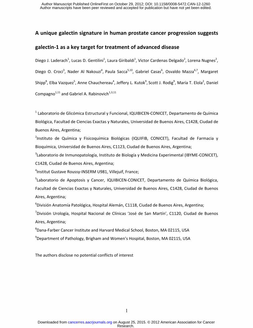
1 A unique galectin signature in human prostate cancer progression suggests galectin-1 as a key target for treatment of advanced disease Diego J. Laderach 1 , Lucas D. Gentilini 1 , Laura Giribaldi 1 , Victor Cardenas Delgado 2 , Lorena Nugnes 2 , Diego O. Croci 3 , Nader Al Nakouzi 4 , Paula Sacca 5,10 , Gabriel Casas 6 , Osvaldo Mazza 6,7 , Margaret Shipp 8 , Elba Vazquez 5 , Anne Chauchereau 4 , Jeffery L. Kutok 9 , Scott J. Rodig 9 , María T. Elola 2 , Daniel Compagno 1,11 and Gabriel A. Rabinovich 1,3,11 1 Laboratorio de Glicómica Estructural y Funcional, IQUIBICEN-CONICET, Departamento de Química Biológica, Facultad de Ciencias Exactas y Naturales, Universidad de Buenos Aires, C1428, Ciudad de Buenos Aires, Argentina; 2 Instituto de Química y Fisicoquímica Biológicas (IQUIFIB, CONICET), Facultad de Farmacia y Bioquímica, Universidad de Buenos Aires, C1123, Ciudad de Buenos Aires, Argentina; 3 Laboratorio de Inmunopatología, Instituto de Biología y Medicina Experimental (IBYME-CONICET), C1428, Ciudad de Buenos Aires, Argentina; 4 Institut Gustave Roussy-INSERM U981, Villejuif, France; 5 Laboratorio de Apoptosis y Cancer, IQUIBICEN-CONICET, Departamento de Química Biológica, Facultad de Ciencias Exactas y Naturales, Universidad de Buenos Aires, C1428, Ciudad de Buenos Aires, Argentina; 6 División Anatomía Patológica, Hospital Alemán, C1118, Ciudad de Buenos Aires, Argentina; 7 División Urología, Hospital Nacional de Clínicas ‘José de San Martín’, C1120, Ciudad de Buenos Aires, Argentina; 8 Dana-Farber Cancer Institute and Harvard Medical School, Boston, MA 02115, USA 9 Department of Pathology, Brigham and Women’s Hospital, Boston, MA 02115, USA The authors disclose no potential conflicts of interest Research. on August 25, 2015. © 2012 American Association for Cancer cancerres.aacrjournals.org Downloaded from Author manuscripts have been peer reviewed and accepted for publication but have not yet been edited. Author Manuscript Published OnlineFirst on October 29, 2012; DOI: 10.1158/0008-5472.CAN-12-1260
-
Upload
independent -
Category
Documents
-
view
0 -
download
0
Transcript of A Unique Galectin Signature in Human Prostate Cancer Progression Suggests Galectin-1 as a Key Target...
1
A unique galectin signature in human prostate cancer progression suggests
galectin-1 as a key target for treatment of advanced disease
Diego J. Laderach1, Lucas D. Gentilini1, Laura Giribaldi1, Victor Cardenas Delgado2, Lorena Nugnes2,
Diego O. Croci3, Nader Al Nakouzi4, Paula Sacca5,10, Gabriel Casas6, Osvaldo Mazza6,7, Margaret
Shipp8, Elba Vazquez5, Anne Chauchereau4, Jeffery L. Kutok9, Scott J. Rodig9, María T. Elola2, Daniel
Compagno1,11 and Gabriel A. Rabinovich1,3,11
1 Laboratorio de Glicómica Estructural y Funcional, IQUIBICEN-CONICET, Departamento de Química
Biológica, Facultad de Ciencias Exactas y Naturales, Universidad de Buenos Aires, C1428, Ciudad de
Buenos Aires, Argentina; 2Instituto de Química y Fisicoquímica Biológicas (IQUIFIB, CONICET), Facultad de Farmacia y
Bioquímica, Universidad de Buenos Aires, C1123, Ciudad de Buenos Aires, Argentina; 3Laboratorio de Inmunopatología, Instituto de Biología y Medicina Experimental (IBYME-CONICET),
C1428, Ciudad de Buenos Aires, Argentina; 4Institut Gustave Roussy-INSERM U981, Villejuif, France; 5Laboratorio de Apoptosis y Cancer, IQUIBICEN-CONICET, Departamento de Química Biológica,
Facultad de Ciencias Exactas y Naturales, Universidad de Buenos Aires, C1428, Ciudad de Buenos
Aires, Argentina; 6División Anatomía Patológica, Hospital Alemán, C1118, Ciudad de Buenos Aires, Argentina; 7División Urología, Hospital Nacional de Clínicas ‘José de San Martín’, C1120, Ciudad de Buenos
Aires, Argentina; 8Dana-Farber Cancer Institute and Harvard Medical School, Boston, MA 02115, USA 9Department of Pathology, Brigham and Women’s Hospital, Boston, MA 02115, USA
The authors disclose no potential conflicts of interest
Research. on August 25, 2015. © 2012 American Association for Cancercancerres.aacrjournals.org Downloaded from
Author manuscripts have been peer reviewed and accepted for publication but have not yet been edited. Author Manuscript Published OnlineFirst on October 29, 2012; DOI: 10.1158/0008-5472.CAN-12-1260
2
Footnotes: 10Current Affiliation: Instituto de Biología y Medicina Experimental (IBYME/ CONICET), C1428
Ciudad de Buenos Aires, Argentina
11D.C. and G.A.R. should be considered as co-senior authors.
Correspondence:
Diego J. Laderach: [email protected]
Daniel Compagno: [email protected]
Gabriel A. Rabinovich: [email protected]
Key words: Tumor microenvironment, Prostate Cancer, Galectins, Angiogenesis.
Running Title: Galectin signature of human prostate cancer
Research. on August 25, 2015. © 2012 American Association for Cancercancerres.aacrjournals.org Downloaded from
Author manuscripts have been peer reviewed and accepted for publication but have not yet been edited. Author Manuscript Published OnlineFirst on October 29, 2012; DOI: 10.1158/0008-5472.CAN-12-1260
3
ABSTRACT
Galectins, a family of glycan-binding proteins, influence tumor progression by modulating
interactions between tumor, endothelial, stromal and immune cells. Despite considerable progress
in identifying the roles of individual galectins in tumor biology, an integrated portrait of the
galectin network in different tumor microenvironments is still missing. We undertook this study to
analyze the 'galectin signature' of the human prostate cancer (PCa) microenvironment with the
overarching goal of selecting novel molecular targets for prognostic and therapeutic purposes. In
examining androgen-responsive and castration-resistant PCa cells and primary tumors
representing different stages of the disease, we found that galectin-1 (Gal-1) was the most
abundantly expressed galectin in PCa tissue and was markedly upregulated during disease
progression. In contrast, all other galectins were expressed at lower levels: Gal-3, -4, -9 and -12
were downregulated during disease evolution, while expression of Gal-8 was unchanged. Given
the prominent regulation of Gal-1 during PCa progression and its predominant localization at the
tumor-vascular interface, we analyzed the potential role of this endogenous lectin in PCa
angiogenesis. In human PCa tissue arrays, Gal-1 expression correlated with the presence of blood
vessels, particularly in advanced stages of the disease. Silencing Gal-1 in PCa cells reduced tumor
vascularization without altering expression of other angiogenesis-related genes. Collectively, our
findings identify a dynamically regulated 'galectin-specific signature’ that accompanies disease
evolution in PCa, and they highlight a major role for Gal-1 as a tractable target for anti-angiogenic
therapy in advanced stages of the disease.
Research. on August 25, 2015. © 2012 American Association for Cancercancerres.aacrjournals.org Downloaded from
Author manuscripts have been peer reviewed and accepted for publication but have not yet been edited. Author Manuscript Published OnlineFirst on October 29, 2012; DOI: 10.1158/0008-5472.CAN-12-1260

4
PRECIS
The dynamically regulated expression signature for an important class of cell surface glycan-
binding molecules in prostate cancer suggests a tractable target for anti-angiogenic therapy in
advanced disease.
Research. on August 25, 2015. © 2012 American Association for Cancercancerres.aacrjournals.org Downloaded from
Author manuscripts have been peer reviewed and accepted for publication but have not yet been edited. Author Manuscript Published OnlineFirst on October 29, 2012; DOI: 10.1158/0008-5472.CAN-12-1260
5
INTRODUCTION
Prostate cancer (PCa) is the second most common cancer in men, and represents a
significant cause of mortality worldwide (1). Localized PCa is efficiently treated by association of
surgery with radiation therapy and androgen ablation. However, PCa evolves towards stages in
which tumor cells acquire properties allowing their distant dissemination (2) and castration-
resistant growth (3). No current treatments are applicable to these situations and the prospect for
cure decreases radically. These particular features urge the search of novel prognosis strategies
that could delineate the transition from hormone-sensitive toward hormone-resistant tumor
growth and innovative therapeutic approaches suitable for castration-refractory stages of the
disease.
Effective cancer therapies typically capitalize on molecular differences between healthy
and neoplastic tissues. In the post-genomic era, the study of the glycome has enabled the
association of specific glycan structures with the transition from normal to neoplastic tissue (4, 5).
The function of deciphering the biological information encoded by the glycome is assigned to
endogenous glycan-binding proteins or lectins, whose expression and function are regulated
during tumor progression (5). Galectins, a family of glycan-binding proteins, play pivotal roles as
regulators of tumor biology by directly influencing tumor transformation, invasiveness,
angiogenesis and tumor-immune escape (6,7). These lectins are defined by a common structural
fold and a conserved carbohydrate recognition domain (CRD) that recognizes N- and O-glycans
expressing the disaccharide N-acetyllactosamine (Galβ(1-4)-GlcNAc), although differences in
glycan-binding preferences of individual members of the family have been reported (7). Galectins
that are traditionally classified as ‘proto-type’ (Gal-1, -2, -5, -7, -10, -11, -13, -14, -15) have one
CRD that can dimerize, whereas ‘tandem-repeat’ galectins (Gal-4, -6, -8, -9 and -12) contain two
homologous CRDs in tandem in a single polypeptide chain. Gal-3 is unique in that it contains a CRD
Research. on August 25, 2015. © 2012 American Association for Cancercancerres.aacrjournals.org Downloaded from
Author manuscripts have been peer reviewed and accepted for publication but have not yet been edited. Author Manuscript Published OnlineFirst on October 29, 2012; DOI: 10.1158/0008-5472.CAN-12-1260
6
connected to a non-lectin N-terminal region that is responsible for oligomerization (7).
Extracellularly, galectins interact with cell surface glycoconjugates and trigger cellular signaling to
control migration, immunity and angiogenesis. Intracellularly, galectins can control tumor
transformation, proliferation and survival (7, 8).
Previous studies have identified galectins as key components of the prostate cancer
microenvironment (9-11). Expression of Gal-1 controls the differentiation and survival of PCa cells
(9, 12) and inhibits T-cell transmigration (13). On the other hand, Gal-3 controls homotypic and
heterotypic aggregation of PCa cells (14-16) and controls their viability (17). Tumor cell expression
of Gal-3 has been proposed to delineate the transition from benign stages to castration-resistant
malignant disease (18) and its regulated expression is associated with promoter methylation (19).
Silencing Gal-3 results in decreased migration, invasion and proliferation of PCa cells (20).
Moreover, Gal-8, which was originally identified as prostate cancer tumor antigen 1 (PCTA1) (21),
can modulate integrin-mediated cell-extracellular matrix interactions (22). However, in spite of
considerable progress in dissecting the functions of individual members of the galectin family,
there is still no integrated portrait of the ‘galectin signature’ of the human prostate cancer
microenvironment.
Our findings identify a unique galectin expression profile which delineates different stages
of PCa progression. From all galectins analyzed, Gal-1 is uniquely expressed at high levels in human
PCa and contributes to tumor progression by promoting neovascularization. These results
underscore the importance of Gal-1 as an attractive therapeutic target in advanced stages of PCa.
Research. on August 25, 2015. © 2012 American Association for Cancercancerres.aacrjournals.org Downloaded from
Author manuscripts have been peer reviewed and accepted for publication but have not yet been edited. Author Manuscript Published OnlineFirst on October 29, 2012; DOI: 10.1158/0008-5472.CAN-12-1260
7
MATERIALS AND METHODS
Human samples
Radical prostatectomies were obtained from the archival tissue bank of the Department of
Pathology, Hospital Alemán (Buenos Aires, Argentina). Samples were classified according to TNM
classification (UICC, 2002) by two independent pathologists (G.C., O.M.). Specimens (n=61)
covered all stages of PCa evolution (T1, T2, T3 and T4) in addition to benign cases (BHP) (Table 1).
None of these patients received pre-operative therapy. Protocols were approved by the Local
Ethics Committee (Hospital de Clínicas ‘José de San Martín’).
Cells and animals
Human prostate cancer cell lines used included: the hormone-responsive LNCaP cell line and the
castration-resistant cell lines 22Rv1 and PC-3 with or without androgen receptor (AR) expression
respectively. The LNCaP and 22Rv1 cell lines were provided by Anne Chauchereau (Institute
Gustave Roussy, France). LNCaP cells were also provided by Elba Vazquez (University of Buenos
Aires, Argentina). These cell lines were originally obtained from the American Type Culture
Collection (ATCC). Cell morphology was evaluated by microscopic examination on a daily basis.
Growth properties of LNCaP cells were regularly tested through their responsiveness to androgens
using MTT assay. Cells were incubated for 24 h in red phenol-free RPMI, 10% Charcoal-treated
serum and medium was supplemented with 10-10M R1881 (AR agonist) for 3 days before analyzing
growth and gene expression. PSA induction was evaluated by real time RT-PCR. Routine tests for
22Rv1 cells included examination of androgen-insensitive growth (MTT method) and PSA induction
by R1881 (real time RT-PCR). The PC-3 cell line was provided by Elba Vazquez (University of Buenos
Aires, Argentina). Growth of these cells was routinely tested for androgen sensitivity and the AR
and PSA phenotypes by real time RT-PCR. Bovine aortic endothelial cells (BAEC) were provided by
Research. on August 25, 2015. © 2012 American Association for Cancercancerres.aacrjournals.org Downloaded from
Author manuscripts have been peer reviewed and accepted for publication but have not yet been edited. Author Manuscript Published OnlineFirst on October 29, 2012; DOI: 10.1158/0008-5472.CAN-12-1260
8
Maria T. Elola (University of Buenos Aires, Argentina). BAEC were tested for their ability to form
tubular structures in the presence of vascular endothelial growth factor (VEGF). Each cell line was
routinely tested for Mycoplasma contamination by genomic PCR. LNCaP, 22Rv1 and PC-3 cells
were cultured in RPMI and BAEC were cultured in DMEM. Medium was supplemented with 10%
heat-inactivated fetal bovine serum (FBS) (PAA, Cell Culture, Austria), 2mM L-glutamine, 100
μg/ml streptomycin and 100 U/ml penicillin. BAEC were used at passage 14 or less. For some
experiments, 22Rv1 cells (50,000) were plated into 12-well plates, cultured for 2 days under
normal oxygen supply, and then exposed to hypoxic (1% O2) or normoxic conditions for an
additional 15 h. Nude mice were obtained from The National University of La Plata (La Plata,
Argentina) and maintained in accordance with the Institutional Animal Care and Use Committee
guidelines (IBYME, Buenos Aires, Argentina).
Reagents
The following anti-galectin antibodies (Santa Cruz Biotechnology, Inc., USA) were used: rabbit anti-
Gal-1 (H-45), anti-Gal-8 (H-80), anti-Gal-3 (H-160), anti-Gal-4 (T-20), anti-Gal-12 (H-166) and goat
anti-Gal-9 (C-20) antibodies. A purified anti-Gal-1 polyclonal rabbit IgG generated in G.A.R’s
laboratory was used (23,24). Anti-human carbonic anhydrase IX polyclonal antibody (H-120) was
obtained from Santa Cruz. Media and trypsin/EDTA were obtained from Gibco-Invitrogen (Life
Technologies). Blocking anti-Gal-1 monoclonal antibody (mAb; F8.G7) was generated and validated
as described (25, 26). Growth factor-reduced Matrigel was obtained from BD Biosciences.
Immunohistochemistry
Immunohistochemistry was performed on paraffin-embedded tissue samples. Samples were
deparaffinized by 5 min incubation in xylene, 100%, 95% and 75% ethanol. Endogenous peroxidase
Research. on August 25, 2015. © 2012 American Association for Cancercancerres.aacrjournals.org Downloaded from
Author manuscripts have been peer reviewed and accepted for publication but have not yet been edited. Author Manuscript Published OnlineFirst on October 29, 2012; DOI: 10.1158/0008-5472.CAN-12-1260
9
activity was quenched by 10 min incubation with 1% H2O2. Non-specific binding was blocked using
normal horse serum in 0.05% saponin. Samples were incubated with the appropriate antibodies at
the optimal dilutions for 1 h at room temperature. The following antibodies were used: polyclonal
anti-Gal antibodies and preimmune sera from Santa Cruz (1:200 dilution) and purified anti-Gal-1
rabbit IgG (1:1,500). Immunoreactions were developed using the avidin-biotin-peroxidase
Vectastain ABC Kit (Vector). Galectin expression was graded as follows: 0 (negative); 1+ (poor
intensity); 2+ (moderate intensity); 3+ (high intensity) and 4+ (very high intensity). PCa cell lines
were adhered to poly-L-lysine (Sigma)-coated coverslips for 2 h at 37°C (50,000 cells per coverslip),
fixed with 4% paraformaldehyde for 5 min and processed for immunocytochemistry as described
for tissues.
Immunohistochemistry of tissue microarrays was performed using 4 µm-thick formalin-
fixed, paraffin-embedded sections of tissue microarray slides containing 29 paired cores (2
different areas of each single tumor from 29 tumors analyzed) of invasive PCa (BC19013; US
Biomax) (Table 1B). Slides were soaked in xylene and passed through graded alcohols and distilled
water prior to use. Slides were then pre-treated with citrate buffer pH 6.0 (Invitrogen) in a steam
pressure cooker (Decloaking Chamber CD2008US, Biocare Biomedical) according to
manufacturers’ recommended settings (127°C for 30 sec, followed by 90°C). Slides were blocked
for peroxidase activity using a specific blocker (DAKO) and washed for 5 min in buffer. Individual
slides were incubated with a mouse anti-human CD34 mAb (clone QBEND-10, RTU, Immunotech)
and a rabbit anti-human Gal-1 polyclonal antibody (1:10,000) generated and used as described
(23,24). After 1 h incubation, slides were washed and processed by the appropriate Envision+ kit
(DAKO) as per manufacturer’s instructions, developed using a DAB chromogen (DAKO) and
counterstained with hematoxylin. Stained slides were digitally scanned using Aperio ScanScope XT
workstation at the 20X setting (Aperio Technology, Inc.). Core images were then analyzed using
Research. on August 25, 2015. © 2012 American Association for Cancercancerres.aacrjournals.org Downloaded from
Author manuscripts have been peer reviewed and accepted for publication but have not yet been edited. Author Manuscript Published OnlineFirst on October 29, 2012; DOI: 10.1158/0008-5472.CAN-12-1260
10
ImageScope software (version 10.0.35.1800, Aperio Technology). Briefly, pathologists (S.J.R., J.L.K.)
identified areas of tumors as regions of interest (ROI) and excluded areas without significant
tumor using standard ImageScope software functions. The ROIs were then analyzed using a
standard analysis algorithm (color deconvolution v9.0, Aperio Technology) to quantify the average
optical density of Gal-1 staining and the percentage of positive pixels in the annotated tumor area.
Real time RT-PCR
Transcriptional profile of galectins was analyzed in human PCa cell lines (log phase of growth) that
are representative of different stages of tumour progression. Transcriptional profile of
angiogenesis-related genes was analyzed in plugs generated by injection of Gal-1-sufficient or Gal-
1-silenced human 22Rv1 cells into nude mice. In all cases, RNA was purified using TRIzol reagent
(Invitrogen) coupled to DNAse (RQ1, Promega) treatment. Four hundred ng of total RNA was used
for the reverse transcription reaction by using SuperScript III Reverse Transcriptase, random
hexamers (2.5 µg/ml) and dNTPs (500 nM) according to manufacturer’s instructions (Invitrogen)
during 50 min at 45°C, following by RNAse H treatment for 30 min at 37°C. One µl of a 1:25
dilution of cDNA was used as template in real time PCR. Relative gene expression was analyzed
using SYBR Green PCR kit (Applied Biosystem, Life Technologies). PCR conditions were as follows: 5
min 95°C, 40 cycles 30 sec at 95°C, 32 sec at 59°C, 45 sec at 72°C. Amplification fragments were
analyzed by electrophoresis on a 2% agarose TAE gel and by thermal dissociation curves (Tm) to
characterize the amplicon corresponding to each primer couple. Primers used are listed in Tables
S1 and S2. Cyclophilin A was used as an internal reference gene (27). Equivalent amounts of RNA
were tested to rule out genomic DNA contamination.
Research. on August 25, 2015. © 2012 American Association for Cancercancerres.aacrjournals.org Downloaded from
Author manuscripts have been peer reviewed and accepted for publication but have not yet been edited. Author Manuscript Published OnlineFirst on October 29, 2012; DOI: 10.1158/0008-5472.CAN-12-1260
11
Immunoblotting
Specificity of anti-galectin antibodies was evaluated by immunoblotting (Figure S2). Cells were
lysed in RIPA buffer (50 mM Tris-HCl-pH 8, 150 mM NaCl, 1% IGEPAL, 0.5% sodium deoxycholate,
0.1% SDS, 10 mM EDTA, 1 mM sodium vanadate and Protease Inhibitor Cocktail Set III
(Calbiochem). Equal amounts of protein (10-30 µg) were resolved by 15% SDS-PAGE, blotted onto
PVDF membranes (GE Healthcare), blocked with 5% BSA and probed with anti-galectin or anti-β-
tubulin (H-235 1:200, Santa Cruz) antibodies or preimmune rabbit antiserum. The following
dilutions of antibodies were used: anti-Gal-1 (1:500), Gal-3 (1:400), Gal-4 (1:100), Gal-8 (1:400),
Gal-9 (1:100), Gal-12 (1:100) and rabbit IgG anti-Gal-1 at 1:1,500). Bound antibodies were
detected with peroxidase-labeled anti-rabbit total immunoglobulins (1:3,000; Sigma) or by
peroxidase-labeled rabbit anti-goat IgG (1:2,000; Sigma). Peroxidase activity was detected using a
luminol-based method and chemiluminescence was determined using a Fuji Photo Film Analyzer.
Lentivirus vector production and transduction
pLv-HTM plasmid (provided by Trono Didier, Geneva University) is a self-inactivation third
generation HIV-1-derived vector (28). Annealed oligonucleotides coding for shRNA were ligated
into ClaI and MluI double-restricted plasmids by standard cloning. Restriction enzymes and T4 DNA
ligase were from New England BioLabs. Production of siRNA was under the control of H1 (RNA
polymerase type II) promoter. As reporter gene, green fluorescent protein (GFP) was expressed
under the control of eukaryotic EF-1α promoter. Plasmids were verified by sequence analysis.
Lentiviral particles were produced by transient transfection of 293T cells. Briefly, subconfluent
293T cells were co-transfected with 20 μg plasmid vector, 15 μg pCMVR8.74 and 5 μg pMD.G
(pseudotyped VSVG) using calcium phosphate. Supernatants were harvested at 48 h and 72 h and
stored at -80°C until use. Viral titers expressed as TU/ml were determined by assessing
Research. on August 25, 2015. © 2012 American Association for Cancercancerres.aacrjournals.org Downloaded from
Author manuscripts have been peer reviewed and accepted for publication but have not yet been edited. Author Manuscript Published OnlineFirst on October 29, 2012; DOI: 10.1158/0008-5472.CAN-12-1260
12
transduction of 22Rv1 cells with serial dilutions of virion preparations. 22Rv1 PCa cells were
transduced with virus at MOI=5 in the presence of 5 µg/ml protamine sulfate (Sigma). After one
week, transduced cells (GFP+) were purified using a FACSAria II cell sorter (BD Bioscience).
In vitro capillary-like tube formation and in vivo Matrigel plug assay
Matrigel (150 µl) (BD Biosciences) was added to 24-well plates and allowed to polymerize for 2 h
at 37°C. Conditioned media were added to wells and 2.5 x 10⁴ BAEC were seeded on each well.
Tube formation was evaluated in 5 different fields of each well and photographed at 18 h using an
inverted microscope. For in vivo assays, cold Matrigel was mixed with 5 x 10⁶ 22Rv1 cells in the
absence or presence of a Gal-1 blocking or isotype control mAb (7.5 mg/kg). The mixture (500 µl)
was subcutaneously injected into six week-old male nude mice. Five days later, Matrigel plugs
were harvested and photographed. Matrigel plugs were homogenized in 500 µl H2O on ice and
cleared by centrifugation at 200 x g for 6 min at 4°C. Hemoglobin content was determined using
the Drabkin’s reagent (Wiener Lab, Buenos Aires, Argentina).
Statistical analysis
Data are presented as mean ± standard deviation (S.D.) of at least three independent experiments
in triplicate. Comparisons between two groups were performed by using paired Student’s t-test or
Spearman correlation test as indicated. Differences were considered significant when P values
were less than 0.05.
Research. on August 25, 2015. © 2012 American Association for Cancercancerres.aacrjournals.org Downloaded from
Author manuscripts have been peer reviewed and accepted for publication but have not yet been edited. Author Manuscript Published OnlineFirst on October 29, 2012; DOI: 10.1158/0008-5472.CAN-12-1260
13
RESULTS
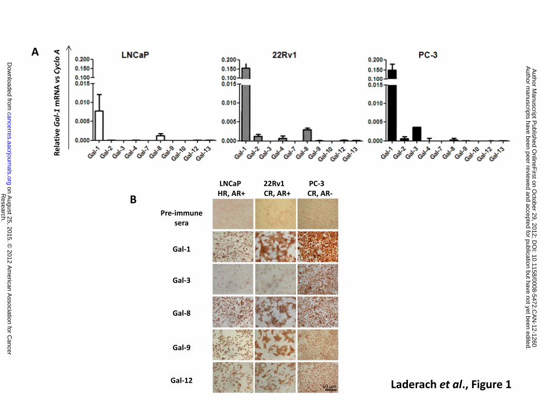
Identification of ‘the galectin signature’ of human prostate cancer cells
To delineate the galectin expression profile during PCa progression, we first examined the
galectin transcriptional pattern of several human PCa cell lines, which are representative of
different stages of the disease. These include the hormone-responsive cell line LNCaP and the
castration-resistant cell lines 22Rv1 and PC-3, which are androgen receptor (AR) positive (22Rv1)
or negative (PC-3) respectively. Total RNA was extracted in the log phase of growth and analyzed
by quantitative RT-PCR (Figure 1A). Gal-1 was found to be the most abundantly expressed galectin
in all cells analyzed and its expression was higher in PCa cells exhibiting more aggressive behavior
in vivo (22Rv1 and PC-3) (27). Transcripts for Gal-8, which has been postulated as PCa marker (21,
29), was expressed at moderate levels in all PCa cell lines tested. Gal-3 mRNA was only detected in
castration-resistant, AR negative PC-3 cells. Transcripts for all other galectin family members (Gal-
2, -4, -7, -9, -10, -12 and -13) were expressed at very low levels.
To further delineate the ‘galectin-specific PCa signature’, we assessed the expression of
galectin family members at the protein level (focusing on galectins with higher transcript
abundance). Immunocytochemical analysis confirmed that Gal-1 was the most abundantly
expressed galectin in the PCa cell lines analyzed, showing up-regulated expression in the most
aggressive cell lines (Figure 1B). On the other hand, Gal-3 was selectively expressed in the PC-3 cell
line, Gal-8 was detected in all the three cell lines and Gal-9 and Gal-12 showed a modest
expression in all cell lines analyzed. These results indicate a fine regulation of galectin expression
in PCa cell lines characterized by differences in phenotype, hormone-dependency and
aggressiveness.
Research. on August 25, 2015. © 2012 American Association for Cancercancerres.aacrjournals.org Downloaded from
Author manuscripts have been peer reviewed and accepted for publication but have not yet been edited. Author Manuscript Published OnlineFirst on October 29, 2012; DOI: 10.1158/0008-5472.CAN-12-1260
14
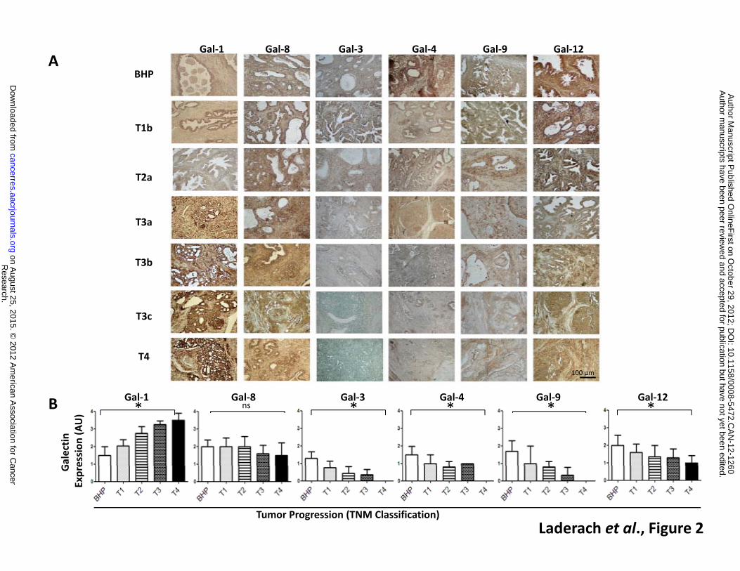
Analysis of the galectin expression profile of human primary prostate tumors
The differential expression of galectins in PCa cell lines prompted us to investigate the
galectin profile in prostatectomies obtained from 61 patients with newly diagnosed untreated
disease. Samples included a large spectrum of PCa stages (T1, T2, T3 and T4 according to TNM
classification; UICC, 2002) in addition to a benign stage (BHP) (Table 1). Similar to PCa cell lines,
Gal-1 was highly expressed in primary tumors and its expression was up-regulated in more
advanced lesions (Figure 2). On the other hand, although typically expressed at lower levels, Gal-3,
-4, -9 and -12 decreased gradually as the disease progressed toward more aggressive stages.
Conversely, Gal-8 was expressed at moderate levels in lesions corresponding to all stages. These
data delineate a ‘galectin-specific signature’ characterized by selective up- or down-regulation of
galectins during PCa progression and highlight a potential role for Gal-1 as a sensitive biomarker in
advanced stages of the disease.
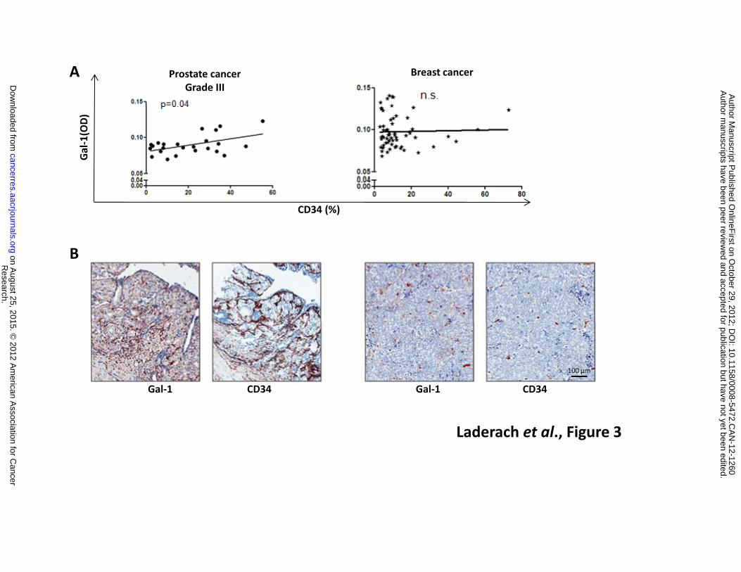
Galectin-1 is a novel target for anti-angiogenic therapies in advanced human PCa
Because Gal-1 expression is associated with PCa aggressiveness and has emerged as a novel
pro-angiogenic factor in other tumor types (30, 31), we asked whether this lectin was differentially
expressed in tumor areas associated to blood vessels in human prostate cancer. To address this issue,
we investigated whether a correlation exists between Gal-1 and CD34 expression using a human
tissue array comprised of 29 paired cores of invasive PCa classified according to proliferation rates and
cell morphology. A positive correlation was found between Gal-1 and CD34 expression in arrays
representing advanced stages of human PCa (Figure 3A). This correlation was not observed in arrays
of human breast cancer (Figure 3A), suggesting tissue-specific pro-angiogenic effects of this lectin.
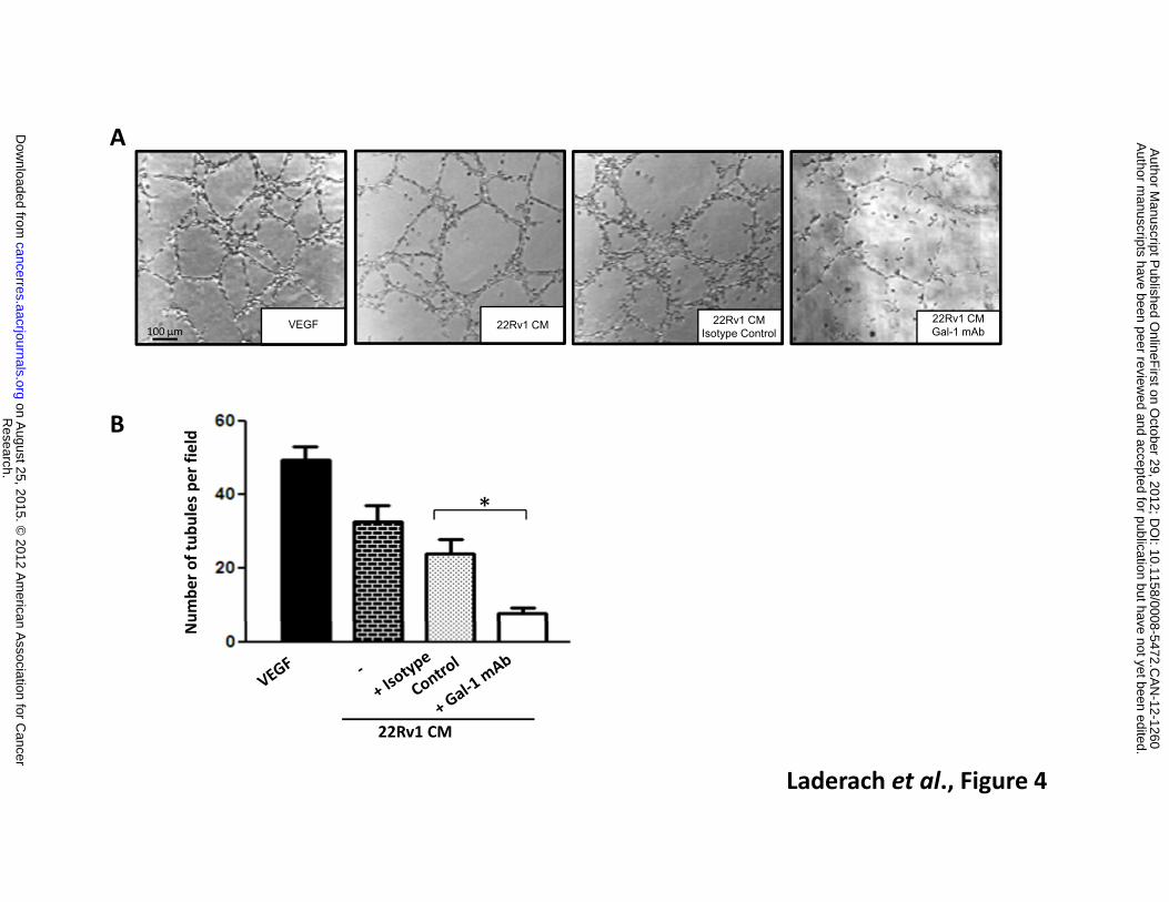
Given the promising value of anti-angiogenic therapies in castration-resistant advanced PCa
(32), we examined the role of Gal-1 in PCa angiogenesis. We evaluated the effect of conditioned
Research. on August 25, 2015. © 2012 American Association for Cancercancerres.aacrjournals.org Downloaded from
Author manuscripts have been peer reviewed and accepted for publication but have not yet been edited. Author Manuscript Published OnlineFirst on October 29, 2012; DOI: 10.1158/0008-5472.CAN-12-1260
15
medium (CM) obtained from a Gal-1-positive PCa cell line (22Rv1 CM) on in vitro BAEC tubulogenesis.
As shown in Figure 4, CM from 22Rv1 cells (Gal-1 concentration: 10.7 ng/ml) induced the formation of
tubular structures reflective of EC morphogenesis. To evaluate the involvement of Gal-1, we exposed
BAEC to CM from 22Rv1 PCa cells in the presence of an anti-Gal-1 neutralizing mAb (25,26).
Neutralization of soluble Gal-1 considerably reduced tube formation (3.76 ± 1.50 fold; n=10)
compared to BAEC exposed to PCa CM in the presence of a control isotype Ab (Figure 4A and B).
The in vitro effects of Gal-1-expressing PCa cells on EC morphogenesis prompted us to
investigate the role of this endogenous lectin in angiogenesis in vivo using two different approaches in
order to differentiate the source of Gal-1 (tumor and microenvironment vs. tumor alone). First, we
injected a Matrigel mixture containing 22Rv1 PCa cells and a blocking anti-Gal-1 mAb (or its isotype
control) into nude mice. Second, we used 22Rv1 tumor cells that were transduced with a human
specific Gal-1 shRNA-coding lentivirus (Gal-1-shRNA-LV) purified to homogeneity by cell sorting. Non-
sorted bulk transduced 22Rv1 cells, with partial down-regulation of Gal-1, were also tested (Figure
5A). A marked reduction of microvessel density was observed using both experimental approaches
(anti-Gal-1 mAb and Gal-1 shRNA-LV), indicating that tumor cells were the main source of Gal-1, at
least at early time points of tumor implantation (Figure 5B and C). Intermediate effects were observed
when Gal-1 was partially down-regulated in PCa cells (Figure 5B and C). Altogether, our results reveal
a key role for Gal-1 as a mediator of PCa-induced angiogenesis.
As angiogenesis relies on the expression of hypoxia-regulated genes and Gal-1 is regulated by
hypoxia in different tumor types (26,33,34), we then examined the effects of hypoxia on the galectin
expression profile of human PCa. For this purpose, 22Rv1 PCa cells were cultured under hypoxic or
normoxic conditions and the galectin transcriptional profile was evaluated. We could observe no
significant modification of the galectin expression profile except for Gal-1 that was modestly up-
regulated in response to hypoxia (1.3-fold, p=0.015) (data not shown). To further understand the
Research. on August 25, 2015. © 2012 American Association for Cancercancerres.aacrjournals.org Downloaded from
Author manuscripts have been peer reviewed and accepted for publication but have not yet been edited. Author Manuscript Published OnlineFirst on October 29, 2012; DOI: 10.1158/0008-5472.CAN-12-1260

16
molecular mechanisms underlying Gal-1-mediating angiogenesis, we screened different molecules
classically associated with angiogenesis (bFGF, VEGF-A, CD142, uPA, CXCR4, PDGF-AA and MMP-9 as
activators of angiogenesis; TSP-1, TIMP-1, CXCL10 and SPP1 as inhibitors of angiogenesis; and CA-IX as
a marker of hypoxia) in Gal-1-silenced human PCa tumors. RNA was purified from in vivo plugs
generated by injection of Gal-1-sufficient and Gal-1-silenced human 22Rv1 cells into nude mice and
angiogenesis-related genes were determined by real time RT-PCR. Silencing Gal-1 in PCa cells growing
in Matrigel plugs in vivo did not alter expression of angiogenesis-related genes neither in the tumor
itself (human) nor in the tumor microenvironment (mouse) (Figure 6). These results suggest that Gal-
1-induced PCa angiogenesis is independent of the up-regulation or down-regulation of classical pro-
angiogenic or anti-angiogenic factors and places Gal-1 as a critical mediator of tumor angiogenesis.
Research. on August 25, 2015. © 2012 American Association for Cancercancerres.aacrjournals.org Downloaded from
Author manuscripts have been peer reviewed and accepted for publication but have not yet been edited. Author Manuscript Published OnlineFirst on October 29, 2012; DOI: 10.1158/0008-5472.CAN-12-1260
17
DISCUSSION
Prostate cancer is no longer viewed as a disease of abnormally proliferating epithelial cells,
but rather as a disease involving complex interactions between prostate cancer epithelial cells and
the tumor microenvironment. Multiple signaling pathways and biological events mediate tumor
growth, including androgen receptor signaling, tyrosine kinase receptor signaling, angiogenesis
and tumor-immune escape (35). Interactions between multivalent lectins and glycans participate
in this complex network by modulating stromal, endothelial and immune cell compartments (6, 7).
Although original assumptions based on conserved carbohydrate specificity and structural
homology suggested that galectins may have redundant functions, recent information challenged
this view demonstrating specific roles for each member of the galectin family in the regulation of
tumor cell invasiveness, inflammation and angiogenesis (7). In search for novel biomarkers and
therapeutic targets, here we identified a ‘galectin-specific signature’ associated with PCa
progression. Galectin expression was profiled in PCa cell lines with diverse androgen-dependence
properties and AR expression and in human primary tumors obtained from treatment-free
patients at different stages of the disease.
Originally described as Prostate Carcinoma Tumor Antigen-1 (PCTA-1), Gal-8 has been
reported to be ubiquitously expressed in several tissues, but is up-regulated in PCa (29). Our
results confirm that this ´tandem-repeat’ galectin is expressed by PCa cell lines and primary
tumors, but indicate that the degree of Gal-8 expression is comparable in all tumor stages. Given
the complexity of Gal-8 isoforms, the variability of its intracellular localization and its ubiquitous
expression pattern, this galectin is likely to be an important component of PCa biology (29),
including modulation of cell proliferation, adhesion and angiogenesis (22, 36).
Interestingly, our results reveal that Gal-3, -4, -9 and -12 are down-modulated in advanced
stage primary tumors. These data are consistent with earlier reports showing that Gal-3
Research. on August 25, 2015. © 2012 American Association for Cancercancerres.aacrjournals.org Downloaded from
Author manuscripts have been peer reviewed and accepted for publication but have not yet been edited. Author Manuscript Published OnlineFirst on October 29, 2012; DOI: 10.1158/0008-5472.CAN-12-1260
18
expression decreases during tumor growth (10, 18) through mechanisms including promoter
methylation (19) and metalloproteinase-mediated protein cleavage (20). Depending on its
selective intracellular or extracellular localization, different biological properties have been
assigned to Gal-3, resulting in a dual pro- or anti-tumorigenic effect (10, 37). Our results suggest
that down-regulation of Gal-3, combined with the expression of other galectin members, is a
hallmark of PCa progression. More importantly, our findings demonstrate that Gal-1 is the most
abundantly expressed galectin in PCa and its expression correlates with disease severity,
underscoring the relevance of this endogenous lectin as a possible biomarker and therapeutic
target in high grade castration-refractory PCa.
Previous studies aimed at delineating the galectin transcriptional profile of a panel of
human tumor cell lines revealed that all PCa cell lines analyzed (DU145, PC-3 and LNCaP) were
negative for Gal-2, Gal-4, Gal-7 and Gal-9, but expressed considerable amounts of Gal-8 (38).
Moreover, Gal-1 and Gal-3 were expressed in DU.145 and PC-3, but not in the LNCaP cell line (38).
In contrast to these findings, we detected significant expression of Gal-1 in LNCaP cells both at the
mRNA and protein levels, although at 20-fold lower levels than the androgen unresponsive 22Rv1
and PC-3 tumor cells. Moreover, Gal-1 expression augmented when LNCaP cells were cultured for
several weeks in the absence of hormones (LNCaP-CR) (Figure S1A). As castration-sensitive or
resistant LNCaP cells were both PSA-positive and responsive to an androgen receptor agonist
(R1881) (Figure S2B), the discrepancies in Gal-1 expression among different studies might reflect
different culture conditions, cell line sources or selection protocols. In this regard, our studies
were performed on PCa cells isolated during the log phase of growth and the results were
substantiated using primary tumors isolated at different stages of the disease.
Given the pleiotropic functions of Gal-1 in the tumor microenvironment, including its role
in angiogenesis (26,31,39,40), cell adhesion and invasiveness (16,30) and immunosuppression (23-
Research. on August 25, 2015. © 2012 American Association for Cancercancerres.aacrjournals.org Downloaded from
Author manuscripts have been peer reviewed and accepted for publication but have not yet been edited. Author Manuscript Published OnlineFirst on October 29, 2012; DOI: 10.1158/0008-5472.CAN-12-1260

19
25, 41), up-regulation of Gal-1 may dramatically influence PCa progression. In this regard, Gal-1 is
expressed in ECs (42-44) and is up-regulated in various cancer types (6). Here, we demonstrate
that Gal-1 is the most highly expressed and regulated galectin in the PCa microenvironment and
plays essential roles in PCa angiogenesis. The role of Gal-1 in angiogenesis seems to be tissue-
specific as Gal-1 expression correlates with EC markers in advanced PCa but not in human breast
cancer. These findings are consistent with the ability of Gal-1 to induce angiogenesis in
oligodendroglioma (30), B16 melanoma (31), and Kaposi’s sarcoma (26), but not in other tumor
types such as Lewis lung carcinoma (41).
Selective silencing strategies in tumors clearly demonstrated that the main cellular source
of Gal-1 is represented by tumor cells. However, mechanisms by which ECs capture Gal-1 from the
tumor microenvironment or tumor-induced EC activation up-regulates Gal-1 expression have also
been described (31,39). Moreover, as Gal-3 and Gal-8 also contribute to angiogenesis in other
tumor types, the spatiotemporal regulation of distinct members of the galectin family might
ultimately dictate the vascularization phenotype (36,45, 46,47). Finally, Gal-1 silencing in PCa cells
did not alter the expression of classical pro-angiogenic or anti-angiogenic mediators neither in
tumor cells nor in the tumor microenvironment, highlighting a direct and critical role of this lectin
in PCa angiogenesis.
In summary, our findings identify a distinctive ‘galectin signature’, which delineates tumor
progression in human PCa and highlight a major role of Gal-1 as a novel target of anti-angiogenic
therapies in advanced castration-resistant stages of PCa, where effective treatments are still
lacking.
Research. on August 25, 2015. © 2012 American Association for Cancercancerres.aacrjournals.org Downloaded from
Author manuscripts have been peer reviewed and accepted for publication but have not yet been edited. Author Manuscript Published OnlineFirst on October 29, 2012; DOI: 10.1158/0008-5472.CAN-12-1260

20
ACKNOWLEDGEMENTS
We thank Drs. Karim Fizazi and Catherine Gaudin (INSERM U981; IGR-France), Geraldine Gueron
(University of Buenos Aires) and Carla Saleh (Pasteur Institute, France) for help and advice.
GRANT SUPPORT
Supported by grants from Prostate Action (UK) to G.A.R, D.J.L. and D.C, Agencia Nacional de
Promoción Científica y Técnica Argentina (ANPCyT; PICT 2008-134 to D.J.L.; PICT 2010-870 to
G.A.R.), Programa de Cooperación Franco-Argentino ECOS-Sud (A10S03 to G.A.R., A.C., D.J.L and
D.C.), Fundación Sales to G.A.R, University of Buenos Aires to G.A.R., and Association pour la
Recherche sur les Tumeurs de la Prostate -ARTP, France- to D.C.
AUTHORS’ CONTRIBUTIONS
Acquisition, analysis and interpretation of data: D.J.L.; L.D.G; L.G.; V.C.D.; L.N.; N.A.N.; J.L.K; S.J.K.;
M.T.E and D.C.
Material support: D.C.; P.S.; G.C.; O.M.; M.S.; E.V. and A.C.
Development of methodology: D.C.; D.O.C.
Conception; design and supervision of the study: D.J.L; D.C.; G.A.R.
Writing of the paper: D.J.L.; D.C.; G.A.R.
Research. on August 25, 2015. © 2012 American Association for Cancercancerres.aacrjournals.org Downloaded from
Author manuscripts have been peer reviewed and accepted for publication but have not yet been edited. Author Manuscript Published OnlineFirst on October 29, 2012; DOI: 10.1158/0008-5472.CAN-12-1260
21
REFERENCES 1. Jemal A, Bray F, Center MM, Ferlay J, Ward E, and Forman D. Global cancer statistics. CA
Cancer J Clin 2011; 61: 69-90. 2. Logothetis CJ, Navone NM, and Lin SH. Understanding the biology of bone metastases: key to
the effective treatment of prostate cancer. Clin Cancer Res 2008; 14: 1599-602. 3. Denmeade SR and Isaacs JT. A history of prostate cancer treatment. Nat Rev Cancer 2002; 2:
389-96. 4. Rillahan CD and Paulson JC. Glycan microarrays for decoding the glycome. Annu Rev Biochem
2011; 80: 797-23. 5. Dube DH and Bertozzi CR. Glycans in cancer and inflammation--potential for therapeutics and
diagnostics. Nat Rev Drug Discov. 2005; 4:477-88. 6. Liu FT and Rabinovich GA. Galectins as modulators of tumour progression. Nat Rev Cancer
2005; 5: 29-41. 7. Rabinovich GA and Croci DO. Regulatory circuits mediated by lectin-glycan interactions in
autoimmunity and cancer. Immunity 2012; 36: 322-35. 8. Levy R, Biran A, Poirier F, Raz A, and Kloog Y. Galectin-3 mediates cross-talk between K-Ras
and Let-7c tumor suppressor microRNA. PLoS One 2011; 6: e27490. 9. Ellerhorst J, Troncoso P, Xu XC, Lee J, and Lotan R. Galectin-1 and galectin-3 expression in
human prostate tissue and prostate cancer. Urol Res 1999; 27: 362-67. 10. van den Brule FA, Waltregny D, Liu FT, and Castronovo V. Alteration of the
cytoplasmic/nuclear expression pattern of galectin-3 correlates with prostate carcinoma progression. Int J Cancer 2000; 89: 361-67.
11. van den Brule FA, Waltregny D, and Castronovo V. Increased expression of galectin-1 in carcinoma-associated stroma predicts poor outcome in prostate carcinoma patients. J Pathol 2001; 193: 80-87.
12. Valenzuela HF, Pace KE, Cabrera PV, White R, Porvari K, Kaija H, et al. O-glycosylation regulates LNCaP prostate cancer cell susceptibility to apoptosis induced by galectin-1. Cancer Res 2007; 67: 6155-62.
13. He J and Baum LG. Endothelial cell expression of galectin-1 induced by prostate cancer cells inhibits T-cell transendothelial migration. Lab Invest 2006; 86: 578-90.
14. Ellerhorst JA, Stephens LC, Nguyen T, and Xu XC. Effects of galectin-3 expression on growth and tumorigenicity of the prostate cancer cell line LNCaP. Prostate 2002; 50: 64-70.
15. Glinsky VV, Glinsky GV, Glinskii OV, Huxley VH, Turk JR, Mossine VV, et al. Intravascular metastatic cancer cell homotypic aggregation at the sites of primary attachment to the endothelium. Cancer Res 2003; 63: 3805-11.
16. Clausse N, van den Brule F, Waltregny D, Garnier F, and Castronovo V. Galectin-1 expression in prostate tumor-associated capillary endothelial cells is increased by prostate carcinoma cells and modulates heterotypic cell-cell adhesion. Angiogenesis 1999; 3: 317-25.
17. Fukumori T, Oka N, Takenaka Y, Nangia-Makker P, Elsamman E, Kasai T, et al. Galectin-3 regulates mitochondrial stability and antiapoptotic function in response to anticancer drug in prostate cancer. Cancer Res 2006; 66: 3114-19.
18. Merseburger AS, Kramer MW, Hennenlotter J, Simon P, Knapp J, Hartmann JT, et al. Involvement of decreased Galectin-3 expression in the pathogenesis and progression of prostate cancer. Prostate 2008; 68: 72-77.
19. Ahmed H, Banerjee PP, and Vasta GR. Differential expression of galectins in normal, benign and malignant prostate epithelial cells: silencing of galectin-3 expression in prostate cancer by its promoter methylation. Biochem Biophys Res Commun 2007; 358: 241-46.
Research. on August 25, 2015. © 2012 American Association for Cancercancerres.aacrjournals.org Downloaded from
Author manuscripts have been peer reviewed and accepted for publication but have not yet been edited. Author Manuscript Published OnlineFirst on October 29, 2012; DOI: 10.1158/0008-5472.CAN-12-1260
22
20. Wang Y, Nangia-Makker P, Tait L, Balan V, Hogan V, Pienta KJ, et al. Regulation of prostate cancer progression by galectin-3. Am J Pathol 2009; 174: 1515-23.
21. Su ZZ, Lin J, Shen R, Fisher PE, Goldstein NI, and Fisher PB. Surface-epitope masking and expression cloning identifies the human prostate carcinoma tumor antigen gene PCTA-1 a member of the galectin gene family. Proc Natl Acad Sci U S A 1996; 93: 7252-57.
22. Zick Y, Eisenstein M, Goren RA, Hadari YR, Levy Y, and Ronen D. Role of galectin-8 as a modulator of cell adhesion and cell growth. Glycoconj J 2004; 19: 517-26.
23. Rubinstein N, Alvarez M, Zwirner NW, Toscano MA, Ilarregui JM, Bravo A, et al. Targeted inhibition of galectin-1 gene expression in tumor cells results in heightened T cell-mediated rejection; A potential mechanism of tumor-immune privilege. Cancer Cell 2004; 5: 241-51.
24. Juszczynski P, Ouyang J, Monti S, Rodig SJ, Takeyama K, Abramson J, et al. The AP1-dependent secretion of galectin-1 by Reed Sternberg cells fosters immune privilege in classical Hodgkin lymphoma. Proc Natl Acad Sci U S A 2007; 104: 13134-39.
25. Ouyang J, Juszczynski P, Rodig SJ, Green MR, O'Donnell E, Currie T, et al. Viral induction and targeted inhibition of galectin-1 in EBV+ posttransplant lymphoproliferative disorders. Blood 2011; 117: 4315-22.
26. Croci DO, Salatino M, Rubinstein N, Cerliani JP, Cavallin LE, Leung HJ, et al. Disrupting galectin-1 interactions with N-glycans suppresses hypoxia-driven angiogenesis and tumorigenesis in Kaposi’s sarcoma. J Exp Med 2012; in press.
27. Compagno D, Merle C, Morin A, Gilbert C, Mathieu JR, Bozec A, et al. SIRNA-directed in vivo silencing of androgen receptor inhibits the growth of castration-resistant prostate carcinomas. PLoS One 2007; 2: e1006.
28. Wiznerowicz M and Trono D. Conditional suppression of cellular genes: lentivirus vector-mediated drug-inducible RNA interference. J Virol. 2003; 77: 8957-61.
29. Gopalkrishnan RV, Roberts T, Tuli S, Kang D, Christiansen KA, and Fisher PB. Molecular characterization of prostate carcinoma tumor antigen-1, PCTA-1, a human galectin-8 related gene. Oncogene 2000; 19: 4405-16.
30. Le Mercier M, Fortin S, Mathieu V, Roland I, Spiegl-Kreinecker S, Haibe-Kains B, et al. Galectin 1 proangiogenic and promigratory effects in the Hs683 oligodendroglioma model are partly mediated through the control of BEX2 expression. Neoplasia 2009; 11: 485-96.
31. Thijssen VL, Barkan B, Shoji H, Aries IM, Mathieu V, Deltour L, et al. Tumor cells secrete galectin-1 to enhance endothelial cell activity. Cancer Res 2010; 70: 6216-24.
32. Karlou M, Tzelepi V, and Efstathiou E. Therapeutic targeting of the prostate cancer microenvironment. Nat Rev Urol 2010; 7: 494-509.
33. Le QT, Shi G, Cao H, Nelson DW, Wang Y, Chen EY, et al. Galectin-1: a link between tumor hypoxia and tumor immune privilege. J Clin Oncol 2005; 23: 8932-41.
34. Zhao XY, Chen TT, Xia L, Guo M, Xu Y, Yue F, et al. Hypoxia inducible factor-1 mediates expression of galectin-1: the potential role in migration/invasion of colorectal cancer cells. Carcinogenesis. 2010; 31: 1367-75.
35. Ramsay AK and Leung HY. Signalling pathways in prostate carcinogenesis: potentials for molecular-targeted therapy. Clin Sci (Lond) 2009; 117: 209-28.
36. Delgado VM, Nugnes LG, Colombo LL, Troncoso MF, Fernandez MM, Malchiodi EL, et al. Modulation of endothelial cell migration and angiogenesis: a novel function for the "tandem-repeat" lectin galectin-8. Faseb J 2011; 25: 242-54.
37. Califice S, Castronovo V, Bracke M, and van den Brule F. Dual activities of galectin-3 in human prostate cancer: tumor suppression of nuclear galectin-3 vs tumor promotion of cytoplasmic galectin-3. Oncogene 2004; 23: 7527-36.
Research. on August 25, 2015. © 2012 American Association for Cancercancerres.aacrjournals.org Downloaded from
Author manuscripts have been peer reviewed and accepted for publication but have not yet been edited. Author Manuscript Published OnlineFirst on October 29, 2012; DOI: 10.1158/0008-5472.CAN-12-1260

23
38. Lahm H, André S, Hoeflich A, Fischer JR, Sordat B, Kaltner H, et al. Comprehensive galectin fingerprinting in a panel of 61 human tumor cell lines by RT-PCR and its implications for diagnostic and therapeutic procedures. J Cancer Res Clin Oncol. 2001; 127:375-86.
39. Thijssen VL, Hulsmans S, and Griffioen AW. The galectin profile of the endothelium: altered expression and localization in activated and tumor endothelial cells. Am J Pathol 2008; 172: 545-53.
40. Baum LG, Seilhamer JJ, Pang M, Levine WB, Beynon D, and Berliner J. A. Synthesis of an endogeneous lectin, galectin-1, by human endothelial cells is up-regulated by endothelial cell activation. Glycoconj J 1995; 12: 63-8.
41. Saussez S, Camby I, Toubeau G, and Kiss R. Galectins as modulators of tumor progression in head and neck squamous cell carcinomas. Head Neck 2007; 29: 874-84.
42. Lotan R, Belloni PN, Tressler RJ, Lotan D, Xu XC, and Nicolson GL. Expression of galectins on microvessel endothelial cells and their involvement in tumour cell adhesion. Glycoconj J 1994; 11: 462-68.
43. Hsieh SH, Ying NW, Wu MH, Chiang WF, Hsu CL, Wong TY, et al. Galectin-1, a novel ligand of neuropilin-1, activates VEGFR-2 signaling and modulates the migration of vascular endothelial cells. Oncogene 2008; 27: 3746-53.
44. Banh A, Zhang J, Cao H, Bouley DM, Kwok S, Kong C, et al. Tumor galectin-1 mediates tumor growth and metastasis through regulation of T-cell apoptosis. Cancer Res 2011; 71: 4423-31.
45. Thijssen VL, Postel R, Brandwijk RJ, Dings RP, Nesmelova I, Satijn S, et al. Galectin-1 is essential in tumor angiogenesis and is a target for antiangiogenesis therapy. Proc Natl Acad Sci U S A 2006; 103: 15975-80.
46. Nangia-Makker P, Honjo Y, Sarvis R, Akahani S, Hogan V, Pienta KJ, et al. Galectin-3 induces endothelial cell morphogenesis and angiogenesis. Am J Pathol 2000; 156: 899-09.
47. Markowska AI, Liu FT, and Panjwani N. Galectin-3 is an important mediator of VEGF- and bFGF-mediated angiogenic response. J Exp Med 2010; 207: 1981-93.
Research. on August 25, 2015. © 2012 American Association for Cancercancerres.aacrjournals.org Downloaded from
Author manuscripts have been peer reviewed and accepted for publication but have not yet been edited. Author Manuscript Published OnlineFirst on October 29, 2012; DOI: 10.1158/0008-5472.CAN-12-1260
24
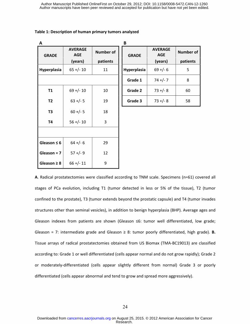
Table 1: Description of human primary tumors analyzed A B
GRADE AVERAGE
AGE Number of
GRADE AVERAGE
AGE Number of
(years) patients (years) patients
Hyperplasia 65 +/- 10 11 Hyperplasia 69 +/- 6 5
Grade 1 74 +/- 7 8
T1 69 +/- 10 10 Grade 2 73 +/- 8 60
T2 63 +/- 5 19 Grade 3 73 +/- 8 58
T3 60 +/- 5 18
T4 56 +/- 10 3
Gleason ≤ 6 64 +/- 6 29
Gleason = 7 57 +/- 9 12
Gleason ≥ 8 66 +/- 11 9 A. Radical prostatectomies were classified according to TNM scale. Specimens (n=61) covered all
stages of PCa evolution, including T1 (tumor detected in less or 5% of the tissue), T2 (tumor
confined to the prostate), T3 (tumor extends beyond the prostatic capsule) and T4 (tumor invades
structures other than seminal vesicles), in addition to benign hyperplasia (BHP). Average ages and
Gleason indexes from patients are shown (Gleason ≤6: tumor well differentiated, low grade;
Gleason = 7: intermediate grade and Gleason ≥ 8: tumor poorly differentiated, high grade). B.
Tissue arrays of radical prostatectomies obtained from US Biomax (TMA-BC19013) are classified
according to: Grade 1 or well differentiated (cells appear normal and do not grow rapidly); Grade 2
or moderately-differentiated (cells appear slightly different from normal) Grade 3 or poorly
differentiated (cells appear abnormal and tend to grow and spread more aggressively).
Research. on August 25, 2015. © 2012 American Association for Cancercancerres.aacrjournals.org Downloaded from
Author manuscripts have been peer reviewed and accepted for publication but have not yet been edited. Author Manuscript Published OnlineFirst on October 29, 2012; DOI: 10.1158/0008-5472.CAN-12-1260
25
FIGURE LEGENDS
Figure 1: Galectin expression profile in human PCa cell lines. A. Transcriptional profile of galectins
by real-time RT-PCR. Results are expressed as galectin mRNA relative to Cyclophilin A. Cell lines are
presented according to androgen sensitivity and androgen receptor (AR) expression. Left, LNCaP
cells (hormone responsive HR, AR+); Middle, 22Rv1 cells (castration resistant CR, AR+); Right, the
more aggressive PC-3 cells (CR and AR-). Data are expressed as mean + SD of four independent
experiments. B. Immunocytochemical analysis of galectins in PCa cells adhered onto poly-L-Lysine-
coated glasses (Magnification X100).
Figure 2: Galectin expression profile of human primary prostate tumors. A. Radical
prostatectomies from naive patients (n=50 carcinomas and n=11 benign hyperplasia) were
arranged according to T grade (TNM classification; UICC). Galectin expression was analyzed by
immunohistochemistry in paraffin-embedded tissue sections from patients (Table 1).
Magnification: 400 X. B. Galectin expression in patient samples was graded as follows: 0= negative;
1=low intensity; 2=moderate intensity; 3=high intensity and 4=very high intensity. Results are
representative (A) or are the mean ± S.D. (B) of four independent experiments. * P <0.05
(Student’s t test), n.s = non-statistically different.
Figure 3: Gal-1 expression positively correlates with the number of CD34+ blood vessels in
advanced human prostate cancer. Expression of Gal-1 and CD34 was evaluated in an invasive PCa
tissue-microarray (slides containing 29 paired cores: BC19013, US Biomax) by
immunohistochemistry. Stained slides were digitally scanned using Aperio ScanScope XT
Research. on August 25, 2015. © 2012 American Association for Cancercancerres.aacrjournals.org Downloaded from
Author manuscripts have been peer reviewed and accepted for publication but have not yet been edited. Author Manuscript Published OnlineFirst on October 29, 2012; DOI: 10.1158/0008-5472.CAN-12-1260
26
workstation and evaluated by ImageScope software. A. Correlation between Gal-1 and CD34
expression in tumor areas of grade III PCa (classification based on grade of proliferation and cell
morphology as described in Materials and methods). Breast cancer tissue was analyzed for
comparison purposes. B. Examples of PCa samples with intense or low Gal-1 and CD34 expression.
Results are representative of three independent experiments. * P <0.05 (Spearman correlation
test).
Figure 4: Prostate cancer-derived Gal-1 promotes EC morphogenesis. A. In vitro formation of
tubular structures by BAEC cultured in Matrigel with 22Rv1-conditioned media (22Rv1 CM) in the
absence or presence of an anti-Gal-1 mAb (F8.G7) or isotype control mAb. Recombinant VEGF was
used as a positive control. B. Quantification of the number of tubular structures per field. Results
are representative (A) or are the mean ± S.D. (B) of four independent experiments. * P <0.001
(Student’s t test).
Figure 5: Prostate cancer-derived Gal-1 promotes angiogenesis in vivo. Nude mice were injected
with 5 x 10⁶ 22Rv1 PCa cells incorporated in Matrigel plugs. The role of Gal-1 was assessed by two
different strategies: a) using 22Rv1 cells transduced with Gal-1 shRNA-coding lentivirus (bulk; n=3
or sorted cells; n=8 with ∼40 and 80% Gal-1 down-regulation respectively) compared to control
shRNA-LV cells (n=8); or b) by adding an anti-Gal-1 mAb (n=3) or isotype control (n=3). A.
Immunoblot of Gal-1 in 22Rv1 cells. First lane: cells transduced with a control shRNA-LV and sorted
according to GFP expression; second lane: Gal-1 shRNA-LV (bulk); third lane: cells transduced with
a Gal-1 shRNA-LV and sorted. B. Representative photographs of in vivo plugs at day 5. C.
Hemoglobin content in plugs at day 5, normalized to µg of protein. Results are representative (A,B)
or are the mean ± S.D. (C) of four independent experiments.** P <0.005 (Student’s t test).
Research. on August 25, 2015. © 2012 American Association for Cancercancerres.aacrjournals.org Downloaded from
Author manuscripts have been peer reviewed and accepted for publication but have not yet been edited. Author Manuscript Published OnlineFirst on October 29, 2012; DOI: 10.1158/0008-5472.CAN-12-1260

27
Figure 6: Gal-1 silencing does not alter expression of angiogenesis-related genes in PCa. Total
RNA was extracted from plugs generated in vivo by injection of Gal-1-sufficient (n=5) and Gal-1-
silenced (n=5) human 22Rv1 cells into nude mice. Angiogenesis-related genes, including activators
of angiogenesis (bFGF, VEGF-A, CD142, uPA, CXCR4, PDGF-AA and MMP-9), inhibitors of
angiogenesis (TSP-1, TIMP-1, CXCL10 and SPP1) and a marker of hypoxia (CA-IX ) were screened by
real time RT-PCR adapted to a gene array platform. This screening allows the distinction of human
genes (derived from the tumor) and mouse genes (reflecting the mouse microenvironment).
Lower panel includes genes derived from both tumor and microenvironment, as these primers
react with both human and mouse genes. Data show individual samples analyzed and the mean of
individual plugs. *P<0.05 (Student’s t test).
Research. on August 25, 2015. © 2012 American Association for Cancercancerres.aacrjournals.org Downloaded from
Author manuscripts have been peer reviewed and accepted for publication but have not yet been edited. Author Manuscript Published OnlineFirst on October 29, 2012; DOI: 10.1158/0008-5472.CAN-12-1260
As
Cycl
o A
e G
al-1
mRN
A v
s
LNCaP 22Rv1 PC 3
Rela
tive
Pre-immunesera
LNCaP 22Rv1 PC-3HR, AR+ CR, AR+ CR, AR-
B
Gal-1
Gal-3
Gal-8
Gal-9
Gal-12 Laderach et al., Figure 150 μm
Research.
on August 25, 2015. ©
2012 Am
erican Association for C
ancercancerres.aacrjournals.org
Dow
nloaded from
Author m
anuscripts have been peer reviewed and accepted for publication but have not yet been edited.
Author M
anuscript Published O
nlineFirst on O
ctober 29, 2012; DO
I: 10.1158/0008-5472.CA
N-12-1260
ABHP
Gal-1 Gal-8 Gal-3 Gal-4 Gal-9 Gal-12
T1b
T2aT2a
T3a
T3b
T3c
G l 1 G l 8 G l 3 G l 4 G l 9 G l 12
T4100 μm
B
Gal
ecti
n es
sion
(AU
)
Gal-1 Gal-8 Gal-3 Gal-4 Gal-9 Gal-12* * * * *ns
GEx
pre
Tumor Progression (TNM Classification)Laderach et al., Figure 2
Research.
on August 25, 2015. ©
2012 Am
erican Association for C
ancercancerres.aacrjournals.org
Dow
nloaded from
Author m
anuscripts have been peer reviewed and accepted for publication but have not yet been edited.
Author M
anuscript Published O
nlineFirst on O
ctober 29, 2012; DO
I: 10.1158/0008-5472.CA
N-12-1260
A Prostate cancerGrade III
Breast cancerG
al-1
(OD
)
B
CD34 (%)
B
Gal-1 CD34 Gal-1 CD34
100 μm
Laderach et al., Figure 3
Research.
on August 25, 2015. ©
2012 Am
erican Association for C
ancercancerres.aacrjournals.org
Dow
nloaded from
Author m
anuscripts have been peer reviewed and accepted for publication but have not yet been edited.
Author M
anuscript Published O
nlineFirst on O
ctober 29, 2012; DO
I: 10.1158/0008-5472.CA
N-12-1260
A
22Rv1 CM 22Rv1 CMIsotype Control
VEGF 22Rv1 CMGal-1 mAb100 μm
B
*es p
er fi
eld
umbe
r of
tub
ule
22Rv1 CM
N
Laderach et al., Figure 4
22Rv1 CM
Research.
on August 25, 2015. ©
2012 Am
erican Association for C
ancercancerres.aacrjournals.org
Dow
nloaded from
Author m
anuscripts have been peer reviewed and accepted for publication but have not yet been edited.
Author M
anuscript Published O
nlineFirst on O
ctober 29, 2012; DO
I: 10.1158/0008-5472.CA
N-12-1260

A C
Gal 1 µg p
rote
in) **
Gal-1
β-tubulin
Hem
oglo
bin
(g/µ **
B
H
Isotype mAbcontrol shRNA-LV
Gal-1 shRNA-LV (bulk)
Gal-1 shRNA-LV (sorted)
Gal-1 mAb
(sorted)
Laderach et al., Figure 5
Research.
on August 25, 2015. ©
2012 Am
erican Association for C
ancercancerres.aacrjournals.org
Dow
nloaded from
Author m
anuscripts have been peer reviewed and accepted for publication but have not yet been edited.
Author M
anuscript Published O
nlineFirst on O
ctober 29, 2012; DO
I: 10.1158/0008-5472.CA
N-12-1260

Gal-1 TIMP-1 VEGF-A CD142 uPA CXCR4 CXCL10 PDGF-AA MMP-9 SPP1
T
sion
Gal 1 TIMP 1 VEGF A CD142 uPA CXCR4 CXCL10
shCONT shGAL-1 shCONT shGAL-1shCONT shGAL-1 shCONT shGAL-1 shCONT shGAL-1 shCONT shGAL-1 shCONT shGAL-1 shCONT shGAL-1 shCONT shGAL-1 shCONT shGAL-1
Tumor (human)
ive
Gen
e Ex
pres
shCONT shGAL-1
Gal-1 TIMP-1 VEGF-A CD142 uPA CXCR4 CXCL10
shCONT shGAL-1 shCONT shGAL-1 shCONT shGAL-1shCONT shGAL-1 shCONT shGAL-1 shCONT shGAL-1
Microenvironment(mouse)
Rela
ti
hCONT hGAL 1hCONT hGAL 1hCONT hGAL 1
bFGF TSP-1 CA-IXTumor and
microenvironment(human and mouse)
shCONT shGAL-1shCONT shGAL-1shCONT shGAL-1
Laderach et al., Figure 6
Research.
on August 25, 2015. ©
2012 Am
erican Association for C
ancercancerres.aacrjournals.org
Dow
nloaded from
Author m
anuscripts have been peer reviewed and accepted for publication but have not yet been edited.
Author M
anuscript Published O
nlineFirst on O
ctober 29, 2012; DO
I: 10.1158/0008-5472.CA
N-12-1260
Published OnlineFirst October 29, 2012.Cancer Res Diego J Laderach, Lucas Gentilini, Laura Giribaldi, et al. advanced diseaseprogression suggests galectin-1 as a key target for treatment of A unique galectin signature in human prostate cancer
Updated version
10.1158/0008-5472.CAN-12-1260doi:
Access the most recent version of this article at:
Material
Supplementary
http://cancerres.aacrjournals.org/content/suppl/2012/10/26/0008-5472.CAN-12-1260.DC1.html
Access the most recent supplemental material at:
Manuscript
Authoredited. Author manuscripts have been peer reviewed and accepted for publication but have not yet been
E-mail alerts related to this article or journal.Sign up to receive free email-alerts
Subscriptions
Reprints and
To order reprints of this article or to subscribe to the journal, contact the AACR Publications
Permissions
To request permission to re-use all or part of this article, contact the AACR Publications
Research. on August 25, 2015. © 2012 American Association for Cancercancerres.aacrjournals.org Downloaded from
Author manuscripts have been peer reviewed and accepted for publication but have not yet been edited. Author Manuscript Published OnlineFirst on October 29, 2012; DOI: 10.1158/0008-5472.CAN-12-1260