Novel mTORC1 Mechanism Suggests Therapeutic Targets for ...
15
MOLECULAR PATHOGENESIS OF GENETIC AND INHERITED DISEASES Novel mTORC1 Mechanism Suggests Therapeutic Targets for COMPopathies Karen L. Posey,* Francoise Coustry,* Alka C. Veerisetty,* Mohammad G. Hossain,* Michael J. Gambello, y and Jacqueline T. Hecht* z From the Department of Pediatrics,* McGovern Medical School, and the School of Dentistry, z University of Texas Health Science Center, Houston, Texas; and Human Genetics and Pediatrics, y Emory University School of Medicine, Atlanta, Georgia Accepted for publication September 17, 2018. Address correspondence to Karen L. Posey, Ph.D., Department of Pediatrics, McGovern Medical School at The University of Texas Health Science Center at Houston (UTHealth), 6431 Fannin St., Houston, TX 77030. E-mail: [email protected]. Cartilage oligomeric matrix protein (COMP) is a large, multifunctional extracellular protein that, when mutated, is retained in the rough endoplasmic reticulum (ER). This retention elicits ER stress, inflammation, and oxidative stress, resulting in dysfunction and death of growth plate chondrocytes. While identifying the cellular pathologic mechanisms underlying the murine mutant (MT)-COMP model of pseudoachondroplasia, increased midline-1 (MID1) expression and mammalian target of rapamycin complex 1 (mTORC1) signaling was found. This novel role for MID1/mTORC1 signaling was investigated since treatments shown to repress the pathology also reduced Mid1/mTORC1. Although ER stresse inducing drugs or tumor necrosis factor a (TNFa) in rat chondrosarcoma cells increased Mid1, oxidative stress did not, establishing that ER stresse or TNFa-driven inflammation alone is sufficient to elevate MID1 expression. Since MID1 ubiquitinates protein phosphatase 2A (PP2A), a negative regulator of mTORC1, PP2A was evaluated in MT-COMP growth plate chondrocytes. PP2A was decreased, indicating de-repression of mTORC1 signaling. Rapamycin treatment in MT-COMP mice reduced mTORC1 signaling and intracellular retention of COMP, and increased proliferation, but did not change inflammatory markers IL-16 and eosinophil peroxidase. Lastly, mRNA from tuberous sclerosise1/2-null mice brain tissue exhibiting ER stress had increased Mid1 expression, confirming the relationship between ER stress and MID1/mTORC1 signaling. These findings suggest a mechanistic link between ER stress and MID1/ mTORC1 signaling that has implications extending to other conditions involving ER stress. (Am J Pathol 2019, 189: 132e146; https://doi.org/10.1016/j.ajpath.2018.09.008) Cartilage oligomeric matrix protein (COMP) is a large, pentameric extracellular matrix protein that is a member of the thrombospondin gene family. These proteins have adhesive properties that are known to mediate cellecell and cellematrix interactions. 1e7 Reported functions of COMP include: i) production, maintenance, and homeostasis of cartilage, ii) regulation of type II collagen fibril assembly, and iii) enhancement of chondrocyte attachment and proliferation. 8e14 Interestingly, more is known about the results of mutant (MT)-COMP in growth plate chondrocytes than the functions of wild-type COMP. Mutations in COMP cause pseudoachondroplasia (PSACH), a severe dwarfing condition characterized by disproportionate short stature, joint laxity, pain, and early-onset osteoarthritis. 2,10,15e36 PSACH is not evident at birth, being identified by 2 years of age, when slow linear growth leads to diagnosis. The hallmark of PSACH growth plate chondrocytes is massive retention of MT-COMP in rough endoplasmic reticulum (ER) cisternae. 20 MT-COMP prematurely assembles into an ordered matrix composed of types II and IX collagen and matrilin-3 and other extracellular matrix proteins, resulting in intracellular protein accumulation. 30,37,38 Approximately 97% of COMP pentamers are predicted to contain at least Supported by the National Institute of Arthritis and Musculoskeletal and Skin Diseases, NIH grant RO1-AR057117-05 (J.T.H. and K.L.P.); and the Leah Lewis Family Foundation (J.T.H.). Disclosures: None declared. The content is solely the responsibility of the authors and does not necessarily represent the official views of the US National Institutes of Health. Copyright ª 2019 American Society for Investigative Pathology. Published by Elsevier Inc. All rights reserved. https://doi.org/10.1016/j.ajpath.2018.09.008 ajp.amjpathol.org The American Journal of Pathology, Vol. 189, No. 1, January 2019
-
Upload
khangminh22 -
Category
Documents
-
view
1 -
download
0
Transcript of Novel mTORC1 Mechanism Suggests Therapeutic Targets for ...
The American Journal of Pathology, Vol. 189, No. 1, January 2019
ajp.amjpathol.org
MOLECULAR PATHOGENESIS OF GENETIC AND INHERITED DISEASES
Novel mTORC1 Mechanism Suggests
Therapeutic Targets for COMPopathies Karen L. Posey,* Francoise Coustry,* Alka C. Veerisetty,* Mohammad G. Hossain,* Michael J. Gambello,y andJacqueline T. Hecht*zFrom the Department of Pediatrics,* McGovern Medical School, and the School of Dentistry,z University of Texas Health Science Center, Houston, Texas; andHuman Genetics and Pediatrics,y Emory University School of Medicine, Atlanta, Georgia
Accepted for publication
C
h
September 17, 2018.
Address correspondence toKaren L. Posey, Ph.D.,Department of Pediatrics,McGovern Medical School atThe University of Texas HealthScience Center at Houston(UTHealth), 6431 Fannin St.,Houston, TX 77030. E-mail:[email protected].
opyright ª 2019 American Society for Inve
ttps://doi.org/10.1016/j.ajpath.2018.09.008
Cartilage oligomeric matrix protein (COMP) is a large, multifunctional extracellular protein that, whenmutated, is retained in the rough endoplasmic reticulum (ER). This retention elicits ER stress,inflammation, and oxidative stress, resulting in dysfunction and death of growth plate chondrocytes.While identifying the cellular pathologic mechanisms underlying the murine mutant (MT)-COMP modelof pseudoachondroplasia, increased midline-1 (MID1) expression and mammalian target of rapamycincomplex 1 (mTORC1) signaling was found. This novel role for MID1/mTORC1 signaling was investigatedsince treatments shown to repress the pathology also reduced Mid1/mTORC1. Although ER stresseinducing drugs or tumor necrosis factor a (TNFa) in rat chondrosarcoma cells increased Mid1, oxidativestress did not, establishing that ER stresse or TNFa-driven inflammation alone is sufficient to elevateMID1 expression. Since MID1 ubiquitinates protein phosphatase 2A (PP2A), a negative regulator ofmTORC1, PP2A was evaluated in MT-COMP growth plate chondrocytes. PP2A was decreased, indicatingde-repression of mTORC1 signaling. Rapamycin treatment in MT-COMP mice reduced mTORC1 signalingand intracellular retention of COMP, and increased proliferation, but did not change inflammatorymarkers IL-16 and eosinophil peroxidase. Lastly, mRNA from tuberous sclerosise1/2-null mice braintissue exhibiting ER stress had increased Mid1 expression, confirming the relationship between ER stressand MID1/mTORC1 signaling. These findings suggest a mechanistic link between ER stress and MID1/mTORC1 signaling that has implications extending to other conditions involving ER stress. (Am J Pathol2019, 189: 132e146; https://doi.org/10.1016/j.ajpath.2018.09.008)
Supported by the National Institute of Arthritis and Musculoskeletal andSkin Diseases, NIH grant RO1-AR057117-05 (J.T.H. and K.L.P.); and theLeah Lewis Family Foundation (J.T.H.).Disclosures: None declared.The content is solely the responsibility of the authors and does not
necessarily represent the official views of the US National Institutes ofHealth.
Cartilage oligomeric matrix protein (COMP) is a large,pentameric extracellular matrix protein that is a member ofthe thrombospondin gene family. These proteins haveadhesive properties that are known to mediate cellecell andcellematrix interactions.1e7 Reported functions of COMPinclude: i) production, maintenance, and homeostasis ofcartilage, ii) regulation of type II collagen fibril assembly,and iii) enhancement of chondrocyte attachment andproliferation.8e14 Interestingly, more is known about theresults of mutant (MT)-COMP in growth plate chondrocytesthan the functions of wild-type COMP. Mutations in COMPcause pseudoachondroplasia (PSACH), a severe dwarfingcondition characterized by disproportionate short stature,joint laxity, pain, and early-onset osteoarthritis.2,10,15e36
PSACH is not evident at birth, being identified by 2 years
stigative Pathology. Published by Elsevier Inc
of age, when slow linear growth leads to diagnosis. Thehallmark of PSACH growth plate chondrocytes is massiveretention of MT-COMP in rough endoplasmic reticulum(ER) cisternae.20 MT-COMP prematurely assembles into anordered matrix composed of types II and IX collagen andmatrilin-3 and other extracellular matrix proteins, resultingin intracellular protein accumulation.30,37,38 Approximately97% of COMP pentamers are predicted to contain at least
. All rights reserved.

MID1 and ER Stress
one mutant subunit causing a dominant negative effect thatresults in the protein being trapped in the ER.39 Ultimately,the rough ER of affected chondrocytes enlarge, filling thecytoplasmic space and being cytotoxic to growth platechondrocytes.2,20,32,34
Previously, a doxycycline-inducible mouse model thatexpresses MT-COMP (D469del) in growth plate chon-drocytes, and that recapitulates the clinical phenotype andchondrocyte pathology of PSACH, was generated,validated, and used to identify the pathologic mecha-nisms.38,40,41 Those studies showed that the retention ofMT-COMP induced ER stress, initiating a self-perpetuatingstress loop involving oxidative stress and inflammation,which led to DNA damage, necroptosis, and loss of growthplate chondrocytes.40e42 Although intracellular retentionwas detected as early as embryonic day 15, the loss ofgrowth plate chondrocytes affected only postnatal long bonegrowth.38,41 By postnatal day (P) 14, COMP intracellularretention stimulated detectable chondrocyte loss that peakedbetween P21 and P28, when inflammation and oxidativestress are also at their highest levels.38,41
A number of novel observations related to changes inmRNA expression were made during the investigation ofthe development of the pathology in the MT-COMP growthplate chondrocytes.38,40,41 Of particular interest was the up-regulation of a microtubule stabilizer, midline 1 (MID1),43
which was elevated in the transcriptome of MT-COMPmice from P1 to P28 at all ages. The maximal levels ofMid1 expression were correlated with high levels ofintracellular retention of MT-COMP and chondrocytedeath,41 suggesting a link between the MID1 and MT-COMP pathologic processes. Recent work has shown thatpersistent ER stress stimulates the stabilization of themicrotubule network in an effort to maintain cellularviability.44,45 Moreover, this finding was intriguing sinceMID1 is a stimulator of mammalian target of rapamycincomplex 1 (mTORC1) signaling. This association led to theassessment of the role of MID1 in the MT-COMP chon-drocyte pathology.
Material and Methods
Generation of Bigenic Mice and CHOP-Null BigenicMice
The bigenic MT-COMP mice were generated using twoplasmids, pTRE-COMP (coding sequence of humanCOMPþFLAG tag driven by the tetracycline responsiveelement promoter) and pTET-On-Col II (reversetetracycline-controlled transactivator coding sequencedriven by a type II collagen promoter) as previouslydescribed.38,41 Standard breeding was used to generatebigenic animals and bigenic animals in the CCAAT/enhancer-binding proteinehomologous protein (CHOP)-null background. Genotypes of the transgenic offspringwere verified by PCR.46 All mice in these studies were
The American Journal of Pathology - ajp.amjpathol.org
male. Mice were pre-/postnatally administered 500 ng/mLdoxycycline through drinking water. All animal studieswere approved by the Animal Welfare Committee at theUniversity of Texas Health Science Center (Houston, TX).
Microarray Analysis
Total RNA was extracted from both hind-limb knee jointsand purified using TRIzol and RNAeasy columns (Qiagen,Hilden, Germany). RNA (300 ng) was amplified using theTotal Prep RNA Amplification Kit (Illumina, San Diego,CA), and microarray analysis was performed as previouslydescribed.41 A P value of <0.05 was considered significant.
IHC Analysis
Hind limbs from MT-COMP and C57BL/6 control micewere collected and tibial growth plates analyzed aspreviously described.38 Briefly, the limbs were fixed in95% ethanol, and pepsin (1 mg/mL in 0.1 N HCl) was usedfor antigen retrieval. MID1 (catalog number SC 55247; goatpolyclonal M-16; Santa Cruz Biotechnologies, Dallas, TX;1:400), protein phosphatase (PP)-2A (catalog number GTX113523; rabbit polyclonal; Genetex, Irvine, CA; 1:200),tumor necrosis factor (TNF)-a (catalog number ab6671;rabbit polyclonal; Abcam, Cambridge, UK; 1:200), TNF-related apoptosis-inducing ligand (TRAIL; catalog numberab42243; rabbit polyclonal; Abcam; 1:200), mTOR (catalognumber 2976S; rabbit monoclonal; Cell Signaling Tech-nology, Danvers, MA; 1:200), phosphorylated S6 ribosomalprotein (pS6; catalog number 2215S; rabbit polyclonal; CellSignaling Technology; 1:200), AKT (catalog number3787S; rabbit monoclonal; Cell Signaling Technology;1:200), glioma-associated oncogene homolog 2 (zinc fingerprotein) (Gli2; catalog number PA1-28838; rabbit poly-clonal; Invitrogen, Carlsbad, CA; 1:300), and parathyroidhormoneerelated peptide (PTHrP; catalog numberSC-20728; rabbit polyclonal; Santa Cruz Biotechnologies;1:300) antibodies were incubated with sections overnight at4�C for immunostaining. Species-specific secondaryantibodies were incubated with sections for 50 minutes.Coverslips were mounted with ProLong Gold antifadereagent (Molecular Probes, Eugene, OR).
Treatments to Induce ER and Oxidative Stress in RCSCells
Rat chondrosarcoma (RCS) cells (300,000 per 22.1-mmwell) were treated with tunicamycin, thapsigargin, or per-oxynitrite. ER stress was induced by either tunicamycin orthapsigargin. Tunicamycin was added to culture media (0.1,0.5, 1, 1.5, and 2 mg/mL) and incubated for 8 hours.Thapsigargin treatment was 36 hours of exposure of 0.001,0.05, 0.4, or 0.8 mmol/L thapsigargin. Oxidative stress wasinduced by peroxynitrite treatment of 5, 50, 200, or1000 mmol/L for 4 hours. RCS cells were harvested after
133

Posey et al
treatments and RNA prepared for RT-PCR analysis. Allexperiments were repeated in triplicate.
TNFa Treatment of RCS Cells to Evaluate Mid1 and TrailResponses
RCS cells (300,000 per 22.1-mm well) were treated with200 ng/mL murine TNFa (PeproTech, Rocky Hill, NJ) for24 or 72 hours. RCS cells were harvested after TNFatreatment, and RNA was prepared for RT-PCR analysis.
mRNA Quantification
RNA was prepared as previously described.40 Quantitativereal-time RT-PCR was performed utilizing the ABI-7900RT-PCR system (Applied Biosystems, Foster City, CA).Each assay was replicated three times, and each sample wasmeasured in triplicate. The data were normalized to Gapdh(percentage of the normalizer transcript). The followingprimers were used in this study: MT-COMP, 50-GCAAT-GACACCATCCCAGAG-30 (forward) and 50-CTTGTCA-TCGTCGTCCTTGTAGTC-30 (reverse); Chop, 50-CCAGC-AGAGGTCACAAGCAC-30 (forward) and 50-CGCACT-GACCACTCTGTTTC-30 (reverse); Gadd34, 50-AATCAG-GACCCTGAGATTCCT-30 (forward) and 50-CTGGTCC-TGCCCAGACAG-30 (reverse); Gadd45a, 50-CCGAAAG-GATGGACACGGTG-30 (forward) and 50-TTATCGGGGT-CTACGTTGAGC-30 (reverse); Ero1b, 50-CAAGGAAGC-CAACCTCCTT-30 (forward) and 50-GTGTTTCGTCCACT-GAAGAAC-30 (reverse); Mid1, 50-CACTATACTGTGC-ATGGCCTAC-30 (forward) and 50-TCGATGAGCAGATT-TGGGATC-30 (reverse); Trail, 50-ACTCCAAAATCG-GACTAGCTTG-30 (forward) and 50-TCTCAAAGGTTCT-CAAAGTCACC-30 (reverse); Gapdh, 50-AGTTCAACGG-CACAGTCAAG-30 (forward) and 50-TACTCAGCAC-CAGCATCACC-30 (reverse).
Human Chondrocyte Nodule Culture
Cartilage nodules were established and maintained fromcontrol and D469del, G427E, and D511Y chondrocytes aspreviously described.2,33 The nodules were collected after 6weeks in culture, fixed in 4% paraformaldehyde, embeddedin paraffin, and sectioned for immunohistochemistryanalysis.
Generation of RCS Cells That Express Human D469del-MT-COMP
RCS cells expressing human D469del-MT-COMP weregenerated using the Lenti-X-Tet-On advanced inducibleexpression system according to the manufacturer’s protocol(Takara Bio Company, Mountain View, CA). Briefly, RCScells were infected with high-level lentiviral preparations ofpLVX-Tet-On advanced vector or pLVX-Tight-humanD469del-MT-COMP vector; at a multiplicity of infection
134
value of 1 to ensure that single-colony integrants could beisolated. Cells were cultured for 3 days in nonselectivemedia. Puromycin and G418 were then added to the mediafor the selection of stable integrants. Single colonies weremoved to separate dishes and grown in standard culturemedia with G418 and puromycin for these experiments. Theexpression of human D469del-MT-COMP in RCS cells wasvalidated by Western blot with anti-Flag antibody for therecognition of the tagged D469del-MT-COMP (catalognumber F7425 rabbit polyclonal; Sigma-Aldrich, SaintLouis, MO; 1:5000).
Mid1 siRNAs and Transfection
RCS cells expressing human D469del-MT-COMP weretransfected with various concentrations of Mid1 siRNAsusing Mission siRNA transfection reagent following themanufacturer’s recommendations (catalog number S1452;Sigma-Aldrich). All siRNAs target rat Mid1 NM_022972and were purchased from Sigma-Aldrich (Mid1 siRNA 84:SASI_Rn01_00087200 targets beginning at nucleotide1528; Mid1 siRNA 86: SASI_Rn01_00087197 targetsbeginning at nucleotide 1051; Mid1 siRNA 88:SASI_Rn01_00087196 targets beginning at nucleotide 899;Mid1 siRNA 90: SASI_Rn01_00087198 targets beginningat nucleotide 903). RCS cells were harvested after treat-ments and RNA prepared for RT-PCR analysis.
MID1 Overexpression
RCS cells expressing human D469del-MT-COMP weretransfected with 1.5 mg of MID1-overexpression plasmidpCS2-xMID147 (kindly provided by Dr. Makoto Suzuki)using Fugene 6 reagent (Promega, Madison, WI) accordingto the manufacturer’s protocol. After treatments, RCS cellswere harvested and RNA prepared for RT-PCR analysis.
Results
MT-COMP Increases MID1 Expression
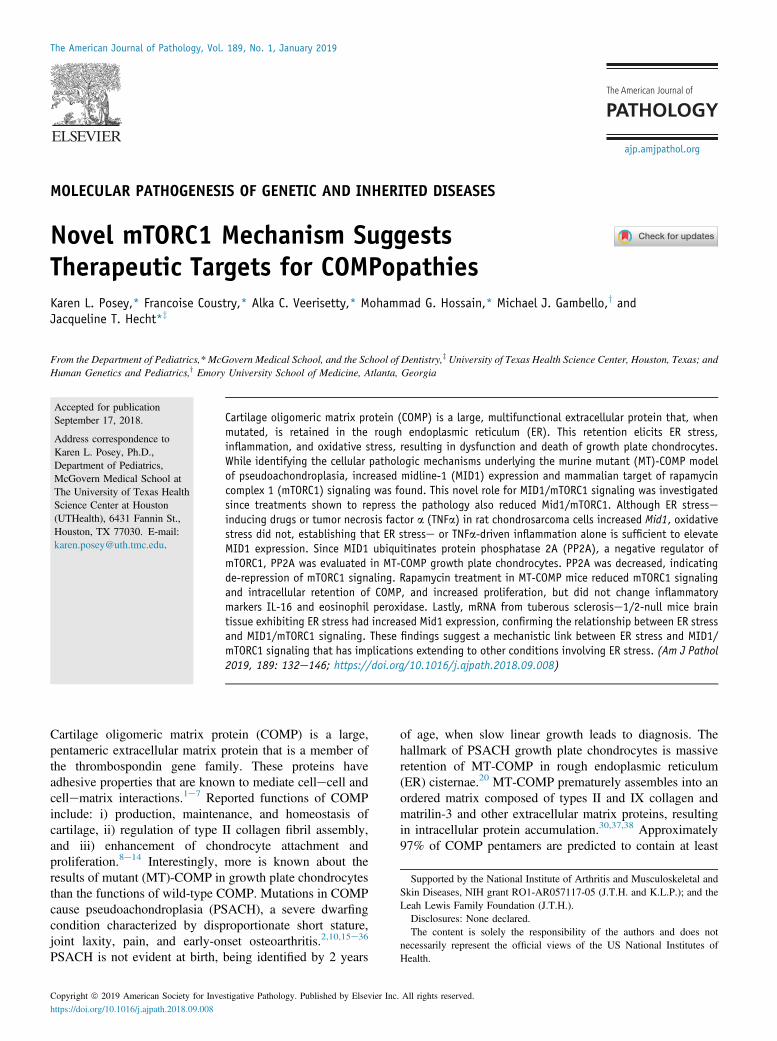
To identify the molecular mechanisms that underlie the MT-COMP chondrocyte pathology, transcriptome analysis wasperformed.41 RNA was collected from the hind-limb kneejoints of MT-COMP and control mice at P1, P7, P14, P21,and P28 and subjected to microarray expression analysis.Mid1 transcript level was significantly increased inMT-COMP mice at all ages compared to controls(Figure 1A). This increase was confirmed by quantitativereal-time RT-PCR (data not shown), and immunostaining ofMT-COMP growth plate chondrocytes showed increases inMid1 protein in MT-COMP compared to the controls at allages (Figure 1, BeI).It was next determined whether this increase in MID1
expression was also present in human PSACH chon-drocytes. Cartilage nodules grown in three-dimensional
ajp.amjpathol.org - The American Journal of Pathology
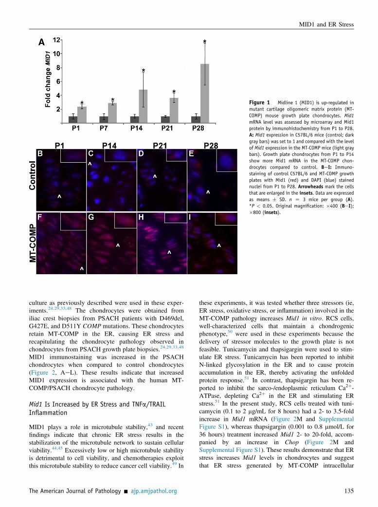
Figure 1 Midline 1 (MID1) is up-regulated inmutant cartilage oligomeric matrix protein (MT-COMP) mouse growth plate chondrocytes. Mid1mRNA level was assessed by microarray and Mid1protein by immunohistochemistry from P1 to P28.A: Mid1 expression in C57BL/6 mice (control; darkgray bars) was set to 1 and compared with the levelof Mid1 expression in the MT-COMP mice (light graybars). Growth plate chondrocytes from P1 to P14show more Mid1 mRNA in the MT-COMP chon-drocytes compared to control. BeI: Immuno-staining of control C57BL/6 and MT-COMP growthplates with Mid1 (red) and DAPI (blue) stainednuclei from P1 to P28. Arrowheads mark the cellsthat are enlarged in the insets. Data are expressedas means � SD. n Z 3 mice per group (A).*P < 0.05. Original magnification: �400 (BeI);�800 (insets).
MID1 and ER Stress
culture as previously described were used in these exper-iments.24,29,33,48 The chondrocytes were obtained fromiliac crest biopsies from PSACH patients with D469del,G427E, and D511Y COMP mutations. These chondrocytesretain MT-COMP in the ER, causing ER stress andrecapitulating the chondrocyte pathology observed inchondrocytes from PSACH growth plate biopsies.24,29,33,48
MID1 immunostaining was increased in the PSACHchondrocytes when compared to control chondrocytes(Figure 2, AeL). These results indicate that increasedMID1 expression is associated with the human MT-COMP/PSACH chondrocyte pathology.
Mid1 Is Increased by ER Stress and TNFa/TRAILInflammation
MID1 plays a role in microtubule stability,43 and recentfindings indicate that chronic ER stress results in thestabilization of the microtubule network to sustain cellularviability.44,45 Excessively low or high microtubule stabilityis detrimental to cell viability, and chemotherapies exploitthis microtubule stability to reduce cancer cell viability.49 In
The American Journal of Pathology - ajp.amjpathol.org
these experiments, it was tested whether three stressors (ie,ER stress, oxidative stress, or inflammation) involved in theMT-COMP pathology increases Mid1 in vitro. RCS cells,well-characterized cells that maintain a chondrogenicphenotype,50 were used in these experiments because thedelivery of stressor molecules to the growth plate is notfeasible. Tunicamycin and thapsigargin were used to stim-ulate ER stress. Tunicamycin has been reported to inhibitN-linked glycosylation in the ER and to cause proteinaccumulation in the ER, thereby activating the unfoldedprotein response.51 In contrast, thapsigargin has been re-ported to inhibit the sarco-/endoplasmic reticulum Ca2þ-ATPase, depleting Ca2þ in the ER and stimulating ERstress.51 In the present study, RCS cells treated with tuni-camycin (0.1 to 2 mg/mL for 8 hours) had a 2- to 3.5-foldincrease in Mid1 mRNA (Figure 2M and SupplementalFigure S1), whereas thapsigargin (0.001 to 0.8 mmol/L for36 hours) treatment increased Mid1 2- to 20-fold, accom-panied by an increase in Chop (Figure 2M andSupplemental Figure S1). These results demonstrate that ERstress increases Mid1 levels in chondrocytes and suggestthat ER stress generated by MT-COMP intracellular
135

Figure 2 Midline (MID)-1 is increased in human pseudoachondroplasia(PSACH) chondrocytes and by endoplasmic reticulum stress in rat chon-drosarcoma (RCS) cells. AeL: Chondrocytes from control and three differentPSACH patients [with D511Y, G427E, and D469del cartilage oligomericmatrix protein (COMP) gene mutations] were grown in a three-dimensionalculture system as previously described2,33 to generate nodules that wereimmunostained with MID1 antibody. DAPI (blue signal) marks the nuclei ofchondrocytes. Arrowheads indicate the cells that are enlarged in the in-sets; dashed circles indicate the nuclei in the insets. M: Mid1 mRNA levelsin RCS cells treated with thapsigargin or tunicamycin (gray bars) arecompared to that in untreated cells (control; black bars). Data areexpressed as means � SD. n > 100 cells (AeL); n Z 9 (M). **P < 0.005,***P < 0.0005. Original magnification: �600 (AeL); �1500 (insets).
Posey et al
retention increases MID1 levels in MT-COMP growth platechondrocytes. Oxidative stress is also involved in theMT-COMP chondrocyte pathology. Peroxynitrite, anendogenous peroxide, was used to generate free-radical in-termediates in RCS cells, and Mid1 mRNA levels weremeasured.52 Peroxynitrite treatment (5 to 1000 mmol/L for 4hours) showed no significant increase in Mid1 mRNA(Supplemental Figure S1), suggesting that increases in Mid1result from ER stress but not oxidative stress.
136
MT-COMP expression has been reported to be associatedwith the inflammatory process driven by TNFa and IL-1b.42,53 TNFa/TRAIL drive MID1 expression in asthmaand esophageal eosinophilia.54,55 It was determined whetherTNFa increases Trail and Mid1 independent of MT-COMPexpression in RCS cells, as this TNFa increase in MID1 waspreviously shown in the epithelial cells lining the airway.55
TNFa treatment (200 ng/mL) in RCS cells for 72 hoursincreased both Trail and Mid1 mRNAs by 33- and 16-fold,respectively (Figure 3K). Consistent with these findings,both TNFa and TRAIL immunostaining were also increasedin MT-COMP mice compared to controls (Figure 3, AeJ).These results demonstrate that ER stress and TNFa/TRAILincreased Mid1 in chondrocytes in vitro, and this finding isconsistent with the up-regulation of Mid1 observed in MT-COMP mice growth plate chondrocytes.
Increased MID1 Coincides with a Decrease in PP2ALevel and Increased mTORC1 Signaling in MT-COMPChondrocytes
Since MID1 acts as a negative regulator of PP2A in manycell types,56,57 it was evaluated whether MID1 has a similarfunction in MT-COMP growth plate chondrocytes in mice.MID1 has been established as a ubiquitin ligase that targetsPP2A for degradation,56,57 and therefore high levels ofMID1 would be expected to decrease PP2A. PP2A wasdiminished in the MT-COMP growth plate, consistent withthe role of MID1 as a negative regulator of PP2A (Figure 4,AeJ).58,59
MID1 is known to regulate mTORC1 but notmTORC2.60 Since MID1 is up-regulated in chondrocytesexpressing MT-COMP and MID1 stimulates mTORC1,mTORC1 signaling and phosphorylated (p)-AKT wereassessed in MT-COMP mice growth plates. pS661 andpmTOR were used as readouts for mTORC1 signaling andpAKT for activated AKT. MT-COMP growth plates at P28showed strong pS6, pAKT, and pmTOR expression levels,which were absent in the controls (Figure 4, KeY). MID1modulates mTORC1 through at least two mechanisms: byregulating 3-phosphoinositideedependent protein kinase(PDPK)-1 upstream of AKT and mTORC1,62 and regulatingPP2A levels, which influences mTOR/regulatory associatedprotein of mTOR complex formation.60 These findingsindicate that mTORC1 signaling is stimulated inMT-COMP mouse growth plate chondrocytes by increasedlevels of pAKT and repression of PP2A, which inhibitsmTORC1 formation.The tuberous sclerosis (TSC) mouse models, which lack
Tsc1, Tsc2, or both, exhibit ER stress and increasedmTORC1 signaling in their brain tissue.58,59 These TSCmice were used to test whether Mid1 was increased inthe context of elevated ER stress and mTORC1 signalingin TSC mice (Figure 4Z). Mid1 mRNA was increasedthree- to fivefold in all TSC mice, similar to the increaseobserved in the MT-COMP mice (Figure 4Z and
ajp.amjpathol.org - The American Journal of Pathology
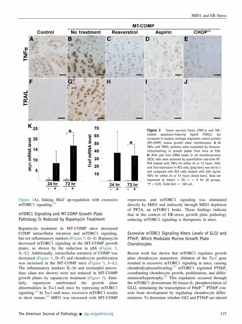
Figure 3 Tumor necrosis factor (TNF)-a and TNF-related apoptosis-inducing ligand (TRAIL) areincreased in mutant cartilage oligomeric matrix protein(MT-COMP) mouse growth plate chondrocytes. AeJ:TNFa and TRAIL proteins were evaluated by immuno-histochemistry in growth plates from mice at P28.K: Mid1 and Trail mRNA levels in rat chondrosarcoma(RCS) cells were assessed by quantitative real-time RT-PCR treated with TNFa for either 24 or 72 hours. Mid1and Trail expression in RCS cells (gray bars) was set to 1and compared with RCS cells treated with 200 ng/mLTNFa for either 24 or 72 hours (black bars). Data areexpressed as means � SD. n Z 9 for all groups.*P < 0.05. Scale bars Z 100 mm.
MID1 and ER Stress
Figure 1A), linking Mid1 up-regulation with excessivemTORC1 signaling.63
mTORC1 Signaling and MT-COMP Growth PlatePathology Is Reduced by Rapamycin Treatment
Rapamycin treatment in MT-COMP mice decreasedCOMP intracellular retention and mTORC1 signaling,but not inflammatory markers (Figure 5, GeI). Rapamycindecreased mTORC1 signaling in the MT-COMP growthplates, as shown by the reduction in pS6 (Figure 5,AeC). Additionally, intracellular retention of COMP wasdecreased (Figure 5, DeF) and chondrocyte proliferationwas increased in the MT-COMP mice (Figure 5, JeL).The inflammatory markers IL-16 and eosinophil peroxi-dase (data not shown) were not reduced in MT-COMPgrowth plates by rapamycin treatment (Figure 5). Simi-larly, rapamycin ameliorated the growth plateabnormalities in Tsc1-null mice by repressing mTORC1signaling.63 In Tsc1-null mice, excessive mTORC1 resultsin short stature.63 MID1 was increased with MT-COMP
The American Journal of Pathology - ajp.amjpathol.org
expression, and mTORC1 signaling was stimulateddirectly by MID1 and indirectly through MID1 depletionof PP2A, an mTORC1 brake. These findings indicatethat in the context of ER-stress growth plate pathologyreducing mTORC1 signaling is therapeutic in mice.
Excessive mTORC1 Signaling Alters Levels of GLI2 andPTHrP, Which Modulate Murine Growth PlateChondrocytes
Recent work has shown that mTORC1 regulates growthplate chondrocyte maturation. Ablation of the Tsc1 generesulted in excessive mTORC1 signaling in mice, causingchondrodysplasia/dwarfing.63 mTORC1 regulated PTHrP,coordinating chondrocyte growth, proliferation, and differ-entiation/hypertrophy.63 This regulation occurred throughthe mTORC1 downstream S6 kinase-b1 phosphorylation ofGLI2, stimulating the transcription of PthrP.63 PTHrP con-trols bone development by regulating chondrocyte differ-entiation. To determine whether Gli2 and PTHrP are altered
137

Figure 4 Increased midline 1 (MID1) decreases protein phosphatase (PP)-2a in mutant cartilage oligomeric matrix protein (MT-COMP) mouse growth platechondrocytes. AeY: MID1 (AeE), PP2A (FeJ), phosphorylated AKT (KeO), phosphorylated mammalian target of rapamycin (pmTOR; PeT), and phosphor-ylated S6 ribosomal protein (pS6; UeY) assessed by immunohistochemistry in P28 control (C57BL/6), MT-COMP with no treatment, MT-COMP treated withaspirin or resveratrol, and MT-COMP/CHOP-/- growth plate chondrocytes. AKT, mTOR, and pS6 are all components of the mTORC1 signaling pathway. Z: RT-PCR performed on RNA extracted from tuberous sclerosis complex (TSC)-1enull, TSC2-null, TSC1/2edouble null, and control mouse brains. Data areexpressed as means � SD. n > 3 (AeY); n Z 3 (Z). *P < 0.05, ***P < 0.0005 versus control. Scale bar Z 100 mm (all images).
Posey et al
138 ajp.amjpathol.org - The American Journal of Pathology
Figure 5 Rapamycin treatment of mutant cartilageoligomeric matrix protein (MT-COMP) mice decreasesintracellular retention of COMP and mammalian target ofrapamycin complex 1 (mTORC1) signaling but notinflammation. Control, untreated MT-COMP, andrapamycin-treated MT-COMP growth plates at P28 immu-nostained with antibodies for phosphorylated S6 ribo-somal protein (pS6) (AeC), human (h) COMP (DeF), IL-16 (GeI), and DNA proliferation cell nuclear antigen(PCNA) (JeL). Rapamycin treatment reduces pS6 andintracellular COMP and improves chondrocyte prolifera-tion. The inflammatory marker IL-16 is not reduced byrapamycin treatment. Scale bars Z 100 mm.
MID1 and ER Stress
in MT-COMP mice, growth plates were assessed at P28 inMT-COMP mice. pS6, a readout for mTORC1 signaling,was increased (Figure 5B). GLI2 and PTHrP expressionlevels were also increased, consistent with elevatedmTORC1 signaling in MT-COMP growth plates comparedto controls (Figure 6). This finding suggests that excessivemTORC1/GLI2/PTHrP stimulated by MT-COMP expres-sion represses growth plate chondrocyte hypertrophy.64e66
Anti-Inflammatory or Antioxidant Treatments orElimination of CHOP-ER Stress Response DecreasesMid1 Expression
Aspirin or resveratrol treatments have been reported toreduce intracellular accumulation of COMP, decrease
The American Journal of Pathology - ajp.amjpathol.org
inflammation markers and chondrocyte death, and increaseproliferation and limb length.53 Additionally, the absenceof CHOP, a key component of MT-COMP ER stress, inMT-COMP/CHOP�/� mice was reported to have dimin-ished the negative effects of MT-COMP expression.40,41
The expression levels of MID1 and PP2A in MT-COMPgrowth plates in mice treated with aspirin or resveratrol orloss of CHOP were next assessed. All of these therapeuticinterventions restored normal levels of MID1 and PP2A inthe growth plate (Figure 4, AeJ), confirming that MID1increases are related to MT-COMP chondrocyte pathology.Aspirin or resveratrol treatments or Chop gene ablation, allof which have been reported to dampen the MT-COMPchondrocyte pathology,53 also decreased the expressionlevels of TNFa and TRAIL, which drive increases in MID1
139

Figure 6 Glioma-associated oncogene homolog 2 (Gli2) and parathyroid hormone-related protein (PTHrP) are increased with mammalian target ofrapamycin complex 1 (mTORC1) signaling in mutant cartilage oligomeric matrix protein (MT-COMP) growth plates. Control (C57BL/6), untreated MT-COMP, MT-COMP treated with resveratrol or aspirin, and MT-Comp/CHOP�/� growth plates from P28 mice were immunostained for phosphorylated GLI2 (AeE) and PTHrP(FeJ). Scale bar Z 100 mm (all images). CHOP, CCAAT/enhancer-binding proteinehomologous protein.
Posey et al
(Figure 3, AeJ). These treatments also normalizedmTORC1 signaling (Figure 4), likely due to decreases in thePP2A level. Additionally, resveratrol or aspirin treatment orelimination of CHOP (MT-COMP/CHOP�/�) normalizedGLI2 and PTHrP levels (Figure 6), also likely due to down-regulation of mTORC1 signaling.
Overexpression of MID1 Decreases Chop mRNA Levelsin the Presence of MT-COMP
To define the relationship between MID1 and CHOP, MID1was overexpressed and knocked down in RSC cellsexpressing MT-COMP. Overexpression of MID1 inMT-COMP RCS cells (Figure 7A) was accompanied bydecreased Chop mRNA levels (Figure 7B). MT-COMPexpression has been associated with increases in Chop,growth arrest, and DNA damageeinducible proteins 34and 45a and ER oxidoreductin protein1b mRNAs.40 siRNAknockdown of Mid1 in MT-COMP RCS cells resulted inincreases in MT-COMP, Chop, Gadd34, Gadd45a, andEro1b (Figure 7, CeH).
Discussion
This is the first study linking ER stress to MID1/mTORC1signaling in chondrocytes and other cell types, a finding thathas significant implications for cellular functions includingautophagy, protein synthesis, and potentially cellular
140
viability. Moreover, these results identify new therapeutictargets for this pathologic process in a wide spectrum ofER-stress disorders. The pathologic mechanistic modelshows that MT-COMP expression and ER retention stimu-late unrelenting ER stress that initiates TNFa/TRAILinflammation and MID1 up-regulation, resulting inincreased mTORC1 signaling (Figure 8).MT-COMPeinduced chondrocyte death occurs through
necroptosis (a form of cell death).40,41 This process beginswith intracellular accumulation of MT-COMP, which in-duces ER stress and the unfolded protein response. Therefolding and degradation branches of the unfolded proteinresponse are not fully engaged by MT-COMP retention, andonly the protein kinase RNA-activatedelike endoplasmicreticulum kinase/CHOP branch proceeds beyond initialactivation.40 Reactive oxygen species are generated byCHOP/ERO1b along with NADPH oxidase 4, creatingDNA damage and the release of phosphorylated histone2AX.40 MT-COMPeinduced mitochondrial dysfunctionstimulates the release of cell deatheinducing factor and itstranslocation to the nucleus, where it interacts with phos-phorylated histone 2AX, inducing necroptosis.40,41 MID1, amicrotubule stabilizer, which was increased, may beactivated to sustain chondrocyte viability in this pathologicprocess.47,67 The ER is inextricably linked to microtubules,as microtubules are the transport highways used by the ERto export its cargo.68 Persistent ER stress is known tostabilize the microtubule network, thereby providing resis-tance to apoptosis.69e77 Additionally, in our study, the
ajp.amjpathol.org - The American Journal of Pathology

Figure 7 Overexpression or knockdown of Mid1 alters Chop mRNA levels. Aand B: Rat chondrosarcoma (RCS) cells that express human mutant cartilageoligomeric matrix protein (h-MT-COMP) were transfected with midline (MID)-1expression plasmid and Mid1 (A) and Chop (B) mRNA levels were assessed. C:Mid1 siRNAs were transfected into RCS cells that express h-MT-COMP, resulting indecreased Mid1 levels. DeH: mRNA levels of h-MT-COMP (D), Chop (E), Gadd34(F), Gadd45a (G), and Ero1b (H) are shown. Data are expressed as means � SD. nZ 9 in each group. *P < 0.05, **P < 0.005, and ***P < 0.0005 versusMT-COMP.
MID1 and ER Stress
overexpression of MID1 was associated with a decrease inChop, whereas knockdown of Mid1 was associated with anincrease in the stress-related transcripts Chop, Gadd34,Gadd45a, and Ero1b. It is not clear whether the increase inChop when MID1 was knocked down was due to decreasedMid1 or increased MT-COMP. MID1 increases stability and
The American Journal of Pathology - ajp.amjpathol.org
translational efficiency of select mRNAs through MID1-associated sequence.62 However, COMP mRNA does notcontain a MID1-binding domain, and the inverse relation-ship between MID1 and MT-COMP suggests that MID1does not directly regulate COMP mRNA levels. Based onthis information, increased MID1 in ER stresserelated
141

Figure 8 Model depicting the roles of midline 1 (MID1), mammaliantarget of rapamycin, complex 1 (mTORC1), and parathyroid hormone-related protein (PTHrP) in mutant cartilage oligomeric matrix protein(MT-COMP) chondrocyte pathology. A: MT-COMP expression elicitsendoplasmic reticulum (ER) stress through PERK/CHOP, which leads tooxidative stress and inflammation. The inflammatory process driven inpart by tumor necrosis factor (TNF)-a increases TNF-related apoptosis-inducing ligand (TRAIL) and MID1. CHOP down-regulates generalprotein synthesis as part of the unfolded protein response and up-regulates AKT. AKT and MID1 stimulate mTORC1 signaling, along witha decrease in the mTORC1 brake, protein phosphatase (PP)-2A. mTORC1signaling drives protein synthesis through protein S6 kinase/phospho-S6 and likely generates additional ER stress. mTORC1 up-regulatesglioma-associated oncogene homolog 2 (zinc finger protein) (GLI)-2and PTHrP, which alters chondrocyte proliferation and hypertrophy. B:Resveratrol counteracts several processes involved in the MT-COMPchondrocyte pathology including oxidative stress, inflammation, andMID1/a4 complex. Aspirin, a cyclooxygenase-2 inhibitor, dampensinflammation and diminishes the negative effects of MT-COMPexpression (red). Rapamycin decreases intracellular retention of MT-COMP and mTORC1 signaling but does not improve inflammation andproliferation. Other drugs to potentially reduce the MT-COMP chon-drocyte pathology are shown in blue. BHA, butylated hydroxyanisole;BIX, immunoglobulin heavy-chainebinding protein inducer X; CHOP,CCAAT/enhancer-binding proteinehomologous protein; PBA, 4-phenylbutyrate; PDPK-1, protein 3-phosphoinositide-dependent pro-tein kinase-1; PERK, protein kinase RNA-activatedelike endoplasmicreticulum kinase TUDCA, tauroursodeoxycholic acid.
Posey et al
142
conditions likely slows but does not ultimately eliminatechondrocyte death.2,33,36,38,40,66,78
It has been tested whether MID1/mTORC1 signaling isinvolved in other ER stresserelated conditions, includingAlzheimer, Huntington, and Parkinson diseases; amyo-trophic lateral sclerosis; cancer; type II diabetes79; andTSC.58 Both PSACH and Alzheimer disease involve proteinmisfolding, oxidative stress, inflammation, and prematurecell death.36,41,80e87 MID1 is elevated in Alzheimer-diseased brains compared to healthy controls.88 Consistentwith these findings, mTORC1 is increased and PP2A, whichis negatively regulated by MID1, is decreased in Alzheimerdisease.89 Furthermore, in human TSC or mice lackingeither Tsc1 or Tsc2 or both Tsc genes, unrestrained mTORsignaling generates ER stress with a concurrent increase inmTOR activation.90 Mid1 mRNA was increased in the braintissue of Tsc1-, Tsc2-, and Tsc1/2edouble null mice,providing further evidence that MID1/mTORC1 is linked toER stress (Figure 4Z). Moreover, dampening mTORC1signaling was reported to improve Parkinson disease out-comes in mice, preventing the development of dyskinesia.91
All together, these combined observations establish that up-regulation of MID1 accompanies elevated mTORC1signaling in a variety of ER stresserelated conditions.92
mTORC1 is a master regulator of growth in response tonutritional status, energy levels, cellular stress, growthfactors, and amino acids, and regulates general proteintranslation and autophagy.93 Dysfunction of secretory cellsor misfolded proteins (chondrocytes, b-islet cells, andneurons) are a significant cause of ER stresserelatedconditions. Secretory cells synthesize large amounts ofexported proteins that are necessary for organ/tissue func-tioning. Since mTORC1 regulates general protein synthesis,increases in mTORC1 signaling may sustain proteinsynthesis during periods of ER stress, allowing cells tocontinue functioning. During ER stress, the unfolded proteinresponse down-regulates general translation to allowclearance of misfolded protein(s). In contrast, mTORC1stimulates general protein synthesis, which may ultimatelyexacerbate ER stress and inhibit ER clearance. Additionally,increased mTORC1 signaling represses autophagy, elimi-nating another mechanism to clear misfolded proteins andresolve ER stress. Collectively, these results indicate thatincreased MID1 during ER stress functions to prolongcellular survival as well as stimulates excess mTORC1signaling, which ultimately may be detrimental.Besides playing a role in ER stresserelated disorders,
mTORC1 signaling regulates PTHrP and GLI2 modulationof chondrocyte hypertrophy, and increased mTORC1signaling is associated with osteoarthritis joint degenera-tion.94 In the present study, PTHrP and GL12 were bothincreased in growth plate chondrocytes in MT-COMP mice(Figure 6). This finding suggests that the lack of hypertro-phic chondrocytes in the PSACH growth plate and theearly-onset osteoarthritis in PSACH are additional conse-quences of increased mTORC1 signaling.
ajp.amjpathol.org - The American Journal of Pathology

MID1 and ER Stress
This work identifies the mTORC1 pathway as a potentialtherapeutic target in ER-stress conditions. It has beenreported that aspirin and resveratrol treatments dampen MT-COMP chondrocyte pathology by reducing inflammationand oxidative stress.53 In the present study, these treatmentsalso reduced Mid1, TNFa, Trail, and mTORC1 signaling inmice. Although rapamycin targeting of mTORC1 in theMT-COMP mouse decreased pS6 and MT-COMP intra-cellular accumulation, and increased DNA proliferation, itdid not reduce inflammation (eosinophil peroxidase or IL-16). This is an expected result since MID1 up-regulationis downstream of TNFa/TRAIL inflammation and thereforerapamycin may be more effective when used in combinationwith anti-inflammatory medications such as aspirin. How-ever, there are significant side effects (suppression of theimmune system, increased risks for infections and cancer,and impaired wound healing) with chronic rapamycintreatments that may limit their current therapeutic utility(Food andDrugAdministration, https://www.accessdata.fda.gov/drugsatfda_docs/label/2010/021110s058lbl.pdf, lastaccessed September, 17, 2018).
Given the numerous conditions that involve ERdysfunction, a complete understanding all of the conse-quences of ER stress is crucial for the development ofmechanism-driven therapeutic approaches. The results ofthis work show that TNFa inflammation, MID1, mTORC1signaling, the microtubule network, protein synthesis, andautophagy form a complex, multifaceted response to proteinaccumulation in the ER when the clearance efforts fail, andMID1 may act as a prosurvival factor to extend chondrocytelife. Importantly, therapeutics interrupting the upstream self-perpetuating pathologic loop between ER stress, inflam-mation, and oxidative stress may be most effective.
Acknowledgments
We thank Frankie Chiu for technical assistance, and Dr.Makoto Suzuki for providing the MID1-overexpressionplasmid.
K.L.P. designed the study, analyzed the data, performedliterature search, generated the figures, and wrote themanuscript; F.C., A.C.V., and M.G.H. collected andanalyzed data; M.J.G. collected data and reviewed themanuscript; J.T.H. designed the study, interpreted data, andwrote the manuscript.
Supplemental Data
Supplemental material for this article can be found athttps://doi.org/10.1016/j.ajpath.2018.09.008.
References
1. DiCesare PE, Morgelin M, Mann K, Paulsson M: Cartilage oligo-meric matrix protein and thrombospondin 1. Purification from
The American Journal of Pathology - ajp.amjpathol.org
articular cartilage, electron microscopic structure, and chondrocytebinding. Eur J Biochem 1994, 223:927e937
2. Hecht JT, Deere M, Putnam E, Cole W, Vertel B, Chen H, Lawler J:Characterization of cartilage oligomeric matrix protein (COMP) inhuman normal and pseudoachondroplasia musculoskeletal tissues.Matrix Biol 1998, 17:269e278
3. Hedbom E, Antonsson P, Hjerpe A, Aeschlimann D, Paulsson M,Rosa-Pimentel E, Sommarin Y, Wendel M, Oldberg A,Heinegard D: Cartilage matrix proteins. An acidic oligomericprotein (COMP) detected only in cartilage. J Biol Chem 1992,267:6132e6136
4. Urban JP, Maroudas A, Bayliss MT, Dillon J: Swelling pressures ofproteoglycans at the concentrations found in cartilaginous tissues.Biorheology 1979, 16:447e464
5. Kempson GE, Freeman MA, Swanson SA: Tensile properties ofarticular cartilage. Nature 1968, 220:1127e1128
6. Schmidt MB, Mow VC, Chun LE, Eyre DR: Effects of proteoglycanextraction on the tensile behavior of articular cartilage. J Orthop Res1990, 8:353e363
7. Adams JC, Tucker RP, Lawler J: The Thrombospondin Gene Family.Austin, TX, R.G. Landes Co., 1995
8. Holden P, Meadows RS, Chapman KL, Grant ME, Kadler KE,Briggs MD: Cartilage oligomeric matrix protein interacts with typeIX collagen, and disruptions to these interactions identify a patho-genetic mechanism in a bone dysplasia family. J Biol Chem 2001,276:6046e6055
9. Mann HH, Ozbek S, Engel J, Paulsson M, Wagener R: Interactionsbetween the cartilage oligomeric matrix protein and matrilins.Implications for matrix assembly and the pathogenesis of chon-drodysplasias. J Biol Chem 2004, 279:25294e25298
10. Thur J, Rosenberg K, Nitsche DP, Pihlajamaa T, Ala-Kokko L,Heinegard D, Paulsson M, Maurer P: Mutations in cartilage oligo-meric matrix protein causing pseudoachondroplasia and multipleepiphyseal dysplasia affect binding of calcium and collagen I, II, andIX. J Biol Chem 2001, 276:6083e6092
11. Rosenberg K, Olsson H, Morgelin M, Heinegard D: Cartilageoligomeric matrix protein shows high affinity zinc-dependentinteraction with triple helical collagen. J Biol Chem 1998, 273:20397e20403
12. Kipnes J, Carlberg AL, Loredo GA, Lawler J, Tuan RS, Hall DJ:Effect of cartilage oligomeric matrix protein on mesenchymal chon-drogenesis in vitro. Osteoarthritis Cartilage 2003, 11:442e454
13. Xu K, Zhang Y, Ilalov K, Carlson CS, Feng JQ, Di Cesare PE,Liu CJ: Cartilage oligomeric matrix protein associates withgranulin-epithelin precursor (GEP) and potentiates GEP-stimulated chondrocyte proliferation. J Biol Chem 2007, 282:11347e11355
14. Chen FH, Thomas AO, Hecht JT, Goldring MB, Lawler J: Cartilageoligomeric matrix protein/thrombospondin 5 supports chondrocyteattachment through interaction with integrins. J Biol Chem 2005, 280:32655e32661
15. Briggs MD, Chapman KL: Pseudoachondroplasia and multipleepiphyseal dysplasia: mutation review, molecular interactions,and genotype to phenotype correlations. Hum Mutat 2002, 19:465e478
16. Briggs MD, Hoffman SM, King LM, Olsen AS, Mohrenweiser H,Leroy JG, Mortier GR, Rimoin DL, Lachman RS, Gaines ES:Pseudoachondroplasia and multiple epiphyseal dysplasia due to mu-tations in the cartilage oligomeric matrix protein gene. Nat Genet1995, 10:330e336
17. Briggs MD, Mortier GR, Cole WG, King LM, Golik SS,Bonaventure J, Nuytinck L, De Paepe A, Leroy JG, Biesecker L,Lipson M, Wilcox WR, Lachman RS, Rimoin DL,Knowlton RG, Cohn DH: Diverse mutations in the gene forcartilage oligomeric matrix protein in the pseudoachondroplasia-multiple epiphyseal dysplasia disease spectrum. Am J HumGenet 1998, 62:311e319
143

Posey et al
18. Chen H, Deere M, Hecht JT, Lawler J: Cartilage oligomeric matrixprotein is a calcium-binding protein, and a mutation in its type 3repeats causes conformational changes. J Biol Chem 2000, 275:26538e26544
19. Chen TL, Stevens JW, Cole WG, Hecht JT, Vertel BM: Cell-typespecific trafficking of expressed mutant COMP in a cell culture modelfor PSACH. Matrix Biol 2004, 23:433e444
20. Cooper RR, Ponseti IV, Maynard JA: Pseudoachondroplasticdwarfism. A rough-surfaced endoplasmic reticulum storage disorder.J Bone Joint Surg Am 1973, 55:475e484
21. Delot E, Brodie SG, King LM, Wilcox WR, Cohn DH: Physiologicaland pathological secretion of cartilage oligomeric matrix protein bycells in culture. J Biol Chem 1998, 273:26692e26697
22. DiCesare PE, Morgelin M, Carlson CS, Pasumarti S,Paulsson M: Cartilage oligomeric matrix protein: isolation andcharacterization from human articular cartilage. J Orthop Res1995, 13:422e428
23. Dinser R, Zaucke F, Kreppel F, Hultenby K, Kochanek S,Paulsson M, Maurer P: Pseudoachondroplasia is caused through bothintra- and extracellular pathogenic pathways. J Clin Invest 2002, 110:505e513
24. Duke J, Montufar-Solis D, Underwood S, Lalani Z, Hecht JT:Apoptosis staining in cultured pseudoachondroplasia chondrocytes.Apoptosis 2003, 8:191e197
25. Ikegawa S, Ohashi H, Nishimura G, Kim KC, Sannohe A,Kimizuka M, Fukushima Y, Nagai T, Nakamura Y: Novel andrecurrent COMP (cartilage oligomeric matrix protein) mutations inpseudoachondroplasia and multiple epiphyseal dysplasia. Hum Genet1998, 103:633e638
26. Kleerekoper Q, Hecht JT, Putkey JA: Disease-causing mutations incartilage oligomeric matrix protein cause an unstructured Ca2þbinding domain. J Biol Chem 2002, 277:10581e10589
27. Maddox BK, Mokashi A, Keene DR, Bachinger HP: A cartilageoligomeric matrix protein mutation associated with pseudoachon-droplasia changes the structural and functional properties of the type 3domain. J Biol Chem 2000, 275:11412e11417
28. McKeand J, Rotta J, Hecht JT: Natural history study of pseu-doachondroplasia. Am J Med Genet 1996, 63:406e410
29. Merritt TM, Alcorn JL, Haynes R, Hecht JT: Expression of mutantcartilage oligomeric matrix protein in human chondrocytes inducesthe pseudoachondroplasia phenotype. J Orthop Res 2006, 24:700e707
30. Merritt TM, Bick R, Poindexter BJ, Alcorn JL, Hecht JT: Uniquematrix structure in the rough endoplasmic reticulum cisternae ofpseudoachondroplasia chondrocytes. Am J Pathol 2007, 170:293e300
31. Unger S, Hecht JT: Pseudoachondroplasia and multiple epiphysealdysplasia: new etiologic developments. Am J Med Genet 2001, 106:244
32. Hecht JT, Montufar-Solis D, Decker G, Lawler J, Daniels K,Duke PJ: Retention of cartilage oligomeric matrix protein (COMP)and cell death in redifferentiated pseudoachondroplasia chondrocytes.Matrix Biol 1998, 17:625e633
33. Hecht JT, Hayes E, Haynes R, Cole WG: COMP mutations, chon-drocyte function and cartilage matrix. Matrix Biol 2005, 23:525e533
34. Hecht JT, Hayes E, Snuggs M, Decker G, Montufar-Solis D,Doege K, Mwalle F, Poole R, Stevens J, Duke PJ: Calreticulin, PDI,Grp94 and BiP chaperone proteins are associated with retainedCOMP in pseudoachondroplasia chondrocytes. Matrix Biol 2001, 20:251e262
35. Hecht JT, Nelson LD, Crowder E, Wang Y, Elder FF, Harrison WR,Francomano CA, Prange CK, Lennon GG, Deere M, Lawler J:Mutations in exon 17B of cartilage oligomeric matrix protein(COMP) cause pseudoachondroplasia. Nat Genet 1995, 10:325e329
36. Posey KL, Hayes E, Haynes R, Hecht JT: Role of TSP-5/COMP inpseudoachondroplasia. Int J Biochem Cell Biol 2004, 36:1005e1012
144
37. Posey KL, Hecht JT: Novel therapeutic interventions for pseu-doachondroplasia. Bone 2017, 102:60e68
38. Posey KL, Veerisetty AC, Liu P, Wang HR, Poindexter BJ, Bick R,Alcorn JL, Hecht JT: An inducible cartilage oligomeric matrix proteinmouse model recapitulates human pseudoachondroplasia phenotype.Am J Pathol 2009, 175:1555e1563
39. Posey KL, Coustry F, Veerisetty AC, Hossain M, Gattis D, Booten S,Alcorn JL, Seth PP, Hecht JT: Antisense reduction of mutant COMPreduces growth plate chondrocyte pathology. Mol Ther 2017, 25:705e714
40. Coustry F, Posey KL, Liu P, Alcorn JL, Hecht JT: D469del-COMPretention in chondrocytes stimulates caspase-independent necroptosis.Am J Pathol 2012, 180:738e748
41. Posey KL, Coustry F, Veerisetty AC, Liu P, Alcorn JL, Hecht JT:Chop (Ddit3) is essential for D469del-COMP retention and cell deathin chondrocytes in an inducible transgenic mouse model of pseu-doachondroplasia. Am J Pathol 2012, 180:727e737
42. Posey KL, Coustry F, Veerisetty AC, Liu P, Alcorn JL, Hecht JT:Chondrocyte-specific pathology during skeletal growth and thera-peutics in a murine model of pseudoachondroplasia. J Bone MinerRes 2014, 29:1258e1268
43. Short KM, Hopwood B, Yi Z, Cox TC: MID1 and MID2 homo- andheterodimerise to tether the rapamycin-sensitive PP2A regulatorysubunit, alpha 4, to microtubules: implications for the clinical vari-ability of X-linked Opitz GBBB syndrome and other developmentaldisorders. BMC Cell Biol 2002, 3:1
44. Jiang CC, Yang F, Thorne RF, Zhu BK, Hersey P, Zhang XD:Human melanoma cells under endoplasmic reticulum stress acquireresistance to microtubule-targeting drugs through XBP-1-mediatedactivation of Akt. Neoplasia 2009, 11:436e447
45. Placido AI, Pereira CM, Correira SC, Carvalho C, Oliveira CR,Moreira PI: Phosphatase 2A inhibition affects endoplasmic reticulumand mitochondria homeostasis via cytoskeletal alterations in brainendothelial cells. Mol Neurobiol 2017, 54:154e168
46. Marciniak SJ, Yun CY, Oyadomari S, Novoa I, Zhang Y, Jungreis R,Nagata K, Harding HP, Ron D: CHOP induces death by promotingprotein synthesis and oxidation in the stressed endoplasmic reticulum.Genes Dev 2004, 18:3066e3077
47. Suzuki M, Hara Y, Takagi C, Yamamoto TS, Ueno N: MID1 andMID2 are required for Xenopus neural tube closure through theregulation of microtubule organization. Development 2010, 137:2329e2339
48. Hecht JT, Sage EH: Retention of the matricellular protein SPARC inthe endoplasmic reticulum of chondrocytes from patients with pseu-doachondroplasia. J Histochem Cytochem 2006, 54:269e274
49. Parker AL, Kavallaris M, McCarroll JA: Microtubules and their rolein cellular stress in cancer. Front Oncol 2014, 4:153
50. King KB, Kimura JH: The establishment and characterization of animmortal cell line with a stable chondrocytic phenotype. J Cell Bio-chem 2003, 89:992e1004
51. Oslowski CM, Urano F: Measuring ER stress and the unfolded pro-tein response using mammalian tissue culture system. MethodsEnzymol 2011, 490:71e92
52. Radi R: Peroxynitrite, a stealthy biological oxidant. J Biol Chem2013, 288:26464e26472
53. Posey KL, Coustry F, Veerisetty AC, Hossain M, Alcorn JL,Hecht JT: Antioxidant and anti-inflammatory agents mitigate pa-thology in a mouse model of pseudoachondroplasia. Hum Mol Genet2015, 24:3918e3928
54. Collison A, Hatchwell L, Verrills N, Wark PA, de Siqueira AP,Tooze M, Carpenter H, Don AS, Morris JC, Zimmermann N,Bartlett NW, Rothenberg ME, Johnston SL, Foster PS, Mattes J: TheE3 ubiquitin ligase midline 1 promotes allergen and rhinovirus-induced asthma by inhibiting protein phosphatase 2A activity. NatMed 2013, 19:232e237
55. Collison AM, Sokulsky LA, Sherrill JD, Nightingale S, Hatchwell L,Talley NJ, Walker MM, Rothenberg ME, Mattes J: TNF-related
ajp.amjpathol.org - The American Journal of Pathology

MID1 and ER Stress
apoptosis-inducing ligand (TRAIL) regulates midline-1, thymicstromal lymphopoietin, inflammation, and remodeling in experi-mental eosinophilic esophagitis. J Allergy Clin Immunol 2015, 136:971e982
56. Trockenbacher A, Suckow V, Foerster J, Winter J, Krauss S,Ropers HH, Schneider R, Schweiger S: MID1, mutated in Opitzsyndrome, encodes an ubiquitin ligase that targets phosphatase 2A fordegradation. Nat Genet 2001, 29:287e294
57. Liu J, Prickett TD, Elliott E, Meroni G, Brautigan DL: Phosphory-lation and microtubule association of the Opitz syndrome proteinmid-1 is regulated by protein phosphatase 2A via binding to theregulatory subunit alpha 4. Proc Natl Acad Sci U S A 2001, 98:6650e6655
58. Reith RM, Way S, McKenna J 3rd, Haines K, Gambello MJ: Loss ofthe tuberous sclerosis complex protein tuberin causes Purkinje celldegeneration. Neurobiol Dis 2011, 43:113e122
59. Ozcan U, Ozcan L, Yilmaz E, Duvel K, Sahin M, Manning BD,Hotamisligil GS: Loss of the tuberous sclerosis complextumor suppressors triggers the unfolded protein response toregulate insulin signaling and apoptosis. Mol Cell 2008, 29:541e551
60. Liu E, Knutzen CA, Krauss S, Schweiger S, Chiang GG: Control ofmTORC1 signaling by the Opitz syndrome protein MID1. Proc NatlAcad Sci U S A 2011, 108:8680e8685
61. Iwenofu OH, Lackman RD, Staddon AP, Goodwin DG, Haupt HM,Brooks JS: Phospho-S6 ribosomal protein: a potential new predictivesarcoma marker for targeted mTOR therapy. Mod Pathol 2008, 21:231e237
62. Aranda-Orgilles B, Rutschow D, Zeller R, Karagiannidis AI,Kohler A, Chen C, Wilson T, Krause S, Roepcke S, Lilley D,Schneider R, Schweiger S: Protein phosphatase 2A (PP2A)-specificubiquitin ligase MID1 is a sequence-dependent regulator of trans-lation efficiency controlling 3-phosphoinositide-dependent proteinkinase-1 (PDPK-1). J Biol Chem 2011, 286:39945e39957
63. Yan B, Zhang Z, Jin D, Cai C, Jia C, Liu W, Wang T, Li S, Zhang H,Huang B, Lai P, Wang H, Liu A, Zeng C, Cai D, Jiang Y, Bai X:mTORC1 regulates PTHrP to coordinate chondrocyte growth, pro-liferation and differentiation. Nat Commun 2016, 7:11151
64. Lindseth RE, Danigelis JA, Murray DG, Wray JB: Spondylo-epiph-yseal dysplasia (pseudoachondroplastic type). Case report withpathologic and metabolic investigations. Am J Dis Child 1967, 113:721e726
65. Stanescu V, Stanescu R, Maroteaux P: Pathogenic mechanisms inosteochondrodysplasias. J Bone Joint Surg Am 1984, 66:817e836
66. Hecht JT, Makitie O, Hayes E, Haynes R, Susic M, Montufar-Solis D, Duke PJ, Cole WG: Chondrocyte cell death and intracellulardistribution of COMP and type IX collagen in the pseudoachon-droplasia growth plate. J Orthop Res 2004, 22:759e767
67. Schweiger S, Foerster J, Lehmann T, Suckow V, Muller YA,Walter G, Davies T, Porter H, van Bokhoven H, Lunt PW, Traub P,Ropers HH: The Opitz syndrome gene product, MID1, associateswith microtubules. Proc Natl Acad Sci U S A 1999, 96:2794e2799
68. Palmer KJ, Watson P, Stephens DJ: The role of microtubules intransport between the endoplasmic reticulum and Golgi apparatus inmammalian cells. Biochem Soc Symp 2005, 72:1e13
69. Gately DP, Howell SB: Paclitaxel activation of the GADD153promoter through a cellular injury response element containingan essential Sp1 binding site. J Biol Chem 1996, 271:20588e20593
70. Seyb KI, Ansar S, Bean J, Michaelis ML: beta-Amyloid and endo-plasmic reticulum stress responses in primary neurons: effects ofdrugs that interact with the cytoskeleton. J Mol Neurosci 2006, 28:111e123
71. Gately DP, Sharma A, Christen RD, Howell SB: Cisplatin and taxolactivate different signal pathways regulating cellular injury-inducedexpression of GADD153. Br J Cancer 1996, 73:18e23
The American Journal of Pathology - ajp.amjpathol.org
72. Jiang P, Gan M, Ebrahim AS, Lin WL, Melrose HL, Yen SH: ERstress response plays an important role in aggregation of alpha-syn-uclein. Mol Neurodegener 2010, 5:56
73. Kim R, Ohi Y, Inoue H, Aogi K, Toge T: Introduction of gadd153gene into gastric cancer cells can modulate sensitivity to anticanceragents in association with apoptosis. Anticancer Res 1999, 19:1779e1783
74. Stockwell SR, Platt G, Barrie SE, Zoumpoulidou G, Te Poele RH,Aherne GW, Wilson SC, Sheldrake P, McDonald E, Venet M,Soudy C, Elustondo F, Rigoreau L, Blagg J, Workman P,Garrett MD, Mittnacht S: Mechanism-based screen for G1/S check-point activators identifies a selective activator of EIF2AK3/PERKsignalling. PLoS One 2012, 7:e28568
75. Swanton C, Marani M, Pardo O, Warne PH, Kelly G, Sahai E,Elustondo F, Chang J, Temple J, Ahmed AA, Brenton JD,Downward J, Nicke B: Regulators of mitotic arrest and ceramidemetabolism are determinants of sensitivity to paclitaxel and otherchemotherapeutic drugs. Cancer Cell 2007, 11:498e512
76. Wu Y, Fabritius M, Ip C: Chemotherapeutic sensitization by endo-plasmic reticulum stress: increasing the efficacy of taxane againstprostate cancer. Cancer Biol Ther 2009, 8:146e152
77. Ho CT, Chang YJ, Yang LX, Wei PL, Liu TZ, Liu JJ: A novelmicrotubule-disrupting agent induces endoplasmic reticular stress-mediated cell death in human hepatocellular carcinoma cells. PLoSOne 2015, 10:e0136340
78. Posey KL, Alcorn JL, Hecht JT: Pseudoachondroplasia/COMP -translating from the bench to the bedside. Matrix Biol 2014, 37:167e173
79. Schonthal AH: Endoplasmic reticulum stress: its role in diseaseand novel prospects for therapy. Scientifica (Cairo) 2012, 2012:857516
80. Posey KL, Hecht JT: The role of cartilage oligomeric matrixprotein (COMP) in skeletal disease. Curr Drug Targets 2008, 9:869e877
81. Greig NH, Mattson MP, Perry T, Chan SL, Giordano T,Sambamurti K, Rogers JT, Ovadia H, Lahiri DK: New therapeuticstrategies and drug candidates for neurodegenerative diseases: p53and TNF-alpha inhibitors, and GLP-1 receptor agonists. Ann N YAcad Sci 2004, 1035:290e315
82. Yankner BA, Duffy LK, Kirschner DA: Neurotrophic and neurotoxiceffects of amyloid beta protein: reversal by tachykinin neuropeptides.Science 1990, 250:279e282
83. Hernandez F, Avila J: Tauopathies. Cell Mol Life Sci 2007, 64:2219e2233
84. Hashimoto M, Rockenstein E, Crews L, Masliah E: Role of proteinaggregation in mitochondrial dysfunction and neurodegeneration inAlzheimer’s and Parkinson’s diseases. Neuromolecular Med 2003, 4:21e36
85. Su B, Wang X, Nunomura A, Moreira PI, Lee HG, Perry G,Smith MA, Zhu X: Oxidative stress signaling in Alzheimer’s disease.Curr Alzheimer Res 2008, 5:525e532
86. Iqbal K, Alonso Adel C, Chen S, Chohan MO, El-Akkad E,Gong CX, Khatoon S, Li B, Liu F, Rahman A, Tanimukai H,Grundke-Iqbal I: Tau pathology in Alzheimer disease and othertauopathies. Biochim Biophys Acta 2005, 1739:198e210
87. Goedert M, Spillantini MG, Crowther RA: Tau proteins and neuro-fibrillary degeneration. Brain Pathol 1991, 1:279e286
88. Schweiger S, Matthes F, Posey K, Kickstein E, Weber S,Hettich MM, Pfurtscheller S, Ehninger D, Schneider R, Krauss S:Resveratrol induces dephosphorylation of Tau by interfering with theMID1-PP2A complex. Sci Rep 2017, 7:13753
89. Vogelsberg-Ragaglia V, Schuck T, Trojanowski JQ, Lee VM: PP2AmRNA expression is quantitatively decreased in Alzheimer’s diseasehippocampus. Exp Neurol 2001, 168:402e412
90. Zeng LH, Rensing NR, Zhang B, Gutmann DH, Gambello MJ,Wong M: Tsc2 gene inactivation causes a more severe epi-lepsy phenotype than Tsc1 inactivation in a mouse model of
145

Posey et al
tuberous sclerosis complex. Hum Mol Genet 2011, 20:445e454
91. Santini E, Heiman M, Greengard P, Valjent E, Fisone G: Inhibition ofmTOR signaling in Parkinson’s disease prevents L-DOPA-induceddyskinesia. Sci Signal 2009, 2:ra36
92. Castellani RJ, Gupta Y, Sheng B, Siedlak SL, Harris PL, Coller JM,Perry G, Lee HG, Tabaton M, Smith MA, Wang X, Zhu X: A novel
146
origin for granulovacuolar degeneration in aging and Alzheimer’sdisease: parallels to stress granules. Lab Invest 2011, 91:1777e1786
93. Laplante M, Sabatini DM: mTOR signaling. Cold Spring Harb Per-spect Biol 2012, 4. a011593
94. Pal B, Endisha H, Zhang Y, Kapoor M: mTOR: a potential thera-peutic target in osteoarthritis? Drugs R D 2015, 15:27e36
ajp.amjpathol.org - The American Journal of Pathology