L'Enhanced Liver Fibrosis (ELF) Test nella NAFLD e nella NASH

Research Article
Galectin-3 ablation protects mice from diet-induced NASH: Amajor scavenging role for galectin-3 in liver
Carla Iacobini1,�, Stefano Menini1,�, Carlo Ricci1,�, Claudia Blasetti Fantauzzi1, Angela Scipioni1,Laura Salvi1, Samantha Cordone1, Francesca Delucchi1, Matteo Serino2, Massimo Federici2,
Flavia Pricci3, Giuseppe Pugliese1,⇑
1Department of Clinical Sciences, ‘‘La Sapienza’’ University, Rome, Italy; 2Department of Internal Medicine, ‘‘TorVergata’’ University, Rome, Italy;3Department of Cell Biology and Neurosciences, National Institute of Health of Italy, Rome, Italy
Background & Aims: Excess fatty acid oxidation and generation � 2010 European Association for the Study of the Liver. Published
of reactive carbonyls with formation of advanced lipoxidationendproducts (ALEs) is involved in nonalcoholic steatohepatitis(NASH) by triggering inflammation, hepatocyte injury, and fibro-sis. This study aimed at verifying the hypothesis that ablation ofthe ALE-receptor galectin-3 prevents experimental NASH byreducing receptor-mediated ALE clearance and downstreamevents.Methods: Galectin-3-deficient (Lgals3�/�) and wild type (Lgals3+/+)mice received an atherogenic diet or standard chow for8 months. Liver tissue was analyzed for morphology, inflamma-tion, cell and matrix turnover, lipid metabolism, ALEs, and ALE-receptors.Results: Steatosis was significantly less pronounced in Lgals3�/�than Lgals3+/+ animals on atherogenic diet. NASH, invariablydetected in Lgals3+/+ mice, was observed, to a lower extent, onlyin 3/8 Lgals3�/� mice, showing less inflammatory, degenerative,and fibrotic phenomena than Lgals3+/+ mice. This was associatedwith higher circulating ALE levels and lower tissue ALE accumu-lation and expression of other ALE-receptors. Up-regulation ofhepatic fatty acid synthesis and oxidation, inflammatory cellinfiltration, pro-inflammatory cytokines, endoplasmic reticulumstress, hepatocyte apoptosis, myofibroblast transdifferentiation,and impaired Akt phosphorylation were also significantly attenu-ated in Lgals3�/� animals. Galectin-3 silencing in liver endothelialcells resulted in reduced Ne-carboxymethyllysine-modified albu-min uptake and ALE-receptor expression.Conclusions: Galectin-3 ablation protects from diet-inducedNASH by decreasing hepatic ALE accumulation, with attenuationof inflammation, hepatocyte injury, and fibrosis. It also reducedup-regulation of lipid synthesis and oxidation causing less fatdeposition, oxidative stress, and possibly insulin resistance. Thesedata suggest that galectin-3 is a major receptor involved in ALEuptake by the liver.
Journal of Hepatology 20
Keywords: Steatosis; Inflammation; Fibrosis; Advanced lipoxidation endprod-ucts; Oxidative stress.Received 28 December 2009; received in revised form 8 September 2010; accepted 14September 2010⇑ Corresponding author. Address: Department of Clinical Sciences, ‘‘La Sapienza’’University of Rome, Viale del Policlinico, 155-00161 Rome, Italy. Tel.: +390633775049; fax: +39 0633775001.E-mail address: [email protected] (G. Pugliese).
� These contributed equally to this work.
Please cite this article in press as: Iacobini C et al. Galectin-3 ablation protectsliver. J Hepatol (2011), doi:10.1016/j.jhep.2010.09.020
by Elsevier B.V. All rights reserved.
Introduction
Non-alcoholic fatty liver disease (NAFLD) encompasses variousdisease conditions, from simple steatosis to nonalcoholic steato-hepatitis (NASH), cirrhosis, and possibly hepatocellular carci-noma [1]. It is associated with insulin resistance and themetabolic syndrome [2], which causes fat accumulation withinthe liver via increased fatty acid (FA) delivery from adipose tissueand enhanced hepatic FA import and synthesis exceeding the rateof FA export and catabolism [3].
Based on the ‘‘two hits’’ hypothesis, transition from steatosisto NASH is dependent on the superimposition of oxidative phe-nomena (‘‘second hit’’) on top of fat accumulation (‘‘first hit’’),which sensitizes the liver to oxidant-dependent injury resultingin tissue inflammation and hepatocyte degeneration [4]. Morerecently, it has been suggested that FA metabolism itself maybe responsible for this transition [5], via induction of endoplas-mic reticulum (ER) stress and increased production of reactiveoxygen species (ROS) by mitochondrial b-oxidation and, whenits capacity becomes overwhelmed, also by peroxisomal b-oxida-tion and endoplasmic reticulum x-oxidation of FAs [4]. In turn,ROS would trigger the self-reinforcing inhibitor jB kinase-b/nuclear factor jB (NFjB)/tumour necrosis factor-a (TNF)-a cycle[6], which initiates and maintains a T helper 1 (Th1)- and macro-phage 1 (M1)-mediated inflammatory response [7]. Further pro-gression to cirrhosis may require a ‘‘third hit’’, which specificallypromotes fibrosis by inducing myofibroblast transdifferentiationof hepatic stellate cells (HSCs), a step involving a shift of theinnate immune system toward a Th2- and M2-phenotype [7].
ROS also peroxidize unsaturated lipids generating endoperox-ides, which are further metabolized to reactive carbonyl species(RCS), such as 4-hydroxy-2-nonenal (HNE) and glyoxal. RCS andthe advanced lipoxidation endproduct (ALE) adducts or cross-links, generated by their reaction with proteins [8], are more per-sistent than ROS, thus causing more inflammation and injury andalso direct activation of fibrogenesis [9].
The liver is the main catabolic site for ALEs and advanced glyca-tion endproducts (AGEs), as indicated by the findings that plasma
11 vol. xxx j xxx–xxx
mice from diet-induced NASH: A major scavenging role for galectin-3 in

Research Article
ALE/AGE levels increase markedly in patients with liver cirrhosis,correlate with disease severity and inversely with residual liverfunction, and decrease after liver transplantation [10]. Tracer stud-ies indicated that BSA-AGE injected into rats is mainly eliminatedby sinusoidal liver cells, particularly endothelial more than Kupffercells [11]. AGE-BSA undergoes receptor-mediated endocytosis [11]and subsequent degradation by detoxifying enzymes [8].Progressive fat accumulation results in increased ALE forma-tion and receptor-mediated endocytosis, which triggers signal-ling pathways leading to a chronic low-grade inflammatorysetting which may drive the development of NASH [12]. It alsocauses impaired scavenging function of liver cells [13], thus ham-pering hepatic removal of ALEs/AGEs and favouring their deposi-tion in various extra-hepatic organs, including vessels, whichmight explain the increased cardiovascular risk of patients suffer-ing from NAFLD/NASH [14].
ALEs/AGEs are recognized by the pattern recognition recep-tors of innate immunity [15], which include the scavengerreceptors (SRs), the toll-like receptors (TLRs), and the classicalAGE receptors. Peritoneal macrophages from SR-A knockoutand SR-A/CD36 double knockout mice showed reduced uptakeand degradation of AGE-BSA and/or modified LDLs, as comparedwith wild-type macrophages [16,17], thus suggesting a majorrole for these receptors in AGE/ALE endocytosis. However,endocytic uptake of AGEs by liver endothelial cells (LECs) wasshown to be mediated by a receptor distinct from SR-A [16]and CD36 [18], but with closely similar ligand specificity.Receptor for AGEs (RAGE) is found predominantly in hepato-cytes, whereas galectin-3 is highly expressed in Kupffer cells,and the levels of both receptors were reported to increase withthe extent of liver damage [19]. RAGE blockade was shown toinhibit experimental hepatic fibrosis [20], whereas it was foundto be up-regulated during HSC myofibroblast transdifferentia-tion [21]. Deletion of galectin-3 gene was shown to block myo-fibroblast activation and matrix production both in vitro andin vivo [22], though other investigators have reported that micewith galectin-3 disruption develop NAFLD/NASH spontaneouslywith aging [23].
We previously showed that galectin-3 ablation acceleratesatherosclerosis and renal disease induced by an atherogenicdiet and that this is associated with increased circulating andtissue ALE/AGE levels [24,25]. Since this diet is known to induceNASH [4], we verified whether, in these mice, deletion of galec-tin-3 accelerates NASH, as in other ALE-related disease condi-tions, or rather prevents the development of liver disease byimpairing hepatic ALE uptake, thus reducing ALE-induced tissueinjury. A corollary of the latter hypothesis is that galectin-3plays a major role in the removal of circulating ALEs/AGEs bythe liver.
Materials and methods
Design
Adult (aged 3 months) female galectin-3 knockout (Lgals3�/�) and coeval C57BL/6wild type (Lgals3+/+) mice were fed for 8 months with a Paigen’s atherogenic high-fat diet (HFD, Mucedola, Settimo Milanese, Italy), containing 15% fat, 1.25% cho-lesterol, and 0.5% Na-cholate, or a standard normal-fat diet (NFD, Charles RiverItalia, Calco, Italy), containing 4% fat [24,25]. This study was approved by the localEthics Committee. The animals were housed and cared for in accordance with the‘‘Principles of Laboratory Animal Care’’ (NIH Publication no. 85–23, revised 1985)and received water and food at libitum. At the end of the 8-month period, the
Please cite this article in press as: Iacobini C et al. Galectin-3 ablation protectsliver. J Hepatol (2011), doi:10.1016/j.jhep.2010.09.020
2 Journal of Hepatology 201
animals were anesthetized with intraperitoneal ketamine (Imalgene�, 60 mg/kgbody weight) and xylazine (Rompum�, 7.5 mg/kg body weight); a blood samplewas obtained and the liver was removed. Eight animals per group were examinedfor liver pathology. Additional mice were injected with 5 IU insulin (Actrapid@,Novo Nordisk, Copenhagen, DK) or vehicle 5 min prior to sacrifice, then the liverand quadricep muscles were sampled. To assess the role of galectin-3 in ALE/AGEliver uptake, human LECs were plated onto fibronectin-coated dishes and cul-tured in Endothelial Cell Medium supplemented with 5% FBS, antibiotics, andEndothelial Cell Growth Supplement (ScienceCell Research Laboratories, Carlsbad,CA, USA), at 37 �C in 95% air-5% CO2 humidified atmosphere. Then, galectin-3 wassilenced using Lipofectamine RNAiMAX (Invitrogen, Carlsbad, CA, USA) and Silen-cer Select Pre-designed siRNA (S8149, Ambion, Austin, TX, USA), according to themanufacturer’s instructions. After 48 h, LECs were probed with 5–50 lg/ml Ne-carboxymethyllysine (CML)-modified human serum albumin (HSA), prepared aspreviously described [26].
Liver morphology/morphometry
Liver morphology was assessed based on the American Association for the Studyof Liver Disease Guidelines [27]. In hematoxylin and eosin-stained sections, stea-tosis grading was assessed based on the percentage of parenchyma involved(grades 0 to 3 as follows: 0, no fat; 1, <33%; 2, 33–66%; 3, >66%). The steatosisgrade and the presence of inflammation, hepatocyte degeneration (acidophil orCouncilman’s bodies, ballooning and Mallory’s hyaline), and fibrosis were thenconsidered for NAFLD staging. Subsequently, samples from NAFLD stage 3 and4 mice (i.e. NASH) were graded based on the type of fat (macrovesicular, micro-vesicular, or mixed), and the extent of inflammation (scored 0 to 3 as follows: 0,no inflammation; 1, mild; 2, moderate; 3, severe), and hepatocyte degeneration ornecrosis. Finally, sections stained with Masson’s trichrome were staged for NASHby assessing the extent and distribution of fibrosis.
Biochemistry
Blood glucose was measured with the aid of an automated colorimetric instru-ment (Glucocard G meter�, Menarini, Florence, Italy), free FAs using the NEFA Ckit (Wako, Osaka, Japan), and serum cholesterol, triglycerides, aspartate transam-inase (AST), and alanine transaminase (ALT) by standard chemical methods(VITROS 5, 1 FS Chemistry System, Ortho-Clinical Diagnostics, Rochester, NY).
ELISA
Serum ALE/AGE levels were assessed by a competitive ELISA technique, using amouse monoclonal antibody recognizing also CML which, under these conditions,originates primarily from lipoxidation-derived glyoxal [24,25]. The activation ofNFjB/p65 was assessed using the Mercury TransFactor kit (BD Biosciences Clon-tech, Palo Alto, CA) on nuclear protein liver extracts.
Immunohistochemistry and Western blot
Immunohistochemical analysis [24,25] was performed to assess the liver contentand distribution of: (a) the markers of murine macrophage activation (F4/80), Tlymphocytes (CD3), Th1/M1-mediated inflammatory response (CX chemokinereceptor 3, CXCR3, and monocyte chemoattractant protein-1, MCP-1), HSC myo-fibroblast differentiation (a-smooth muscle actin, a-SMA), and apoptotic cells(active caspase-3); (b) the lipoxidation products HNE adducts and CML; and (c)the ALE/AGE-receptors CD36, RAGE, and galectin-3. Sections were analyzed usingthe Optimas 6.5 image analysis system. Total and phosphorylated Akt expressionin liver and quadriceps muscle tissue from mice injected with insulin or vehicleand CML content in LEC monolayers (and conditioned media) probed withCML-HSA were assessed by Western blot analysis (see Supplemental Table 1for primary antibodies used in these assays).
RT-PCR
The following transcripts were measured by competitive RT-PCR [24,25]: (a) theinflammatory mediators CXCR3, MCP-1, cycloxygenase-2 (Cox-2), TNF-a, inter-feron-c (IFN-c), and interleukin (IL)-4, 6, and 10; (b) the extracellular matrix pro-teins fibronectin and collagen I, and the pro-fibrotic cytokines transforminggrowth factor-b (TGF-b) and connective tissue growth factor (CTGF); (c) the tran-scription factors regulating lipid metabolism, sterol regulatory element bindingprotein (SREBP)-1c and 2, peroxisome proliferator-activated receptor (PPAR)-a
mice from diet-induced NASH: A major scavenging role for galectin-3 in
1 vol. xxx j xxx–xxx

Table 1. Metabolic parameters. Body weight, blood glucose, cholesterol, triglycerides, free FAs, ALT, ALT, and ALEs/AGEs in NFD- and HFD-fed Lgals3+/+ and Lgals3�/� mice(mean ± SD; n = 8 per group, except n = 5 for free FAs, AST, and ALT). ⁄p < 0.001 vs. NFD-fed mice; �p < 0.001 or �p < 0.01 vs. Lgals3+/+ mice.
Body weight (g) Blood glucose (mg/dl)Cholesterol (mg/dl)Triglycerides (mg/dl) Free FAs (mEq/L)AST (IU/L)ALT (IU/L)ALEs/AGEs (U/ml)
Lgals3+/+-NFD
26.98 ± 0.91 111.38 ± 5.42 61.25 ± 8.53 74.88 ± 14.78 0.33 ± 0.08 36.40 ± 5.03 19.60 ± 4.56 3.12 ± 0.69
Lgals3-/--NFD
26.09 ± 1.52 110.75 ± 7.52 61.13 ± 6.51 75.38 ± 8.77 0.32 ± 0.10 35.60 ± 4.62 18.00 ± 3.39 3.34 ± 0.87
Lgals3+/+-HFD
26.93 ± 1.21 117.00 ± 7.67 221.75 ± 16.18* 243.13 ± 16.09* 0.42 ± 0.11 88.40 ± 15.65* 113.20 ± 14.24* 7.68 ± 0.50*
Lgals3-/--HFD
25.83 ± 1.12 116.25 ± 7.21 219.75 ± 27.19* 249.63 ± 26.14* 0.38 ± 0.10 56.20 ± 9.88*‡ 58.20 ± 8.07*† 11.75 ± 2.43*†
JOURNAL OF HEPATOLOGY
and c, and liver X receptor (LXR)- a and b; (d) the enzymes of FA synthesis, acetyl-CoA carboxylase (ACC) and FA synthase (FAS), peroxisomal b-oxidation, acyl-CoAoxidase (ACO), ER x-oxidation cytochrome P450-2E1 (CYP2E1), and cholesterolsynthesis, hydroxymethylglutaryl (HMG)-CoA reductase; (e) the protein respon-sible for triglyceride transfer to apolipoprotein B100, microsomal triglyceridetransfer protein (MTTP); (f) the regulator of ER stress-mediated apoptosisCCAAT/enhancer binding protein (C/EBP) homologous protein (CHOP); and (g)the ALE/AGE-receptors SR-A1, CD36, TLR-2 and 4, RAGE, and galectin-3 (see Sup-plemental Table 2 for primers). Results were quantified by scanning densitometryusing the ImageJ software and expressed as the ratio of each target to 18S rRNAlevel.
Statistical analysis
Results are expressed as mean ± SD. Statistical significance was evaluated byone-way ANOVA followed by the Student–Newman–Keuls test for multiplecomparisons. A p-value <0.05 was considered significant. All statistical tests wereperformed on raw data.
LgD B
B
M
INF
Lgals3 +/+-HFD LgC A
CV
P
P CV
Lgals3 +/+-HFD
Fig. 1. NAFLD grading and staging. Liver sections from representative HFD-fed Lgals3+/+ (magnification A–C 100�, B–D 400�); and steatosis grading (E) and NAFLD staging (F)p = portal area; CV = centrolobular vein; B = ballooning degeneration; M = Mallory’s body
Please cite this article in press as: Iacobini C et al. Galectin-3 ablation protectsliver. J Hepatol (2011), doi:10.1016/j.jhep.2010.09.020
Journal of Hepatology 201
Results
Metabolic parameters
Body weight and blood glucose levels did not differ amonggroups, whereas serum cholesterol and triglycerides increasedsignificantly and while free FAs did not in HFD- vs. NFD-mice,with no difference between the two genotypes (Table 1). We pre-viously reported that HFD-fed mice showed biochemical featuresof insulin resistance, including increased insulin and HOMA-IR,decreased adiponectin, and a blunted blood glucose surge andfall, respectively, in response to intraperitoneal glucose and theinsulin tolerance test; changes vs. NFD-fed animals were signifi-cant only in Lgals+/+ mice, though no difference was detectedbetween HFD-fed Lgals+/+ and Lgals�/� mice [24,25].
als3 -/--HFD
als3 -/--HFD
P
F
NAF
LD s
tagi
ng
(sco
re)
0
1
2
3
4 *
* †
E
Stea
tosi
s gr
adin
g (s
core
)
0
1
2
3 * * †
Lgals3+/+-HFD Lgals3-/--HFD
Lgals3+/+-NFD Lgals3-/--NFD
A and B) and Lgals3�/� (C and D) mice stained with hematoxylin and eosin (originalin NFD- and HFD-fed Lgals3+/+ and Lgals3�/� mice (mean ± SD; n = 8 per group).; INF = inflammation. ⁄p <0.001 vs. NFD-fed mice; �p <0.001 vs. Lgals3+/+ mice.
mice from diet-induced NASH: A major scavenging role for galectin-3 in
1 vol. xxx j xxx–xxx 3

Lgals3+/+-HFD A B Lgals3+/+-HFD
E
Lgals3+/+-HFD G
N A
Lgals3+/+-HFD F I
H
*
C Lgals3+/+-HFD
*
D Lgals3-/--HFD Lgals3-/--HFD
NAS
H s
tagi
ng
(sco
re)
NAS
H g
radi
ng
(sco
re)
0
1
2
3
4
0
1
2
3
Lgals3+/+-HFD Lgals3-/--HFD
Fig. 2. NASH grading and staging. Liver sections from representative HFD-fed Lgals3+/+ (A) perisinusoidal/pericellular fibrosis; (B) perivenular/centrolobular fibrosis; (C)bridging fibrosis; and Lgals3�/� (D–E) mild focal perisinusoidal/pericellular fibrosis with normal centrolobular vein in (E) mice stained with Masson’s trichrome and fromrepresentative HFD-fed Lgals3+/+ mice stained with hematoxylin and eosin (F) acidophil Councilman’s body [A], focal necrosis [N], Kupffer cell activation, and perisinusoidalfibrosis; and G: portal inflammation with mononuclear cell infiltration and lobular inflammation with neutrophils around an area of hepatocyte degeneration) (originalmagnification 250�, except C, 100�, and F, 400�); and NASH grading (H) and staging (I) in the HFD-fed Lgals3+/+ (n = 8) and Lgals3�/� (n = 3) mice fulfilling NASH criteria(mean ± SD). ⁄p <0.001 vs. Lgals3+/+ mice.
Research Article
NAFLD
Steatosis was detected only in HFD-fed mice, but it was of highergrade in Lgals3+/+ than in Lgals3�/� mice. While all Lgals3+/+-HFDmice showed stage 4 NAFLD (macrovesicular steatosis, associatedwith portal and lobular inflammation, Councilman’s bodies, bal-looning degeneration, Mallory’s hyaline, and fibrosis), only 3Lgals3�/� animals fell into stage 3 (mixed steatosis with lobularinflammation and ballooning degeneration). The remainingLgals3�/� mice did not fulfil NASH criteria, with 2 and 3 animalsshowing stage 2 (predominantly microvesicular steatosis withmild lobular inflammation) and 1 (simple steatosis) NAFLD,respectively (Fig. 1).
NASH
Of the Lgals3+/+ animals fed a HFD, 3 showed grade 2 (moderate)and 5 grade 3 (severe or florid) NASH, whereas 5 exhibited stage2 (zone 3 portal/periportal, perivenular/centrolobular, perisinu-soidal/pericellular fibrosis; focal or extensive) and 3 stage 3
Please cite this article in press as: Iacobini C et al. Galectin-3 ablation protectsliver. J Hepatol (2011), doi:10.1016/j.jhep.2010.09.020
4 Journal of Hepatology 201
(bridging fibrosis, focal or extensive) fibrosis. The 3 Lgals3�/�
mice fulfilling NASH criteria were graded 1 (mild) for NASH andstaged 1 (i.e. as stage 2, except portal fibrosis) for fibrosis (Fig. 2).
Inflammatory cells and mediators
A more marked increase in monocyte–macrophage, lymphocyte,and CXCR3 and MCP-1 protein content was detected in Lgals3+/+
vs. Lgals3�/� mice fed a HFD, especially in periportal areas (Sup-plemental Fig. 1A–L). The activation of NFjB/p65 and the geneexpression levels of both Th1/M1 (CXCR3, MCP-1, TNF-a, IL-6,IFN-c) and Th2/M2 (IL-4 and IL-10) cytokines increased moremarkedly or exclusively in Lgals3+/+ mice, as compared withLgals3�/� animals (Table 2).
Liver cell and matrix turnover
The rate of apoptosis was negligible in NFD-fed mice andincreased significantly only in Lgals3+/+ mice fed a HFD(Fig. 3A–C). Upon HFD diet, gene expression of fibronectin and
mice from diet-induced NASH: A major scavenging role for galectin-3 in
1 vol. xxx j xxx–xxx

Table 2. Inflammation, fibrosis, lipid metabolism, ER stress, and ALE/AGE-receptors. Liver NFjB/p65 activation and gene expression of CXCR3, MCP-1, Cox-2, TNF-a, IFN-c, IL-4, 6, and 10, fibronectin, collagen I, TGF-b, CTGF, SREBP-1c and 2, PPAR-a and c, LXR-a and b, ACC, FAS, ACO, CYP2E1, HMG-CoA reductase, MTTP, CHOP, SR-A1, CD36,TLR-2 and 4, RAGE, and galectin-3 in NFD- and HFD-fed Lgals3+/+ and Lgals3�/�mice (mean ± SD; n = 4 per group). ⁄p <0.001 or �p <0.05 vs. NFD-fed mice; �p <0.001, §p <0.01or #p <0.05 vs. Lgals3+/+ mice.
NF B/p65 CXCR3 MCP-1 Cox-2 TNF-Il-6 IFN-IL-4 IL-10
fibronectin collagen I TGF-CTGF SREBP-1c SREBP-2 PPAR-PPAR-LXR-LXR-ACC FAS ACO CYP2E1 MTTP HMG-CoA red. CHOP SR-A1 CD36 TLR-2 TLR-4 RAGE galectin-3
Lgals3+/+-NFD
0.15 ± 0.03
1.12 ± 0.36 0.09 ± 0.03 0.18 ± 0.08 0.29 ± 0.15
0.72 ± 0.23 0.23 ± 0.06
0.92 ± 0.23 0.25 ± 0.07
1.22 ± 0.15 0.45 ± 0.08 0.34 ± 0.08 1.09 ± 0.43 0.62 ± 0.07 0.66 ± 0.11 0.36 ± 0.17 0.51 ± 0.07 0.76 ± 0.11 0.71 ± 0.08 0.69 ± 0.10 0.79 ± 0.07 1.00 ± 0.04 0.95 ± 0.10 1.09 ± 0.15 1.29 ± 0.13 1.15 ± 0.35 0.38 ± 0.06 0.35 ± 0.08 0.94 ± 0.07 0.99 ± 0.25 0.62 ± 0.07 0.21 ± 0.11
Lgals3-/--NFD
0.14 ± 0.02
1.14 ± 0.30 0.08 ± 0.03 0.19 ± 0.04 0.18 ± 0.07
0.67 ± 0.08 0.23 ± 0.06
0.88 ± 0.19 0.18 ± 0.04
1.02 ± 0.16 0.34 ± 0.13 0.26 ± 0.09 1.03 ± 0.26 0.56 ± 0.07 0.58 ± 0.18 0.23 ± 0.14 0.98 ± 0.31# 0.70 ± 0.13 0.66 ± 0.07 0.64 ± 0.12 0.80 ± 0.07 0.93 ± 0.24 0.86 ± 0.09 0.93 ± 0.25 1.16 ± 0.13 1.09 ± 0.44 0.39 ± 0.14 0.37 ± 0.09 0.71 ± 0.24 0.72 ± 0.20 0.56 ± 0.07 ND
Lgals3+/+-HFD
0.73 ± 0.16*
20.82 ± 3.81* 1.16 ± 0.07* 1.26 ± 0.18* 1.25 ± 0.19*
2.71 ± 1.10* 1.03 ± 0.19*
2.13 ± 0.56* 0.67 ± 0.10*
1.89 ± 0.15* 1.03 ± 0.15* 2.42 ± 0.57* 5.01 ± 1.26* 1.12 ± 0.27* 0.45 ± 0.09 0.26 ± 0.11 1.35 ± 0.12* 1.09 ± 0.14† 0.83 ± 0.13 0.62 ± 0.15 1.03 ± 0.08* 2.31 ± 0.79* 1.57 ± 0.38* 1.13 ± 0.39 0.79 ± 0.04* 4.59 ± 0.68* 2.02 ± 0.18* 1.78 ± 0.63* 3.06 ± 0.50* 3.75 ± 1.56* 1.12 ± 0.27* 2.10 ± 0.21*
Lgals3-/--HFD
0.29 ± 0.10
11.52 ± 3.83*‡ 0.66 ± 0.06*‡ 0.84 ± 0.18*‡ 0.50 ± 0.05*‡
0.88 ± 0.11‡0.33 ± 0.08‡
0.97 ± 0.14‡ 0.45 ± 0.07*‡
1.30 ± 0.14‡ 0.76 ± 0.04*§ 1.14 ± 0.13*‡ 1.27 ± 0.26‡ 0.63 ± 0.08‡ 0.35 ± 0.08 0.31 ± 0.04 2.31 ± 0.34*‡ 0.77 ± 0.11§ 0.72 ± 0.09 0.63 ± 0.03 0.89 ± 0.03# 1.65 ± 0.58 1.08 ± 0.18‡ 0.93 ± 0.280.75 ± 0.06* 3.66 ± 0.50*§ 1.04 ± 0.06*‡ 0.49 ± 0.19‡ 2.09 ± 0.54*‡ 2.14 ± 1.04# 0.63 ± 0.08‡ ND
JOURNAL OF HEPATOLOGY
CTGF increased significantly only in Lgals3+/+ animals, whereascontent of a-SMA-positive cells and collagen I and TGF-b mRNAlevels were up-regulated in both genotypes, though significantlymore in Lgals3+/+ vs. Lgals3�/� mice (Fig. 3D–F and Table 2).
Liver lipid metabolism
PPAR-c mRNA expression was higher in Lgals3�/� vs. Lgals3+/+
animals fed a NFD. Upon HFD, gene expression for SREBP-1, LXRa,FAS, ACO, and CYP2E1 increased significantly only in Lgals3+/+
mice, whereas transcripts for PPAR-c increased and those ofHMG-CoA reductase decreased significantly in both genotypes,with higher increments in PPAR-c in Lgals3+/+ vs. Lgals3�/� mice(Table 2).
Please cite this article in press as: Iacobini C et al. Galectin-3 ablation protectsliver. J Hepatol (2011), doi:10.1016/j.jhep.2010.09.020
Journal of Hepatology 201
Lipoxidation products and oxidative and ER stress
As previously shown [24,25], circulating ALE/AGE levels in miceassessed for NASH were markedly increased in HFD-fed animals,with significantly higher values in Lgals3�/� than in Lgals3+/+ mice(Table 1). A diffuse liver positivity for both HNE adducts and CMLwas observed in HFD-fed mice, though it was significantly higherin Lgals3+/+ vs. Lgals3�/� animals (Fig. 4). CHOP mRNA levels alsoincreased significantly more in Lgals3+/+ vs. Lgals3�/�mice (Table 2).
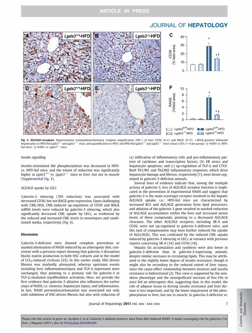
ALE/AGE-receptor expression
RAGE and CD36 protein content and mRNA levels increased sig-nificantly only in Lgals3+/+ mice fed a HFD, with immunoreactivity
mice from diet-induced NASH: A major scavenging role for galectin-3 in
1 vol. xxx j xxx–xxx 5

F
E
Lgals3+/+-HFD Lgals3-/--HFD E D F
Lgals3+/+-HFD Lgals3-/--HFD B A C
α-SM
A(%
field
are
a)
0
1
2
3
5
4 *
† ‡
Cas
p-3
(n/fi
eld
area
)
0.0
0.5
1.0
1.5
2.5
2.0
*
‡
Lgals3+/+-HFD Lgals3-/--HFD
Lgals3+/+-NFD Lgals3-/--NFD
Fig. 3. Apoptosis and HSC myofibroblast differentiation. Representative immunohistochemistry of liver active caspase-3 (A-C), original magnification 250�), and a-SMA(D–F), original magnification 400�) in HFD-fed Lgals3+/+ and Lgals3�/� mice; and quantification in NFD- and HFD-fed Lgals3+/+ and Lgals3�/� mice (mean ± SD; n = 4 pergroup). ⁄p <0.001 or �p <0.01 vs. NFD-fed mice; �p <0.001 vs. Lgals3+/+ mice.
Research Article
for RAGE involving inflammatory cells, especially in the portalarea, and some ballooned hepatocytes, and that for CD36 remain-ing confined to sinusoidal endothelial cells (Fig. 5 and Table 2).SR-A1 and TLR-2 were up-regulated in mice fed a HFD, with sig-nificantly higher increments in Lgals3+/+ vs. Lgals3�/� mice,
B A
D E
Lgals3+/+-HFD
Lgals3+/+-HFD
Fig. 4. ALE content. Representative immunohistochemistry (original magnification 250mice; and quantification in NFD- and HFD-fed Lgals3+/+ and Lgals3�/� mice (mean ± SD;
Please cite this article in press as: Iacobini C et al. Galectin-3 ablation protectsliver. J Hepatol (2011), doi:10.1016/j.jhep.2010.09.020
6 Journal of Hepatology 201
whereas TLR-4 mRNA levels increased significantly only inLgals3+/+ animals (Table 2). Galectin-3 protein expression, whichwas limited to Kupffer cells and biliary ducts in NFD-fed Lgals3+/+
mice, increased significantly in HFD-fed animals also showingpositivity for hepatocytes (Supplemental Fig. 1M–O and Table 2).
F
C Lgals3-/--HFD
Lgals3-/--HFD
CM
L(%
field
are
a)
HN
E ad
duct
s(%
fiel
d ar
ea)
0 5
10 15
25 30
20 *
* †
0 5
10 15
25 30
20
*
* †
Lgals3+/+-HFD Lgals3-/--HFD
Lgals3+/+-NFD Lgals3-/--NFD
�) of liver HNE adducts (A–C) and CML (D–F) in HFD-fed Lgals3+/+ and Lgals3�/�
n = 4 per group). ⁄p <0.001 vs. NFD-fed mice; �p <0.001 vs. Lgals3+/+ mice.
mice from diet-induced NASH: A major scavenging role for galectin-3 in
1 vol. xxx j xxx–xxx
B
D E F
C Lgals3+/+-HFD Lgals3-/--HFD
Lgals3+/+-HFD Lgals3-/--HFD
A
RAG
E(%
field
are
a)
CD
36(%
fiel
d ar
ea)
0
10
20
40
30 *
* †
0
2
4
6
10
8 *
* †
Lgals3+/+-HFD Lgals3-/--HFD
Lgals3+/+-NFD Lgals3-/--NFD
Fig. 5. ALE/AGE-receptors. Representative immunohistochemistry (original magnification 250�) of liver CD36 (A–C) and RAGE (D–F); = RAGE-positive balloonedhepatocytes) in HFD-fed Lgals3+/+ and Lgals3�/� mice; and quantification in NFD- and HFD-fed Lgals3+/+ and Lgals3�/� mice (mean ± SD; n = 4 per group). ⁄p <0.001 vs. NFD-fed mice; �p <0.001 vs. Lgals3+/+ mice.
JOURNAL OF HEPATOLOGY
Insulin signalling
Insulin-stimulated Akt phosphorylation was decreased in HFD-vs. NFD-fed mice, and the extent of reduction was significantlyhigher in Lgals3+/+ vs. Lgals3�/� mice in liver, but not in muscle(Supplemental Fig. 2).
ALE/AGE uptake by LECs
Galectin-3 silencing (70% reduction) was associated withdecreased CD36, but not RAGE gene expression. Upon challengingwith CML-HSA, CML-induced up-regulation of CD36 and RAGEmRNA levels were reduced by galectin-3 silencing, which alsosignificantly decreased CML uptake by LECs, as evidenced bythe reduced and increased CML levels in monolayers and condi-tioned media, respectively (Fig. 6).
Discussion
Galectin-3-deficient mice showed complete prevention ormarked attenuation of NASH induced by an atherogenic diet, con-sistent with a previous report showing that galectin-3 disruptionblocks matrix production in both HSC cultures and in the modelof CCL4-induced cirrhosis [22]. In this earlier study, HSC-drivenfibrosis was markedly attenuated, whereas upstream eventsincluding liver inflammation/injury and TGF-b expression wereunchanged, thus pointing to a primary role for galectin-3 inTGF-b-mediated myofibroblast activation. Here, we provide thefirst evidence that galectin-3 ablation also influences the earliersteps of NASH, i.e. steatosis, hepatocyte injury, and inflammation.In fact, NASH prevention/attenuation was associated not onlywith inhibition of HSC-driven fibrosis but also with reduction of
Please cite this article in press as: Iacobini C et al. Galectin-3 ablation protectsliver. J Hepatol (2011), doi:10.1016/j.jhep.2010.09.020
Journal of Hepatology 201
(a) infiltration of inflammatory cells and pro-inflammatory pat-tern of cytokines and transcription factors, (b) ER stress andhepatocyte apoptosis; and (c) up-regulation of TGF-b and CTGF.Both Th1/M1 and Th2/M2 inflammatory responses, which drivehepatocyte damage and fibrosis, respectively [7], were down-reg-ulated in galectin-3-deficient animals.
Several lines of evidence indicate that, among the multipleactions of galectin-3, loss of ALE/AGE receptor function is impli-cated in the prevention of experimental NASH and suggest thatgalectin-3 is the main scavenger receptor involved in the hepaticALE/AGE uptake, i.e.: HFD-fed mice are characterized byincreased RCS and ALE/AGE generation from lipid precursors,and ablation of the galectin-3 gene resulted in marked reductionof ALE/AGE accumulation within the liver and increased serumlevels of these compounds, pointing to a decreased ALE/AGEclearance. The other AGE/ALE receptors, including SR-A andCD36, were not up-regulated in galectin-3-deficient mice, andthis lack of compensation may have further reduced the uptakeof ALEs/AGEs. This was confirmed by the reduced CML uptakeinduced by galectin-3 silencing in LECs, at variance with previousreports concerning SR-A [16] and CD36 [18].
Hepatic fat accumulation and synthesis were also lower ingalectin-3-deficient than in galectin-3-expressing animals,despite similar increases in circulating lipids. This may be attrib-uted to the slightly lower degree of insulin resistance, though itmight also be secondary to the reduced extent of liver injury,since the cause-effect relationship between steatosis and insulinresistance is bidirectional [2]. This view is supported by the non-obese phenotype and the nonsignificant increase of free FAs inmice fed an atherogenic diet, suggesting that, in this model, therole of adipose tissue in driving insulin resistance and liver dis-ease is less important, and by the lower impairment of Akt phos-phorylation in liver, but not in muscle, in galectin-3-deficient vs.
mice from diet-induced NASH: A major scavenging role for galectin-3 in
1 vol. xxx j xxx–xxx 7

Gal
-3(/β
-act
in))
0
2
1
3
5
4
*
CM
L(/β
-act
in)
CM
L
0
2
1
3
5
4
*
0 -
1h +
2h +
24h +
CML 50 µg/ml
- 70
CML-HSA
CML-HSA
- 70
- 42
A
B
C F
E
D Galectin-3
- 30
- 42
β-actin
β-actin
kDa
Ctr Si
Ctr Si
Ctr Si
Nonsilenced (Ctr) Galectin-3-silenced (Si)
0.0
0.5
1.0
2.0
1.5
*
Gal
actin
-3(/1
8s)
0.0
0.4
0.8
1.2
1.6
* * * *
CD
36(/1
8s)
0.0
0.4
0.8
1.2
1.6
* * * *
RAG
E(/1
8s)
0.0
0.4
0.8
1.2
1.6 * ††
Fig. 6. Galectin-3 silencing in LECs. Galectin-3, CD36, and RAGE mRNA levels in galectin-3-silenced and nonsilenced LECs probed with CML-HSA (A–C); representativeWestern blots and quantification of galectin-3 protein in cell extracts (D) and CML content in cell extracts (E) and conditioned media (F) from galectin-3-silenced andnonsilenced LECs incubated with CML-HSA for 5 h (optical density; mean ± SD; n = 3). ⁄p <0.001 or �p <0.01 vs. nonsilenced LECs.
Research Article
wild type animals. Reduced FA uptake due to lack of up-regula-tion of CD36, which serves as an FA translocase, may have con-tributed to attenuated steatosis, and the blunted up-regulationof FA oxidation associated with decreased fat deposition mayhave further decreased ALE/AGE accumulation by reducing ROSgeneration driving lipid peroxidation.
These data, as well as those by Henderson et al. [22], are incontrast with the report of spontaneous NAFLD/NASH occurringwith aging in galectin-3-deficient mice [23]. We have no obviousexplanation for this discrepancy. In fact, we have followed galec-tin-3-deficient mice of both sexes for up to 24 months anddetected more marked age-related renal changes [28], but noliver disease.
At variance with the liver, in the aorta and kidney of galectin-3-deficient mice fed an atherogenic diet, ALEs/AGEs content wasincreased, consistent with circulating levels, and other receptorssuch as RAGE, TLRs, and SRs were up-regulated [24,25]. More-over, according to the major role of ALEs/AGEs in diet-inducedatherosclerosis and renal disease, both these conditions weremarkedly accelerated [24,25]. Deletion of the galectin-3 gene alsoaggravated glomerular injury caused by diabetes [29], CML injec-tion [26], and aging [28], all conditions characterized byincreased AGE/ALE levels. Taken together, these data suggestthat, in mice lacking galectin-3, impaired AGE/ALE removal bythe liver causes increased circulating levels of these compoundsand enhanced uptake by vascular and renal tissues via RAGE
Please cite this article in press as: Iacobini C et al. Galectin-3 ablation protectsliver. J Hepatol (2011), doi:10.1016/j.jhep.2010.09.020
8 Journal of Hepatology 201
and other receptors, which trigger pro-oxidant and pro-inflam-matory pathways leading to injury in these extra-hepatic tissues.Thus, tissue discrepancies in the consequences of galectin-3 abla-tion seem to reflect tissue differences in the function of the ALE/AGE receptor system, which, in the liver, is mainly implicated inALE/AGE removal/detoxification. This interpretation is in keepingwith the view that the role of galectin-3 in vivo is dependent on thetype of injurious stimulus and the context of organ damage [30].
In conclusion, galectin-3 ablation prevents the developmentof NASH induced by an atherogenic diet by inhibiting not onlyHSC myofibroblast transdifferentiation and fibrosis but also oxi-dant-dependent tissue inflammation and hepatocyte injury. Italso reduced up-regulation of lipid synthesis and oxidation, caus-ing less fat deposition (and possibly insulin resistance) and oxida-tive stress (with reduced ALE generation), respectively. Theseeffects can be attributed to a reduced LEC uptake and hepaticALE/AGE accumulation, which suggests that galectin-3 is a majorscavenger receptor involved in ALE/AGE uptake by the liver, acentral mechanism for clearing these compounds from the circu-lation and preventing deposition in extra-hepatic tissues.
Financial support
This work was supported by grants from the European Founda-tion for the Study of Diabetes/Juvenile Diabetes Foundation Inter-national/Novo Nordisk, the Telethon Foundation, the Ministry of
mice from diet-induced NASH: A major scavenging role for galectin-3 in
1 vol. xxx j xxx–xxx

JOURNAL OF HEPATOLOGY
University and Research of Italy, and the Diabetes, Endocrinologyand Metabolism Foundation, Rome, Italy.Conflict of interest
The authors who have taken part in this study declared that theydo not have anything to disclose regarding funding or conflict ofinterest with respect to this manuscript.
Supplementary data
Supplementary data associated with this article can be found, inthe online version, at doi:10.1016/j.jhep.2010.09.020.
References
[1] Erickson SK. Nonalcoholic fatty liver disease. J Lipid Res 2009;50:S412–416.[2] Marchesini G, Bugianesi E, Forlani G, Cerrelli F, Lenzi M, Manini R, et al.
Nonalcoholic fatty liver, steatohepatitis, and the metabolic syndrome.Hepatology 2003;37:917–923.
[3] Bradbury MW, Berk PD. Lipid metabolism in hepatic steatosis. Clin Liver Dis2004;8:639–671.
[4] Anstee QM, Goldin RD. Mouse models in non-alcoholic fatty liver disease andsteatohepatitis research. Int J Exp Pathol 2006;87:1–16.
[5] Maher JJ, Leon P, Ryan JC. Beyond insulin resistance. Innate immunity innonalcoholic steatohepatitis. Hepatology 2008;48:670–678.
[6] Koteish A, Diehl AM. Animal models of steatosis. Semin Liver Dis2001;21:89–104.
[7] Choi S, Diehl AM. Role of inflammation in nonalcoholic steatohepatitis. CurrOpin Gastroenterol 2005;21:702–707.
[8] Negre-Salvayre A, Coatrieux C, Ingueneau C, Salvayre R. Advanced lipidperoxidation end products in oxidative damage to proteins. Potential role indiseases and therapeutic prospects for the inhibitors. Br. J. Pharmacol.2008;153:6–20.
[9] George J, Pera N, Phung N, Leclercq I, Yun Hou J, Farrell G. Lipid peroxidation,stellate cell activation and hepatic fibrogenesis in a rat model of chronicsteatohepatitis. J Hepatol 2003;39:756–764.
[10] Sebeková K, Kupcová V, Schinzel R, Heidland A. Markedly elevated levels ofplasma advanced glycation end products in patients with liver cirrhosis–amelioration by liver transplantation. J Hepatol 2002;36:66–71.
[11] Smedsrød B, Melkko J, Araki N, Sano H, Horiuchi S. Advanced glycation endproducts are eliminated by scavenger-receptor-mediated endocytosis inhepatic sinusoidal Kupffer and endothelial cells. Biochem J 1997;322:567–573.
[12] Moore KJ. Freeman MW: Scavenger receptors in atherosclerosis: beyondlipid uptake. Arterioscler Thromb Vasc Biol 2006;26:1702–1711.
[13] Hansen B, Svistounov D, Olsen R, Nagai R, Horiuchi S, Smedsrød B. Advancedglycation end products impair the scavenger function of rat hepaticsinusoidal endothelial cells. Diabetologia 2002;45:1379–1388.
[14] Targher G, Marra F, Marchesini G. Increased risk of cardiovascular disease innon-alcoholic fatty liver disease: causal effect or epiphenomenon? Diabet-ologia 2008;51:1947–1953.
Please cite this article in press as: Iacobini C et al. Galectin-3 ablation protectsliver. J Hepatol (2011), doi:10.1016/j.jhep.2010.09.020
Journal of Hepatology 201
[15] Chou MY, Hartvigsen K, Hansen LF, Fogelstrand L, Shaw PX, Boullier A, et al.Oxidation-specific epitopes are important targets of innate immunity. JIntern Med 2008;263:479–488.
[16] Matsumoto K, Sano H, Nagai R, Suzuki H, Kodama T, Yoshida M, et al.Endocytic uptake of advanced glycation end products by mouse liversinusoidal cells is mediated by a scavenger receptor distinct from themacrophage scavenger receptor class A. Biochem J 2000;352:233–240.
[17] Kunjathoor VV, Febbraio M, Podrez EA, Moore KJ, Andersson L, Koehn S,et al. Scavenger receptors class A-I/II and CD36 are the principalreceptors responsible for the uptake of modified low density lipoproteinleading to lipid loading in macrophages. J Biol Chem 2002;277:49982–49988.
[18] Nakajou K, Horiuchi S, Sakai M, Hirata K, Tanaka M, Takeya M, et al. CD36 isnot involved in scavenger receptor–mediated endocytic uptake of glycolal-dehyde- and methylglyoxal-modified proteins by liver endothelial cells. JBiochem 2005;137:607–616.
[19] Butscheid M, Hauptvogel P, Fritz P, Klotz U, Alscher D. Hepatic expression ofGalectin-3 and RAGE in patients with liver disease. J Clin Pathol2007;60:415–418.
[20] Xia J-R, Liu N-F, Zhu N-X. Specific siRNA targeting the receptor for advancedglycation end products inhibits experimental hepatic fibrosis in rats. Int JMol Sci 2008;9:638–661.
[21] Fehrenbach H, Weiskirchen R, Kasper M, Gressner AM. Up-regulatedexpression of the receptor for advanced glycation end products in culturedrat hepatic stellate cells during transdifferentiation to myofibroblasts.Hepatology 2001;34:943–952.
[22] Henderson NC, Mackinnon AC, Farnworth SL, Poirier F, Russo FP, Iredale JP,et al. Galectin-3 regulates myofibroblast activation and hepatic fibrosis. ProcNatl Acad Sci USA 2006;103:5060–5065.
[23] Nomoto K, Tsuneyama K, Abdel Aziz HO, Takahashi H, Murai Y, Cui Z-G, et al.Disrupted galectin-3 causes non-alcoholic fatty liver disease in male mice. JPathol 2006;210:469–477.
[24] Iacobini C, Menini S, Ricci C, Scipioni A, Sansoni V, Cordone S, et al.Accelerated lipid-induced atherogenesis in galectin-3-deficient mice. role oflipoxidation via receptor-mediated mechanisms. Arterioscler Thromb VascBiol 2009;29:831–836.
[25] Iacobini C, Menini S, Ricci C, Scipioni A, Sansoni V, Mazzitelli G, et al.Advanced lipoxidation end-products mediate lipid-induced glomerularinjury: role of receptor-mediated mechanisms. J Pathol 2009;218:360–369.
[26] Iacobini C, Menini S, Oddi G, Ricci C, Amadio L, Pricci F, et al. Galectin-3/AGE-receptor 3 knockout mice show accelerated AGE-induced glomerular injury.Evidence for a protective role of galectin-3 as an AGE-receptor. FASEB J2004;18:1773–1775.
[27] Neuschwander-Tetri BA, Caldwell SH. Nonalcoholic steatohepatitis: sum-mary of an AASLD Single Topic Conference. Hepatology 2003;37:1202–1219.
[28] Iacobini C, Oddi G, Menini S, Amadio L, Ricci C, Barsotti P, et al. Developmentof age-dependent glomerular lesions in galectin-3/AGE-receptor-3 knockoutand wild type mice. Am J Physiol 2005;289:F611–F621.
[29] Pugliese G, Pricci F, Iacobini C, Leto G, Amadio L, Barsotti P, et al. Accelerateddiabetic glomerulopathy in galectin-3/AGE-receptor-3 knockout mice.FASEB J 2001;15:2471–2479.
[30] Henderson NC, Sethi T. The regulation of inflammation by galectin-3.Immunol Rev 2009;230:160–171.
mice from diet-induced NASH: A major scavenging role for galectin-3 in
1 vol. xxx j xxx–xxx 9
Copyright © 2022 FDOKUMEN