A Cyclin-Binding Motif within the Amino-Terminal Homology Domain of EBNA3C Binds Cyclin A and...
12
10.1128/JVI.78.23.12857-12867.2004. 2004, 78(23):12857. DOI: J. Virol. S. Robertson Jason S. Knight, Nikhil Sharma, Danielle E. Kalman and Erle Epstein-Barr Virus-Infected Cells Cyclin A-Dependent Kinase Activity in EBNA3C Binds Cyclin A and Modulates Amino-Terminal Homology Domain of A Cyclin-Binding Motif within the http://jvi.asm.org/content/78/23/12857 Updated information and services can be found at: These include: REFERENCES http://jvi.asm.org/content/78/23/12857#ref-list-1 at: This article cites 32 articles, 18 of which can be accessed free CONTENT ALERTS more» articles cite this article), Receive: RSS Feeds, eTOCs, free email alerts (when new http://journals.asm.org/site/misc/reprints.xhtml Information about commercial reprint orders: http://journals.asm.org/site/subscriptions/ To subscribe to to another ASM Journal go to: on April 3, 2014 by guest http://jvi.asm.org/ Downloaded from on April 3, 2014 by guest http://jvi.asm.org/ Downloaded from
-
Upload
independent -
Category
Documents
-
view
3 -
download
0
Transcript of A Cyclin-Binding Motif within the Amino-Terminal Homology Domain of EBNA3C Binds Cyclin A and...
10.1128/JVI.78.23.12857-12867.2004.
2004, 78(23):12857. DOI:J. Virol. S. RobertsonJason S. Knight, Nikhil Sharma, Danielle E. Kalman and Erle Epstein-Barr Virus-Infected CellsCyclin A-Dependent Kinase Activity in EBNA3C Binds Cyclin A and ModulatesAmino-Terminal Homology Domain of A Cyclin-Binding Motif within the
http://jvi.asm.org/content/78/23/12857Updated information and services can be found at:
These include:
REFERENCEShttp://jvi.asm.org/content/78/23/12857#ref-list-1at:
This article cites 32 articles, 18 of which can be accessed free
CONTENT ALERTS more»articles cite this article),
Receive: RSS Feeds, eTOCs, free email alerts (when new
http://journals.asm.org/site/misc/reprints.xhtmlInformation about commercial reprint orders: http://journals.asm.org/site/subscriptions/To subscribe to to another ASM Journal go to:
on April 3, 2014 by guest
http://jvi.asm.org/
Dow
nloaded from
on April 3, 2014 by guest
http://jvi.asm.org/
Dow
nloaded from

JOURNAL OF VIROLOGY, Dec. 2004, p. 12857–12867 Vol. 78, No. 230022-538X/04/$08.00�0 DOI: 10.1128/JVI.78.23.12857–12867.2004Copyright © 2004, American Society for Microbiology. All Rights Reserved.
A Cyclin-Binding Motif within the Amino-Terminal Homology Domainof EBNA3C Binds Cyclin A and Modulates Cyclin A-Dependent
Kinase Activity in Epstein-Barr Virus-Infected CellsJason S. Knight, Nikhil Sharma, Danielle E. Kalman, and Erle S. Robertson*
Department of Microbiology and the Abramson Comprehensive Cancer Center, University of PennsylvaniaMedical School, Philadelphia, Pennsylvania
Received 16 February 2004/Accepted 22 July 2004
The Epstein-Barr virus (EBV) nuclear antigen 3C (EBNA3C) is a virus-encoded latent antigen essential forprimary B-cell transformation. In this report we demonstrate that although the carboxy terminus of EBNA3Cpredominantly regulates cyclin A-dependent kinase activity, the region of greatest affinity for cyclin A lieswithin the EBNA3 amino-terminal homology domain of EBNA3C. Detailed mapping studies employing both invitro binding assays and coimmunoprecipitation experiments implicated a small region of EBNA3C, aminoacids 130 to 159 within the EBNA3 homology domain, as having the greatest affinity for cyclin A. The EBNA3homology domain has the highest degree of amino acid similarity (approximately 30%) between the EBNA3proteins, and, indeed, EBNA3B, but not EBNA3A, showed binding activity with cyclin A. We also show thatEBNA3C binds to the �1 helix of the highly conserved mammalian cyclin box, with cyclin A amino acids 206to 226 required for strong binding to EBNA3C amino acids 130 to 159. Interestingly, EBNA3C also boundhuman cyclins D1 and E in vitro, although the affinity was approximately 30% of that seen for cyclin A.Previously it was demonstrated that full-length EBNA3C rescues p27-mediated suppression of cyclin A-de-pendent kinase activity (J. S. Knight and E. S. Robertson, J. Virol. 78:1981-1991, 2004). It was also demon-strated that the carboxy terminus of EBNA3C recapitulates this phenotype. Surprisingly, the amino terminusof EBNA3C with the highest affinity for cyclin A was unable to rescue p27 suppression of kinase activity andactually downregulates cyclin A activity when introduced into EBV-infected cells. The data presented heresuggests that the amino terminus of EBNA3C may play an important role in recruiting cyclin A complexes,while the carboxy terminus of EBNA3C is necessary for the functional modulation of cyclin A complex kinaseactivity.
Epstein-Barr virus (EBV) has been associated with humancancer since the early 1960s. EBV was discovered in 1962 byelectron microscopy of cells cultured from Burkitt’s lymphoma,an endemic and aggressive malignancy that is predominantlyseen in equatorial Africa (7). EBV has since been implicated asthe likely causative agent in other malignancies, including na-sopharyngeal carcinoma, posttransplant lymphomas, and someforms of Hodgkins disease (11, 22). Further, EBV has beenmore tenuously linked to additional cancers such as gastriccarcinoma and, somewhat controversially, metastatic breastcarcinoma (11, 22, 28). Early studies of EBV noted its potentability to transform resting primary B lymphocytes in cell cul-ture. Subsequently, in vitro recombination studies identifiedfive viral latency genes that are absolutely required for thetransformation phenotype (11, 22). These include LMP1,EBNA1, EBNA2, EBNA3A, and EBNA3C (11, 22). Of theseessential gene products, the EBNA3 proteins are clearly theleast well understood in terms of their contribution to B-lym-phocyte transformation.
EBNA3C of type 1 EBV is a 992-amino-acid protein thatlocalizes to the nucleus in either a diffuse or punctate pattern
depending on the cell type and expression level (12, 25, 28).EBNA3C has been shown to play a complex regulatory role inthe transcription of viral and cellular genes. EBNA3C antag-onizes EBNA2-mediated transactivation by competing withEBNA2 for RBP-J� binding (10, 19, 24), and, conversely, co-operates with EBNA2 in the upregulation of some promoters,like the EBV LMP1 promoter (2, 3, 33). Additionally,EBNA3C recruits both histone acetylase and deacetylase ac-tivities (12, 20, 29), in part through its association with thesmall acidic nuclear protein prothymosin-alpha (5). Further,EBNA3C associates with an antimetastatic factor, Nm23-H1,and may modulate the transcription of cellular genes involvedin cell migration and invasion (28, 30).
In addition to this somewhat diffuse transcriptional regula-tory picture, EBNA3C also modulates the mammalian cellcycle, presumably through direct protein-protein interactions,by targeting multiple checkpoint regulatory proteins (17, 18).To this end, EBNA3C has previously been shown to indirectlytarget pathways regulated by the retinoblastoma tumor sup-pressor (Rb) (17, 18). EBNA3C activates E2F-dependent pro-moters and induces foci formation similar to papillomavirus E7in a colony formation assay (17). Additionally, EBNA3C over-comes the ability of the cyclin-dependent kinase inhibitorp16INK4A to block transformation and dramatically drives se-rum-starved cells through the G1/S restriction point (17, 18).
Despite this evidence, a clear molecular link between the Rbprotein and EBNA3C has yet to be demonstrated in vivo.
* Corresponding author. Mailing address: Department of Microbi-ology and the Abramson Comprehensive Cancer Center, University ofPennsylvania Medical School, 201E Johnson Pavilion, 3610 HamiltonWalk, Philadelphia, PA 19104. Phone: (215) 746-0114 or (215) 746-0116. Fax: (215) 898 -9557. E-mail: [email protected].
12857
on April 3, 2014 by guest
http://jvi.asm.org/
Dow
nloaded from
More recently, it was shown that EBNA3C may indirectlytarget Rb by associating with cyclin A complexes in EBV-transformed lymphoblastoid cell lines (13). Cell cycle progres-sion is partially dependent on the activity of cyclin-dependentkinases (cdks). These kinases are allosterically activated bybinding to cyclins, a family of proteins whose levels oscillate insynchrony with cell cycle progression (14). Cyclin A, synthe-sized beginning in G1 and maximally at the onset of S phase, isnecessary for both progression through S phase and for entryinto mitosis (15). Additionally, exogenous expression of cyclinA has been shown to accelerate exit from G1 (21). EBNA3Cstimulates cyclin A-dependent kinase activity and rescues p27-mediated inhibition of cyclin A/Cdk2 kinase activity by de-creasing the molecular association between cyclin A and p27 incells (13). Cyclin A functionally interacts with a region of thecarboxy terminus of EBNA3C shown to be important for bothstimulation of cyclin A-dependent kinase activity and for cellcycle progression (13).
The molecular association between EBNA3C and cyclin Acomplexes is further examined in this study. Surprisingly, wehave identified an additional and perhaps predominant cyclinA binding site at the amino terminus of EBNA3C not previ-
ously investigated in the initial report of Knight and Roberston(13) identifying cyclin A as an EBNA3C binding protein. Anin-depth understanding of the different domains of EBNA3Cthat modulate cyclin A complexes will lead to a better under-standing of the basic mechanism underlying the regulation ofthe mammalian cell cycle by EBV.
MATERIALS AND METHODS
Plasmids, antibodies, and cell lines. pA3M-E3C constructs expressing eitherfull-length EBNA3C or EBNA3C truncations 1-365, 366-620, and 621-992 witha carboxy-terminal myc tag have been described previously (5). pA3M-E3Cconstructs expressing amino acids 1 to 100 and 1 to 200 were prepared by cloningPCR-amplified cDNAs into the previously described pA3M vector (4). Gluta-thione S transferase (GST)-EBNA3C truncation mutants were prepared by clon-ing PCR-amplified cDNAs into pGEX-2TK. Point mutations in the EBNA3Cgene were prepared by a standard PCR primer mutagenesis method. pSG5-EBNA3A, pSG5-EBNA3B, and pSG5-EBNA3C have been described previously(24). pA3M-cyclin A constructs expressing either full-length cyclin A or cyclin Atruncations with a carboxy-terminal myc tag were prepared by cloning PCR-amplified cDNAs into the previously described pA3M vector (4). GST-cyclin Afusions were prepared by cloning PCR amplified fragments into the pGEX-2TKvector: the cyclin box clone represents amino acids 206 to 310, �1 representsamino acids 206 to 227, and �2-5 represents amino acids 228 to 310. pCMV-Cdk2, RC-cyclin A, and RC-cyclin E were kindly provided by Philip Hinds (8).
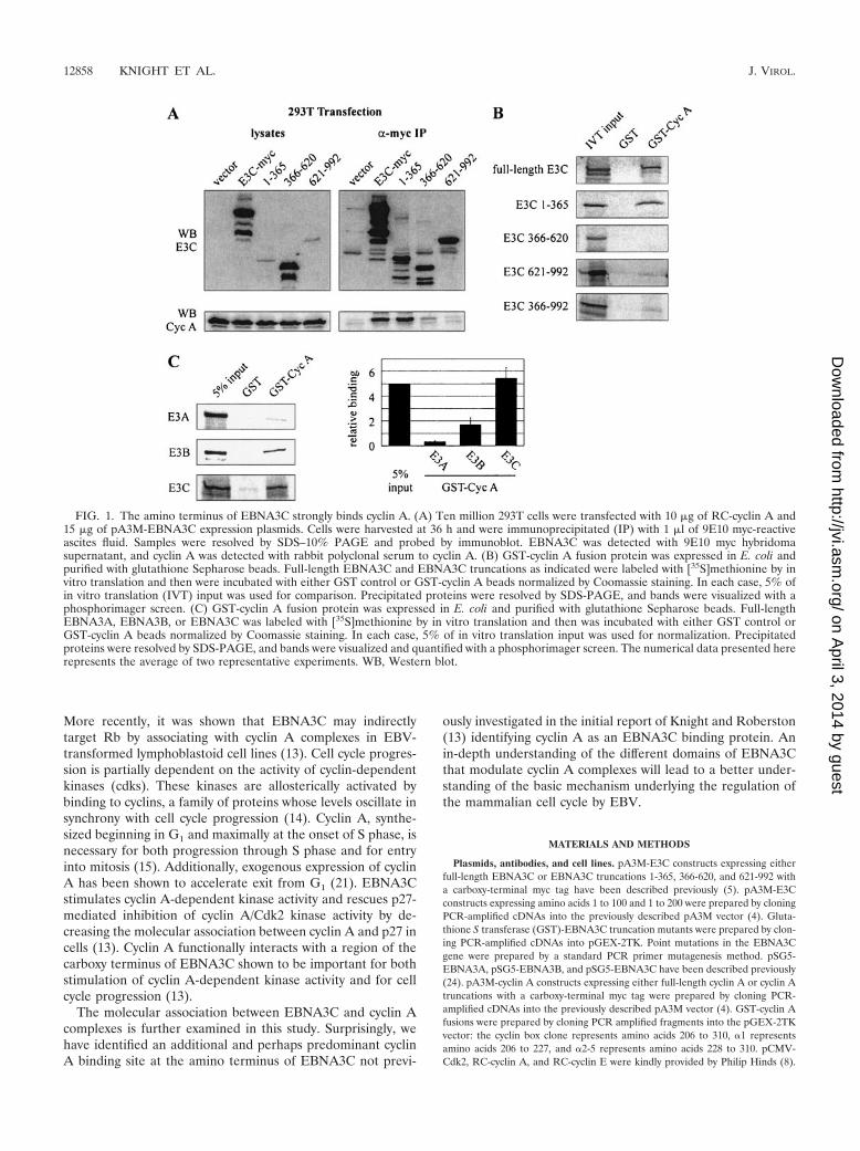
FIG. 1. The amino terminus of EBNA3C strongly binds cyclin A. (A) Ten million 293T cells were transfected with 10 �g of RC-cyclin A and15 �g of pA3M-EBNA3C expression plasmids. Cells were harvested at 36 h and were immunoprecipitated (IP) with 1 �l of 9E10 myc-reactiveascites fluid. Samples were resolved by SDS–10% PAGE and probed by immunoblot. EBNA3C was detected with 9E10 myc hybridomasupernatant, and cyclin A was detected with rabbit polyclonal serum to cyclin A. (B) GST-cyclin A fusion protein was expressed in E. coli andpurified with glutathione Sepharose beads. Full-length EBNA3C and EBNA3C truncations as indicated were labeled with [35S]methionine by invitro translation and then were incubated with either GST control or GST-cyclin A beads normalized by Coomassie staining. In each case, 5% ofin vitro translation (IVT) input was used for comparison. Precipitated proteins were resolved by SDS-PAGE, and bands were visualized with aphosphorimager screen. (C) GST-cyclin A fusion protein was expressed in E. coli and purified with glutathione Sepharose beads. Full-lengthEBNA3A, EBNA3B, or EBNA3C was labeled with [35S]methionine by in vitro translation and then was incubated with either GST control orGST-cyclin A beads normalized by Coomassie staining. In each case, 5% of in vitro translation input was used for normalization. Precipitatedproteins were resolved by SDS-PAGE, and bands were visualized and quantified with a phosphorimager screen. The numerical data presented hererepresents the average of two representative experiments. WB, Western blot.
12858 KNIGHT ET AL. J. VIROL.
on April 3, 2014 by guest
http://jvi.asm.org/
Dow
nloaded from
The construct expressing GST-cyclin A was provided by Maria Mudryj (6).pCDNA3-p27 was provided by Michele Pagano (16). pCDNA3-cyclin D1 wasprovided by Alan Diehl (University of Pennsylvania). Rabbit polyclonal antibod-ies reactive to cyclin A, cyclin E, Cdk2, and p27Kip1 were purchased from Santa
Cruz Biotechnology, Inc. A10 monoclonal and rabbit polyclonal antibodies re-active to EBNA3C have been previously described (5). HEK 293 cells are humanembryonic kidney cells transformed by adenovirus type 5 DNA; HEK 293T cellsstably express the simian virus 40 large T antigen (1). U2OS is a human osteo-
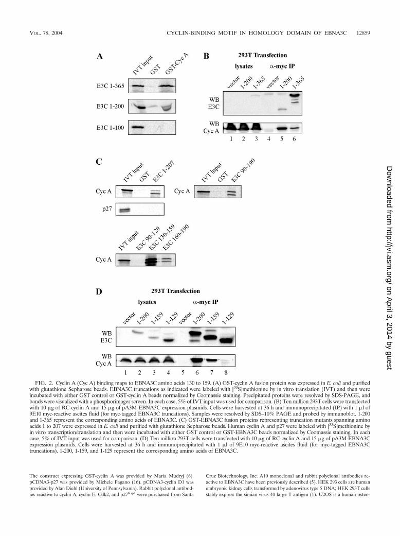
FIG. 2. Cyclin A (Cyc A) binding maps to EBNA3C amino acids 130 to 159. (A) GST-cyclin A fusion protein was expressed in E. coli and purifiedwith glutathione Sepharose beads. EBNA3C truncations as indicated were labeled with [35S]methionine by in vitro translation (IVT) and then wereincubated with either GST control or GST-cyclin A beads normalized by Coomassie staining. Precipitated proteins were resolved by SDS-PAGE, andbands were visualized with a phosphorimager screen. In each case, 5% of IVT input was used for comparison. (B) Ten million 293T cells were transfectedwith 10 �g of RC-cyclin A and 15 �g of pA3M-EBNA3C expression plasmids. Cells were harvested at 36 h and immunoprecipitated (IP) with 1 �l of9E10 myc-reactive ascites fluid (for myc-tagged EBNA3C truncations). Samples were resolved by SDS–10% PAGE and probed by immunoblot. 1-200and 1-365 represent the corresponding amino acids of EBNA3C. (C) GST-EBNA3C fusion proteins representing truncation mutants spanning aminoacids 1 to 207 were expressed in E. coli and purified with glutathione Sepharose beads. Human cyclin A and p27 were labeled with [35S]methionine byin vitro transcription/translation and then were incubated with either GST control or GST-EBNA3C beads normalized by Coomassie staining. In eachcase, 5% of IVT input was used for comparison. (D) Ten million 293T cells were transfected with 10 �g of RC-cyclin A and 15 �g of pA3M-EBNA3Cexpression plasmids. Cells were harvested at 36 h and immunoprecipitated with 1 �l of 9E10 myc-reactive ascites fluid (for myc-tagged EBNA3Ctruncations). 1-200, 1-159, and 1-129 represent the corresponding amino acids of EBNA3C.
VOL. 78, 2004 CYCLIN-BINDING MOTIF IN HOMOLOGY DOMAIN OF EBNA3C 12859
on April 3, 2014 by guest
http://jvi.asm.org/
Dow
nloaded from
sarcoma cell line. HEK 293T and U2OS cells were grown in Dulbecco’s modifiedEagle’s medium (DMEM; Gibco-BRL) with 10% fetal bovine serum unlessotherwise indicated. For most experiments, 10 million 293T cells were trans-fected by electroporation at 210 V and 975 �F in a 0.4-�m cuvette.
GST pull-down assays. GST fusion proteins were purified from bulk Esche-richia coli cultures following induction with IPTG as described previously (5). Forin vitro binding experiments, GST fusion proteins were incubated with 35S-labeled in vitro-translated protein in binding buffer (1� phosphate-bufferedsaline [PBS], 0.1% NP-40, 0.5 mM dithiothreitol [DTT], 10% glycerol, supple-mented with protease inhibitors). In vitro translation was with the TNT T7 QuickCoupled Transcription/Translation System (Promega Inc., Madison, Wis.) ac-cording to the manufacturer’s instructions.
Immunoprecipitation assays. Immunoprecipitation was essentially performedas described previously (5). Proteins were fractionated by electrophoresis on asodium dodecyl sulfate (SDS)-polyacrylamide gel and then transferred to a0.45-�m nitrocellulose membrane. The membrane was blotted with appropriaterabbit polyclonal antibodies followed by horseradish peroxidase-linked Protein A(Amersham Biosciences, Piscataway, N.J.) at a 1:5,000 dilution in PBS.
In vitro kinase assay. U2OS cells were seeded into 6-well plates and grown toconfluence in 0.5% FBS for 48 h prior to transfection. Cells were transfected withLipofectamine 2000 reagent (Invitrogen Corporation, Bethesda, Md.), harvestedafter 24 h with a cell scraper, washed with PBS, and lysed on ice in 500 �l ofradioimmunoprecipitation assay (RIPA) buffer (0.5% NP-40, 10 mM Tris [pH7.5], 2 mM EDTA, 150 mM NaCl, 1 mM EGTA, plus protease and phosphataseinhibitors). Lysates were precleared and then rotated with 1 �g of cyclin Aantibody overnight at 4°C. Cyclin A complexes were captured by rotating withProtein A-Sepharose beads and were washed with RIPA buffer. Cyclin A com-plexes were then washed with histone wash buffer (25 mM Tris [pH 7.5], 70 mMNaCl, 10 mM MgCl2, 1 mM EGTA, 1 mM DTT, plus protease and phosphataseinhibitors). Complexes were incubated in 30 �l of histone wash buffer supple-mented with 4 �g of histone H1 (Upstate Biotechnology, Inc., Lake Placid,N.Y.), 10 mM cold ATP, and 0.2 �Ci of [�-32P]-ATP/�l for 30 min at 37°C. Thereaction was stopped by adding SDS-lysis buffer and heating to 95°C for 10 min.Labeled Histone H1 was resolved by SDS–12% polyacrylamide gel electrophore-sis (PAGE). Quantitation was performed with ImageQuant software (AmershamBiosciences).
LCLs were transfected by electroporation with the Bio-Rad Gene Pulser II at220 V and 975 �F, and transfection efficiency was monitored by using a cotrans-fected green fluorescent protein (GFP) expression plasmid. Prior to transfection,LCLs were grown exponentially and tested by trypan blue staining for live cells.All transfections were done when cells were greater than 90% positive forexclusion of trypan blue. Transfected cells were incubated in RPMI 1640 plus10% FBS for 24 h and then were treated in the same manner as that for U2OScells.
RESULTS
In a previous study, a truncated form of EBNA3C corre-sponding to amino acids 365 to 992 was fused in frame with theGAL4 DNA binding domain and was tested against an LCL-derived cDNA library in a yeast two-hybrid screen (5). Partialsequences from two positive cDNA clones identified codingsequences that were located within the cyclin A gene (13).Based on this initial screen, a region at the extreme carboxyterminus of EBNA3C was identified as functionally regulatingcyclin A-dependent kinase activity while also possessing affinityfor cyclin A, as determined by in vitro binding assays (13).Interestingly, in follow-up studies mutational analysis of theEBNA3C-cyclin A interaction revealed that another bindingsite with greater affinity for cyclin A lies at the amino terminusof EBNA3C. The following experiments dissect this interactionand attempt to understand the functional role of the aminoterminus in modulation of cyclin A-dependent kinase activity.
Binding studies identify the amino terminus of EBNA3C asthe predominant binding site for cyclin A. To assess the asso-ciation between different regions of EBNA3C and cyclin A inthe context of cell lysates, 293T cells were transfected withexpression constructs for cyclin A and either full-length
EBNA3C, EBNA3C 1-365, EBNA3C 366-620, or EBNA3C621-992. All EBNA3C expression constructs placed a myc tagin frame with the carboxy terminus of the protein. Cyclin Acoimmunoprecipitated with relatively high affinity with bothfull-length EBNA3C and EBNA3C 1-365 (Fig. 1A). However,little to no coimmunoprecipiation of cyclin A above back-ground was detected with EBNA3C 366-620. Moreover, andsomewhat surprisingly, EBNA3C 621-992 showed significantlyreduced coimmunoprecipitation in this assay. This suggeststhat the predominant region of interaction between cyclin Aand EBNA3C lies in the amino terminus and that the interac-tion previously seen with the carboxy terminus using in vitrobinding assays (13) may be somewhat transient or masked intransiently transfected cells expressing the truncated proteins(Fig. 1A).
Bacterially expressed cyclin A binds the amino terminus ofEBNA3C. Coimmunoprecipitation experiments suggested thatthe amino terminus of EBNA3C is the predominant interact-ing site for cyclin A in cells. To corroborate this finding, theinteraction with the amino-terminal domain was tested in thecontext of bacterially expressed human cyclin A and comparedto that seen with the central and carboxy-terminal domains.GST-cyclin A beads strongly precipitated in vitro-translated,
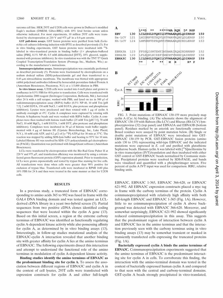
FIG. 3. Point mutations of EBNA3C 130-159 more precisely mapcyclin A (Cyc A) binding. (A) The schematic shows the alignment ofEBNA3C 130-159 with Baboon (Ba-LCV) and Rhesus (Rh-LCV) lym-phocryptovirus (32) (top panel) and EBNA3A and EBNA3B (bottompanel). Residues marked by an asterisk are functionally conserved.Boxed residues were assayed by point mutation below. (B) Single ordouble point mutations of EBNA3C were introduced into GST-EBNA3C 130-159 by PCR. GST-EBNA3C fusion proteins (aminoacids 130 to 159) representing wild-type EBNA3C and the indicatedmutations were expressed in E. coli and purified with glutathioneSepharose beads. Human cyclin A was labeled with [35S]methionine byin vitro transcription (IVT)/translation and then incubated with eitherGST control or GST-EBNA3C beads normalized by Coomassie stain-ing. Precipitated proteins were resolved by SDS-PAGE, and bandswere visualized and quantified with a phosphorimager screen. Fivepercent of cyclin A IVT input was used for comparison. RBU, relativebinding units.
12860 KNIGHT ET AL. J. VIROL.
on April 3, 2014 by guest
http://jvi.asm.org/
Dow
nloaded from
35S-labeled EBNA3C, as has been shown previously (13). In-terestingly, in vitro-translated EBNA3C 1-365 also precipi-tated strongly in this experiment, while both the central andcarboxy-terminal domains of EBNA3C showed significantlyless affinity for cyclin A than the amino terminus; however, thecarboxy terminus did have slightly greater affinity than thecentral domain (Fig. 1B).
EBNA3A and EBNA3B interact weakly with bacterially ex-pressed cyclin A. The juxtaposition and colinear homology ofthe three EBNA3 family genes suggests gene duplication of acommon ancestral gene (11, 22). While studies have revealed
shared properties among the three EBNA3 gene products,such as association with the RBP-J� transcription factor (24),the proteins have clearly diverged functionally to some extent.This is emphasized by the fact that EBNA3A and EBNA3C areessential for primary B-cell transformation in vitro whileEBNA3B appears to be dispensable (31). Previous sequencingand characterization of the EBNA3 family of proteins hasidentified a region known as the homology domain, locatedwithin amino acids 90 to 320 (23). As the EBNA3C-cyclin Ainteraction potentially falls within this region, we decided totest whether other EBNA3 family members might also interact
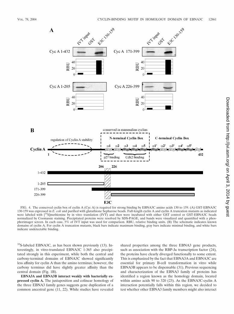
FIG. 4. The conserved cyclin box of cyclin A (Cyc A) is required for strong binding by EBNA3C amino acids 130 to 159. (A) GST-EBNA3C130-159 was expressed in E. coli and purified with glutathione Sepharose beads. Full-length cyclin A and cyclin A truncation mutants as indicatedwere labeled with [35S]methionine by in vitro translation (IVT) and then were incubated with either GST control or GST-EBNA3C beadsnormalized by Coomassie staining. Precipitated proteins were resolved by SDS-PAGE, and bands were visualized and quantified with a phos-phorimager screen. In each case, 5% of IVT input was used for comparison. RBU, relative binding units. (B) The schematic indicates knowndomains of cyclin A. For cyclin A truncation mutants, black bars indicate maximum binding, gray bars indicate minimal binding, and white barsindicate undetectable binding.
VOL. 78, 2004 CYCLIN-BINDING MOTIF IN HOMOLOGY DOMAIN OF EBNA3C 12861
on April 3, 2014 by guest
http://jvi.asm.org/
Dow
nloaded from

with bacterially expressed cyclin A. As described above, GST-cyclin A strongly precipitated in vitro-translated EBNA3C(Fig. 1C). While EBNA3A, an essential latent protein for B-cell transformation, showed an association only slightly abovebackground levels, EBNA3B was reproducibly precipitatedwith GST-cyclin A. This assay was repeated multiple times withindependently prepared GST-cyclin A to ensure that the asso-ciation with EBNA3B was significantly above that for eitherEBNA3A or GST control. The data presented in Fig. 1C is thequantification and average of two representative experiments.This finding suggests that the cyclin A binding site on EBNA3Cmay be conserved to some extent in EBNA3B and also thatEBNA3B may have some limited role in regulating cyclin Acomplexes in virally infected cells.
Amino acids 130 to 159 of EBNA3C interact with high af-finity with cyclin A. To more finely map the region of EBNA3Cresponsible for cyclin A binding in the amino-terminal regionof EBNA3C, additional truncation constructs of EBNA3Cwithin the amino-terminal 365 amino acids were prepared. Invitro precipitation experiments with bacterially expressedGST-cyclin A showed that EBNA3C amino acids 1 to 200, butnot 1 to 100, precipitated strongly with cyclin A (Fig. 2A). Thisresult suggested that EBNA3C amino acids 101 to 200 containthe predominant mediator of cyclin A binding. Indeed, cyclin
A coimmunoprecipitated strongly with EBNA3C 1-200 whenexpressed in cells (Fig. 2B). In repeated coimmunoprecipita-tion experiments, the association between cyclin A andEBNA3C 1-200 was actually stronger than the association be-tween cyclin A and EBNA3C 1-365 (Fig. 2B). This result wasin spite of the fact that 1-200 did not express as well as 1-365in cells (compare lanes 5 and 6 in Fig. 2B). This suggests thatEBNA3C sequences which lie outside of the primary bindingdomain play a potentially negative regulatory role in the func-tional association between EBNA3C and cyclin A.
GST-EBNA3C truncation constructs were next used to fur-ther refine the association between cyclin A and EBNA3C. Wefirst confirmed that, as expected, GST-EBNA3C fusion pro-teins corresponding to amino acids 1 to 207 and 90 to 190 wereboth able to strongly precipitate in vitro-translated cyclin A(Fig. 2C). This domain did not interact with in vitro-translatedp27, a known cyclin A-interacting protein (Fig. 2C). We nexttested EBNA3C fusion proteins corresponding to amino acids90 to 129, 130 to 159, and 160 to 190. In this assay, amino acids130 to 159 were the primary mediator of cyclin A binding, withamino acids 160 to 190 also having some affinity for cyclin A(Fig. 2C). To further confirm the importance of EBNA3Camino acids 130 to 159, cyclin A and EBNA3C truncationswere expressed in cells for coimmunoprecipitation analysis. As
FIG. 5. The cyclin box of cyclin A binds the amino terminus of EBNA3C. (A) GST-cyclin A cyclin box fusion protein (amino acids 206 to 310)was expressed in E. coli and purified with glutathione Sepharose beads. Full-length EBNA3C and EBNA3C truncations as indicated were labeledwith [35S]methionine by in vitro translation (IVT) and then were incubated with either GST control or GST-cyclin A cyclin box (Cyc A Box) beadsnormalized by Coomassie staining. Precipitated proteins were resolved by SDS-PAGE, and bands were visualized with a phosphorimager screen.In each case, 5% of IVT input was used for comparison. (B) Ten million 293T cells were transfected with 15 �g of pCMV-Cdk2 or 15 �g ofpA3M-EBNA3C 1-365 expression plasmid. Lysates were prepared at 36 h and incubated with either GST alone or GST-cyclin A cyclin box (CycA Box) fusion protein as described above. Samples were resolved by SDS–12% PAGE and probed by immunoblot. EBNA3C 1-365 was detectedwith 9E10 myc hybridoma supernatant, and Cdk2 was detected with rabbit polyclonal serum to Cdk2. (C) GST fusion proteins corresponding tothe �1 helix of the cyclin A cyclin box (amino acids 206 to 225) or helices �2 to �5 (amino acids 226 to 310) were expressed in E. coli and werepurified with glutathione Sepharose beads. EBNA3C amino acids 1-365 was labeled with [35S]methionine by in vitro translation (IVT) and thenwere incubated with GST control and either �1 (Cyc A Box �1) or �2 to �5 (Cyc A Box �2-5) beads normalized by Coomassie staining. In eachcase, 5% of IVT input was used for comparison.
12862 KNIGHT ET AL. J. VIROL.
on April 3, 2014 by guest
http://jvi.asm.org/
Dow
nloaded from
described above, immunoprecipitation of EBNA3C amino ac-ids 1 to 200 also strongly precipitated cyclin A (Fig. 2D).EBNA3C 1-159 also precipitated cyclin A, although at a levelless than that for EBNA3C 1-200 (Fig. 2D, compare lanes 6and 7). Importantly, EBNA3C 1-129 was unable to precipitatecyclin A (Fig. 2D, lane 8). These coimmunoprecipitation datasuggest that, consistent with the above GST pull-down data,EBNA3C amino acids 160 to 200 as well as 130 to 159 playsome role in binding cyclin A.
To identify individual residues mediating this association, wealigned EBV EBNA3C amino acids 130 to 159 with the known3C homologues of Baboon and Rhesus lymphocryptovirus(LCV) (Fig. 3A). This region of EBNA3C was chosen becauseit demonstrated the strongest association with cyclin A in GSTpull-down experiments (Fig. 2C). It has previously been dem-onstrated that transcriptional regulatory regions withinEBNA3C are conserved in these EBNA3C homologues, and itis possible that cyclin A regulatory motifs might be similarlyconserved (32). It is also possible that, given the associationbetween cyclin A and EBNA3B, some of the residues impor-tant for the EBNA3C-cyclin A interaction would be conservedin EBNA3B but not EBNA3A (Fig. 3A). Amino acids 130 to159 were about 70% conserved in Baboon and Rhesus LCVbut were conserved less so in EBNA3A and EBNA3B. Toaddress the role of individual residues, point mutations wereconstructed in the context of GST-EBNA3C 130-159. Mutatedmotifs are indicated by boxes in Fig. 3A. Mutation of phenyl-alanine 144 dramatically reduced cyclin A binding, suggestingthat this residue is especially critical (Fig. 3B). Double muta-tion of arginine 149 and arginine 151 also significantly reducedcyclin A binding (Fig. 3B). These data confirm that specificconserved residues within EBNA3C 130-159 mediate cyclin Abinding, with phenylalanine 144 playing an especially criticalrole in the EBNA3C-cyclin A association. However, additionalmutational studies may be necessary to identify mutations in
this domain capable of completely eliminating cyclin A bindingto EBNA3C.
EBNA3C binds cyclin A at the conserved cyclin box. Whilelittle is known about the tertiary and quaternary structure ofEBNA3C, cyclin A is well understood structurally and, impor-tantly, is understood in the context of structure-function rela-tionships (9, 26). In an attempt to gain insights into the func-tionality of the association between cyclin A and EBNA3C, anumber of cyclin A truncations were tested for their ability tobind GST-EBNA3C 130-159. Representative truncations aredepicted here with cyclin A 1-205 showing no binding, cyclin A171-399 showing strong binding similar to that of full-lengthcyclin A, and cyclin A 226-399 showing significantly weakerbinding than full-length cyclin A (Fig. 4A). Other truncations,including cyclin A 1-170, showed no binding, and cyclin A311-399 showed binding only slightly above background (datanot shown). Given that cyclin A 1-205 shows no binding anddeletion of cyclin A amino acids 171 to 225 significantly re-duces binding affinity (Fig. 4A, compare right two panels),these data implicate cyclin A amino acids 206 to 225 as animportant determinant of EBNA3C binding (Fig. 5B). Thisregion of cyclin A is notable because it represents the �1 helixof the so-called cyclin box of cyclin A (9) (Fig. 4B). This helixcontains a highly conserved MRAIL motif and has also beenshown to be a primary determinant of both substrate recruit-ment and binding of the Cip/Kip family of inhibitors includingp27 (26, 27).
To confirm the importance of the cyclin A cyclin box andspecifically the �1 helix in EBNA3C binding, we constructedGST fusions containing the full-length cyclin A cyclin box(amino acids 206 to 310), the �1 helix (206 to 227), or helices�2 to �5 (228 to 310). As expected, in vitro-translated proteincorresponding to both full-length EBNA3C and EBNA3C1-365 bound strongly to a GST-cyclin A cyclin box fusion butnot to GST control (Fig. 5A). As seen in Fig. 1B, EBNA3C
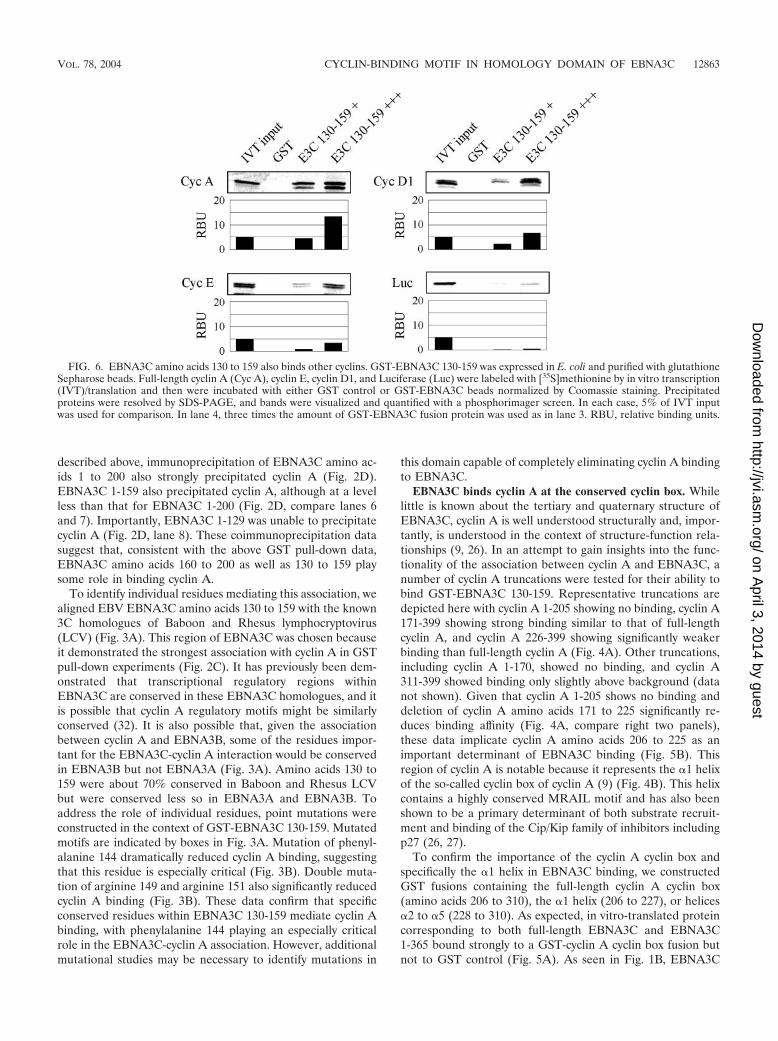
FIG. 6. EBNA3C amino acids 130 to 159 also binds other cyclins. GST-EBNA3C 130-159 was expressed in E. coli and purified with glutathioneSepharose beads. Full-length cyclin A (Cyc A), cyclin E, cyclin D1, and Luciferase (Luc) were labeled with [35S]methionine by in vitro transcription(IVT)/translation and then were incubated with either GST control or GST-EBNA3C beads normalized by Coomassie staining. Precipitatedproteins were resolved by SDS-PAGE, and bands were visualized and quantified with a phosphorimager screen. In each case, 5% of IVT inputwas used for comparison. In lane 4, three times the amount of GST-EBNA3C fusion protein was used as in lane 3. RBU, relative binding units.
VOL. 78, 2004 CYCLIN-BINDING MOTIF IN HOMOLOGY DOMAIN OF EBNA3C 12863
on April 3, 2014 by guest
http://jvi.asm.org/
Dow
nloaded from
621-992 also showed limited binding slightly above background(Fig. 5A). In order to compare the affinity of EBNA3C 1-365to a known functional interacting partner, the cyclin A cyclinbox was used to precipitate proteins from the lysates of 293Tcells transfected with expression plasmids for either Cdk2 orEBNA3C 1-365 (Fig. 5B). While Cdk2 precipitated morestrongly with the cyclin A cyclin box, EBNA3C 1-365 precipi-tation was of a similar magnitude, hinting at the potentialfunctionality of this interaction. Finally, we assessed the rela-tive binding of the cyclin A �1 helix compared to that of helices�2 to �5 (Fig. 5C). In this in vitro binding assay, both elements
of the cyclin A cyclin box confer binding to EBNA3C 1-365,consistent with the GST-EBNA3C 130-159 pull-downs of cy-clin A truncation mutants (Fig. 4). In Fig. 5C, we show that theaffinities of these two elements for the amino terminus ofEBNA3C were approximately the same. This may suggest that,in the context of EBNA3C 130-159, the �1 helix is the pre-dominant partner (Fig. 4) while the �2 to �5 helices may bindother regions of the amino terminus.
EBNA3C amino acids 130 to 159 bind other mammaliancyclins in addition to cyclin A. The above truncation studieswith cyclin A suggested that the amino terminus of EBNA3Ctargets the �1 helix of the cyclin box, a region of cyclin Aconserved not only in cyclin A homologues but also in othermammalian cyclins. As such, we decided to test whether othermammalian cyclins can bind EBNA3C 130-159 in an in vitrobinding assay. In this assay, we occasionally saw backgroundprecipitation of luciferase control when maximum amounts ofGST-EBNA3C 130-159 protein were used (Fig. 6, lower rightpanel). However, this binding was always lower than that forthe cyclins and was never seen when less fusion protein wasused. Both cyclin E and cyclin D1 showed binding to EBNA3C130-159 that was clearly above both GST control and the afore-mentioned luciferase background (Fig. 6). In repeated exper-iments, average binding was typically 30% of that seen withcyclin A, suggesting that residues unique to cyclin A confersome specificity for EBNA3C binding (Fig. 6). However, thisdata also suggests that EBNA3C may target other mammaliancyclins in addition to cyclin A. A previous study attemptedrescue of p27-mediated suppression of cyclin E-dependent ki-nase activity similar to that seen for cyclin A (13). Theseattempts were unsuccessful, suggesting that the mechanism bywhich EBNA3C regulates other cyclins, such as cyclin E, islikely distinct from that seen for cyclin A.
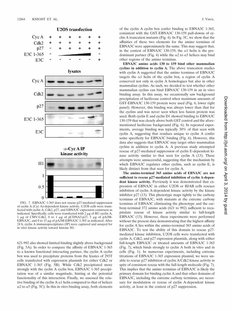
The amino-terminal 365 amino acids of EBNA3C are notsufficient to rescue p27-mediated inhibition of cyclin A-depen-dent kinase activity. Previously it was demonstrated that ex-pression of EBNA3C in either U2OS or BJAB cells rescuesinhibition of cyclin A-dependent kinase activity by the kinaseinhibitor p27 (13). This phenotype maps tightly to the carboxyterminus of EBNA3C, with mutants at the extreme carboxyterminus of EBNA3C eliminating the phenotype and the car-boxy-terminal 372 amino acids (621 to 992) sufficient to reca-pitulate rescue of kinase activity similar to full-lengthEBNA3C (13). However, these experiments were performedwithout the present data demonstrating that the highest affinityfor cyclin A lies within the amino-terminal 365 amino acids ofEBNA3C. To test the ability of this domain to rescue p27-mediated kinase inhibition, U2OS cells were transfected withcyclin A, Cdk2, and p27 expression plasmids, along with eitherfull-length EBNA3C or titrated amounts of EBNA3C 1-365(Fig. 7), which binds strongly to cyclin A both in vitro and incells (Fig. 1). In numerous experiments, including extremetitrations of EBNA3C 1-365 expression plasmid, we were un-able to rescue p27 inhibition of cyclin A/Cdk2 kinase activity inspite of consistent rescue with the full-length molecule (Fig. 7).This implies that the amino terminus of EBNA3C is likely theprimary domain for binding cyclin A and that other domains ofEBNA3C, including the extreme carboxy terminus, are neces-sary for modulation or rescue of cyclin A-dependent kinaseactivity, at least in the context of p27 suppression.
FIG. 7. EBNA3C 1-365 does not rescue p27-mediated suppressionof cyclin A (Cyc A)-dependent kinase activity. U2OS cells were trans-fected with cyclin A, Cdk2, p27, and EBNA3C expression constructs asindicated. Specifically, cells were transfected with 2 �g of RC-cyclin A,1 �g of CMV-Cdk2, 0 to 1 �g of pCDNA3-p27, 5 �g of pA3M-EBNA3C, and 0 to 15 �g of pA3M-EBNA3C 1-365 as indicated. After24 h, cyclin A immunoprecipitates (IP) were captured and assayed forin vitro kinase activity toward histone H1.
12864 KNIGHT ET AL. J. VIROL.
on April 3, 2014 by guest
http://jvi.asm.org/
Dow
nloaded from

The amino-terminal 365 amino acids of EBNA3C suppresscyclin A-dependent kinase activity when introduced into LCLs.Previously it was demonstrated that the introduction of a stopcodon at amino acid 365 in EBNA3C eliminates the ability ofEBV to transform B lymphocytes (31). When a mixed popu-lation of wild-type and mutant virus was used to infect primaryB cells, the resulting LCLs only harbored mutant EBNA3Cwhen coinfected with wild-type (31). Further, in these coin-fected LCLs the recombinant EBNA3C was lost when culturedcontinually (31). These studies hint that the amino-terminaldomain of EBNA3C not only fails to mimic full-lengthEBNA3C but may also negatively regulate the effects of full-length EBNA3C in coinfected LCLs.
As EBNA3C amino acids 1 to 365 do not rescue p27 sup-pression of cyclin A-dependent kinase activity as full-lengthEBNA3C does, we tested whether 1-365 would actually sup-press cyclin A kinase activity when introduced into LCLs.LCLs were transfected by electroporation with vector control,EBNA3C 1-365, or EBNA3C 1-129, which does not bind cyclinA. With this system, we achieved transfection efficiencies rang-ing from 10 to 20% in multiple experiments using GFP as amonitor. After 24 h, cyclin A complexes were immunoprecipi-tated and assayed for kinase activity toward histone H1. Ex-pression of EBNA3C 1-365 reproducibly reduced cyclin A-de-pendent kinase activity, with representative data shown in Fig.8. EBNA3C 1-365 suppressed the activity by 25 to 50% de-pending on the individual experiment, and, importantly,EBNA3C 1-129 (Fig. 8) and full-length EBNA3C (data notshown) did not reduce the kinase activity in this assay.
DISCUSSION
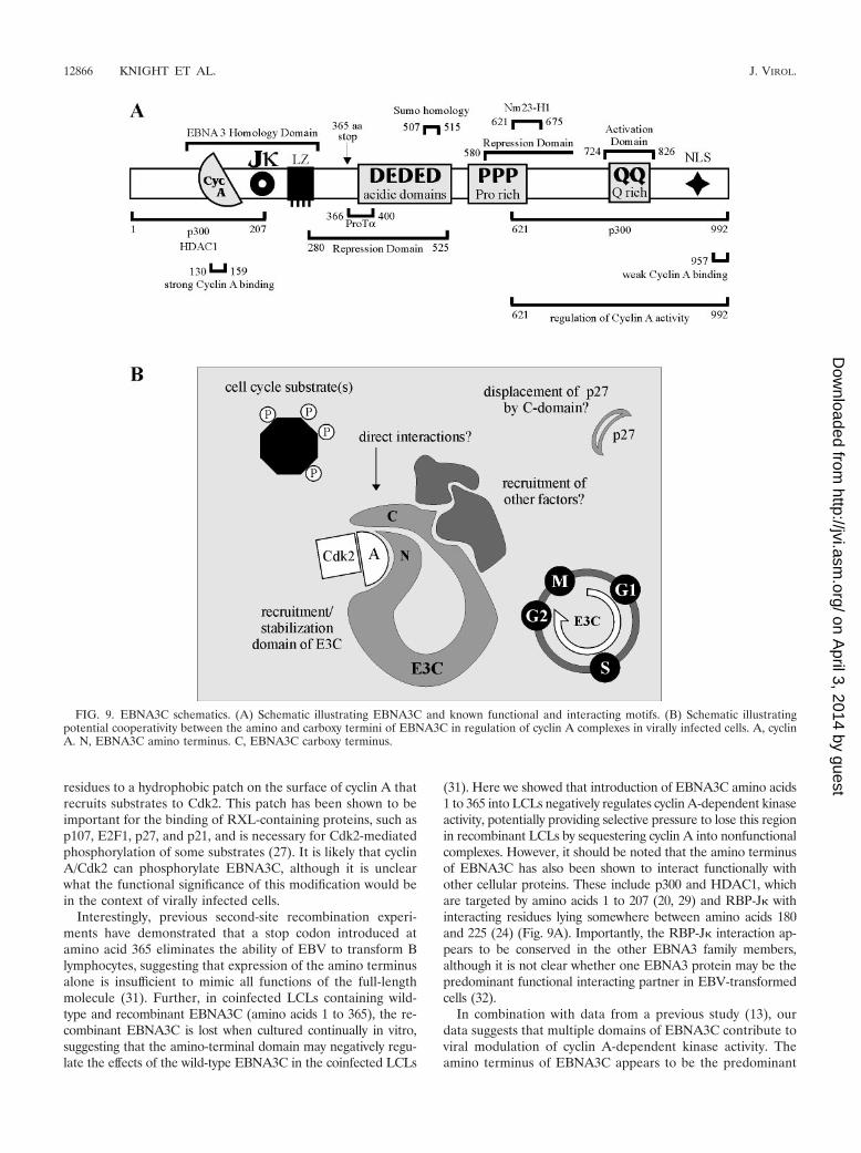
In a previous study, a yeast two-hybrid screen with the car-boxy terminus of EBNA3C as bait identified cyclin A as apotential EBNA3C-interacting protein. It was subsequentlydemonstrated that EBNA3C binds cyclin A in vitro and targetscyclin A kinase complexes in lymphoblastoid cell lines (13).Further, in the context of an experimental system, EBNA3Cdecreases the molecular association between cyclin A and thekinase inhibitor p27 and rescues p27-mediated inhibition ofcyclin A-dependent kinase activity (13). This phenotype was atleast partially mapped to the carboxy terminus of EBNA3C,which was consistent with the region of EBNA3C used as baitin the yeast two-hybrid screen (Fig. 9A). Subsequent to thisstudy, we performed a more detailed dissection of theEBNA3C-cyclin A interaction in an attempt to better under-stand the functional relationship between these two molecules.Surprisingly, we found that the predominant interaction be-tween EBNA3C and cyclin A actually occurred at the aminoterminus of EBNA3C, a region of the molecule not included inthe yeast two-hybrid screen (Fig. 9A). The cyclin A-interactingdomain was mapped to amino acids 130 to 159 of EBNA3C, aregion conserved in 3C homologues of other lymphocryptovi-rus species, Baboon lymphocryptovirus and Rhesus lymphoc-ryptovirus (32). Specifically, 21 of 30 residues are either iden-tical or demonstrate potential functional similarity, consistentwith a conserved and potentially critical role for this region inthe biology of EBV. This region of EBNA3C also falls withinthe so-called EBNA3 homology domain from amino acids 90to 320, and, indeed, 10 of 30 residues show conservation be-
tween EBNA3A, 3B, and 3C. In multiple trials, EBNA3B, butnot EBNA3A, showed some binding affinity for cyclin A, sug-gesting that the EBNA3C residues conserved in EBNA3B, butnot EBNA3A, may be most important for cyclin A binding. Wehave yet to show a role for EBNA3B, individually, in modu-lating cyclin A function, although more thorough studies toevaluate potential synergism between EBNA3B and EBNA3C,both in vitro and in virally infected cells, are still needed.
The strong interaction between EBNA3C 130-159 and cyclinA is at least partially dependent on the �1 helix of the con-served cyclin box of cyclin A. This helix has been shown to becritical for cyclin A function in at least two regards. First, thehelix and especially its conserved MRAIL motif provide manyof the critical residues required for binding of Cip/Kip familykinase inhibitors, such as p27 (26). Indeed, the crystal structureof cyclin A/Cdk2/p27 shows a hydrophobic groove in cyclin Athat receives p27 (26). Among the residues contributing vander Waals contacts to p27 are Met 210, Ile 213, Leu 214, andTrp 217 (26). This observation is particularly noteworthy, as aprevious study demonstrated that EBNA3C reduces the mo-lecular association between cyclin A and p27 in cells. As such,an attractive model is that EBNA3C directly competes withp27 for cyclin A binding. However, we have yet to demonstratesuch competition with either in vitro-translated or bacteriallyexpressed proteins, suggesting that our observed competitionin coimmunoprecipitation experiments may require other fac-tors present in cell lysates, potentially recruited to cyclin Acomplexes by EBNA3C (Fig. 9B). We are presently seeking toidentify such factors. In addition to mediating the binding ofcyclin A inhibitors such as p27, the �1 helix also contributes
FIG. 8. EBNA3C amino acids 1 to 365 downregulate cyclin A (CycA)-dependent kinase activity when expressed in LCLs. LCLs weretransfected by electroporation with 10 �g of RC-cyclin A and 20 �g ofpA3M vector, pA3M-EBNA3C 1-365, or pA3M-EBNA3C 1-129. Af-ter 24 h, cyclin A immunoprecipitates (IP) were captured and assayedfor in vitro kinase activity toward histone H1.
VOL. 78, 2004 CYCLIN-BINDING MOTIF IN HOMOLOGY DOMAIN OF EBNA3C 12865
on April 3, 2014 by guest
http://jvi.asm.org/
Dow
nloaded from
residues to a hydrophobic patch on the surface of cyclin A thatrecruits substrates to Cdk2. This patch has been shown to beimportant for the binding of RXL-containing proteins, such asp107, E2F1, p27, and p21, and is necessary for Cdk2-mediatedphosphorylation of some substrates (27). It is likely that cyclinA/Cdk2 can phosphorylate EBNA3C, although it is unclearwhat the functional significance of this modification would bein the context of virally infected cells.
Interestingly, previous second-site recombination experi-ments have demonstrated that a stop codon introduced atamino acid 365 eliminates the ability of EBV to transform Blymphocytes, suggesting that expression of the amino terminusalone is insufficient to mimic all functions of the full-lengthmolecule (31). Further, in coinfected LCLs containing wild-type and recombinant EBNA3C (amino acids 1 to 365), the re-combinant EBNA3C is lost when cultured continually in vitro,suggesting that the amino-terminal domain may negatively regu-late the effects of the wild-type EBNA3C in the coinfected LCLs
(31). Here we showed that introduction of EBNA3C amino acids1 to 365 into LCLs negatively regulates cyclin A-dependent kinaseactivity, potentially providing selective pressure to lose this regionin recombinant LCLs by sequestering cyclin A into nonfunctionalcomplexes. However, it should be noted that the amino terminusof EBNA3C has also been shown to interact functionally withother cellular proteins. These include p300 and HDAC1, whichare targeted by amino acids 1 to 207 (20, 29) and RBP-J� withinteracting residues lying somewhere between amino acids 180and 225 (24) (Fig. 9A). Importantly, the RBP-J� interaction ap-pears to be conserved in the other EBNA3 family members,although it is not clear whether one EBNA3 protein may be thepredominant functional interacting partner in EBV-transformedcells (32).
In combination with data from a previous study (13), ourdata suggests that multiple domains of EBNA3C contribute toviral modulation of cyclin A-dependent kinase activity. Theamino terminus of EBNA3C appears to be the predominant
FIG. 9. EBNA3C schematics. (A) Schematic illustrating EBNA3C and known functional and interacting motifs. (B) Schematic illustratingpotential cooperativity between the amino and carboxy termini of EBNA3C in regulation of cyclin A complexes in virally infected cells. A, cyclinA. N, EBNA3C amino terminus. C, EBNA3C carboxy terminus.
12866 KNIGHT ET AL. J. VIROL.
on April 3, 2014 by guest
http://jvi.asm.org/
Dow
nloaded from
recruitment domain, with both in vitro binding and coimmu-noprecipitation studies suggesting that the amino terminuscontains the strongest and predominant cyclin A binding do-main. However, the previous work of Knight and Robertsondemonstrated that the carboxy terminus of EBNA3C is theprimary functional domain, with EBNA3C 621-992 able torecapitulate the ability of full-length EBNA3C to rescue p27-mediated inhibition of cyclin A-dependent kinase activity (13).It must be noted that these functional rescue experimentsrequired exogenous expression of the EBNA3C polypeptides.With overexpression of the amino terminus of EBNA3C, cyclinA may be sequestered or recruited into complexes or compart-ments that are not particularly accessible to the carboxy ter-minus. It is possible that in the context of viral infection, whereEBNA3C levels are undoubtedly significantly lower, strongcyclin A binding by the amino-terminal domain may be re-quired to place the carboxy terminus in the proximity of cyclinA for functional modulation (Fig. 9B). We will continue topursue the role of the carboxy terminus of EBNA3C in thesecomplexes and also attempt to identify additional EBNA3C-recruited molecules in an attempt to better understand bothEBNA3C cell cycle modulation and the ability of EBV totransform primary cells.
ACKNOWLEDGMENTS
We thank Ed Harlow, Philip Hinds, Elliot Kieff, Maria Mudryj,Michele Pagano, Martin Rowe, and Alan Diehl for generously provid-ing reagents.
J.S.K. is supported by the Lady Tata Memorial Trust. The project issupported by NIH grants NCI CA72150-07, NCI CA91792-01, andNIDCR DE14136-01 to E.S.R. E.S.R. is a scholar of the Leukemia andLymphoma Society of America.
REFERENCES
1. Aiello, L., R. Guilfoyle, K. Huebner, and R. Weinmann. 1979. Adenovirus 5DNA sequences present and RNA sequences transcribed in transformedhuman embryo kidney cells (HEK-Ad-5 or 293). Virology 94:460–469.
2. Allday, M. J., D. H. Crawford, and J. A. Thomas. 1993. Epstein-Barr virus(EBV) nuclear antigen 6 induces expression of the EBV latent membraneprotein and an activated phenotype in Raji cells. J. Gen. Virol. 74:361–369.
3. Allday, M. J., and P. J. Farrell. 1994. Epstein-Barr virus nuclear antigenEBNA3C/6 expression maintains the level of latent membrane protein 1 inG1-arrested cells. J. Virol. 68:3491–3498.
4. Aster, J. C., E. S. Robertson, R. P. Hasserjian, J. R. Turner, E. Kieff, and J.Sklar. 1997. Oncogenic forms of NOTCH1 lacking either the primary bind-ing site for RBP-J� or nuclear localization sequences retain the ability toassociate with RBP-J� and activate transcription. J. Biol. Chem. 272:11336–11343.
5. Cotter, M. A., Jr., and E. S. Robertson. 2000. Modulation of histone acetyl-transferase activity through interaction of Epstein-Barr nuclear antigen 3Cwith prothymosin alpha. Mol. Cell. Biol. 20:5722–5735.
6. Devoto, S. H., M. Mudryj, J. Pines, T. Hunter, and J. R. Nevins. 1992. Acyclin A-protein kinase complex possesses sequence-specific DNA bindingactivity: p33cdk2 is a component of the E2F-cyclin A complex. Cell 68:167–176.
7. Epstein, M. A., B. G. Achong, and Y. M. Barr. 1964. Virus particles incultured lymphoblasts from Burkitt’s lymphoma. Lancet i:702–703.
8. Hinds, P. W., S. Mittnacht, V. Dulic, A. Arnold, S. I. Reed, and R. A.Weinberg. 1992. Regulation of retinoblastoma protein functions by ectopicexpression of human cyclins. Cell 70:993–1006.
9. Jeffrey, P. D., A. A. Russo, K. Polyak, E. Gibbs, J. Hurwitz, J. Massague, andN. P. Pavletich. 1995. Mechanism of CDK activation revealed by the struc-ture of a cyclinA-CDK2 complex. Nature 376:313–320.
10. Johannsen, E., C. L. Miller, S. R. Grossman, and E. Kieff. 1996. EBNA-2and EBNA-3C extensively and mutually exclusively associate withRBPJkappa in Epstein-Barr virus-transformed B lymphocytes. J. Virol. 70:4179–4183.
11. Kieff, E., and A. B. Rickinson. 2002. Epstein-Barr virus and its replication, p.2511–2573. In D. Knipe and P. Howley (ed.), Fields virology, 4th ed., vol. 2.Lippincott Williams & Wilkins, Philadelphia, Pa.
12. Knight, J. S., K. Lan, C. Subramanian, and E. S. Robertson. 2003. Epstein-Barr virus nuclear antigen 3C recruits histone deacetylase activity and asso-ciates with the corepressors mSin3A and NCoR in human B-cell lines.J. Virol. 77:4261–4272.
13. Knight, J. S., and E. S. Robertson. 2004. Epstein-Barr virus nuclear antigen3C regulates cyclin A/p27 complexes and enhances cyclin A-dependent ki-nase activity. J. Virol. 78:1981–1991.
14. Morgan, D. O. 1997. Cyclin-dependent kinases: engines, clocks, and micro-processors. Annu. Rev. Cell Dev. Biol. 13:261–291.
15. Pagano, M., R. Pepperkok, F. Verde, W. Ansorge, and G. Draetta. 1992.Cyclin A is required at two points in the human cell cycle. EMBO J. 11:961–971.
16. Pagano, M., S. W. Tam, A. M. Theodoras, P. Beer-Romero, G. Del Sal, V.Chau, P. R. Yew, G. F. Draetta, and M. Rolfe. 1995. Role of the ubiquitin-proteasome pathway in regulating abundance of the cyclin-dependent kinaseinhibitor p27. Science 269:682–685.
17. Parker, G. A., T. Crook, M. Bain, E. A. Sara, P. J. Farrell, and M. J. Allday.1996. Epstein-Barr virus nuclear antigen (EBNA)3C is an immortalizingoncoprotein with similar properties to adenovirus E1A and papillomavirusE7. Oncogene 13:2541–2549.
18. Parker, G. A., R. Touitou, and M. J. Allday. 2000. Epstein-Barr virusEBNA3C can disrupt multiple cell cycle checkpoints and induce nucleardivision divorced from cytokinesis. Oncogene 19:700–709.
19. Radkov, S. A., M. Bain, P. J. Farrell, M. West, M. Rowe, and M. J. Allday.1997. Epstein-Barr virus EBNA3C represses Cp, the major promoter forEBNA expression, but has no effect on the promoter of the cell gene CD21.J. Virol. 71:8552–8562.
20. Radkov, S. A., R. Touitou, A. Brehm, M. Rowe, M. West, T. Kouzarides, andM. J. Allday. 1999. Epstein-Barr virus nuclear antigen 3C interacts withhistone deacetylase to repress transcription. J. Virol. 73:5688–5697.
21. Resnitzky, D., L. Hengst, and S. I. Reed. 1995. Cyclin A-associated kinaseactivity is rate limiting for entrance into S phase and is negatively regulatedin G1 by p27Kip1. Mol. Cell. Biol. 15:4347–4352.
22. Rickinson, A. B., and E. Kieff. 2002. Epstein-Barr Virus, p. 2575–2627. In D.Knipe and P. Howley (ed.), Fields Virology, 4th ed., vol. 2. LippincottWilliams & Wilkins, Philadelphia, Pa.
23. Robertson, E. S. 1997. The Epstein-Barr Virus EBNA3 protein family asregulators of transcription. Epstein-Barr Vir. Rep. 4:143–150.
24. Robertson, E. S., J. Lin, and E. Kieff. 1996. The amino-terminal domains ofEpstein-Barr virus nuclear proteins 3A, 3B, and 3C interact with RBPJ(�).J. Virol. 70:3068–3074.
25. Rosendorff, A., D. Illanes, G. David, J. Lin, E. Kieff, and E. Johannsen. 2004.EBNA3C coactivation with EBNA2 requires a SUMO homology domain.J. Virol. 78:367–377.
26. Russo, A. A., P. D. Jeffrey, A. K. Patten, J. Massague, and N. P. Pavletich.1996. Crystal structure of the p27Kip1 cyclin-dependent-kinase inhibitorbound to the cyclin A-Cdk2 complex. Nature 382:325–331.
27. Schulman, B. A., D. L. Lindstrom, and E. Harlow. 1998. Substrate recruit-ment to cyclin-dependent kinase 2 by a multipurpose docking site on cyclinA. Proc. Natl. Acad. Sci. USA 95:10453–10458.
28. Subramanian, C., M. A. Cotter, Jr., and E. S. Robertson. 2001. Epstein-Barrvirus nuclear protein EBNA-3C interacts with the human metastatic sup-pressor Nm23-H1: a molecular link to cancer metastasis. Nat. Med. 7:350–355.
29. Subramanian, C., S. Hasan, M. Rowe, M. Hottiger, R. Orre, and E. S.Robertson. 2002. Epstein-Barr virus nuclear antigen 3C and prothymosinalpha interact with the p300 transcriptional coactivator at the CH1 andCH3/HAT domains and cooperate in regulation of transcription and histoneacetylation. J. Virol. 76:4699–4708.
30. Subramanian, C., J. S. Knight, and E. S. Robertson. 2002. The Epstein Barrnuclear antigen EBNA3C regulates transcription, cell transformation andcell migration. Front. Biosci. 7:704–716.
31. Tomkinson, B., E. Robertson, and E. Kieff. 1993. Epstein-Barr virus nuclearproteins EBNA-3A and EBNA-3C are essential for B-lymphocyte growthtransformation. J. Virol. 67:2014–2025.
32. Zhao, B., R. Dalbies-Tran, H. Jiang, I. K. Ruf, J. T. Sample, F. Wang, andC. E. Sample. 2003. Transcriptional regulatory properties of Epstein-Barrvirus nuclear antigen 3C are conserved in simian lymphocryptoviruses. J. Vi-rol. 77:5639–5648.
33. Zhao, B., and C. E. Sample. 2000. Epstein-barr virus nuclear antigen 3Cactivates the latent membrane protein 1 promoter in the presence of Ep-stein-Barr virus nuclear antigen 2 through sequences encompassing an Spi-1/Spi-B binding site. J. Virol. 74:5151–5160.
VOL. 78, 2004 CYCLIN-BINDING MOTIF IN HOMOLOGY DOMAIN OF EBNA3C 12867
on April 3, 2014 by guest
http://jvi.asm.org/
Dow
nloaded from