Mutational Analyses of Epstein-Barr Virus Glycoprotein 42 Reveal Functional Domains Not Involved in...
12
See discussions, stats, and author profiles for this publication at: https://www.researchgate.net/publication/8565041 Mutational Analyses of Epstein-Barr Virus Glycoprotein 42 Reveal Functional Domains Not Involved in Receptor... Article in Journal of Virology · July 2004 DOI: 10.1128/JVI.78.11.5946-5956.2004 · Source: PubMed CITATIONS 38 READS 28 4 authors, including: Jasmina Smajlovic University Clinical Center Tuzla 10 PUBLICATIONS 182 CITATIONS SEE PROFILE All content following this page was uploaded by Jasmina Smajlovic on 04 December 2016. The user has requested enhancement of the downloaded file. All in-text references underlined in blue are added to the original document and are linked to publications on ResearchGate, letting you access and read them immediately.
-
Upload
independent -
Category
Documents
-
view
1 -
download
0
Transcript of Mutational Analyses of Epstein-Barr Virus Glycoprotein 42 Reveal Functional Domains Not Involved in...
Seediscussions,stats,andauthorprofilesforthispublicationat:https://www.researchgate.net/publication/8565041
MutationalAnalysesofEpstein-BarrVirusGlycoprotein42RevealFunctionalDomainsNotInvolvedinReceptor...
ArticleinJournalofVirology·July2004
DOI:10.1128/JVI.78.11.5946-5956.2004·Source:PubMed
CITATIONS
38
READS
28
4authors,including:
JasminaSmajlovic
UniversityClinicalCenterTuzla
10PUBLICATIONS182CITATIONS
SEEPROFILE
AllcontentfollowingthispagewasuploadedbyJasminaSmajlovicon04December2016.
Theuserhasrequestedenhancementofthedownloadedfile.Allin-textreferencesunderlinedinblueareaddedtotheoriginaldocumentandarelinkedtopublicationsonResearchGate,lettingyouaccessandreadthemimmediately.

JOURNAL OF VIROLOGY, June 2004, p. 5946–5956 Vol. 78, No. 110022-538X/04/$08.00�0 DOI: 10.1128/JVI.78.11.5946–5956.2004Copyright © 2004, American Society for Microbiology. All Rights Reserved.
Mutational Analyses of Epstein-Barr Virus Glycoprotein 42 RevealFunctional Domains Not Involved in Receptor Binding but
Required for Membrane FusionAmanda L. Silva,1 Jasmina Omerovic,1 Theodore S. Jardetzky,2 and Richard Longnecker1*
Department of Microbiology and Immunology, Northwestern University Medical School, Chicago,Illinois 60611,1 and Department of Biochemistry, Molecular Biology and Cell Biology,
Northwestern University, Evanston, Illinois 602082
Received 30 October 2003/Accepted 15 January 2004
Epstein-Barr virus (EBV) is a human gammaherpesvirus associated with malignancies of both epithelial andlymphoid origin. Efficient infection of the latent host reservoir B lymphocytes involves the binding of glyco-proteins gp350/220 for initial attachment, followed by the concerted action of gH, gL, gB, and gp42 formembrane fusion. The type II membrane protein gp42 is required for infection of B cells and assembles intoa complex with gH and gL. The cellular host receptor for gp42, class II human leukocyte antigen (HLA), hasbeen structurally verified by crystallization analyses of gp42 bound to HLA-DR1. Interestingly, the crystalstructure revealed a hydrophobic pocket consisting of many aromatic and aliphatic residues from the predictedC-type lectin domain of gp42 that in other members of the C-type lectin family binds major histocompatibilitycomplex class I or other diverse ligands. Although the hydrophobic pocket does not bind HLA class II,mutational analyses presented here indicate that this domain is essential for EBV-induced membrane fusion.In addition, mutational analysis of the region of gp42 contacting HLA class II in the gp42-HLA-DR1 cocrystalconfirms that this region interacts with HLA class II and that this interaction is also important for EBV-induced membrane fusion.
Epstein-Barr virus (EBV) is a ubiquitous member of thehuman gammaherpesvirus subfamily that is able to establishlifelong latency in host B cells. The double-stranded envelopedvirus has been associated with diseases of both lymphoid andepithelial origin (3, 9). Infection in infancy is generally asymp-tomatic but during adolescence can cause infectious mononu-cleosis. EBV infection is associated with Burkitt’s lymphoma,Hodgkin’s disease, and lymphoproliferative diseases in AIDSpatients (for reviews, see references 25 and 32). Epithelialdiseases associated with EBV infection include oral hairy leu-koplakia and nasopharyngeal carcinoma (for a review, see ref-erence 32). Although EBV encodes as many as 11 glycopro-teins, efficient entry of EBV into B cells requires only 5glycoproteins: gp350/220, gH, gL, gB, and gp42 (for reviews,see references 16, 33, and 34). The type II membrane proteingp42 of the BZLF2 open reading frame is required for entryinto B lymphocytes but not epithelial cells and has two alter-nately processed forms: a 42- and a 38-kDa protein (1, 24, 37).The receptor of gp350 has been identified as complementreceptor 2 (CR2) or CD21 (10, 29, 36). Expression of gp350 isnot required for membrane fusion, but as a binding factor, itgreatly enhances infection efficiency, similar to herpes simplexvirus (HSV) gC (17, 20). Transmembrane gH dimerizes withgL, which exists as membrane-bound and soluble forms and isrequired for correct folding and transport of gH (14, 24, 40).These dimers have been demonstrated to bind gp42 (38). Theminimal glycoproteins required for membrane fusion of B cells
using experimentally transfected cells are gB, gH, gL, and gp42(12). It was previously demonstrated that gB is required forlytic replication as well as the production of transforming virus(15, 22). EBV gB has been shown to be functionally distinctfrom other human herpesvirus subfamily gBs, and this could bedue to the different regulatory domains of the cytoplasmic tailthat are involved in membrane fusion and virion transport (12,21, 22). Recently, Neuhierl et al. demonstrated that EBV gBdramatically enhances infection of human cells and constitutesan important virulence factor that determines infection ofnon-B cells (30).
It has been theorized that expression levels of gp42 on thesurface of the virion decrease or increase as the virus alternatesbetween infection of epithelial cells and lymphocytes in thehuman host, the so-called “cell-switching and kissing” model(2). It was demonstrated in vitro that when the virus is pro-duced in epithelial cells, the virion contains abundant gp42,which allows very efficient infection of B cells. However, asgp42 is not required for entry into epithelial cells, the highsurface expression might sterically interfere with receptorbinding, which would lead to the reduced efficiency of epithe-lial cell infection that was observed. Conversely, when the virusis produced in B cells, the amount of gp42 in the virion isreduced, possibly due to colocalization with the EBV B-cellreceptor, which allows efficient infection of epithelial cells butdecreased infection of other B cells, as was demonstrated. Thisprovides a plausible route of oral EBV infection in humans viaepithelial cells and then B cells, where the virus can establishlatency. The B-cell receptor of gp42 has been identified as thehuman leukocyte antigen (HLA) class II, and the DR, DP, andDQ alleles are all functional (11, 23, 35). Interestingly,HLA-DQ �*02 is the only functional DQ allele (13).
* Corresponding author. Mailing address: Department of Microbi-ology and Immunology, Northwestern University Medical School,Ward 6-231, Chicago, IL 60611. Phone: (312) 503-0467. Fax: (312)503-1339. E-mail: [email protected].
5946
Recently, the crystal structure of a baculovirus-producedsoluble form of gp42 (sgp42) bound to the HLA-DR1 allelewas solved (28). The predicted C-type lectin domain (CTLD)lies between residues 94 and 223 of the protein, and though itshows some structural homology to other CTLD-containingproteins, such as Ly49A, it was revealed that binding of gp42/HLA-DR1 was not homologous to Ly49A binding to its recep-tor, the major histocompatibility complex (MHC) class I mol-ecule H-2Dd, nor any other CTLD-containing protein and itsreceptor. Interestingly, gp42 dimerized in the crystal form be-tween residues 87 and 94, a region of the ectodomain. Dimer-ization of gp42, either before or after binding to HLA class II,may be important for fusion since it would result in morereceptors being available to trigger fusion. The initiation offusion by gp42 mediated by the coordinated action of gB, gH,and gL may result from conformational changes in gp42 afterits binding to HLA class II. This conformational change mayallow other EBV-encoded glycoproteins or additional cellularreceptors to bind to the fusion complex, or it may rearrangereceptors on the virion or cell surface. Crystallization studies ofgp42 alone should reveal if such a conformational change oc-curs upon binding with HLA class II. Adding to its inimitablecharacter as a novel herpesvirus glycoprotein is a region ofseveral aliphatic and aromatic residues that create a hydropho-bic pocket with a yet unknown binding partner. The specifictropism that gp42 lends EBV, along with its unique bindingstructure and hydrophobic pocket, makes it an interesting tar-get for functional studies of membrane fusion to not onlycharacterize EBV fusion mechanisms but also increase ouroverall understanding of herpesvirus infection. We have un-dertaken mutational studies of gp42 to confirm the predictedinteraction of gp42 with HLA class II from structural studiesand to investigate the function of other gp42 domains such asthe hydrophobic pocket in EBV-induced membrane fusion.
MATERIALS AND METHODS
Cells and plasmids. Chinese hamster ovary cells (CHO-K1), kindly providedby Nanette Susmarski, were grown in Ham’s F-12 media with 10% fetal bovineserum and 1% penicillin-streptomycin, referred to as complete media (all fromBioWhittaker). EBV-positive HLA class II- and CD21-expressing Daudi B lym-phocytes were obtained from American Type Culture Collection (Manassas, Va.)and were grown in RPMI complete media (BioWhittaker). To more easilymonitor membrane fusion, a Daudi cell line stably transfected and expressing T7RNA polymerase was constructed. Briefly, Daudi cells were electroporated with40 �g of pOS2 plasmid DNA, containing a G418 selectable marker and bacte-riophage T7 RNA polymerase under transcriptional control of the simian virus40 promoter. This plasmid was kindly provided by Stanley Lemon (39). Followingtransfection, the cells were plated at 10,000 and 50,000 cells per well in 96-wellplates and selected with G418 (0.9, 1.1, or 1.3 mg/ml). Clones emerged approx-imately 3 to 4 weeks postplating. Approximately 50 clones were expanded fromthe 96-well plates and were tested in the fusion assay as described below. Of thetested clones, cell lines 22, 29, 31, and 36 provided the highest levels of luciferaseexpression in the fusion assay. Of these lines, lines 29 and 36 were used for allsubsequent experiments and were maintained in RPMI complete media with 1.1and 1.3 mg of G418/ml, respectively. Cells were grown in 75-cm2 cell cultureflasks (Corning), and adherent cells were detached by using either trypsin-Versene (BioWhittaker) or Versene (phosphate-buffered saline [PBS]–1 mMEDTA). The various plasmids used in the present studies are shown in Table 1.
Generation of mutants. Mutations were generated using either a GPS-LSlinker scanning system (New England Biolabs [NEB]) or a QuikChange kit(Stratagene) on an EBV gp42-containing pCAGGS/MCS vector (19) and iso-lated by cesium chloride density gradients. The NEB kit uses a transposon-basedrandom mutagenesis to introduce a selection marker. After selection, clones arediagnostically digested and then sequenced to determine the exact location of the
mutation. Collapse of the marker leaves a 5-amino-acid insertion introducing arare PmeI site and a repeat of five host base pairs. The QuikChange kit uses PCRto introduce a specific mutation via primers designed with a silent mutation fordiagnostic purposes. The suggested PCR protocol was followed to generatemutant clones, which were then diagnostically digested and sequenced.
Transfection. (i) Fusion assay. CHO cells were seeded in plastic 24-well plates(Corning), grown 24 h to approximately 90% confluency, and transiently trans-fected with 0.125 �g each of EBV gH, gL, and gB, 0.5 �g of gp42 or gp42 mutant,and 0.2 �g of a luciferase-containing reporter plasmid with a T7 promoter (31).Transfections utilized 700 �l of Opti-Mem (Gibco) and 1 �l of Lipofectamine2000 (Invitrogen) per well.
(ii) Western blotting and cell-based enzyme-linked immunosorbent assay(CELISA). CHO cells were seeded into six-well plastic plates (Corning) andtransfected with 4 �g of plasmid DNA with 2.5 ml of Opti-Mem and 5 �l ofLipofectamine 2000. The transfection efficiency was always simultaneously as-sessed by transfection of pEGFP-N1 and expression of enhanced green fluores-cent protein.
Expression of mutants. (i) Western blotting. CHO cells were transfected aspreviously stated. Media was changed 12 h later and cells were collected 24 hlater. Cells were scraped, washed 2 to 3 times in PBS, and lysed using a 1% TritonX buffer with 1 mM sodium vanadate, 10 mM sodium fluoride, leupeptin (0.5mg/ml), pepstatin (0.7 mg/ml), and 0.2 mM phenylmethylsulfonyl fluoride. Ly-sates were run on Bio-Rad 12.5% criterion gels in sodium dodecyl sulfate run-ning buffer at 120 V for 90 min. Proteins were transferred to Immobilon-Pmembranes in transfer buffer at 90 V for 90 min with cooling or at 15 Vovernight. Blots were blocked in Tris-buffered saline with Tween with 3% milkfor an hour at room temperature (RT) or overnight at 4°C and then incubatedfor an hour at RT with a rabbit polyclonal anti-gp42 antibody (PB1114) diluted1:1,000 in blocking solution. Blots were washed, and a secondary protein A-horseradish peroxidase (HRP)-conjugated antibody (Amersham) was applied forhalf an hour at RT. Blots were then mixed in equal volumes of ECL solutions andexposed to hyperfilm (Amersham Biosciences).
(ii) CELISA. CHO cells were transfected as previously stated. After 12 hmedia was changed, and then 12 h later cells were detached with Versene andtransferred to Corning 96-well plates, 3 wells per sample. After 16 h of incuba-tion at 37oC, cells were washed with PBS-ABC, incubated for 30 min at RT witha rabbit polyclonal anti-gp42 antibody (PB1112) diluted 1:1,000 in PBS-ABCwith 3% bovine serum albumin (PBS-BSA), and then fixed for 10 min in PBSwith 2% formaldehyde and 0.2% glutaraldehyde. Cells were washed three timeswith PBS-BSA, incubated with a biotinylated goat anti-rabbit immunoglobulin G(IgG) (Sigma) at 1:500 for 30 min, washed five times, and then incubated with astreptavidin-HRP antibody (1:20,000) (Amersham) for 30 min, all at RT. Cellswere then mixed with a peroxide substrate (BioFX Laboratories) and read witha Victor plate reader at 370 nm for 0.1 s.
Cell-cell membrane fusion assay. The cell-cell membrane fusion assay wasslightly modified from a previously published protocol (12). Briefly, CHO cellswere transfected as stated previously. After 12 h, these cells were washed andoverlaid with 5.0 � 105 target HLA class II-expressing Daudi B cells that hadbeen stably transfected to express T7 RNA polymerase. After 24 h of incubation,the cells were washed twice with PBS and lysed, and 100 �l of firefly luciferinsubstrate was added to 20 �l of lysate (Promega luciferase assay system). Rela-tive luciferase activity was measured in Visibottom 96-well plates by a Victorplate reader at 370 nm for 0.1 s.
Quantification of sgp42 levels by enzyme-linked immunosorbent assay(ELISA). A baculovirus-generated soluble gp42 (sgp42) was used as a positivecontrol in addition to transfected soluble gp42. Twenty-five microliters of trans-fected CHO cell supernatants diluted in PBSN (PBS with 0.02% sodium azide)was added to Nunc round-bottom plates (3 wells per samples), covered withplastic wrap, and allowed to incubate overnight at RT. Samples were blockedwith PBS–1% BSA (blocking buffer) for 1 h at 37°C, followed by PB1112 diluted1:1,000 in blocking buffer for 2 h at 37oC, goat anti-rabbit IgG diluted 1:500 inblocking buffer for 2 h at 37°C, and streptavidin-HRP diluted 1:20,000 in block-ing buffer for 2 h at 37°C. Peroxide substrate was added, and plates were read ona Victor plate reader at 405 nm for 0.1 s.
Binding of sgp42 to HLA class II-expressing cells by flow cytometry. This assaywas slightly modified from the previously published protocol (26). After mea-suring relative levels of soluble gp42 in CHO cell supernatants by ELISA,supernatants were diluted in Ham’s F-12 complete media and incubated with 5.0� 105 Daudi cells in approximately 250 �l of RPMI complete media whilerotating at 4°C for 20 min. Cells were then washed with fluorescence-activatedcell sorting (FACS) buffer (PBS with 0.01% sodium azide, 1% HEPES buffer[BioWhittaker], and 1% fetal bovine serum), followed by incubation with PB1114(diluted in FACS buffer to 1:250) and a biotinylated anti-DQ Ia3 antibody
VOL. 78, 2004 ANALYSIS OF EBV gp42 BINDING AND FUSION 5947
(1:100) on ice for 15 min. After washing with FACS buffer, secondary antibodiesused were fluorescein isothiocyanate (FITC)-conjugated goat anti-rabbit wholeIgG and allophycocyanin-conjugated streptavidin (both at 1:250) (Pharmingen),also on ice for 15 min. Cells were washed and resuspended in 350 �l of FACSbuffer. Immunofluorescence was measured using a Becton-Dickinson FACS-Sortcytometer and analyzed by using Cell Quest Pro. As a positive control, a bacu-lovirus-generated sgp42 of residues 33 to 223 was utilized.
RESULTS
Construction of gp42 mutants. In order to better understandthe mechanism of EBV-induced membrane fusion, we wantedto define functional domains of gp42. Initially, we utilized atransposon-based linker insertion mutagenesis to generate ran-dom 5-amino-acid inserts spaced throughout gp42. The NEBGPS-LS linker scanning system creates mutants with an inser-tion of five amino acids containing a 5-bp repeat from the hostsequence along with the addition of a unique PmeI site. Duringthis time, the crystal structure of a baculovirus-expressed sol-uble form of gp42 (sgp42) from residues 33 through 223 boundto HLA class II was solved, which revealed a short stretch ofresidues 104 through 107, an �-helix of residues 154 through160, and arginine 220 all as areas of interaction with the HLA-DR1 �-chain (Fig. 1A). The HLA class II ��46 forms a saltbridge with gp42 R220 and hydrogen bonds with T104 and
Y107, R�72 forms hydrogen bonds with T104 and Y107, andN�62, S�63, and K�65 all form hydrogen bonds with the �-he-lix (26, 28) (Fig. 1B). Although the ectodomain up to residue85 was disordered, other structural features were also discov-ered. Ten cysteine residues of the native protein form fivedisulfide bonds (99 with 138, 102 and 115, 128 and 214, 132 and216, and 192 with 208), and there are four potential N-linkedglycosylation sites at residues 64, 93, 98, and 173. A potentialdimerization site was delineated between residues 87 and 94,and a hydrophobic pocket was observed consisting of severalaliphatic and aromatic residues between residues 161 and 201(Fig. 1C). The proposed binding site with the gH/gL dimerlocated in the amino terminus was in the disordered region.Although the linker insertion mutants spanned all of thesedomains, we constructed additional site-directed mutations ofindividual residues to introduce specific mutations within cer-tain domains. For these mutations, a Stratagene QuikChangekit was utilized. For each site-specific mutation, a unique re-striction site was silently incorporated into the reading frameto allow easy identification of each mutant. More detailedprocedures and the various plasmid clones used in these stud-ies are described in Materials and Methods and in Table 1.Plasmid DNA was isolated by cesium chloride density gradi-
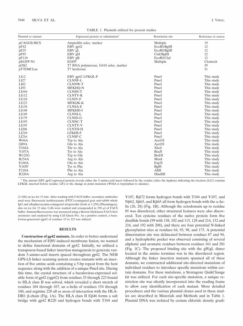
TABLE 1. Plasmids utilized for present studies
Plasmid or mutant Expressed protein or substitutiona Restriction site Reference or source
pCAGGS.MCS Ampicillin selec. marker Multiple 19pF42 EBV gp42 EcoRI/BgIII 12pF25 EBV gL EcoRI/BgIII 12pF85 EBV gH ClaI/BgIII 12pF110 EBV gB EcoRI/ClaI 12pEGFP-N1 EGFP Multiple ClontechpOS2 T7 RNA polymerase, G418 selec. marker 39pT7EMCLuc T7 luciferase 31
LI12 EBV gp42 LFKQL-F PmeI This studyLI27 CLNNF-L PmeI This studyLI82 CLNNW-T PmeI This studyLI93 MFKHQ-N PmeI This studyLI104 CLNSN-T PmeI This studyLI112 CLNTY-K PmeI This studyLI118 CLNIY-F PmeI This studyLI122 MFKQK-K PmeI This studyLI134 CLNSA-E PmeI This studyLI148 MFKHD-I PmeI This studyLI149 CLNNI-L PmeI This studyLI179 CLNID-G PmeI This studyLI193 CLNNC-T PmeI This studyLI195 CLNTY-V PmeI This studyLI206 CLNTH-H PmeI This studyLI210 LFKHS-F PmeI This studyLI216 CLNSF-C PmeI This studyW44A Trp to Ala Acc65I This studyQ89A Gln to Ala Acc65I This studyT104A Thr to Ala XhoI This studyY107A Tyr to Ala BsaJI This studyW125G Trp to Gly BsrDI This studyR154A Arg to Ala MunI This studyE160A Glu to Ala Esp3I This studyY185F Tyr to Phe BglII This studyF210A Phe to Ala AflII This studyR220A Arg to Ala HindIII This study
a The mutant EBV gp42-expressed protein reveals either the 5-amino-acid insert followed by the residue (after the hyphen) indicating the location (LI12 containsLFKQL inserted before residue 12F) or the change in point mutation (W44A is tryptophan to alanine).
5948 SILVA ET AL. J. VIROL.

FIG. 1. Important structural features of EBV gp42 and residue sequence mapping location of mutations. (A) Three-dimensional ribbon modelof soluble EBV gp42 (rose) bound to class II HLA-DR1 (beige). The amino terminus of residues 33 to 85 was disordered in the crystal structure.Backbones of residues interacting with HLA class II are yellow, oxygen atoms are red, and nitrogen atoms are blue. The orange and green arrowsindicate the regions enlarged in panels B and C, respectively. (B) Close-up depicting key gp42 contact residues at the HLA class II interface. Thecolor scheme is the same as in panel A. (C) Close-up of gp42 hydrophobic pocket highlighting aliphatic and aromatic residues. (D) The residuesequence of gp42 reveals the locations of point mutations above residues and linker insertion mutations containing 5-amino-acid inserts abovearrows. Linker insertion mutants are named based on the residue that their mutation precedes, e.g., LI12, and point mutants reveal original andmutation residue, e.g., W44A. The baculovirus-produced soluble gp42 (sgp42) used as a positive control in many experiments spans residues 33through 223 and was kindly provided by Maureen Mullen. Residues of the potential gH/gL binding site are in green. HLA class II contact sitesare indicated as follows: aromatic ring residues in red, �-2-helix in purple, and arginine 220 in blue. Hydrophobic residues in the pocket are inyellow. Mutants are color-coded by their function in the fusion assay: those in green have levels similar to the wild-type levels, those in orange havereduced levels of fusion, and those in red do not mediate fusion.
VOL. 78, 2004 ANALYSIS OF EBV gp42 BINDING AND FUSION 5949

ents and sequenced to locate and confirm the nature of eachmutation shown in Fig. 1D. Linker insertion mutants arenamed based on the residue number that follows the 5-amino-acid insert and are shown along with the locations of the pointmutations.
Analysis of cell surface expression, whole-cell expression,and secretion of the gp42 mutants. Prior to using the gp42mutants in a cell-based fusion assay, we verified the expressionof each of the mutants following transfection in CHO cells.Cell surface expression for each of the mutants was detected byCELISA at 40 h posttransfection. Only a subset of the mutantsare shown in Fig. 2A, but all were tested. All mutants wereexpressed on the surface at detectable levels (threefold abovebackground at the left of Fig. 2A), and many were expressed atlevels similar to that of wild-type gp42. The W125G mutantwas expressed the least, with an approximate 50% reductioncompared to wild-type gp42. Total cellular expression of thegp42 mutants was measured by Western immunoblotting, andalthough the expression level for each of the mutants wassomewhat variable, each was expressed at levels greater thanthat observed for wild-type gp42. Fourteen of the seventeenlinker insertion mutants are shown in Fig. 2B. The linker in-sertion mutant LI193 consistently migrated higher than wild-type or other mutant gp42 samples on a gel (Fig. 2B, laneLI193). This mutant contains two asparagine residues in theinsert, likely introducing a new glycosylation site at the firstasparagine, resulting in the altered mobility. Finally, we inves-
tigated the levels of a secreted form of gp42 found in the mediaof the CHO-transfected cells 24 h posttransfection. As shownin Fig. 2C, the relative levels of gp42 in the medium superna-tant for each of the gp42 linker insertion mutants were equal toor greater than that observed for wild-type gp42. Analyzed butnot shown, the gp42 site-specific mutants also had expressionlevels similar to those of linker insertion mutants shown in Fig.2C, except for W125G, which showed reduced levels similar tothose obtained with the CELISA.
Function of gp42 mutants in membrane fusion. Since mostgp42 mutants were expressed with levels similar to that of thewild type, we screened them for their ability to mediate mem-brane fusion in a virus-free assay (Fig. 3) as previously de-scribed (12). Briefly, effector CHO cells were transfected withexpression constructs for gH, gL, gB, and either wild-type orone of the mutant forms of gp42. In addition, a plasmid withthe luciferase gene under control of the T7 RNA polymerasewas included. The effector cells were then overlaid with Daudicells constitutively expressing T7 RNA polymerase. The T7RNA polymerase-expressing cells were constructed as detailedin Materials and Methods. Upon fusion, luciferase expressionis upregulated, which provides a quantitative measure of fusionbetween the two cell types. The fusion assay was repeated atleast five times for each mutant, with clear patterns of thefusion competence of each mutant emerging. Results from arepresentative experiment with all of the gp42 mutants areshown in Fig. 3. Although surface expression levels vary as
FIG. 2. Verification of mutant gp42 surface expression by CELISA and of whole-cell expression by Western blotting. (A) CHO cells weretransiently transfected to express wild-type or mutant gp42 and transferred to 96 wells, 3 wells per sample. Shown is a representative CELISAexperiment with a random sampling of linker insertion mutants followed by point mutants in sequential order from amino terminus to carboxylterminus. Positive expression is at least a threefold higher average reading of triplicate samples than the vector control shown at left. OD, opticaldensity. (B and C) Mutant gp42 expression was verified as both a transmembrane form in whole lysates (B) and a secreted form in supernatants(C) of transiently transfected CHO cells by Western blotting. Molecular mass (kDa) is noted to the left of blots.
5950 SILVA ET AL. J. VIROL.

demonstrated in Fig. 2A, it is not a completely accurate gaugeof fusion capability. Fusion levels consistently appear similareven if one mutant surface expression level is measurablyhigher than another, indicating a quantitative threshold offusion and that detectable levels of surface expression aresufficient to assess the ability to mediate membrane fusion(compare LI93 and Y185F in Fig. 2A and 3). The mutants canbe categorized by their fusion activity and, as might be ex-pected from the crystallization studies, the various groups ofmutations localized to specific domains of gp42, some in do-mains known to be functionally important and, interestingly, inothers with no function currently ascribed. The first group ofmutants, all located within the amino-terminal domain of gp42or near the membrane-spanning domain of gp42, is competentin mediating fusion (LI12, LI27, LI82, LI93, W44A, and Q89A[Fig. 3]). Of the nine gp42 mutants with mutations localized inareas predicted to be important for binding to HLA class II, sixhad clear defects in fusion (LI104, LI112, LI148, LI149,Y107A, and E160A [Fig. 3]). Two of the gp42 mutations lo-calized in these regions had no effect on membrane fusion(T104A and R154A [Fig. 3]), with the mutants having levels offusion similar to that of wild-type gp42, whereas the R220Amutant was reduced in membrane fusion when compared towild-type gp42. With the exception of the Y185F and LI210gp42 mutants, the gp42 mutants localized to the hydrophobicpocket dramatically reduced membrane fusion when comparedto wild-type gp42 (LI193, LI195, LI206, and F210A [Fig. 3]).All the remaining mutants, with the exception of LI134, weregreatly reduced in fusion (LI118, LI122, LI179, LI216, andW125G [Fig. 3]). LI134 had a defect leading to a roughlyfivefold reduction in membrane fusion. These mutations,which do not localize to the gp42 hydrophobic pocket or theHLA class II binding site, can be classified as “core/distant”mutations, since they are localized distant from these two gp42functional domains. They likely interrupt important structuraldeterminants of gp42, as they either are buried in the central
part of the protein or are located in key structural determi-nants, and they most likely contribute to the overall structuralintegrity of gp42. They will be more fully discussed later. Asummary of the results of the fusion assay is listed in Table 2.
Binding of gp42 mutants to HLA class II-positive B cells. Inorder to further characterize gp42 mutants, we assayed theirability to bind HLA class II. We reasoned that it was unlikelythat all mutants unable to mediate membrane fusion wouldalso be defective in HLA class II binding and that the resultsmight reveal another requirement for membrane fusion. Sim-ilar to previous studies in which the binding of a baculovirus-expressed gp42 was assayed (26), the Daudi human B-cell line,which expresses abundant levels of all alleles of HLA class II,was used (18). In this assay, supernatants of transfected CHOcells were collected, and the amount of secreted gp42 wasquantified by ELISA (data not shown) and diluted so thatsimilar amounts of the soluble gp42 were contained in eachsample. The concentration-adjusted gp42 was then added toDaudi cells at 4°C to allow binding of gp42 to HLA class II onthe cell surface. Binding of gp42 to HLA class II was detectedby using rabbit serum directed against gp42 followed by FITC-conjugated secondary antibody. HLA class II expression wasmonitored by using a biotinylated anti-HLA class II antibodyfollowed by allophycocyanin-conjugated streptavidin antibody.The cells were then analyzed by FACS. Controls are shown inthe first column of Fig. 4. No FITC-positive cells were detectedwhen media alone (Ham’s) or cell supernatants from vectorcontrol-transfected cells (pCAGGS) were tested, despite theabundance of HLA class II-positive cells. When either thebaculovirus-expressed gp42 or supernatants from wild-typegp42 were tested, binding was readily detected (Fig. 4, sgp42[baculovirus] and gp42 [CHO cells]). Previous studies haveshown that gp42 binding to target cells is dependent on HLAclass II expression (23, 35).
Binding of a subset of gp42 mutants to HLA class II is shownin Fig. 4, and the remaining data with all of the gp42 mutants
FIG. 3. Mutant gp42s vary in ability to mediate cell-cell fusion. CHO cells transiently transfected to express EBV gH, gL, gB, wild-type ormutant gp42, and luciferase driven by a T7 promoter were overlaid with Daudi cells stably transfected to express T7 RNA polymerase. Mutantsare in order of location from the amino terminus to carboxyl terminus, linker insertion mutants are followed by point mutants, and readings areaverages of two samples.
VOL. 78, 2004 ANALYSIS OF EBV gp42 BINDING AND FUSION 5951
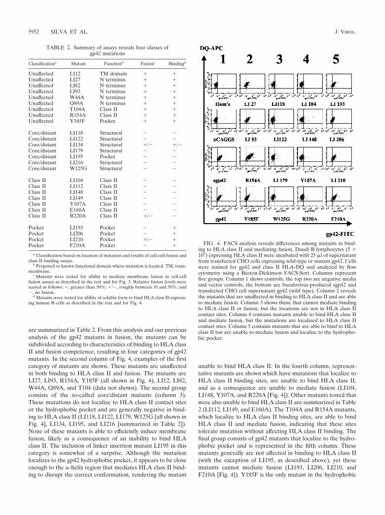
are summarized in Table 2. From this analysis and our previousanalysis of the gp42 mutants in fusion, the mutants can besubdivided according to characteristics of binding to HLA classII and fusion competence, resulting in four categories of gp42mutants. In the second column of Fig. 4, examples of the firstcategory of mutants are shown. These mutants are unaffectedin both binding to HLA class II and fusion. The mutants areLI27, LI93, R154A, Y185F (all shown in Fig. 4), LI12, LI82,W44A, Q89A, and T104 (data not shown). The second groupconsists of the so-called core/distant mutants (column 3).These mutations do not localize to HLA class II contact sitesor the hydrophobic pocket and are generally negative in bind-ing to HLA class II (LI118, LI122, LI179, W125G [all shown inFig. 4], LI134, LI195, and LI216 [summarized in Table 2]).None of these mutants is able to efficiently induce membranefusion, likely as a consequence of an inability to bind HLAclass II. The inclusion of linker insertion mutant LI195 in thiscategory is somewhat of a surprise. Although the mutationlocalizes to the gp42 hydrophobic pocket, it appears to be closeenough to the �-helix region that mediates HLA class II bind-ing to disrupt the correct conformation, rendering the mutant
unable to bind HLA class II. In the fourth column, represen-tative mutants are shown which have mutations that localize toHLA class II binding sites, are unable to bind HLA class II,and as a consequence are unable to mediate fusion (LI104,LI148, Y107A, and R220A [Fig. 4]). Other mutants tested thatwere also unable to bind HLA class II are summarized in Table2 (LI112, LI149, and E160A). The T104A and R154A mutants,which localize to HLA class II binding sites, are able to bindHLA class II and mediate fusion, indicating that these sitestolerate mutation without affecting HLA class II binding. Thefinal group consists of gp42 mutants that localize to the hydro-phobic pocket and is represented in the fifth column. Thesemutants generally are not affected in binding to HLA class II(with the exception of LI195, as described above), yet thesemutants cannot mediate fusion (LI193, LI206, LI210, andF210A [Fig. 4]). Y185F is the only mutant in the hydrophobic
FIG. 4. FACS analysis reveals differences among mutants in bind-ing to HLA class II and mediating fusion. Daudi B lymphocytes (5 �105) expressing HLA class II were incubated with 25 �l of supernatantfrom transfected CHO cells expressing wild-type or mutant gp42. Cellswere stained for gp42 and class II HLA-DQ and analyzed by flowcytometry using a Becton-Dickinson FACS-Sort. Columns representfive groups. Column 1 shows controls; the top two are negative mediaand vector controls, the bottom are baculovirus-produced sgp42 andtransfected CHO cell supernatant gp42 (wild type). Column 2 revealsthe mutants that are unaffected in binding to HLA class II and are ableto mediate fusion. Column 3 shows those that cannot mediate bindingto HLA class II or fusion, but the locations are not in HLA class IIcontact sites. Column 4 contains mutants unable to bind HLA class IIand mediate fusion, but the mutations are localized to HLA class IIcontact sites. Column 5 contains mutants that are able to bind to HLAclass II but are unable to mediate fusion and localize to the hydropho-bic pocket.
TABLE 2. Summary of assays reveals four classes ofgp42 mutations
Classificationa Mutant Functionb Fusionc Bindingd
Unaffected LI12 TM domain � �Unaffected LI27 N terminus � �Unaffected LI82 N terminus � �Unaffected LI93 N terminus � �Unaffected W44A N terminus � �Unaffected Q89A N terminus � �Unaffected T104A Class II � �Unaffected R154A Class II � �Unaffected Y185F Pocket � �
Core/distant LI118 Structural � �Core/distant LI122 Structural � �Core/distant LI134 Structural �/� �/�Core/distant LI179 Structural � �Core/distant LI195 Pocket � �Core/distant LI216 Structural � �Core/distant W125G Structural � �
Class II LI104 Class II � �Class II LI112 Class II � �Class II LI148 Class II � �Class II LI149 Class II � �Class II Y107A Class II � �Class II E160A Class II � �Class II R220A Class II �/� �
Pocket LI193 Pocket � �Pocket LI206 Pocket � �Pocket LI210 Pocket �/� �Pocket F210A Pocket � �
a Classification based on location of mutation and results of cell-cell fusion andclass II binding assays.
b Proposed or known functional domain where mutation is located. TM, trans-membrane.
c Mutants were tested for ability to mediate membrane fusion in cell-cellfusion assays as described in the text and for Fig. 3. Relative fusion levels werescored as follows: �, greater than 50%; �/�, roughly between 10 and 50%; and�, no fusion.
d Mutants were tested for ability of soluble form to bind HLA class II-express-ing human B cells as described in the text and for Fig. 4.
5952 SILVA ET AL. J. VIROL.

pocket able to mediate fusion similar to the wild type (sum-marized in Table 2).
DISCUSSION
We wished to confirm crystallization studies of EBV gp42bound to HLA class II and also ascertain if domains not bind-ing HLA class II were involved in membrane fusion. Theanalyses of gp42 mutants provide additional clues as to howgp42 may be involved in the initiation of membrane fusion, anessential process for the entry of EBV into B lymphocytes.From these studies, four general categories of gp42 mutantswere identified and are summarized in Table 2. Figure 5 showsthree-dimensional ribbon models with the locations of the mu-tations based on these categories that are explained furtherbelow. The four categories define mutants that (i) are unaf-fected for binding and fusion, (ii) likely induce folding defectsin the gp42 structure, disrupting HLA class II binding andfusion, (iii) are located in the HLA class II binding site, do notbind to HLA class II, and also do not mediate fusion, or (iv)are located in the hydrophobic pocket and are able to bindHLA class II but unable to mediate fusion. Interestingly, foreach of these categories we were able to identify a single pointmutant.
Mutations in the transmembrane domain and amino-termi-nal domain located near the membrane-spanning domain areunaffected in HLA class II binding and fusion (Fig. 5, upperleft). LI12, LI27, LI82, LI93, W44A, and Q89A, in all of whichthe mutations located within the amino-terminal domain ofgp42, were unaffected in their ability to bind HLA class II andmediate fusion. These results are compatible with results fromour structural studies of gp42 bound to HLA class II thatindicate that this region is not involved in the contact of gp42with HLA class II. Interestingly, this region may be importantfor the dimerization of gp42, as observed in our previous struc-tural studies, and the interaction of gp42 with the gH and gLcomplex. Previous studies have shown by deletion that a regionin the first 58 amino acids of gp42 is critical for the interactionof gp42 with gH and gL (38). In addition, this gp42 deletionmutant was unable to complement a gp42-deficient recombi-nant virus, indicating that complex formation between gp42and gH/gL is likely essential for EBV infectivity. None of thepresent mutants within this region of gp42, the HLA class IIinterface, or the hydrophobic pocket had any defect in theassociation of gp42 with gH/gL (data not shown), indicatingthat additional mutants should be isolated to determine thefunctional significance of the interaction of gp42 with gH/gL
FIG. 5. Ribbon models identifying locations of all gp42 mutants based on classes identified from Table 2. The classes are inconsequential,core/distant, HLA class II binding, or hydrophobic pocket. In each panel, the sites of the linker insertion (LI) mutants are indicated in blue andthe site-specific mutants are indicated in yellow. Abbreviations and color scheme follow those in previous figures.
VOL. 78, 2004 ANALYSIS OF EBV gp42 BINDING AND FUSION 5953

and its importance in fusion. Although gp42 was observed asdimers in the crystal structure, formation of gp42 dimers hasnot been detected by other analyses; thus, specific mutantswithin the dimerization region of gp42 may directly test thefunctional significance of gp42 dimer formation.
Mutations that localize to neither the HLA class II bindingsite nor the hydrophobic pocket are unable to mediate fusionand binding (Fig. 5, upper right). Surprisingly, several mutantswith linker insertions in regions that neither make contact withHLA class II nor are located in the hydrophobic pocket do notbind class II HLA-DQ and are unable to mediate fusion. Theseinclude LI118, LI122, LI134, LI179, and LI216, although LI134seems to allow low levels of binding and fusion. Since each ofthe mutations is distant to the HLA class II binding site butblocks HLA class II binding, with the possible exception ofLI134, it is likely that these mutants alter the overall structureof gp42. Compatible with this idea, many of these mutants fallwithin major structural features of gp42. LI118 and LI216 fallwithin two antiparallel �-sheets, for which the insertion of the5-amino-acid linker would be expected to have dramatic struc-tural consequences. LI134 is located at the base of one of two�-helices within the gp42 structure. Although not within thecore, this mutation may also be expected to alter the gp42conformation. Compatible with this, LI134 binds weakly toHLA class II and does not mediate fusion at wild-type levels.
The linker insertion site for the mutants LI122 and LI179 iscontained within exterior loops that connect structural deter-minants of the gp42 core. Since both insertion sites are fairlydistant from the HLA class II binding site and are not nearcysteines forming gp42 disulfide bonds, these results may indi-cate that the overall gp42 structure is easily perturbed, asindicated by the inability to bind HLA class II, but still suffi-ciently folded to result in expression and secretion of gp42, aswas observed with all of the mutants. The point mutant W125Gwould also appear to globally alter the gp42 structure, as dem-onstrated by lack of binding to HLA class II and fusion com-petence. The tryptophan residue is within the core of gp42 andmay act as a platform for the gp42 hydrophobic pocket to reston. The dramatic substitution to a glycine residue, if the tryp-tophan does serve as a major structural determinant, may alterthe stability of the protein. Consistent with this idea, of all ofthe mutations described in the present study, the W125G mu-tant exhibited both a decrease in cell surface expression and inthe supernatant of transfected cells.
Not all mutants localized to HLA class II binding domainsdisrupt gp42 function (Fig. 5, lower left). Y107A, E160A, andR220A mutants all lost their ability to bind HLA class II andare unable to mediate fusion, highlighting the requirement ofgp42 binding to HLA class II in fusion. Surprisingly, low levelsof fusion are consistently detected with the R220A mutant,which does not detectably bind to HLA class II expressed onDaudi cells. One possible explanation is that perhaps the saltbridge is not as critical as the hydrogen bonds and some low-level binding may occur that is not readily apparent in thebinding assay, allowing for some measurable fusion to occur.All of the linker insertion mutants that are within or close toHLA class II contact sites are unable to bind HLA class II andare also unable to mediate fusion (LI104, LI112, LI148, andLI149). A 5-amino-acid insert clearly eliminates all bindingactivity in this region. Although T104A and R154A localize to
HLA class II binding sites, they are able to bind to HLA classII as well as mediate fusion. Previous structural studies of thegp42 HLA class II complex indicated that the carbonyl oxygensfrom the main chains of T104 and Y107 of gp42 form hydrogenbonds with R�72 of HLA class II (28). Mutational analysis ofR�72 of HLA class II indicated that this residue is essential forgp42 binding to HLA class II and EBV entry (26). The T104Amutation, which would be predicted not to disrupt the hydro-gen bond between T104 and R�72, has no effect on gp42binding to HLA class II or EBV-induced membrane fusion, asmight be expected. However, mutation of Y107 blocks both,probably due to multiple effects by potentially changing thelocal conformation of the region that binds HLA class II aswell as blocking a hydrogen bond with E�46. In regard to themutation of R154 of gp42, previous mutational studies of HLAclass II S�63, which hydrogen bonds with gp42 R154, indicatedthat this interaction of R154 with S�63 is not important forgp42 binding to HLA class II or EBV entry since mutation ofS�63 to either an alanine or lysine did not block gp42 bindingto HLA class II or EBV entry (26). This result is furthersupported by the present results indicating that R154 of gp42is not important for gp42 binding to HLA class II or EBV-mediated fusion.
Though gp42 is structurally similar to some other C-typelectin superfamily members, the gp42/HLA class II interactionis unique compared to other natural killer (NK) receptor com-plexes, namely, Ly49A/MHC class I and NKG2D/MHC class Ihomolog (MICA) (28). The interaction is also different fromthe HSV gD/HveA binding complex. McShane et al. clearlyshowed that a single residue mutation of HLA-DQ could elim-inate sgp42 binding and EBV infection, whereas single muta-tions of HveA did not affect HSV entry, suggesting that mostcontact residues contribute to receptor function collectivelyrather than individually (26).
Many mutations that localize to the hydrophobic pocket areunaffected in HLA class II binding but do not mediate fusion(Fig. 5, lower right). LI193, LI206, LI210, Y185F, and F210Aare all able to bind to class II-expressing cells. However, ofthese, only Y185F is able to efficiently mediate fusion similarlyto the wild type. In the hydrophobic pocket, it is interesting tosee that a single point mutant can distinguish HLA class IIbinding from the ability to mediate membrane fusion. It isperhaps not surprising that the relatively conservative muta-tion of tyrosine to phenylalanine at amino acid 185 does notalter fusion activity. In contrast, the more drastic change ofphenylalanine to alanine at amino acid 210 does disrupt fusionsignificantly. It is likely, as with the case of HLA class IIbinding, that some of these hydrophobic residues are morecritical than others for generating a strong interaction with apotential unknown binding partner. Interestingly, all of thelinker insertion mutants within the gp42 hydrophobic pockethave greatly reduced fusion when compared to that of the wildtype, LI210 clearly being positive for fusion but at reducedlevels, whereas LI193 and LI206 have fusion consistently abovebackground but not nearly as high as that of LI210. Of thelinker insertions, only LI195 appears to be entirely negative forfusion. Importantly, the site of this insertion is close to the�-helix HLA class II binding site, resulting in a mutant gp42that is unable to bind HLA class II. This may be the primarydefect in this mutant and not due to the gp42 hydrophobic
5954 SILVA ET AL. J. VIROL.
pocket. It is likely that proximity of the mutation to the HLAclass II binding site coupled with the insertion of five aminoacids disrupts the HLA class II binding function of gp42 in theLI195 mutant. Therefore, this mutant has been classified as acore/distant mutant, as it does not show the hydrophobicpocket mutant phenotype, that is, the ability to bind HLA classII but not mediate fusion.
The mutants described in this report have helped to definefunctional domains required for fusion. We have demonstratedthat, in addition to a functional gp42/HLA class II binding site,a structurally intact hydrophobic pocket is required to initiatemembrane fusion. The discovery of this hydrophobic pocket isinteresting for two reasons: the functional requirement (i) addsto our current model of EBV-induced membrane fusion and(ii) provides a potential target for antiviral strategies. Like theHSV gD/HveA interface, only a handful of the EBV gp42/HLA class II contact residues that comprise the interface areactually required for binding (5). The structural studies of gDby Carfı et al. suggested that the N-terminal gD hairpin wasconformationally flexible and that a conformational changeaccompanying binding might be part of the viral entry mech-anism (4). Although structurally distinct from gD, gp42 couldhave a similar functional mechanism, and such a conforma-tional change might also be required for membrane fusion.Binding of HLA class II might induce a conformational changethat reveals the hydrophobic pocket, which is then able to bindits viral or cellular ligand. If the pocket is occupied beforebinding to HLA class II, the structure could reposition the viralligand to come in contact with the host membrane. The func-tional requirement of the hydrophobic pocket makes it anexcellent target for antiviral inhibitors. Modis et al. discovereda ligand-binding hydrophobic pocket in the dengue virus en-velope glycoprotein E that is briefly exposed as fusion-compe-tent trimers form, implicating a multiconformational mecha-nism of membrane fusion (27). This hydrophobic pocket wasdiscovered to bind a single molecule of the detergent n-octyl-�-D-glucoside, indicating a potential site for small-moleculeinhibitors. The hydrophobic pocket of the human immunode-ficiency virus gp120/41 trimer has already proven fruitful inboth identifying synthetic peptides and screening small mole-cules to inhibit binding and fusion (7, 8). Such results areencouraging for future studies of potential inhibitors of gp42-mediated membrane fusion. The mutants generated fromthese experiments will be used to supplement our knowledgeof binding requirements with the gH/gL complex and with gBif there is a direct interaction. We will next use these mutantsto identify the ligand of this hydrophobic pocket, which couldbe viral or cellular, which would further characterize require-ments and mechanisms involved with initiating membrane fu-sion and provide a target to block viral entry.
ACKNOWLEDGMENTS
We thank Gant Luxton, George Stanton, and Asheera Alvarez forhelping generate mutants; Charu Deshpande for help in generatingstably transfected Daudi cells; and members of the Longnecker andSpear laboratories for a surplus of technical advice, especially CherylJogger, Alina Fridberg, Keith Haan, and Marisa McShane.
R.L. is a Stohlman Scholar of the Leukemia and Lymphoma Societyof America and supported by the Public Health Service grantsCA62234, CA73507, and CA93444 from the National Cancer Instituteand DE13127 from the National Institute of Dental and Craniofacial
Research. T.S.J. is a Scholar of the Leukemia and Lymphoma Societyof America and supported by the Public Health Service grantsGM61050, AI38972, and CA93444 from the National Cancer Institute.A.L.S. is supported by the training program in the Cellular and Mo-lecular Basis of Disease (T32 GM08061) from the National Institutesof Health.
REFERENCES
1. Baer, R., A. T. Bankier, M. D. Biggin, P. L. Deininger, P. J. Farrell, T. J.Gibson, G. Hatfull, G. S. Hudson, S. C. Satchwell, C. Seguin, et al. 1984.DNA sequence and expression of the B95-8 Epstein-Barr virus genome.Nature 310:207–211.
2. Borza, C. M., and L. M. Hutt-Fletcher. 2002. Alternate replication in B cellsand epithelial cells switches tropism of Epstein-Barr virus. Nat. Med. 8:594–599.
3. Burkitt, D. 1962. A children’s cancer dependent on climatic factors. Nauchni.Tr. Vissh. Med. Inst. Sofiia 194:232–234.
4. Carfı, A., S. H. Willis, J. C. Whitbeck, C. Krummenacher, G. H. Cohen, R. J.Eisenberg, and D. C. Wiley. 2001. Herpes simplex virus glycoprotein Dbound to the human receptor HveA. Mol. Cell 8:169–179.
5. Connolly, S. A., D. J. Landsburg, A. Carfi, D. C. Wiley, G. H. Cohen, andR. J. Eisenberg. 2003. Structure-based mutagenesis of herpes simplex virusglycoprotein D defines three critical regions at the gD-HveA/HVEM bindinginterface. J. Virol. 77:8127–8140.
6. Connolly, S. A., D. J. Landsburg, A. Carfi, D. C. Wiley, R. J. Eisenberg, andG. H. Cohen. 2002. Structure-based analysis of the herpes simplex virusglycoprotein D binding site present on herpesvirus entry mediator HveA(HVEM). J. Virol. 76:10894–10904.
7. Debnath, A. K., L. Radigan, and S. Jiang. 1999. Structure-based identifica-tion of small molecule antiviral compounds targeted to the gp41 core struc-ture of the human immunodeficiency virus type 1. J. Med. Chem. 42:3203–3209.
8. Eckert, D. M., V. N. Malashkevich, L. H. Hong, P. A. Carr, and P. S. Kim.1999. Inhibiting HIV-1 entry: discovery of D-peptide inhibitors that targetthe gp41 coiled-coil pocket. Cell 99:103–115.
9. Epstein, M. A., B. G. Achong, and Y. M. Barr. 1964. Virus particles incultured lymphoblasts from Burkitt’s lymphoma. Lancet 15:702–703.
10. Fingeroth, J. D., J. J. Weis, T. F. Tedder, J. L. Strominger, P. A. Biro, andD. T. Fearon. 1984. Epstein-Barr virus receptor of human B lymphocytes isthe C3d receptor CR2. Proc. Natl. Acad. Sci. USA 81:4510–4514.
11. Haan, K. M., W. W. Kwok, R. Longnecker, and P. Speck. 2000. Epstein-Barrvirus entry utilizing HLA-DP or HLA-DQ as a coreceptor. J. Virol. 74:2451–2454.
12. Haan, K. M., S. K. Lee, and R. Longnecker. 2001. Different functionaldomains in the cytoplasmic tail of glycoprotein B are involved in Epstein-Barr virus-induced membrane fusion. Virology 290:106–114.
13. Haan, K. M., and R. Longnecker. 2000. Coreceptor restriction within theHLA-DQ locus for Epstein-Barr virus infection. Proc. Natl. Acad. Sci. USA97:9252–9257.
14. Heineman, T., M. Gong, J. Sample, and E. Kieff. 1988. Identification of theEpstein-Barr virus gp85 gene. J. Virol. 62:1101–1107.
15. Herrold, R. E., A. Marchini, S. Fruehling, and R. Longnecker. 1996. Glyco-protein 110, the Epstein-Barr virus homolog of herpes simplex virus glyco-protein B, is essential for Epstein-Barr virus replication in vivo. J. Virol.70:2049–2054.
16. Hutt-Fletcher, L. M., and C. M. Lake. 2001. Two Epstein-Barr virus glyco-protein complexes. Curr. Top. Microbiol. Immunol. 258:51–64.
17. Janz, A., M. Oezel, C. Kurzeder, J. Mautner, D. Pich, M. Kost, W. Ham-merschmidt, and H. J. Delecluse. 2000. Infectious Epstein-Barr virus lackingmajor glycoprotein BLLF1 (gp350/220) demonstrates the existence of addi-tional viral ligands. J. Virol. 74:10142–10152.
18. Klein, G., G. Clements, J. Zeuthen, and A. Westman. 1976. Somatic cellhybrids between human lymphoma lines. II. Spontaneous and induced pat-terns of the Epstein-Barr virus (EBV) cycle. Int. J. Cancer 17:715–724.
19. Kobasa, D., M. Rodgers, K. Wells, and Y. Kawaoka. 1997. Neuraminidasehemadsorption activity, conserved in avian influenza A viruses, does notinfluence viral replication in ducks. J. Virol. 71:6706–6713.
20. Langeland, N., A. M. Oyan, H. S. Marsden, A. Cross, J. C. Glorioso, L. J.Moore, and L. Haarr. 1990. Localization on the herpes simplex virus type 1genome of a region encoding proteins involved in adsorption to the cellularreceptor. J. Virol. 64:1271–1277.
21. Lee, S. K., T. Compton, and R. Longnecker. 1997. Failure to complementinfectivity of EBV and HSV-1 glycoprotein B (gB) deletion mutants with gBsfrom different human herpesvirus subfamilies. Virology 237:170–181.
22. Lee, S. K., and R. Longnecker. 1997. The Epstein-Barr virus glycoprotein 110carboxy-terminal tail domain is essential for lytic virus replication. J. Virol.71:4092–4097.
23. Li, Q., M. K. Spriggs, S. Kovats, S. M. Turk, M. R. Comeau, B. Nepom, andL. M. Hutt-Fletcher. 1997. Epstein-Barr virus uses HLA class II as a cofactorfor infection of B lymphocytes. J. Virol. 71:4657–4662.
24. Li, Q., S. M. Turk, and L. M. Hutt-Fletcher. 1995. The Epstein-Barr virus
VOL. 78, 2004 ANALYSIS OF EBV gp42 BINDING AND FUSION 5955

(EBV) BZLF2 gene product associates with the gH and gL homologs ofEBV and carries an epitope critical to infection of B cells but not of epithe-lial cells. J. Virol. 69:3987–3994.
25. Longnecker, R. 1998. Molecular biology of Epstein-Barr virus, p. 133–174. InD. J. McCance (ed.), Human tumor viruses. American Society for Microbi-ology, Washington, D.C.
26. McShane, M. P., M. M. Mullen, K. M. Haan, T. S. Jardetzky, and R.Longnecker. 2003. Mutational analysis of the HLA class II interaction withEpstein-Barr virus glycoprotein 42. J. Virol. 77:7655–7662.
27. Modis, Y., S. Ogata, D. Clements, and S. C. Harrison. 2003. A ligand-bindingpocket in the dengue virus envelope glycoprotein. Proc. Natl. Acad. Sci. USA100:6986–6991.
28. Mullen, M. M., K. M. Haan, R. Longnecker, and T. S. Jardetzky. 2002.Structure of the Epstein-Barr virus gp42 protein bound to the MHC class IIreceptor HLA-DR1. Mol. Cell 9:375–385.
29. Nemerow, G. R., C. Mold, V. K. Schwend, V. Tollefson, and N. R. Cooper.1987. Identification of gp350 as the viral glycoprotein mediating attachmentof Epstein-Barr virus (EBV) to the EBV/C3d receptor of B cells: sequencehomology of gp350 and C3 complement fragment C3d. J. Virol. 61:1416–1420.
30. Neuhierl, B., R. Feederle, W. Hammerschmidt, and H. J. Delecluse. 2002.Glycoprotein gp110 of Epstein-Barr virus determines viral tropism and ef-ficiency of infection. Proc. Natl. Acad. Sci. USA 99:15036–15041.
31. Okuma, K., M. Nakamura, S. Nakano, Y. Niho, and Y. Matsuura. 1999. Hostrange of human T-cell leukemia virus type I analyzed by a cell fusion-dependent reporter gene activation assay. Virology 254:235–244.
32. Rickinson, A. B., and E. Kieff. 1996. Epstein-Barr virus, p. 2397–2446. InB. N. Fields, D. M. Knipe, and P. M. Howley (ed.), Virology, 3rd ed. RavenPress, New York, N.Y.
33. Spear, P. G., and R. Longnecker. 2003. Herpesvirus entry: an update. J. Vi-rol. 77:10179–10185.
34. Speck, P., K. M. Haan, and R. Longnecker. 2000. Epstein-Barr virus entryinto cells. Virology 277:1–5.
35. Spriggs, M. K., R. J. Armitage, M. R. Comeau, L. Strockbine, T. Farrah, B.Macduff, D. Ulrich, M. R. Alderson, J. Mullberg, and J. I. Cohen. 1996. Theextracellular domain of the Epstein-Barr virus BZLF2 protein binds theHLA-DR � chain and inhibits antigen presentation. J. Virol. 70:5557–5563.
36. Tanner, J., J. Weis, D. Fearon, Y. Whang, and E. Kieff. 1987. Epstein-Barrvirus gp350/220 binding to the B lymphocyte C3d receptor mediates adsorp-tion, capping, and endocytosis. Cell 50:203–213.
37. Wang, X., and L. M. Hutt-Fletcher. 1998. Epstein-Barr virus lacking glyco-protein gp42 can bind to B cells but is not able to infect. J. Virol. 72:158–163.
38. Wang, X., W. J. Kenyon, Q. Li, J. Mullberg, and L. M. Hutt-Fletcher. 1998.Epstein-Barr virus uses different complexes of glycoproteins gH and gL toinfect B lymphocytes and epithelial cells. J. Virol. 72:5552–5558.
39. Whetter, L. E., S. P. Day, E. A. Brown, O. Elroy-Stein, and S. M. Lemon.1994. Analysis of hepatitis A virus translation in a T7 polymerase-expressingcell line. Arch. Virol. Suppl. 9:291–298.
40. Yaswen, L. R., E. B. Stephens, L. C. Davenport, and L. M. Hutt-Fletcher.1993. Epstein-Barr virus glycoprotein gp85 associates with the BKRF2 geneproduct and is incompletely processed as a recombinant protein. Virology195:387–396.
5956 SILVA ET AL. J. VIROL.