Expression and Processing of a Small Nucleolar RNA from the Epstein-Barr Virus Genome
13
Expression and Processing of a Small Nucleolar RNA from the Epstein-Barr Virus Genome Roland Hutzinger 1. , Regina Feederle 2. , Jan Mrazek 3 , Natalia Schiefermeier 4 , Piotr J. Balwierz 5 , Mihaela Zavolan 5 , Norbert Polacek 1 , Henri-Jacques Delecluse 2 *, Alexander Hu ¨ ttenhofer 1 * 1 Innsbruck Biocenter, Division of Genomics and RNomics, Innsbruck Medical University, Innsbruck, Austria, 2 German Cancer Research Center, Department of Virus- Associated Tumours, Heidelberg, Germany, 3 Department of Biological Chemistry, David Geffen School of Medicine, University of California Los Angeles, Los Angeles, California, United States of America, 4 Innsbruck Biocenter, Division of Cell Biology, Innsbruck Medical University, Innsbruck, Austria, 5 Biozentrum, Swiss Institute of Bioinformatics, University of Basel, Basel, Switzerland Abstract Small nucleolar RNAs (snoRNAs) are localized within the nucleolus, a sub-nuclear compartment, in which they guide ribosomal or spliceosomal RNA modifications, respectively. Up until now, snoRNAs have only been identified in eukaryal and archaeal genomes, but are notably absent in bacteria. By screening B lymphocytes for expression of non-coding RNAs (ncRNAs) induced by the Epstein-Barr virus (EBV), we here report, for the first time, the identification of a snoRNA gene within a viral genome, designated as v-snoRNA1. This genetic element displays all hallmark sequence motifs of a canonical C/D box snoRNA, namely C/C9- as well as D/D9-boxes. The nucleolar localization of v-snoRNA1 was verified by in situ hybridisation of EBV-infected cells. We also confirmed binding of the three canonical snoRNA proteins, fibrillarin, Nop56 and Nop58, to v-snoRNA1. The C-box motif of v-snoRNA1 was shown to be crucial for the stability of the viral snoRNA; its selective deletion in the viral genome led to a complete down-regulation of v-snoRNA1 expression levels within EBV- infected B cells. We further provide evidence that v-snoRNA1 might serve as a miRNA-like precursor, which is processed into 24 nt sized RNA species, designated as v-snoRNA1 24pp . A potential target site of v-snoRNA1 24pp was identified within the 39- UTR of BALF5 mRNA which encodes the viral DNA polymerase. V-snoRNA1 was found to be expressed in all investigated EBV-positive cell lines, including lymphoblastoid cell lines (LCL). Interestingly, induction of the lytic cycle markedly up- regulated expression levels of v-snoRNA1 up to 30-fold. By a computational approach, we identified a v-snoRNA1 homolog in the rhesus lymphocryptovirus genome. This evolutionary conservation suggests an important role of v-snoRNA1 during c-herpesvirus infection. Citation: Hutzinger R, Feederle R, Mrazek J, Schiefermeier N, Balwierz PJ, et al. (2009) Expression and Processing of a Small Nucleolar RNA from the Epstein-Barr Virus Genome. PLoS Pathog 5(8): e1000547. doi:10.1371/journal.ppat.1000547 Editor: Bryan R. Cullen, Duke University Medical Center, United States of America Received May 5, 2009; Accepted July 20, 2009; Published August 14, 2009 Copyright: ß 2009 Hutzinger et al. This is an open-access article distributed under the terms of the Creative Commons Attribution License, which permits unrestricted use, distribution, and reproduction in any medium, provided the original author and source are credited. Funding: This work was supported by two Austrian genome research grants (Gen-Au grant D-110420-011-013) to A.H. and N.P. and (D-11420-011-015) to A.H. and by DKFZ grant to H-J.D. The funders had no role in study design, data collection and analysis, decision to publish, or preparation of the manuscript. Competing Interests: The authors have declared that no competing interests exist. * E-mail: [email protected] (H-JD); [email protected] (AH) . These authors contributed equally to this work. Introduction The Epstein-Barr virus (EBV), a member of the c-herpesvirus subfamily, possesses a large (170 to 180 kb) double-stranded DNA genome. EBV infection is etiologically linked with various cancers of the lymphoid and epithelial lineages that include Burkitt’s lymphoma (BL), Hodgkin’s disease, nasopharyngeal carcinoma (NPC) and post-transplant lymphoproliferate disease (PTLD) [1– 4]. In vitro and in vivo, EBV transforms normal B cells through establishment of a type III latency during which a restricted set of viral genes is expressed (eight Epstein-Barr nuclear antigens and two latent membrane proteins) [5]. More restricted expression patterns such as latency type II in NPC and latency type I in BL have also been characterized. In fact, recent work on Burkitt’s lymphoma has shown that a subset of these tumours display a latency pattern intermediate between latency I and III showing that the boundaries between the latency types are not always sharply established as initially thought [6]. More then two decades ago, the group of J. Steitz discovered two highly abundant ,170-nt long non-coding RNAs (ncRNAs) in the EBV genome, designated as Epstein-Barr encoded RNAs (EBER1 and EBER2) [7]. EBER RNAs have subsequently been shown to bind to human ribosomal protein L22. However, no unequivocal biological functions could be assigned to EBER transcripts, up till now [8]. The list of non-coding RNAs encoded by EBV has since rapidly expanded with the recent discovery of 25 microRNAs (miRNAs) [9–14]. In addition to miRNAs, numerous other ncRNAs have been discovered in all three domains of life, i.e. Archaea, Bacteria and Eukarya, as well as in various viruses [15,16]. A large number of these ncRNA species were found to be involved in multiple regulatory functions including cellular differentiation and development, chro- matin architecture, transcription and translation, alternative splicing, RNA editing, virulence and stress responses [17–20]. Small nucleolar RNAs (snoRNAs) consist of more than 200 stable ncRNA species in Eukarya of about 60 to 300 nt in size which are located in a sub-nuclear compartment, the nucleolus [21,22]. SnoRNAs guide nucleotide modifications within ribo- somal RNAs (rRNAs) or spliceosomal RNAs (snRNAs), i.e. 29-O- PLoS Pathogens | www.plospathogens.org 1 August 2009 | Volume 5 | Issue 8 | e1000547
Transcript of Expression and Processing of a Small Nucleolar RNA from the Epstein-Barr Virus Genome
Expression and Processing of a Small Nucleolar RNA fromthe Epstein-Barr Virus GenomeRoland Hutzinger1., Regina Feederle2., Jan Mrazek3, Natalia Schiefermeier4, Piotr J. Balwierz5, Mihaela
Zavolan5, Norbert Polacek1, Henri-Jacques Delecluse2*, Alexander Huttenhofer1*
1 Innsbruck Biocenter, Division of Genomics and RNomics, Innsbruck Medical University, Innsbruck, Austria, 2 German Cancer Research Center, Department of Virus-
Associated Tumours, Heidelberg, Germany, 3 Department of Biological Chemistry, David Geffen School of Medicine, University of California Los Angeles, Los Angeles,
California, United States of America, 4 Innsbruck Biocenter, Division of Cell Biology, Innsbruck Medical University, Innsbruck, Austria, 5 Biozentrum, Swiss Institute of
Bioinformatics, University of Basel, Basel, Switzerland
Abstract
Small nucleolar RNAs (snoRNAs) are localized within the nucleolus, a sub-nuclear compartment, in which they guideribosomal or spliceosomal RNA modifications, respectively. Up until now, snoRNAs have only been identified in eukaryal andarchaeal genomes, but are notably absent in bacteria. By screening B lymphocytes for expression of non-coding RNAs(ncRNAs) induced by the Epstein-Barr virus (EBV), we here report, for the first time, the identification of a snoRNA genewithin a viral genome, designated as v-snoRNA1. This genetic element displays all hallmark sequence motifs of a canonicalC/D box snoRNA, namely C/C9- as well as D/D9-boxes. The nucleolar localization of v-snoRNA1 was verified by in situhybridisation of EBV-infected cells. We also confirmed binding of the three canonical snoRNA proteins, fibrillarin, Nop56 andNop58, to v-snoRNA1. The C-box motif of v-snoRNA1 was shown to be crucial for the stability of the viral snoRNA; itsselective deletion in the viral genome led to a complete down-regulation of v-snoRNA1 expression levels within EBV-infected B cells. We further provide evidence that v-snoRNA1 might serve as a miRNA-like precursor, which is processed into24 nt sized RNA species, designated as v-snoRNA124pp. A potential target site of v-snoRNA124pp was identified within the 39-UTR of BALF5 mRNA which encodes the viral DNA polymerase. V-snoRNA1 was found to be expressed in all investigatedEBV-positive cell lines, including lymphoblastoid cell lines (LCL). Interestingly, induction of the lytic cycle markedly up-regulated expression levels of v-snoRNA1 up to 30-fold. By a computational approach, we identified a v-snoRNA1 homologin the rhesus lymphocryptovirus genome. This evolutionary conservation suggests an important role of v-snoRNA1 duringc-herpesvirus infection.
Citation: Hutzinger R, Feederle R, Mrazek J, Schiefermeier N, Balwierz PJ, et al. (2009) Expression and Processing of a Small Nucleolar RNA from the Epstein-BarrVirus Genome. PLoS Pathog 5(8): e1000547. doi:10.1371/journal.ppat.1000547
Editor: Bryan R. Cullen, Duke University Medical Center, United States of America
Received May 5, 2009; Accepted July 20, 2009; Published August 14, 2009
Copyright: � 2009 Hutzinger et al. This is an open-access article distributed under the terms of the Creative Commons Attribution License, which permitsunrestricted use, distribution, and reproduction in any medium, provided the original author and source are credited.
Funding: This work was supported by two Austrian genome research grants (Gen-Au grant D-110420-011-013) to A.H. and N.P. and (D-11420-011-015) to A.H.and by DKFZ grant to H-J.D. The funders had no role in study design, data collection and analysis, decision to publish, or preparation of the manuscript.
Competing Interests: The authors have declared that no competing interests exist.
* E-mail: [email protected] (H-JD); [email protected] (AH)
. These authors contributed equally to this work.
IntroductionThe Epstein-Barr virus (EBV), a member of the c-herpesvirus
subfamily, possesses a large (170 to 180 kb) double-stranded DNA
genome. EBV infection is etiologically linked with various cancers
of the lymphoid and epithelial lineages that include Burkitt’s
lymphoma (BL), Hodgkin’s disease, nasopharyngeal carcinoma
(NPC) and post-transplant lymphoproliferate disease (PTLD) [1–
4]. In vitro and in vivo, EBV transforms normal B cells through
establishment of a type III latency during which a restricted set of
viral genes is expressed (eight Epstein-Barr nuclear antigens and
two latent membrane proteins) [5]. More restricted expression
patterns such as latency type II in NPC and latency type I in BL
have also been characterized. In fact, recent work on Burkitt’s
lymphoma has shown that a subset of these tumours display a
latency pattern intermediate between latency I and III showing
that the boundaries between the latency types are not always
sharply established as initially thought [6].
More then two decades ago, the group of J. Steitz discovered
two highly abundant ,170-nt long non-coding RNAs (ncRNAs) in
the EBV genome, designated as Epstein-Barr encoded RNAs
(EBER1 and EBER2) [7]. EBER RNAs have subsequently been
shown to bind to human ribosomal protein L22. However, no
unequivocal biological functions could be assigned to EBER
transcripts, up till now [8]. The list of non-coding RNAs encoded
by EBV has since rapidly expanded with the recent discovery of 25
microRNAs (miRNAs) [9–14].
In addition to miRNAs, numerous other ncRNAs have been
discovered in all three domains of life, i.e. Archaea, Bacteria and
Eukarya, as well as in various viruses [15,16]. A large number of these
ncRNA species were found to be involved in multiple regulatory
functions including cellular differentiation and development, chro-
matin architecture, transcription and translation, alternative splicing,
RNA editing, virulence and stress responses [17–20].
Small nucleolar RNAs (snoRNAs) consist of more than 200
stable ncRNA species in Eukarya of about 60 to 300 nt in size
which are located in a sub-nuclear compartment, the nucleolus
[21,22]. SnoRNAs guide nucleotide modifications within ribo-
somal RNAs (rRNAs) or spliceosomal RNAs (snRNAs), i.e. 29-O-
PLoS Pathogens | www.plospathogens.org 1 August 2009 | Volume 5 | Issue 8 | e1000547
ribose methylation or pseudouridylation, respectively. The
snoRNA class has been identified in Archaea and Eukarya, but
not in Bacteria, and is subdivided into box C/D and box H/ACA
snoRNAs. In Eukarya, the majority of snoRNAs is located within
introns of protein-coding genes and is processed by splicing
followed by endo- and exonucleolytic cleavage [19,23,24].
Each member of the box C/D snoRNA family possesses
characteristic sequence elements called box C (PuUGAUGA) and
box D (CUGA), optional degenerate C9/D9 boxes and a short 59-39
terminal stem structure [24,25]. 10–21 nt long sequence-specific
antisense elements upstream of the boxes D/D9 guide the box C/D
snoRNA core proteins fibrillarin, a RNA methyltransferase, Nop56,
Nop58 and the 15.5 kD protein to the target RNA. 29-O-methylation
of the ribose at the fifth nucleotide upstream of the D/D9 box on the
target RNA is carried out by the fibrillarin core protein [24]. Box H/
ACA snoRNAs possess a distinctive common ACA sequence motif at
their 39-terminus and one to two stem-loop structures linked by a
hinge (the so-called H-box motif: ANANNA, with N being any
nucleotide), and guide the conversion of uridine to pseudouridine
within the RNA target [26,27]. The large number of conserved
modifications in functionally conserved regions of rRNAs, such as the
peptidyl-transferase centre, has suggested an important role for rRNA
modifications in fine-tuning the structure and/or function of rRNAs
[28]. It is important to note that a significant number of so-called
‘‘orphan’’ snoRNAs, lacking rRNA or snRNA targets, have been
identified in Eukarya [29,30]. However, the biological functions of
orphan snoRNAs are still elusive.
In this study, we report, for the first time, the identification of a
functional C/D box snoRNA within the EBV genome. We
demonstrate that this viral snoRNA exhibits all bona fide box C/D
snoRNA features with respect to its processing and expression,
nucleolar localization as well as to canonical core protein binding
partners. We also provide evidence that v-snoRNA1 is processed into
a 24 nt long miRNA-like species which might target the 39-UTR of
the viral DNA polymerase mRNA.
Results
Identification of v-snoRNA1 by cDNA cloning andexpression analysis
We have established an experimental strategy, designated as
SHORT, to identify viral-induced ncRNAs in cord blood
lymphocytes (CBL) infected with the EBV strain B95.8 [31].
The SHORT method is based on subtractive hybridisation of
ncRNA populations of virus-infected cells from non-infected cells.
NcRNAs, selectively expressed in the infected cell population,
were subsequently converted into cDNAs. Sequencing of a small
number, i.e. about 500 cDNA clones, allowed identification of
several ncRNAs from the human as well as from the EBV genome
whose expression was up-regulated upon viral infection [32].
Deep-sequencing analysis of 40.000 cDNA clones from this
subtracted cDNA library further extended the list of differentially
expressed ncRNAs (Hutzinger et al., manuscript in preparation).
Interestingly, one of these sequences was represented by 95
cDNAs and exhibited all defining features of canonical C/D box
snoRNA sequence motifs, i.e. C, C9, D9 and D boxes [24,25].
Crucially, this potentially novel snoRNA species mapped to the
EBV genome and was therefore designated as v-snoRNA1
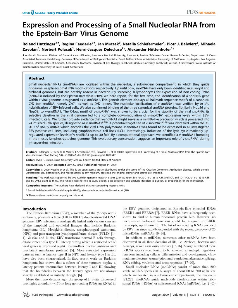
(Figure 1A, and see above; Accession number FN376861). It is
noteworthy that the canonical terminal stem-structure, formed by
the 59 and 39 ends of eukaryal snoRNAs, was absent in the viral
snoRNA, a feature shared with snoRNAs identified from archaeal
or fungal species [33,34].
To assess expression of v-snoRNA1, northern blot analysis was
performed employing RNA from EBV-positive cell lines (Rael,
Raji, BL2-B95.8, BL41-B95.8 and a LCL generated in vitro with
the B95.8 virus) or EBV-negative cell lines (BL2 and BL41;
Figure 1B). As expected, v-snoRNA1 could only be detected in
infected cells but not in the EBV-negative control cells.
Comparison with an internal RNA marker showed that the
hybridized RNA species was 65 nt in size, which fully matched the
size suggested by the original sequence obtained by cDNA cloning
(see above and Figure 1B). Repeated attempts to identify v-
snoRNA1-precursor transcripts by northern blot analysis were
unsuccessful (unpublished data), suggesting that they are subjected
to rapid processing.
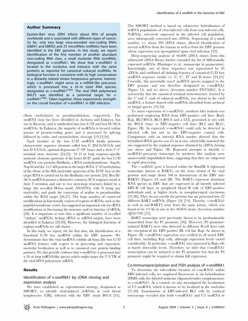
The v-snoRNA1 gene is located within the BamHI A rightward
transcripts, known as BARTs, on the sense strand of the viral
genome and maps about 100 nt downstream of the EBV mir-
BART2 (Figures 2A and 2B). The BARTs represent abundant
RNA species in EBV that are expressed in all latently infected
EBV-B cell lines, in peripheral blood B cells of EBV-positive
individuals and, at higher levels, in nasopharyngeal carcinoma
[35,36]. They do not encode for proteins but are processed into 22
different BART miRNAs (Figure 2A) [14]. Thereby, v-snoRNA1
as well as mir-BART2 arise from the same intron, which was
found to be 4.9 kb in size in the AG876 strain (Accession number
AJ507799) [35].
BART transcripts were previously shown to be predominantly
transcribed from the P1 promoter [36]. However, P2 promoter-
initiated BARTs were also detected in different B-cell lines with
the exception of the EBV-positive BL cell line Raji. As shown in
Figure 1B, v-snoRNA1 expression was verified in all tested EBV
cell lines, including Raji cells, although expression levels varied
considerably. In particular, v-snoRNA1 was expressed in Raji cells
at barely detectable levels. Therefore, we infer that v-snoRNA1
transcription can be initiated at the P1 promoter but that the P2
promoter might be required to obtain full expression.
Co-Immunoprecipitation and FISH analysis of v-snoRNA1To determine the sub-cellular location of v-snoRNA1 within
EBV-infected cells, we employed fluorescent in situ hybridization
(FISH) with dye-labeled antisense oligonucleotides complementary
to v-snoRNA1. As a control, we also investigated the localization
of U3 snoRNA, which is known to be localized in the nucleolus
[37,38]. Examination of EBV-infected BL2 cells by confocal
microscopy revealed that both v-snoRNA1 and U3 snoRNA in
Author Summary
Epstein-Barr virus (EBV) infects about 90% of peopleworldwide and is associated with different types of cancer.So far, only two large virus-encoded non-coding RNAs(EBER1 and EBER2) and 25 microRNAs (miRNAs) have beenidentified in the EBV genome. In this study, we reportidentification of the first member of another abundantnon-coding RNA class, a small nucleolar RNA (snoRNA),designated as v-snoRNA1. We show that v-snoRNA1 islocated in the nucleolus and interacts with the sameproteins as reported for canonical eukaryal snoRNAs. Itsbiological function is consistent with its high conservationin a distantly related simian herpesvirus genome. Interest-ingly, v-snoRNA1 might serve as a miRNA-like precursor,which is processed into a 24 nt sized RNA species,designated as v-snoRNA124pp. The viral DNA polymeraseBALF5 was identified as a potential target for v-snoRNA124pp. Taken together, these experiments strength-en the crucial function of v-snoRNA1 in EBV infection.
Identification of a snoRNA in the Genome of EBV
PLoS Pathogens | www.plospathogens.org 2 August 2009 | Volume 5 | Issue 8 | e1000547
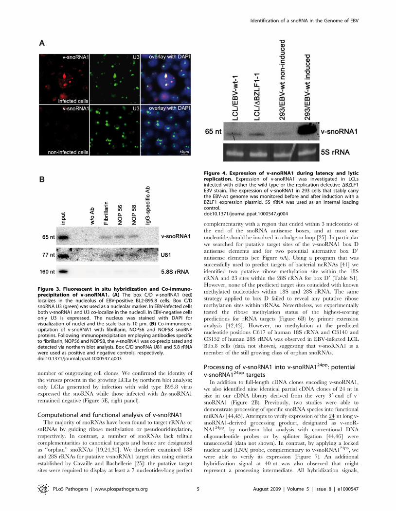
fact co-localized to the nucleolus (Figure 3A). In contrast, a v-
snoRNA1 hybridization signal was absent in non-infected B cells.
Canonical C/D box snoRNAs have previously been shown to
bind to four snoRNA core proteins: fibrillarin, Nop56, Nop58, and
the 15.5 K protein, respectively. These proteins have previously
been shown to be strictly required for RNA maturation,
stabilization and function [22,39]. The C/D box proteins
assemble with snoRNAs thus forming ribonucleo-protein com-
plexes (snoRNPs) that localize to the nucleolus. In order to assess
whether v-snoRNA1 assembles into a canonical C/D box
snoRNP, binding of v-snoRNA1 to three of these canonical
snoRNA-binding proteins (fibrillarin, Nop56 and Nop58) was
assessed by co-immunoprecipitation using specific antibodies.
Immuno-precipitated samples were subsequently analyzed for
the presence of v-snoRNA1 by northern blot analysis. These assays
demonstrated that v-snoRNA1 and the canonical U81 snoRNA,
used as a positive control, were both co-immunoprecipitated with
similar efficiencies with antibodies against all three snoRNA-
binding proteins (Figure 3B). In contrast, none of the snoRNAs
was precipitated in controls without antibodies or employing an
IgG-specific antibody. Hybridization with an oligonucleotide
specific for 5.8S rRNA was used to test for the specificity of the
employed antibodies. Thereby, a faint, unspecific signal was
detected in all samples after antibody addition, except the control
without an antibody. This is likely caused by the high expression
levels of 5.8S rRNA in our samples. From these results we
conclude that the newly identified 65 nt long viral RNA transcript
displays all hallmark features of a genuine box C/D snoRNA.
v-snoRNA1 expression is strongly stimulated in the lyticcycle
A common trait shared by all herpesviruses is their ability to
infect their target cells under several modes; cells can support lytic
replication during which new virus progeny is replicated or instead
induce virus latency. Viral proteins used in both modes are usually,
but not always, distinct. We therefore assayed v-snoRNA1
expression in latently infected cells or in cells undergoing lytic
replication. We took advantage of LCLs established with viruses
that are devoid of the lytic immediate early gene BZLF1 (DBZLF1)
and therefore cannot initiate lytic replication [40] and examined v-
snoRNA1 expression in these cells by northern blot analysis
(Figure 4). Northern blot signals were clearly visible in these cells
thereby demonstrating that v-snoRNA1 is a latent transcript. We
then performed the same experiment with replication-competent
293/EBV-wt cells lytically induced by transfection of the BZLF1
gene (Figure 4). Comparison with non-induced cells showed that
the v-snoRNA1 expression levels were up-regulated up to 30-fold
following induction (Figure 4). V-snoRNA1 is therefore especially
part of the EBV lytic expression programme.
Phenotypic traits of a recombinant virus lacking v-snoRNA1
In an attempt to discover the function of v-snoRNA1 during the
EBV life cycle, we constructed a recombinant virus that lacks a
functional v-snoRNA1. To this aim, the C-box motif of v-snoRNA1
from the B95.8 strain was exchanged against the sequence of the
kanamycin resistance gene flanked by two FLP recombinase
recognition sites (Figure 5A). Excision of this cassette left an
unrelated bacterial sequence containing a HindIII restriction site in
place of the box C of v-snoRNA1 (Figure 5A and 5B, lane 2). DNA
from the recombinant virus was stably transfected into 293 cells to
generate a virus producer cell line, here referred to as 293/Dv-
snoRNA1. Multiple clones were screened for their ability to support
virus replication. One of the replication-competent clones was
chosen at random for further experiments. Recombinant episomes
purified from this producer cell line and transformed into E. coli cells
were found to be intact as assessed by restriction analysis (Figure 5B,
lane 3). Sequencing of the recombination site on these rescued
episomes confirmed exchange of the Box C against unrelated DNA
TTTCCCGCGCCAAGCTTCAAAAGCGCTCTGAAGTTCC
TATACTTTCTAGAGAATAGGAACTTCGGAATAGGAAC
TTCCAACC (EBV DNA around the insertion is indicated in
bold). A northern blot, performed on 293/Dv-snoRNA1 cells using
a v-snoRNA1-specific probe, yielded negative results while signals
could be clearly identified in the 293/EBV-wt positive control
(Figure 5E, left panel). We therefore conclude that the Dv-snoRNA1
Figure 1. Sequence and expression profile of v-snoRNA1. (A) Sequences of the newly identified 65 nt long v-snoRNA1 and the canonical boxC/D from snoRNA U80 used as a control are shown here. The position of C/D boxes and C9/D9 boxes are indicated in red. (B) Northern blot analysisshowing expression of v-snoRNA1 in a panel of EBV-positive (BL2-B95.8, BL41-B95.8, LCL-B95.8, Raji, Rael) and EBV-negative (BL2, BL41) cell lines. 5SrRNA served as internal loading control.doi:10.1371/journal.ppat.1000547.g001
Identification of a snoRNA in the Genome of EBV
PLoS Pathogens | www.plospathogens.org 3 August 2009 | Volume 5 | Issue 8 | e1000547
virus is devoid of the viral snoRNA and that destruction of the
putative C box of v-snoRNA1 is sufficient to exert this effect.
We then conducted a series of experiments aiming at defining
phenotypic traits of the mutant strain. We first assessed the ability
of the 293/Dv-snoRNA1 to support viral replication. Viral titres
were quantified either as packaged viral genome-equivalents
(physical titres) or as green Raji units, i.e. as the concentration
of viruses able to infect the Raji cell line determined by exposure to
a limiting dilution of the viral supernatants (functional titres). Both
assays revealed nearly identical titres for both the mutant and the
wild type control (Figure 5D). The Dv-snoRNA1 viruses and
producer cell line were then examined in electron microscopy;
both displayed normal morphological features: encapsidation,
primary and secondary egress were unchanged in the absence of
the viral snoRNA (unpublished data). We further evaluated viral
gene expression by western blot or immunostains (BZLF1, EA/D-
BMRF1, gp350). Again, we could not discern any differences
between the mutant and its wild type counterpart (unpublished
data).
We then exposed various established cell lines or primary cells
to the Dv-snoRNA1 mutant and monitored the efficiency of
infection by counting the percentage of GFP-positive (293 cell line,
primary epithelial cells) or EBNA2-positive (primary B cells)
lymphocytes three days post-infection. The rate of infection was
nearly identical in both wild type and mutant viruses (unpublished
data). We finally investigated the transforming capacity of the
mutant by performing infections of normal resting B cells from
three different normal individuals at decreasing multiplicity of
infections (Figure 5D). Wild type and mutant viruses both
exhibited a transforming potential that resulted in a very similar
Figure 2. Schematic representation of the Epstein-Barr-virus genome. The location of ncRNA genes, latent genes and the precise location ofv-snoRNA1 is indicated. (A) Location and transcription of EBV ncRNA genes (black lines with blue lettering) and EBV latent genes (grey bars with blacklettering). The v-snoRNA1 is indicated in red, the neighboring miRNA BART2 in orange and the viral DNA polymerase BALF5 is depicted in green (forcoding region) and brown (for 39-UTR). The promoters are shown in grey lines and lettering, the BARTs region as a grey bar and the B95.8 deletion arealso indicated. (B) Close-up of v-snoRNA1 location within the 39-UTR of the viral DNA polymerase gene. The v-snoRNA1 is located on the sense strandabout 60 nt downstream of the mir-BART2 precursor transcript and complementary to the BALF5 39UTR that is situated on the antisense strand. V-snoRNA124pp is indicated in blue, other transcripts are indicated in the same colors as described above. The black line illustrates the cleavage site ofmir-BART2. Corresponding EBV coordinates refer to the EBV B95.8 deletion strain (Accession number V01555.2).doi:10.1371/journal.ppat.1000547.g002
Identification of a snoRNA in the Genome of EBV
PLoS Pathogens | www.plospathogens.org 4 August 2009 | Volume 5 | Issue 8 | e1000547
number of outgrowing cell clones. We confirmed the identity of
the viruses present in the growing LCLs by northern blot analysis;
only LCLs generated by infection with wild type B95.8 virus
expressed the snoRNA while those infected with Dv-snoRNA1
remained negative (Figure 5E, right panel).
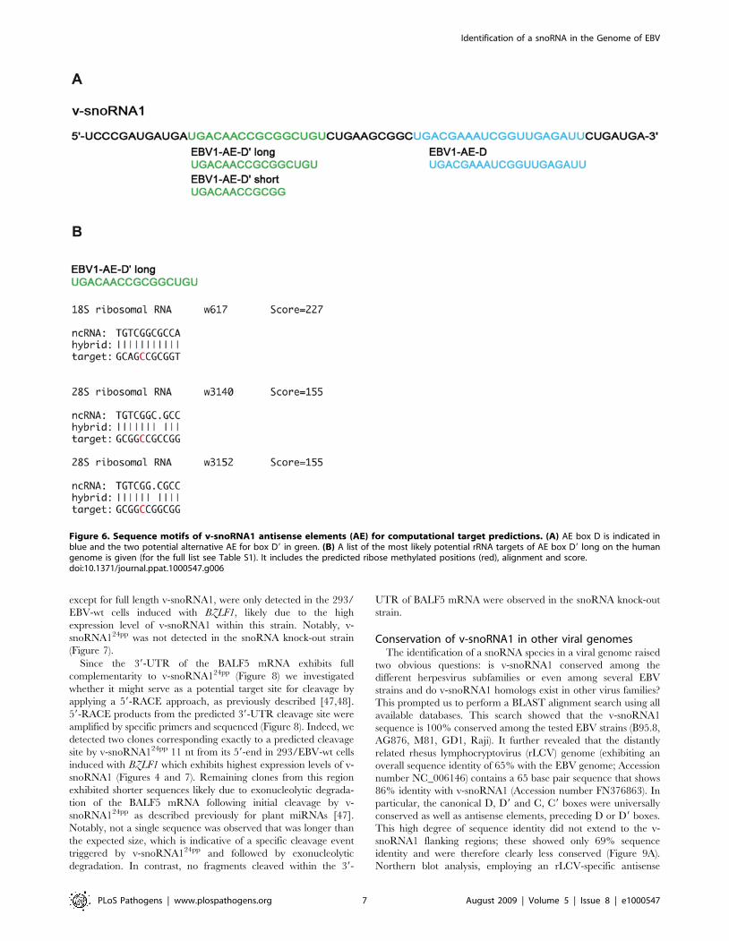
Computational and functional analysis of v-snoRNA1The majority of snoRNAs have been found to target rRNAs or
snRNAs by guiding ribose methylation or pseudouridinylation,
respectively. In contrast, a number of snoRNAs lack telltale
complementarities to canonical targets and hence are designated
as ‘‘orphan’’ snoRNAs [19,24,30]. We therefore examined 18S
and 28S rRNAs for putative v-snoRNA1 target sites using criteria
established by Cavaille and Bachellerie [25]: the putative target
sites were required to display at least a 7 nucleotides-long perfect
complementarity with a region that ended within 3 nucleotides of
the end of the snoRNA antisense boxes, and at most one
nucleotide should be involved in a bulge or loop [25]. In particular
we searched for putative target sites of the v-snoRNA1 box D
antisense elements and for two potential alternative box D9
antisense elements (see Figure 6A). Using a program that was
successfully used to predict targets of bacterial ncRNAs [41] we
identified two putative ribose methylation site within the 18S
rRNA and 23 sites within the 28S rRNA for box D9 (Table S1).
However, none of the predicted target sites coincided with known
methylated nucleotides within 18S and 28S rRNA. The same
strategy applied to box D failed to reveal any putative ribose
methylation sites within rRNAs. Nevertheless, we experimentally
tested the ribose methylation status of the highest-scoring
predictions for rRNA targets (Figure 6B) by primer extension
analysis [42,43]. However, no methylation at the predicted
nucleotide positions C617 of human 18S rRNA and C3140 and
C3152 of human 28S rRNA was observed in EBV-infected LCL
B95.8 cells (data not shown), suggesting that v-snoRNA1 is a
member of the still growing class of orphan snoRNAs.
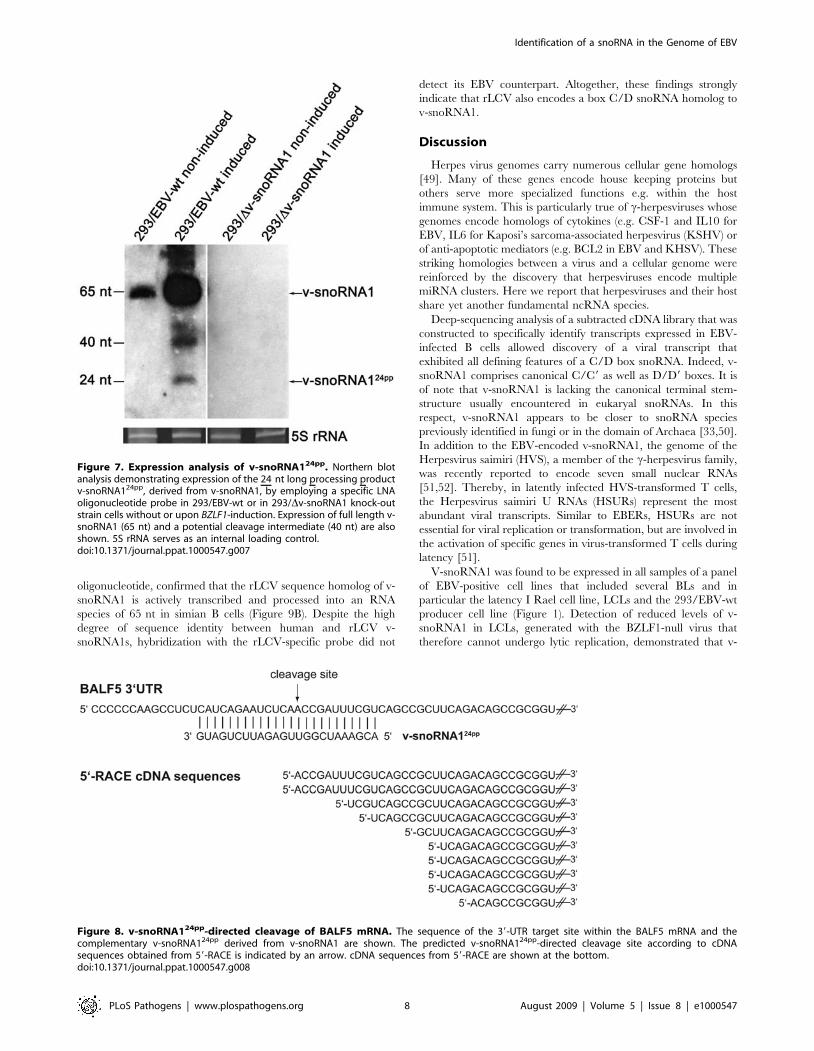
Processing of v-snoRNA1 into v-snoRNA124pp: potentialv-snoRNA124pp targets
In addition to full-length cDNA clones encoding v-snoRNA1,
we also identified nine identical partial cDNA clones of 24 nt in
size in our cDNA library derived from the very 39-end of v-
snoRNA1 (Figure 2B). Previously, two studies were able to
demonstrate processing of specific snoRNA species into functional
miRNAs [44,45]. Attempts to verify expression of the 24 nt long v-
snoRNA1-derived processing product, designated as v-snoR-
NA124pp, by northern blot analysis with conventional DNA
oligonucleotide probes or by splinter ligation [44,46] were
unsuccessful (data not shown). In contrast, by applying a locked
nucleic acid (LNA) probe, complementary to v-snoRNA124pp, we
were able to verify its expression (Figure 7). An additional
hybridization signal at 40 nt was also observed that might
represent a processing intermediate. All hybridization signals,
Figure 3. Fluorescent in situ hybridization and Co-immuno-precipitation of v-snoRNA1. (A) The box C/D v-snoRNA1 (red)localizes in the nucleolus of EBV-positive BL2-B95.8 cells. Box C/DsnoRNA U3 (green) was used as a nucleolar marker. In EBV-infected cellsboth v-snoRNA1 and U3 co-localize in the nucleoli. In EBV-negative cellsonly U3 is expressed. The nucleus was stained with DAPI forvisualization of nuclei and the scale bar is 10 mm. (B) Co-immunopre-cipitation of v-snoRNA1 with fibrillarin, NOP56 and NOP58 snoRNPproteins. Following immunoprecipitation employing antibodies specificto fibrillarin, NOP56 and NOP58, the v-snoRNA1 was co-precipitated anddetected via northern blot analysis. Box C/D snoRNA U81 and 5.8 rRNAwere used as positive and negative controls, respectively.doi:10.1371/journal.ppat.1000547.g003
Figure 4. Expression of v-snoRNA1 during latency and lyticreplication. Expression of v-snoRNA1 was investigated in LCLsinfected with either the wild type or the replication-defective DBZLF1EBV strain. The expression of v-snoRNA1 in 293 cells that stably carrythe EBV-wt genome was monitored before and after induction with aBZLF1 expression plasmid. 5S rRNA was used as an internal loadingcontrol.doi:10.1371/journal.ppat.1000547.g004
Identification of a snoRNA in the Genome of EBV
PLoS Pathogens | www.plospathogens.org 5 August 2009 | Volume 5 | Issue 8 | e1000547
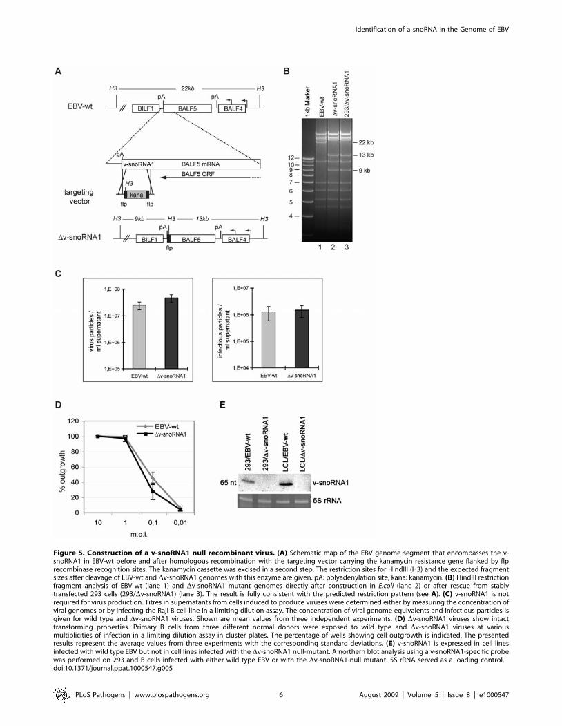
Figure 5. Construction of a v-snoRNA1 null recombinant virus. (A) Schematic map of the EBV genome segment that encompasses the v-snoRNA1 in EBV-wt before and after homologous recombination with the targeting vector carrying the kanamycin resistance gene flanked by flprecombinase recognition sites. The kanamycin cassette was excised in a second step. The restriction sites for HindIII (H3) and the expected fragmentsizes after cleavage of EBV-wt and Dv-snoRNA1 genomes with this enzyme are given. pA: polyadenylation site, kana: kanamycin. (B) HindIII restrictionfragment analysis of EBV-wt (lane 1) and Dv-snoRNA1 mutant genomes directly after construction in E.coli (lane 2) or after rescue from stablytransfected 293 cells (293/Dv-snoRNA1) (lane 3). The result is fully consistent with the predicted restriction pattern (see A). (C) v-snoRNA1 is notrequired for virus production. Titres in supernatants from cells induced to produce viruses were determined either by measuring the concentration ofviral genomes or by infecting the Raji B cell line in a limiting dilution assay. The concentration of viral genome equivalents and infectious particles isgiven for wild type and Dv-snoRNA1 viruses. Shown are mean values from three independent experiments. (D) Dv-snoRNA1 viruses show intacttransforming properties. Primary B cells from three different normal donors were exposed to wild type and Dv-snoRNA1 viruses at variousmultiplicities of infection in a limiting dilution assay in cluster plates. The percentage of wells showing cell outgrowth is indicated. The presentedresults represent the average values from three experiments with the corresponding standard deviations. (E) v-snoRNA1 is expressed in cell linesinfected with wild type EBV but not in cell lines infected with the Dv-snoRNA1 null-mutant. A northern blot analysis using a v-snoRNA1-specific probewas performed on 293 and B cells infected with either wild type EBV or with the Dv-snoRNA1-null mutant. 5S rRNA served as a loading control.doi:10.1371/journal.ppat.1000547.g005
Identification of a snoRNA in the Genome of EBV
PLoS Pathogens | www.plospathogens.org 6 August 2009 | Volume 5 | Issue 8 | e1000547
except for full length v-snoRNA1, were only detected in the 293/
EBV-wt cells induced with BZLF1, likely due to the high
expression level of v-snoRNA1 within this strain. Notably, v-
snoRNA124pp was not detected in the snoRNA knock-out strain
(Figure 7).
Since the 39-UTR of the BALF5 mRNA exhibits full
complementarity to v-snoRNA124pp (Figure 8) we investigated
whether it might serve as a potential target site for cleavage by
applying a 59-RACE approach, as previously described [47,48].
59-RACE products from the predicted 39-UTR cleavage site were
amplified by specific primers and sequenced (Figure 8). Indeed, we
detected two clones corresponding exactly to a predicted cleavage
site by v-snoRNA124pp 11 nt from its 59-end in 293/EBV-wt cells
induced with BZLF1 which exhibits highest expression levels of v-
snoRNA1 (Figures 4 and 7). Remaining clones from this region
exhibited shorter sequences likely due to exonucleolytic degrada-
tion of the BALF5 mRNA following initial cleavage by v-
snoRNA124pp as described previously for plant miRNAs [47].
Notably, not a single sequence was observed that was longer than
the expected size, which is indicative of a specific cleavage event
triggered by v-snoRNA124pp and followed by exonucleolytic
degradation. In contrast, no fragments cleaved within the 39-
UTR of BALF5 mRNA were observed in the snoRNA knock-out
strain.
Conservation of v-snoRNA1 in other viral genomesThe identification of a snoRNA species in a viral genome raised
two obvious questions: is v-snoRNA1 conserved among the
different herpesvirus subfamilies or even among several EBV
strains and do v-snoRNA1 homologs exist in other virus families?
This prompted us to perform a BLAST alignment search using all
available databases. This search showed that the v-snoRNA1
sequence is 100% conserved among the tested EBV strains (B95.8,
AG876, M81, GD1, Raji). It further revealed that the distantly
related rhesus lymphocryptovirus (rLCV) genome (exhibiting an
overall sequence identity of 65% with the EBV genome; Accession
number NC_006146) contains a 65 base pair sequence that shows
86% identity with v-snoRNA1 (Accession number FN376863). In
particular, the canonical D, D9 and C, C9 boxes were universally
conserved as well as antisense elements, preceding D or D9 boxes.
This high degree of sequence identity did not extend to the v-
snoRNA1 flanking regions; these showed only 69% sequence
identity and were therefore clearly less conserved (Figure 9A).
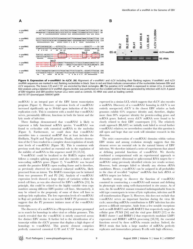
Northern blot analysis, employing an rLCV-specific antisense
Figure 6. Sequence motifs of v-snoRNA1 antisense elements (AE) for computational target predictions. (A) AE box D is indicated inblue and the two potential alternative AE for box D9 in green. (B) A list of the most likely potential rRNA targets of AE box D9 long on the humangenome is given (for the full list see Table S1). It includes the predicted ribose methylated positions (red), alignment and score.doi:10.1371/journal.ppat.1000547.g006
Identification of a snoRNA in the Genome of EBV
PLoS Pathogens | www.plospathogens.org 7 August 2009 | Volume 5 | Issue 8 | e1000547
oligonucleotide, confirmed that the rLCV sequence homolog of v-
snoRNA1 is actively transcribed and processed into an RNA
species of 65 nt in simian B cells (Figure 9B). Despite the high
degree of sequence identity between human and rLCV v-
snoRNA1s, hybridization with the rLCV-specific probe did not
detect its EBV counterpart. Altogether, these findings strongly
indicate that rLCV also encodes a box C/D snoRNA homolog to
v-snoRNA1.
Discussion
Herpes virus genomes carry numerous cellular gene homologs
[49]. Many of these genes encode house keeping proteins but
others serve more specialized functions e.g. within the host
immune system. This is particularly true of c-herpesviruses whose
genomes encode homologs of cytokines (e.g. CSF-1 and IL10 for
EBV, IL6 for Kaposi’s sarcoma-associated herpesvirus (KSHV) or
of anti-apoptotic mediators (e.g. BCL2 in EBV and KHSV). These
striking homologies between a virus and a cellular genome were
reinforced by the discovery that herpesviruses encode multiple
miRNA clusters. Here we report that herpesviruses and their host
share yet another fundamental ncRNA species.
Deep-sequencing analysis of a subtracted cDNA library that was
constructed to specifically identify transcripts expressed in EBV-
infected B cells allowed discovery of a viral transcript that
exhibited all defining features of a C/D box snoRNA. Indeed, v-
snoRNA1 comprises canonical C/C9 as well as D/D9 boxes. It is
of note that v-snoRNA1 is lacking the canonical terminal stem-
structure usually encountered in eukaryal snoRNAs. In this
respect, v-snoRNA1 appears to be closer to snoRNA species
previously identified in fungi or in the domain of Archaea [33,50].
In addition to the EBV-encoded v-snoRNA1, the genome of the
Herpesvirus saimiri (HVS), a member of the c-herpesvirus family,
was recently reported to encode seven small nuclear RNAs
[51,52]. Thereby, in latently infected HVS-transformed T cells,
the Herpesvirus saimiri U RNAs (HSURs) represent the most
abundant viral transcripts. Similar to EBERs, HSURs are not
essential for viral replication or transformation, but are involved in
the activation of specific genes in virus-transformed T cells during
latency [51].
V-snoRNA1 was found to be expressed in all samples of a panel
of EBV-positive cell lines that included several BLs and in
particular the latency I Rael cell line, LCLs and the 293/EBV-wt
producer cell line (Figure 1). Detection of reduced levels of v-
snoRNA1 in LCLs, generated with the BZLF1-null virus that
therefore cannot undergo lytic replication, demonstrated that v-
Figure 7. Expression analysis of v-snoRNA124pp. Northern blotanalysis demonstrating expression of the 24 nt long processing productv-snoRNA124pp, derived from v-snoRNA1, by employing a specific LNAoligonucleotide probe in 293/EBV-wt or in 293/Dv-snoRNA1 knock-outstrain cells without or upon BZLF1-induction. Expression of full length v-snoRNA1 (65 nt) and a potential cleavage intermediate (40 nt) are alsoshown. 5S rRNA serves as an internal loading control.doi:10.1371/journal.ppat.1000547.g007
Figure 8. v-snoRNA124pp-directed cleavage of BALF5 mRNA. The sequence of the 39-UTR target site within the BALF5 mRNA and thecomplementary v-snoRNA124pp derived from v-snoRNA1 are shown. The predicted v-snoRNA124pp-directed cleavage site according to cDNAsequences obtained from 59-RACE is indicated by an arrow. cDNA sequences from 59-RACE are shown at the bottom.doi:10.1371/journal.ppat.1000547.g008
Identification of a snoRNA in the Genome of EBV
PLoS Pathogens | www.plospathogens.org 8 August 2009 | Volume 5 | Issue 8 | e1000547
snoRNA1 is an integral part of the EBV latent transcription
program (Figure 4). However, expression levels of v-snoRNA1
increased significantly up to 30-fold upon induction of the lytic
replication cycle. This is consistent with a model that v-snoRNA1
serves, presumably different, functions in both the latent and the
lytic mode of infection.
Three findings demonstrated that v-snoRNA1 is likely to
represent a fully functional ncRNA species. V-snoRNA1 was
found to co-localize with canonical snoRNA to the nucleolus
(Figure 3). Furthermore, we could show that v-snoRNA1
assembles into a canonical snoRNP that at least includes the
fibrillarin, Nop56 and Nop58 proteins. Finally, selective destruc-
tion of the C box resulted in a complete down-regulation of steady
state levels of v-snoRNA1 (Figure 5E). This is consistent with
previous work that ascribed an essential role in the regulation of
the stability of snoRNA to this sequence motif [21,53,54].
V-snoRNA1 could be localized to the BARTs region which
follows a complex splicing pattern and also encodes a cluster of
non-coding miRNA genes (Figure 2). V-snoRNA1 was located
outside the putative BARTs open reading frame and is therefore,
as previously observed for canonical eukaryal snoRNAs, likely
processed from an intron. The BARTs transcripts can be initiated
from two promoters P1 and P2 [36]. Analysis of v-snoRNA1
expression levels showed a large degree of variation within the
tested cell lines, as was also observed for EBV’s miRNAs [55]. In
principle, this could be related to the highly variable virus copy
numbers among different EBV-positive cell lines. Alternatively, it
may be related to the propensity of some of these cell lines to
undergo lytic replication. The low expression levels of v-snoRNA1
in Raji are probably due to an inactive BART P2 promoter; this
suggests that the P2 promoter initiates most of the v-snoRNA1
transcripts.
The discovery of a snoRNA in a Herpesvirus genome prompted
us to search for homologs in other viral or cellular genomes. This
search revealed that the v-snoRNA1 is strictly conserved across
five distinct EBV strains. It further led to the identification of a
transcript within the rLCV genome that displays a high degree of
homology to v-snoRNA1. This genetic element comprises
perfectly conserved canonical C/D and C9/D9 boxes and was
expressed in a simian LCL which suggests that rLCV also encodes
a snoRNA. Discovery of a v-snoRNA1 homolog in rLCV is not
entirely unexpected; rLCV is the closest EBV relative as both
genomes exhibit 65% sequence identity and, therefore, display
more than 80% sequence identity for protein-coding genes and
ncRNA genes. Indeed, seven rLCV miRNA were found to be
closely related to their EBV counterparts [11]. The relatively
crude approach (BLAST) we initially took failed to reveal further
v-snoRNA1 relatives; we nevertheless consider that this question is
still open and hope that our work will stimulate research in this
direction.
The strict conservation of v-snoRNA1 domains within various
EBV strains and among evolution strongly suggests that this
element serves an essential role in the natural history of EBV
infection. We therefore initiated a series of experiments that aimed
at defining potential functions of v-snoRNA1. We thereby
combined a computational with an experimental approach to
determine putative ribosomal or spliceosomal RNA targets for v-
snoRNA1 using previously identified criteria (see results section).
However, both attempts failed to identify any obvious rRNA
candidates. Hence, v-snoRNA1 can be assigned in all probability
to the class of so-called ‘‘orphan’’ snoRNAs that lack rRNA or
snRNA targets (see below).
Another strategy to discover the function of v-snoRNA1
consisted in constructing a v-snoRNA1-null mutant and defining
its phenotypic traits using well-characterized in vitro assays. As of
now, the Dv-snoRNA1 mutant remained indistinguishable from its
wild type counterparts in terms of lytic replication, infection and B
cell transformation (Figure 5). However, this does not exclude that
v-snoRNA1 serves an important function during the virus life
cycle; unraveling miRNAs contributions to EBV infection has also
proven a difficult enterprise. Aside from a few notable exceptions
such as miR-BART5 and miR-BART2 that respectively target the
cellular gene PUMA [56] and the viral gene BALF5 [57] or the
BART cluster 1 and BHRF1-2 that respectively modulate LMP1
expression and BHRF1 mRNA processing [58,59], the essential
functions served by these ncRNAs remain unclear. Indeed, the
B95.8 strain that lacks a large number of miRNAs perfectly
replicates and immortalizes primary B cells with high efficiency.
Figure 9. Expression of v-snoRNA1 in rLCV. (A) Alignment of v-snoRNA1 and rLCV including their flanking regions. V-snoRNA1 and rLCVsnoRNA sequences are marked in red, flanking nucleotides in black. Stars in red and black indicate conservation of the nucleotides between EBV andrLCV sequences. The boxes C/D and C9/D9 are encircled by black rectangles. (B) The putative rLCV snoRNA is expressed in simian LCLs. A northernblot analysis using a labeled rLCV snoRNA oligonucleotide was performed on the LCL8664 cell line that was generated by infection with rLCV. A panelof EBV-negative and EBV-positive human LCLs were used as controls. 5S rRNA was used as loading control.doi:10.1371/journal.ppat.1000547.g009
Identification of a snoRNA in the Genome of EBV
PLoS Pathogens | www.plospathogens.org 9 August 2009 | Volume 5 | Issue 8 | e1000547
Recently, specific snoRNA species have been characterized as
miRNA precursors, which are processed to mature miRNAs and
assemble into a functional RNA induced silencing complex
[60,61]. Indeed, by deep-sequencing we identified nine identical
cDNA clones of 24 nt in size, that mapped to the very 39-end of v-
snoRNA1. The expression of v-snoRNA124pp was verified by
northern blot analysis employing a specific LNA oligonucleotide
antisense probe (Figure 7). Thereby, the hybridization signal was
especially apparent in 293/EBV cells induced by BZLF1, which
results in a 30-fold up-regulation of v-snoRNA1 expression; the
hybridization signal was absent, however, in non-induced wild
type cells. This could be explained by lower v-snoRNA1
expression levels in non-induced 293/EBV cells, compared to
BZLF1-induced cells (Figure 7), resulting in reduced processing of
v-snoRNA124pp below the northern blot detection limit. Alterna-
tively, this finding could result from preferential processing of v-
snoRNA1 into v-snoRNA124pp during lytic replication.
Subsequently, by a 59-RACE approach we also investigated a
potential target for snoRNA124pp. Since the RNA species maps in
antisense orientation to the 39-UTR of the BALF5 mRNA, which
encodes the viral DNA polymerase, BALF5 mRNA might
represent a likely target site. As has been shown previously, the
39-UTR of the BALF5 mRNA encodes in antisense orientation, in
addition to v-snoRNA124pp, a bona fide EBV miRNA, designated
as mir-BART2. Thereby, it has been reported that mir-BART2
down-regulates the mRNA levels by cleavage within the BALF5
39-UTR [57]. According to the proposed model, mir-BART2
thereby inhibits the transition from latent to lytic viral replication.
By 59-RACE analysis, we provide evidence that v-snoRNA124pp
might also target BALF5 mRNA for cleavage and subsequent
degradation. In contrast to mir-BART2, however, expression of v-
snoRNA124pp was only apparent upon induction of the viral lytic
cycle by BZLF1 (Figure 7). Future experiments will focus on the
function of v-snoRNA1 and v-snoRNA124pp especially in respect
to its function in the latent and lytic cycles of EBV infection.
Materials and Methods
Cell lines and EBV strainsThe cell lines BL2, BL2 B95.8, BL41, BL41 B95.8, CBL B95.8,
LCL B95.8, Raji, Rael, HEK293 and LCL8664 were cultured in
RPMI 1640 supplemented with 10% FCS, L-glutamine
(2 mM ml21) and antibiotics (100 U penicillin ml21 and 100 mg
streptomycin ml21). BL2 and BL41 are EBV-negative Burkitt’s
Lymphoma cell lines, BL2 B95.8 and BL41 B95.8 are cell lines
infected with EBV strain B95.8, Raji and Rael are EBV-positive
Burkitt’s lymphoma cell lines [62,63]. CBL B95.8 and LCL B95.8
cells were obtained after in vitro transformation of cord blood
lymphocytes (CBL) or primary human blood lymphocytes (LCLs)
with the B95.8 strain of EBV. The EBV deletion strain B95.8,
used in this study, lacks a 12 kb large portion of the genome [64].
LCL8664 is a rhesus LCV (cercopithicine herpesvirus 15)-infected
B-cell line derived from a retro-orbital B-cell lymphoma in a
rhesus monkey [65]. 293/EBV-wt contains the wild type EBV
B95.8 genome in a recombinant form. Two LCL/EBV-wt and
LCL/DBZLF1 pairs were established by immortalization of B cells
from two different donors with either wild type or BZLF1-negative
recombinant viruses [40].
RNA preparation and northern blot analysisTotal RNA from EBV-infected and non-infected cells was
isolated by using the Tri Reagent method according to the
manufacturers protocol. Northern blot analysis was performed as
described in Mrazek et. al [32] applying a mix of five
oligonucleotides (F1: CCTCTCATCAGAATCTCAACC, F2:
TCTCAACCGATTTCGTCAGC, F3: CGTCAGCCGCTTCA-
GACAG, F4: GACAGCCGCGGTTGTCATC, F5: GGTTG-
TCATCATCATCGGGAA) covering the whole v-snoRNA1
sequence. For the detection of the homologous rhesus lympho-
cryptovirus a rLCV-specific v-snoRNA1 oligonucleotide (59-
AATCTCAACCAATTTCCTCAGC-39) was used. Detection of
v-snoRNA124pp by an LNA oligonucleotide (59-CATCA-
GAATCTCAACCGATTTCGT-39, Exiqon) was performed ac-
cording to the standard protocol, except 60 mg RNA was loaded
and membrane was washed under stringent conditions. 5.8S
rRNA antisense oligonucleotide 59-TCCTGCAATTCACAT-
TAATTCTCGCAGCTAGC-39 was used as negative control in
immunoprecipitations. Ethidium bromide-stained 5S rRNA were
used as loading control for normalization after polyacrylamid gel
electrophoresis. Northern blot signals were either put onto Kodak
MS-1 film, using an intensifier screen or analyzed with a
Molecular Dynamics Storm PhosphorImager (Image quant
software version 5.0).
Fluorescent in situ hybridizationFor the detection of the viral and U3 snoRNA the following
amino-modified DNA oligonucleotides were used:AT*CTCAA-
CCGATT*TCGTCAGCCGCT*TCAGACAGCCGCGGT*TG-
TCATCAT*CAT for v-snoRNA1 and GT*TCTCTCCCT-
CT*CACTCCCCAAT*ACGGAGAGAAGAACGAT*CATCAA-
TGGCT*G for U3 (the amino-modified T nucleotides are marked
with asterisks). The probes were labeled with CY3 (v-snoRNA1;
Amersham Biosciences) or Oregon Green 488 (U3; Molecular
Probes) according to the manufacturers protocol.
BL2 and BL2-B95.8 cells were washed in 16 PBS (PBS:
100 mM Na2HPO4, 20 mM KH2PO4, 137 mM NaCl, 27 mM
KCl, pH 7.4) and diluted in 16 PBS to an appropriate
concentration. The cell suspension was dropped onto glass slides
and hybridized according to [21,66]. The slides were washed 3
times for 20 min after hybridization and mounted with 15 ml
Mowiol containing 0.1 mg/ml DAPI. Slides were analyzed by
confocal fluorescence microscopy (LSM 510 META, Carl Zeiss
GmbH) using Zeiss LSM Software, version 3.2.
Co-ImmunoprecipitationFor the preparation of snoRNP extract, BL2-B95.8 cells were
washed in 16PBS, lysed in 5-fold amount of 16RNP lysis buffer
(25 mM Tris-HCl pH 7.5, 150 mM NaCl, 0.2 mM EDTA
pH 8.0, 0.2% TritonX-100), sonicated and incubated for 1 h on
ice. After centrifugation steps at 180006g, 4uC, 10 min and
300006g, 4uC, 30 min the snoRNP extract was used for co-
immunoprecipitation.
250 ml Protein A/G PLUS-Agarose (Santa Cruz Biotechnology
Inc.) was washed three times in 16PBS and resuspended with 16RNP lysis buffer to receive a final volume of 125 ml. 50 ml of the
suspension was added to the total cell lysate containing 500 mg
protein extract for each approach and precleared for 1 h at 6uCduring rotation. The precleared supernatant was equally distrib-
uted and incubated with specific antibodies for fibrillarin (ab5821;
Abcam), NOP56, NOP58, and IgG (Santa Cruz Biotechnology
Inc.) for 1 h at 6uC. After addition of 12 ml of washed beads to
each approach and rotation for 4 h at 6uC, samples were
centrifuged at 8006g at 4uC for 5 min. The supernatant was
used as a control for unbound RNA and the remaining beads were
washed four times with 1 ml 16 RNP lysis buffer and the co-
immunoprecipitated RNA was eluted with 200 ml IP elution buffer
(100 mM Tris-HCl pH 7.5, 150 mM NaCl, 12.5 mM EDTA
pH 8.0, 20% SDS) after heating for 5 min at 95uC. The
Identification of a snoRNA in the Genome of EBV
PLoS Pathogens | www.plospathogens.org 10 August 2009 | Volume 5 | Issue 8 | e1000547
supernatant was used to perform phenol-chloroform-isoamylalco-
hol (Fluka) extraction, RNA was ethanol-precipitated over night
and analyzed by northern blot analysis.
Recombinant EBV genomesThe wild type EBV recombinant plasmid (p2089) is cloned onto
the prokaryotic F factor origin of replication and carries the green
fluorescent protein (gfp), the chloramphenicol (cam) resistance
gene and the hygromycin (hyg) resistance gene [67]. The EBV
snoRNA mutant was constructed by replacing the C box sequence
motif (B95.8 coordinates 153331–153341) with the kanamycin
(kan) resistance gene using homologous recombination [68].
Composite primers were used whose internal parts (underlined)
are specific for the kan resistance gene, and whose external parts
(40 bp) are specific for the snoRNA gene (59-ACGCTC-
CCCTGGGGGCTTCATGATCCCACCGCCTTTCfCCGCG-
CCAAGCTTCAAAAGCGCTC-39; 59-CTCAACCGATTTCG-
TCAGCCGCTTCAGACAGCCGCGGTTGGAAGTTCCTA-
TTCCGAAGTTCC-39).
These primers allowed PCR-mediated amplification of the kan
resistance gene through their internal sequences and then
homologous recombination of the amplified PCR product with
the EBV wild type genome via their external sequences. PCR
amplification products were incubated with the restriction enzyme
DpnI to remove traces of the parental plasmid and introduced by
electroporation (1000 V, 25 mF, 100 V) into E. coli DH10B cells
carrying the recombinant virus p2089 and the temperature
sensitive pKD46 helper plasmid encoding the phage lambda red
recombinase to foster homologous recombination. Cells were
grown in LB with cam (15 mg/ml) at 37uC for an hour and then
plated onto LB agar plates containing cam (15 mg/ml) and kan
(10 mg/ml). Incubation at 42uC induced the loss of the helper
plasmid. DNA of positive clones was purified and analyzed with
HindIII restriction enzyme to confirm correct recombination. The
kan resistance gene was excised using the Flp recombinase cloned
onto the temperature-sensitive plasmid pCP20 [69] which also
carries the amp resistance gene. The bacterial clones that resulted
from selection on cam/amp plates were further grown on cam
plates at 42uC to induce the loss of the pCP20 plasmid. Resistant
clones were then submitted to restriction analysis to confirm the
expected restriction pattern. Sequencing further confirmed
successful recombination and the intactness of the flanking regions.
Stable clone selectionHEK293 cells were transfected with the properly recombined
mutant viral DNA (clone B 253) using Lipofectamine (Invitrogen)
as described [70]. Selection of stable 293 cell clones carrying the
EBV recombinant plasmid was performed by addition of
hygromycin to the culture medium (100 mg/ml). Cell clones
surviving selection were first assessed for GFP fluorescence and the
positive clones were further expanded. Fifteen clones were assessed
for their ability to support lytic replication by qPCR. Ten of those
were found to produce virus at high levels, one of which was
selected for further analysis. The cell clone used in this study is
referred to as 293/Dv-snoRNA1. Viral episomes from this clone
were transferred back in E.coli and submitted to restriction
analysis and sequencing.
Plasmid rescue in E. coliCircular plasmid DNA from 293/DsnoRNA and was extracted
using a denaturation-renaturation method as described previously
[71]. E.coli strain DH10B was transformed with the viral
recombinant DNA by electroporation as described before [68]
and clones were selected on LB plates containing cam (15 mg/ml).
Single bacterial colonies were expanded and DNA plasmid
preparation submitted to digestion with restriction enzyme
HindIII.
Virus induction and infection of target cellsProducer cell clones 293/EBV-wt (carrying p2089) and 293/
Dv-snoRNA1 were transfected with a BZLF1 (Accession number
NC_007605.1) expression plasmid (0.5 mg/well) to induce lytic
cycle [72] using lipid micelles (Metafectene, Biontex) according to
manufactures instructions. Virus supernatants were harvested four
days post transfection, filtered through a 0.8 mm filter and stored
at 280uC. Viral titers were determined by infecting 104 Raji cells
with increasing dilutions of EBV-wt or Dv-snoRNA1 supernatants.
Three days after infection, gfp-positive Raji cells were counted
using a fluorescent microscope (Leica). For immortalization assays,
primary B cells were mixed with infectious supernatants at various
multiplicities of infections (MOI) and seeded into U-bottom 96-
well plates coated with gamma-irradiated WI38 feeder cells [73] at
a concentration of 102 cells per well. Wells containing outgrowing
LCL clones were counted.
Quantitative real-time PCRDetection of viral DNA and calculation of viral titers was
carried out by quantitative real-time PCR (qPCR) using BALF5-
specific primers and probe as described [74]. The DNA content
was calculated using a serial dilution of Namalwa DNA, a human
Burkitt’s lymphoma cell line that contains two EBV genome copies
per cell, as a standard curve.
Computational prediction of target sites in rRNAsWe predicted putative rRNA target sites for the snoRNAs in this
study as follows. We first downloaded from Genbank the
sequences of the human 18S (Accession NR_003286) and 28S
(Accession NR_003287) rRNAs. The sequences of the antisense
D-box (TGACGAAATCGGTTGAGATT) and D9-box (TGA-
CAACCGCGGCTGT) were used to search for subsequences with
good complementarity to the rRNAs with the program described
in Mandin P et al. [41]. As the study of Cavaille & Bachellerie [25]
indicated that snoRNA-rRNA interactions involve regions of at
least 7 nucleotides complementarity that are located at most 3
nucleotides from the end of the snoRNA antisense box, and that
bulges and loops of more than 1 nucleotide are disfavored, we
implemented these constraints in our programs. That is, we first
used relatively large penalties for the introduction and extension of
bulges and loops (a score penalty of 8), and we restricted the
maximum size of loops and bulges to 1 nucleotide. The energy
parameters of nucleotide-nucleotide interactions were kept with
their default values coded in the program. We then extracted only
hybrids that contained at least 7 nucleotide-nucleotide pairs, that
ended within 3 nucleotides of the end of the antisense box, and
that did not contain more than one bulge or loop.
Primer extension assay2-OH ribose methylation of rRNA was assayed as follows.
Oligonucleotides (0.6 pM) were 59-end-labeled with 32P-c-ATP
and heat-denaturated after addition of 3 mg of total RNA (LCL
B95.8) for 2 min at 96uC. Primer annealing was performed in
presence of 30 mM KCl and 25 mM Tris-HCl pH 8.4 for 30 min
at 42uC. Reverse transcription was carried out for 45 min at 42uCin buffer containing 100 mM Tris/HCl pH 8.4, 10 mM MgCl2,
15 mM KCl, 10 mM DTT, 0.5/0.02/0.005 mM dNTPs and
0.4 U AMV reverse transcriptase. Additionally, a final concen-
tration of 0.0625 mM dideoxynucleotides was added to the
Identification of a snoRNA in the Genome of EBV
PLoS Pathogens | www.plospathogens.org 11 August 2009 | Volume 5 | Issue 8 | e1000547
sequencing reactions. The reactions were stopped by addition of
twice volume of 4 M NH4Ac and 20 mM EDTA, cDNA products
were precipitated, resolved on a 10% denaturating polyacrylamide
gel and visualized by autoradiography.
59 RACE of the viral BALF5Total RNA of 293/Dv-snoRNA1 and 293/EBV-wt induced by
BZLF1 and was adaptor-ligated and reverse transcribed using a
gene-specific primer (59- TTCGCCCTTGCGTGTCCATTGT-
39) according to the FirstChoice RLM-RACE Kit (Ambion).
cDNA was PCR-amplified with the non-specific 59 RACE outer
primer and the same reverse primer and further amplified by
nested PCR using the 59 RACE inner primer and a second gene-
specific reverse primer (59- GCAAGGAGCGATTTGGA-
GAAAATAAAC-39). PCR DNA was gel purified, cloned
(pGEM-T Easy Vector System I, Promega) and subjected to
Sanger sequencing employing the ABI Prism 3100 capillary
sequencer (Perkin Elmer).
Accession numbersv-snoRNA1: FN376861; BZLF1: NC_007605.1; 18S rRNA:
NR_003286; 28S rRNA: NR_003287; Epstein-Barr-Virus ge-
nome, strain AG876: AJ507799; Rhesus lymphocryptovirus
genome: NC_006146
Supporting Information
Table S1 Potential target sites of 18S and 28S rRNA
complementary to v-snoRNA1 AE D9.
Found at: doi:10.1371/journal.ppat.1000547.s001 (0.05 MB PDF)
Acknowledgments
We thank F. Grasser for providing BL2, BL2 B95.8, BL41, BL41 B95.8,
Raji, Rael and LCL B95.8 cell lines. We thank also F. Wang for providing
LCL8664 rhesus lymphocryptovirus cell line and R. Luhrmann for
providing NOP56 and NOP58 antibodies.
Author Contributions
Conceived and designed the experiments: RH HJD AH. Performed the
experiments: RH RF JM PJB. Analyzed the data: RH RF NP HJD AH.
Contributed reagents/materials/analysis tools: NS MZ AH. Wrote the
paper: RH HJD AH.
References
1. Murray PG, Young LS (2001) Epstein-Barr virus infection: basis of malignancyand potential for therapy. Expert Rev Mol Med 3: 1–20.
2. Straathof KC, Bollard CM, Rooney CM, Heslop HE (2003) Immunotherapy forEpstein-Barr virus-associated cancers in children. Oncologist 8: 83–98.
3. Hislop AD, Taylor GS, Sauce D, Rickinson AB (2007) Cellular responses to viral
infection in humans: lessons from Epstein-Barr virus. Annu Rev Immunol 25:
587–617.
4. Rickinson AB, Kieff E (2007) Epstein-Barr Virus. Fields Virology 5th edition. pp2655–2700.
5. Miller G, Lipman M (1973) Release of infectious Epstein-Barr virus by
transformed marmoset leukocytes. Proc Natl Acad Sci U S A 70: 190–194.
6. Kelly G, Bell A, Rickinson A (2002) Epstein-Barr virus-associated Burkitt
lymphomagenesis selects for downregulation of the nuclear antigen EBNA2. NatMed 8: 1098–1104.
7. Lerner MR, Andrews NC, Miller G, Steitz JA (1981) Two small RNAs encodedby Epstein-Barr virus and complexed with protein are precipitated by antibodies
from patients with systemic lupus erythematosus. Proc Natl Acad Sci U S A 78:805–809.
8. Fok V, Friend K, Steitz JA (2006) Epstein-Barr virus noncoding RNAs are
confined to the nucleus, whereas their partner, the human La protein, undergoes
nucleocytoplasmic shuttling. J Cell Biol 173: 319–325.
9. Kim do N, Chae HS, Oh ST, Kang JH, Park CH, et al. (2007) Expression ofviral microRNAs in Epstein-Barr virus-associated gastric carcinoma. J Virol 81:
1033–1036.
10. Landgraf P, Rusu M, Sheridan R, Sewer A, Iovino N, et al. (2007) A
mammalian microRNA expression atlas based on small RNA librarysequencing. Cell 129: 1401–1414.
11. Cai X, Schafer A, Lu S, Bilello JP, Desrosiers RC, et al. (2006) Epstein-Barr
virus microRNAs are evolutionarily conserved and differentially expressed.
PLoS Pathog 2: e23. doi:10.1371/journal.ppat.0020023.
12. Pfeffer S, Zavolan M, Grasser FA, Chien M, Russo JJ, et al. (2004) Identificationof virus-encoded microRNAs. Science 304: 734–736.
13. Pfeffer S, Voinnet O (2006) Viruses, microRNAs and cancer. Oncogene 25:
6211–6219.
14. Zhu JY, Pfuhl T, Motsch N, Barth S, Nicholls J, et al. (2009) Identification of
novel Epstein-Barr virus microRNA genes from nasopharyngeal carcinomas.J Virol 83: 3333–3341.
15. Mattick JS (2004) RNA regulation: a new genetics? Nat Rev Genet 5: 316–323.
16. Sullivan CS (2008) New roles for large and small viral RNAs in evading hostdefences. Nat Rev Genet 9: 503–507.
17. Romby P, Vandenesch F, Wagner EG (2006) The role of RNAs in the
regulation of virulence-gene expression. Curr Opin Microbiol 9: 229–236.
18. Amaral PP, Dinger ME, Mercer TR, Mattick JS (2008) The eukaryotic genome
as an RNA machine. Science 319: 1787–1789.
19. Huttenhofer A, Schattner P, Polacek N (2005) Non-coding RNAs: hope or hype?Trends Genet 21: 289–297.
20. Kishore S, Stamm S (2006) The snoRNA HBII-52 regulates alternative splicingof the serotonin receptor 2C. Science 311: 230–232.
21. Samarsky DA, Fournier MJ, Singer RH, Bertrand E (1998) The snoRNA box
C/D motif directs nucleolar targeting and also couples snoRNA synthesis and
localization. Embo J 17: 3747–3757.
22. Matera AG, Terns RM, Terns MP (2007) Non-coding RNAs: lessons from the
small nuclear and small nucleolar RNAs. Nat Rev Mol Cell Biol 8: 209–220.
23. Huttenhofer A, Schattner P (2006) The principles of guiding by RNA: chimericRNA-protein enzymes. Nat Rev Genet 7: 475–482.
24. Huttenhofer A, Brosius J, Bachellerie JP (2002) RNomics: identification andfunction of small, non-messenger RNAs. Curr Opin Chem Biol 6: 835–843.
25. Cavaille J, Bachellerie JP (1998) SnoRNA-guided ribose methylation of rRNA:
structural features of the guide RNA duplex influencing the extent of thereaction. Nucleic Acids Res 26: 1576–1587.
26. Ganot P, Bortolin ML, Kiss T (1997) Site-specific pseudouridine formation in
preribosomal RNA is guided by small nucleolar RNAs. Cell 89: 799–809.
27. Kiss AM, Jady BE, Bertrand E, Kiss T (2004) Human box H/ACA
pseudouridylation guide RNA machinery. Mol Cell Biol 24: 5797–5807.
28. Bachellerie JP, Cavaille J, Huttenhofer A (2002) The expanding snoRNA world.Biochimie 84: 775–790.
29. Huttenhofer A, Kiefmann M, Meier-Ewert S, O’Brien J, Lehrach H, et al.
(2001) RNomics: an experimental approach that identifies 201 candidates fornovel, small, non-messenger RNAs in mouse. Embo J 20: 2943–2953.
30. Kiss T (2001) Small nucleolar RNA-guided post-transcriptional modification of
cellular RNAs. Embo J 20: 3617–3622.
31. Frech B, Zimber-Strobl U, Suentzenich KO, Pavlish O, Lenoir GM, et al.
(1990) Identification of Epstein-Barr virus terminal protein 1 (TP1) in extracts of
four lymphoid cell lines, expression in insect cells, and detection of antibodies inhuman sera. J Virol 64: 2759–2767.
32. Mrazek J, Kreutmayer SB, Grasser FA, Polacek N, Huttenhofer A (2007)
Subtractive hybridization identifies novel differentially expressed ncRNA speciesin EBV-infected human B cells. Nucleic Acids Res 35: e73.
33. Jochl C, Rederstorff M, Hertel J, Stadler PF, Hofacker IL, et al. (2008) SmallncRNA transcriptome analysis from Aspergillus fumigatus suggests a novel
mechanism for regulation of protein synthesis. Nucleic Acids Res 36:
2677–2689.
34. Aspegren A, Hinas A, Larsson P, Larsson A, Soderbom F (2004) Novel non-
coding RNAs in Dictyostelium discoideum and their expression during
development. Nucleic Acids Res 32: 4646–4656.
35. Edwards RH, Marquitz AR, Raab-Traub N (2008) Epstein-Barr virus BART
microRNAs are produced from a large intron prior to splicing. J Virol 82:9094–9106.
36. Chen H, Huang J, Wu FY, Liao G, Hutt-Fletcher L, et al. (2005) Regulation of
expression of the Epstein-Barr virus BamHI-A rightward transcripts. J Virol 79:1724–1733.
37. Dragon F, Gallagher JE, Compagnone-Post PA, Mitchell BM, Porwancher KA,
et al. (2002) A large nucleolar U3 ribonucleoprotein required for 18S ribosomalRNA biogenesis. Nature 417: 967–970.
38. Michienzi A, Cagnon L, Bahner I, Rossi JJ (2000) Ribozyme-mediated
inhibition of HIV 1 suggests nucleolar trafficking of HIV-1 RNA. Proc NatlAcad Sci U S A 97: 8955–8960.
39. Kiss T (2002) Small nucleolar RNAs: an abundant group of noncoding RNAs
with diverse cellular functions. Cell 109: 145–148.
40. Feederle R, Kost M, Baumann M, Janz A, Drouet E, et al. (2000) The Epstein-
Barr virus lytic program is controlled by the co-operative functions of two
transactivators. Embo J 19: 3080–3089.
Identification of a snoRNA in the Genome of EBV
PLoS Pathogens | www.plospathogens.org 12 August 2009 | Volume 5 | Issue 8 | e1000547
41. Mandin P, Repoila F, Vergassola M, Geissmann T, Cossart P (2007)
Identification of new noncoding RNAs in Listeria monocytogenes andprediction of mRNA targets. Nucleic Acids Res 35: 962–974.
42. Maden BE, Corbett ME, Heeney PA, Pugh K, Ajuh PM (1995) Classical and
novel approaches to the detection and localization of the numerous modifiednucleotides in eukaryotic ribosomal RNA. Biochimie 77: 22–29.
43. Kiss-Laszlo Z, Henry Y, Bachellerie JP, Caizergues-Ferrer M, Kiss T (1996)Site-specific ribose methylation of preribosomal RNA: a novel function for small
nucleolar RNAs. Cell 85: 1077–1088.
44. Saraiya AA, Wang CC (2008) snoRNA, a Novel Precursor of microRNA inGiardia lamblia. PLoS Pathog 4: e1000224. doi:10.1371/journal.ppat.1000224.
45. Ender C, Krek A, Friedlander MR, Beitzinger M, Weinmann L, et al. (2008) Ahuman snoRNA with microRNA-like functions. Mol Cell 32: 519–528.
46. Maroney PA, Chamnongpol S, Souret F, Nilsen TW (2007) A rapid,quantitative assay for direct detection of microRNAs and other small RNAs
using splinted ligation. Rna 13: 930–936.
47. Yekta S, Shih IH, Bartel DP (2004) MicroRNA-directed cleavage of HOXB8mRNA. Science 304: 594–596.
48. Llave C, Xie Z, Kasschau KD, Carrington JC (2002) Cleavage of Scarecrow-likemRNA targets directed by a class of Arabidopsis miRNA. Science 297:
2053–2056.
49. Holzerlandt R, Orengo C, Kellam P, Alba MM (2002) Identification of newherpesvirus gene homologs in the human genome. Genome Res 12: 1739–1748.
50. Omer AD, Lowe TM, Russell AG, Ebhardt H, Eddy SR, et al. (2000) Homologsof small nucleolar RNAs in Archaea. Science 288: 517–522.
51. Cook HL, Lytle JR, Mischo HE, Li MJ, Rossi JJ, et al. (2005) Small nuclearRNAs encoded by Herpesvirus saimiri upregulate the expression of genes linked
to T cell activation in virally transformed T cells. Curr Biol 15: 974–979.
52. Cook HL, Mischo HE, Steitz JA (2004) The Herpesvirus saimiri small nuclearRNAs recruit AU-rich element-binding proteins but do not alter host AU-rich
element-containing mRNA levels in virally transformed T cells. Mol Cell Biol24: 4522–4533.
53. Huang GM, Jarmolowski A, Struck JC, Fournier MJ (1992) Accumulation of
U14 small nuclear RNA in Saccharomyces cerevisiae requires box C, box D,and a 59, 39 terminal stem. Mol Cell Biol 12: 4456–4463.
54. Jarmolowski A, Zagorski J, Li HV, Fournier MJ (1990) Identification of essentialelements in U14 RNA of Saccharomyces cerevisiae. Embo J 9: 4503–4509.
55. Pratt ZL, Kuzembayeva M, Sengupta S, Sugden B (2009) The microRNAs ofEpstein-Barr Virus are expressed at dramatically differing levels among cell lines.
Virology.
56. Choy EY, Siu KL, Kok KH, Lung RW, Tsang CM, et al. (2008) An Epstein-Barr virus-encoded microRNA targets PUMA to promote host cell survival.
J Exp Med 205: 2551–2560.57. Barth S, Pfuhl T, Mamiani A, Ehses C, Roemer K, et al. (2008) Epstein-Barr
virus-encoded microRNA miR-BART2 down-regulates the viral DNA poly-
merase BALF5. Nucleic Acids Res 36: 666–675.
58. Lo AK, To KF, Lo KW, Lung RW, Hui JW, et al. (2007) Modulation of LMP1
protein expression by EBV-encoded microRNAs. Proc Natl Acad Sci U S A 104:
16164–16169.
59. Xing L, Kieff E (2007) Epstein-Barr virus BHRF1 micro- and stable RNAs
during latency III and after induction of replication. J Virol 81: 9967–9975.
60. Bernstein E, Caudy AA, Hammond SM, Hannon GJ (2001) Role for a bidentate
ribonuclease in the initiation step of RNA interference. Nature 409: 363–366.
61. Gregory RI, Chendrimada TP, Cooch N, Shiekhattar R (2005) Human RISC
couples microRNA biogenesis and posttranscriptional gene silencing. Cell 123:
631–640.
62. Day L, Chau CM, Nebozhyn M, Rennekamp AJ, Showe M, et al. (2007)
Chromatin profiling of Epstein-Barr virus latency control region. J Virol 81:
6389–6401.
63. Falk KI, Szekely L, Aleman A, Ernberg I (1998) Specific methylation patterns in
two control regions of Epstein-Barr virus latency: the LMP-1-coding upstream
regulatory region and an origin of DNA replication (oriP). J Virol 72:
2969–2974.
64. Miller G, Shope T, Lisco H, Stitt D, Lipman M (1972) Epstein-Barr virus:
transformation, cytopathic changes, and viral antigens in squirrel monkey and
marmoset leukocytes. Proc Natl Acad Sci U S A 69: 383–387.
65. Rangan SR, Martin LN, Bozelka BE, Wang N, Gormus BJ (1986) Epstein-Barr
virus-related herpesvirus from a rhesus monkey (Macaca mulatta) with
malignant lymphoma. Int J Cancer 38: 425–432.
66. http://singerlab.aecom.yu.edu/protocols/.
67. Delecluse HJ, Hilsendegen T, Pich D, Zeidler R, Hammerschmidt W (1998)
Propagation and recovery of intact, infectious Epstein-Barr virus from
prokaryotic to human cells. Proc Natl Acad Sci U S A 95: 8245–8250.
68. Neuhierl B, Delecluse HJ (2005) Molecular genetics of DNA viruses:
recombinant virus technology. Methods Mol Biol 292: 353–370.
69. Cherepanov PP, Wackernagel W (1995) Gene disruption in Escherichia coli:
TcR and KmR cassettes with the option of Flp-catalyzed excision of the
antibiotic-resistance determinant. Gene 158: 9–14.
70. Janz A, Oezel M, Kurzeder C, Mautner J, Pich D, et al. (2000) Infectious
Epstein-Barr virus lacking major glycoprotein BLLF1 (gp350/220) demonstrates
the existence of additional viral ligands. J Virol 74: 10142–10152.
71. Griffin BE, Bjorck E, Bjursell G, Lindahl T (1981) Sequence complexity of
circular Epstein-Bar virus DNA in transformed cells. J Virol 40: 11–19.
72. Hammerschmidt W, Sugden B (1988) Identification and characterization of
oriLyt, a lytic origin of DNA replication of Epstein-Barr virus. Cell 55: 427–433.
73. Hayflick L (1965) The Limited in Vitro Lifetime of Human Diploid Cell Strains.
Exp Cell Res 37: 614–636.
74. Feederle R, Bannert H, Lips H, Muller-Lantzsch N, Delecluse HJ (2009) The
Epstein-Barr virus alkaline exonuclease BGLF5 serves pleiotropic functions in
virus replication. J Virol.
Identification of a snoRNA in the Genome of EBV
PLoS Pathogens | www.plospathogens.org 13 August 2009 | Volume 5 | Issue 8 | e1000547