Mediation of Epstein–Barr virus EBNA-LP transcriptional coactivation by Sp100
11
Mediation of Epstein–Barr virus EBNA-LP transcriptional coactivation by Sp100 Paul D Ling 1, *, Rong Sheng Peng 1 , Ayako Nakajima 2 , Jiang H Yu 2 , Jie Tan 1 , Stephanie M Moses 1 , Wei-Hong Yang 2 , Bo Zhao 3 , Elliott Kieff 3 , Kenneth D Bloch 4 and Donald B Bloch 2, * 1 Department of Molecular Virology and Microbiology, Baylor College of Medicine, Houston, TX, USA, 2 Department of Medicine, Harvard Medical School and Center for Immunology and Inflammatory Diseases of the General Medical Services, Massachusetts General Hospital, Boston, MA, USA, 3 Departments of Medicine and Microbiology and Molecular Genetics, Channing Laboratory, Brigham and Women’s Hospital and Harvard Medical School, Boston, MA, USA and 4 Department of Medicine, Harvard Medical School and Cardiovascular Research Center of the General Medical Services, Massachusetts General Hospital, Boston, MA, USA The Epstein–Barr virus (EBV) EBNA-LP protein is important for EBV-mediated B-cell immortalization and is a potent gene-specific coactivator of the viral transcriptional activa- tor, EBNA2. The mechanism(s) by which EBNA-LP func- tions as a coactivator remains an important question in the biology of EBV-induced B-cell immortalization. In this study, we found that EBNA-LP interacts with the promyelocytic leukemia nuclear body (PML NB)-associated protein Sp100 and displaces Sp100 and heterochromatin protein 1a (HP1a) from PML NBs. Interaction between EBNA-LP and Sp100 was mediated through conserved region 3 in EBNA-LP and the PML NB targeting domain in Sp100. Overexpression of Sp100 lacking the N-terminal PML NB targeting domain, but not a mutant form of Sp100 lacking the HP1a interaction domain, was sufficient to coactivate EBNA2 in a gene-specific man- ner independent of EBNA-LP. These findings suggest that Sp100 is a major mediator of EBNA-LP coactivation. These studies indicate that modulation of PML NB-associated pro- teins may be important for establishment of latent viral infections, and also identify a convenient model system to investigate the functions of Sp100. The EMBO Journal (2005) 24, 3565–3575. doi:10.1038/ sj.emboj.7600820; Published online 22 September 2005 Subject Categories: chromatin & transcription; molecular biology of disease Keywords: Epstein–Barr virus; gene regulation; latency; nuclear bodies; Sp100 Introduction Epstein–Barr virus (EBV) is a causative agent or cofactor in the etiology of several human malignancies including ende- mic Burkitt’s lymphoma, nasopharyngeal carcinoma, some forms of Hodgkin’s disease, and lymphomas in immunosup- pressed patients (Crawford, 2001; Rickinson and Kieff, 2001). In vitro, the virus establishes a latent infection in human B cells and has an intrinsic ability to immortalize these cells through expression of several latent cycle gene products (Bornkamm and Hammerschmidt, 2001). The functions of some of these proteins have already been elucidated. EBNA2 is a transcriptional activating protein that controls viral latent and cellular gene expression via mimicry of cellular Notch signaling pathways (Bornkamm and Hammerschmidt, 2001; Kieff and Rickinson, 2001). Latent membrane protein 1 (LMP-1) functions through interaction with tumor necrosis- associated factors (TRAFs) and resembles a constitutively active CD40 receptor (Bornkamm and Hammerschmidt, 2001; Kieff and Rickinson, 2001). LMP2A operates through B-cell- receptor signaling pathways by its association with lyn and syk (Bornkamm and Hammerschmidt, 2001; Kieff and Rickinson, 2001). In contrast, the mechanistic contributions of EBNA-LP to EBV-induced immortalization remain less well characterized. EBNA-LP is an unusual protein composed of 22 and 44 amino-acid repeats derived from the W1 and W2 exons found in the large internal repeated region in the virus (IR1) and two unique exons known as Y1 and Y2 (Sample et al, 1986; Speck et al, 1986). Illustrations of the EBNA-LP coding region relative to the rest of the EBV genome, and of the structure of the EBNA-LP transcript, can be found in Figure S1A. Although EBNA-LP localizes predominantly in the nucleus, the distribution of EBNA-LP within the nucleus is variable. During early infection in B cells and following transient or constitutive expression of EBNA-LP in type I Burkitt’s lym- phoma cell lines (Rickinson and Kieff, 2001), EBNA-LP is distributed diffusely throughout the nucleus (Szekely et al, 1995b, 1996; Nitsche et al, 1997). In contrast, in established LCLs, EBNA-LP localizes to promyelocytic leukemia nuclear bodies (PML NBs) (Szekely et al, 1995b, 1996). EBNA-LP function has been investigated using genetic and cell-based assays. A mutant EBV, with a deletion of the carboxy-terminal 45 amino-acid residues encoded by Y1 and Y2, immortalized cells only in the presence of feeder cells (Hammerschmidt and Sugden, 1989; Mannick et al, 1991). Once established, however, these cell lines did not differ from wild-type (wt) virus-immortalized cells in pheno- type or growth properties (Allan et al, 1992). More recent studies found that EBNA-LP stimulates EBNA2-mediated activation of viral latent membrane proteins 1 and 2B (LMP-1 and LMP2B) as well as the C promoter (Cp), a major promoter of latent transcription in immortalized cells (Figure S1A) (Harada and Kieff, 1997; Nitsche et al, 1997; Peng et al, 2005). Several nonhuman primate lympho- cryptoviruses (LCVs) also encode EBNA-LP homologs Received: 8 March 2005; accepted: 25 August 2005; published online: 22 September 2005 *Corresponding authors. PD Ling, Department of Molecular Virology and Microbiology, Baylor College of Medicine, Mail Stop BCM-385, One Baylor Plaza, Houston, TX 77030, USA. Tel.: þ 1 713 798 8474; Fax: þ 1 713 798 3586; E-mail: [email protected] or DB Bloch, Department of Medicine, Harvard Medical School and the Center for Immunology and Inflammatory Diseases, Massachusetts General Hospital-East, CNY 149 13th Street, Charlestown, MA 02129, USA. Tel: þ 1 617 726 3780; Fax: þ 1 617 726 5651; E-mail: [email protected] The EMBO Journal (2005) 24, 3565–3575 | & 2005 European Molecular Biology Organization | All Rights Reserved 0261-4189/05 www.embojournal.org & 2005 European Molecular Biology Organization The EMBO Journal VOL 24 | NO 20 | 2005 EMBO THE EMBO JOURNAL THE EMBO JOURNAL 3565
-
Upload
independent -
Category
Documents
-
view
3 -
download
0
Transcript of Mediation of Epstein–Barr virus EBNA-LP transcriptional coactivation by Sp100
Mediation of Epstein–Barr virus EBNA-LPtranscriptional coactivation by Sp100
Paul D Ling1,*, Rong Sheng Peng1,Ayako Nakajima2, Jiang H Yu2, Jie Tan1,Stephanie M Moses1, Wei-Hong Yang2,Bo Zhao3, Elliott Kieff3, Kenneth D Bloch4
and Donald B Bloch2,*1Department of Molecular Virology and Microbiology, Baylor College ofMedicine, Houston, TX, USA, 2Department of Medicine, HarvardMedical School and Center for Immunology and Inflammatory Diseasesof the General Medical Services, Massachusetts General Hospital,Boston, MA, USA, 3Departments of Medicine and Microbiology andMolecular Genetics, Channing Laboratory, Brigham and Women’sHospital and Harvard Medical School, Boston, MA, USA and4Department of Medicine, Harvard Medical School and CardiovascularResearch Center of the General Medical Services, Massachusetts GeneralHospital, Boston, MA, USA
The Epstein–Barr virus (EBV) EBNA-LP protein is important
for EBV-mediated B-cell immortalization and is a potent
gene-specific coactivator of the viral transcriptional activa-
tor, EBNA2. The mechanism(s) by which EBNA-LP func-
tions as a coactivator remains an important question in the
biology of EBV-induced B-cell immortalization. In this study,
we found that EBNA-LP interacts with the promyelocytic
leukemia nuclear body (PML NB)-associated protein Sp100
and displaces Sp100 and heterochromatin protein 1a (HP1a)
from PML NBs. Interaction between EBNA-LP and Sp100 was
mediated through conserved region 3 in EBNA-LP and the
PML NB targeting domain in Sp100. Overexpression of Sp100
lacking the N-terminal PML NB targeting domain, but not a
mutant form of Sp100 lacking the HP1a interaction domain,
was sufficient to coactivate EBNA2 in a gene-specific man-
ner independent of EBNA-LP. These findings suggest that
Sp100 is a major mediator of EBNA-LP coactivation. These
studies indicate that modulation of PML NB-associated pro-
teins may be important for establishment of latent viral
infections, and also identify a convenient model system to
investigate the functions of Sp100.
The EMBO Journal (2005) 24, 3565–3575. doi:10.1038/
sj.emboj.7600820; Published online 22 September 2005
Subject Categories: chromatin & transcription; molecular
biology of disease
Keywords: Epstein–Barr virus; gene regulation; latency;
nuclear bodies; Sp100
Introduction
Epstein–Barr virus (EBV) is a causative agent or cofactor in
the etiology of several human malignancies including ende-
mic Burkitt’s lymphoma, nasopharyngeal carcinoma, some
forms of Hodgkin’s disease, and lymphomas in immunosup-
pressed patients (Crawford, 2001; Rickinson and Kieff, 2001).
In vitro, the virus establishes a latent infection in human B
cells and has an intrinsic ability to immortalize these cells
through expression of several latent cycle gene products
(Bornkamm and Hammerschmidt, 2001). The functions of
some of these proteins have already been elucidated. EBNA2
is a transcriptional activating protein that controls viral latent
and cellular gene expression via mimicry of cellular Notch
signaling pathways (Bornkamm and Hammerschmidt, 2001;
Kieff and Rickinson, 2001). Latent membrane protein 1
(LMP-1) functions through interaction with tumor necrosis-
associated factors (TRAFs) and resembles a constitutively
active CD40 receptor (Bornkamm and Hammerschmidt, 2001;
Kieff and Rickinson, 2001). LMP2A operates through B-cell-
receptor signaling pathways by its association with lyn and syk
(Bornkamm and Hammerschmidt, 2001; Kieff and Rickinson,
2001). In contrast, the mechanistic contributions of EBNA-LP to
EBV-induced immortalization remain less well characterized.
EBNA-LP is an unusual protein composed of 22 and 44
amino-acid repeats derived from the W1 and W2 exons found
in the large internal repeated region in the virus (IR1) and
two unique exons known as Y1 and Y2 (Sample et al, 1986;
Speck et al, 1986). Illustrations of the EBNA-LP coding region
relative to the rest of the EBV genome, and of the structure
of the EBNA-LP transcript, can be found in Figure S1A.
Although EBNA-LP localizes predominantly in the nucleus,
the distribution of EBNA-LP within the nucleus is variable.
During early infection in B cells and following transient or
constitutive expression of EBNA-LP in type I Burkitt’s lym-
phoma cell lines (Rickinson and Kieff, 2001), EBNA-LP is
distributed diffusely throughout the nucleus (Szekely et al,
1995b, 1996; Nitsche et al, 1997). In contrast, in established
LCLs, EBNA-LP localizes to promyelocytic leukemia nuclear
bodies (PML NBs) (Szekely et al, 1995b, 1996).
EBNA-LP function has been investigated using genetic and
cell-based assays. A mutant EBV, with a deletion of the
carboxy-terminal 45 amino-acid residues encoded by Y1
and Y2, immortalized cells only in the presence of feeder
cells (Hammerschmidt and Sugden, 1989; Mannick et al,
1991). Once established, however, these cell lines did not
differ from wild-type (wt) virus-immortalized cells in pheno-
type or growth properties (Allan et al, 1992). More recent
studies found that EBNA-LP stimulates EBNA2-mediated
activation of viral latent membrane proteins 1 and 2B
(LMP-1 and LMP2B) as well as the C promoter (Cp), a
major promoter of latent transcription in immortalized
cells (Figure S1A) (Harada and Kieff, 1997; Nitsche et al,
1997; Peng et al, 2005). Several nonhuman primate lympho-
cryptoviruses (LCVs) also encode EBNA-LP homologsReceived: 8 March 2005; accepted: 25 August 2005; published online:22 September 2005
*Corresponding authors. PD Ling, Department of Molecular Virologyand Microbiology, Baylor College of Medicine, Mail Stop BCM-385,One Baylor Plaza, Houston, TX 77030, USA. Tel.: þ 1 713 798 8474;Fax: þ 1 713 798 3586; E-mail: [email protected] or DB Bloch,Department of Medicine, Harvard Medical School and the Center forImmunology and Inflammatory Diseases, Massachusetts GeneralHospital-East, CNY 149 13th Street, Charlestown, MA 02129, USA.Tel: þ 1 617 726 3780; Fax: þ 1 617 726 5651;E-mail: [email protected]
The EMBO Journal (2005) 24, 3565–3575 | & 2005 European Molecular Biology Organization | All Rights Reserved 0261-4189/05
www.embojournal.org
&2005 European Molecular Biology Organization The EMBO Journal VOL 24 | NO 20 | 2005
EMBO
THE
EMBOJOURNAL
THE
EMBOJOURNAL
3565
and the ability to coactivate EBNA2 is conserved (Peng et al,
2000a).
The functional domains in the W1W2 repeats have been
identified (Peng et al, 2000b; McCann et al, 2001). EBNA-LP
has a bipartite nuclear localization signal (NLS), which, with
a region known as conserved region 3 (CR3), is critical for
coactivation function (Peng et al, 2000b; McCann et al, 2001;
Yokoyama et al, 2001). At least two copies of the W1W2
repeats are required for coactivation function. The Y1Y2
domains are not needed for this activity (Harada and Kieff,
1997; Nitsche et al, 1997; Peng et al, 2000b).
Host cell proteins that have been reported to interact with
EBNA-LP include pRb, p53, hsp72/hsc73, hsp27, Hax-1,
ERR1, p14ARF, DNA-Pkcs, a-tubulin, b-tubulin, prolyl-4-
hydroxylase, and HA95 (Jiang et al, 1991; Szekely et al,
1993, 1995a; Mannick et al, 1995; Kitay and Rowe, 1996;
Kawaguchi et al, 2000; Dufva et al, 2001; Han et al, 2001,
2002; Igarashi et al, 2003; Kashuba et al, 2003). So far,
however, no correlation has been made between EBNA-LP
association with these factors and the ability of EBNA-LP to
coactivate EBNA2. Based on the observation that EBNA-LP
associated with PML NBs in established LCLs (Szekely et al,
1996), we hypothesized that proteins found in these struc-
tures might be candidate cofactors for, or mediators of, EBNA-
LP function.
The PML NB is a cellular structure that appears to be
involved in the pathogenesis of a variety of human diseases
including acute promyelocytic leukemia (APL) and viral
infections (Sternsdorf et al, 1997; Hodges et al, 1998;
Melnick and Licht, 1999; Pandolfi, 2001). In addition, com-
ponents of PML NBs, including Sp100, PML, and Sp140, are
the targets of autoantibodies in the serum of patients with
the autoimmune disease primary biliary cirrhosis (PBC)
(Szostecki et al, 1987, 1990; Sternsdorf et al, 1995; Bloch
et al, 1996). Proteins that localize to PML NBs include factors
involved in gene transcription (PML, CBP, Sp100, heterochro-
matin protein 1a (HP1a), Sp110, and Sp140), in genomic
stability (BLM, MRE11, and p95), in cell-cycle regulation
(p53 and pRb), and in apoptosis (PML and Daxx) (Lehming
et al, 1998; Bloch et al, 1999, 2000; Doucas et al, 1999; Ishov
et al, 1999; Guo et al, 2000; Li et al, 2000; Nicewonger et al,
2004). The presence of this diverse group of proteins within
PML NBs suggests that this structure may affect or regulate a
wide variety of cellular processes.
Herpesviruses interact with and modify PML NBs. Human
cytomegalovirus (CMV) genomes initially localize to PML
NBs and the CMV Immediate-Early protein 1 (IE1) displaces
Sp100 and PML from these structures (Korioth et al, 1996;
Ahn and Hayward, 1997). Herpes simplex virus (HSV)-1
Immediate-Early Protein Zero (ICP0) disrupts PML NBs by
inducing proteosome-dependent degradation of PML and
Sp100 (Everett, 2001; Hagglund and Roizman, 2004).
During the course of lytic EBV infection, Sp100 and PML
are sequentially displaced from PML NBs (Bell et al, 2000;
Adamson and Kenney, 2001). During viral productive cycles,
interactions between viral and PML NB proteins may facil-
itate viral DNA transcription and replication. Alternatively,
disruption of PML NBs may circumvent or disable innate
cellular defenses. In this study, we provide evidence that
EBNA-LP coactivates EBNA2 through binding Sp100 and
displacing it and HP1a from PML NBs.
Results
EBNA-LP displaces Sp100 from PML NBs
To examine the effects of EBNA-LP on the cellular location of
components of the PML NB, EBNA-LP was transfected into
Hep-2 cells, and cells were stained with mouse anti-EBNA-LP
and rabbit anti-Sp100. In these studies, we used EBNA-LP
isoforms composed of two W1W2 repeats. This is the mini-
mal isoform of EBNA-LP compatible with B-cell transforma-
tion and coactivation (Nitsche et al, 1997; Yoo et al, 1997;
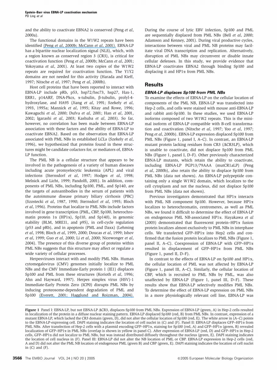
Peng et al, 2000b). EBNA-LP expression displaced Sp100 from
PML NBs (Figure 1, panel I, A–C). In contrast, an EBNA-LP
mutant protein lacking residues from CR3 (DCR3LP), which
is unable to coactivate, did not displace Sp100 from PML
NBs (Figure 1, panel I, D–F). Other previously characterized
EBNA-LP mutants, which retain the ability to coactivate,
including EBNA-LP PGP13/79AAA (mutCR1aLP) (Peng
et al, 2000b), also retain the ability to displace Sp100 from
PML NBs (data not shown). An EBNA-LP polypeptide con-
taining only a single W1W2 domain, which localizes to the
cell cytoplasm and not the nucleus, did not displace Sp100
from PML NBs (data not shown).
Previous investigators demonstrated that HP1a interacts
with PML NB component Sp100. However, because HP1alocalizes to heterochromatin, centromeres, as well as PML
NBs, we found it difficult to determine the effect of EBNA-LP
on endogenous PML NB-associated HP1a. Hayakawa et al
(2003) demonstrated that fluorescent protein–HP1a fusion
protein localizes almost exclusively to PML NBs in interphase
cells. We transfected GFP–HP1a into Hep2 cells and con-
firmed that the fusion protein localizes to PML NBs (Figure 1,
panel II, A–C). Coexpression of EBNA-LP with GFP–HP1aresulted in displacement of GFP–HP1a from PML NBs
(Figure 1, panel II, D–F).
In contrast to the effects of EBNA-LP on Sp100 and HP1a,
the cellular location of PML was not affected by EBNA-LP
(Figure 1, panel III, A–C). Similarly, the cellular location of
CBP, which is recruited to PML NBs by PML, was also
unaffected by EBNA-LP (Figure 1, panel III, D–F). These
results show that EBNA-LP selectively modifies PML NBs.
To determine the effect of EBNA-LP expression on PML NBs
in a more physiologically relevant cell line, EBNA-LP was
Figure 1 Panel I: EBNA-LP, but not EBNA-LP DCR3, displaces Sp100 from PML NBs. Expression of EBNA-LP (green, A) in Hep-2 cells resultedin localization of the protein in a diffuse nuclear staining pattern. EBNA-LP displaced Sp100 (red, B) from PML NBs. In contrast, expression of amutant EBNA-LP, which lacked the CR3 domain (green, D), did not alter the cellular location of Sp100 (red, E). The white arrow in (A–C) pointsto the EBNA-LP-expressing cell. DAPI staining indicates the location of cell nuclei in (C) and (F). Panel II: EBNA-LP displaces GFP–HP1a fromPML NBs. After transfection of Hep-2 cells with a plasmid encoding GFP–HP1a, staining for Sp100 (red, A) and GFP–HP1a (green, B) revealedlocalization of GFP–HP1a in PML NBs (overlap is shown in yellow in panel C). After expression of EBNA-LP (red, D) and GFP–HP1a in Hep-2cells, GFP–HP1a did not localize to PML NBs, but was instead distributed diffusely throughout the nucleus (green, E). DAPI staining indicatesthe location of cell nucleus in (F). Panel III: EBNA-LP did not alter the NB location of PML or CBP. EBNA-LP expression in Hep-2 cells (red,A and D) did not alter the PML NB location of endogenous PML (green B) and CBP (green, E). DAPI staining indicates the location of cell nucleiin (C) and (F).
Epstein–Barr virus EBNA-LP coactivation mechanismPD Ling et al
The EMBO Journal VOL 24 | NO 20 | 2005 &2005 European Molecular Biology Organization3566
transiently expressed in EBV-negative DG75 B cells. EBNA-LP
displaced Sp100, but not PML, from PML NBs in these cells
(Figure S2 A–F).
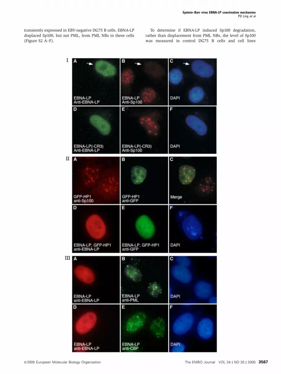
To determine if EBNA-LP induced Sp100 degradation,
rather than displacement from PML NBs, the level of Sp100
was measured in control DG75 B cells and cell lines
Epstein–Barr virus EBNA-LP coactivation mechanismPD Ling et al
&2005 European Molecular Biology Organization The EMBO Journal VOL 24 | NO 20 | 2005 3567
constitutively expressing wt EBNA-LP or DCR3 EBNA-LP.
There was no difference in the level of Sp100 in these
cell lines, indicating that EBNA-LP does not mediate Sp100
destruction (Figure 2).
EBNA-LP interacts with Sp100
To determine whether EBNA-LP-induced displacement of
Sp100 from PML NBs was mediated through interactions
with PML NB-associated proteins, we overexpressed EBNA-
LP with several proteins including Sp100, Daxx, and PML. In
these studies, eukaryotic expression vectors encoding the HA
epitope fused to Sp100, Daxx, or PML were expressed in EBV-
negative Burkitt’s lymphoma cells with a Flag epitope fused
to EBNA-LP (EBNA-LP-Flag). Cell lysates were incubated with
anti-Flag epitope antibodies directed against EBNA-LP-Flag or
anti-HA epitope antibodies directed against HA fused to PML,
Daxx, or Sp100, and the immunoprecipitated (IP) proteins
were assayed by Western blot. EBNA2-HA and EBNA-LP-Flag,
which do not interact in this assay (Peng et al, 2004, 2005),
were used as a negative control. The results showed that
EBNA-LP-Flag was able to co-precipitate Sp100 but not Daxx,
PML, or EBNA2 (Figure S3).
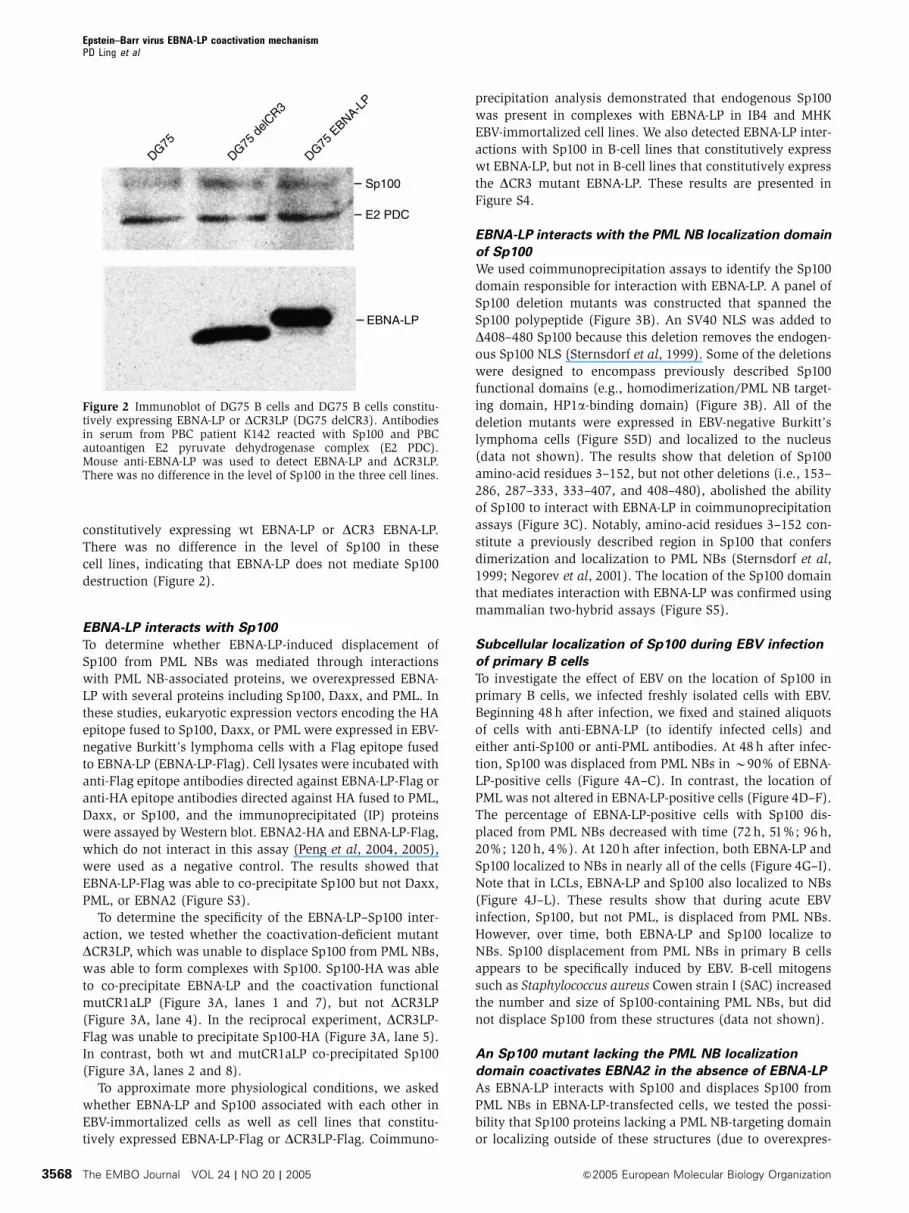
To determine the specificity of the EBNA-LP–Sp100 inter-
action, we tested whether the coactivation-deficient mutant
DCR3LP, which was unable to displace Sp100 from PML NBs,
was able to form complexes with Sp100. Sp100-HA was able
to co-precipitate EBNA-LP and the coactivation functional
mutCR1aLP (Figure 3A, lanes 1 and 7), but not DCR3LP
(Figure 3A, lane 4). In the reciprocal experiment, DCR3LP-
Flag was unable to precipitate Sp100-HA (Figure 3A, lane 5).
In contrast, both wt and mutCR1aLP co-precipitated Sp100
(Figure 3A, lanes 2 and 8).
To approximate more physiological conditions, we asked
whether EBNA-LP and Sp100 associated with each other in
EBV-immortalized cells as well as cell lines that constitu-
tively expressed EBNA-LP-Flag or DCR3LP-Flag. Coimmuno-
precipitation analysis demonstrated that endogenous Sp100
was present in complexes with EBNA-LP in IB4 and MHK
EBV-immortalized cell lines. We also detected EBNA-LP inter-
actions with Sp100 in B-cell lines that constitutively express
wt EBNA-LP, but not in B-cell lines that constitutively express
the DCR3 mutant EBNA-LP. These results are presented in
Figure S4.
EBNA-LP interacts with the PML NB localization domain
of Sp100
We used coimmunoprecipitation assays to identify the Sp100
domain responsible for interaction with EBNA-LP. A panel of
Sp100 deletion mutants was constructed that spanned the
Sp100 polypeptide (Figure 3B). An SV40 NLS was added to
D408–480 Sp100 because this deletion removes the endogen-
ous Sp100 NLS (Sternsdorf et al, 1999). Some of the deletions
were designed to encompass previously described Sp100
functional domains (e.g., homodimerization/PML NB target-
ing domain, HP1a-binding domain) (Figure 3B). All of the
deletion mutants were expressed in EBV-negative Burkitt’s
lymphoma cells (Figure S5D) and localized to the nucleus
(data not shown). The results show that deletion of Sp100
amino-acid residues 3–152, but not other deletions (i.e., 153–
286, 287–333, 333–407, and 408–480), abolished the ability
of Sp100 to interact with EBNA-LP in coimmunoprecipitation
assays (Figure 3C). Notably, amino-acid residues 3–152 con-
stitute a previously described region in Sp100 that confers
dimerization and localization to PML NBs (Sternsdorf et al,
1999; Negorev et al, 2001). The location of the Sp100 domain
that mediates interaction with EBNA-LP was confirmed using
mammalian two-hybrid assays (Figure S5).
Subcellular localization of Sp100 during EBV infection
of primary B cells
To investigate the effect of EBV on the location of Sp100 in
primary B cells, we infected freshly isolated cells with EBV.
Beginning 48 h after infection, we fixed and stained aliquots
of cells with anti-EBNA-LP (to identify infected cells) and
either anti-Sp100 or anti-PML antibodies. At 48 h after infec-
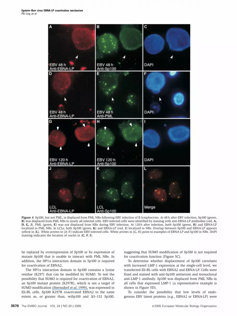
tion, Sp100 was displaced from PML NBs in B90% of EBNA-
LP-positive cells (Figure 4A–C). In contrast, the location of
PML was not altered in EBNA-LP-positive cells (Figure 4D–F).
The percentage of EBNA-LP-positive cells with Sp100 dis-
placed from PML NBs decreased with time (72 h, 51%; 96 h,
20%; 120 h, 4%). At 120 h after infection, both EBNA-LP and
Sp100 localized to NBs in nearly all of the cells (Figure 4G–I).
Note that in LCLs, EBNA-LP and Sp100 also localized to NBs
(Figure 4J–L). These results show that during acute EBV
infection, Sp100, but not PML, is displaced from PML NBs.
However, over time, both EBNA-LP and Sp100 localize to
NBs. Sp100 displacement from PML NBs in primary B cells
appears to be specifically induced by EBV. B-cell mitogens
such as Staphylococcus aureus Cowen strain I (SAC) increased
the number and size of Sp100-containing PML NBs, but did
not displace Sp100 from these structures (data not shown).
An Sp100 mutant lacking the PML NB localization
domain coactivates EBNA2 in the absence of EBNA-LP
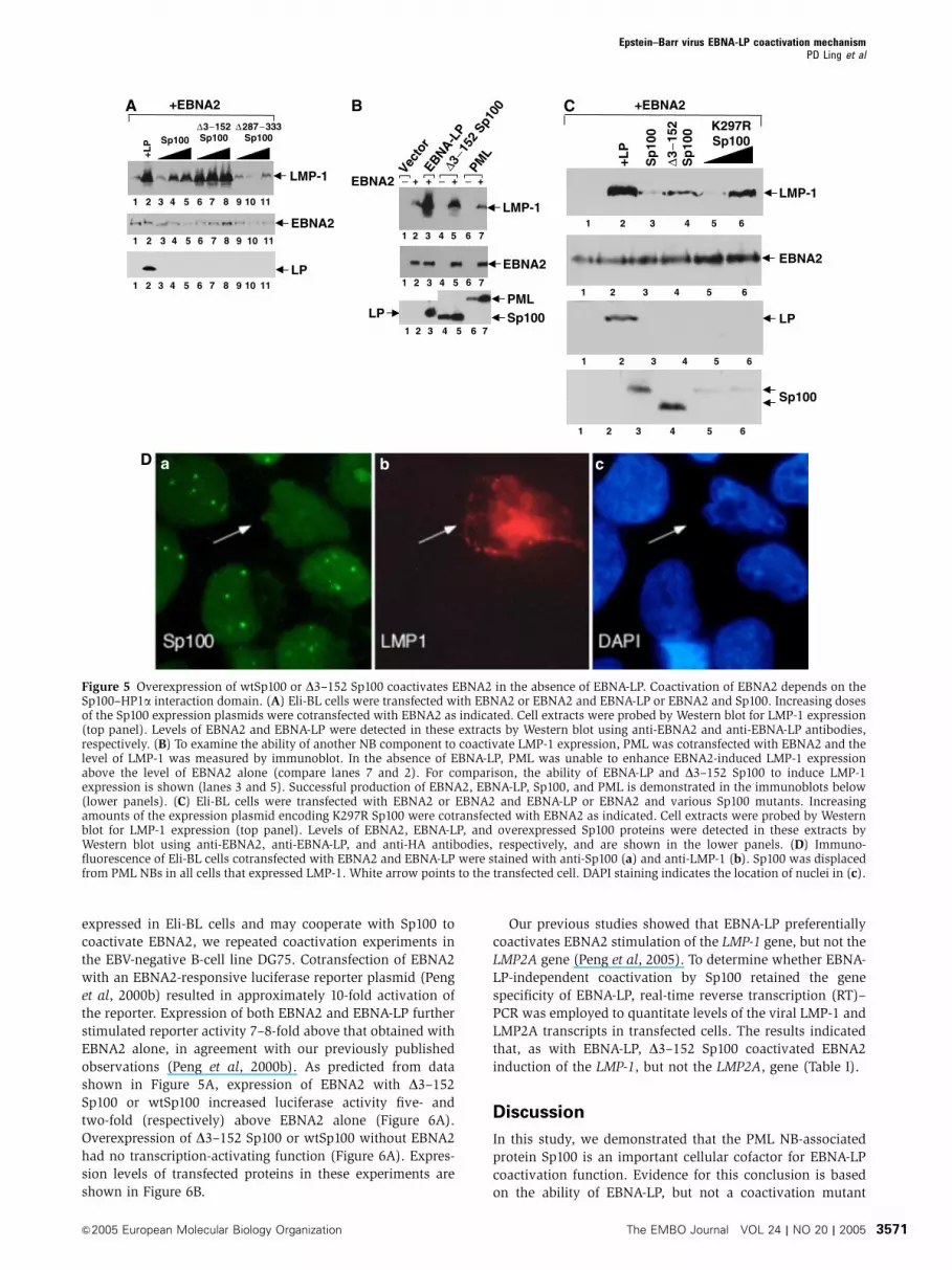
As EBNA-LP interacts with Sp100 and displaces Sp100 from
PML NBs in EBNA-LP-transfected cells, we tested the possi-
bility that Sp100 proteins lacking a PML NB-targeting domain
or localizing outside of these structures (due to overexpres-
DG75
DG75 d
elCR3
DG75 E
BNA-LP
Sp100
E2 PDC
EBNA-LP
Figure 2 Immunoblot of DG75 B cells and DG75 B cells constitu-tively expressing EBNA-LP or DCR3LP (DG75 delCR3). Antibodiesin serum from PBC patient K142 reacted with Sp100 and PBCautoantigen E2 pyruvate dehydrogenase complex (E2 PDC).Mouse anti-EBNA-LP was used to detect EBNA-LP and DCR3LP.There was no difference in the level of Sp100 in the three cell lines.
Epstein–Barr virus EBNA-LP coactivation mechanismPD Ling et al
The EMBO Journal VOL 24 | NO 20 | 2005 &2005 European Molecular Biology Organization3568
sion (Negorev et al, 2001)) might be able to coactivate EBNA2
in the absence of EBNA-LP. In transient cotransfection experi-
ments in Eli-BL cells, we found that both wtSp100 and, to a
greater extent, D3–152 Sp100 were able to coactivate EBNA2
induction of LMP-1 in the absence of EBNA-LP (Figure 5A,
top panel, lanes 3–5 and 6–8, respectively). We also tested
the other Sp100 deletion mutants shown in Figure 3B for
EBNA-LP-independent coactivation. Overexpression of one of
the mutants, D287–333 Sp100, which lacks the interaction
domain for binding HP1a, was unable to coactivate EBNA2 in
these assays (Figure 5A, top panel, lanes 9–11). In addition,
neither PML (Figure 5B, top panel, lane 7) nor Daxx (data not
shown) was able to coactivate EBNA2 in these assays. These
results show that the coactivation function of EBNA-LP can
Sp100
EBNA-LP
F HH H FF No
Ab
W: anti-HA
W: anti-LP
IP:
Ig H chain
2 31 4 65 87 9
2 31 4 65 87 9
No
Ab
No
Ab
LP-Flag+
Sp100-HA
∆CR3-LP-Flag
+Sp100-HA
mutCR1a-LP-Flag
+Sp100-HA
IP: H FH FHF
W: anti-HA
Ig H chain
Ig H chain
W: anti-HA
H F H F H F H FIP:
∗
∗∆153−286 Sp100-HA
wtSp100-HA+
LP-Flag
∆3−152Sp100-HA
+LP-Flag
∆153−286Sp100-HA
+LP-Flag
wtSp100-HA+
LP-Flag
∆287−333Sp100-HA
+LP-Flag
∆334−407Sp100-HA
+LP-Flag
∆408−480Sp100-HA
+LP-Flag
No
Ab
No
Ab
No
Ab
No
Ab
No
Ab
No
Ab
No
Ab
∗
PML-ND10 NB targeting; dimerization HP1α NLS
K297-SUMO
Sp100A
∆3−152
∆153−286
∆287−333
∆334−407
∆408−480
NLS+HA
Amino acids1 100 200 300 400 480
B
A C
Figure 3 (A) EBNA-LP interacts with Sp100. DG75 cells cotransfected with Sp100-HA and LP-Flag, or Sp100-HA and DCR3LP-Flag, or Sp100-HA and mutCR1aLP-Flag were lysed and precipitated with anti-HA or anti-Flag antibodies. The extracts were divided into equal parts, resolvedby SDS–PAGE, and the proteins were detected by immunoblotting with anti-HA (top panel) or anti-EBNA-LP (bottom panel) antibodies. Themigration of Sp100, EBNA-LP, and immunoglobulin heavy (Ig H) chain from the primary antibody used in the IP is indicated. As a control, eachextract was also treated with Staph A beads alone (no Ab). The coexpressed proteins contained in each extract are indicated above the panel.Precipitation with anti-HA or anti-Flag antibody is indicated above each lane. (B) Schematic of Sp100. Functional domains in Sp100 includeamino-acid residues 1–152 (PML NB targeting domain and Sp100 homodimerization region), 287–333 (HP1a interaction domain and SUMOmodification site), and 444–450 (nuclear localization sequence). Five Sp100 deletion mutants were used in coimmunoprecipitation andfunctional studies as indicated. (C) EBNA-LP interacts with the PML NB-targeting domain in Sp100. Lysates from cells cotransfected withEBNA-LP-Flag and one of each of the Sp100-HA deletion mutants were immunoprecipitated with anti-HA (H) or anti-Flag (F) antibodies and theresulting precipitates were probed for Sp100 using anti-HA antibodies. The D153–286 Sp100 mutant migrated just below the Ig H chain and isdesignated by asterisks. Each cell extract was also mock-precipitated as a control (no Ab).
Epstein–Barr virus EBNA-LP coactivation mechanismPD Ling et al
&2005 European Molecular Biology Organization The EMBO Journal VOL 24 | NO 20 | 2005 3569
be replaced by overexpression of Sp100 or by expression of
mutant Sp100 that is unable to interact with PML NBs. In
addition, the HP1a interaction domain in Sp100 is required
for coactivation of EBNA2.
The HP1a interaction domain in Sp100 contains a lysine
residue (K297) that can be modified by SUMO. To test the
possibility that SUMO is required for coactivation of EBNA2,
an Sp100 mutant protein (K297R), which is not a target of
SUMO modification (Sternsdorf et al, 1999), was expressed in
Eli-BL cells. Sp100 K297R coactivated EBNA2 to the same
extent as, or greater than, wtSp100 and D3–152 Sp100,
suggesting that SUMO modification of Sp100 is not required
for coactivation function (Figure 5C).
To determine whether displacement of Sp100 correlates
with increased LMP-1 expression at the single-cell level, we
transfected Eli-BL cells with EBNA2 and EBNA-LP. Cells were
fixed and stained with anti-Sp100 antiserum and monoclonal
anti-LMP-1 antibody. Sp100 was displaced from PML NBs in
all cells that expressed LMP-1 (a representative example is
shown in Figure 5D).
To consider the possibility that low levels of endo-
genous EBV latent proteins (e.g., EBNA2 or EBNA-LP) were
Figure 4 Sp100, but not PML, is displaced from PML NBs following EBV infection of B lymphocytes. At 48 h after EBV infection, Sp100 (green,B) was displaced from PML NBs in nearly all infected cells. EBV-infected cells were identified by staining with anti-EBNA-LP antibodies (red, A,D, G, J). PML (green, E) was not displaced from NBs during EBV infection. At 120 h after infection, both Sp100 (green, H) and EBNA-LPlocalized to PML NBs. In LCLs, both Sp100 (green, K) and EBNA-LP (red, J) localized to NBs. Overlap between Sp100 and EBNA-LP appearsyellow in (L). White arrows in (A–F) indicate EBV-infected cells. White arrows in (G, H) point to examples of EBNA-LP and Sp100 in NBs. DAPIstaining indicates the location of nuclei in (C, F, I).
Epstein–Barr virus EBNA-LP coactivation mechanismPD Ling et al
The EMBO Journal VOL 24 | NO 20 | 2005 &2005 European Molecular Biology Organization3570
expressed in Eli-BL cells and may cooperate with Sp100 to
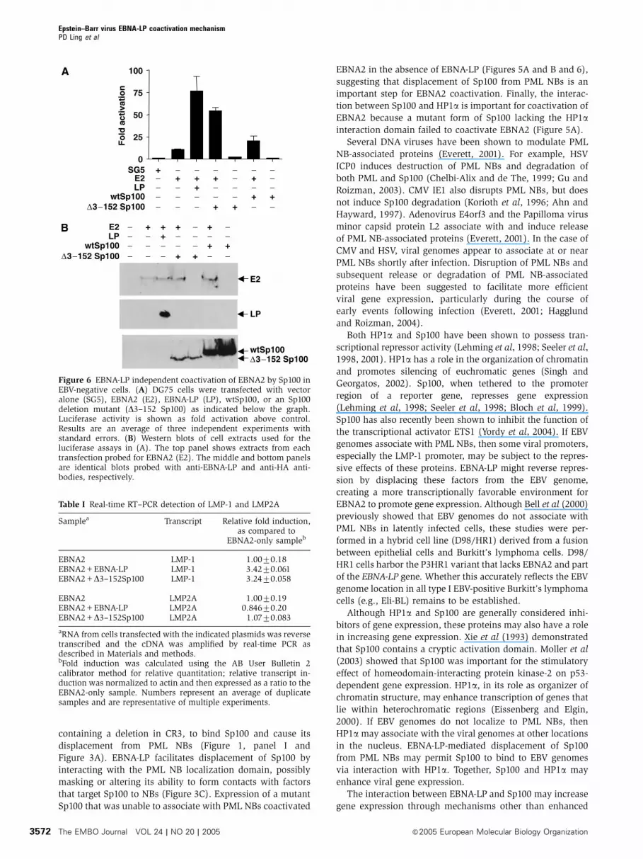
coactivate EBNA2, we repeated coactivation experiments in
the EBV-negative B-cell line DG75. Cotransfection of EBNA2
with an EBNA2-responsive luciferase reporter plasmid (Peng
et al, 2000b) resulted in approximately 10-fold activation of
the reporter. Expression of both EBNA2 and EBNA-LP further
stimulated reporter activity 7–8-fold above that obtained with
EBNA2 alone, in agreement with our previously published
observations (Peng et al, 2000b). As predicted from data
shown in Figure 5A, expression of EBNA2 with D3–152
Sp100 or wtSp100 increased luciferase activity five- and
two-fold (respectively) above EBNA2 alone (Figure 6A).
Overexpression of D3–152 Sp100 or wtSp100 without EBNA2
had no transcription-activating function (Figure 6A). Expres-
sion levels of transfected proteins in these experiments are
shown in Figure 6B.
Our previous studies showed that EBNA-LP preferentially
coactivates EBNA2 stimulation of the LMP-1 gene, but not the
LMP2A gene (Peng et al, 2005). To determine whether EBNA-
LP-independent coactivation by Sp100 retained the gene
specificity of EBNA-LP, real-time reverse transcription (RT)–
PCR was employed to quantitate levels of the viral LMP-1 and
LMP2A transcripts in transfected cells. The results indicated
that, as with EBNA-LP, D3–152 Sp100 coactivated EBNA2
induction of the LMP-1, but not the LMP2A, gene (Table I).
Discussion
In this study, we demonstrated that the PML NB-associated
protein Sp100 is an important cellular cofactor for EBNA-LP
coactivation function. Evidence for this conclusion is based
on the ability of EBNA-LP, but not a coactivation mutant
LMP-1
Sp100
2 31 4 5 76
2 31 4 5 76
PML
4 5 76
EBNA2
21 3
LMP-1
LP
LP
+EBNA2
EBNA2 + + + +− −−
∆3−1
52 S
p100
PML
Vecto
rEBNA-L
P
EBNA2
2 31 4 5 76 8 109 11
2 31 4 5 76 8 109 11
2 31 4 5 76 8 109 11
+LP Sp100
∆3−152Sp100
∆287−333Sp100
LMP-1
LP
+EBNA2
EBNA2
2 31 4 5 6
+LP
K297RSp100
2 31 4 5 6
2 31 4 5 6
2 31 4 5 6
Sp100
Sp
100
∆3
−152
Sp
100
A B C
D a b c
Figure 5 Overexpression of wtSp100 or D3–152 Sp100 coactivates EBNA2 in the absence of EBNA-LP. Coactivation of EBNA2 depends on theSp100–HP1a interaction domain. (A) Eli-BL cells were transfected with EBNA2 or EBNA2 and EBNA-LP or EBNA2 and Sp100. Increasing dosesof the Sp100 expression plasmids were cotransfected with EBNA2 as indicated. Cell extracts were probed by Western blot for LMP-1 expression(top panel). Levels of EBNA2 and EBNA-LP were detected in these extracts by Western blot using anti-EBNA2 and anti-EBNA-LP antibodies,respectively. (B) To examine the ability of another NB component to coactivate LMP-1 expression, PML was cotransfected with EBNA2 and thelevel of LMP-1 was measured by immunoblot. In the absence of EBNA-LP, PML was unable to enhance EBNA2-induced LMP-1 expressionabove the level of EBNA2 alone (compare lanes 7 and 2). For comparison, the ability of EBNA-LP and D3–152 Sp100 to induce LMP-1expression is shown (lanes 3 and 5). Successful production of EBNA2, EBNA-LP, Sp100, and PML is demonstrated in the immunoblots below(lower panels). (C) Eli-BL cells were transfected with EBNA2 or EBNA2 and EBNA-LP or EBNA2 and various Sp100 mutants. Increasingamounts of the expression plasmid encoding K297R Sp100 were cotransfected with EBNA2 as indicated. Cell extracts were probed by Westernblot for LMP-1 expression (top panel). Levels of EBNA2, EBNA-LP, and overexpressed Sp100 proteins were detected in these extracts byWestern blot using anti-EBNA2, anti-EBNA-LP, and anti-HA antibodies, respectively, and are shown in the lower panels. (D) Immuno-fluorescence of Eli-BL cells cotransfected with EBNA2 and EBNA-LP were stained with anti-Sp100 (a) and anti-LMP-1 (b). Sp100 was displacedfrom PML NBs in all cells that expressed LMP-1. White arrow points to the transfected cell. DAPI staining indicates the location of nuclei in (c).
Epstein–Barr virus EBNA-LP coactivation mechanismPD Ling et al
&2005 European Molecular Biology Organization The EMBO Journal VOL 24 | NO 20 | 2005 3571
containing a deletion in CR3, to bind Sp100 and cause its
displacement from PML NBs (Figure 1, panel I and
Figure 3A). EBNA-LP facilitates displacement of Sp100 by
interacting with the PML NB localization domain, possibly
masking or altering its ability to form contacts with factors
that target Sp100 to NBs (Figure 3C). Expression of a mutant
Sp100 that was unable to associate with PML NBs coactivated
EBNA2 in the absence of EBNA-LP (Figures 5A and B and 6),
suggesting that displacement of Sp100 from PML NBs is an
important step for EBNA2 coactivation. Finally, the interac-
tion between Sp100 and HP1a is important for coactivation of
EBNA2 because a mutant form of Sp100 lacking the HP1ainteraction domain failed to coactivate EBNA2 (Figure 5A).
Several DNA viruses have been shown to modulate PML
NB-associated proteins (Everett, 2001). For example, HSV
ICP0 induces destruction of PML NBs and degradation of
both PML and Sp100 (Chelbi-Alix and de The, 1999; Gu and
Roizman, 2003). CMV IE1 also disrupts PML NBs, but does
not induce Sp100 degradation (Korioth et al, 1996; Ahn and
Hayward, 1997). Adenovirus E4orf3 and the Papilloma virus
minor capsid protein L2 associate with and induce release
of PML NB-associated proteins (Everett, 2001). In the case of
CMV and HSV, viral genomes appear to associate at or near
PML NBs shortly after infection. Disruption of PML NBs and
subsequent release or degradation of PML NB-associated
proteins have been suggested to facilitate more efficient
viral gene expression, particularly during the course of
early events following infection (Everett, 2001; Hagglund
and Roizman, 2004).
Both HP1a and Sp100 have been shown to possess tran-
scriptional repressor activity (Lehming et al, 1998; Seeler et al,
1998, 2001). HP1a has a role in the organization of chromatin
and promotes silencing of euchromatic genes (Singh and
Georgatos, 2002). Sp100, when tethered to the promoter
region of a reporter gene, represses gene expression
(Lehming et al, 1998; Seeler et al, 1998; Bloch et al, 1999).
Sp100 has also recently been shown to inhibit the function of
the transcriptional activator ETS1 (Yordy et al, 2004). If EBV
genomes associate with PML NBs, then some viral promoters,
especially the LMP-1 promoter, may be subject to the repres-
sive effects of these proteins. EBNA-LP might reverse repres-
sion by displacing these factors from the EBV genome,
creating a more transcriptionally favorable environment for
EBNA2 to promote gene expression. Although Bell et al (2000)
previously showed that EBV genomes do not associate with
PML NBs in latently infected cells, these studies were per-
formed in a hybrid cell line (D98/HR1) derived from a fusion
between epithelial cells and Burkitt’s lymphoma cells. D98/
HR1 cells harbor the P3HR1 variant that lacks EBNA2 and part
of the EBNA-LP gene. Whether this accurately reflects the EBV
genome location in all type I EBV-positive Burkitt’s lymphoma
cells (e.g., Eli-BL) remains to be established.
Although HP1a and Sp100 are generally considered inhi-
bitors of gene expression, these proteins may also have a role
in increasing gene expression. Xie et al (1993) demonstrated
that Sp100 contains a cryptic activation domain. Moller et al
(2003) showed that Sp100 was important for the stimulatory
effect of homeodomain-interacting protein kinase-2 on p53-
dependent gene expression. HP1a, in its role as organizer of
chromatin structure, may enhance transcription of genes that
lie within heterochromatic regions (Eissenberg and Elgin,
2000). If EBV genomes do not localize to PML NBs, then
HP1a may associate with the viral genomes at other locations
in the nucleus. EBNA-LP-mediated displacement of Sp100
from PML NBs may permit Sp100 to bind to EBV genomes
via interaction with HP1a. Together, Sp100 and HP1a may
enhance viral gene expression.
The interaction between EBNA-LP and Sp100 may increase
gene expression through mechanisms other than enhanced
0
25
50
75
100
Fo
ld a
ctiv
atio
n
SG5E2LP
wtSp100∆3−152 Sp100
E2LP
wtSp100∆3−152 Sp100
E2
LP
wtSp100∆3−152 Sp100
+ − − − −− −−
−−
−−
− −− ++
+ + +
+++
− −− − −−
− − − − −
+
−−
−−
−− −− +
+
+ + +
+++
− −− − −−
− − − − −
+
A
B
Figure 6 EBNA-LP independent coactivation of EBNA2 by Sp100 inEBV-negative cells. (A) DG75 cells were transfected with vectoralone (SG5), EBNA2 (E2), EBNA-LP (LP), wtSp100, or an Sp100deletion mutant (D3–152 Sp100) as indicated below the graph.Luciferase activity is shown as fold activation above control.Results are an average of three independent experiments withstandard errors. (B) Western blots of cell extracts used for theluciferase assays in (A). The top panel shows extracts from eachtransfection probed for EBNA2 (E2). The middle and bottom panelsare identical blots probed with anti-EBNA-LP and anti-HA anti-bodies, respectively.
Table I Real-time RT–PCR detection of LMP-1 and LMP2A
Samplea Transcript Relative fold induction,as compared to
EBNA2-only sampleb
EBNA2 LMP-1 1.0070.18EBNA2+EBNA-LP LMP-1 3.4270.061EBNA2+D3–152Sp100 LMP-1 3.2470.058
EBNA2 LMP2A 1.0070.19EBNA2+EBNA-LP LMP2A 0.84670.20EBNA2+D3–152Sp100 LMP2A 1.0770.083
aRNA from cells transfected with the indicated plasmids was reversetranscribed and the cDNA was amplified by real-time PCR asdescribed in Materials and methods.bFold induction was calculated using the AB User Bulletin 2calibrator method for relative quantitation; relative transcript in-duction was normalized to actin and then expressed as a ratio to theEBNA2-only sample. Numbers represent an average of duplicatesamples and are representative of multiple experiments.
Epstein–Barr virus EBNA-LP coactivation mechanismPD Ling et al
The EMBO Journal VOL 24 | NO 20 | 2005 &2005 European Molecular Biology Organization3572
gene transcription. A recent study showed that Sp110b, a
component of the PML NB and a member of the Sp100 family
of proteins, interacts with the EBV lytic cycle protein SM
(Nicewonger et al, 2004). The EBV SM–Sp110b interaction
resulted in increased stability of SM-regulated viral tran-
scripts. It is possible that the EBNA-LP–Sp100 interaction,
like that of EBV SM–Sp110b, may also alter gene expression
through post-transcriptional mechanisms.
HP1a has a complex pattern of intracellular localization,
depending on the presence or absence of a variety of inter-
acting proteins and the phase of the cell cycle (Everett et al,
1999; Hayakawa et al, 2003). As endogenous HP1a localizes
to several different cellular domains, including heterochro-
matin, centromeres, as well as PML NBs, we were unable to
determine the effect of EBNA-LP on the PML NB localization
of endogenous HP1a. However, Hayakawa et al showed,
and we have confirmed in this study, that expression of a
GFP–HP1a fusion protein results in localization of HP1a to
PML NBs in nearly all interphase cells. Coexpression of
EBNA-LP and GFP–HP1a resulted in displacement of GFP–
HP1a from PML NBs. In view of the previous studies showing
that HP1a interacts with Sp100, and our findings that EBNA-
LP displaces Sp100 and GFP–HP1a from PML NBs, it seems
likely that EBNA-LP also alters the cellular location of
endogenous, PML NB-associated HP1a.
Similar to the other herpesvirus immediate early proteins,
EBNA-LP coactivation function is likely to play an important
role during early events of infection. EBNA-LP and EBNA2 are
the earliest latent cycle proteins detected following infection
of primary B cells. It is likely that EBV genomes are subject to
similar host cell modifications encountered by other herpes-
viruses following infection. EBNA-LP may mitigate transcrip-
tional barriers that prevent efficient expression of viral latent
genes important for establishing latent infection. Although
EBNA-LP and Sp100 are distributed diffusely throughout the
nucleus during early infection, EBNA-LP and Sp100 localize
to PML NBs in established lymphoblastoid cell lines immor-
talized by EBV. It is possible that other viral latent cycle
proteins may act to regulate EBNA-LP–Sp100 activity as latent
infection becomes established. This may be particularly im-
portant because latent viral genes like LMP-1 are cytostatic
when overexpressed (Floettmann et al, 1996). Inhibition of
EBNA-LP activity would prevent overly robust LMP-1 expres-
sion, which would negatively affect LCL growth. Thus, EBNA
-LP may play an important role during early EBV infection,
but may not be required for maintenance of (or in fact may be
detrimental to) immortalization in established LCLs.
Identification of cellular cofactors that mediate EBV latent
protein functions has been a central question in EBV biology.
Our data are the first to show that modulation of PML NBs
might be required for establishment of nonproductive or
latent herpesvirus infections. In this study, we used EBNA-
LP isoforms with only two W1W2 repeats. Most EBV strains
synthesize EBNA-LP polypeptides with several W1W2 re-
peats. The existence of multiple Sp100 interaction domains
in a single EBNA-LP polypeptide may make it particularly
adept at binding and displacing Sp100 from PML NBs. In
contrast to CMV IE1 or HSV ICP0; EBNA-LP does not appear
to displace PML from NBs, but instead seems to selectively
displace Sp100 and HP1a from these structures. Thus, EBNA-
LP appears to modulate PML NBs by a more subtle mechan-
ism than observed for ICP0 and IE1. This may be important
for EBV, as it tends to favor coexistence with the host cell
rather than host cell destruction. At present it is unclear why
EBNA-LP and IE1 redistribute Sp100, while HSV ICP0 induces
Sp100 degradation.
Historically, the function of numerous cellular proteins has
been elucidated through investigation of their interactions
with viral proteins. Induction of LMP-1 expression in Eli-BL
cells, which is potentiated through interactions between
EBNA-LP and Sp100, provides a unique system to unravel
how the cellular Sp100 protein regulates gene expression and
to identify the cellular genes that are targeted for regulation
by this protein.
Materials and methods
Cell culture, cell lines, transfections, and plasmidsEli-BL, an EBV type I Burkitt’s lymphoma line, DG75, an EBV-negative Burkitt’s cell line, and Hep-2 cells were maintained andtransfected as described previously (Bloch et al, 1999; Peng et al,2000b, 2005). To generate cell lines constitutively expressing EBNA-LP, we cotransfected plasmids pRSP438 or pJT125 with a plasmidexpressing the puromycin-N-acetyl-transferase gene (pGK3PURO),and cell clones emerging under puromycin selection were screenedfor EBNA-LP expression. Plasmids used in this study were generatedusing standard procedures (see Supplementary data for details).
B-cell isolation and EBV infectionPrimary human B cells were isolated from healthy donor buffy coats(Gulf coast regional blood center). The buffy coats were diluted 1:2in PBS and the lymphocytes were purified on Ficoll gradients.B cells were selected using CD19 magnetic beads (Miltenyi). B cells(1�106) were incubated with 10 ml of virus-containing supernatantderived from B95-8 cells as described (Ling and Hulls, 2005). Theinfected cells were incubated at 371C. Aliquots of infected cells wereharvested at the indicated time-points and prepared for indirectimmunofluorescence as described below.
RNA extraction and RT–PCRRNA was prepared from transfected cells using the TRIZOL(Invitrogen) extraction method. Complementary DNA was preparedfrom 0.5–1mg of RNA using AMV reverse transcriptase (Invitrogen).Subsequent PCR reactions were performed using oligonucleotideprimers as described previously (Peng et al, 2005).
Western blots, IPs, and indirect immunofluorescenceWestern blotting was carried out as described previously (Penget al, 2000b, 2005). Serum from patient K142 with PBC containsantibodies directed against Sp100 and PBC autoantigen E2 pyruvatedehydrogenase complex (Bloch et al, 1999). For IPs, transfectedcells were lysed in either RIPA buffer or a 1% NP40 buffer (10 mMTris-Cl (pH 7.4), 1 mM EDTA, 150 mM NaCl, 3% glycerol, 1 mMphenylmethylsulfonyl fluoride, 5 mg/ml leupeptin, 10 mg/ml apro-tinin). Transfected cell lysates were incubated with a primaryantibody at 41C overnight, followed by incubation with Staph Asepharose beads (Pierce) for 1 h at room temperature. The beadswere washed in IP lysis buffer and the bound proteins weresolubilized by addition of 2� Laemmli sample buffer and boilingfor 5 min. The proteins were subjected to SDS–PAGE and Westernblotting. Indirect immunofluorescence was performed as describedpreviously (Bloch et al, 1999).
Supplementary dataSupplementary data are available at The EMBO Journal Online.
Acknowledgements
We thank Drs Cliona Rooney, Andrew Rice, and Richard Sutton forcritical reading of the manuscript and Drs Thomas Sternsdorf andHans Will for the K297R Sp100 plasmid. This work was supportedby a grant to PDL from the American Cancer Society. DBB wassupported by grants from the Arthritis Foundation, the NationalInstitutes of Health (DK-051179) and an Established InvestigatorGrant from the American Heart Association.
Epstein–Barr virus EBNA-LP coactivation mechanismPD Ling et al
&2005 European Molecular Biology Organization The EMBO Journal VOL 24 | NO 20 | 2005 3573
References
Adamson AL, Kenney S (2001) Epstein–Barr virus immediate-earlyprotein BZLF1 is SUMO-1 modified and disrupts promyelocyticleukemia bodies. J Virol 75: 2388–2399
Ahn JH, Hayward GS (1997) The major immediate-early proteinsIE1 and IE2 of human cytomegalovirus colocalize with anddisrupt PML-associated nuclear bodies at very early times ininfected permissive cells. J Virol 71: 4599–4613
Allan GJ, Inman GJ, Parker BD, Rowe DT, Farrell PJ (1992) Cellgrowth effects of Epstein–Barr virus leader protein. J Gen Virol73: 1547–1551
Bell P, Lieberman PM, Maul GG (2000) Lytic but not latent replica-tion of Epstein–Barr virus is associated with PML and inducessequential release of nuclear domain 10 proteins. J Virol 74:11800–11810
Bloch DB, Chiche JD, Orth D, de la Monte SM, Rosenzweig A, BlochKD (1999) Structural and functional heterogeneity of nuclearbodies. Mol Cell Biol 19: 4423–4430
Bloch DB, de la Monte SM, Guigaouri P, Filippov A, Bloch KD(1996) Identification and characterization of a leukocyte-specific component of the nuclear body. J Biol Chem 271:29198–29204
Bloch DB, Nakajima A, Gulick T, Chiche JD, Orth D, de La MonteSM, Bloch KD (2000) Sp110 localizes to the PML-Sp100 nuclearbody and may function as a nuclear hormone receptor transcrip-tional coactivator. Mol Cell Biol 20: 6138–6146
Bornkamm GW, Hammerschmidt W (2001) Molecular virology ofEpstein–Barr virus. Philos Trans R Soc Lond B Biol Sci 356:437–459
Chelbi-Alix MK, de The H (1999) Herpes virus induced proteasome-dependent degradation of the nuclear bodies-associated PML andSp100 proteins. Oncogene 18: 935–941
Crawford DH (2001) Biology and disease associations of Epstein–Barr virus. Philos Trans R Soc Lond B Biol Sci 356: 461–473
Doucas V, Tini M, Egan DA, Evans RM (1999) Modulation of CREBbinding protein function by the promyelocytic (PML) oncoproteinsuggests a role for nuclear bodies in hormone signaling. Proc NatlAcad Sci USA 96: 2627–2632
Dufva M, Olsson M, Rymo L (2001) Epstein–Barr virus nuclearantigen 5 interacts with HAX-1, a possible component of theB-cell receptor signalling pathway. J Gen Virol 82: 1581–1587
Eissenberg JC, Elgin SC (2000) The HP1 protein family: getting agrip on chromatin. Curr Opin Genet Dev 10: 204–210
Everett RD (2001) DNA viruses and viral proteins that interact withPML nuclear bodies. Oncogene 20: 7266–7273
Everett RD, Earnshaw WC, Pluta AF, Sternsdorf T, Ainsztein AM,Carmena M, Ruchaud S, Hsu WL, Orr A (1999) A dynamicconnection between centromeres and ND10 proteins. J Cell Sci112 (Part 20): 3443–3454
Floettmann JE, Ward K, Rickinson AB, Rowe M (1996) Cytostaticeffect of Epstein–Barr virus latent membrane protein-1 analyzedusing tetracycline-regulated expression in B cell lines. Virology223: 29–40
Gu H, Roizman B (2003) The degradation of promyelocytic leuke-mia and Sp100 proteins by herpes simplex virus 1 is mediated bythe ubiquitin-conjugating enzyme UbcH5a. Proc Natl Acad SciUSA 100: 8963–8968
Guo A, Salomoni P, Luo J, Shih A, Zhong S, Gu W, Paolo Pandolfi P(2000) The function of PML in p53-dependent apoptosis. Nat CellBiol 2: 730–736
Hagglund R, Roizman B (2004) Role of ICP0 in the strategy ofconquest of the host cell by herpes simplex virus 1. J Virol 78:2169–2178
Hammerschmidt W, Sugden B (1989) Genetic analysis of immorta-lizing functions of Epstein–Barr virus in human B lymphocytes.Nature 340: 393–397
Han I, Harada S, Weaver D, Xue Y, Lane W, Orstavik S, Skalhegg B,Kieff E (2001) EBNA-LP associates with cellular proteins includingDNA-PK and HA95. J Virol 75: 2475–2481
Han I, Xue Y, Harada S, Orstavik S, Skalhegg B, Kieff E (2002)Protein kinase A associates with HA95 and affects transcriptionalcoactivation by Epstein–Barr virus nuclear proteins. Mol Cell Biol22: 2136–2146
Harada S, Kieff E (1997) Epstein–Barr virus nuclear protein LPstimulates EBNA-2 acidic domain-mediated transcriptionalactivation. J Virol 71: 6611–6618
Hayakawa T, Haraguchi T, Masumoto H, Hiraoka Y (2003) Cell cyclebehavior of human HP1 subtypes: distinct molecular domains ofHP1 are required for their centromeric localization during inter-phase and metaphase. J Cell Sci 116: 3327–3338
Hodges M, Tissot C, Howe K, Grimwade D, Freemont PS (1998)Structure, organization, and dynamics of promyelocytic leukemiaprotein nuclear bodies. Am J Hum Genet 63: 297–304
Igarashi M, Kawaguchi Y, Hirai K, Mizuno F (2003) Physicalinteraction of Epstein–Barr virus (EBV) nuclear antigen leaderprotein (EBNA-LP) with human oestrogen-related receptor 1(hERR1): hERR1 interacts with a conserved domain of EBNA-LPthat is critical for EBV-induced B-cell immortalization. J Gen Virol84: 319–327
Ishov AM, Sotnikov AG, Negorev D, Vladimirova OV, Neff N,Kamitani T, Yeh ET, Strauss III JF, Maul GG (1999) PML is criticalfor ND10 formation and recruits the PML-interacting protein daxxto this nuclear structure when modified by SUMO-1. J Cell Biol147: 221–234
Jiang WQ, Szekely L, Wendel-Hansen V, Ringertz N, Klein G, RosenA (1991) Co-localization of the retinoblastoma protein and theEpstein–Barr virus-encoded nuclear antigen EBNA-5. Exp Cell Res197: 314–318
Kashuba E, Mattsson K, Pokrovskaja K, Kiss C, Protopopova M,Ehlin-Henriksson B, Klein G, Szekely L (2003) EBV-encodedEBNA-5 associates with P14ARF in extranucleolar inclusionsand prolongs the survival of P14ARF-expressing cells. Int JCancer 105: 644–653
Kawaguchi Y, Nakajima K, Igarashi M, Morita T, Tanaka M,Suzuki M, Yokoyama A, Matsuda G, Kato K, Kanamori M, HiraiK (2000) Interaction of Epstein–Barr virus nuclear antigenleader protein (EBNA-LP) with HS1-associated protein X-1:implication of cytoplasmic function of EBNA-LP. J Virol 74:10104–10111
Kieff E, Rickinson AB (2001) Epstein–Barr virus and its replication.In Virology, Knipe DM, Howley PM (eds), Vol. 2, pp 2511–2573.Philadelphia: Lippencott-Raven Publishers
Kitay MK, Rowe DT (1996) Protein–protein interactions betweenEpstein–Barr virus nuclear antigen-LP and cellular gene products:binding of 70-kilodalton heat shock proteins. Virology 220:91–99
Korioth F, Maul GG, Plachter B, Stamminger T, Frey J (1996) Thenuclear domain 10 (ND10) is disrupted by the human cytomega-lovirus gene product IE1. Exp Cell Res 229: 155–158
Lehming N, Le Saux A, Schuller J, Ptashne M (1998) Chromatincomponents as part of a putative transcriptional repressing com-plex. Proc Natl Acad Sci USA 95: 7322–7326
Li H, Leo C, Zhu J, Wu X, O’Neil J, Park EJ, Chen JD (2000)Sequestration and inhibition of Daxx-mediated transcriptionalrepression by PML. Mol Cell Biol 20: 1784–1796
Ling PD, Hulls HH (2005) Isolation and immortalization of lympho-cytes. In Current Protocols in Molecular Biology, Ausubel FM,Brent R, Kingston RE, Moore DD, Seidman JG, Struhl K (eds).New York: John Wiley & Sons Inc
Mannick JB, Cohen JI, Birkenbach M, Marchini A, Kieff E (1991)The Epstein–Barr virus nuclear protein encoded by the leader ofthe EBNA RNAs is important in B-lymphocyte transformation.J Virol 65: 6826–6837
Mannick JB, Tong X, Hemnes A, Kieff E (1995) The Epstein–Barrvirus nuclear antigen leader protein associates with hsp72/hsc73.J Virol 69: 8169–8172
McCann EM, Kelly GL, Rickinson AB, Bell AI (2001) Geneticanalysis of the Epstein–Barr virus-coded leader proteinEBNA-LP as a co-activator of EBNA2 function. J Gen Virol 82:3067–3079
Melnick A, Licht JD (1999) Deconstructing a disease: RARalpha, itsfusion partners, and their roles in the pathogenesis of acutepromyelocytic leukemia. Blood 93: 3167–3215
Moller A, Sirma H, Hofmann TG, Staege H, Gresko E, Ludi KS,Klimczak E, Droge W, Will H, Schmitz ML (2003) Sp100 isimportant for the stimulatory effect of homeodomain-interactingprotein kinase-2 on p53-dependent gene expression. Oncogene22: 8731–8737
Negorev D, Ishov AM, Maul GG (2001) Evidence for separate ND10-binding and homo-oligomerization domains of Sp100. J Cell Sci114: 59–68
Epstein–Barr virus EBNA-LP coactivation mechanismPD Ling et al
The EMBO Journal VOL 24 | NO 20 | 2005 &2005 European Molecular Biology Organization3574
Nicewonger J, Suck G, Bloch D, Swaminathan S (2004) Epstein–Barr virus (EBV) SM protein induces and recruits cellular Sp110bto stabilize mRNAs and enhance EBV lytic gene expression.J Virol 78: 9412–9422
Nitsche F, Bell A, Rickinson A (1997) Epstein–Barr virus leaderprotein enhances EBNA-2-mediated transactivation of latentmembrane protein 1 expression: a role for the W1W2 repeatdomain. J Virol 71: 6619–6628
Pandolfi PP (2001) Oncogenes and tumor suppressors in the mole-cular pathogenesis of acute promyelocytic leukemia. Hum MolGenet 10: 769–775
Peng CW, Xue Y, Zhao B, Johannsen E, Kieff E, Harada S(2004) Direct interactions between Epstein–Barr virus leaderprotein LP and the EBNA2 acidic domain underlie coordi-nate transcriptional regulation. Proc Natl Acad Sci USA 101:1033–1038
Peng R, Gordadze AV, Fuentes Panana EM, Wang F, Zong J,Hayward GS, Tan J, Ling PD (2000a) Sequence and functionalanalysis of EBNA-LP and EBNA2 proteins from nonhumanprimate lymphocryptoviruses. J Virol 74: 379–389
Peng R, Tan J, Ling PD (2000b) Conserved regions in the Epstein–Barr virus leader protein define distinct domains required fornuclear localization and transcriptional cooperation with EBNA2.J Virol 74: 9953–9963
Peng RS, Moses SC, Tan J, Kremmer E, Ling PD (2005) TheEpstein–Barr virus EBNA-LP protein preferentially co-activatesEBNA2-mediated stimulation of latent membrane proteinsexpressed from the viral divergent promoter. J Virol 79:4492–4505
Rickinson AB, Kieff E (2001) Epstein–Barr virus. In Virology, KnipeDM, Howley PM (eds), Vol. 2, pp 2575–2627. Philadelphia:Lippincott-Raven Publishers
Sample J, Hummel M, Braun D, Birkenbach M, Kieff E (1986)Nucleotide sequences of mRNAs encoding Epstein–Barr virusnuclear proteins: a probable transcriptional initiation site. ProcNatl Acad Sci USA 83: 5096–5100
Seeler JS, Marchio A, Losson R, Desterro JM, Hay RT, Chambon P,Dejean A (2001) Common properties of nuclear body proteinSP100 and TIF1alpha chromatin factor: role of SUMO modifica-tion. Mol Cell Biol 21: 3314–3324
Seeler JS, Marchio A, Sitterlin D, Transy C, Dejean A (1998)Interaction of SP100 with HP1 proteins: a link between thepromyelocytic leukemia-associated nuclear bodies and the chro-matin compartment. Proc Natl Acad Sci USA 95: 7316–7321
Singh PB, Georgatos SD (2002) HP1: facts, open questions, andspeculation. J Struct Biol 140: 10–16
Speck SH, Pfitzner A, Strominger JL (1986) An Epstein–Barr virustranscript from a latently infected, growth-transformed B-cell lineencodes a highly repetitive polypeptide. Proc Natl Acad Sci USA83: 9298–9302
Sternsdorf T, Grotzinger T, Jensen K, Will H (1997) Nuclear dots:actors on many stages. Immunobiology 198: 307–331
Sternsdorf T, Guldner HH, Szostecki C, Grotzinger T, Will H (1995)Two nuclear dot-associated proteins, PML and Sp100, are oftenco-autoimmunogenic in patients with primary biliary cirrhosis.Scand J Immunol 42: 257–268
Sternsdorf T, Jensen K, Reich B, Will H (1999) The nuclear dotprotein sp100, characterization of domains necessary for dimer-ization, subcellular localization, and modification by small ubi-quitin-like modifiers. J Biol Chem 274: 12555–12566
Szekely L, Jiang WQ, Pokrovskaja K, Wiman KG, Klein G, RingertzN (1995a) Reversible nucleolar translocation of Epstein–Barrvirus-encoded EBNA-5 and hsp70 proteins after exposure toheat shock or cell density congestion. J Gen Virol 76: 2423–2432
Szekely L, Pokrovskaja K, Jiang WQ, de The H, Ringertz N, Klein G(1996) The Epstein–Barr virus-encoded nuclear antigen EBNA-5accumulates in PML-containing bodies. J Virol 70: 2562–2568
Szekely L, Pokrovskaja K, Jiang WQ, Selivanova G, Lowbeer M,Ringertz N, Wiman KG, Klein G (1995b) Resting B-cells, EBV-infected B-blasts and established lymphoblastoid cell lines differin their Rb, p53 and EBNA-5 expression patterns. Oncogene 10:1869–1874
Szekely L, Selivanova G, Magnusson KP, Klein G, Wiman KG (1993)EBNA-5, an Epstein–Barr virus-encoded nuclear antigen, binds tothe retinoblastoma and p53 proteins. Proc Natl Acad Sci USA 90:5455–5459
Szostecki C, Guldner HH, Netter HJ, Will H (1990) Isolation andcharacterization of cDNA encoding a human nuclear antigenpredominantly recognized by autoantibodies from patients withprimary biliary cirrhosis. J Immunol 145: 4338–4347
Szostecki C, Krippner H, Penner E, Bautz FA (1987) Autoimmunesera recognize a 100 kD nuclear protein antigen (sp-100). Clin ExpImmunol 68: 108–116
Xie K, Lambie EJ, Snyder M (1993) Nuclear dot antigens mayspecify transcriptional domains in the nucleus. Mol Cell Biol 13:6170–6179
Yokoyama A, Tanaka M, Matsuda G, Kato K, Kanamori M, KawasakiH, Hirano H, Kitabayashi I, Ohki M, Hirai K, Kawaguchi Y (2001)Identification of major phosphorylation sites of Epstein–Barrvirus nuclear antigen leader protein (EBNA-LP): ability ofEBNA-LP to induce latent membrane protein 1 cooperativelywith EBNA-2 is regulated by phosphorylation. J Virol 75:5119–5128
Yoo LI, Mooney M, Puglielli MT, Speck SH (1997) B-cell linesimmortalized with an Epstein–Barr virus mutant lacking theCp EBNA2 enhancer are biased toward utilization of theoriP-Proximal EBNA gene promoter Wp1. J Virol 71: 9134–9142
Yordy JS, Li R, Sementchenko VI, Pei H, Muise-Helmericks RC,Watson DK (2004) SP100 expression modulates ETS1 transcrip-tional activity and inhibits cell invasion. Oncogene 23: 6654–6665
Epstein–Barr virus EBNA-LP coactivation mechanismPD Ling et al
&2005 European Molecular Biology Organization The EMBO Journal VOL 24 | NO 20 | 2005 3575